Integrated evaluation of relation between coronary lesion features and stress echocardiography...
10
Integrated Evaluation of Relation Between Coronary Lesion Features and Stress Echocardiography Results: The Importance of Coronary Lesion Morphology Branko D. Beleslin, MD, Miodrag Ostojic, MD, PHD, FESC, FACC, Ana Djordjevic-Dikic, MD, Rade Babic, MD, Milan Nedeljkovic, MD, Goran Stankovic, MD, Sinisa Stojkovic, MD, Jelena Marinkovic, PHD, Ivana Nedeljkovic, MD, Jelena Stepanovic, MD, Jovica Saponjski, MD, Zorica Petrasinovic, MD, Srecko Nedeljkovic, MD, PHD, FESC, FACC, Vladimir Kanjuh, MD, PHD Belgrade, Yugoslavia OBJECTIVES The aim of this study was to analyze, in the same group of patients, the relationship between multiple variables of coronary lesion and results of exercise, dobutamine and dipyridamole stress echocardiography tests. BACKGROUND Integrated evaluation of the relation between stress echocardiography results and angio- graphic variables should include not only the assessment of stenosis severity but also evaluation of other quantitative and qualitative features of coronary stenosis. METHODS Study population consisted of 168 (138 male, 30 female, mean age 51 6 9 years) patients, on whom exercise (Bruce treadmill protocol), dobutamine (up to 40 mcg/kg/min) and dipyrid- amole (0.84 mg/kg over 10 min) stress echocardiography tests were performed. Stress echocar- diography test was considered positive for myocardial ischemia when a new wall motion abnormality was observed. One-vessel coronary stenosis ranging from mild stenosis to complete obstruction of the vessel was present in 153 patients, and 15 patients had normal coronary arteries. The observed angiographic variables included particular coronary vessel, stenosis location, the presence of collaterals, plaque morphology according to Ambrose classification, percent diameter stenosis and obstruction diameter as assessed by quantitative coronary arteriography. RESULTS Covariates significantly associated with the results of physical and pharmacological stress tests included for all three stress modalities presence of collateral circulation, percent diameter stenosis and obstruction diameter, as well as lesion morphology (p , 0.05 for all, except collaterals for dobutamine stress test, p 5 0.06). By stepwise multiple logistic regression analysis, the strongest predictor of the outcome of exercise echocardiography test was only percent diameter stenosis (p 5 0.0002). However, both dobutamine and particularly dipyridamole stress echocardiography results were associated not only with stenosis severity - percent diameter stenosis (dobutamine, p 5 0.04; dipyridamole, p 5 0.003) - but also, and even more strongly, with lesion morphology (dobutamine, p 5 0.006; dipyridamole, p 5 0.0009). As all of stress echocardiog- raphy results were significantly associated with percent diameter stenosis, the best angiographic cutoff in relation to the results of stress echocardiography test was: exercise, 54%; dobutamine, 58% and dipyridamole, 60% (p , 0.05 vs. exercise). CONCLUSIONS Integrated evaluation of angiographic variables have shown that the results of dobutamine and dipyridamole stress echocardiography are not only influenced by stenosis severity but also, and even more importantly, by plaque morphology. The results of exercise stress echocardiogra- phy, although separately influenced by plaque morphology, are predominantly influenced by stenosis severity, due to a stronger exercise capacity in provoking myocardial ischemia in milder forms of coronary stenosis. (J Am Coll Cardiol 1999;33:717–26) © 1999 by the American College of Cardiology The diagnostic value of the vast majority of noninvasive echocardiographic or nuclear stress tests in detection of coronary artery disease has been customarily related to coronary artery stenosis distribution and severity. The def- inition and description of significant coronary stenosis has been usually based on visual and, more recently, quantitative estimation (1–3), regardless of the appreciation that the pure anatomic-geometric approach renders just one di- mension of coronary stenosis. In fact, the variables of coronary stenosis, as plaque morphology and endothelial function, have shown to be of significant importance in From the University Institute for Cardiovascular Diseases, Clinical Center of Serbia, Department for Diagnostic and Catheterization Labs., 8 Koste Todorovica, 11000 Belgrade, Yugoslavia. Manuscript received May 13, 1998; revised manuscript received October 9, 1998, accepted November 16, 1998. Journal of the American College of Cardiology Vol. 33, No. 3, 1999 © 1999 by the American College of Cardiology ISSN 0735-1097/99/$20.00 Published by Elsevier Science Inc. PII S0735-1097(98)00613-5
-
Upload
independent -
Category
Documents
-
view
0 -
download
0
Transcript of Integrated evaluation of relation between coronary lesion features and stress echocardiography...

Integrated Evaluation of RelationBetween Coronary Lesion Featuresand Stress Echocardiography Results:The Importance of Coronary Lesion MorphologyBranko D. Beleslin, MD, Miodrag Ostojic, MD, PHD, FESC, FACC, Ana Djordjevic-Dikic, MD,Rade Babic, MD, Milan Nedeljkovic, MD, Goran Stankovic, MD, Sinisa Stojkovic, MD,Jelena Marinkovic, PHD, Ivana Nedeljkovic, MD, Jelena Stepanovic, MD, Jovica Saponjski, MD,Zorica Petrasinovic, MD, Srecko Nedeljkovic, MD, PHD, FESC, FACC, Vladimir Kanjuh, MD, PHDBelgrade, Yugoslavia
OBJECTIVES The aim of this study was to analyze, in the same group of patients, the relationship betweenmultiple variables of coronary lesion and results of exercise, dobutamine and dipyridamolestress echocardiography tests.
BACKGROUND Integrated evaluation of the relation between stress echocardiography results and angio-graphic variables should include not only the assessment of stenosis severity but alsoevaluation of other quantitative and qualitative features of coronary stenosis.
METHODS Study population consisted of 168 (138 male, 30 female, mean age 51 6 9 years) patients, onwhom exercise (Bruce treadmill protocol), dobutamine (up to 40 mcg/kg/min) and dipyrid-amole (0.84 mg/kg over 10 min) stress echocardiography tests were performed. Stress echocar-diography test was considered positive for myocardial ischemia when a new wall motionabnormality was observed. One-vessel coronary stenosis ranging from mild stenosis to completeobstruction of the vessel was present in 153 patients, and 15 patients had normal coronary arteries.The observed angiographic variables included particular coronary vessel, stenosis location, thepresence of collaterals, plaque morphology according to Ambrose classification, percent diameterstenosis and obstruction diameter as assessed by quantitative coronary arteriography.
RESULTS Covariates significantly associated with the results of physical and pharmacological stress testsincluded for all three stress modalities presence of collateral circulation, percent diameterstenosis and obstruction diameter, as well as lesion morphology (p , 0.05 for all, exceptcollaterals for dobutamine stress test, p 5 0.06). By stepwise multiple logistic regressionanalysis, the strongest predictor of the outcome of exercise echocardiography test was only percentdiameter stenosis (p 5 0.0002). However, both dobutamine and particularly dipyridamole stressechocardiography results were associated not only with stenosis severity - percent diameter stenosis(dobutamine, p 5 0.04; dipyridamole, p 5 0.003) - but also, and even more strongly, with lesionmorphology (dobutamine, p 5 0.006; dipyridamole, p 5 0.0009). As all of stress echocardiog-raphy results were significantly associated with percent diameter stenosis, the best angiographiccutoff in relation to the results of stress echocardiography test was: exercise, 54%; dobutamine, 58%and dipyridamole, 60% (p , 0.05 vs. exercise).
CONCLUSIONS Integrated evaluation of angiographic variables have shown that the results of dobutamine anddipyridamole stress echocardiography are not only influenced by stenosis severity but also, andeven more importantly, by plaque morphology. The results of exercise stress echocardiogra-phy, although separately influenced by plaque morphology, are predominantly influenced bystenosis severity, due to a stronger exercise capacity in provoking myocardial ischemia inmilder forms of coronary stenosis. (J Am Coll Cardiol 1999;33:717–26) © 1999 by theAmerican College of Cardiology
The diagnostic value of the vast majority of noninvasiveechocardiographic or nuclear stress tests in detection ofcoronary artery disease has been customarily related to
coronary artery stenosis distribution and severity. The def-inition and description of significant coronary stenosis hasbeen usually based on visual and, more recently, quantitativeestimation (1–3), regardless of the appreciation that thepure anatomic-geometric approach renders just one di-mension of coronary stenosis. In fact, the variables ofcoronary stenosis, as plaque morphology and endothelialfunction, have shown to be of significant importance in
From the University Institute for Cardiovascular Diseases, Clinical Center ofSerbia, Department for Diagnostic and Catheterization Labs., 8 Koste Todorovica,11000 Belgrade, Yugoslavia.
Manuscript received May 13, 1998; revised manuscript received October 9, 1998,accepted November 16, 1998.
Journal of the American College of Cardiology Vol. 33, No. 3, 1999© 1999 by the American College of Cardiology ISSN 0735-1097/99/$20.00Published by Elsevier Science Inc. PII S0735-1097(98)00613-5

coronary artery disease pathophysiology and clinical out-come (4,5).
Several studies have found a fair relation between coro-nary stenosis severity as determined by quantitative arteriog-raphy variables (percent diameter stenosis, minimal luminaldiameter) and the results of stress echocardiography (6–8).Also, it has been shown that integrated evaluation of therelation between dipyridamole stress echocardiography re-sults and angiographic variables includes not only theassessment of stenosis severity but also the evaluation ofplaque morphology (9). However, it is indecisive whetherthe relation between specific type of lesion morphology anddipyridamole echocardiography result may be extrapolatedto the other physical or pharmacological forms of stresstesting. Thus, the aim of this study was to analyze, in thesame group of patients, the relationship between multiplevariables of coronary lesion and the results of exercise,dobutamine and dipyridamole stress echocardiography tests.
METHODS
Study population. The initial group consisted of 191consecutive patients scheduled for coronary angiography onwhom exercise, dobutamine and dipyridamole stress echo-cardiography tests were performed. None of them sufferedfrom congestive heart failure, severe congenital or valvularheart disease, or documented cardiomyopathy. The patientswith severe hypertension (systolic pressure .200 mm Hgand diastolic pressure .120 mm Hg), recent malignantventricular arrhythmia and severe asthmatic or chronicobstructive pulmonary disease were not considered for thestudy, since these conditions represent a contraindication todobutamine and dipyridamole stress testing. Stress echocar-diography tests were performed in random order within twoto three days in patients on their prescribed antianginaltherapy. Our institution’s human use committee approvedthe study, and the informed consent was obtained from allthe patients.
Of this initial set of 191 consecutive patients, 23 patientswere ruled out from further analysis because of the presenceof multivessel coronary artery disease which would makecorrelation complex and uncertain. The results of exercise,dobutamine and dipyridamole stress echocardiography test-ing were analyzed in the remaining 168 patients (mean age51 6 9 years; range 28 to 71 years; 130 male, 30 female).Coronary stenosis, ranging from mild to complete obstruc-tion of the coronary vessel, was found in 153 out of 168patients. In 15 patients no identifiable lesion was found,thus the relation between stress echocardiography resultsand quantitative and qualitative angiographic features ofcoronary stenosis was restricted to 153 patients. Quantita-tive and qualitative parameters of coronary stenosis evalu-ated in the analysis included: particular stenotic coronaryartery vessel, stenosis localization, presence of collateralcirculation, obstruction diameter and percent diameter ste-nosis as determined by quantitative coronary arteriography
and the type of coronary lesion morphology. Coronarylesion morphology was defined as simple: correspondent toAmbrose I and IIa classification, including lesions withsmooth and regular borders without intraluminal fillingdefects, and complex: correspondent to Ambrose IIb and IIIclassification, included lesions with irregular, rough borders,ulceration with or without intraluminal filling defects sug-gestive of thrombus, as well as long atherosclerotic lesionswith severe narrowing in series (9,10). Of the 168 patients,40 (24%) had had a previous non-Q wave myocardialinfarction, 44 (26%) had had a Q-wave myocardial infarc-tion, 73 (44%) had angina pectoris and 11 (7%) patientscomplained of atypical chest pain. Stress echocardiographytests were performed at least 14 days after myocardialinfarction and at least 3 days after stabilization of clinicalsymptoms in patients who presented with unstable angina.In 89% of patients, the clinically prescribed antianginalmedications (single or combined) were continued and werethe same during each stress test. These consisted of nitratesin 130 (77%) patients, calcium antagonists in 68 (40%)patients, and beta-adrenergic blocking agents in 52 (31%)patients. Theophylline and caffeine-containing productswere not allowed for at least 12 hours before the tests.
Stress protocols. Exercise testing was performed accordingto the maximal Bruce treadmill protocol. Criteria for inter-rupting the test were severe chest pain, development ofmarked ST segment depression, age-predicted maximaltarget heart rate, systolic hypotension (decrease in systolicblood pressure .20 mm Hg) or hypertension (increase insystolic blood pressure .220 mm Hg, and diastolic bloodpressure .120 mm Hg), appearance of frequent or complexventricular arrhythmia, exercise limiting dyspnea, fatigue,claudication or other noncardiac symptoms necessitatingcessation of exercise.
Dobutamine was infused intravenously using a mechan-ical pump, starting at the dose of 5 mcg/kg/min for 3 min,increasing to 10, 20 and 30 mcg/kg/min at 3 min intervals,to a maximal rate of 40 mcg/kg/min administered over5 min. Dipyridamole was infused intravenously at the doseof 0.56 mg/kg over 5 min, followed by 4 min of no dose, andthen, if the test was still negative, 0.28 mg/kg over 2 min.The cumulative dose was therefore 0.84 mg/kg over 10 min.The infusions were stopped before reaching maximal dosefor any of the following reasons: obvious echocardiographicpositivity, progressive and severe angina accompanied bymarked ST segment changes, symptomatic hypotension(decrease in systolic blood pressure .20 mm Hg accompa-nied by nausea, sweating, dizziness, etc.) or hypertension(same criteria as during exercise), severe ECG rhythm orconduction abnormalities or intolerable symptoms. Intrave-nous aminophylline (250 mg) and intravenous propranolol(0.5 mg) were available after dipyridamole and dobutamineinfusions, respectively.
A 12-lead electrocardiogram and blood pressure record-ing were performed at baseline and thereafter at the end of
718 Beleslin et al. JACC Vol. 33, No. 3, 1999Lesion Morphology and Stress Echocardiography March 1, 1999:717–26

each stress protocol stage or before the premature cessationof the test. Electrocardiographic tracing was considereddiagnostic for myocardial ischemia in the presence of STsegment horizontal or downsloping depression of at least0.1 mV, 0.08 s after J point compared to baseline. Rate-pressure product was calculated by multiplying systolicblood pressure and heart rate. All electrocardiographicresults were reviewed without the knowledge of clinical dataor the results of echocardiographic or angiographic findings.
Echocardiographic analyses. Two-dimensional echocar-diography was performed with the patient in the left lateraldecubitus position, before and immediately after exercisetest and continuously before, during, and after dobutamineand dipyridamole infusion. At the end of each stage andwhen new wall motion abnormalities developed, all viewsobtained at baseline were imaged. Following the cessationof the tests, echocardiographic imaging was continued untilthe return of hemodynamic parameters and/or left ventric-ular wall motion to the basal state. Interpretable echocar-diograms were obtained in all patients. In each patient theapical approach (two chamber, four chamber and fivechamber views) as well as parasternal long- and short-axisviews were utilized, depending on the patient’s visually mostuseful acoustic window. All echocardiograms were per-formed using a commercially available imaging system(Toshiba SSH60A; Diasonics). A stress echocardiographytest was interpreted from the off line digitized videotapes(ImageView, ATL), having these images, at rest and peakstress pharmacological test, as well as rest and immediatepost exercise test, displayed in split screen cine-loop mode.Two observers, unaware of the patient’s clinical data, an-giography findings or other echocardiographic test resultsanalyzed the videotapes. For the purpose of analysis the leftventricular walls were divided into a 16-segment model.Segmental wall motion was evaluated using the method bythe American Society of Echocardiography (11) as: normal,hypokinetic, akinetic or dyskinetic. If a consensus in readingstress echocardiography study could not be reached, thejudgment of a third observer was obtained. A stress echo-cardiography test was considered positive when new orworsening of preexisting wall motion abnormality wasobserved. Only echocardiographic criteria (a new wall mo-tion abnormality) were considered as positive tests. We hadpreviously reported a high level of inter- and intraobserverreproducibility for the part of the initial group of patients(12).
Coronary angiography and angiographic analyses. Allthe patients underwent selective coronary angiography usingthe Judkins technique, within seven days of the stressechocardiography tests. Multiple views of each coronaryartery were obtained. For the purpose of analysis, after visualinspection of the coronary artery in all views, the frame ofoptimal clarity in the end-diastolic part of cardiac cycle, inthe view best showing coronary stenosis, was selected. Theimage was digitized and analyzed with the quantitative
coronary angiography imaging system (Medis CMS, Neth-erlands, software version 1.11) by an observer unaware ofthe patient clinical data and echocardiographic results. Forthe purpose of the analysis, we utilized percent diameterstenosis and obstruction diameter as quantitative parametersof stenosis severity. Inter- and intraobserver variability ofquantitative coronary arteriography measurement of end-diastolic approach for 20 randomly assigned patients havebeen evaluated by mean signed difference (representingaccuracy) between the repeated measurements 6 standarddeviations of these differences (representing precision) andcorrelation coefficient (13). For interobserver variability, themean difference (accuracy) 6 standard deviation (precision)was 0.01 6 0.16 mm, 0.97 6 3.30% and 1.9 6 6.6 mm, forobstruction diameter, percent diameter stenosis and lengthof the lesion, respectively, and the correlation coefficient was0.94, 0.97 and 0.66 for obstruction diameter, percentdiameter stenosis and length of the lesion, respectively. Forintraobserver variability, the mean difference (accuracy) 6standard deviation (precision) was 0.03 6 0.19 mm, 0.75 63.59% and 1.2 6 7.2 mm for obstruction diameter, percentdiameter stenosis and length of the lesion, respectively andcorrelation coefficient was 0.92, 0.96 and 0.62 for obstruc-tion diameter, percent diameter stenosis and length of thelesion, respectively. Therefore, the length of the lesion wasnot observed in further analysis because of the suboptimaland not acceptable level of reproducibility (,0.80 correla-tion coefficient, and low accuracy and precision in absoluteterms). Significant coronary artery stenosis was consideredpresent when 50% diameter stenosis of at least one majorcoronary artery was observed. Stenosis location was evalu-ated by utilizing a simplified coronary tree schema with theleft main coronary artery, left anterior descending, leftcircumflex and right coronary arteries divided into proximal,middle and distal segments (14). Diagonal, marginal andother branches were classified as distal (or middle) segmentsof left anterior descending and left circumflex, correspond-ingly. Utilizing quantitative coronary analysis equipment,magnification of the coronary segment of interest andqualitative angiographic evaluation was performed accord-ing to the Ambrose classification (10). The qualitativeevaluation was performed independently by two observers,and, in case of disagreement, the decision was based on thejudgment of the third more experienced observer (M.O.).Interobserver variability for plaque morphology evaluationwas 91% while the intraobserver variability was 94%.
Statistical analyses. The data are expressed as mean 6 SD.Continuous variables were compared using the analyses ofvariance and Newman-Keuls procedure; t test for twoindependent groups was used where appropriate. Dichoto-mous variables were compared using a chi-square (McNe-mar’s test for paired proportions). Calculations of sensitiv-ity, specificity and diagnostic accuracy were calculated in thestandard manner. A P value less than 0.05 was considered tobe statistically significant. Logistic regression analysis based
719JACC Vol. 33, No. 3, 1999 Beleslin et al.March 1, 1999:717–26 Lesion Morphology and Stress Echocardiography

on the maximum-likelihood estimate was used to evaluatethe relation between angiographic variables and the resultsof stress echocardiography tests. Categorical stress echocar-diography results were regarded as the dependent variable,and all other variables (particular vessel, stenosis location,presence of collaterals, percent diameter stenosis, obstruc-tion diameter, lesion morphology, antianginal medicationand presence of resting wall motion abnormality) wereregarded as independent variables, appropriately classified asquantitative or categorical. To select covariates indepen-dently associated with stress echocardiography results, sig-nificant univariate predictors were reassessed by a forwardstepwise multivariate analysis with values for inclusion andelimination set at p , 0.05. Also, in order to determine thebest angiographic cutoff value for identification of the resultsof each stress echocardiography test, we have determinedthe point of the maximum difference in achieving sensitivityand specificity, respectively, based on the probability calcu-lated out of McNemar’s test. This technique is independentof definitions of cutoff values. Ninety-five percent confi-dence intervals were calculated for comparison of predictivepower of the best angiographic cutoff value for each stresstest.
RESULTS
Angiographic characteristics. Significant one-vessel coro-nary artery disease ($50% diameter stenosis) was present in128 patients, in 25 patients coronary lesion was ,50%diameter stenosis and in 15 patients coronary lesion was notidentified. The distribution of affected vessels was: leftanterior descending 5 103 patients, circumflex artery 5 20patients and right coronary artery 5 30 patients. Proximalcoronary lesion was identified in 74 patients (left anteriordescending coronary artery 5 65, left circumflex 5 4, rightcoronary artery 5 5), middle coronary lesion was identifiedin 76 patients (left anterior descending coronary artery 538, left circumflex 5 16, right coronary artery 5 22), anddistal coronary lesion was identified in 3 patients (all rightcoronary artery). The complete obstruction of the coronaryvessel was present in eight patients. Distribution of coronarystenosis according to quantitatively assessed diameter ste-nosis was presented in Figure 1. Mean obstruction diameterwas 1.09 6 0.50 mm, and mean percent diameter stenosiswas 64 6 15%. Simple lesion morphology was present in 63patients (I–28 patients; IIa–35 patients) and complex lesionmorphology was found in 90 patients (IIb–73 patients;III–17 patients). Collateral circulation was present in 40 andabsent in 113 patients.
Hemodynamics and feasibility. During exercise testingand dobutamine infusion all observed hemodynamic values(heart rate, systolic and diastolic blood pressure and rate-pressure product) increased significantly (p , 0.01), whereasduring dipyridamole infusion only heart rate and rate-pressure product significantly increased (p , 0.01). Byanalysis of variance, heart rate, blood pressure and rate-
pressure product reached significantly higher levels duringexercise than during dobutamine, and also during dipyrid-amole (peak rate-pressure product: exercise, 268 6 59;dobutamine 178 6 58; dipyridamole, 132 6 29 mm Hgxb/min/100; p , 0.01 for all intergroup differences).
The feasibility of exercise, dobutamine and dipyridamolestress echocardiography tests were 93%, 97% and 99%,respectively (p 5 NS), i.e., only 2, 5 and 12 patients duringdipyridamole, dobutamine, and exercise were considered tohave nondiagnostic stress test (negative test in patients notreaching age-predicted heart rate, or not completing fullpharmacological test). The incidence of significant sideeffects were highest (16 patients) during dobutamine infu-sion and included mainly nonsustained ventricular tachycar-dia and hypotension and/or bradycardia. Significant sideeffects during all three stress tests were well controlled byimmediate cessation of the test or administration of specificantidotes.
Overall diagnostic value of stress echocardiography tests.The sensitivity of exercise stress echocardiography (83%)tests for identification of myocardial ischemia was higherthan that of dobutamine (77%) and dipyridamole (61%)stress echocardiography (p , 0.05 for all intergroup differ-ences). The new or worsening of preexisting wall motionabnormalities seen in these patients corresponded always tothe territory of the affected coronary vessel. Specificity washighest for dipyridamole stress echocardiography (88%)than that of exercise (77%) and dobutamine (79%) testing,but the difference did not reach statistical significance (p 5NS). The diagnostic accuracy of stress echocardiographytests was as follows: exercise 82%, dobutamine 74% anddipyridamole 67% (p , 0.05 for all intergroup differences).
Relation between multiple variables of coronary lesionand stress echocardiography results. Among the angio-graphical characteristics which are presented in Table 1,covariates significantly associated with the results of physicaland pharmacological stress echocardiography results in-cluded for all three stress tests presence of collateral circu-lation, quantitatively determined percent diameter stenosis
Figure 1. Bar graph showing the distribution of coronary stenosisand plaque morphology types in 153 patients. Striped columns 5simple; dotted columns 5 complex.
720 Beleslin et al. JACC Vol. 33, No. 3, 1999Lesion Morphology and Stress Echocardiography March 1, 1999:717–26

and obstruction diameter, as well as lesion morphology (p ,0.05 for all, except collaterals for dobutamine stress test, p 50.06). We have also analyzed in this manner, the value ofprevious myocardial infarction with resting wall motionabnormalities, as well as concomitant beta-blocker andcalcium antagonist therapy during testing, but none of thisclinical covariates were significantly associated with theoutcome of stress test results (exercise: previous infarction,p 5 0.83; beta-blocker therapy, p 5 0.62; calcium antago-
nists, p 5 0.74; dobutamine: previous infarction, p 5 0.16;beta-blocker therapy, p 5 0.39; calcium antagonists, p 50.52; dipyridamole: previous infarction, p 5 0.76; beta-blocker therapy, p 5 0.11; calcium antagonists, p 5 0.25).Stepwise multiple logistic regression analysis was used toidentify the strongest parameter determining stress echocar-diography results (Table 2). The strongest predictor of theoutcome of exercise echocardiography test was only percentdiameter stenosis (p 5 0.0002, OR adjusted for other
Table 1. Angiographic Covariates Associated with the Outcome of Stress EchocardiographyTests by Univariate Logistic Regression Analysis
1a. Exercise Stress EchocardiographyRegressionCoefficient
StandardError p
Particular coronary artery 0.52Stenosis localization 0.24Collaterals 1.22 0.57 0.03Obstruction diameter 21.50 0.44 0.0006% Diameter stenosis 0.07 0.02 0.0002Stenosis morphology 1.32 0.41 0.0012
1b. Dobutamine Stress EchocardiographyRegressionCoefficient
StandardError
p
Particular coronary artery 0.77Stenosis localization 0.11Collaterals 0.06Obstruction diameter 21.29 0.40 0.0012% Diameter stenosis 0.05 0.01 0.0006Stenosis morphology 1.49 0.37 0.0001
1c. Dipyridamole Stress EchocardiographyRegressionCoefficient
StandardError
p
Particular coronary artery 0.42Stenosis localization 0.54Collaterals 1.12 0.41 0.007Obstruction diameter 21.61 0.38 0.0001% Diameter stenosis 0.07 0.02 , 0.0001Stenosis morphology 1.78 0.36 , 0.0001
1a 5 exercise; 1b 5 dobutamine; 1c 5 dipyridamole.
Table 2. Multivariate Predictors of the Outcome of Stress Echocardiography Tests by LogisticRegression Analysis
VariableStandardized
CoefficientStandard
Error pAdjusted
OR
CorrectClassification
Rate
Exercise% Diameter stenosis 0.07 0.02 0.0002 1.07 78%
Dobutamine% Diameter stenosis 0.03 0.02 0.04 1.03 69%Stenosis morphology 1.12 0.40 0.006 3.07 73%
Dipyridamole% Diameter stenosis 0.05 0.02 0.003 1.05 71%Stenosis morphology 1.29 0.39 0.0009 3.64 73%
721JACC Vol. 33, No. 3, 1999 Beleslin et al.March 1, 1999:717–26 Lesion Morphology and Stress Echocardiography

variables in the model 1.07). However, both dobutamineand particularly dipyridamole stress echocardiography re-sults were associated not only with stenosis severity—percent diameter stenosis (dobutamine, p 5 0.04, adjustedOR 1.03; dipyridamole, p 5 0.003, adjusted OR 1.05) butalso, and even more importantly, with lesion morphology(dobutamine, p 5 0.006, adjusted OR 3.07; dipyridamole,p 5 0.0009, adjusted OR 3.64).
As all of stress echocardiography results were significantlyassociated with percent diameter stenosis, in Figure 2 wehave presented the relation between sensitivity and speci-ficity for each of the angiographic indexes as a function of
stenosis severity. The best angiographic cutoff in relation tothe outcome of stress echocardiography test was exercise,54% (95% CI, 49-57%, diagnostic accuracy 78%), dobut-amine, 58% (55% CI, 55-61%, diagnostic accuracy 69%)and dipyridamole, 60% (95% CI, 59-63%, diagnostic accu-racy 69%; p , 0.05 vs. exercise).
Diagnostic value in relation to lesion morphology. Sen-sitivity of exercise, dobutamine and dipyridamole echocar-diography in patients with simple plaque morphology was77%, 56% and 40%, respectively (p , 0.05 for all intergroupdifferences). Sensitivity of exercise, dobutamine and dipyr-idamole in patients with complex plaque morphology was88%, 82% and 74%, respectively (p 5 0.014 for exercise vs.dipyridamole). Furthermore, sensitivity of dipyridamole wassignificantly (p , 0.001) higher in the group of patientswith complex, rather than simple, lesion morphology. Sen-sitivity of dobutamine was also higher in the group ofpatients with complex plaque morphology (p 5 0.003)while the sensitivity of exercise was higher in the complexmorphology group, but the difference did not reach statis-tical significance (Fig. 3).
DISCUSSION
The principal finding of this study is that the results ofpharmacological stress echocardiography test for detectionof myocardial ischemia are dependent not only on absolutestenosis severity, but also, and even more importantly, onthe morphological pattern of the lesion. Our results areemphasizing and extending the findings of the previousstudy (9) showing that dipyridamole as well as dobutamineexert more frequently myocardial ischemia in the patientswith complex coronary lesion morphology. The results ofexercise echocardiography test, although separately influ-
Figure 2. Relation between the sensitivity (solid triangles), spec-ificity (asterisks) and diagnostic accuracy (solid squares) of theexercise (A), dobutamine (B), and dipyridamole (C) stress echo-cardiography as a function of quantitatively assessed percentdiameter stenosis (n 5 153). The best angiographic cutoff valuewith 95% confidence intervals (CI) for each stress echocardiogra-phy test is presented.
Figure 3. Bar graph showing the sensitivity of exercise (Ex),dobutamine (Dob) and dipyridamole (Dip) in the patients withsimple and complex lesion morphology. Sensitivity of dipyrida-mole and dobutamine was significantly higher (p , 0.01) in thegroup of patients with complex lesion morphology. There was asignificant difference between all three stress echocardiographytests (p , 0.05) for simple lesion types while for complex lesionsthe difference was statistically significant (p 5 0.014) only betweenexercise and dipyridamole.
722 Beleslin et al. JACC Vol. 33, No. 3, 1999Lesion Morphology and Stress Echocardiography March 1, 1999:717–26

enced by plaque morphology, are predominantly influencedby coronary stenosis severity.
Relation between stress echocardiography results andlesion morphology. The mechanism of provoking myocar-dial ischemia of dipyridamole on one side, and dobutamineand exercise on the other, is different: dipyridamole isreducing myocardial supply through flow maldistributionphenomenon, while the exercise and dobutamine causesmyocardial ischemia through a marked increase in myocar-dial oxygen demand (15–17). Due to the different mecha-nism of action, which may induce different degrees ofmyocardial supply-demand mismatch, the ischemic poten-tial of the respective tests is different. The previous findingshave shown that the diagnostic potential is higher forexercise, than dobutamine and dipyridamole (12,15,18).With appreciation that coronary stenosis severity is not theonly variable of coronary stenosis, it is suggestive thatischemic threshold is not the same for simple and complexcoronary lesions. In fact for any given stenosis severity, theischemic threshold may be different and dependent not onlyon anatomic severity but also on morphologic characteris-tics. It was noted that in the presence of moderate reductionin coronary reserve, the flow maldistribution provoked bydipyridamole might not occur or might not be severeenough to induce subendocardial ischemia (18,19). Thus,dobutamine and particularly dipyridamole with a lowerischemic potential may not reach ischemic ceiling in thepresence of simple lesion morphology, while exercise mayachieve it due to the higher inherent ischemic potential. Onthe other side in patients with a complex lesion of similarseverity, the ischemic ceiling may be reached by all threetests due to the lower ischemic threshold of the lesion.
Mechanism of ischemia. Arteriolar dilation induced byphysical or pharmacological stress consequently increasesblood flow through the coronary artery in order to providebalance between myocardial demand and supply. However,in the presence of coronary stenosis, an increase in flow isaccompanied by a pressure fall across the stenosis, and fromthe fluid dynamics point of view the magnitude of thesechanges is mostly dependent on the tube diameter or, in thiscase, stenosis diameter. Thus, the sensitivity of diagnostictests would depend on their own ischemic potential andstenosis severity. However, it is suggestive from our resultsthat the coronary stenosis morphology may significantlyinfluence the results of pharmacological stress testing. Thismay be explained partly by the previous report showing thatcomplex coronary stenosis morphology are associated withmore profound endothelial damage (20,21), i.e., loss ofendothelial integrity in complex lesions will reduce localplaque vasodilatation properties increasing the pressure dropacross the stenosis (22). In addition, by fluid dynamics, thepressure drop across the stenosis will be higher in presenceof complex lesion morphology due to the higher energy lossin this type of lesion (23). Thus, the lower ischemicthreshold of complex lesions may be the consequence of the
higher pressure drop across this morphological type oflesions resulting from the combined biological (endotheliumdependent) and mechanical factors.
Comparison to previous studies. The application of quan-titative coronary arteriography has made it possible toobjectively study angiographic images, and it has becomethe main methodological tool in a number of coronary arterydisease investigations (24). Bartunek et al. (8) have shownthat the magnitude of wall motion abnormalities induced bydobutamine infusion correlates well with angiographic in-dexes of stenosis severity even though a wide scatter wasobserved. However, they have not found any relationbetween qualitative descriptors of stenosis morphology anddegree of dyssynergy. Also, Baptista et al. (7) in the study ofrelation between dobutamine-atropine stress echocardiog-raphy results and quantitative features of stenosis havepresented a nice relation between them with the angio-graphic cutoff value best predicting the development of wallmotion abnormalities of 52%. This is in concordance withour findings on dobutamine stress echocardiography testwith angiographic cutoff point at 58% diameter stenosis, asthe addition of atropine to dobutamine negative patientswould identify less severe coronary stenosis. However,Heyman et al. (25) in patients with nonocclusive single-vessel disease, have not found association between complextype coronary stenosis and greater vulnerability todobutamine-atropine induced ischemia. Most importantly,in the study comparing the dipyridamole stress echocardi-ography findings and qualitative features of the lesion (9),Lu et al. have found that dipyridamole sensitivity wasassociated with complex type coronary morphology, indi-cating that physiological consequences of a stenosis cannotalways be predicted with a simple anatomic-geometricapproach. The findings of our study are correspondent totheir results, and even more, extrapolated to other investi-gated test modalities. Varga et al. (26) have confirmed therelation between complex lesion morphology and dipyrida-mole echocardiography test positivity. On the other hand,less severe stenosis was more frequently detected bydobutamine-atropine stress echocardiography (26). Differ-ences to our results, in regard to dobutamine, may be relatedto addition of atropine in dobutamine negative patients notreaching target heart rate (in our study group only 14% ofpatients reached target heart rate during dobutamine and noadditional atropine was administered), as well as differentselection criteria (multivessel disease included) and smallersample size (25,26).
We have shown that collateral circulation is univariatelyrelated significantly to the results of exercise and dipyrida-mole stress echocardiography and with borderline signifi-cance for dobutamine echocardiography. After vasodilata-tion, which may be induced either directly by vasodilatingagent or indirectly by an increase in myocardial oxygendemand, the flow in the collateral circulation is decreasedbecause the arteriolar bed of the nonstenotic artery com-
723JACC Vol. 33, No. 3, 1999 Beleslin et al.March 1, 1999:717–26 Lesion Morphology and Stress Echocardiography

petes with the arteriolar bed of the stenotic artery that hasalready exhausted vasodilatory reserve (15,27). The findingsof borderline significance of collateral circulation for dobut-amine stress test may be explained by the significantly lowerincrease in myocardial oxygen demand (as expressed byrate-pressure product) during dobutamine infusion in rela-tion to exercise. In addition, the lack of multivariateassociation between collateral circulation and stress echo-cardiography, particularly vasodilator, test results may bealso due to the evidence that the majority of collateralcirculation is not visible on an angiogram due to inherentresolution of the angiography. The findings of poor associ-ation of concomitant beta-blocker therapy with dipyrida-mole stress results may be explained by the nonexistence ofsignificant resting and peak hemodynamic difference be-tween patients on and off beta-blocker therapy—thus, anumber of patients in this study group were most probableundermedicated with beta-blockers.
Study limitations. This was a retrospective analysis of ourstress echocardiography data (12). However, the data acqui-sition was prospective and blinded to the present analysis inwhich the results of exercise, dobutamine and dipyridamoleechocardiography testing were correlated to quantitative andqualitative features of coronary stenosis. Although we areroutinely performing third generation stress echocardiogra-phy protocols (28–32), we have not included these tests inthe study, because of: 1) methodological reasons—in thatcase the study population would be comprised of noncon-secutive and selected group of patients from two studyprotocols; 2) conceptual, and more important—the aim ofthe study was to evaluate the influence of various stenoticfeatures on the results of stress echocardiography tests tryingto understand the pathophysiological basis for developmentof myocardial ischemia during particular stress agent. Fur-thermore, the difference in sensitivity and diagnostic accu-racy between dipyridamole and dobutamine observed in thisstudy (linked to the skewed nature of the study populationtowards one-vessel coronary artery disease) evaporate withcoadministration of atropine, as previously reported (30).However, further analysis is required to establish the rela-tion between third generation stress echocardiography pro-tocols, particularly dipyridamole-atropine and qualitativefeatures of the coronary lesion.
It has been shown that quantitative arteriography analy-ses, apart from the technical issues, may show variabilityassociated with the operators’ and patients’ dependentfeatures (24). However, it is currently the most reproducibletool for analyses of angiograms, and with appreciation of thepossible obstacles, many of operators’ dependent limitationsmay be overcome. Among basic anatomic parameters whichwere considered for analysis, including obstruction diame-ter, percent diameter stenosis and length, the length of thelesion was not observed in further analysis due to theunacceptable level of reproducibility (,80%), as well as theevidence that angiography systematically underestimates the
size of the plaque. The qualitative features of coronarystenosis, although appreciated, have been routinely ignoredin clinical classification of coronary stenosis probably due tothe deficiency of quantitative classification criteria withrespective variability (33). The qualitative assessment ofplaque morphology was visual, but adequate level of inter-and intraobserver agreement has been presented, probablydue to interpretation and visualization of coronary stenosisfrom quantitative coronary arteriography system.
The use of echocardiography positivity as an end pointmight have important impact on the results of pharmaco-logical stress techniques, when such a response may bereadily detected during examination, compared with exer-cise technique, when the patient is more likely to exercise toa symptomatic end point and then have ischemia identifiedafter stress. This alone might explain the difference insensitivity between exercise and other techniques and theabsence of impact of stenosis morphology on exerciseinduced ischemia by multivariate analysis.
Clinical implication. It has been shown that the degree ofarterial obstruction does not correlate with the risk forsubsequent acute ischemic events (34), and the growingevidence indicates that the degree of coronary obstructioneven precisely measured is just one, maybe not the mostimportant, feature of coronary stenosis, and that plaquemorphology as well as endothelial function and plaquecomposition are other significant determinants of coronarystenosis. Together, not by themselves, they determine theoutcome of the coronary stenosis. We have shown in thisgroup of patients with discrete one-vessel coronary stenosisthat dipyridamole, as well as dobutamine, more frequentlyexert myocardial ischemia in the patients with complexcoronary lesion morphology. The results of stronger stressor(exercise) are less, if at all, affected by stenosis lesionmorphology due to the higher ischemic potential and due toa lack of echocardiography monitoring during exercisewhere the overshooting of ischemic threshold during exer-cise occurs in probably most of the patients. Thus, thefindings of positive, particularly dipyridamole and dobut-amine, stress echocardiography tests may suggest severe andcomplex coronary artery stenosis requiring expeditious fur-ther diagnostic evaluation as it has been shown that suchlesions are more likely to undergo occlusion and myocardialinfarction (35,36). On the other hand, exercise (ordobutamine-atropine [25,26]) stress echocardiographymay identify less severe forms of coronary stenosis. Ourresults are indirectly supporting the concept that maybe itis not most important to detect geometrically significantstenosis but a complex one which may be troublesome inthe follow up period. In expert hands, the results of stressechocardiography testing may actually provide subtle“master mind” algorhythm for clinical evaluation anddecision making.
724 Beleslin et al. JACC Vol. 33, No. 3, 1999Lesion Morphology and Stress Echocardiography March 1, 1999:717–26

Reprint requests and correspondence: Dr. Miodrag Ostojic,University Institute for Cardiovascular Diseases, Clinical Center ofSerbia, Department of Diagnostic and Catheterization Labs., 8Koste Todorovica, 11000 Belgrade, Yugoslavia. E-mail: [email protected].
REFERENCES
1. Zijlstra F, Fioretti P, Reiber JHC, Serruys PW. Whichcineangiographically assessed anatomic variable correlates bestwith functional measurements of stenosis severity? A compar-ison of quantitative analyses of the coronary cineangiogramwith measured coronary flow reserve and exercise/redistribution thallium-201 scintigraphy. J Am Coll Cardiol1988;12:686–691.
2. Sheikh KH, Bengtson JR, Helmy S, et al. Relation ofquantitative coronary lesion measurements to the developmentof exercise-induced ischemia assessed by exercise echocardi-ography. J Am Coll Cardiol 1990;15:1043–51.
3. Picano E, Parodi O, Lattanzi F, et al. Assessment of anatomicand physiologic severity of single-vessel coronary artery lesionsby dipyridamole echocardiography. Comparison with positronemission tomography and quantitative arteriography. Circula-tion 1994;89:753–61.
4. Fuster V, Badimon L, Cohen M, Ambrose JA, Badimon JJ,Chesebro JH. Insight into the pathogenesis of acute ischemicsyndromes. Circulation 1988;77:1213–20.
5. Kimbal BP, Cohen EA, Adelman AG, for the CanadianCoronary Atherectomy. Influence of stenotic lesion morphol-ogy on immediate and long term (6 months) angiographicoutcome: comparative analyses of directional coronaryatherectomy versus standard balloon angioplasty. J Am CollCardiol 1996;27:543–51.
6. Segar DS, Brown SE, Sawada SG, Ryan T, Feigenbaum H.Dobutamine stress echocardiography: correlation with coro-nary lesion severity as determined by quantitative angiography.J Am Coll Cardiol 1992;19:1197–202.
7. Baptista J, Arnese M, Roelandt JRTC, et al. Quantitativecoronary angiography in the estimation of the functional signif-icance of coronary stenosis: correlations with dobutamine-atropine stress test. J Am Coll Cardiol 1994;23:1434–9.
8. Bartunek J, Marwick TH, Rodrigues ACT, et al. Dobutamine-induced wall motion abnormalities: correlations with myocar-dial fractional flow reserve and quantitative coronary angiog-raphy. J Am Coll Cardiol 1996;27:1429–36.
9. Lu C, Picano E, Pingitore A, et al. Complex coronary arterylesion morphology influences results of stress echocardiogra-phy. Circulation 1995;91:1669–75.
10. Ambrose JA, Israel DH. Angiography in unstable angina.Am J Cardiol 1991;68:78B–84B.
11. Schiller NB, Shah PM, Crawford M, et al. Recommendationsfor quantitation of the left ventricle by two-dimensionalechocardiography. J Am Soc Echocardiogr 1989;2:358–67.
12. Beleslin BD, Ostojic M, Stepanovic J, et al. Stress echocardi-ography in the detection of myocardial ischemia: head-to-head comparison of exercise, dobutamine, and dipyridamoletests. Circulation 1994;90:1168–76.
13. Reiber JHC, van der Zwet MJ, von Land CD, et al. Quan-titative coronary arteriography: equipment and technical re-quirements. In: Reiber JHC, Serruys PW, editors. Advancesin Quantitative Coronary Arteriography. Dodrecht, Boston,London: Kluwer Academic Publishers, 1993: 75–111.
14. Ellis SG, Popma JJ, Buchbinder M, et al. Relation of clinicalpresentation, stenosis morphology, and operator technique tothe procedural results of rotational atherectomy and rotational
atherectomy-facilitated angioplasty. Circulation 1994;89:882–92.
15. Picano E. Stress echocardiography: from pathophysiologicaltoy to diagnostic tool. Circulation 1992;85:1604–12.
16. Fung AY, Gallagher KP, Buda AJ. The physiologic basis ofdobutamine as compared to dipyridamole stress interventionsin the assessment of critical coronary stenosis. Circulation1987;76:943–51.
17. Carlson RE, Kavanaugh KM, Buda AJ. The effect of differentmechanisms of myocardial ischemia on left ventricular func-tion. Am Heart J 1988;116:536–45.
18. Previtali M, Lanzarini L, Ferrario M, Tortorici M, MussiniM, Montemartini C. Dobutamine versus dipyridamole echo-cardiography in coronary artery disease. Circulation 1991;83;III27–III21.
19. Martin TW, Seaworth JF, Johns JP, Pupa LE, Condos WR.Comparison of adenosine, dipyridamole and dobutamine instress echocardiography. Ann Intern Med 1992;116:190–6.
20. Ambrose JA, Winters SL, Arora RR, et al. Coronary angio-graphic morphology in myocardial infarction: a link betweenthe pathogenesis of unstable angina and myocardial infarction.J Am Coll Cardiol 1985;6:1233–8.
21. Ambrose JA, Winters SL, Stern A, et al. Angiographicmorphology and the pathogenesis of unstable angina pectoris.J Am Coll Cardiol 1985;5:609–16.
22. Cohen RA. The role of nitric oxide and other endothelium-derived vasoactive substances in vascular disease. Prog Car-diovasc Dis 1995;38:105–28.
23. Spaan JAE. Limitation of coronary flow reserve by a stenosis.In: Spaan JAE, editor. Coronary Blood Flow. Mechanics,Distribution, and Control. Dordrecht/Boston/London: Klu-wer Academic Publishers, 1991:333–62.
24. Foley DP, Escaned J, Strauss BH, et al. Quantitative coronaryangiography (QCA) in interventional cardiology: clinical ap-plication of QCA measurements. Prog Cardiovasc Dis 1994;36:363–84.
25. Heyman J, Salvade P, Picano E, et al. The elusive linkbetween coronary lesion morphology and dobutamine stressechocardiography results. The EDIC (Echo DobutamineInternational Cooperative) Study Group. Int J Card Imaging1997;13:395–401.
26. Varga A, Picano E, Sicari R, Gliozheni E, Palmieri C,Marzilli M. Relative role of coronary stenosis severity andmorphology in determining pharmacological stress echo pos-itivity. Am J Cardiol 1998;82:166–71.
27. Gliozheni E, Picano E, Bernardino L, Pingitore A, Sicari R,Marzilli M. Angiographically assessed coronary collateralcirculation increases vulnerability to myocardial ischemia dur-ing vasodilator stress testing. Am J Cardiol 1996;78:1419–24.
28. Picano E, Pingitore A, Conti U, et al. Enhanced sensitivity fordetection of coronary artery disease by addition of atropine todipyridamole echocardiography. Eur Heart J 1993;14:1216–22.
29. Ostojic M, Picano E, Beleslin BD, et al. Dipyridamole-dobutamine echocardiography: A novel test for detection ofmilder forms of coronary artery disease. J Am Coll Cardiol1994;23:1115–22.
30. Pingitore A, Picano E, Colloso MQ, et al., for EPIC andEDIC Study Groups. The atropine factor in pharmacologicstress echocardiography. Echo Persantina (EPIC) and EchoDobutamine International Cooperative (EDIC) StudyGroups. J Am Coll Cardiol 1996;27:1164–70.
31. Picano E, Lattanzi F, Masini M, Distante A, L’Abbate A.Usefulness of the dipyridamole-exercise echocardiography testfor diagnosis of coronary artery disease. Am J Cardiol 1988;62:67–70.
32. Fioretti P, Poldermans D, Salustri A, et al. Atropine increases
725JACC Vol. 33, No. 3, 1999 Beleslin et al.March 1, 1999:717–26 Lesion Morphology and Stress Echocardiography
the accuracy of dobutamine stress echocardiography in pa-tients taking beta-blockers. Eur Heart J 1994;15:355–60.
33. Lesperance J, Hudon G, Theroux P, Waters D. Are qualita-tive features of coronary artery lesions useful in predictingprogression? In: Reiber JHC, Serruys PW, editors. Quantita-tive coronary arteriography. Kluwer Academic Publishers,1994:307–24.
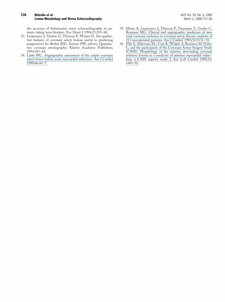
34. Little WC. Angiographic assessment of the culprit coronaryartery lesion before acute myocardial infarction. Am J Cardiol1990;66:44–7.
35. Moise A, Lesperance J, Theroux P, Taeymans Y, Goulet C,Bourassa MG. Clinical and angiographic predictors of newtotal coronary occlusion in coronary artery disease: analyses of313 nonoperated patients. Am J Cardiol 1984;54:1176–81.
36. Ellis S, Alderman EL, Cain K, Wright A, Bourassa M, FisherL, and the participants of the Coronary Artery Surgery Study(CASS). Morphology of the anterior descending coronaryterritory lesions as a predictor of anterior myocardial infarc-tion: a CASS registry study. J Am Coll Cardiol 1989;13:1481–91.
726 Beleslin et al. JACC Vol. 33, No. 3, 1999Lesion Morphology and Stress Echocardiography March 1, 1999:717–26