Coronary CT angiography: Dose reduction strategies
8
TOPIC HIGHLIGHT World Journal of Cardiology WJC Online Submissions: http://www.wjgnet.com/esps/ bpgoffi[email protected] doi:��.����/wjc.��.i��.��� ��.����/wjc.��.i��.��� /wjc.��.i��.��� World J �ard�ol �ard�ol ���� December ��; �(��): ���-�7� ISSN ����-���� (online) ����-���� (online) (online) © ���� Baishideng Publishing Group Co., Limited. All rights reser�ed. Coronary CT angiography: Dose reduction strategies Akmal Sabarudin, Zhonghua Sun Akmal Sabarudin, Diagnostic Imaging and Radiotherapy Pro- gram, School of Diagnostic and Applied Health Sciences, Fac- ulty of Health Sciences, Universiti Kebangsaan Malaysia, Kuala Lumpur 50300, Malaysia Zhonghua Sun, Discipline of Medical Imaging, Department of Imaging and Applied Physics, Curtin University, Perth 6845, Western Australia, Australia Author contributions: Both authors wrote the paper. Correspondence to: Zhonghua Sun, �h�, Associate �ro� Zhonghua Sun, �h�, Associate �ro� fessor, Discipline of Medical Imaging, Department of Imaging and Applied Physics, Curtin University, GPO Box U1987, Perth 6845, Western Australia, Australia. [email protected] Telephone: +61-8-92667509 Fax: +61-8-92662377 Received: July 3, 2013 Revised: July 24, 2013 Accepted: August 20, 2013 August 20, 2013 �ublished online: December 26, 2013 Abstract With the introduction of 64� and post�64 slice com� puted tomography (CT) technology, coronary CT angi� ography has been increasingly used as a less invasive modality for the diagnosis of coronary artery disease. �espite its high diagnostic value and promising results compared to invasive coronary angiography, coronary CT angiography is associated with high radiation dose, leading to potential risk of radiation�induced cancer. A variety of dose�reduction strategies have been re� ported recently to reduce radiation dose with effective outcomes having been achieved. This article presents an overview of the various methods currently used for radiation dose reduction. © 2013 Baishideng �ublishing Group Co., Limited. All rights reserved. Key words: Coronary artery disease; Coronary com� puted tomography angiography; Multislice computed tomography; Radiation dose; �ose reduction Core tip: Various dose�reduction strategies of coronary computed tomography angiography have been discussed in this article with the aim of providing readers with a comprehensive summary of the effectiveness of these radiation reduction approaches. Sabarudin A, Sun Z. Coronary CT angiography: Dose reduction strategies. World J Cardiol 2013; 5(12): 465-472 Available from: URL: http://www.wjgnet.com/1949-8462/full/v5/i12/465.htm DOI: http://dx.doi.org/10.4330/wjc.v5.i12.465 INTRODUCTION Coronary computed tomography angiography (CCTA) pro- cedure has been known as an effective technique in non- invasive coronary artery assessment. With high accuracy in the detection of coronary artery disease, this makes CCTA accepted as a widely used diagnostic tool in cardiac imag- ing [1-4] . However, radiation dose of CCTA that has been reported in the literature is the greatest concern and varies a great deal depending on the scanning parameter settings. There are many factors influencing the overall radiation exposure including tube voltage, tube current, scan range, scanner geometry, the electrocardiogram (ECG)-gating ap- plication either prospective or retrospective ECG-gating, slice thickness and pitch value selection (for helical scan mode). Most of the parameters are controlled, monitored and modulated by the computed tomography (CT) operator during the procedure in order to obtain an optimum image quality. Therefore, all factors need to be taken into consid- eration in minimizing the radiation exposure to achieve the goal of “as low as reasonably possible”. Previous studies have also reported that standard CCTA procedure with the use of retrospective ECG-gated technique results in very high radiation dose, which ranged from 13.4 to 31.4 mSv [5-7] . This has raised serious concerns in the literature due to the potential risk of radiation-induced malignancy result- ing from CCTA. Therefore, several dose-saving strategies have been introduced to deal with radiation dose issues, and Zhonghua Sun, PhD, Associate Professor, Series Editor ��� December ��, ����|Volume �|Issue ��| WJC|www.wjgnet.com
Transcript of Coronary CT angiography: Dose reduction strategies
TOPIC HIGHLIGHT
World Journal of CardiologyW J C
Online Submissions: http://www.wjgnet.com/esps/[email protected]:��.����/wjc.��.i��.�����.����/wjc.��.i��.���/wjc.��.i��.���
World J �ard�ol�ard�ol ���� December ��; �(��): ���-�7�ISSN ����-���� (online)����-���� (online) (online)
© ���� Baishideng Publishing Group Co., Limited. All rights reser�ed.
Coronary CT angiography: Dose reduction strategies
Akmal Sabarudin, Zhonghua Sun
Akmal Sabarudin, Diagnostic Imaging and Radiotherapy Pro-gram, School of Diagnostic and Applied Health Sciences, Fac-ulty of Health Sciences, Universiti Kebangsaan Malaysia, Kuala Lumpur 50300, MalaysiaZhonghua Sun, Discipline of Medical Imaging, Department of Imaging and Applied Physics, Curtin University, Perth 6845, Western Australia, AustraliaAuthor contributions: Both authors wrote the paper.Correspondence to: Zhonghua Sun, �h�, Associate �ro�Zhonghua Sun, �h�, Associate �ro�fessor, Discipline of Medical Imaging, Department of Imaging and Applied Physics, Curtin University, GPO Box U1987, Perth 6845, Western Australia, Australia. [email protected]: +61-8-92667509 Fax: +61-8-92662377Received: July 3, 2013 Revised: July 24, 2013Accepted: August 20, 2013August 20, 2013�ublished online: December 26, 2013
AbstractWith the introduction of 64� and post�64 slice com�puted tomography (CT) technology, coronary CT angi�ography has been increasingly used as a less invasive modality for the diagnosis of coronary artery disease. �espite its high diagnostic value and promising results compared to invasive coronary angiography, coronary CT angiography is associated with high radiation dose, leading to potential risk of radiation�induced cancer. A variety of dose�reduction strategies have been re�ported recently to reduce radiation dose with effective outcomes having been achieved. This article presents an overview of the various methods currently used for radiation dose reduction.
© 2013 Baishideng �ublishing Group Co., Limited. All rights reserved.
Key words: Coronary artery disease; Coronary com�puted tomography angiography; Multislice computed tomography; Radiation dose; �ose reduction
Core tip: Various dose�reduction strategies of coronary computed tomography angiography have been discussed
in this article with the aim of providing readers with a comprehensive summary of the effectiveness of these radiation reduction approaches.
Sabarudin A, Sun Z. Coronary CT angiography: Dose reduction strategies. World J Cardiol 2013; 5(12): 465-472 Available from: URL: http://www.wjgnet.com/1949-8462/full/v5/i12/465.htm DOI: http://dx.doi.org/10.4330/wjc.v5.i12.465
INTRODUCTIONCoronary computed tomography angiography (CCTA) pro-cedure has been known as an effective technique in non-invasive coronary artery assessment. With high accuracy in the detection of coronary artery disease, this makes CCTA accepted as a widely used diagnostic tool in cardiac imag-ing[1-4]. However, radiation dose of CCTA that has been reported in the literature is the greatest concern and varies a great deal depending on the scanning parameter settings. There are many factors influencing the overall radiation exposure including tube voltage, tube current, scan range, scanner geometry, the electrocardiogram (ECG)-gating ap-plication either prospective or retrospective ECG-gating, slice thickness and pitch value selection (for helical scan mode).
Most of the parameters are controlled, monitored and modulated by the computed tomography (CT) operator during the procedure in order to obtain an optimum image quality. Therefore, all factors need to be taken into consid-eration in minimizing the radiation exposure to achieve the goal of “as low as reasonably possible”. Previous studies have also reported that standard CCTA procedure with the use of retrospective ECG-gated technique results in very high radiation dose, which ranged from 13.4 to 31.4 mSv[5-7]. This has raised serious concerns in the literature due to the potential risk of radiation-induced malignancy result-ing from CCTA. Therefore, several dose-saving strategies have been introduced to deal with radiation dose issues, and
Zhonghua Sun, PhD, Associate Professor, Series Editor
��� December ��, ����|Volume �|Issue ��|WJC|www.wjgnet.com
Sabarudin A et al . Coronary CT angiography
these techniques include anatomy-based tube current mod-ulation[8,9], ECG-controlled tube current modulation[10,11], tube voltage reduction[12,13], a high-pitch scanning[14,15] and prospective ECG-triggered CCTA[16,17]. This article is writ-ten purposely to provide information about the strategies that could be used to further reduce the radiation dose to patient during CCTA procedure.
STRATEGIES FOR RADIATION DOSE REDUCTION IN CCTAAnatomy-based tube current modulationTube current is an important element that is directly relat-ed to radiation dose and image quality. With rapid devel-opments of CT technology, implementation of automatic tube current modulation allows significant reduction in ra-diation dose for CT examinations. In CT examination, au-tomatic tube current modulation can be defined as a series of techniques that enable automatic adjustment of the tube current in x-, y-plane (angular modulation) or z-plane (z-axis modulation), according to the size and attenuation characteristics of the human body. The purpose of these adjustments is to achieve optimum image quality with low radiation dose. The term automatic tube current modula-tion is similar to automatic exposure-control that is com-monly used in conventional radiography[18,19]. Anatomy-based tube current modulation is then divided into two modes namely angular modulation and z-axis modulation.
Angular modulation (x-y plane)Since the shape of patients body is not symmetrical [antero-posterior (AP) vs lateral], angular-modulation techniques au-tomatically adjust the tube current for each projection angle to the appropriate attenuation according to patient’s ana-tomical structures. Without angular modulation, the tube current is held constant over the 360° rotation, regardless of the patient attenuation profile. The angular-modulation technique reduces tube current as a function of projection angles for low-attenuation projections (AP vs lateral pro-jections). This technique calculates the modulation func-tion from the online attenuation profile of the patient. The modulation function data are processed and sent to the generator control for further tube current modulation with a delay of 180° from the X-ray generation angle. In asymmetrical regions being scanned such as the shoulders in chest CT, the X-ray attenuation is substantially less in the AP than in the lateral direction. The radiation dose reduction could be achieved up to 90% with application of the angular-modulation technique[20]. Therefore, the technique of angular modulation helps in improving dose efficiency in the x- and y-axis by reducing radiation expo-sure in a particular scanning plane.
Z-axis modulationThe principle of z-axis-modulation technique is different from that of angular modulation. Unlike angular modula-tion, the z-axis modulation technique adjusts the tube cur-rent automatically to maintain a user-specified quantum
noise level in the image data. It provides a noise index to allow users to select the amount of X-ray noise that will be presented in the reconstructed images. Using a local-izer radiograph, the scanner computes the tube current re-quired obtaining images with a selected noise level. Hence, z-axis modulation attempts to make all images have a similar noise irrespective of patient size and anatomy. The noise index value is approximately equal to the im-age noise (standard deviation) in the central region of an image of a uniform phantom. However, the actual noise measured on the image by drawing a region of interest that will differ from the noise index selected for scanning. This is due to the fact that noise index settings only adjust the tube current, whereas the standard deviation is also affected by other parameters, including the reconstruction algorithm, the reconstructed section thickness (if different from the prospective thickness), the use of image space filters, variations in patient anatomy and patient motion, and the presence of beam-hardening artifacts.
The CARE Dose 4D protocol (Siemens, Medical Solu-tions, Erlangen, Germany) was then introduced in order to adapt the tube current to the patient’s individual anatomy and modulate the tube current in the section with the low-est dose levels. Previous studies have shown that 20%-60% dose reduction was achieved depending on the anatomic region and patient habitus, with improved image quality[21]. Another study combining angular and z-axis modulation (3D Auto mA; GE Yokogowa Medical Systems, Tokyo, Japan) reported significant dose reductions (60%) in abdominal-pelvic CT examinations[22]. This technique uses a single localizer radiograph to determine patient asymmetry and appropriate angular and z-axis modulation for the patient. The investigators added noise (computer modification of original raw scan data to simulate lower tube current noise levels) to patients’ scan data to produce images and calcu-late the radiation dose reduction.
A lower minimum tube current may result in reduced exposure to patients, which occasionally increases im-age noise in smaller patients scanned with a substantially reduced tube current. Generally, larger patients receive higher tube current with z-axis modulation if a fixed-tube-current technique used in order to maintain the selected image noise. In contrast, with automatic tube current modulation, the tube current is inconsistent throughout the scan and thus results in the diagnostic image quality with reduced radiation dose. The main limitation of auto-matic tube current modulation is the lack of uniformity between techniques developed by different vendors.
ECG-CONTROLLED TUBE CURRENT MODULATIONThe idea of decreasing radiation doses associated with tube current modulation in CT stimulates manufacturers to improve the CCTA examinations. One of the most recently developed methods, CARE dose 4D by Siemens Medical Solutions, which combining the effects of angu-lar and z-axis modulation techniques[23]. Virtually all ana-
��� December ��, ����|Volume �|Issue ��|WJC|www.wjgnet.com
tomic regions in the thorax, abdomen, and pelvis have benefited from these sophisticated techniques that result in considerable significant dose reduction[10,24].
However, the z-axis modulation principle in CARE dose 4D was not compatible with ECG pulsing. ECG-pulsed tube current modulation is the most significant improvement in minimizing radiation from CT technol-ogy and it is the only technique dedicated to cardiac im-aging. ECG pulsing is performed online during cardiac CT examination which allows a decrease in radiation exposure of between 30% and 50%. The radiation dose is reduced by modulating the tube current output during the systolic phase[25]. Moreover, the algorithm for ECG-dependent dose modulation also represents a very effec-tive tool for limiting radiation dose in the vast majority of patients undergoing cardiac CT studies.
In ECG-controlled tube current modulation technique, a high tube current with optimal image quality is applied only during the diastolic phase of the cardiac cycle, in which images are most likely to be reconstructed with minimal artifacts, while in the systolic phase, a low tube current (50% of normal tube current) is applied. Image reconstruction during cardiac CT examinations is usually performed in ventricular mid-diastole phase due to less cardiac motion that causes blurring of cardiac structures. Thus, high quality diagnostic images can be acquired dur-ing the diastolic phase[26]. However, this method totally depends on the patient’s heart rate and requires a regular sinus rhythm in order to prevent poor image quality. Un-fortunately, the ECG-controlled tube current modulation algorithm cannot be performed in the presence of ar-rhythmias such as premature extra beats. Thus, this algo-rithm may not be useful in patients with arrhythmias.
LOW TUBE VOLTAGESince radiation dose varies with the square of tube volt-age, an application of lower tube voltage during CT data acquisition is another approach for radiation dose reduction. A previous study by Huda et al[27] showed that reducing the X-ray tube potential from 140 to 80 kVp at constant tube current decreased the radiation dose by a factor of about 3.4. Consequently, image contrast and image noise will definitely be increased because of fewer numbers of photons produced[27-29]. However, since the contrast-to-noise ratio (CNR) and signal-to-noise ratio are the key factor of CT image quality, noise is rather irrel-evant if the level of contrast or amount of signals are too high[28]. The change in image contrast is dependent on the anatomic number (Z) of the structures being investigated. The image structure with high-anatomic-number becomes significantly more prominent than image of low-anatom-ic-number structures (soft tissue) in the application of low tube voltages[27].
It has been confirmed that diagnostic image quality was not affected by lower tube voltages in pediatric CT in-vestigations. Similarly, in a phantom study by Siegel et al[29] showed that reduced beam energy in contrast-enhanced
pediatric CT decreased the radiation dose without affect-ing image contrast and image noise. Moreover, the inter-relationship between beam energy and tube output has been described by Boone et al[30] in the context of image noise characterization in CT techniques by using tube voltages of 80-140 kVp and tube currents of 10-300 mA. Provided the tube current-time product was appropri-ately adapted, radiation dose can be significantly reduced at lower tube voltage while CNR remained at a constant level. Cody et al[31] reported that the use of 80-kVp tube voltage resulted in beam-hardening artifacts and thus rec-ommended the use of 100- to 120-kVp settings in pediat-ric patients. For non-cardiac CT studies with kilovoltage reduction, an increase of the tube current by 50% has been proposed to maintain image quality and to reduce the dose estimation concurrently[31]. However, a further increase in tube current is limited with the available stan-dard protocols for cardiac CT scanning on the studied CT scanners. Therefore, a trade-off between dose saving and increased image noise has to be considered with cur-rent cardiac CT protocols.
Previous study compared the diagnostic image quality of the coronary artery segments in order to detect steno-sis in various scan protocols[32]. In this qualitative analysis, no deterioration of image quality was detected in most of the scan protocols inclusive of the ECG-dose modulation and the 100-kVp tube voltage for both 16- and 64-slice CT scanners. The value of this analysis is only limited by a potential selection bias of the scanning protocols. Image obtained with 120-kVp scan protocol without ECG mod-ulation (on patients with arrhythmia) are likely to present with more non-diagnostic coronary segments, even when no dose-saving algorithms were applied. However, the impact of dose-saving algorithms on the detection of cal-cified and non-calcified plaques remains unknown. There-fore, further studies are needed to investigate the balance between dose savings and maintained diagnostic image quality for CCTA investigations.
HIGH PITCH VALUEWith the recent advent of second-generation of dual-source, another low-dose technique has been introduced for cardiac CT which is high-pitch scanning mode[33]. This technique was successfully tested with dual-source 128-slice CT in retrospective ECG-gating protocol. In this technique, the data are acquired in a spiral mode while the X-ray table runs with a very high pitch of 3.4 equaling to a table feed of 46 cm/s. When this high-pitch mode is used, the entire heart is scanned within one single cardiac cycle, generally during the diastolic phase (75% R-R interval). The temporal resolution for this system is 75 ms, with the gantry rotation time of 280 ms and only quarter rota-tions for data reconstruction. Early reports on phantom studies have shown that the purpose of this scan mode is to deliver images of diagnostic quality at a low radia-tion dose. Moreover, two studies have successfully proved that feasibility of this high-pitch mode technique also in
��7 December ��, ����|Volume �|Issue ��|WJC|www.wjgnet.com
Sabarudin A et al . Coronary CT angiography

patients by using the remodeled first generation of dual-source 64-slice CT scanners with effective dose less than 1 mSv[15,34]. Then, several recent studies also have reported similar results[35-37]. In addition to low dose aspect, high diagnostic accuracy has been achieved with the high-pitch dual-source CT[38].
In order to apply the high-pitch mode, several re-quirements must be fulfilled. Firstly, dual-source geom-etry is necessary in order to obtain the projection data by the second detector for gaps fill-up due to rapid table movement. In this way, the pitch can be increased up to 3.4 while allowing image reconstruction, although the limited field of view is covered by both detectors. A quarter rotation of data per measurement is used for image reconstruction, and each of the individual axial images has a temporal resolution of a quarter of the rotation time trot/4. Thus, the overlapping of radiation exposure can be avoided with the application of high pitch resulting in radiation dose reduction to the mini-mum level[39]. Secondly, a higher temporal resolution is essential to enable single cardiac cycle reconstruction without image distortion due to motion artifacts. Third-ly, patient’s heart rate must be regular and consistent in order to obtain a good image quality. With used of high pitch mode, the examination table is accelerated to the maximum speed during data acquisition which is trig-gered by the R-peak of the heartbeat. The examination table could not be accelerated in an infinitely small time period; therefore, it has to be set in motion sufficiently earlier prior to scanning acquisition. Inconstant heart rates lead to inaccurate positioning of the data acquisi-tion window, with data being acquired either too early (if heart rate decreases) or too late (if heart rate increases) in the cardiac cycle. Inconsistent heart rates would com-promise image quality by stair-step artifacts.
Finally, high pitch mode requires patient with low heart rates (< 65 bpm). In order to obtain a motion-free artifact, CT data acquisition can possibly be performed during a single diastolic period if the patient heart rate is constantly lower than 65 bpm[39]. On the other hand, patients with high heart rates may not yield diagnostic image quality of the coronary arteries due to a narrow diastolic exposure of R-R interval window and therefore, tube current mod-ulation is required for adjustment accordingly[35].
ITERATIVE RECONSTRUCTION METHODSSAlternative image reconstruction techniques such as it-erative reconstruction have been used mainly in nuclear medicine studies[40,41]. In CCTA, iterative reconstruction such as adaptive statistical iterative reconstruction (ASIR) (GE Healthcare) has been introduced as a new recon-struction algorithm[42]. Iterative reconstruction is a meth-od to reconstruct 2D and 3D images from measured projections of an object. However, unlike filtered back projection, iterative reconstruction starts with an initial estimate of the object which is subsequently improved in
a stepwise fashion by comparing the synthesized image to the one acquired with projection data and improving the previous estimation.
Moreover, iterative reconstruction reduces image noise by iteratively comparing the acquired image to a mod-eled projection. This reconstruction algorithm is used to help deal with one of the primary issues of dose and tube current reduction for CCTA. Since iterative reconstruc-tion has been consistently associated with image quality improvement, especially improving CNR, it has the pos-sibility of improving spatial resolution[43,44]. With faster computer technologies and adapted techniques, the use of iterative reconstruction for cardiac CT imaging has been increasingly studied and the reconstruction speed now al-lows its use in clinical practice. Iterative reconstruction has been shown to reduce noise, improve image quality and reduce radiation dose not only in body CT but in coro-nary CT. The ASIR technique was reported to provide about 27% of radiation dose reduction compared to that standard filtered back projection reconstruction[43]. In ad-dition, image quality and the proportion of interpretable segments were also improved with the application of 40% or 60% ASIR in CCTA reconstruction compared to that filtered back projection reconstruction[43]. Another study using the similar reconstruction method with different no-menclature, namely iterative reconstruction in image space (IRIS) also resulted in significant reduction of image noise and improved subjective image quality[45]. However, the main limitation to its routine use is the high computational cost, which can be 100-1000 times higher than for filtered back projection[46].
Moreover, iterative reconstruction does not assume that the measured signal is free of noise due to x-ray photon statistics or electronic noise but rather uses more accurate statistical modeling during the reconstruc-tion process[42]. This enables improved noise properties in the reconstructed images, while maintaining spatial resolution and other image quality parameters. The use of iterative reconstruction techniques is expected to in-crease in CT as computational processing improves and algorithms become more robust and easy to apply. Ow-ing to more powerful iterative reconstruction algorithms are emerging, the impact of these techniques may show greater noise reduction and thereby permit further re-ductions in radiation exposure to patients.
PROSPECTIVELY ECG-TRIGGERED CORONARY CT ANGIOGRAPHY
Various strategies have been developed to reduce radi-ation exposure to patients, and prospectively ECG-gated CT coronary angiography is remained as the most impor-tant and effective in reducing the radiation dose which also called step-and-shoot mode. The step-and-shoot mode is characterized by turning on the x-ray tube only at a predefined time point of the cardiac cycle, usually in mid-diastole, while keeping the patient table station-ary. The x-ray exposure time of this technique is short,
��� December ��, ����|Volume �|Issue ��|WJC|www.wjgnet.com
Sabarudin A et al . Coronary CT angiography
and thus, low radiation doses ranging between 1.2 and 4.3 mSv have been reported using various 64-slice and first-generation of dual-source 64-slice CT[32,47]. Most importantly, this low-dose step-and-shoot method is still being able to produce high diagnostic accuracy for the detection of coronary stenosis[32,48].
Unlike standard retrospective ECG-gating, where the tube output (in mA) is constant throughout the data ac-quisition during spiral CT which results in high radiation dose, prospective triggering is performed with sequential scans. In prospective triggering, the tube current is turned off for most of the scan period and is triggered by the ECG to be ‘‘on’’ only for a short period during diastole. Thus, this results in remarkable reduction in radiation dose[49]. With application of prospective ECG triggering, the radiation dose of CCTA can be reduced by up to 83% when compared to that standard retrospective ECG gat-ing technique[47,49].
Prospectively ECG-triggered technique uses axial im-ages and an incrementally moving table to cover the heart with minimal overlap of axial slices. Cardiac imaging with electron beam CT also uses prospective data acquisition triggered by ECG. Prospective triggered technique in car-diac CT is not new and it was actually being used in early 1980 by Dr. Godfrey Hounsfield with conventional single-slice CT[50]. It was recognized that CT image synchroniza-tion with heart diastolic phase was optimal for imaging the heart. Unfortunately, the findings were not being achieved when the patient heart rate increases.
When a 64-slice system is used, the scan is prescribed by using 3-5 incremental of 64 mm × 0.625 mm (40 mm) image groups which requires 2-4 incremental table translations of 35 mm. Thus, allow for 5 mm of overlap. The minimum interscan delay is approximately between 0.6 and 1.0 second which normally requires skipping a cardiac cycle between data acquisitions which results in one image acquisition per 2 cardiac R-R cycles[49]. How-ever, the process will be faster with larger detectors (128-, 256- or 320-slice CT) being used. The detector width de-
termines the number of steps/scans to cover the entire heart and complete an examination. For instance, the dual-source 64-slice CT has a narrower detector array (32 mm × 2 mm × 0.6 mm = 38.4 mm per acquisition); thus, it takes more incremental steps (normally 4-5 car-diac cycles) to cover the heart and complete an examina-tion than with the 320-row system (320 × 0.5 mm = 160 mm) which covers the heart in a single acquisition[51].
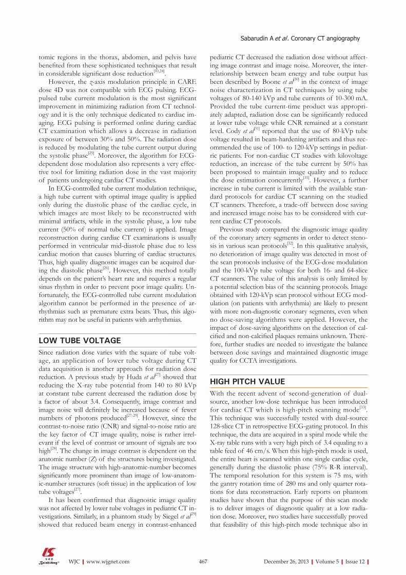
Prospectively ECG-triggered technique has a limited number of cardiac phases available for reconstruction. Therefore, mid-diastolic phase (75% of R-R interval) was always being selected for data acquisition for all subjects. In addition, by using add-on ‘padding’ will al-low more cardiac phases for reconstruction. Padding technique is described as prolonging the acquisition window in order to allow the reconstruction to adapt with minor heart rate variations and to produce consistent im-age quality. Padding turns the X-ray tube on before and after the minimum or actual acquisition time (millisec-onds) required. Available padding options with current software ranges from 0 to 200 ms (Figure 1). No padding is required for patient with stable heart rates and minimal heart rate variability. However, radiation dose also will increase with application of padding window due to ex-pense of radiation exposure on the particular windows phase[24,49].
Other than adjusting prospective triggering parameters in order to adapt with high heart rates, application of β-blockade for heart rate control is also commonly used in CCTA to produce better results. However, precautions have to be taken in patients who are contraindicated to β-blockage agent. Alternatively, calcium channel blocker could be used in order to reduce the heart rate. The maxi-mum of 15 mg of intravenous metoprolol (β-blocker) or 40 mg of intravenous diltiazem (calcium channel blocker) is recommended prior to the scan in order to control the heart rat[49,52].
The major drawback of prospective ECG triggering is that cardiac functional analysis is unavailable. Since pro-
Minimum acquisition window (no padding)
�adding windows �adding windows100 ms 100 ms50 ms 50 ms0 ms 0 ms
Figure 1 Use of extra tube-on time to acquire image data during additional cardiac phases. Padding turns tube on prior to minimum half-scan time and leaves it on afterwards. It is recommended in cases when heart rate varies during examination.
��� December ��, ����|Volume �|Issue ��|WJC|www.wjgnet.com
Sabarudin A et al . Coronary CT angiography
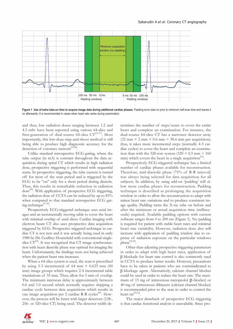
Table 1 Dose reduction strategies and corresponding effectiveness in dose reduction in coronary computer tomography angiography
spective technique acquires data during a limited portion of the cardiac cycle, it cannot be used to evaluate cardiac function. Both quantitative and qualitative functions, ei-ther global or regional, require images to be reconstructed throughout the entire cardiac cycle. If the clinical scenario or referring physician requires information about cardiac function, then retrospective gating must be undertaken. Heart rate variability is another limitation for the prospec-tive ECG triggered technique. Heart rate variability of > 5 beat/min is considered not applicable for prospective triggering. Therefore, the scan has to be reverted into retrospective ECG gating technique if patients’ heart rate elevated or heart rate variability does not meet the requirement after β-blocker has been given[49]. However, the prospective ECG triggered technique in patients with higher heart rates still produces diagnostic images. CT scanner with higher detector arrays is an alternative to obtain CCTA in patients with high or irregular heart rates. It has been reported that high diagnostic value could be achieved with 320-slice CT angiography in the diagnosis of CAD, with image quality independent of heart rate[51]. The improved temporal resolution (175 ms) and increased coverage scan value (160 mm) of 320-slice CT results in robust image quality within a wide range of heart rates; thus providing the opportunity to image patients with higher heart rates without requiring pre-examination beta-blockage[51].
CONCLUSIONRecent technological developments have led coronary CT to be used widely and the acceptable indications for CCTA imaging become broaden. However, despite the strength of CCTA, the potential risk of radiation- induced malignancy has received attention in scientific publications although it may be unproven. Therefore, appropriate referral of CT studies, lowering tube voltage, using tube current modula-
tion, increasing the pitch value, applying iterative recon-struction technique and implementation of prospective ECG-triggering CCTA enable CCTA to be performed at a low dose while preserving good image quality and diagnos-tic accuracy. Table 1 summarises above-mentioned dose-reduction strategies and corresponding effectiveness in the reduction of radiation dose associated with CCTA.
REFERENCES� Buth J, Disselhoff B, Sommeling C, Stam L. Color-flow duplex
criteria for grading stenosis in infrainguinal �ein grafts. J Vasc Surg ����; 14: 7��-7��; discussion 7��-7�� [PMID: ������� DOI: ��.����/j.jacc.����.��.��7]
� Sun Z, Ng KH. Diagnostic �alue of coronary CT angiography with prospecti�e ECG-gating in the diagnosis of coronary artery disease: a systematic re�iew and meta-analysis. Int J �ard�ovasc Imag�ng ����; 28: ����-���� [PMID: �������� DOI: ��.���7/s�����-���-����-�]
� Sun Z, Choo GH, Ng KH. Coronary CT angiography: current status and continuing challenges. Br J Rad�ol ����; 85: ���-��� [PMID: ��������]
� Sun Z. Multislice CT angiography in coronary artery disease: Technical de�elopments, radiation dose and diagnostic �alue. World J �ard�ol ����; 2: ���-��� [PMID: ��������]
� d'Agostino AG, Remy-Jardin M, Khalil C, Delannoy-Deken V, Flohr T, Duhamel A, Remy J. Low-dose ECG-gated ��-slices he-lical CT angiography of the chest: e�aluation of image quality in ��� patients. Eur Rad�ol ����; 16: ���7-���� [PMID: ��������]
� Johnson TR, Nikolaou K, Wintersperger BJ, Leber AW, �on Ziegler F, Rist C, Buhmann S, Knez A, Reiser MF, Becker CR. Dual-source CT cardiac imaging: initial experience. Eur Rad�ol ����; 16: ����-���� [PMID: ��77����]
7 Poll LW, Cohnen M, Brachten S, Ewen K, Mödder U. Dose re-duction in multi-slice CT of the heart by use of ECG-controlled tube current modulation (“ECG pulsing”): phantom measure-ments. Rofo ����; 174: ����-���� [PMID: ���7����]
� Jung B, Mahnken AH, Stargardt A, Simon J, Flohr TG, Schaller S, Koos R, Günther RW, Wildberger JE. Indi�idually weight-adapt-Indi�idually weight-adapt-ed examination protocol in retrospectively ECG-gated MSCT of the heart. Eur Rad�ol ����; 13: ����-���� [PMID: ��������]
� Starck G, Lönn L, Cederblad A, Forssell-Aronsson E, Sjöström L, Alpsten M. A method to obtain the same le�els of CT image
Techniques Advantages Pitfalls Dose reduction
Tube current modulation: anatomy-based Suitable for unsymmetrical body habitus No apparent reduction in CCTA proce-dure due to homogeneity of the body
thickness in the cardiac region
��%-��%�
Tube current modulation: ECG- controlled
Dedicated for cardiac imagingModulates tube current output during systolic phase
Heart rate must be regular ��%-��%
Low tube �oltage (kVp) Image structure with high-atomic number becomes more prominent than that with low-atomic number
Beam hardening artifacts may occurMay increase image noise which leads
to suboptimal image quality
Up to ��%
High pitch �alue Fast image acquisitionReduce motion artifacts
Patient heart rate must at < �� bpm and regular
Can only be performed on second gen-eration of dual-source CT scanner
Up to ��%
Iterati�e reconstruction algorithms Impro�e contrast-to-noise ratio and spatial resolutionReduce image noise
High computational cost Up to ��%
Prospecti�ely ECG- triggered CCTA High sensiti�ity in the detection of CADTube current is only ‘on’ in a short period during dia-
stolic phase
Limited number for cardiac reconstruc-tion phases
No cardiac functional analysis
Up to ��%
�Applied to the abdominal-pel�ic region. CT: Computed tomography; CCTA: Coronary CT angiography; CAD: Coronary artery disease; ECG: Electrocar-diogram.
�7� December ��, ����|Volume �|Issue ��|WJC|www.wjgnet.com
Sabarudin A et al . Coronary CT angiography

noise for patients of �arious sizes, to minimize radiation dose. Br J Rad�ol ����; 75: ���-��� [PMID: ��������]
�� Abada HT, Larchez C, Daoud B, Sigal-Cinqualbre A, Paul JF. MDCT of the coronary arteries: feasibility of low-dose CT with ECG-pulsed tube current modulation to reduce radiation dose. AJR Am J Roentgenol ����; 186: S��7-S��� [PMID: ��7�����]
�� Wintersperger B, Jakobs T, Herzog P, Schaller S, Nikolaou K, Suess C, Weber C, Reiser M, Becker C. Aorto-iliac multide-tector-row CT angiography with low kV settings: impro�ed �essel enhancement and simultaneous reduction of radia-tion dose. Eur Rad�ol ����; 15: ���-��� [PMID: ������7�]
�� Geleijns J, Sal�adó Artells M, Veldkamp WJ, López Tortosa M, Calzado Cantera A. Quantitati�e assessment of selec-ti�e in-plane shielding of tissues in computed tomography through e�aluation of absorbed dose and image quality. Eur Rad�ol ����; 16: ����-���� [PMID: ��������]
�� Hohl C, Mühlenbruch G, Wildberger JE, Leidecker C, Süss C, Schmidt T, Günther RW, Mahnken AH. Estimation of radia-Estimation of radia-tion exposure in low-dose multislice computed tomography of the heart and comparison with a calculation program. Eur Rad�ol ����; 16: ����-���� [PMID: ��������]
�� Achenbach S, Marwan M, Ropers D, Schepis T, Pflederer T, Anders K, Kuettner A, Daniel WG, Uder M, Lell MM. Coro-Coro-nary computed tomography angiography with a consistent dose below � mS� using prospecti�ely electrocardiogram-triggered high-pitch spiral acquisition. Eur Heart J ����; 31: ���-��� [PMID: ����7��7 DOI: ��.����/eurheartj/ehp�7�]
�� Achenbach S, Marwan M, Schepis T, Pflederer T, Bruder H, All-mendinger T, Petersilka M, Anders K, Lell M, Kuettner A, Ropers D, Daniel WG, Flohr T. High-pitch spiral acquisition: a new scan mode for coronary CT angiography. J �ard�ovasc �omput Tomogr ����; 3: ��7-��� [PMID: �������� DOI: ��.����/j.jcct.����.��.���]
�� Paul JF, Abada HT. Strategies for reduction of radiation dose in cardiac multislice CT. Eur Rad�ol ���7; 17: ����-���7 [PMID: �7������]
�7 Sun Z, Ng KH. Prospecti�e �ersus retrospecti�e ECG-gated multislice CT coronary angiography: a systematic re�iew of radiation dose and diagnostic accuracy. Eur J Rad�ol ����; 81: e��-��� [PMID: �������7]
�� Deetjen A, Möllmann S, Conradi G, Rolf A, Schmermund A, Hamm CW, Dill T. Use of automatic exposure control in mul-tislice computed tomography of the coronaries: comparison of ��-slice and ��-slice scanner data with con�entional coronary angiography. Heart ���7; 93: ����-���� [PMID: �7�����7]
�� Kalra MK, Maher MM, Toth TL, Schmidt B, Westerman BL, Morgan HT, Saini S. Techniques and applications of automatic tube current modulation for CT. Rad�ology ����; 233: ���-��7 [PMID: ��������]
�� Greess H, Nömayr A, Wolf H, Baum U, Lell M, Böwing B, Kal-ender W, Bautz WA. Dose reduction in CT examination of chil-dren by an attenuation-based on-line modulation of tube current (CARE Dose). Eur Rad�ol ����; 12: ��7�-��7� [PMID: ������7�]
�� Suess C, Chen X. Dose optimization in pediatric CT: current technology and future inno�ations. Ped�atr Rad�ol ����; 32: 7��-7��; discussion 7��-7�� [PMID: ��������]
�� Horiuchi T. Study on �D modulation auto mA. Jpn Soc Rad�ol Technol ����; 78: ���
�� Schwartzman D, Lacomis J, Wigginton WG. Characterization of left atrium and distal pulmonary �ein morphology using multidimensional computed tomography. J Am �oll �ard�ol ����; 41: ����-���7 [PMID: ��7�����]
�� Hausleiter J, Meyer T, Hadamitzky M, Huber E, Zankl M, Martinoff S, Kastrati A, Schömig A. Radiation dose estimates from cardiac multislice computed tomography in daily prac-tice: impact of different scanning protocols on effecti�e dose estimates. ��rculat�on ����; 113: ����-���� [PMID: ��������]
�� Leber AW, Knez A, �on Ziegler F, Becker A, Nikolaou K, Paul S, Wintersperger B, Reiser M, Becker CR, Steinbeck G, Boekste-gers P. Quantification of obstructive and nonobstructive coro-nary lesions by ��-slice computed tomography: a comparati�e
study with quantitati�e coronary angiography and intra�as-cular ultrasound. J Am �oll �ard�ol ����; 46: ��7-��� [PMID: ��������]
�� Earls JP, Berman EL, Urban BA, Curry CA, Lane JL, Jennings RS, McCulloch CC, Hsieh J, Londt JH. Prospecti�ely gated trans-�erse coronary CT angiography �ersus retrospecti�ely gated helical technique: impro�ed image quality and reduced radia-tion dose. Rad�ology ����; 246: 7��-7�� [PMID: �������� DOI: ��.����/radiol.�����7����]
�7 Huda W, Scalzetti EM, Le�in G. Technique factors and im-age quality as functions of patient weight at abdominal CT. Rad�ology ����; 217: ���-��� [PMID: ��������]
�� Huda W. Dose and image quality in CT. Ped�atr Rad�ol ����; 32: 7��-7��; discussion 7��-7�� [PMID: ��������]
�� Siegel MJ, Schmidt B, Bradley D, Suess C, Hildebolt C. Radia-tion dose and image quality in pediatric CT: effect of techni-cal factors and phantom size and shape. Rad�ology ����; 233: ���-��� [PMID: �������7]
�� Boone JM, Geraghty EM, Seibert JA, Wootton-Gorges SL. Dose reduction in pediatric CT: a rational approach. Rad�ology ����; 228: ���-��� [PMID: �������7]
�� Cody DD, Moxley DM, Krugh KT, O’Daniel JC, Wagner LK, Eftekhari F. Strategies for formulating appropriate MDCT tech-niques when imaging the chest, abdomen, and pel�is in pediatric patients. AJR Am J Roentgenol ����; 182: ���-��� [PMID: ��������]
�� Herzog BA, Husmann L, Burkhard N, Gaemperli O, Valenta I, Tatsugami F, Wyss CA, Landmesser U, Kaufmann PA. Ac-curacy of low-dose computed tomography coronary angiogra-phy using prospective electrocardiogram-triggering: first clini-cal experience. Eur Heart J ����; 29: ���7-���� [PMID: �������� DOI: ��.����/eurheartj/ehn���]
�� Ertel D, Lell MM, Harig F, Flohr T, Schmidt B, Kalender WA. Cardiac spiral dual-source CT with high pitch: a feasibility study. Eur Rad�ol ����; 19: ���7-���� [PMID: �������� DOI: ��.���7/s�����-���-����-�]
�� Hausleiter J, Bischoff B, Hein F, Meyer T, Hadamitzky M, Thierfelder C, Allmendinger T, Flohr TG, Schömig A, Marti-noff S. Feasibility of dual-source cardiac CT angiography with high-pitch scan protocols. J �ard�ovasc �omput Tomogr ����; 3: ���-��� [PMID: ���77��� DOI: ��.����/j.jcct.����.��.���]
�� Goetti R, Leschka S, Baumüller S, Plass A, Wieser M, Desbio-lles L, Stolzmann P, Falk V, Marincek B, Alkadhi H, Feuchtner G. Low dose high-pitch spiral acquisition ���-slice dual-source computed tomography for the e�aluation of coronary artery bypass graft patency. Invest Rad�ol ����; 45: ���-��� [PMID: �����7�� DOI: ��.���7/RLI.�b���e����dfa�7e]
�� Lell M, Hinkmann F, Anders K, Deak P, Kalender WA, Uder M, Achenbach S. High-pitch electrocardiogram-triggered comput-ed tomography of the chest: initial results. Invest Rad�ol ����; 44: 7��-7�� [PMID: �������� DOI: ��.���7/RLI.�b���e����b�df7e]
�7 Lell M, Marwan M, Schepis T, Pflederer T, Anders K, Flohr T, Allmendinger T, Kalender W, Ertel D, Thierfelder C, Kuettner A, Ropers D, Daniel WG, Achenbach S. Prospecti�ely ECG-triggered high-pitch spiral acquisition for coronary CT angiog-raphy using dual source CT: technique and initial experience. Eur Rad�ol ����; 19: ��7�-���� [PMID: ��7����� DOI: ��.���7/s�����-���-����-�]
�� Leschka S, Stolzmann P, Desbiolles L, Baumueller S, Goetti R, Schertler T, Scheffel H, Plass A, Falk V, Feuchtner G, Marincek B, Alkadhi H. Diagnostic accuracy of high-pitch dual-source CT for the assessment of coronary stenoses: first experience. Eur Rad�ol ����; 19: ����-���� [PMID: ��7����� DOI: ��.���7/s�����-���-����-�]
�� Alkadhi H, Stolzmann P, Desbiolles L, Baumueller S, Goetti R, Plass A, Scheffel H, Feuchtner G, Falk V, Marincek B, Leschka S. Low-dose, ���-slice, dual-source CT coronary angiography: accuracy and radiation dose of the high-pitch and the step-and-shoot mode. Heart ����; 96: ���-��� [PMID: �������� DOI: ��.����/hrt.����.������]
�� Knesaurek K, Machac J, Vallabhajosula S, Buchsbaum MS. A
�7� December ��, ����|Volume �|Issue ��|WJC|www.wjgnet.com
Sabarudin A et al . Coronary CT angiography

new iterati�e reconstruction technique for attenuation correc-tion in high-resolution positron emission tomography. Eur J Nucl Med ����; 23: ���-��� [PMID: �������]
�� Liow JS, Strother SC, Rehm K, Rottenberg DA. Impro�ed resolu-tion for PET �olume imaging through three-dimensional iterati�e reconstruction. J Nucl Med ���7; 38: ����-���� [PMID: ��7����]
�� Cheng LCY, Fang T, Tyan J. Fast iterati�e adapti�e reconstruc-tion in low-dose CT imaging. In: Proceedings of the Proceed-ings of the IEEE International Conference on Image Processing. Sep ����; Lausanne, ����: ���-���
�� Leipsic J, Labounty TM, Heilbron B, Min JK, Mancini GB, Lin FY, Taylor C, Dunning A, Earls JP. Adapti�e statistical iterati�e reconstruction: assessment of image noise and image quality in coronary CT angiography. AJR Am J Roentgenol ����; 195: ���-��� [PMID: ��7����� DOI: ��.����/AJR.��.����]
�� Thibault JB, Sauer KD, Bouman CA, Hsieh J. A three-dimen-sional statistical approach to impro�ed image quality for mul-tislice helical CT. Med Phys ���7; 34: ����-���� [PMID: ���7����]
�� Bittencourt MS, Schmidt B, Seltmann M, Muschiol G, Ropers D, Daniel WG, Achenbach S. Iterati�e reconstruction in image space (IRIS) in cardiac computed tomography: initial experience. Int J �ard�ovasc Imag�ng ����; 27: ����-���7 [PMID: �������� DOI: ��.���7/s�����-���-�7��-�]
�� Wang G, Yu H, De Man B. An outlook on x-ray CT research and
de�elopment. Med Phys ����; 35: ����-���� [PMID: ��������]�7 Shuman WP, Branch KR, May JM, Mitsumori LM, Lockhart
DW, Dubinsky TJ, Warren BH, Caldwell JH. Prospecti�e �er-sus retrospecti�e ECG gating for ��-detector CT of the coro-nary arteries: comparison of image quality and patient radia-tion dose. Rad�ology ����; 248: ���-��7 [PMID: �������� DOI: ��.����/radiol.�����7����]
�� Scheffel H, Alkadhi H, Leschka S, Plass A, Desbiolles L, Guber I, Krauss T, Gruenenfelder J, Genoni M, Luescher TF, Marincek B, Stolzmann P. Low-dose CT coronary angiography in the step-and-shoot mode: diagnostic performance. Heart ����; 94: ����-���7 [PMID: �������� DOI: ��.����/hrt.����.����7�]
�� Earls JP. How to use a prospecti�e gated technique for cardiac CT. J �ard�ovasc �omput Tomogr ����; 3: ��-�� [PMID: ������7� DOI: ��.����/j.jcct.����.��.���]
�� Hounsfield GN. Computed medical imaging. Sc�ence ����; 210: ��-�� [PMID: ���7���]
�� Hoe J, Toh KH. First experience with 320-row multidetector CT coronary angiography scanning with prospecti�e electrocardio-gram gating to reduce radiation dose. J �ard�ovasc �omput Tomogr ����; 3: ��7-��� [PMID: ���77��� DOI: ��.����/j.jcct.����.��.���]
�� Pannu HK, Al�arez W, Fishman EK. Beta-blockers for cardiac CT: a primer for the radiologist. AJR Am J Roentgenol ����; 186: S���-S��� [PMID: ��7����7]
P- Reviewers: Peteiro J, Ueda H S- Editor: Zhai HH L- Editor: A E- Editor: Liu XM
�7� December ��, ����|Volume �|Issue ��|WJC|www.wjgnet.com
Sabarudin A et al . Coronary CT angiography