Arginine vasopressin compromises gut mucosal microcirculation in septic rats
Upload
independentCategory
view
4download
0
Distal Heme Pocket Residues of B-type Dye-decolorizingPeroxidaseARGININE BUT NOT ASPARTATE IS ESSENTIAL FOR PEROXIDASE ACTIVITY*□S
Received for publication, December 10, 2011, and in revised form, February 2, 2012 Published, JBC Papers in Press, February 3, 2012, DOI 10.1074/jbc.M111.332171
Rahul Singh, Jason C. Grigg, Zachary Armstrong, Michael E. P. Murphy, and Lindsay D. Eltis1
From the Department of Microbiology and Immunology, Life Sciences Institute, University of British Columbia,Vancouver, British Columbia V6T 1Z3, Canada
Background: DypB, a Dyp-type peroxidase, oxidizes Mn(II) and transforms lignin.Results:DypB forms a stable Compound I that rapidly decays to Compound II in the D153A and N246A but is undetectable inthe R244L variant.Conclusion:The requirement of Arg-244 but not Asp-153 to form Compound I indicates that DyPs modulate the peroxidativecycle differently than plant peroxidase.Significance: Understanding DyPs helps harness their biotechnological potential.
DypB from Rhodococcus jostii RHA1 is a bacterial dye-decol-orizing peroxidase (DyP) that oxidizes lignin andMn(II). Threeresidues interact with the iron-bound solvent species in ferricDypB: Asn-246 and the conserved Asp-153 and Arg-244. Sub-stitution of either Asp-153 or Asn-246 with alanine minimallyaffected the second order rate constant for Compound I forma-tion (k1 � 105 M�1s�1) and the specificity constant (kcat/Km) forH2O2. Even in the D153A/N246A double variant, these valueswere reduced less than 30-fold. However, these substitutionsdramatically reduced the stability ofCompound I (t1⁄2 �0.13 s) ascompared with the wild-type enzyme (540 s). By contrast, sub-stitutionofArg-244with leucine abolished the peroxidase activ-ity, and heme iron of the variant showed a pH-dependent tran-sition from high spin (pH 5) to low spin (pH 8.5). Two variantswere designed tomimic the plant peroxidase active site: D153H,which was more than an order of magnitude less reactive withH2O2, and N246H, which had no detectable peroxidase activity.X-ray crystallographic studies revealed that structural changesin the variants are confined to the distal heme environment. Thedata establish an essential role for Arg-244 in Compound I for-mation in DypB, possibly through charge stabilization and pro-ton transfer. The principle roles of Asp-153 andAsn-246 appearto be in modulating the subsequent reactivity of Compound I.These results expand the range of residues known to catalyzeCompound I formation in heme peroxidases.
Dyp-type peroxidases (DyPs)2 are a recently identified familyof heme proteins present in a wide range of bacteria and fungi(1–4). Based on phylogenetic analysis, DyPs have been classi-
fied into four subfamilies: A–C are predominantly bacterial,whereas subfamily D is mostly fungal (2–5). Although DyPswere initially characterized for their ability to degradeanthraquinone dyes (1, 5), the subfamilies have remarkably dif-ferent specificities for reductive substrates, with some DyPscatalyzing the oxidation of methoxylated aromatic compounds(6) and carotenoids. More recently, DypB from Rhodococcusjostii RHA1 was shown to oxidize lignin and Mn(II) (2, 7).Accordingly, there has been burgeoning interest in developingthe biotechnological potential of these enzymes. Nevertheless,the physiological role of these enzymes is unclear, with propos-als ranging from bacterial oxidative stress response (8) to viru-lence factors in plant pathogens (9), iron removal from heme(10), and porphyrinogen oxidation (11). Regardless of theirphysiological role, the peroxidative catalytic cycle of theseenzymes has not been experimentally substantiated.DyPs belong to the CDE (chlorite dismutases, DyPs, EfeB)
superfamily (12), whose fold comprises two copies of a ferre-doxin-like domain containing two �-sheets sandwichedbetween two �-helices. The heme is bound in the C-terminaldomain (5, 7, 13–15). As in plant peroxidases, a histidine servesas the fifth ligand to the heme iron on the proximal face and ishydrogen-bonded to an acidic residue. The distal heme envi-ronment includes two conserved residues, an aspartate and anarginine, numbered 153 and 244 in DypB, respectively. Theaspartate forms a hydrogen bond with the distal solvent speciesand has been proposed to be essential for catalysis, analogous tothe distal histidine in plant peroxidases and distal glutamate ofstructurally distinct chloroperoxidases (5, 7, 13–16). The argi-nine guanidinium side chain interacts electrostatically withheme propionate A (5, 7, 14, 15). Finally, B-type DyPs also havean asparagine in the distal heme pocket (Asn-246 in DypB ofRHA1) that interacts with the distal solvent ligand. In the wellcharacterized D-type DypDec1, this residue is Ser-331 (5).
The peroxidative cycle of DyPs has been proposed to be sim-ilar to that of plant peroxidases (4, 5) whereby the distal aspar-tate serves as the base-acid catalyst. Accordingly, this aspartatetransfers the proton from the proximal oxygen atom of theFe(III)-bound H2O2 to the distal oxygen atom, facilitating het-
* This work was supported by Natural Sciences and Engineering ResearchCouncil of Canada Discovery grants (to L. D. E. and M. E. P. M.), respectively.
□S This article contains supplemental Experimental Procedures, Figs. S1–S9,and Tables S1 and S2.
1 To whom correspondence may be addressed: Dept. of Microbiology andImmunology, Life Sciences Institute, 2350 Health Sciences Mall, Vancou-ver, British Columbia, V6T 1Z3, Canada. Tel.: 604-822-0042; Fax: 604-822-6041; E-mail: [email protected].
2 The abbreviations used are: DyP, dye-decolorizing peroxidase; ABTS, 2,2�-azino-bis(3-ethylbenzthiazoline-6-sulphonic acid.
THE JOURNAL OF BIOLOGICAL CHEMISTRY VOL. 287, NO. 13, pp. 10623–10630, March 23, 2012© 2012 by The American Society for Biochemistry and Molecular Biology, Inc. Published in the U.S.A.
MARCH 23, 2012 • VOLUME 287 • NUMBER 13 JOURNAL OF BIOLOGICAL CHEMISTRY 10623
by guest on June 21, 2016http://w
ww
.jbc.org/D
ownloaded from
by guest on June 21, 2016
http://ww
w.jbc.org/
Dow
nloaded from
by guest on June 21, 2016http://w
ww
.jbc.org/D
ownloaded from
by guest on June 21, 2016
http://ww
w.jbc.org/
Dow
nloaded from
by guest on June 21, 2016http://w
ww
.jbc.org/D
ownloaded from
by guest on June 21, 2016
http://ww
w.jbc.org/
Dow
nloaded from
by guest on June 21, 2016http://w
ww
.jbc.org/D
ownloaded from
by guest on June 21, 2016
http://ww
w.jbc.org/
Dow
nloaded from
by guest on June 21, 2016http://w
ww
.jbc.org/D
ownloaded from
by guest on June 21, 2016
http://ww
w.jbc.org/
Dow
nloaded from
by guest on June 21, 2016http://w
ww
.jbc.org/D
ownloaded from
by guest on June 21, 2016
http://ww
w.jbc.org/
Dow
nloaded from
by guest on June 21, 2016http://w
ww
.jbc.org/D
ownloaded from

erolytic cleavage of the O-O bond to form Compound I, anFe(IV)AO intermediate with an additional oxidizing equiva-lent delocalized over either the porphyrin ([Fe(IV)AO Por�]�)or an oxidizable amino acid. The distal arginine is proposed tostabilize and polarize the negative charge on the Fe(III)-OOHcomplex during formation of Compound I. The reaction ofCompound I with one equivalent of the reducing substrateyields Compound II, an [Fe(IV)AO] species, which furtherreacts with a second equivalent of the reducing substrate yield-ing the resting state Fe(III) peroxidase. Critical aspects of thisproposed mechanism remain unsubstantiated, and the fewstudies to date indicate important differences both with theplant peroxidases and within the four DyP subfamilies. Forexample, the identity of Compound I in DyPs has not beendefinitively established, although the electron absorption spec-trum (7) and the 35-GHz EPR spectrum3 of the green-coloredintermediate are consistent with an [Fe(IV)AOPor�]� species.Regardless of its identity, B- and D-type DyPs stabilize Com-pound I (t1⁄2 � 9 min), which then decays to the ferric enzymewithout detectable Compound II formation (4, 5, 7). Moreover,substitution of the conserved aspartate in DyPDec1 abolishedthe enzyme peroxidase activity (5). By contrast, the equivalentsubstitution in EfeB/YcdB from Escherichia coli, an A-typeDyP, retained �80% of the wild-type peroxidase activity (15),suggesting the residue is not essential for peroxidase activity inall DyPs. Further structure-function studies are required tosubstantiate the peroxidative catalytic cycle in DyPs and toidentify their catalytic determinants.Herein, we investigated the roles of each of the three distal
residues (Fig. 1) in the peroxidative cycle of DypB of R. jostiiRHA1. Asp-153 and Asn-246 were single- and double-substi-tuted with alanine, whereas Arg-244 was substituted with leu-cine. In addition, the D153H and N246H variants were con-structed to engineer histidine in the active site of DypB tomoreclosely resemble the active site of plant peroxidases. The reac-tion of the variant proteins with H2O2 was analyzed usingsteady-state and transient state kinetics. X-ray crystal struc-tures were solved for each of the single variants. The role ofthese residues in the peroxidative cycle of DypB is discussed.
EXPERIMENTAL PROCEDURES
Reagents and Chemicals—HPLC gradeDMSOwas fromAlfaAesar. All other reagents and chemicals were purchased fromSigma, ACROS, MP Bio-medicals, or Fisher and were usedwithout further purification. Water was purified using a Barn-stead NANO pure UV apparatus (Barnstead International,Dubuque, IA) to a resistivity of greater than 17 megaohms cm.Recombinant DypB and Variants—Wild-type DypB (WT)
was heterologously produced in E. coli BL21(DE3) usingpETDYPB1 as described elsewhere (2). The variants wereconstructed using oligonucleotide-directed mutagenesis asdescribed in the supplemental Experimental Procedures. Thevariants were produced, purified, and reconstituted with hemeas described for the WT (2). For reconstitution, 20 mg/mlhemin chloride in DMSO was added dropwise with gentle stir-ring to 20mg/ml protein in to a molar ratio of 2:1. Excess hemewas removed via centrifugation and gel filtration. The proteinsamples were then dialyzed overnight at 4 °C in buffer contain-ing 1 mM EDTA and further purified by anion-exchange chro-matography. Fractions with Rz values � 2.5 were pooled, con-centrated, and flash-frozen as beads in liquid nitrogen andstored at�80 °Cuntil use. Protein concentrationwasmeasuredusing the Micro BCA assay (Thermo Scientific). The molarabsorption coefficients for heme in the DypB variants wasdetermined by using a pyridine hemochromogen assay (17) andwere used to determine the enzyme concentrations in kineticassays. Electronic absorption spectra were recorded using aCary 5000 spectrophotometer (Varian) equipped with a ther-mostatted cuvette holder.Steady-state Kinetic Analysis—Apparent steady-state kinetic
parameters for H2O2 were determined using a previouslydescribed spectrophotometric assay containing 10 mM ABTS,appropriately diluted enzyme, andH2O2 (0.05–5mM) in 1ml of50 mM sodium acetate (pH 5.5) at 25.0 � 0.5 °C (7). Reactionswere initiated by the addition of H2O2, and initial rates weremonitored at 414 nm (�414 � 36.6 mM�1cm�1 (18)). Steady-state kinetic equations were fit to the data using LEONORA(19).Stopped-flow Kinetics—Transient state kinetics were per-
formed using an SX18 stopped-flow spectrophotometer(Applied Photo Physics Ltd.) equippedwith a diode array detec-tor and amonochromator. A circulating water bath was used tomaintain the temperature of the reactant syringes and the mix-ing cell at 25 °C. Typically, 10�M enzymewasmixedwithH2O2at various concentrations at pH 5.5 and 7.5. Multiwavelengthdatawere analyzed using singular value decompositionwith thePro-K.2000 Global Analysis program (Applied Photo Physics)to obtain the transition rates (kn) between intermediates. Reac-tions were then monitored at selected single wavelengths tofollow the formation and decay of intermediates, and multipleexponential equations were fitted to the data to obtain pseudo-first order rate constants (kobs). Second order rate constants forthe formation of Compound I were evaluated from plots of kobsversus H2O2.Crystallization, Data Collection, and Structure Solution—
Crystals of each of the D153H, D153A, N246H, N246A, andR244L variants were grown at room temperature by hanging3 R. Davydov and B. M. Hoffman, personal communication.
FIGURE 1. The distal residues of DypB from R. jostii RHA1. Non-covalentbonds are shown as dashed lines. The atoms are colored as follows: hemecarbon, orange; residue carbons, cyan; nitrogen, blue; oxygen, red; iron,brown; solvent species oxygen, gray. The figure was generated using coordi-nates from PDB ID 3QNR.
Peroxidative Cycle of DypB
10624 JOURNAL OF BIOLOGICAL CHEMISTRY VOLUME 287 • NUMBER 13 • MARCH 23, 2012
by guest on June 21, 2016http://w
ww
.jbc.org/D
ownloaded from
drop vapor diffusion. Drops were made from 2 �l of 18 mg/mlprotein solution in 20mMMOPS (pH7.5) 50mMNaCl, and 2�lof well solution consisting of 0.1 M sodium acetate trihydrate, 3MNaCl (pH4.5). Crystals formed overnight. To prepare crystalsfor mounting, they were briefly soaked in well solution supple-mented with 16% glycerol and then flash-frozen by immersionin liquid nitrogen. Data were collected at the Canadian LightSource on beamline 08B1-1 at 0.97952 Åwavelength. The crys-tals grew in the space group P3221 and crystallized with threemolecules in the asymmetric unit as reported for theWT struc-ture (7). Data were processed using HKL2000 (20). Structureswere solved directly by refinement using the WT P3221 crystalstructure (PDB ID 3QNR) with alanine replacement of activesite residues. Structures were manually edited using Coot (21)and refined using Refmac (22) from the CCP4 program suite(23). Structure figures were generated using PyMol (24). Datacollection and structure refinement statistics are summarizedin Supplemental Table S2.
RESULTS
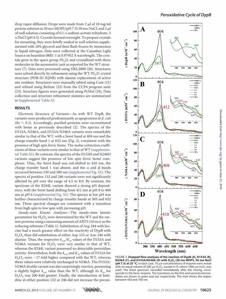
Electronic Structure of Variants—As with WT DypB, thevariants were produced predominantly as apoproteins in E. coli(Rz � 0.1). Accordingly, purified proteins were reconstitutedwith heme as previously described (2). The spectra of theD153A, N246A, and D153A/N246A variants were remarkablysimilar to that of the WT, with a Soret band at 404 nm and thecharge transfer band 1 at 632 nm (Fig. 2), consistent with thepresence of high spin ferric heme. The molar extinction coeffi-cients of these variants were similar to that ofWT (supplemen-tal Table S1). By contrast, the spectra of the D153H andN246Hvariants suggest the presence of low spin ferric heme com-plexes. Thus, the Soret band was red-shifted to 410 nm, thecharge transfer band 1 was absent, and the � and � bandsoccurred between 530 and 580 nm (supplemental Fig. S1). Thespectra of position 153 and 246 variants were not significantlyaffected by pH over the range of 4.5 to 8.0. By contrast, thespectrum of the R244L variant showed a strong pH depend-ence, with the Soret band shifting from 411 nm at pH 8 to 404nm at pH 6 (supplemental Fig. S1). The species at low pH wasfurther characterized by charge transfer bands at 503 and 632nm. These spectral changes are consistent with a transitionfrom high spin to low spin with increasing pH.Steady-state Kinetic Analyses—The steady-state kinetic
parameters for H2O2 were determined for theWT and the var-iant proteins using a saturating amount of ABTS (10mM) as thereducing substrate (Table 1). Substitution of Arg-244 with leu-cine had a much greater effect on the reactivity of DypB withH2O2 than did substitution of either Asp-153 or Asn-246 withalanine. Thus, the respective kcat/Km values of the D153A andN246A variants for H2O2 were very similar to that of WT,whereas the R244L variant possessed no detectable peroxidaseactivity. Nevertheless, both the kcat andKm values of D153A forH2O2 were �17-fold higher compared with the WT, whereasthese values were relatively unchanged in N246A. The D153A/N246Adouble variantwas also surprisingly reactive, possessinga slightly higher kcat value than the WT, although its Km forH2O2 was 200-fold greater. Finally, the introduction of histi-dine at either position 153 or 246 did not increase the peroxi-
FIGURE 2. Stopped-flow analyses of the reaction of DypB (A), D153A (B),N246A (C), and D153A/N246A (D) with H2O2 (20 mM MOPS, 50 mM NaCl(pH 7.5) at 25 °C. In each case, 10 �M concentrations of enzyme were mixedwith an equal volume of 200 �M H2O2, except in D, where 1000 �M H2O2 wasused. The black spectrum, recorded immediately after the mixing, corre-sponds to the ferric enzyme. The transitions to the first and second interme-diates are shown in green and red, respectively. The inset shows the regionbetween 450 and 700 nm.
Peroxidative Cycle of DypB
MARCH 23, 2012 • VOLUME 287 • NUMBER 13 JOURNAL OF BIOLOGICAL CHEMISTRY 10625
by guest on June 21, 2016http://w
ww
.jbc.org/D
ownloaded from

dase activity of DypB. Indeed, the N246H variant did not showany detectable peroxidase activity under the experimentalconditions.Compound I Formation in Variants—Transient intermedi-
ates in DypB and the variants were observed by following thevariants reaction with 5–200 eq of H2O2 using a rapid-mixing,stopped-flow spectrophotometer. In the D153A and N246Avariants at pH 7.5, the spectrum of the first intermediate,observed within �0.1 s upon mixing with H2O2, was almostidentical to Compound I observed in WT (Fig. 2, A–C, thickgreen trace) with a hypochromatic, blue-shifted Soret at 397 nmand a hyperchromic CT band at 649 nm. Similar intermediateswere observed in D153A/N246A and D153H, although the CTband of Compound I in the double variant was less hyperchro-mic (Fig. 2D, thick green trace), and �100 eq of H2O2 wererequired to form it, consistentwith the highKm forH2O2. In theD153H variant, the Soret was less blue-shifted at 406 nm (Fig.3A, thick green trace), and the visible region was less hypochro-matic compared with the WT. Neither R244L nor N246Hdetectably reacted with H2O2 even at 100-fold greater concen-trations of the latter (Fig. 3, B andC, thick red spectrum). More-over, neither the high spin state (pH 6) nor pH 7.5 the low spinstate (pH 7.5) of the R244L variant detectably reacted withH2O2 (Fig. 3B, thick red spectrum). However, the heme of thisvariant was more susceptible to degradation at pH 6 (supple-mental Fig. S2) compared with pH 8.The second order rate constants for Compound I formation
(k1) by theWTand the variants weremeasured by following thechanges in absorption at 404 and 647 nm. The kinetic traceswere described by single order exponentials, and the deter-mined pseudo-first order rate constants, kobs, were similar atthe two wavelengths (supplemental Fig. S5). Plots of kobs versusH2O2 concentration were linear (supplemental Fig. S6). In eachof the studied enzymes, the k1 valueswere comparable at pH7.5(Table 1) and pH 5.5 (data not shown) and were remarkablysimilar to the kcat/Km values for H2O2. Thus, k1 for D153A wasabout half that for WT, whereas that for N246A was 1.6-foldgreater. Similarly, the k1 values for D153H and D153A/N246Awere more than an order of magnitude less than that for WT.Compound II Formation in Variants—Compound I was
much shorter lived in each of the D153H, D153A, and N246Avariants (t1⁄2 � 0.13 s) than in WT (t1⁄2 � 540 s). Moreover, inthese variants, Compound I decayed to an intermediate whosespectra resemble Compound II of peroxidase (Fig. 2, B and C,
and Fig. 3A, thick red spectrum). Thus, the Soret of CompoundII was red-shifted to various extents in each of the three vari-ants, whereas � and � bands appeared near 525 and 555 nm,respectively. Interestingly, the Soret band for Compound II isless red-shifted in the spectrum of N246A compared withD153A. In addition, the spectrum of N246A possessed a chargetransfer band at 649 nm, suggesting the presence of amixture ofCompounds I and II (Fig. 2C, thick red spectrum).In D153A/N246A, Compound I decayed to an intermediate
whose spectrum resembled that of Compound III, with a Soretfor this intermediate located at 410 nm and �/� bands at 538and 574 nm, respectively (Fig. 2D, thick red spectrum). Forma-tion of this intermediate was likely due to the use of higherconcentrations of H2O2 required to produce detectable Com-pound I. Compound III was not observed in either D153A orN246A even upon reaction with 100 eq of H2O2 (supplementalFig. S4).The transient observed in reactions of WT DypB and the
variants with H2O2 were modeled using singular value decom-position, global analysis, and a three-step model (A3 B3 C)(supplemental Fig. S7). The simulated spectra of the interme-diates were consistent with the formation of a single interme-diate in the reaction with WT and two intermediates in thereactions with each of D153A, N246A, D153A/N246A, andD153H.Structural Characterization of Variants—Crystal structures
of each of the four single variants were determined to at least2.64 Å resolution (supplemental Table S2). The average r.m.s.dfor all C� atoms is �0.25 Å when compared with theWT crys-tal structure (PDB ID 3QNS), indicating that the substitutionshadminimal effect on the overall -fold. Generally, the active siteresidues are well defined and are similarly positioned relative tothe wild-type structure (Fig. 4A). The only significant differ-ences in the structures are observed on the distal side of theheme where Asp-153, Arg-244, and Asn-246 interact with theiron-coordinated solvent molecule in the wild-type structure.Difference electron density maps of the D153A and D153H
variants revealed a strong positive peak on the distal side of theheme iron. Refinement of each site as a watermolecule resultedin residual positive electron density and a low B-factor relativeto surrounding atoms. Chloride, present at 3.0 M in the crystal-lization buffer, fully models the electron density and refines atfull occupancywith B-factors comparable with those of the sur-rounding atoms (D153A, Fe(III) �45 Å2 and Cl� �58 Å2;
TABLE 1Kinetic parameters for DypB and the distal residue variantsSteady-state kinetic parameters were determined using 10 mM ABTS, 50 mM sodium acetate (pH 5.5) at 25.0 � 0.5 °C. Pseudo-first order and -second order rate constantswere determined using 20 mM MOPS, 50 mM NaCl (pH 7.5) at 25.0 � 0.5 °C.
Enzyme KmH2O2 kcat kcat/Km k1 k2�M s�1 105 M�1s�1 105 M�1s�1 s�1
DypB 27 � 5 3.2 � 0.03 1.20 � 0.08 2.0 � 0.07 –a
D153H 87 � 7 1.8 � 0.04 0.20 � 0.06 0.12 � 0.05 1.2 � 0.01D153A 460 � 30 51 � 3 1.10 � 0.04 0.94 � 0.01 0.75 � 0.06R244L –b
N246H –b
N246A 15 � 1 5.4 � 0.2 3.60 � 0.20 3.20 � 0.01 0.37 � 0.04D153A/N246A 5000 � 2000 19 � 8 0.04 � 0.002 0.13 � 0.02 –c
a Not measured because Compound I did not transition to Compound II.b No significant activity was detected (detection limit was 0.04 �mole/min/�mol of heme).c Not measured because the high concentrations of H2O2 required to produced Compound I resulted in the conversion of the latter to Compound III without detectable for-mation of Compound II.
Peroxidative Cycle of DypB
10626 JOURNAL OF BIOLOGICAL CHEMISTRY VOLUME 287 • NUMBER 13 • MARCH 23, 2012
by guest on June 21, 2016http://w
ww
.jbc.org/D
ownloaded from

D153H, Fe(III) �39 Å2 and Cl� �58 Å2). The presence of achloride in the Asp-153 variants may be favored in the absenceof the negatively charged carboxylate. In D153A, the chlorideion interacts with the heme iron (�2.3Å), Arg-244N�1 (3.2Å),and Asn-246 N�2 (3.1 Å) (Fig. 4B). Arg-244 is in the same con-formation as in the WT structure, and the orientation of Asn-246 corresponds to one of the two orientations modeled in theWT structure. A boundwatermoleculewas observed to occupy
part of the void created by the aspartate-to-alanine replace-ment. This solvent species is located �4.5 Å from the chlorideion and forms a hydrogen bond with Asp147. Similarly, in theD153H structure, the chloride also interacts with Arg-244 N�1(3.1 Å) and Asn-246 N�2 (2.4 Å in one conformation). How-ever, it also interactswithHis-153 (3.0Å), and the chloride-ironbond is elongated (�2.7 Å) (Fig. 4C). In contrast to D153A, twoconformations are observed for Asn-246 in the D153H struc-ture; one bound to chloride throughN�2 and the other directedinto the distal solvent access channel by an �135° rotationabout �1. The increased chloride-iron bond length in D153Happears to be a result of the imidazole side chain, which steri-cally prevents the chloride from occupying a coordination sitecloser to the iron. This amino acid replacement also causes a0.6-Å shift of Asn-246 N�2 away from position 153 relative toits position in the structure of WT DypB.The replacement of Asn-246 with either alanine or histidine
removes theN�2 atom that is directly bound to the axially coor-dinated solvent molecule in the WT structure (Fig. 4A). Omitdifference (Fo � Fc) electron density maps of N246A andN246H clearly display elongated density on the distal side ofheme-iron, extending toward residue 246 (Figs. 4, D and E).Two water molecules may be refined into the density of eachvariant with one coordinating the iron (2.1–2.2 Å) and the sec-ond hydrogen-bonded to the first (2.5 Å). The B-factors forthese water molecules are in line with those of heme iron(N246A, Fe(III) �35 Å2 and waters both �38 Å2; N246H,Fe(III) �41 Å2 and waters �41 and 50 Å2). Attempts to modelthe density by an acetate molecule (present at 0.1 M in the crys-tallization buffer) were unsuccessful, but the possibility that thestructure contains amixture of coordinated solvent and acetatecannot be excluded. A positionally equivalent second solventmolecule was observed in the high resolutionWT crystal struc-ture (Fig. 4A), where it ismodeled at 40% occupancy, correlatedwith one of two conformations of Asn-246. In contrast to WT,in N246A and N246H the second solvent molecule is shifted�0.7 Å toward residues 246 and forms a hydrogen bond withAsp-153 (3.1–3.3 Å). The remaining residues in N246A activesite overlay well with those of theWT structure, indicating thatthe mutation has little effect on the rest of the heme environ-ment. In the N246H variant, the imidazole of His-246 is orien-tated away from the heme and projects into the distal solventaccess channel, potentially blocking substrate access. The ori-entationofHis-246 appears toperturb surrounding residues.Asp-153 is displaced toward the heme (maximal deviation �1.2 Å atO�1 relative to WT), and the side chain of Arg-244 is displaced�0.5 Å away from the distal coordination site (Fig. 4E). Theremainder of the N246H structure closely resemblesWT.In R244L, the distal ligand is a solvent molecule coordinated
at 2.0Å from the iron (Fig. 4F), similar towhat is observed in theWT structure. The coordinated solvent forms a H-bond withAsn-246 (3.1 Å) but is shifted �0.5 Å away from Asp-153 rela-tive to its position in the WT structure. As a result, Asp-153O�1 is located �4.2 Å from the solvent molecule, outsidehydrogen bonding distance. The guanidinium group of Arg-244 is effectively replaced by two solvent species. One solventmolecule is positioned similarly to Arg-244 N�1 in the WTstructure, bridging the coordinating solvent molecule (2.6 Å)
FIGURE 3. Stopped-flow analyses of the reaction of D153H (A), R244L (B),and N246H (C) with H2O2 (20 mM MOPS, 50 mM NaCl (pH 7.5) at 25 °C). Ineach case, 10 �M concentrations of enzyme were mixed with an equal volumeof 2000 �M H2O2, except in A, where 200 �M H2O2 was used. The black spec-trum, recorded immediately after the mixing, corresponds to the ferricenzyme. The spectra are represented as in Fig. 2.
Peroxidative Cycle of DypB
MARCH 23, 2012 • VOLUME 287 • NUMBER 13 JOURNAL OF BIOLOGICAL CHEMISTRY 10627
by guest on June 21, 2016http://w
ww
.jbc.org/D
ownloaded from

and heme propionateA (2.7Å). This species also forms an addi-tional hydrogen bond with Asp-153 O�1 (2.9 Å). The secondadditional solvent species occupies a position similar to that ofArg-244 N�2 in the WT structure, forming another hydrogenbond with heme propionate A (2.8 Å). The remainder of theactive site is unperturbed by the mutation.
DISCUSSIONThe current study establishes that of the three distal polar
residues, only Arg-244 is essential for peroxidase activity inDypB. Indeed, the replacement of Asp-153 and/or Asn-246with an alanine had a remarkably small effect on the enzymereactivity with H2O2, as evaluated by the apparent specificityconstant (kcat/Km) and the first order rate constant for the for-mation of Compound I (k1). Nevertheless, substitution of theseresidues dramatically decreased the stability of Compound I inDypB. In addition, the data from the D153H and N246H vari-ants suggest that DypB cannot efficiently utilize imidazole as anacid-base catalyst on the distal face of the heme. Overall, this
study indicates that in DypB, the peroxidative cycle is modu-lated very differently than in plant peroxidases.The ability of D153A to form Compound I and to react with
peroxide almost as efficiently asWTDypB is surprising consid-ering that substitution of the equivalent residue, Asp-171, inDyPDec1 reduced the enzyme peroxidase activity to undetect-able levels (5). The latter result led to the proposal that the distalaspartate functions as an acid-base catalyst, similar to the distalhistidine of plant peroxidases and the distal glutamate of chlo-roperoxidase (B: in Fig. 5). Substitution of the distal acid-basecatalyst in plant peroxidases reduces catalytic efficiency by fourto six orders of magnitude (25, 26). The distal aspartate alsodoes not appear to be essential for peroxidase activity in A-typeDyPs. Thus,WT EfeB and the D235N variant oxidized guaiacolat similar rates (15). Interestingly, the variant-oxidized catecholat �20% that of the rate ofWT indicated that the aspartate hasa role in substrate specificity. The lack of the steady-state andtransient state kinetic data from the DyPDec1 and EfeB variants
FIGURE 4. X-ray crystal structures of DypB variants. Shown are wild-type (PDB ID 3QNS) (A), D153A (B), D153H (C), N246A (D), N246H (E), R244L (F). Active sitesresidues are shown as green (carbon), blue (nitrogen), and red (oxygen) sticks. Alternate conformations for Asn-246 (A and C) are highlighted in light green. Water(gray), iron (orange), and chloride (cyan) are shown as spheres. Hydrogen bonds are indicated by dashed lines. Gray mesh represents omit difference (Fo � Fc)maps for the distal ligand contoured at 3 � for all panels except E (2.5 �) and F (2.0 �).
FIGURE 5. The proposed mechanism of peroxidases. The steps leading to Compound I formation are in parentheses. An acid-base catalyst, B:, assists intransferring a proton from the proximal O to the distal O of the iron-bound H2O2, accelerating Compound I formation. This residue is histidine and glutamatein plant peroxidases and chloroperoxidase, respectively. In D- and B-type DyPs, this residue appears to be aspartate and arginine, respectively.
Peroxidative Cycle of DypB
10628 JOURNAL OF BIOLOGICAL CHEMISTRY VOLUME 287 • NUMBER 13 • MARCH 23, 2012
by guest on June 21, 2016http://w
ww
.jbc.org/D
ownloaded from

makes comparison with DypB difficult. Nevertheless, the avail-able data suggest that although the distal aspartate is the essen-tial acid-base catalyst in D-type DyPs, it is not in either A- orB-type DyPs. This may reflect the different physiological sub-strates and/or roles of the different subfamilies of enzymes asdiscussed below.Although the current data indicate that Asn-246 plays a
minor role in Compound I formation, it is positioned remark-ably similarly to the conserved asparagine in catalases (27). Inbovine liver catalase the conserved asparagine (Asn-147) andhistidine (His-74) on the distal face of the heme assist in thebinding of H2O2 to the heme iron (27). In resting state bovineliver catalase, the two residues are hydrogen-bonded to a sharedsolvent molecule, much as Asn-246 and Asp-153 are in DypB.Although the kinetic parameters of theN246A variant (Table 1)establish that Asp-246 is not required for catalysis, those of theD153A/N246A double variant indicate that these two residuesinteract with the H2O2. Further insight into the interaction ofAsn-246 and Asp-153 was obtained by inverse analysis. Thisanalysis uses the double variant as a reference point to evaluatewhether the effect of substituting two residues is additive, coop-erative, anti-cooperative, or antagonistic (28). The effect of theAsp-153 and Asn-246 substitutions was anti-cooperative (sup-plemental Fig. S9), indicating that the two residues functionsynergistically with respect to peroxidase activity.The inability of the R244L variant to form Compound I fur-
ther distinguishes DypB from HRP and cytochrome c peroxi-dase. Although the distal arginine is similarly located in theseenzymes, it forms an electrostatic interaction with heme propi-onate A (supplemental Fig. S8) in DyPs (5, 7, 14). In plant per-oxidases, the propionate is orientated away from the arginineguanidinium group (29). Substitution of this residue with leu-cine in HRP or cytochrome c peroxidase lowers the rate ofCompound I formation by 2–3 orders of magnitude, althoughthis intermediate is still formed (25, 26). Nevertheless, the pH-dependent iron spin state transition of R244L is similar to thatof the R48L variant of cytochrome c peroxidase (26). This tran-sition suggests that the pKa of Asp-153 in R244L is �7, muchhigher than the expected value of 3.5. Indeed, it would be inter-esting to determine the pKa values of Asp-153, which is 0.7 Åfurther away from the distal solvent species in R244L than inWT.The large effect of substituting Arg-244 in DypB indicates
that this residue has a major catalytic role in Compound I for-mation andmay be the requisite acid-base catalyst (B: in Fig. 5).It is unclear what other residue could fulfill this role; other thanthe three residues targeted in this study, the closest residuewiththe requisite functionality is Asp-147, located up to 7 Å awayfrom the heme iron. Although the pKa of the acid-base catalystis expected to be �5, much lower than that of an arginine res-idue (12.5), the pKa of catalytically relevant arginines can besignificantly shifted. For example, Arg-82 of bacteriorhodopsinhas a pKa of 5.8 in the M state (deprotonated retinal) (30), andArg-418 of inosine 5�-monophosphate dehydrogenase has apKa of �8 (31). In both of these cases, the arginine is in closeproximity to an aspartate, as observed inDypB.Moreover, in allthree cases, the arginine is part of a hydrogen bonding networkthat connects the residue to the protein surface. In DypB, the
network involves the iron-bound solvent species, Arg-244,heme propionate A, a structurally conserved solvent molecule,and the highly conserved Arg-208 on the protein surface (sup-plemental Fig. S8). This network is conserved in DyPs and maymediate proton transfer as has been proposed in ascorbate per-oxidase and other enzymes (32, 33). Disruption of this networkmay help explain the lack of peroxidase activity in R244L.Although Asp-153 and Asn-246 have relatively minor roles
in Compound I formation, they appear to have important rolesin stabilizing this intermediate; substitution of either residuedecreased the half-life of Compound I by �3 orders of magni-tude. This is consistent with their close proximity to the hemeiron: �5.0 Å for Asn-246N�2 and 5.1 Å for Asp-153O�1.Although this is similar to the corresponding distances for His-42N�2 in HRP (5.8 Å (29)) and Glu-183O�2 in chloroperoxi-dase (5.1 Å (16)), neither of the latter has an equivalent of Asn-246. At this time, it is unclear whether the stabilization ofCompound I by Asp-153 and Asn-246 is due to kinetic or ther-modynamic effects. Thus, these residues contribute to both theelectrostatic environment and the hydrogen bonding networkof the active site. The precise role of Asp-153 in stabilizingCompound I clearly depends on the residue protonation state.Although the iron spin-state transition in R244L suggests thatthe pKa value of Asp-153 is �7, this is unlikely to be the case inWT DypB.The poor catalytic efficiency of the D153H and N246H vari-
ants is likely due in part to the suboptimal positioning of theimidazole in the distal heme pocket of DypB. In the case ofD153H, the N�2 atom of His-153 is 1.8 Å closer to the hemeiron than His-42 is in HRP (29). This close proximity also likelyfavors the low spin state of the D153H variant. In the case ofN246H, the imidazole is 7.8 Å from the iron. However, it ispositioned at the center of the distal solvent access channel,potentially limiting the access of H2O2 to the heme iron. Inaddition, hydrogen bonding (3.3 Å) between His-246 N�1 andthe main chain carbonyl of Asp-153 would also influence thecatalytic role ofHis-246.Overall, this result highlights howpro-tein active sites are fine-tuned for efficient utilization of specificfunctional groups. Additional amino acid substitutions arerequired to transform DypB into a more efficient peroxidase.This work highlights the differences in themodulation of the
active site residues in the peroxidative cycle of DypB as com-pared with that of D-typeDyPs and plant peroxidases. Our datarule out Asp-153 as the acid-base catalyst in the formation ofCompound I in DypB and identifies Arg-244 as best candidatefor this role. It is possible that the different modulation of theperoxidative cycle in theDyPs reflects their different physiolog-ical roles. For example, C- and D-type DyPs may oxidize a vari-ety of substrates such as dyes (5) and melanin (14), whereas A-and B-type DyPs may oxidize a specific substrate, such asporphyrinogen (11). Thus, although the lignin-and Mn2�-oxi-dizing activities of DypB from R. jostii RHA1 may be biotech-nologically useful, it may not be physiologically relevant. Thispossibility is consistent with the �100-fold lesser activity of A-and B-type DyPs with classic peroxidase substrates (7). Furtherstudies should elucidate the structural bases of the diverseactivities of DyPs and facilitate developing the biotechnologicalpotential of this family of microbial enzymes.
Peroxidative Cycle of DypB
MARCH 23, 2012 • VOLUME 287 • NUMBER 13 JOURNAL OF BIOLOGICAL CHEMISTRY 10629
by guest on June 21, 2016http://w
ww
.jbc.org/D
ownloaded from

Acknowledgments—Part of this work was performed at the CanadianLight Source, which is supported byNatural Sciences and EngineeringResearch Council of Canada, the National Research Council of Can-ada, the Canadian Institutes of Health Research, the Province of Sas-katchewan, Western Economic Diversification Canada, and the Uni-versity of Saskatchewan.
REFERENCES1. Sugano, Y., Sasaki, K., and Shoda, M. (1999) cDNA cloning and genetic
analysis of a novel decolorizing enzyme, peroxidase gene dyp fromGeotri-chum candidum Dec 1. J. Biosci. Bioeng. 87, 411–417
2. Ahmad, M., Roberts, J. N., Hardiman, E. M., Singh, R., Eltis, L. D., andBugg, T.D. (2011) Identification ofDypB fromRhodococcus jostiiRHA1 asa lignin peroxidase. Biochemistry 50, 5096–5107
3. Ogola, H. J., Kamiike, T., Hashimoto, N., Ashida, H., Ishikawa, T., Shibata,H., and Sawa, Y. (2009) Molecular characterization of a novel peroxidasefrom the cyanobacterium Anabaena sp. strain PCC 7120. Appl. Environ.Microbiol. 75, 7509–7518
4. Sugano, Y. (2009) DyP-type peroxidases comprise a novel heme peroxi-dase family. Cell. Mol. Life. Sci. 66, 1387–1403
5. Sugano, Y., Muramatsu, R., Ichiyanagi, A., Sato, T., and Shoda, M. (2007)DyP, a unique dye-decolorizing peroxidase, represents a novel heme per-oxidase family. Asp-171 replaces the distal histidine of classical peroxi-dases. J. Biol. Chem. 282, 36652–36658
6. Liers, C., Bobeth, C., Pecyna, M., Ullrich, R., and Hofrichter, M. (2010)DyP-like peroxidases of the jelly fungus Auricularia auricula-judae oxi-dize nonphenolic ligninmodel compounds and high redox potential dyes.Appl. Microbiol. Biotechnol. 85, 1869–1879
7. Roberts, J. N., Singh, R., Grigg, J. C., Murphy, M. E., Bugg, T. D., and Eltis,L. D. (2011) Characterization of dye-decolorizing peroxidases from Rho-dococcus jostii RHA1. Biochemistry 50, 5108–5119
8. Kaur, A., Van, P. T., Busch, C. R., Robinson, C. K., Pan, M., Pang, W. L.,Reiss, D. J., DiRuggiero, J., and Baliga, N. S. (2010) Coordination of front-line defense mechanisms under severe oxidative stress.Mol. Syst. Biol. 6,393–408
9. Kong, L., Guo, D., Zhou, S., Yu, X., Hou, G., Li, R., and Zhao, B. (2010)Cloning and expression of a toxin gene from Pseudomonas fluorescensGcM5-1A. Arch. Microbiol. 192, 585–593
10. Létoffé, S., Heuck, G., Delepelaire, P., Lange, N., and Wandersman, C.(2009) Bacteria capture iron from heme by keeping tetrapyrrol skeletonintact. Proc. Natl. Acad. Sci. 106, 11719–11724
11. Dailey, H. A., Septer, A. N., Daugherty, L., Thames, D., Gerdes, S., Stabb,E. V., Dunn, A. K., Dailey, T. A., and Phillips, J. D. (2011) The Escherichiacoli protein YfeX functions as a porphyrinogen oxidase, not a heme deche-latase.MBio 2, e00248–00211
12. Goblirsch, B., Kurker, R. C., Streit, B. R., Wilmot, C. M., and DuBois, J. L.(2011) Chlorite dismutases, DyPs, and EfeB. threemicrobial heme enzymefamilies comprise the CDE structural superfamily. J. Mol. Biol. 408,379–398
13. Zubieta, C., Krishna, S. S., Kapoor,M., Kozbial, P.,McMullan, D., Axelrod,H. L., Miller, M. D., Abdubek, P., Ambing, E., Astakhova, T., Carlton, D.,Chiu, H. J., Clayton, T., Deller, M. C., Duan, L., Elsliger, M. A., Feuerhelm,J., Grzechnik, S. K., Hale, J., Hampton, E., Han, G. W., Jaroszewski, L., Jin,K. K., Klock, H. E., Knuth, M. W., Kumar, A., Marciano, D., Morse, A. T.,Nigoghossian, E., Okach, L., Oommachen, S., Reyes, R., Rife, C. L., Schim-mel, P., van den Bedem,H.,Weekes, D.,White, A., Xu, Q., Hodgson, K. O.,Wooley, J., Deacon, A. M., Godzik, A., Lesley, S. A., and Wilson, I. A.(2007) Crystal structures of two novel dye-decolorizing peroxidases reveala �-barrel-fold with a conserved heme binding motif. Proteins 69,223–233
14. Zubieta, C., Joseph, R., Krishna, S. S., McMullan, D., Kapoor, M., Axelrod,
H. L., Miller, M. D., Abdubek, P., Acosta, C., Astakhova, T., Carlton, D.,Chiu, H. J., Clayton, T., Deller, M. C., Duan, L., Elias, Y., Elsliger, M. A.,Feuerhelm, J., Grzechnik, S. K., Hale, J., Han, G. W., Jaroszewski, L., Jin,K. K., Klock, H. E., Knuth, M. W., Kozbial, P., Kumar, A., Marciano, D.,Morse, A. T.,Murphy, K. D., Nigoghossian, E., Okach, L., Oommachen, S.,Reyes, R., Rife, C. L., Schimmel, P., Trout, C. V., van den Bedem, H.,Weekes, D., White, A., Xu, Q., Hodgson, K. O., Wooley, J., Deacon, A. M.,Godzik, A., Lesley, S. A., andWilson, I. A. (2007) Identification and struc-tural characterization of heme binding in a novel dye-decolorizing perox-idase, TyrA. Proteins 69, 234–243
15. Liu, X., Du,Q.,Wang, Z., Zhu,D.,Huang, Y., Li, N.,Wei, T., Xu, S., andGu,L. (2011) Crystal structure and biochemical features of EfeB/YcdB fromEscherichia coliO157. Asp-235 plays divergent roles in different enzyme-catalyzed processes. J. Biol. Chem. 286, 14922–14931
16. Sundaramoorthy,M., Terner, J., and Poulos, T. L. (1995) The crystal struc-ture of chloroperoxidase. A heme peroxidase; cytochrome P450 func-tional hybrid. Structure 3, 1367–1377
17. Falk, J. E. (1964) Porphyrins andMetalloporphyrins pp. 804–807, ElsevierScience Publishing Co., Inc., Amsterdam
18. Childs, R. E., and Bardsley, W. G. (1975) The steady-state kinetics of per-oxidase with 2,2�-azino-di-(3-ethyl-benzthiazoline-6-sulphonic acid) aschromogen. Biochem. J. 145, 93–103
19. Cornish-Bowden, A. (1995)Analysis of EnzymeKinetic Data, OxfordUni-versity Press, New York
20. Otwinowski, Z., andMinor,W. (1997) Processing of x-ray diffraction datacollected in oscillation mode.Methods Enzymol. 276, 307–326
21. Emsley, P., Lohkamp, B., Scott,W. G., and Cowtan, K. (2010) Features anddevelopment of Coot. Acta Crystallogr. D Biol. Crystallogr. 66, 486–501
22. Murshudov, G. N., Vagin, A. A., and Dodson, E. J. (1997) Refinement ofmacromolecular structures by the maximum-likelihood method. ActaCrystallogr. Sect. D Biol. Crystallogr. 53, 240–255
23. Collaborative Computational Project, Number 4 (1994) The CCP4 suite.Programs for protein crystallography. Acta Crystallogr. D. Biol. Crystal-logr. 50, 760–763
24. DeLano, W. L. (2008) The PyMOL Molecular Graphics System, DeLanoScientific LLC, Palo Alto, CA
25. Rodriguez-Lopez, J. N., Smith, A. T., and Thorneley, R. N. (1996) Role ofarginine 38 in horseradish peroxidase. A critical residue for substratebinding and catalysis. J. Biol. Chem. 271, 4023–4030
26. Vitello, L. B., Erman, J. E.,Miller,M.A.,Wang, J., andKraut, J. (1993) Effectof arginine 48 replacement on the reaction between cytochrome c perox-idase and hydrogen peroxide. Biochemistry 32, 9807–9818
27. Fita, I., and Rossmann, M. G. (1985) The active center of catalase. J. Mol.Biol. 185, 21–37
28. Mildvan, A. S. (2004) Inverse thinking about double mutants of enzymes.Biochemistry 43, 14517–14520
29. Gajhede, M., Schuller, D. J., Henriksen, A., Smith, A. T., and Poulos, T. L.(1997) Crystal structure of horseradish peroxidase C at 2.15 A resolution.Nat. Struct. Biol. 4, 1032–1038
30. Xiao, Y., Hutson,M. S., Belenky,M.,Herzfeld, J., andBraiman,M. S. (2004)Role of arginine 82 in fast proton release during the bacteriorhodopsinphotocycle. A time-resolved FT-IR study of purplemembranes containing15N-labeled arginine. Biochemistry 43, 12809–12818
31. Guillén Schlippe, Y. V., and Hedstrom, L. (2005) Is Arg-418 the catalyticbase required for the hydrolysis step of the IMP dehydrogenase reaction?Biochemistry 44, 11700–11707
32. Efimov, I., Badyal, S. K.,Metcalfe, C. L.,Macdonald, I., Gumiero, A., Raven,E. L., and Moody, P. C. (2011) Proton delivery to ferryl heme in a hemeperoxidase. Enzymatic use of theGrotthussmechanism. J. Am. Chem. Soc.133, 15376–15383
33. Sharp, K. H., Mewies, M., Moody, P. C., and Raven, E. L. (2003) Crystalstructure of the ascorbate peroxidase-ascorbate complex. Nat. Struct.Biol. 10, 303–307
Peroxidative Cycle of DypB
10630 JOURNAL OF BIOLOGICAL CHEMISTRY VOLUME 287 • NUMBER 13 • MARCH 23, 2012
by guest on June 21, 2016http://w
ww
.jbc.org/D
ownloaded from

S-1
SUPPLEMENTAL DATA
The distal heme pocket residues of a B-type Dye-decolorizing peroxidase: arginine but not
aspartate is essential for peroxidase activity
Rahul Singh1, Jason C. Grigg
1, Zachary Armstrong
1, Michael E.P. Murphy
1, Lindsay D. Eltis
1*
1Department of Microbiology and Immunology, Life Sciences Institute, University of British
Columbia, Vancouver, British Columbia, Canada.
Table of Contents
Page S-1 – Supplemental Experimental Procedures
Page S-2 – Supplemental Table S1
Page S-3 – Supplemental Table S2
Page S-4 – Supplemental Figures S1and S2
Page S-5 – Supplemental Figure S3
Page S-6 – Supplemental Figure S4
Page S-7 – Supplemental Figure S5
Page S-8 – Supplemental Figure S6
Page S-9 – Supplemental Figure S7
Page S-10 – Supplemental Figure S8
Page S-11 – Supplemental Figure S9
Supplemental Experimental Procedures
Construction of DypB variants: The plasmids carrying genes coding for the D153H
(pETDB153H), D153A (pETDB153A), N246H (pETDB246H) and N246A (pETDB246A) were
constructed using pETDYB1 as the template DNA and the following 5’ phosphorylated forward
primers:
1. oD153A: GGATTCGTCGCGGGCACCGAGAAT
2. oN246A: CCTCCGCGACGCGATGGCATTCG
3. oD153H: GGATTCGTCCACGGCACCGAGAAT
4. oN246H: CCTCCGCGACCACATGGCATTCG
The plasmid carrying the gene encoding the D153A/N246A variant (pETDB153A/246A) was
constructed using pETDB153A as the template DNA and the primer oN246A. Mutagenesis was
performed using the QuikChangeTM
PCR protocol (Agilent Technologies) with minor
modifications. Thus, only forward primer was used for the mutagenesis reaction and the resulting
PCR products were digested with DpnI and transformed into E. coli Nova Blue electro-
competent cells (recA-, endA
-, lacI
q). The desired mutation was confirmed by sequencing the
complete gene length
S-2
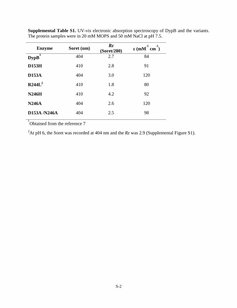
Supplemental Table S1. UV-vis electronic absorption spectroscopy of DypB and the variants.
The protein samples were in 20 mM MOPS and 50 mM NaCl at pH 7.5.
Enzyme Soret (nm) Rz
(Soret/280) ε (mM
-1 cm
-1)
DypB1 404 2.7 84
D153H 410 2.8 91
D153A 404 3.0 120
R244L2 410 1.8 80
N246H 410 4.2 92
N246A 404 2.6 120
D153A /N246A 404 2.5 98
1Obtained from the reference 7
2At pH 6, the Soret was recorded at 404 nm and the Rz was 2.9 (Supplemental Figure S1).

S-3
Supplemental Table S2. Data collection and refinement statistics.
avalues for the highest resolution shell are shown in parenthesis
DypB D153A DypB D153H DypB N246A DypB N246H DypB R244L
Data collectiona
Wavelength (Å) 0.97952 0.97952 0.97952 0.97952 0.97952
Resolution range (Å) 40.0 – 2.6
(2.64 – 2.60)
40.0 – 2.5
(2.54 – 2.50)
40.0 – 2.4
(2.44 – 2.40)
45.0 – 2.64
(2.69 – 2.64)
53.0 – 2.35
(2.43 – 2.35)
Space group P3221 P3221 P3221 P3221 P3221
Unit cell dimensions (Å) a = 132.4,
b = 132.4,
c = 160.6
a = 132.6,
b = 132.6,
c = 159.6
a = 132.9,
b = 132.9,
c = 161.0
a = 132.3,
b = 132.3,
c = 158.8
a = 133.2,
b = 133.2,
c = 158.9
Unique reflections 50668 56625 64436 49698 67842
Completeness (%) 99.1 (100) 99.6 (99.5) 98.0 (98.6) 99.5 (99.8) 99.9 (100)
Average I/σI 21.6 (2.7) 19.5 (2.3) 22.2 (3.5) 19.7 (2.9) 15.6 (3.6)
Redundancy 5.0 (4.7) 5.3 (4.3) 6.3 (6.4) 5.5 (5.2) 6.0 (6.0)
Rmerge 0.062 (0.543) 0.069 (0.627) 0.070 (0.588) 0.074 (0.566) 0.096 (0.516)
Wilson B (Å2) 57.7 54.7 50.0 60.0 44.2
Refinement
Rwork (Rfree) 0.166 (0.213) 0.184 (0.218) 0.184 (0.218) 0.156 (0.205) 0.186 (0.222)
B-factors (Å2)
All atoms 51.6 48.9 44.1 51.3 39.1
Protein 51.9 49.2 44.3 51.6 39.1
Heme 42.8 37.9 33.3 38.7 33.4
Water 44.3 42.1 41.0 45.3 38.2
r.m.s.d. bond length (Å) 0.013 0.013 0.013 0.013 0.013
In most-favorable region 90.5 91.2 91.8 90.0 92.7
In disallowed regions 0.0 0.1 0.4 0.0 0.1
PDB Accession Codes 3VEC 3VED 3VEE 3VEF 3VEG

S-4
Supplemental Figure S1. UV-vis electronic absorption spectrum of purified R244L at pH 8
(black), 7 (blue), and 6 (red). The region between 450-700 nm is shown at greater magnification
in the inset.
Supplemental Figure S2. Reaction of R244L with 1 equivalent of H2O2 (50 mM sodium
succinate, pH 5, 25 ○C). Reaction of 5 µM R244L (black) with 5 µM H2O2. The spectra (thin red
to thick red) were recorded at the following time intervals (s): 5, 120,360, and 1200. The region
between 450-700 nm is shown at greater magnification in the inset.
300 350 400 450 500 550 600 650
0.0
0.2
0.4
0.6
411
Ab
sorb
an
ce
Wavelength (nm)
pH 6
pH 7
pH 8
407
404
500 550 600 650
632
503
536
568
300 350 400 450 500 550 600 650
0.0
0.1
0.2
0.3
0.4
0.5
500 550 600 650
Ab
sorb
an
ce
Wavelength (nm)
404
632
502

S-5
Supplemental Figure S3. Stopped-flow analyses of the reaction of D153A and N246A with
H2O2 (50 mM sodium acetate, pH 5.5, 25 ○C). Reaction of 10 µM D153A (A) or N246A (B)
with an equal volume of 200 µM H2O2. Calculated spectra for D153A (C) and N246A (D)
derived from the SVD analysis of the data from A and B. The region between 450-700 nm is
shown at greater magnification in the inset.
300 350 400 450 500 550 600 650
0.0
2.0x10⁴
4.0x10⁴
6.0x10⁴
8.0x10⁴
1.0x10⁵
1.2x10⁵
300 350 400 450 500 550 600 650
0.0
2.0x10⁴
4.0x10⁴
6.0x10⁴
8.0x10⁴
1.0x10⁵
1.2x10⁵
404
404
397500 550 600 650
502
527
554 647
632
D
Wavelength (nm) Wavelength (nm)
404
404
397500 550 600 650
502
558
525 647
632
C
D
(M
-1cm
-1)
0.0013 s
0.064 s
0.3 s
0.5 s
1 s
2 s
5 s
20 s
404
412
397
A
500 550 600 650
502
525555
647
632
B
(M
-1cm
-1)
0.0013 s
0.064 s
0.3 s
0.5 s
1 s
2 s
5 s
20 s
404
397
404
500 550 600 650
502525
555 584 649
632
S-6
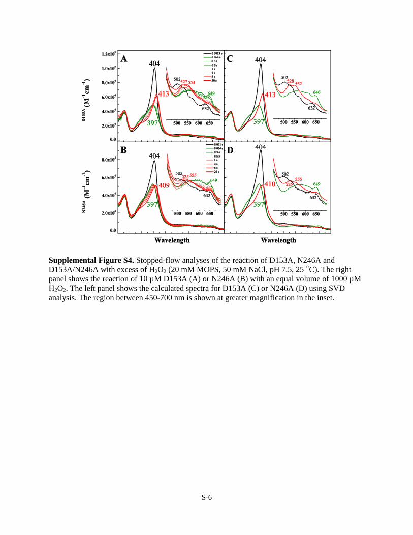
Supplemental Figure S4. Stopped-flow analyses of the reaction of D153A, N246A and
D153A/N246A with excess of H2O2 (20 mM MOPS, 50 mM NaCl, pH 7.5, 25 ○C). The right
panel shows the reaction of 10 µM D153A (A) or N246A (B) with an equal volume of 1000 µM
H2O2. The left panel shows the calculated spectra for D153A (C) or N246A (D) using SVD
analysis. The region between 450-700 nm is shown at greater magnification in the inset.

S-7
Supplemental Figure S5. Time traces of the reaction of DypB variants with H2O2. The reactions
were followed at 404 nm (410 nm for the D153H variants) (left panel) and 649 nm (right panel)
by after mixing 10 µM enzyme with 200 µM H2O2. Red curves show the fits of single-
exponential decays to the data and the residual is shown in grey.

S-8
Supplemental Figure S6. Plots of kobs vs. [H2O2] to determine second order rate constants at
404 nm for DypB (Black open square), D153A (Dark gray close circle), D153H (Black cross)
N246A (Gray close triangle) and D153A/N246A (Light-gray open circle).
S-9
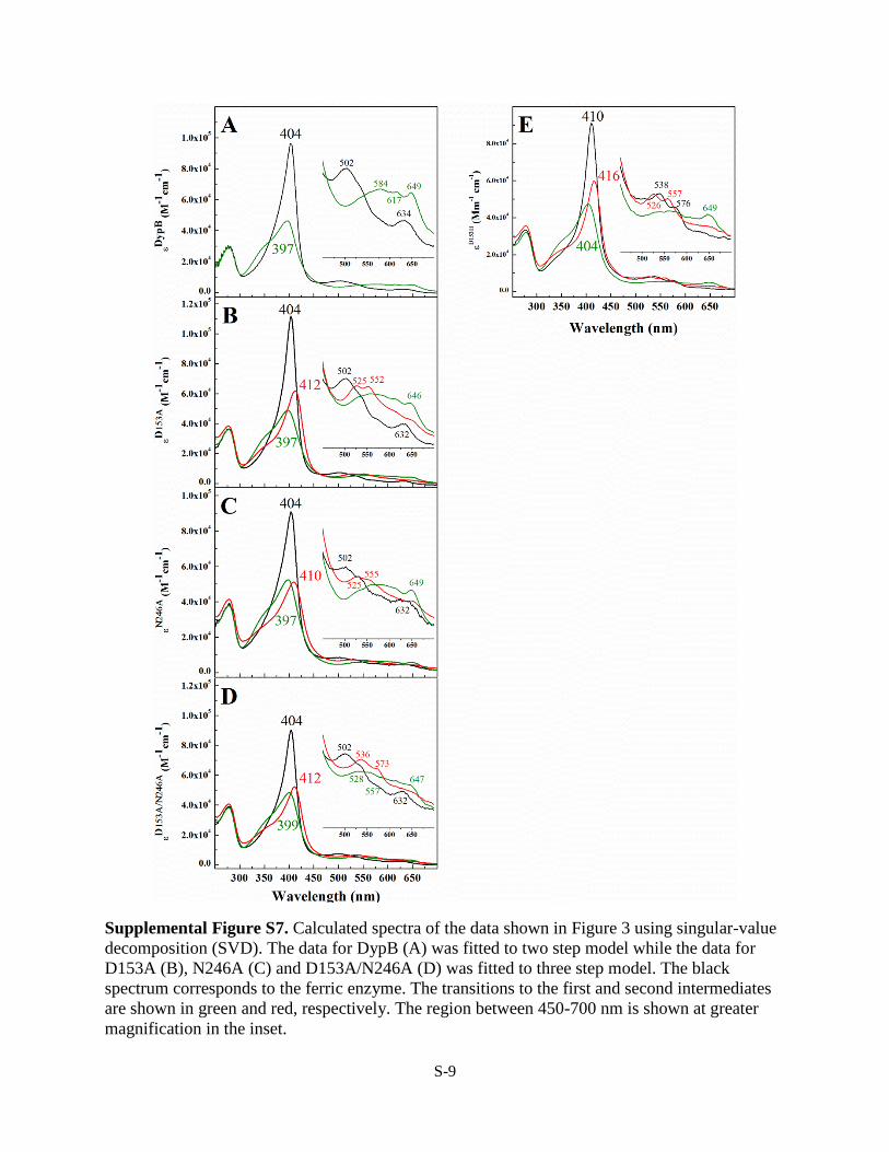
Supplemental Figure S7. Calculated spectra of the data shown in Figure 3 using singular-value
decomposition (SVD). The data for DypB (A) was fitted to two step model while the data for
D153A (B), N246A (C) and D153A/N246A (D) was fitted to three step model. The black
spectrum corresponds to the ferric enzyme. The transitions to the first and second intermediates
are shown in green and red, respectively. The region between 450-700 nm is shown at greater
magnification in the inset.

S-10
Supplemental Figure S8. Conserved hydrogen bond network in DypB involving Asp153, Water
(W) 507, Arg244, Heme propionates, W420, Arg208 and W438. Only side chains of the residues
are shown. Non-covalent bonds are shown in grey as dashed lines and the atomic distances are
shown in Å. The atoms are colored as follows: heme carbon, orange; residue carbons, cyan;
nitrogen, blue; oxygen, red; iron, brown; and solvent species oxygen, gray. The figure was
generated using coordinates from PDB ID 3QNS.

S-11
Supplemental Figure S9. Inverse analysis of Asp153 and Asn246 on Compound I formation. The log
of the k1 values of each variant and WT versus D153A/N246A are plotted. The dashed bars plotted for
WT DypB represent what the relative k1 values of the WT and the D153A/N246A would be if the effects
of the two sets of substitutions was additive.

D. EltisRahul Singh, Jason C. Grigg, Zachary Armstrong, Michael E. P. Murphy and Lindsay
BUT NOT ASPARTATE IS ESSENTIAL FOR PEROXIDASE ACTIVITYDistal Heme Pocket Residues of B-type Dye-decolorizing Peroxidase: ARGININE
doi: 10.1074/jbc.M111.332171 originally published online February 3, 20122012, 287:10623-10630.J. Biol. Chem.
10.1074/jbc.M111.332171Access the most updated version of this article at doi:
Alerts:
When a correction for this article is posted•
When this article is cited•
to choose from all of JBC's e-mail alertsClick here
Supplemental material:
http://www.jbc.org/content/suppl/2012/02/03/M111.332171.DC1.html
http://www.jbc.org/content/287/13/10623.full.html#ref-list-1
This article cites 30 references, 6 of which can be accessed free at
by guest on June 21, 2016http://w
ww
.jbc.org/D
ownloaded from
Copyright © 2022 FDOKUMEN