Healing of diabetic foot ulcers in l-arginine-treated patients
10
Dossier: Diabetes: Basic Research and Clinical Approach Healing of diabetic foot ulcers in L-arginine-treated patients Victor Arana a , Yolanda Paz b , Angélica González c , Verna Méndez d , José D. Méndez e, * a Department of Pathological Anatomy, Hospital General de Zona # 47, IMSS, Mexico City, Mexico b Department of Internal Medicine, Hospital General de Zona # 47, IMSS, Mexico City, Mexico c Department of Surgery, Hospital General de Zona # 1 A, IMSS, Mexico City, Mexico d Department of Internal Medicine, Hospital General de Zona/MF # 2, IMSS, San Luis Potosí, Mexico e Medical Research Unit in Metabolic Diseases, Twentieth Century National Medical Center, Mexican Institute of Social Security (IMSS), P.O. Box A-047, Mexico City 06703, Mexico Received 5 April 2004 Available online 13 October 2004 Abstract Experimentally, we demonstrated the beneficial effects of L-arginine on regulation of hyperglycemia and dyslipidemia in experimental diabetes, in addition to a positive anti-aggregating effect in platelets in animals and humans. Here, the effect of L-arginine on foot ulcers from diabetic patients was studied. Three groups of diabetic patients were included: 11 patients without ulcer received neither treatment and served as controls. Eleven patients with diabetic ulcer received the standard treatment, this group served as diabetic control with diabetic ulcer. Eleven remain patients with diabetic ulcer received 10 mM L-arginine subcutaneously on the site of the wound. Biopsy with punch number 5 on wound site comprising both ulcerative and contiguous undamaged skin were performed in all patients with ulcerative lesions before any treatment. Patients with intact skin had biopsy performed with punch number 5 on external malleolar region of right lower limb. Biopsies were examined by light and confocal microscopy utilizing histochemical and immunohistochemical methods. Initial and final blood samples were collected to determine glucose, triglycerides, total cholesterol, glycated hemoglobin (HbA 1c ), low (LDL), and high density lipoproteins (HDL). Significant differences (P < 0.05) were observed between initial and final serum glucose levels for treated patients, and initial serum glucose levels between treated and control patients without diabetic ulcer. Glycated hemoglobin, triglycerides, cholesterol, and lipoprotein levels showed no significant changes. Eight patients treated with L-arginine reached total wound healing and the remaining three who aban- doned the study because of change of residence showed relevant improvement. Histochemistry and immunohistochemistry methods have shown vascular impairment in both patients with diabetic ulcer (prior to treatment) and control patients without diabetic ulcer. Our observa- tions strongly support efficacy of L-arginine for successful wound healing of diabetic ulcers. © 2004 Elsevier SAS. All rights reserved. Keywords: Wound healing; Diabetic foot; L-arginine; Chronic ulcers 1. Introduction Cellular and biochemical interplay that comprises normal wound healing response is complex and implies intricate inter- actions among a variety of different cell types, structural pro- teins, growth factors, and proteinases [1]. According to meta- bolic situation and patient conditions, these interactions will lead to quick healing through acute inflammation and its reso- lution in a reasonable time or to establishment of a perpetu- ated chronic ulcer. Chronic wounds and their treat- ment are an enormous burden on the health care system, both in terms of cost and intensity of care required [1]. There exists a diversity of predisposal factors such as vascular alterations, infections, cancer, positional ulcers, alterations of immunity, pressure, and altered metabolism such as diabetes mellitus. Perhaps the latter is the most important due to the great num- ber of diabetic patients throughout the world. Diabetic patients frequently ask advice of the physician because of problems related to diabetic foot, especially appearance of ulcers, which if not properly treated will pro- duce amputation of the damaged lower limb [2–19]. Despite educational and preventive efforts to decrease both, fre- quency of diabetic ulcers and lower limb amputations caused by chronic ulcers in diabetic patients [2,3,5,10,12,13,19], the * Corresponding author. E-mail address: [email protected] (J.D. Méndez). Biomedicine & Pharmacotherapy 58 (2004) 588–597 http://france.elsevier.com/direct/BIOPHA/ 0753-3322/$ - see front matter © 2004 Elsevier SAS. All rights reserved. doi:10.1016/j.biopha.2004.09.009
-
Upload
independent -
Category
Documents
-
view
0 -
download
0
Transcript of Healing of diabetic foot ulcers in l-arginine-treated patients
Dossier: Diabetes: Basic Research and Clinical Approach
Healing of diabetic foot ulcers in L-arginine-treated patients
Victor Arana a, Yolanda Paz b, Angélica González c, Verna Méndez d, José D. Méndez e,*a Department of Pathological Anatomy, Hospital General de Zona # 47, IMSS, Mexico City, Mexico
b Department of Internal Medicine, Hospital General de Zona # 47, IMSS, Mexico City, Mexicoc Department of Surgery, Hospital General de Zona # 1 A, IMSS, Mexico City, Mexico
d Department of Internal Medicine, Hospital General de Zona/MF # 2, IMSS, San Luis Potosí, Mexicoe Medical Research Unit in Metabolic Diseases, Twentieth Century National Medical Center, Mexican Institute of Social Security (IMSS),
P.O. Box A-047, Mexico City 06703, Mexico
Received 5 April 2004
Available online 13 October 2004
Abstract
Experimentally, we demonstrated the beneficial effects of L-arginine on regulation of hyperglycemia and dyslipidemia in experimentaldiabetes, in addition to a positive anti-aggregating effect in platelets in animals and humans. Here, the effect of L-arginine on foot ulcers fromdiabetic patients was studied. Three groups of diabetic patients were included: 11 patients without ulcer received neither treatment and servedas controls. Eleven patients with diabetic ulcer received the standard treatment, this group served as diabetic control with diabetic ulcer.Eleven remain patients with diabetic ulcer received 10 mM L-arginine subcutaneously on the site of the wound. Biopsy with punch number5 on wound site comprising both ulcerative and contiguous undamaged skin were performed in all patients with ulcerative lesions before anytreatment. Patients with intact skin had biopsy performed with punch number 5 on external malleolar region of right lower limb. Biopsies wereexamined by light and confocal microscopy utilizing histochemical and immunohistochemical methods. Initial and final blood samples werecollected to determine glucose, triglycerides, total cholesterol, glycated hemoglobin (HbA1c), low (LDL), and high density lipoproteins(HDL). Significant differences (P < 0.05) were observed between initial and final serum glucose levels for treated patients, and initial serumglucose levels between treated and control patients without diabetic ulcer. Glycated hemoglobin, triglycerides, cholesterol, and lipoproteinlevels showed no significant changes. Eight patients treated with L-arginine reached total wound healing and the remaining three who aban-doned the study because of change of residence showed relevant improvement. Histochemistry and immunohistochemistry methods haveshown vascular impairment in both patients with diabetic ulcer (prior to treatment) and control patients without diabetic ulcer. Our observa-tions strongly support efficacy of L-arginine for successful wound healing of diabetic ulcers.© 2004 Elsevier SAS. All rights reserved.
Keywords: Wound healing; Diabetic foot; L-arginine; Chronic ulcers
1. Introduction
Cellular and biochemical interplay that comprises normalwound healing response is complex and implies intricate inter-actions among a variety of different cell types, structural pro-teins, growth factors, and proteinases [1]. According to meta-bolic situation and patient conditions, these interactions willlead to quick healing through acute inflammation and its reso-lution in a reasonable time or to establishment of a perpetu-ated chronic ulcer. Chronic wounds and their treat-
ment are an enormous burden on the health care system, bothin terms of cost and intensity of care required [1]. There existsa diversity of predisposal factors such as vascular alterations,infections, cancer, positional ulcers, alterations of immunity,pressure, and altered metabolism such as diabetes mellitus.Perhaps the latter is the most important due to the great num-ber of diabetic patients throughout the world.
Diabetic patients frequently ask advice of the physicianbecause of problems related to diabetic foot, especiallyappearance of ulcers, which if not properly treated will pro-duce amputation of the damaged lower limb [2–19]. Despiteeducational and preventive efforts to decrease both, fre-quency of diabetic ulcers and lower limb amputations causedby chronic ulcers in diabetic patients [2,3,5,10,12,13,19], the
* Corresponding author.E-mail address: [email protected] (J.D. Méndez).
Biomedicine & Pharmacotherapy 58 (2004) 588–597
http://france.elsevier.com/direct/BIOPHA/
0753-3322/$ - see front matter © 2004 Elsevier SAS. All rights reserved.doi:10.1016/j.biopha.2004.09.009

target of 40% decrease in amputation rates at year 2000 inthe US and Europe was not reached as has stated [19], andrates have actually increased.
In the 1990s, recombinant human platelet-derived growthfactor (rhPDGF-BB) emerged as a promise of successful treat-ment of chronic wounds, especially in diabetic patients. Sev-eral studies were effected to prove its efficacy in animal mod-els [20], toxicology aspects [21], effects of chronic woundfluid on structure and biologic activity [22], clinical safety inhumans [23] and clinical efficacy [24]. However, no conclu-sive results have been obtained and diabetic foot and its com-plications continue as a major problem in public health.
Other treatments have been assayed, such as low densityirradiation [25], human skin equivalent [26], blood-derivedangioblasts [27], hyperbaric oxygen therapy [28], inhibitionof lipid peroxidation [29], and systemic electromagnetic fields[30]. All attempted improvement of the vascular networkinvolved in the ulcerative lesion.
Recent investigations seem to support the beneficial effectof the role of nutrition on wound healing. Adequate processof scar formation has been observed when enteric nutrition isprovided, especially in arterial occlusive disease and chroniculcers of diabetic foot [31]. Thus, a diet containing 1.5−3 g/kgprotein has been recommended, depending severity of lesionand metabolic condition of the patient.
L-Arginine is one of the most versatile amino acids in ani-mal cells. There is evidence to implicate this amino acid as akey component in beneficial effects of low protein diet [32].It has also been shown to possess other numerous unique andpotentially useful pharmacologic effects [33]. L-Arginine is aprecursor of nitric oxide [34] and polyamine biosynthesis[34,35]. In extrahepatic tissues L-arginine is metabolized toL-ornithine and urea by arginase. L-ornithine is also metabo-lized to L-proline which is a substrate for collagen biosynthe-sis [34,35]. In extrahepatic tissues, L-arginine is metabolizedinto L-ornithine and urea by arginase. L-ornithine is alsometabolized into L-proline, which is a substrate for collagen
Table 1Serum glucose levels in treated with diabetic ulcer and non-treated diabetic patients without ulcer
No Treated patients Non-treated patientsName Glucose (mg/dl) Name Glucose (mg/dl)
Initial Final1 RPB 169 88 VHJ 672 RPE 274 177 PRJ 2053 MTA 247 240 IFM 2074 OGG 342 188 RGMM 1405 RFF 178 129 BMA 2446 RMJ 159 nm HRG 2547 FRF 192 110 RBD 1268 GCJ 173 77 PMR 1569 HMA 206 122 PMJ 7010 CAA 282 280 HCC 9611 CGJ 141 nm RPH 44Mean 214.81 156.77 133.54SD 62.92 69.6 73.43
nm = not measured.
Table 2Initial and final serum levels of glycated hemoglobin (HbA1c) and lipid profiles in treated diabetic patients with diabetic ulcer
Patient Initial FinalHbA1c TC HDL LDL TG HbA1c TC HDL LDL TG
1 RPB 7.6 150 38 87 123 8 141 41 77 1172 RPE 8.5 296 39 nm 675 11.4 323 41 117 8253 MTA 13.2 200 33 77 451 15.7 192 42 100 2494 OGG 10.6 168 32 112 122 13.9 161 32 101 1415 FRF 9.2 167 36 94 189 nm nm nm nm nm6 RMJ 5.5 182 50 117 73 nm nm nm nm nm7 FRH 11 226 38 154 171 7 243 58 156 1448 GCJ 7.3 212 42 142 142 6.5 209 46 134 1439 HMA 9.1 181 39 122 101 nm nm nm nm nm10 CAA 11.4 166 43 109 68 nm nm nm nm nm11 CGJ Nm 148 nm nm nm nm nm nm nm nmMean 9.34 191 39 113 212 10.4 212 43 114 270SD 2.26 42.7 5.2 24.9 196 3.84 65.3 8.5 28 276
Values of reference: HbA1c = glycated hemoglobin: 4.8–6.0%; TC = total cholesterol: 0–200 mg/dl; HDL = high density lipoproteins: 35–85 mg/dl; LDL = lowdensity lipoproteins: 50–130 mg/dl; TG = triglycerides: 50–200 mg/dl; nm = not measured.
589V. Arana et al. / Biomedicine & Pharmacotherapy 58 (2004) 588–597

biosynthesis [34–36]. Previously, it was suggested thatL-arginine may be of clinical benefit in improving wound heal-ing and immune response in healthy humans [37,38].
This paper was designed to study the effect of L-arginineon wound healing in diabetic patients with chronic ulcerativelesions on the foot.
2. Materials and methods
2.1. Patients
Thirty-three diabetic patients were consecutively selectedto participate in the study. Of these, 22 patients showedchronic ulcers of one of the lower limbs and 11 correspondedto diabetic patients without ulcerative lesions. We did not con-sider age or sex as exclusion criteria. After signing informedconsent form, patients were distributed into three groups:11 patients without ulcer received neither treatment and servedas non-treated control. Eleven patients with diabetic ulcerreceived the standard treatment available at the hospital. Thistreatment was based on the use of soap, water and iodine, thisgroup served as diabetic control with ulcer. Eleven remainpatients with diabetic ulcer were administered daily 10 mML-arginine dissolved in sterile 0.154 M NaCl around the ulcer(experimental treatment). By ethical reasons when all thepatients were selected the patients with diabetic ulcer receivedinformation about the experimental treatment, and they weredecided by consensus of the assimilation to a group; treatedor non-treated group. So that, the design of the experimentalstudy, was non-randomized between the groups. Initial sizeof the ulcerative lesion was calculated by multiplying lengthby width. Biopsies were performed with Biopsy punch No. 5(Fray Products Corp., Buffalo, NY USA) comprising ulceredand non-ulcered skin in patients with diabetic ulcer and onskin of external maleollar zone in control patients. Tissuesamples were processed by light and confocal microscopy. Itis important to mention that neither patients with non-treated
diabetic ulcer nor patients with treated diabetic ulcer werenot previously submitted to any surgery before to the begin-ning of the study.
2.2. Biochemical determinations
2.2.1. Blood sample collectionTo make initial and final biochemical determinations in
serum, blood samples were taken from forearm vein. Gly-cated hemoglobin (HbA1c) was determined by means of Para-gon Beckman electrophoresis. Total cholesterol [39], glu-cose [40], and triglycerides [41] were assayed enzymatically.In addition, high density lipoproteins (HDL) were measuredby precipitation method [42], and low density lipoproteins(LDL) were calculated by Friedewald formula [43]. Duringtime of study, patients were under metabolic control, receiv-ing medical attention when necessary.
2.2.2. Histological evaluationSamples were fixed in 10% buffered formalin for light
microscopic examination. After fixation, sections of thewound were dehydrated with graded ethanols and embeddedin paraffin. Sections of 5 µ-thick of paraffin-embedded tis-sues were mounted on glass slides, rehydrated with distilledwater, and stained with hematoxylin and eosin. In addition,specimens were stained with Masson trichrome methodaccording to Bancroft and Cook technique [44] to demon-strate collagen fibers.
2.2.3. ImmunofluorescenceSpecimens were fixed in 4% buffered paraformaldehyde,
then washed with phosphate/sucrose buffer. Subsequently,6–8 µ-thick frozen sections were effected and placed on slidespreviously prepared with gelatine. Sections were incubatedin methanol/acetone solution. Prior to labeling, non-specificbinding sites were blocked for 1 h in horse normal serum(Equitech-Bio, Inc., Kerville, TX). They were incubated atroom temperature overnight with primary antibody mono-
Table 3Serum levels of HbA1c and lipid profile of the control without ulcer and treated patients
Cholesterol (mg/dl) HbA1c (%) LDL (mg/dl) HDL (mg/dl) TG (mg/dl)Non-treated Treated Non-treated Treated Non-treated Treated Non-treated Treated Non-treated Treated72 150 17 7.6 115 87 72 38 123 117158 296 9.9 8.5 nm nm nm 39 675 825321 200 11.4 13.2 228 77 39 33 451 249176 168 9.9 10.6 nm 112 nm 32 122 141183 167 8.2 9.2 nm 94 nm 36 189 nm211.5 182 nm 5.5 149 117 40 50 73 nm198 226 nm 11 nm 154 43 38 171 144195 212 nm 7.3 100 142 29 42 142 143218 181 13.2 9.1 128 122 48 39 101 nm201 166 nm 11.4 124 109 38 43 68 nm228 148 12.1 nm nm nm nm nm nm nm
Mean 196.5 190.5 11.5 9.3 140.7 112.7 44.1 39.0 211.5 269.8SD 59.0 42.7 2.9 2.3 45.7 24.9 13.6 5.2 196.4 275.9
Values of reference: HbA1c = glycated hemoglobin: 4.8–6.0%; TC = total cholesterol: 0–200 mg/dl; HDL = high density lipoproteins: 35–85 mg/dl; LDL = lowdensity lipoproteins: 50–130 mg/dl; TG = triglycerides: 50–200 mg/dl; nm = not measured.
590 V. Arana et al. / Biomedicine & Pharmacotherapy 58 (2004) 588–597
clonal anti-a smooth muscle actin (Sigma Chemical Co., St.Louis MO, USA). Sections were then washed with phosphate-buffered saline (PBS)-brij and incubated for 1 h with second-ary biotin-goat anti-mouse antibody (Zymed Laboratories,Inc., San Francisco, CA, USA) diluted 1:200 in PBS. Then,sections were washed in PBS for 30 min, and incubated for40 min in fluorescein isothiocyanate-avidin, washed in PBSfor 30 min, and stained with Evans blue. Samples were thenmounted in Vectashield mounting medium (Vector Laborato-ries, Inc., Burlingame, CA, USA) [45–50].
2.2.4. Confocal microscopySamples were observed with confocal laser microscope
BIO-RAD model MCR-600. Serial 1–2 µ-thick sections wereperformed, with green filter for Evans blue channel and bluefilter for fluorescein channel; fluorescein has green fluores-cence, while Evans blue has red fluorescence.
2.2.5. Statistical analysisMean ± SD was calculated from data obtained from treated
and control group without diabetic ulcer. Statistical signifi-
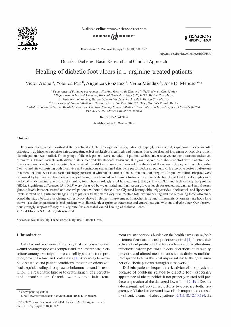
Fig. 1. Sections from skin of diabetic foot ulcers stained by H-E prior to treatment. (A) In this section is observed on the left side the epithelium (E), on the rightside the ulcer (U), the inflammatory process below the ulcer (I), and fibrin deposits (F) 20X. (B) The ulcer is shown (U), in addition to surface epithelium (E),fibrin deposits (F), inflammatory process (I), and loose connective tissue (T) 20X. (C and D) Relevant here are presence of extravasation of erythrocytes (E) andelements from acute and chronic inflammation characterized by polymorphonuclear cells (P), fibroblasts (FB), and loose connective tissue (T) 40X.
591V. Arana et al. / Biomedicine & Pharmacotherapy 58 (2004) 588–597

cance between experimental and control groups was calcu-lated by Student t-test for related and independent samples.Size of ulcers and time of evolution together with time oftreatment were analyzed by Pearson test correlation. Oddsratio was utilized in order to show the association betweenthe treatment or non-treatment from the diabetic ulcer andthe outcome (wound healing or non-wound healing).
3. Results
3.1. Glucose levels
All the treated patients were under initial hyperglycemia.At the end of the study, two patients reached normal serumlevels, the others showed decreasing levels. Significant varia-tions (P < 0.05) were observed between initial and final glu-cose levels. Control patients showed lower values of glucose(P < 0.05) than treated patients (Table 1).
3.2. Glycated hemoglobin
Both treated and control groups showed altered levels ofHbA1c. Initial and final values in treated group did not presentsignificant variations (Tables 2,3).
3.3. Triglycerides
No significant differences were observed between groups(Tables 2,3). Both groups showed certain tendency to in-creased levels.
3.4. Cholesterol
Approximately one-third of patients in both groups hadserum high cholesterol levels without significant differencesbetween groups (Tables 2,3).
3.5. Lipoproteins
No significant change was observed in control and treatedgroups (Tables 2,3).
3.6. Histological evaluation
Histological sections obtained from all the patients withor without ulcers showed little dermal and epidermal organi-zation (Fig. 2). The tissue obtained from the ulcerative lesionsof the patients with diabetic ulcer showed acute and chronicinflammatory processes characterized by polymorphonuclear(PMN) and mononuclear (MN) cells, fibroblasts, and mac-rophages (Fig. 1). Extracellular matrix (ECM) was character-ized by loose connective tissue in an irregular fashion withcollagen fibers distributed in different directions (Fig. 2). Inaddition, prominent edema, extravasation of erythrocytes, andinterstitial fibrin were observed, probably related to vasculardamage (Fig. 1). Little vessels were observed in both groupsof patients, and when vessels were seen diameter of lumenwas narrow or occluded and absence of erythrocytes wasnoted. In addition, we observed basement membrane thick-ening (Figs. 3,4).
3.7. Response to L-arginine
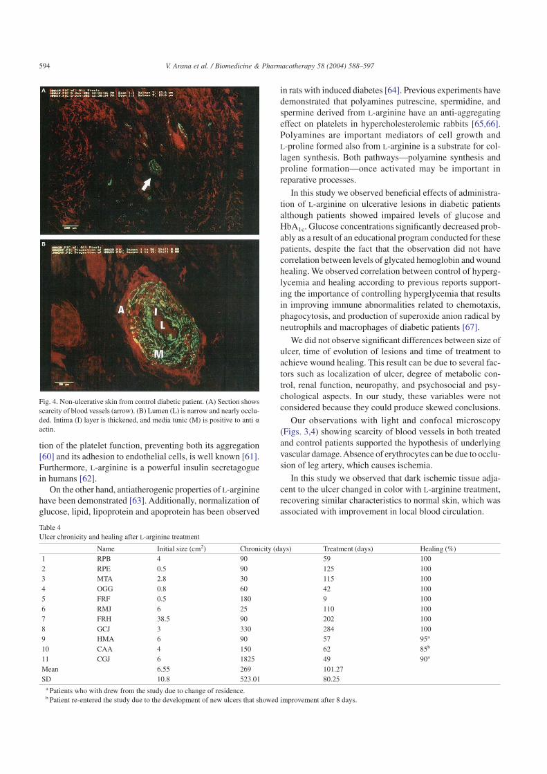
The ulcers measured from 0.5–38.5 cm2 (6.55 ± 10.8 cm2,mean ± SD) (Table 4). Chronicity of ulcers varied from 25 to1825 days (269 ± 523 days, mean ± SD) (Table 4). Time oftreatment varied from 9 to 284 days (101.27 ± 80.25 days,mean ± SD) (Table 4). All patients treated with L-arginineshowed improvement. Eight treated patients reached totalwound healing, three had rates of 95%, 90%, and 85% ofimprovement, respectively. The latter patients mentioneddropped out the study due to change of residence. One patientwho improved 85% re-entered the study due to appearanceof new ulcers, which showed healing during the following8 days. Thereafter, the patient left the study definitely(Table 4).
Table 5 summarizes the values of biochemical parametersfrom the group of non-treated diabetic patients with diabeticulcer obtained at the beginning of the study. Because severalpatients suffered amputation during the study, they went outthemselves the study and was not possible to make the finaldetermination of biochemical parameters in serum.
Fig. 2. Skin from diabetic patients without ulcer (A) and diabetic ulcer (B)stained by trichrome Masson technique. (A) Collagen fibers (C), showing anirregular pattern 40X. (B) Presence of adipose subcutaneous tissue (AST),next to collagen fibers (C), showing depth of lesion 40X.
592 V. Arana et al. / Biomedicine & Pharmacotherapy 58 (2004) 588–597
The follow-up from the patients with non-treated diabeticulcer showed the deterioration of the ulcerative lesion in allof them. These patients underwent surgical interventionswhich consisted since amputation of one toe until amputa-tion of the lower limb. One patient was also subject of ampu-tation of the contralateral limb.
No correlation was observed when Pearson correlation testwas carried out to compare simultaneously variable size ofulcers, time of evolution, and time of treatment.
4. Discussion and conclusion
In diabetic patients, deficiency of insulin can influencedevelopment of atherosclerosis through synergism of patho-logic mechanisms that involve dyslipidemia, advanced gly-cation end-products (AGEs), altered platelet function, abnor-malities in erythrocyte deformability, and abnormalities ofarterial wall function [2,15,51–56]. These mechanisms resultin peripheral vascular disease that produces ischemia.
Ischemia and neuropathy, two common complications ofdiabetes mellitus, are primary underlying risk factors fordevelopment of foot ulcers and their complications. Themajority of diabetic foot ulcers have mixed ischemic and neu-ropathic etiology [57]. However, a vascular etiology has been
postulated, including when neuropathy has been identified asdeterminant factor producing the ulcer [58].
In general, ischemia is considered a global enemy of heal-ing [59]. Wound healing is a complex process involving intri-cate interactions among a variety of different cell types, struc-tural proteins, growth factors, and proteinases that play animportant role in acute and chronic inflammation and tissuerepair. Acute and chronic wounds heal in an orderly progres-sion through artificially defined phases of vascular dilation,coagulation, inflammation, matrix synthesis and deposition,angiogenesis, fibroplasia, epithelization, contraction, andremodeling [1]. These processes imply participation of poly-morphonuclear and mononuclear cells, monocytes, lympho-cytes, fibroblasts, macrophages, and myriad growth, coagu-lation and angiogenic factors, and the cytokines produced bythese.
Adverse effects of malnutrition and diabetes mellitus onwound healing have justified nutritional treatment. Evidenceto implicate L-arginine as a key component in the beneficialeffects of a low-protein diet [32], can be widely explained,because this aminoacid participates in several metabolic path-ways serving as a precursor for synthesis not only of proteinsbut also of nitric oxide (NO), urea, glutamate, creatine,polyamines, proline, and other molecules involved in regu-lating cellular homeostasis [34]. The role of NO on regula-
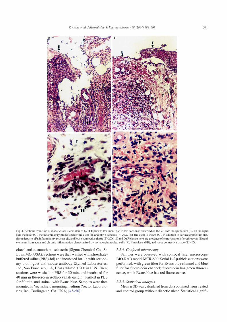
Fig. 3. Sections from ulcerative lesions of skin marked with anti a actin by confocal microscopy prior to treatment. (A and B) Scarce blood vessels (arrow). (Cand D) There are thickening of intima (I), absence of erythrocytes in narrow lumen (L), and smooth muscle cells positive to anti a actin in media tunic (M).
593V. Arana et al. / Biomedicine & Pharmacotherapy 58 (2004) 588–597
tion of the platelet function, preventing both its aggregation[60] and its adhesion to endothelial cells, is well known [61].Furthermore, L-arginine is a powerful insulin secretagoguein humans [62].
On the other hand, antiatherogenic properties of L-argininehave been demonstrated [63]. Additionally, normalization ofglucose, lipid, lipoprotein and apoprotein has been observed
in rats with induced diabetes [64]. Previous experiments havedemonstrated that polyamines putrescine, spermidine, andspermine derived from L-arginine have an anti-aggregatingeffect on platelets in hypercholesterolemic rabbits [65,66].Polyamines are important mediators of cell growth andL-proline formed also from L-arginine is a substrate for col-lagen synthesis. Both pathways—polyamine synthesis andproline formation—once activated may be important inreparative processes.
In this study we observed beneficial effects of administra-tion of L-arginine on ulcerative lesions in diabetic patientsalthough patients showed impaired levels of glucose andHbA1c. Glucose concentrations significantly decreased prob-ably as a result of an educational program conducted for thesepatients, despite the fact that the observation did not havecorrelation between levels of glycated hemoglobin and woundhealing. We observed correlation between control of hyperg-lycemia and healing according to previous reports support-ing the importance of controlling hyperglycemia that resultsin improving immune abnormalities related to chemotaxis,phagocytosis, and production of superoxide anion radical byneutrophils and macrophages of diabetic patients [67].
We did not observe significant differences between size ofulcer, time of evolution of lesions and time of treatment toachieve wound healing. This result can be due to several fac-tors such as localization of ulcer, degree of metabolic con-trol, renal function, neuropathy, and psychosocial and psy-chological aspects. In our study, these variables were notconsidered because they could produce skewed conclusions.
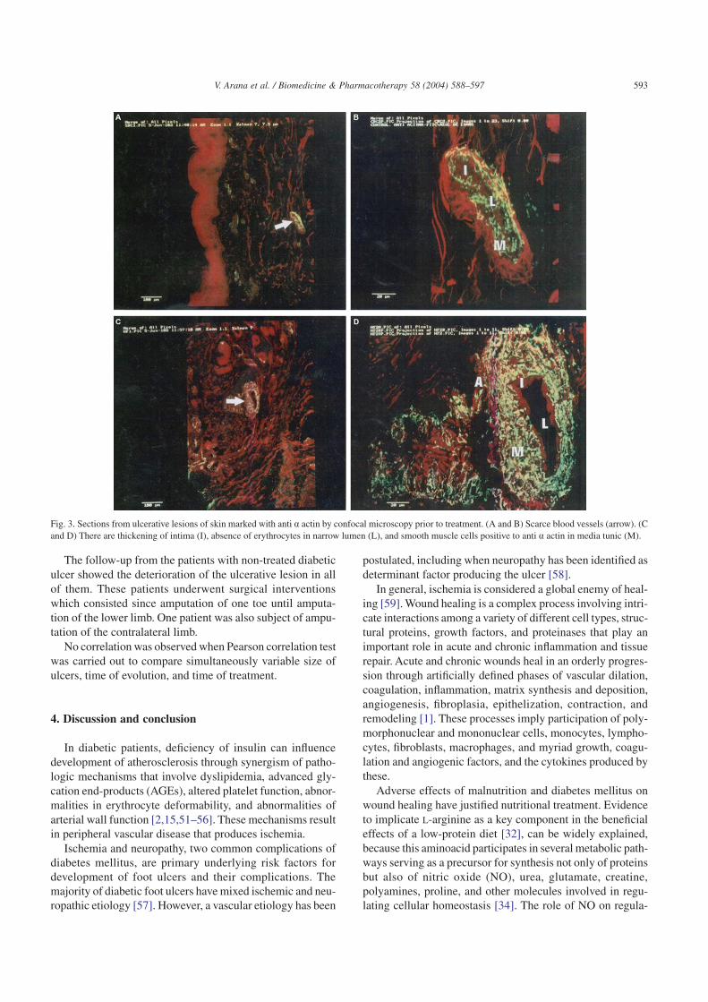
Our observations with light and confocal microscopy(Figs. 3,4) showing scarcity of blood vessels in both treatedand control patients supported the hypothesis of underlyingvascular damage.Absence of erythrocytes can be due to occlu-sion of leg artery, which causes ischemia.
In this study we observed that dark ischemic tissue adja-cent to the ulcer changed in color with L-arginine treatment,recovering similar characteristics to normal skin, which wasassociated with improvement in local blood circulation.
Fig. 4. Non-ulcerative skin from control diabetic patient. (A) Section showsscarcity of blood vessels (arrow). (B) Lumen (L) is narrow and nearly occlu-ded. Intima (I) layer is thickened, and media tunic (M) is positive to anti aactin.
Table 4Ulcer chronicity and healing after L-arginine treatment
Name Initial size (cm2) Chronicity (days) Treatment (days) Healing (%)1 RPB 4 90 59 1002 RPE 0.5 90 125 1003 MTA 2.8 30 115 1004 OGG 0.8 60 42 1005 FRF 0.5 180 9 1006 RMJ 6 25 110 1007 FRH 38.5 90 202 1008 GCJ 3 330 284 1009 HMA 6 90 57 95a
10 CAA 4 150 62 85b
11 CGJ 6 1825 49 90a
Mean 6.55 269 101.27SD 10.8 523.01 80.25
a Patients who with drew from the study due to change of residence.b Patient re-entered the study due to the development of new ulcers that showed improvement after 8 days.
594 V. Arana et al. / Biomedicine & Pharmacotherapy 58 (2004) 588–597

The precise mechanism by which L-arginine exerts itseffects on reparative processes of ulcerative lesion is notknown; however, it can be partially explained by:L-arginine→ornithine→glutamic semialdehyde→prolinepathway, as stated previously [68], although the contributionof polyamines synthesized from L-arginine is not discarded.For ethical reasons it was not possible to take another biopsyto study angiogenesis once ulcerative lesions were healed.This aspect remains to be studied.
In conclusion, our study showed that L-arginine was apotentially successful treatment to prevent lower limb ampu-tation and improve time of healing of chronic ulcer in dia-betic patients. These results support those observations ondietary arginine supplementation in elderly population thatresulted in up-regulation of T-cell activity and increased pro-tein and collagen deposition in experimental wound [37].L-Arginine administered daily was non-toxic and well-tolerated at this pharmacologic dosage, and therefore, can besafely used as auxiliary therapy for diabetic foot ulcers, inaddition to other available treatments.
Acknowledgments
We thank Dr. Ma. de Lourdes Esquivel Guzmán, Head ofthe Central Laboratory of the Specialities Hospital, Twenti-eth Century National Medical Center, Mexican Institute ofSocial Security (IMSS) for performing biochemical assays,Dr. Raúl Mena for use of facilities at the Department of Con-focal Microscopy and for access to his Neuroscience Labo-ratory of the Advanced Studies and Research Center (CIN-VESTAV), IPN, Mexico City, Mexico. V. Arana is aCONACYT doctoral fellow (No. 87745).
References
[1] Stadelman KS, Digenis AG, Tobin GR. Physiology and healingdynamics and chronic cutaneous wounds. Am J Surg 1998;176(Suppl2A):26S–38S.
[2] Levin ME. Preventing amputation in the patient with diabetes. Diabe-tes Care 1995;18(10):383–94.
[3] Ollendorf DA, Kotsanos J, James G, Wishner WJ, Friedman M,Cooper T, et al. Potential economic benefits of lower-extremity ampu-tation prevention strategies in diabetes. Diabetes Care 1998;21(8):1240–5.
[4] Potter PJ. Watch your step! Can Med Assoc J 1998;159(3):218–9.[5] Mayfield JA, Reiber GE, Sanders LJ, Janisse D, Pogach LM. Preven-
tive foot care in people with diabetes. Diabetes Care 1998;21(12):2161–77.
[6] American Diabetes Association. Preventive foot care in people withdiabetes. Diabetes Care 1998;21(12):2178–9.
[7] Hill SL, Holtzman GI, Buse R. The effects of peripeheral vasculardisease with osteomyelitis in the diabetic foot. Am J Surg 1999;177(4):282–6.
[8] Pittet D, Wyssa B, Herter-Clavel C, Kursteiner K, Vaucher J, Lew PD.Outcome of diabetic foot infections treated conservatively: a retropec-tive cohort study with long-term follow up. Arch Intern Med 1999;159(8):851–6.
[9] Armstrong DG, Stacpoole-Shea SB, Nguyen N, Harkless LB. Length-ening of the Achilles tendon in diabetic patients who are at high riskfor ulceration of the foot. J Bone Joint Surg Am 1999;81(A(4):535–8.
[10] Van Gils CC, Wheeler LA, Mellstrom M, Brinton EA, Mason S,Wheeler CG. Amputation prevention by vascular sugery and podiatrycollaboration in high-risk diabetic and nondiabetic patients: theOperation Desert Foot experience. Diabetes Care 1999;22(5):678–83.
[11] Ahroni JH, Boyko EJ, Forsberg RC. Clinical correlates of plantarpressure among diabetic veterans. Diabetes Care 1999;22(6):965–72.
[12] Bloomgarden ZT. The EuropeanAssociation for the Study of DiabetesAnnual Meeting 1998. Complications of diabetes. Diabetes Care1999;22(8):1364–70.
[13] Dargis VP, Olga A, Jonushaite L, Vileikyte, Boulton AJ. Benefits of amultidisciplinary approach in the management of recurrent diabeticfoot ulceration in Lithuania: a prospective study. Diabetes Care 1999;22(9):1428–31.
[14] Pinzur MS. American Orthopaedic Foot and Ankle Society DiabeticShoe Survey. Diabetes Care 1999;22(12):2099–100.
[15] Colwell JA. Peripheral vascular disease in diabetes mellitus. In:Davidson JK, editor. Clinical diabetes mellitus. A problem-orientedapproach. NY: Thieme; 2000. p. 560–1.
[16] Coleman WC. The diabetic foot. In: Davidson JK, editor. Clinicaldiabetes mellitus. A problem-oriented approach. NY: Thieme. 2000.p. 571–80.
[17] Levin ME. Pathophysiology of diabetic foot lesions. In: Davidson JK,editor. Clinical diabetes mellitus. A problem-oriented approach. NY:Thieme; 2000. p. 581–98.
Table 5Serum levels of glucose, glycated hemoglobin and lipid profile from non-treated diabetic patients with diabetic ulcer
No. Name Glucose (mg/dl) HbA1c (%) TC (mg/dl) HDL (mg/dl) LDL (mg/dl) TG (mg/dl)1 ACL 294 17.7 249 nm nm 2362 MLL 262 15.2 149 nm nm 603 GRM 267 16 191 43 nm 2474 MHM 387 12.7 406 75 329 2135 VMR 264 13.1 172 nm nm 756 ZEJ 83 8.9 180 68 97 7517 AGM 117 nm 271 36 165 3528 LRM 370 nm 193 70 90 1679 PCJ 164 10.6 168 32 112 12210 LRE 208 7.9 142 30 83 14411 EAE 72 5.8 137 38 66 167r 2488 107.9 2011.49 392 942 2534Mean 226.18 9.81 182.86 35.64 85.64 230.36SD 107.66 3.99 97.74 18.72 91.29 191.54
Values of reference: HbA1c = glycated hemoglobin: 4.8–6.0%; TC = total cholesterol: 0–200 mg/dl; HDL = high density lipoproteins: 35–85 mg/dl; LDL = lowdensity lipoproteins: 50–130 mg/dl; TG = triglycerides: 50–200 mg/dl; nm = not measured. r = summa; SD = standard deviation.
595V. Arana et al. / Biomedicine & Pharmacotherapy 58 (2004) 588–597

[18] Hobgood E. Conservative therapy of foot abnormalities, infections,and vascular insufficiency. In: Davidson JK, editor. Clinical diabetesmellitus. A problem-oriented approach. NY: Thieme; 2000. p. 599–609.
[19] Bloomgarden ZT. American Diabetes Association 60th Scientific Ses-sions, 2000. The diabetic foot. Diabetes Care 2001;24(5):946–51.
[20] LeGrand EK. Preclinical promise of becaplermin (rhPDGF-BB) inwound healing. Am J Surg 1998;176(Suppl 2A):48S–54S.
[21] Knight EV, Oldham JW, Mohler MA, Liu S, Dooley JA. Review ofnonclinical toxicology studies of becaplermin (rhPDGF-BB). Am JSurg 1998;176(Suppl 2A):55S–60S.
[22] Castronuovo Jr. JJ, Ghobrial I, Giusti AM, Rudolph S, Smiell JM.Effects of chronic wound fluid on the structure and biological activityof becaplermin (rhPDGF-BB) and becaplermin gel. Am J Surg 1988;176(Suppl 2A):61S–67S.
[23] Smiell JM. Clinical safety of becaplermin (rhPDGF-BB) gel. Am JSurg 1988;176(Suppl 2A):68S–73S.
[24] Wieman TJ. Clinical efficacy of becaplermin (rhPDGF-BB) gel. Am JSurg 1998;176(Suppl 2A):74S–79S.
[25] Schindl A, Schindl M, Schon H, Knobler R, Havelec L, Schindl L.Low-intensity laser irradiation improves skin circulation in patientswith diabetic microangiopathy. Diabetes Care 1998;21(4):580–4.
[26] Brem H, Balledux J, Bloom T, Kerstein M, Hollier L. Healing ofdiabetic foot ulcers and pressure ulcers with human skin equivalent: anew paradigm in wound healing. Arch Surg 2000;135(6):627–34.
[27] Schatteman GC, Hanlon HD, Jiao C, Dodds SG, Christy BA. Blood-derived angioblasts accelerate blood-flow restoration in diabetesmice. J Clin Invest 2000;106(4):571–8.
[28] Leslie CA. Randomized controlled trial of topical hyperbaric oxygenfor treatment of diabetic foot ulcers. Diabetes Care 1998;11:111–5.
[29] Altavilla D, SaittaA, Cucinotta D, Galeano M, Deodato B, Colonia M,et al. Inhibition of lipid peroxidation restores impaired vascular endot-elial growth factor expression and stimulates wound healing andangiogenesis in the genetically diabetic mouse. Diabetes 2001;50(3):667–74.
[30] Cañedo-Dorantes L, García-Cantú R, Barrera R, Méndez-Ramírez I,Navarro VH, Serrano G. Healing of chronic arterial and venous legulcers with systemic electromagnetic fields. Arch Med Res 2002;33:281–9.
[31] Kiyama T, Witte MB, Thornton FJ. The route of nutrition supportaffects the early phase of wound healing. J Parenter Enteral Nutr1998;22(5):276.
[32] Narita I, Border WA, Ketteler M, Ruoslahti E, Noble NA. L-Argininemay mediate the therapeutic effects of low protein diets. Proc NatlAcad Sci USA 1995;92:4552–6.
[33] Barbul A. Arginine, biochemistry, physiology, and therapeutic impli-cation. J Parenter Enteral Nutr 1986;10:227–38.
[34] Wu G, Morris SM. Arginine metabolism: nitric oxide and beyond.Biochem J 1998;336:1–7.
[35] Pegg AE. Recent advances in the biochemistry of polyamines ineukaryotes. Biochem J 1986;234:249–62.
[36] Shih VE. Regulation of ornithine metabolism. Enzyme1981;26:254–8.
[37] Barbul A, Lazarou SA, Efron DT, Wasserkrug HL, Efron G. Arginineenhances response wound healing and lymphocyte immune responsesin humans. Surgery 1990;108:331–7.
[38] Kirk SJ, Hurson M, Regan MC, Holt DR, Wasserkrug HL, Barbul A.Arginine stimulates wound healing and immune function in elderlyhuman beings. Surgery 1993;114:155–60.
[39] Katterman R. Rapid determination of total and free cholesterol inserum. Clin Chem Clin Biochem 1984;22:245.
[40] Trinder P. Determination of glucose in blood using oxidase with analternative oxygen acceptor. Ann Clin Biochem 1969;6:24.
[41] Wahlefeld AM. In: Triglycerides, determination after enzymatichydrolysis. In: Bergmeyer HU, editor. Methods of enzymatic analysis.New York: Academic Press; 1974. p. 1831.
[42] Friedewald WT, Levy FI, Fredrickson DS. Estimation of the concen-tration of low-density lipoprotein-cholesterol in plasma without theuse of the preparative ultracentrifuge. Clin Chem 1972;18:499.
[43] Warnick GR, Knopp RH, Branson L. Estimating low-density lipopro-tein cholesterol by the Friedewald equation is adequate for classifyingpatients on the basis of Nationally Recommended Cutpoints. ClinChem 1990;36:15.
[44] Bancroft JD, Cook HC. Connective tissues. In: Manual of histologicaltechniques and their diagnostic application. New York: Churchill-Livingstone; 1994. p. 42–3.
[45] Hermoso M, Sáez JC, Villalón M. Identification of gap junctions in theoviduct and regulation of connexins during development and bysexual hormones. Eur J Cell Biol 1997;74:1–9.
[46] Taylor CR, Shi SR. Fixation, processing, special applications. In:Taylor CR, Cote RJ, editors. Immunomicroscopy: a diagnostic tool forthe surgical pathologist. Philadelphia: W.B. Saunders Company;1994. p. 42–70.
[47] Van der Loos CM. In: Bosher A, editor. Immunoenzyme multiplestaining methods. New York: BIOS Scientific Publishers Limited;1999. p. 1.
[48] Jasani B, Schmid KW. Role and scope of immunocytochemistry inroutine histopathological analysis. In: Horne T, editor. Immunocy-tochemistry in diagnostic histopathology. New York: Churchill-Livingstone; 1993. p. 23–7.
[49] Brandtzaeg P, Halstensen TS, Huitfeldt HS, Valnes KN. Immunofluo-rescence and immunoenzyme histochemistry. In: Johnstone AP,Turner MW, editors. Immunochemistry 1. London: Oxford UniversityPress; 1997. p. 71–130.
[50] Hsu SM. The use of the avidin–biotin interaction in immunocy-tochemistry. In: Cuello AC, editor. Immunohistochemistry II.Chichester: John Wiley and Sons; 1993. p. 169–80.
[51] Brownlee M, Cerami A. The biochemistry of the complications ofdiabetes mellitus. Annu Rev Biochem 1981;50:385–432.
[52] Sobening IA, Tertov VV, Orekhov AN. Atherogenic modified LDL indiabetes. Diabetes 1996;45(3S):35S–39S.
[53] Bloomgarden ZT. The 32nd Annual Meeting of the European Asso-ciation for the Study of Diabetes: Macrovascular disease. DiabetesCare 1997;2(7):1198–201.
[54] Bloomgarden ZT. American European Association Annual Meeting,1997: endothelial dysfunction, neuropathy and the diabetic foot, dia-betic mastopathy, and erectile dysfunction. Diabetes Care 1998;21(1):183–9.
[55] Devaraj S. Low-density lipoprotein postsecretory modification,monocyte function, and circulating adhesion molecules in type 2 dia-betic patients with and without macrovascular complications: theeffect of tocopherol supplementation. Circulation2000;102(2):191–6.
[56] Suzuki LA, Poot M, Gerrity RG, Bornfeldt KE. Diabetes acceleratessmooth muscle accumulation in lesions of atherosclerosis: lack ofdirect growth-promoting effects of high glucose levels. Diabetes2001;50(4):851–60.
[57] Laing P. The development and complications of diabetic foot ulcers.Am J Surg 1998;176(Suppl 2A):11S–19S.
[58] Irwin ST, Gilmore J, McGrann S. Blood flow in diabetics with footlesions due to “small vessel disease”. Br J Surg 1988;75:1201–6.
[59] Bulat T, Kosinski M. Diabetic foot: strategies to prevent and treatcommon problems. Geriatrics 1995;50(2):46–55.
[60] Radomski MW, Palmer RMJ, Moncada S. Characterization of theL-arginine: nitric oxide pathway in human platelets. Br J Pharmacol1990;101:325–8.
[61] Radomski MW, Palmer RMJ, Moncada S. Endogenous nitric oxideinhibits human platelet adhesion to vascular endothelium. Lancet1987;ii:1057–8.
[62] Menchini M, Meschi F, Lambiase R, Puzzovio M, Del Guercio MJ,Chiumelo G. C-Peptide response to arginine stimulation in diabeticchildren. J Pediatr 1980;96(3):362–6.
596 V. Arana et al. / Biomedicine & Pharmacotherapy 58 (2004) 588–597
[63] Cook JP, Singer AH, Tsao P, Zera P, Rowan RA, Billinham ME.Antiatherogenic effects of L-arginine in the hypercholesterolemicrabbit. J Clin Invest 1992;90:1168–72.
[64] Méndez JD, Balderas F. Regulation of hyperglycemia and dyslipi-demia by exogenous l-arginine in diabetic rats. Biochimie 2001;83(5):453–8.
[65] Méndez JD, Zarzoza E. Inhibition of platelet aggregation byL-arginine and polyamines in alloxan treated rats. Biochem Mol BiolInt 1997;43:311–8.
[66] Corona de la Peña N, Sosa-Melgarejo J, Ramos RR, Méndez JD.Inhibition of platelet aggregation by putrescine, spermidine, and sper-mine in hypercholesterolemic rabbits. Arch Med Res 2000;31:546–50.
[67] McMahon M, Rizza R. Nutrition support in hospitalized patients withdiabetes mellitus. Mayo Clin Proc 1996;71:587–94.
[68] Zinker S, Rodkind M. Collagen biosynthesis in the chick embryo, I.The source of free proline and collagen hydroxyproline. ConnectTissue Res 1972;1:275–81.
597V. Arana et al. / Biomedicine & Pharmacotherapy 58 (2004) 588–597