FOOT-AND-MOUTH DISEASE VIRUS - WUR eDepot
139
FOOT-AND-MOUTH DISEASE VIRUS THE ROLE OF INFECTION ROUTES AND SPECIES DIFFERENCES IN THE TRANSMISSION OF FMDV CARLA BRAVO DE RUEDA C. Bravo de Rueda 2015 Foot-and-mouth disease virus The role of infection routes and species differences in the transmission of FMDV
-
Upload
khangminh22 -
Category
Documents
-
view
0 -
download
0
Transcript of FOOT-AND-MOUTH DISEASE VIRUS - WUR eDepot

FOOT-AND-MOUTH DISEASE VIRUS
THE ROLE OF INFECTION ROUTES AND SPECIES DIFFERENCES IN THE TRANSMISSION OF FMDV
CARLA BRAVO DE RUEDA
C. B
ravo de Rued
a 2015Fo
ot-and
-mo
uth disease virus The role of infection routes and
species d
ifferences in the transmission of FM
DV

Foot-and-mouth disease virus
The role of infection routes and species differences in the transmission of FMDV
Carla Bravo de Rueda

Thesis committee
Promotor Prof. Dr M.C.M. de Jong Professor of Quantitative Veterinary Epidemiology Wageningen University Co-promotors Dr A. Dekker Senior Scientist Central Veterinary Institute (CVI), Wageningen UR, Lelystad Dr P.L. Eblé Scientist Central Veterinary Institute (CVI), Wageningen UR, Lelystad Other members
Prof. Dr J.A. Stegeman, Utrecht University Prof. Dr J.M. Vlak, Wageningen University Prof. Dr H. Hogeveen, Wageningen University Prof. Dr M. Nielen, Utrecht University This research was conducted under the auspices of the Graduate School of Wageningen Institute of Animal Sciences (WIAS).

Foot-and-mouth disease virus
The role of infection routes and species differences in the transmission of FMDV
Carla Bravo de Rueda
Thesis submitted in fulfilment of the requirements for the degree of doctor
at Wageningen University by the authority of the Academic Board,
in the presence of the Thesis Committee appointed by the Academic Board
to be defended in public on Monday 1 June 2015
at 4 p.m. in the Aula.

Carla Bravo de Rueda Foot-and-mouth disease virus The role of infection routes and species differences in the transmission of FMDV, 138 pages. PhD thesis, Wageningen University, Wageningen, NL (2015)
With references, with summaries in Dutch and English
ISBN 978-94-6257-328-4

For all those who contributed to my professional development


taBLe oF Contents
Chapter 1 General introduction 9
Chapter 2 Identification of factors associated with increased 25 excretion of foot-and-mouth disease virus
Chapter 3 Quantification of transmission of foot-and-mouth 47 disease virus caused by an environment contaminated with secretions and excretions from infected calves
Chapter 4 Estimation of the transmission of foot-and-mouth 73 disease virus from infected sheep to cattle
Chapter 5 Vaccination of cattle only is sufficient to stop FMDV 95 transmission in mixed populations of sheep and cattle
Chapter 6 General discussion 109
Part I: The role of infection routes in the transmission of FMDV 111
Part II: The role of species differences in the transmission of FMDV and vaccination strategies towards the species 113
Targeting control measures to reduce the transmission of the infection 114
Concluding remarks 116
Implications and further research 117
Chapter 7 Summary & Samenvatting 123
About the author 131
Training and Supervision Plan 133
List of Publications 135
Acknowledgement 137


IGeneral introduction


Ge
ne
ra
l Intr
od
uc
tIon
IFoot-and-mouth disease (FMD) is a contagious viral disease of cloven-hoofed animals, not only of the most important domestic animals i.e. cattle, pigs, sheep, goats and domestic buffalo, but also of wildlife species [1]. Outbreaks of FMD are responsible of large socio-economic losses [2]. The disease is caused by foot-and-mouth disease virus (FMDV), a virus 25-30 nm in size containing a single strand of ribonucleic acid (RNA) inside an icosahedral capsid made of protein. FMDV is classified within the genus Aphtovirus, member of the Picornaviridae family [3] and consists of seven different serotypes (A, O, C, Asia1, SAT1, SAT2, SAT3) with many subtypes [4]. A property of this virus is that it is difficult to stop its transmission [5] which makes it difficult to control and eradicate the disease worldwide. Although the virus can be rapidly inactivated at pH values of less than 7.0 (below neutral pH) [4], it can withstand high temperatures when protected by proteins e.g. from milk [6–9], limiting virus inactivation and therefore enhancing virus persistence in the environment.
history and worLdwide distriBution
FMDV outbreaks have occurred in almost all countries containing FMDV-susceptible animals, with the exception of New Zealand [10].
FMDV used to be endemic in Europe, huge epidemics swept through Europe at the end of the 19th and the beginning of the 20th century. It was reported that FMDV was introduced in the 19th century into South America by imported cattle from Europe [11].
Early control strategies for the control of FMDV transmission involved movement restrictions, slaughter of infected animals, and disinfection of the environment. Later, the use of vaccines was implemented [12]. The first inactivated vaccine against FMDV was produced in the 1930’s by Waldmann and collaborators [13]. But it was not until the late 40’s of the 20th century when Frenkel made the production of vaccines on a larger scale [14] possible. The Netherlands was the first country implementing nationwide prophylactic vaccination in cattle, which was followed by most countries in mainland Europe. The vaccination of cattle only, reduced the number of FMDV outbreaks enormously. The vaccination program resulted in cessation of disease outbreaks in 1989. In 1992, a non-prophylactic vaccination policy was adopted in the EU [15]. The decision was mainly based on economic evaluations, resulting in the reduction in cost of prophylactic vaccination and increasing the market potential of livestock and livestock products [12]. After the ban on prophylactic FMDV vaccination, only minor FMDV outbreaks occurred in Europe until the outbreak of 2001 in the United Kingdom [16]. FMDV control strategies such as movement restrictions, slaughter of infected animals, and disinfection of the environment have remained the basis to control FMD [12, 17]. Also, slaughter of non-infected animals from farms neighboring infected farms (pre-emptive culling) is used as a control measure. In The Netherlands in the outbreak of 2001, emergency vaccination of all FMDV-susceptible animals was used as an additional control measure [18]. However, all vaccinated animals were culled (suppressive vaccination) for economic reasons. Slaughtering and vaccination remain primary lines of defense for FMDV control [12, 19]. It has been suggested that
11

Ge
ne
ra
l Intr
od
uc
tIon
Ithere is a risk that vaccinated animals can become carriers of the virus after exposure to the virus [20]. However to differentiate vaccinated from infected animals (either carriers or not), DIVA (Differentiating Infected from Vaccinated Animals) serological tests for detecting non-structural proteins (NSP) have been developed [21–23]; If the animals are NSP positive they are infected with the virus. Tests for detecting NSPs are highly specific to differentiate non-infected from infected vaccinated animals and are even capable to detect silent infections in populations of either cattle or sheep [24, 25]. Also complementary serological tests for detecting specific immunoglobulin A [26, 27] in infected animals, have been developed. However their use is until now questionable [12].
If vaccination would be used as control strategy and tests for detecting NSPs would be used for differentiating infected from non-infected vaccinated animals, vaccination should be performed with purified vaccines that do not contain NSPs [28, 29].
Fmd CLiniCaL siGns
Typical clinical signs and lesions of animals infected with FMDV include fever, excessive salivation and/or nasal secretion and development of vesicles on epithelia of the mouth, feet and teats. Rupture of vesicles on the hoofs can precede ulcers with secondary bacterial infections causing lameness and pain. Loss of affected hoofs can also be expected and lesions on the udder can lead to mastitis. Ultimately, an FMDV infection can lead to weight loss, decrease in milk production, and loss of draught power [10].
In field conditions, the diagnosis of FMDV infection mostly depends on observing clinical signs. FMDV lesions are most apparent in cattle and pigs and therefore clinical diagnosis of FMDV infections in these species is mostly straightforward. But in sheep and goats, clinical signs are milder and may stay undetected [30] which can make it difficult to detect newly infected herds.
PathoGenesis
After the infection with the virus takes place, large amounts of FMDV are released in secretions and excretions, before [37] and after [38] the appearance of clinical signs. After FMDV is released in secretions and excretions from the infected animals, virus aerosols can be formed [39] which may contaminate the environment some distance from their source [40].
Secretions are defined as material released from glands (i.e. milk, semen, saliva) whereas excretions refer to any other products released from animals (e.g. lesion material, urine, faeces, and material released from the respiratory tract). All secretions and excretions from infected animals can contain FMDV [31, 41]. FMDV can persist in the secretions and excretions from the infected animals in the environment for long periods of time even after the animals have died [39] facilitating transmission of FMDV via the indirect route. The factors associated with increased secretion and excretions of FMDV by the infected animals are still unknown.
12

Ge
ne
ra
l Intr
od
uc
tIon
I
transmission routes For Fmdv
Pathogen transmission includes any mechanism by which an infectious agent is spread from an infected individual to a susceptible host [42]. Even though studies support the idea that FMDV transmission does mostly occur when the animals are manifesting clinical signs of the diseases [43], others have suggested that the presence of clinical signs is not a determinant for FMDV transmission to occur [44]. FMDV in secreted and excreted material can be transmitted either directly, e.g. via contact with an infected host [43, 45–52], or indirectly, e.g. via contact with a contaminated environment with FMDV infected secretions and excretions [41, 53]. Direct (contact) transmission of FMDV occurs when the infected animal(s) and the recipient animal(s) are in close vicinity. Indirect (contact) transmission of FMDV, via the environment, occurs when the recipient animal(s) are in contact with infected secretions and excretions. For example, indirect transmission occurs after feeding animals with contaminated feedstuff and during the movement of animals [12] i.e. bringing animals to environments contaminated previously by infected animals (e.g. trucks, pens used to hold animals for trade etc.). Contamination of the environment and feedstuffs (including concentrates, hay and straw) by saliva, faeces and
Figure 1. FMD clinical signs in cattle and sheep. Top left figure shows excessive nasal discharge in cattle. Top right figure shows a lesion on the coronary band of the hoof from a sheep. Bottom right figure shows a vesicle in the interdigital space of the hoof from a sheep.
13

Ge
ne
ra
l Intr
od
uc
tIon
Iurine, was an important cause of transmission of FMDV when Europe was endemic to the virus in the 20th century [41]. Even though airborne transmission has been suggested as the cause of outbreaks in the 2001 epidemic in Europe [54], its actual contribution to the transmission of FMDV is often overestimated. It has been shown that the probability of infection via the airborne route is very low [55]. The most important factor in the transmission of FMDV has been suggested to be the movement of infected animals [56]. The contribution of secretions and excretions from infected animals in the transmission of FMDV has not been quantified extensively [41]. It is not yet understood which factors could influence the higher secretion and excretion of FMDV by the infected animals. Moreover mathematical quantification of the contribution of these infected secretions and excretions to the transmission of FMDV has not yet been performed. Filling these gaps in the knowledge of FMD could allow better estimation of the contribution of other routes to the transmission of FMDV.
QuantiFiCation oF transmission
Epidemic mathematical models can be used to study the transmission of pathogens. It was in 1766 when Bernoulli used the first mathematical model to estimate life expectancy in individuals infected with smallpox [57]. This was the first mathematical model created to study the spread of a disease. Modern theoretical epidemiology began with Ross who described for the first time a “infection rate parameter” (i.e. the rate of occurrence of new infection cases in a susceptible population) based on the assumption that an infectious individual (re-) infect others in unit of time [58]. Building on the research of Ross, Kermack and McKendrick used a mathematical model to describe the transmission dynamics of infections [59]. For describing transmission, the individuals in a population are often classified according to a simple dynamic system: susceptible (S), infectious (I), or recovered (R). The number of individuals S, I and R will vary in time (t). This system described by Kermack and McKendrick is called the SIR model. An SIR model can be varied to accommodate additional pathways when required [42, 60]. The simplest differential equations that describe the SIR transmission dynamics of infections are:
Where β is the transmission rate parameter (i.e. the number of new infections caused by a typical infected individual per unit of time) and α is the recovery rate parameter. Thus to determine the number of new infections per unit of time, the total number of individuals transmitting infection (βI) is multiplied by the probability that the individuals are susceptible in the population (S/N).
14

Ge
ne
ra
l Intr
od
uc
tIon
IThe average number of infections caused by a single typical infectious individual, called
the reproduction ratio and denoted R0 [61, 62], was first described by Ronald Ross and Kermack and McKendrick. But it was George MacDonald who applied it first in modern epidemiology to describe transmission of malaria [63]. In epidemics, R0 is useful to describe the magnitude of transmission. If R0 is greater than 1, major and minor outbreaks are possible, and if R0 is less than 1 only minor outbreaks are possible [64, 65]. By using a final size approach the final outcome of a transmission experiment [66, 67] can be used to calculate R0. R0 can also be estimated by multiplying the transmission rate parameter β with the average infectious period [68]. In heterologous populations (e.g. with different animal species or vaccination status) a next generation matrix can be used to calculate R0 [69].
To understand the transmission of FMDV and be able to predict FMDV transmission dynamics, quantification of FMDV transmission parameters is essential. Deterministic models [70, 71] and stochastic models [71, 72] have been used to analyze transmission of FMDV. Quantification of R0 for FMDV can be performed either by using field data [73] or by using data from animal experiments [48].
intrasPeCies and intersPeCies transmission oF Fmdv
All cloven-hoofed mammals are susceptible to FMDV but for economic reasons: cattle, sheep, swine, goats and water buffalos have been considered as the epidemiologically most important animal species. Many researchers consider cattle as the most susceptible species [74] and pigs as the most infectious species [75]. Sheep have a role in the epidemiology of FMDV specially because of the difficulty in making a clinical diagnosis in this species and therefore the possibility of silent transmission of FMDV [76]. In sheep populations, even in the absence of clinical signs, high prevalence of antibodies against FMDV have been identified [77].
The reproduction ratio R0 and the transmission rate parameter β have been estimated for analyzing intraspecies transmission of FMDV in cattle [51, 78], sheep [50] and pigs [46]. In these intraspecies studies, the effect of vaccination has also been quantified. Intraspecies transmission of FMDV has also been quantified after the immunization of susceptible animals in other studies [45, 46, 49, 52].
Whereas pigs are resistant to infection via the airborne route [35], infected pigs will rapidly infect other pigs and other animal species in proximity even after vaccination [49]. In 2010, a devastating outbreak in Japan showed the high risk of transmission of FMDV when pigs and cattle are in close vicinity [79]. It is generally accepted that few infected pigs pose a great risk for between herd transmission of FMDV to occur [80]. Moreover, by using data from experimental studies, it has been inferred that the transmission from cattle to pigs has a low transmission rate whereas the transmission from pigs to cattle has a high transmission rate [81, 82]. Interspecies transmission parameters for a mixed population of infected sheep and in-contact pigs have been reported: The interspecies transmission rate parameter β for a population of sub-clinically infected sheep and in-contact pigs has been
15
Ge
ne
ra
l Intr
od
uc
tIon
Ireported as β = 0.24 per day [48]. For mixed populations of cattle and sheep, it has been suggested that when the cattle are vaccinated but suffer occasional outbreaks, the infection in the sheep will be self-limiting (R0<1) [83]. No interspecies transmission parameters for a mixed population of cattle and sheep have been reported.
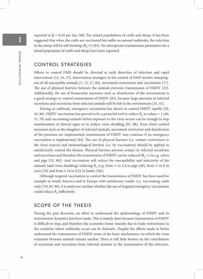
ControL strateGies
Efforts to control FMD should be directed at early detection of infection and rapid intervention [12, 16, 17]. Intervention strategies in the control of FMD involve stamping-out of all susceptible animals [5, 12, 17, 84], movement restrictions and vaccination [17]. The use of physical barriers between the animals prevents transmission of FMDV [52]. Additionally, the use of biosecurity measures such as disinfection of the environment is a good strategy to control transmission of FMDV [85], because large amounts of infected secretions and excretions from infected animals will be left in the environment [31, 41].
During an outbreak, emergency vaccination has shown to control FMDV rapidly [18, 45, 86]. FMDV vaccination has proved to be a powerful tool to reduce R0 to values < 1 [46, 51, 78] and vaccinating animals before exposure to the virus occurs can be enough to stop manifestation of clinical signs or to reduce virus shedding [87, 88]. Even when control measures such as the slaughter of infected animals, movement restriction and disinfection of the premises are implemented, transmission of FMDV may continue if no emergency vaccination is implemented [84]. The use of physical barriers (i.e. contact restrictions to the virus source) and immunological barriers (i.e. by vaccination) should be applied to satisfactorily control the disease. Physical barriers prevent contact to infected secretions and excretions and therefore the transmission of FMDV can be reduced (R0 <1 in e.g. calves and pigs [52, 89]). And vaccination will reduce the susceptibility and infectivity of the animals (and virus shedding) reducing R0 (e.g. from ∞ to 2.4 in pigs [49], from ∞ to 0 in cows [78] and from 1.14 to 0.22 in lambs [50]).
Although targeted vaccination to control the transmission of FMDV has been used for example in South America and in Europe with satisfactory results (i.e. vaccinating cattle only) [19, 83, 90], it is until now unclear whether the use of targeted emergency vaccination could reduce R0 sufficiently.
sCoPe oF the thesis
During the past decennia, an effort to understand the epidemiology of FMDV and its transmission dynamics has been made. This is mainly done because transmission of FMDV is difficult to stop, and therefore the economic losses (mainly due to trade restrictions) in the countries where outbreaks occur can be dramatic. Despite the efforts made to better understand the transmission of FMDV, some of the basic mechanisms via which the virus transmits between animals remain unclear. There is still little known on the contribution of secretions and excretions from infected animals to the transmission of the infection.
16

Ge
ne
ra
l Intr
od
uc
tIon
IAlso the role of different species on the magnitude of transmission of the virus is limitedly understood. Because different species can be differently infectious and susceptible, their relative infectivity and susceptibility should be determined to estimate risks of transmission of FMDV precisely. Interspecies transmission of FMDV has been limitedly quantified. This could be related with the limitations of FMDV research; the biosecurity requirements for working with this virus are highly demanding and thus limiting animal experimentation. However published data can also be used to determine missing parameters in the transmission of FMDV. In this thesis, both published data from animal experiments performed previously and data from new animal experiments were used to analyse the 4 following identified gaps in the transmission of FMDV:
1. It is known that most of the secretions and excretions of the FMDV infected animals can contain virus and thus contamination of the environment with these secretions and excretions is feasible. The main goal of chapter 2 of this thesis was to identify which factors are associated with higher secretion and excretion of FMDV and consequently with a higher contamination of the environment. For this, a multivariate linear regression analysis using published data from several animal experiments was performed. Better knowledge on quantitative data on FMDV in secretions and excretions is needed to identify possible routes of transmission of FMDV and to allow an efficient control planning (e.g. of disinfection) during outbreaks.
2. Since secretions and excretions from FMDV infected animals can contaminate the environment, new infections of FMDV can be caused. Another goal of this thesis is to quantify the contribution of a contaminated environment in the transmission of the infection. In chapter 3 of this thesis, two animal experiments were performed using non-vaccinated and vaccinated calves in both direct and indirect transmission studies set-ups. Using these experimental data, a modified SIR model in which we included an extra compartment for transmission via the environment was used to quantify the transmission rate β, and based upon that, the reproduction ratio R0. These results allowed estimation of the contribution of a contaminated environment to the transmission of FMDV. Understanding how much of the transmission of FMDV occurs via the infected secretions and excretions is needed to implement control strategies towards prevention of contact of susceptible animals to infectious surfaces.
3. For better understanding of the transmission of FMDV and the improvement of spread models, it is necessary to know about the susceptibility and the infectivity of the animal species that could be involved during an outbreak of FMDV. There is little known about the quantitative role of different species in the transmission of FMDV in a mixed population. One other goal of this thesis is to study transmission of FMDV in a mixed population (i.e. cattle and sheep) and to identify relative susceptibility and infectivity for both animal species. To this aim, in chapter 4 of this thesis, an animal experiment was performed using inoculated lambs and in-contact calves. This small-scale transmission study allowed quantification of a partial R0 for transmission of FMDV between infected sheep and contact cattle. These results allowed definition of relative susceptibility and
17

Ge
ne
ra
l Intr
od
uc
tIon
Iinfectivity for both animal species. Accurate measures of susceptibility and infectivity can be used to actualize transmission models of FMDV and to update control strategies targeting determined animal species.
4. Control strategies for FMD involve vaccination of animals. In the past it has been shown that targeting cattle only to vaccination can be sufficient to control FMD. However it is until now unknown if we could extrapolate this strategy to other populations. The last aim of this thesis is to analyse the effect of different vaccination strategies to the control of FMD in mixed populations of cattle and sheep. For this purpose, in chapter 5 of this thesis, published data on intraspecies and interspecies (partial) R estimates were used to determine R for a mixed population of cattle and sheep using a next generation matrix. This method allowed estimation of the effect of different vaccination strategies in different mixed populations.
The results of this thesis add knowledge on the different mechanisms that are involved in the transmission of FMDV. These results can be used to improve FMDV control as for example in implementing better biosecurity measures and updating vaccination plans.
List oF reFerenCes
1 Thomson GR, Vosloo W, Bastos AD: Foot and mouth disease in wildlife. Virus research 2003, 91:145–161.
2 Thompson D, Muriel P, Russell D, Osborne P, Bromley A, Rowland M, Creigh-Tyte S, Brown C: Economic costs of the foot and mouth disease outbreak in the United Kingdom in 2001. Revue scientifique et technique (International Office of Epizootics) 2002, 21:675–687.
3 Belsham GJ: Distinctive features of foot-and-mouth disease virus, a member of the picornavirus family; aspects of virus protein synthesis, protein processing and structure. Progress in biophysics and molecular biology 1993, 60:241–260.
4 Racaniello VR: Picornaviride: The Viruses and Their Replication. 4th ed. Volume 1. Phila-delphia: Lippincott Williams and WIlkins; 2001.
5 Sanson RL: The epidemiology of foot-and-mouth disease: implications for New Zealand. New Zealand veterinary journal 1994, 42:41–53.
6 Dekker A: Inactivation of foot-and-mouth disease virus by heat, formaldehyde, ethylene oxide and gamma radiation. The Veterinary record 1998, 143:168–169.
7 Hyde JL, Blackwell JH, Callis JJ: Effect of pasteurization and evaporation on foot-and-mouth disease virus in whole milk from infected cows. Canadian Veterinary Medical Association 1975, 39:305–309.
8 Blackwell JH, Hyde JL: Effect of heat on foot-and-mouth disease virus (FMDV) in the components of milk from FMDV-infected cows. The Journal of hygiene 1976, 77:77–83.
9 Walker JS, de Leeuw PW, Callis JJ, van Bekkum JG: The thermal death time curve for foot-and-mouth disease virus contained in primarily infected milk. Journal of biological standardization 1984, 12:185–9.
10 Grubman MJ, Baxt B: Foot-and-mouth disease. Clinical Microbiology Reviews 2004, 17:465–493.
11 Machado M: Aftosa: A Historical Survey of Foot-and-Mouth Disease and Inter-American Relations. New York: State University of New York Press; 1969.
12 Sutmoller P, Barteling SS, Olascoaga RC, Sumption KJ: Control and eradication of foot-and-mouth disease. Virus research 2003, 91:101–44.
13 Waldmann D, Kobe K, Pyl G.: Die aktive Immunisierung des Rindes gegen Maul- und Klauenseuche mittels Formolimpfstoff. Zentralblatt für Bakteriologie, Parasitenkunde, Infektionskrankheiten und Hygiene 1937, 138:401–412.
14 Frenkel HS: La culture de virus de la fievre aphteuse sur l’epithelium de la langue des bovides. Bulletin office international des épizooties 1947, 28:155–162.
15 Brown F: New approaches to vaccination against foot-and-mouth disease. Vaccine 1992:1022–1026.
16 Leforban Y: Review of the status of foot and mouth disease and approach to control/
18

Ge
ne
ra
l Intr
od
uc
tIon
Ieradication in Europe and Central Asia. Revue scientifique et technique (International Office of Epizootics) 2002, 21:477–492.
17 FAO: Preparation of Foot-and-Mouth Disease Contingency Plans. Volume 16. Rome: FAO; 2002:100.
18 Pluimers FH, Akkerman AM, van der Wal P, Dekker A, Bianchi A: Lessons from the foot and mouth disease outbreak in The Netherlands in 2001. Revue scientifique et technique (International Office of Epizootics) 2002, 21:711–721.
19 Leforban Y: How predictable were the outbreaks of foot and mouth disease in Europe in 2001 and is vaccination the answer? Revue scientifique et technique (International Office of Epizootics) 2002, 21:549–56, 539–47.
20 Kitching RP: Identification of foot and mouth disease virus carrier and subclinically infected animals and differentiation from vaccinated animals. Revue scientifique et technique (International Office of Epizootics) 2002, 21:531–538.
21 Bergmann IE, de Mello PA, Neitzert E, Beck E, Gomes I: Diagnosis of persistent aphthovirus infection and its differentiation from vaccination response in cattle by use of enzyme-linked immunoelectrotransfer blot analysis with bioengineered nonstructural viral antigens. American journal of veterinary research 1993, 54:825–831.
22 De Diego M, Brocchi E, Mackay D, De Simone F: The non-structural polyprotein 3ABC of foot-and-mouth disease virus as a diagnostic antigen in ELISA to differentiate infected from vaccinated cattle. Archives of virology 1997, 142:2021–2033.
23 Sørensen KJ, de Stricker K, Dyrting KC, Grazioli S, Haas B: Differentiation of foot-and-mouth disease virus infected animals from vaccinated animals using a blocking ELISA based on baculovirus expressed FMDV 3ABC antigen and a 3ABC monoclonal antibody. Archives of virology 2005, 150:805–814.
24 Brocchi E, De Diego M, Berlinzani A, Gamba D, De Simone F: Diagnostic potential of Mab-based ELISAs for antibodies to non-structural proteins of foot-and-mouth disease virus to differentiate infection from vaccination. The Veterinary quarterly 1998, 20:20–24.
25 Brocchi E, Bergmann IE, Dekker A, Paton DJ, Sammin DJ, Greiner M, Grazioli S, De Simone F, Yadin H, Haas B, Bulut N, Malirat V, Neitzert E, Goris N, Parida S, Sørensen K, De Clercq K: Comparative evaluation of six ELISAs for the
detection of antibodies to the non-structural proteins of foot-and-mouth disease virus. Vaccine 2006, 24:6966–6979.
26 Parida S, Anderson J, Cox SJ, Barnett P V., Paton DJ: Secretory IgA as an indicator of oro-pharyngeal foot-and-mouth disease virus replication and as a tool for post vaccination surveillance. Vaccine 2006, 24:1107–1116.
27 Archetti IL, Amadori M, Donn A, Salt J, Lodetti E: Detection of foot-and-mouth disease virus-infected cattle by assessment of antibody response in oropharyngeal fluids. Journal of clinical microbiology 1995, 33:79–84.
28 Bergmann IE, Astudillo V, Malirat V, Neitzert E: Serodiagnostic strategy for estimation of foot-and-mouth disease viral activity through highly sensitive immunoassays using bioengineered nonstructural proteins. The Veterinary quarterly 1998, 20 Suppl 2:S6–9.
29 Doel T.: FMD vaccines. Virus Research 2003, 91:81–99.
30 Alexandersen S, Zhang Z, Donaldson AI, Garland AJ: The pathogenesis and diagnosis of foot-and-mouth disease. Journal of comparative pathology 2003, 129:1–36.
31 Thomson GR: Foot and Mouth Disease. Volume Vol. 2. Capetown: Oxford University Press Southern Africa; .
32 Arzt J, Pacheco JM, Rodriguez LL: The early pathogenesis of foot-and-mouth disease in cattle after aerosol inoculation. Identification of the nasopharynx as the primary site of infection. Veterinary pathology 2010, 47:1048–1063.
33 Alexandersen S, Oleksiewicz MB, Donaldson AI: The early pathogenesis of foot-and-mouth disease in pigs infected by contact: a quantitative time-course study using TaqMan RT-PCR. The Journal of general virology 2001, 82(Pt 4):747–755.
34 Murphy C, Bashiruddin JB, Quan M, Zhang Z, Alexandersen S: Foot-and-mouth disease viral loads in pigs in the early, acute stage of disease. The Veterinary record 2010, 166:10–14.
35 Alexandersen S, Donaldson AI: Further studies to quantify the dose of natural aerosols of foot-and-mouth disease virus for pigs. Epidemiology and infection 2002, 128:313–323.
36 Donaldson AI, Alexandersen S: Predicting the spread of foot and mouth disease by airborne virus. Revue scientifique et technique (International Office of Epizootics) 2002, 21:569–75.
37 Burrows R: Excretion of foot and mouth disease virus prior to the development of lesions. Veterinary Record 1968, 82:387.
19

Ge
ne
ra
l Intr
od
uc
tIon
I38 Sellers RF: Quantitative aspects of the spread of
foot-and-mouth disease. The Veterinary bulletin 1971, 41:431–439.
39 Sellers RF, Parker J: Airborne excretion of foot-and-mouth disease virus. The Journal of hygiene 1969, 67:671–7.
40 Burrows R, Mann JA, Garland AJ, Greig A, Goodridge D: The pathogenesis of natural and simulated natural foot-and-mouth disease infection in cattle. Journal of comparative pathology 1981, 91:599–609.
41 Pharo HJ: Foot-and-mouth disease: an assessment of the risks facing New Zealand. New Zealand veterinary journal 2002, 50:46–55.
42 Neumann EJ: Disease Transmission and Biosecurity. United Kingdom: Wiley-Blackwell ; 2012.
43 Charleston B, Bankowski BM, Gubbins S, Chase-Topping ME, Schley D, Howey R, Barnett P V, Gibson D, Juleff ND, Woolhouse MEJ: Relationship Between Clinical Signs and Transmission of an Infectious Disease and the Implications for Control. Science 2011, 332:726–729.
44 Orsel K, Bouma A, Dekker A, Stegeman JA, de Jong MCM: Foot and mouth disease virus transmission during the incubation period of the disease in piglets, lambs, calves, and dairy cows. Preventive Veterinary Medicine 2009, 88:158–163.
45 Cox SJ, Voyce C, Parida S, Reid SM, Hamblin PA, Paton DJ, Barnett P V: Protection against direct-contact challenge following emergency FMD vaccination of cattle and the effect on virus excretion from the oropharynx. Vaccine 2005, 23:1106–1113.
46 Eblé PL, Bouma A, de Bruin MG, van Hemert-Kluitenberg F, van Oirschot JT, Dekker A: Vaccination of pigs two weeks before infection significantly reduces transmission of foot-and-mouth disease virus. Vaccine 2004, 22:1372–1378.
47 Eblé PL, de Koeijer A, Bouma A, Stegeman A, Dekker A: Quantification of within- and between-pen transmission of Foot-and-Mouth disease virus in pigs. Veterinary research 2006, 37:647–654.
48 Goris NE, Eblé PL, de Jong MCM, De Clercq K, Eblé PL: Quantification of foot-and-mouth disease virus transmission rates using published data. ALTEX 2009, 26:52–54.
49 Orsel K, de Jong MCM, Bouma A, Stegeman JA, Dekker A: Foot and mouth disease virus transmission among vaccinated pigs after exposure to virus shedding pigs. Vaccine 2007, 25:6381–6391.
50 Orsel K, Dekker A, Bouma A, Stegeman JA, de Jong MCM: Quantification of foot and mouth disease virus excretion and transmission within
groups of lambs with and without vaccination. Vaccine 2007, 25:2673–2679.
51 Orsel K, Dekker A, Bouma A, Stegeman JA, de Jong MCM: Vaccination against foot and mouth disease reduces virus transmission in groups of calves. Vaccine 2005, 23:4887–4894.
52 Van Roermund HJ, Eblé PL, de Jong MCM, Dekker A: No between-pen transmission of foot-and-mouth disease virus in vaccinated pigs. Vaccine 2010, 28:4452–4461.
53 Sellers RF, Donaldson AI, Herniman KA: Ihalation, persistence and dispersal f foot-and-mouth disease virus by man. The Journal of hygiene 1970, 68:565–73.
54 Gloster J, Champion HJ, Sorensen JH, Mikkelsen T, Ryall DB, Astrup P, Alexandersen S, Donaldson AI: Airborne transmission of foot-and-mouth disease virus from Burnside Farm, Heddon-on-the-Wall, Northumberland, during the 2001 epidemic in the United Kingdom. Veterinary Record 2003, 152:525–533.
55 French NP, Kelly L, Jones R, Clancy D: Dose-response relationships for foot and mouth disease in cattle and sheep. Epidemiology and infection 2002, 128:325–332.
56 Rweyemamu M, Roeder P, Mackay D, Sumption K, Brownlie J, Leforban Y, Valarcher J-F, Knowles NJ, Saraiva V: Epidemiological patterns of foot-and-mouth disease worldwide. Transboundary and emerging diseases 2008, 55:57–72.
57 Bernoulli D: Essai d’une nouvelle analyse de la mortalite causee par la petite verole et des avantages de l’inoculation pour la prevenir. Mémoires de Mathématique et de Physique, présentés à l’Académie Royale des Sciences 1760:1–45.
58 Ross R: The prevention of Malaria. Springer-Verlag 1911.
59 Becker NG: Analysis of Infectious Disease Data. Volume 1. London: Chapman and Hall Ltd; 1989.
60 Velthuis AG, de Jong MCM, Kamp EM, Stockhofe N, Verheijden JH: Design and analysis of an Actinobacillus pleuropne-umoniae transmission experiment. Preven-tive Veterinary Medicine 2003, 60:53–68.
61 Heffernan JM, Smith RJ, Wahl LM: Perspectives on the basic reproductive ratio. Journal of the Royal Society, Interface 2005, 2:281–293.
62 Anderson RM, May RM: Infectious Diseases of Humans: Dynamics and Control . Oxford : Oxford University Press; 1991.
63 Macdonald G: The Epidemiology and Control of Malaria. London: Oxford University Press; 1957.
20

Ge
ne
ra
l Intr
od
uc
tIon
I64 Diekmann O, Heesterbeek JA, Metz JA: On the
definition and the computation of the basic reproduction ratio R0 in models for infectious diseases in heterogeneous populations. Journal of mathematical biology 1990, 28:365–82.
65 De Jong MCM, Kimman TG: Experimental quantification of vaccine-induced reduction in virus transmission. Vaccine 1994, 8:761–766.
66 Velthuis AG, de Jong MCM, De Bree J: Comparing methods to quantify experimental transmission of infectious agents. Mathematical biosciences 2007, 210:157–176.
67 Klinkenberg D, de Bree J, Laevens H, de Jong MCM: Within- and between-pen transmission of Classical Swine Fever Virus: a new method to estimate the basic reproduction ratio from transmission experiments. Epidemiology and infection 2002, 128:293–299.
68 Velthuis AG, de Jong MCM, de Bree J, Nodelijk G, van Boven M: Quantification of transmission in one-to-one experiments. Epidemiology and infection 2002, 128:193–204.
69 Diekmann O, Heesterbeek JA, Roberts MG: The construction of next-generation matrices for compartmental epidemic models. Journal of the Royal Society, Interface 2010, 7:873–885.
70 Ferguson NM, Donnelly CA, Anderson RM: The foot-and-mouth epidemic in Great Britain: pattern of spread and impact of interventions. Science (New York, NY) 2001, 292:1155–1160.
71 Keeling MJ, Woolhouse ME, Shaw DJ, Matthews L, Chase-Topping M, Haydon DT, Cornell SJ, Kappey J, Wilesmith J, Grenfell BT: Dynamics of the 2001 UK foot and mouth epidemic: stochastic dispersal in a heterogeneous landscape. Science (New York, NY) 2001, 294:813–817.
72 Kao RR: The role of mathematical modelling in the control of the 2001 FMD epidemic in the UK. Trends in Microbiology 2002:279–286.
73 Hagenaars TJ, Dekker A, de Jong MCM, Eblé PL: Estimation of foot and mouth disease transmission parameters, using outbreak data and transmission experiments. Revue scientifique et technique (International Office of Epizootics) 2011, 30:467–77.
74 Porphyre T, Auty HK, Tildesley MJ, Gunn GJ, Woolhouse ME: Vaccination against foot-and-mouth disease: do initial conditions affect its benefit? PLoS One 2013, 8:e77616.
75 Alexandersen S, Quan M, Murphy C, Knight J, Zhang Z: Studies of quantitative parameters of virus excretion and transmission in pigs and cattle experimentally infected with foot-and-
mouth disease virus. Journal of comparative pathology 2003, 129:268–282.
76 Kitching RP, Hughes GJ: Clinical variation in foot and mouth disease: sheep and goats. Revue scientifique et technique (International Office of Epizootics) 2002, 21:505–512.
77 Barnett P V, Cox SJ: The role of small ruminants in the epidemiology and transmission of foot-and-mouth disease. Veterinary journal (London, England : 1997) 1999, 158:6–13.
78 Orsel K, de Jong MCM, Bouma A, Stegeman JA, Dekker A: The effect of vaccination on foot and mouth disease virus transmission among dairy cows. Vaccine 2007, 25:327–335.
79 Muroga N, Kobayashi S, Nishida T, Hayama Y, Kawano T, Yamamoto T, Tsutsui T: Risk factors for the transmission of foot-and-mouth disease during the 2010 outbreak in Japan: a case-control study. BMC veterinary research 2013, 9:150.
80 Donaldson AI, Alexandersen S, Sørensen JH, Mikkelsen T: Relative risks of the uncontrollable (airborne) spread of FMD by different species. The Veterinary record 2001, 148:602–4.
81 De Jong MCM, Hagenaars TJ, Eblé PL: Transmission of FMDV within and between species: Quantification and comparisons. In 13th International Symposium on Veterinary Epidemiology and Economics. Maastricht: Wageningen Academic Publishers; 2012.
82 Cox SJ, de Jong MCM, Barnett P V, Eblé PL: Reusing published data to quantify foot-and-mouth disease transmission parameter β. In Foot and Mouth Disease International Symposium and Workshop. Melbourne; 2010.
83 Donaldson A: The role of sheep in the epidemiology of foot-and-mouth disease and proposasls for control and eradication in animal populations with a high density of sheep. In Session of the Research Group of the Standing Technical Committee of EuFMD. Borovets: FAO of the United Nations; 2000.
84 Muroga N, Hayama Y, Yamamoto T, Kurogi A, Tsuda T, Tsutsui T: The 2010 Foot-and-Mouth Disease Epidemic in Japan. Journal of Veterinary Medical Science 2012:399–404.
85 Foot-and-Mouth Disease [http://www.oie.int/filea dmin/home/eng/Animal_Health_in_the_world/docs/pdf/foot_and_mouth_disease_final.pdf]
86 Cox SJ, Barnett P V, Dani P, Salt JS: Emergency vaccination of sheep against foot-and-mouth disease: protection against disease and reduction in contact transmission. Vaccine 1999, 17:1858–1868.
21

Ge
ne
ra
l Intr
od
uc
tIon
I87 Golde WT, Pacheco JM, Duque H, Doel T,
Penfold B, Ferman GS, Gregg DR, Rodriguez LL: Vaccination against foot-and-mouth disease virus confers complete clinical protection in 7 days and partial protection in 4 days: Use in emergency outbreak response. Vaccine 2005, 23:5775–5782.
88 Pacheco JM, Brum MCS, Moraes MP, Golde WT, Grubman MJ: Rapid protection of cattle from direct challenge with foot-and-mouth disease
virus (FMDV) by a single inoculation with an adenovirus-vectored FMDV subunit vaccine. Virology 2005, 337:205–209.
89 Bouma A, Dekker A, de Jong MCM: No foot-and-mouth disease virus transmission between individually housed calves. Veterinary Microbiology 2004, 98:29–36.
90 Sutmoller P, Barteling SS, Olascoaga RC, Sumption KJ: Control and eradication of foot-and-mouth disease. Virus Res 2003, 91:101–144.
22


IIidentif ication of factors associated with increased excretion
of foot-and-mouth disease virus
Carla Bravo de Rueda, Aldo Dekker, Phaedra L. Eblé, Mart C.M. de Jong
Preventive Veterinary Medicine 113 (2014) 23–33

Ide
ntIfIc
atIo
n o
f fac
tor
s asso
cIa
ted
wIth
Inc
re
ase
d e
xc
re
tIon
of fo
ot-a
nd
-mo
uth
dIse
ase
vIr
us
II
aBstraCt
We investigated which variables possibly influence the amount of foot-and-mouth disease virus (FMDV) shed in secretions and excretions by FMDV infected animals, as it is likely that the amount of FMDV shed is related to transmission risk. First, in a separate analysis of laboratory data, we showed that the total amount of FMDV in secretions and excretions from infected animals is highly correlated with maximum titres of FMDV. Next, we collected data from 32 published scientific articles in which FMDV infection experiments were described. The maximum titres of FMDV reported in different secretions and excretions (the response variable) and the experimental conditions in which they occurred (the explanatory variables), were recorded in a database and analysed using multivariate regression models with and without random effects. In both types of models, maximum titres of FMDV were significantly (p < 0.05) associated with types of secretions and excretions, animal species, stage of the disease and days post infection. These results can be used to prioritize biosecurity measures in contingency plans.
26

Ide
ntIfIc
atIo
n o
f fac
tor
s asso
cIa
ted
wIth
Inc
re
ase
d e
xc
re
tIon
of fo
ot-a
nd
-mo
uth
dIse
ase
vIr
us
II
introduCtion
Foot-and-mouth disease (FMD) is a contagious viral disease of cloven-hoofed animals, both domestic (cattle, pigs, sheep, goats and domestic buffalo) and wild [1]. The FMD virus (FMDV) can be transmitted by several routes [2, 3], with direct contact between animals considered the most important. The virus can also be transmitted by several indirect routes. In the European Union, an outbreak of FMD invokes an obligatory stand-still of animal transport [4]. During such a stand-still, direct contact between infected animals in one farm and non-infected animals in another farm is theoretically impossible, leaving indirect transmission via contaminated material the most likely remaining route of transmission. In this respect, airborne transmission has been also considered [5].
During epidemics, even when there is a complete standstill of animal transport, transmission between farms has been shown [6]. That indirect routes play a role in such transmission is clear from the observation that veterinarians were involved in the transmission of FMDV in outbreaks both in Denmark in 1982, and in Italy in 1993, either by using contaminated surgical equipment or by visiting farms after visiting an infected farm. Similarly, during the 2001 FMD outbreak in the United Kingdom, it was suggested that farmers were involved in the transmission of the virus between sheep flocks [7]. In the 2001 United Kingdom outbreak, the basic reproduction number remained above 1, that is, FMDV transmission continued despite the standstill in animal transport [8]. Thus indirect transmission of FMDV can have enormous consequences.
It can be assumed that the risk of indirect transmission of FMDV is related to the total amount of FMDV present in the environment through contamination by secretions and excretions from FMDV infected animals. Here, secretions include material released from glands (e.g. milk, semen, saliva) whereas excretions refer to any other products released from animals (e.g. faeces, material released from the respiratory tract, urine, probang samples, nasal discharge and blood). The concentrations of FMDV in infected secretions and excretions have been reviewed [9]. However we analysed the quantitative relationship between possible explanatory variables and the amount of FMDV in infected secretions and excretions.
materiaLs and methods
materials Laboratory data Laboratory reports from animal studies performed at the Central Veterinary Institute (The Netherlands) were mined for all available daily data on virus secretion in milk from cattle [10] and on virus secretion and excretion in oropharyngeal fluid (OPF) swabs from cattle [11], sheep [12, 13] and pigs [14, 15, 16, 17, 18]. These data were used to identify the response variable for our multivariate regression analysis.
27

Ide
ntIfIc
atIo
n o
f fac
tor
s asso
cIa
ted
wIth
Inc
re
ase
d e
xc
re
tIon
of fo
ot-a
nd
-mo
uth
dIse
ase
vIr
us
II
Literature data Data on FMDV in secretions and excretions were collected from 32 scientific articles published between 1965 and 2007 (see Annex) found in internal databases and through the electronic (external) databases Scopus and PubMed in 2010, all reporting experimental trials involving FMDV infection. The electronic databases were explored using the keywords: foot-and-mouth disease, virus, infection and excretion. References cited in retrieved articles were reviewed to identify additional ones. The articles had to meet two basic criteria for their inclusion in the analysis: be written either in English, Spanish or French, and contain data on animal experiments with FMDV. They needed to contain information on the maximum titre(s) of FMDV detected in secretions and/or excretions, and additional information on one or more of the following: the type(s) of secretion or excretion in which the virus was detected, route of infection, animal species, FMDV serotype, stage of disease (clinical and non-clinical), dose of infection and/or days post infection at which the maxi- mum secretion or excretion occurred. Missing data on one or more of these variables were recorded as not available (N.A.). These data were used as the response and possible explanatory variables for our multivariate regression analysis.
Per individual animal, the maximum titre of FMDV (including the experimental conditions) was recorded. Virus titres were reported as 10log TCID50/ml. Plaque forming units (PFU) were converted to TCID50 [19]. Median doses, such as 50% cattle infection dose (CID50), 50% mouse infection dose (MID50) or 50% mouse lethal dose (MID50) per ml, were considered equal to 50% tissue culture infective dose (TCID50/ml) [20]. The maximum recorded titre was the maximum titre over time for an individual animal. If the maximum titre was reported per group of animals, this resulted in one observation (from blood in Alexandersen et al. (2003); from airborne excretion in Alexandersen et al. (2002), Alexandersen and Donaldson (2002), Donaldson et al. (1970, 1981, 1982), Gloster et al. (2007), and Sellers and Parker (1969) [21]; from probang, milk, faeces and blood in Burrows (1968); from milk in Burrows (1971); and, from probang and nasal discharge in Burrows (1972)). Data on airborne excretion were recorded as 10log TCID50/animal/day.
The recorded secretion or excretion types were airborne, faeces, milk, probang, semen, urine, blood, nasal discharge, oropharyngeal fluid (OPF) swabs, and saliva. The category faeces contains data on material collected from the rectum (Burrows et al., 1968) and from rectal swabs (Garland, 1974). Probang refers to oropharyngeal samples that were obtained after scraping the oropharynx with a sampling cup.
Routes of infection were recorded as: contact (if an infected donor and a susceptible contact animal shared a common experimental unit); intranasal (IN, if the animals were infected via the intranasal route) or parenteral (if the animals were infected intravenously (IV), intramuscularly (IM), intralingually, intracutaneously (IC), intramammary or intradermally (ID)).
Animal species were recorded as cattle (bull, steer, ox, cow, calf and heifer), swine (pigs) or small ruminants (sheep, lambs and goats). The FMD viruses used for infection were recorded based on FMDV serotype, i.e. A, O, C, Asia-1, SAT 1, SAT 2, SAT 3, but no subdivision was made to the level of subtypes. The stage of disease was recorded as ‘clinical’ when lesions or clinical signs (including fever) were reported; otherwise it was recorded as ‘non-clinical’.
28
Ide
ntIfIc
atIo
n o
f fac
tor
s asso
cIa
ted
wIth
Inc
re
ase
d e
xc
re
tIon
of fo
ot-a
nd
-mo
uth
dIse
ase
vIr
us
II
Dose of infection (ranging from 0.95 to 10.15 TCID50/ml) was recorded. Days post infection was recorded as the day when the maximum titres in the secretion or excretion were observed (ranging from 0.33 to 28 dpi).
methods Identifying the response variable for the multivariate regression analysis A proxy for the total amount of FMDV secreted and excreted by the infected animals was established using available laboratory data from OPF swab samples and milk samples. The total amount of secreted and excreted FMDV (per individual animal) was calculated by summing the observed viral amounts (without logarithmic trans- formation) from consecutive observations (area under the curve, AUC). In a univariate regression analysis, the logarithm of the AUC (10log AUC) was used as the response variable. Three explanatory variables were analysed: (1) the maximum virus titre (max 10log TCID50/ml), (2) the time when the maximum virus titre occurred (10log days post infection) and, (3) their product 10log (max TCID50/ml × days post infection) which is equal to max 10log TCID50/ml + 10log days post infection. For each univariate model, the r2 values were calculated. An F-test (in ANOVA) was used to test the significance of each variable. The best explanatory variable was used as response variable in the multivariate regression analysis.
Identifying the explanatory variables for the multivariate regression analysis A dataset was built using the information found in the literature. Descriptive statistics of the data can be found in Tables 1A–1C.
Per individual animal, several categorical variables were recorded: type of secretion and excretion, route of infection, animal species, FMDV serotype and stage of disease and, two continuous variables: dose of infection and days post infection (Table 2). Categories in which a limited number of observations were present were combined with another category where this made biological sense (e.g. URT secretions and excretions, FMDV serotype SAT) [22].
Multivariate regression analysis Under the assumption that all the included FMDV infection experiments share a common true effect size, we used a model in which we did not adjust for variability between data sources (a linear model without random effects). Under the assumption that some of the FMDV infection experiments from the different data sources differ from each other in ways that could impact on the effect in the model, we used a model in which we adjusted for variability between the data sources (a linear model with random effects). Three different random effects were evaluated: “article” (articles included in the analysis, see Annex), “laboratory” (laboratories where the original analyses had been performed) and their nested effect. All random effects were assumed to follow a Gaussian distribution [23]. The models were compared by computing the AIC (Table 3).
29

Ide
ntIfIc
atIo
n o
f fac
tor
s asso
cIa
ted
wIth
Inc
re
ase
d e
xc
re
tIon
of fo
ot-a
nd
-mo
uth
dIse
ase
vIr
us
II
Table 1A. Descriptive statistics on data retrieved from the literature on maximum virus excretion from cattle.
FMDVInfection variables
Number of observations
Maximum titre average (range)
TCID50/ml *
Maximum titre Standard deviation
TCID50/ml*
Total 220 4.51 (0.95,8.65) 1.66
Type of secretion and excretion
Airborne 9 4.33 (3.88, 5.08) 0.36
Blood 47 4.03 (0.95 ,6.20) 1.18
Faeces 5 1.55 (1.50, 1.75) 0.10
Milk 40 4.48 (2.15, 7.35) 1.46
URT (OPF swabs, saliva and nasal discharge) 33 5.70 (1.25, 8.50) 1.66
Nasal discharge only 7 6.09 (2.75, 7.85) 1.61
Probang 68 4.91 (2.20, 8.65) 1.53
Semen 8 4.55 (2.10 , 6.20) 1.33
Urine 10 1.93 (1.00 , 3.80) 0.87
Route of infection
Intranasal 37 4.68 (0.95, 8.65) 1.76
Parenteral 95 4.75 (1.25, 8.50) 1.63
Contact 88 4.17 (1.00, 8.05) 1.57
Undetermined 1 4.60 (NA) NA
FMDV serotype
A 38 3.98 (2.10, 8.05) 1.40
O 140 4.54 (0.95, 8.65) 1.68
Asia-1 4 4.10 (2.80, 5.00) 0.80
C 6 4.6 (2.10, 7.00) 1.80
SAT (1, 2, 3) 12 4.26 (2.10, 6.00) 1.06
Undetermined 20 5.52 (1.25, 8.15) 1.81
Stage of disease
Non-clinical 61 4.52 (0.95, 8.65) 1.66
Clinical 123 4.62 (1.00, 8.50) 1.72
Undetermined 36 4.11 (1.15, 7.15) 1.36
Dose of infection (below/above median: 5.5 TCID50/ml)
0.95 - 5.4 TCID50/ml 51 4.94 (0.95, 8.65) 1.71
5.5 - 10.15 TCID50/ml 59 4.30 (2.10, 7.20) 1.48
Undetermined 110 4.43 (1.00, 8.15) 1.69
Days post infection (dpi; below/above median: 3 dpi)
0.3 to 2.8 dpi 65 4.82 (1.00, 8.50) 1.69
3 to 28 dpi 115 4.07 (0.95, 8.65) 1.49
Undetermined 40 5.28 (1.25, 8.15) 1.67
Total refers to all the maximum titres observations that were encountered.* TCID50 per animal per day for airborne excretion; dose of infection and days post infection were divided as above and below the median of the maximum titre calculated using the maximum titres when either the dose of infection or the days post infection were available.
30

Ide
ntIfIc
atIo
n o
f fac
tor
s asso
cIa
ted
wIth
Inc
re
ase
d e
xc
re
tIon
of fo
ot-a
nd
-mo
uth
dIse
ase
vIr
us
II
Table 1B. Descriptive statistics on maximum virus excretion from swine.
FMDVInfection variables
Number of observations
Maximum titre average (range) TCID50/ml *
Maximum titre standard deviation TCID50/ml*
Total 71 5.15 (3.41,8.60) 0.98
Type of secretion and excretion
Airborne 22 6.00 (4.48, 8.60) 0.89
Blood 6 5.18 (3.90, 6.50) 1.07
OPF (swabs and saliva) 43 4.70 (3.41, 6.45) 0.66
Route of infection
Parenteral 39 5.44 (3.85, 8.08) 0.84
Contact 32 4.78 (3.41, 8.60) 1.01
FMDV serotype
A 5 5.65 (4.48, 6.68) 0.70
O 64 5.01 (3.41, 6.54) 0.81
C 2 8.34 (8.08, 8.60) 0.26
Stage of disease
Non-clinical 5 5.80 (5.30, 6.54) 0.50
Clinical 43 5.09 (3.41, 8.60) 1.04
Undetermined 23 5.11 (3.85, 8.08) 0.89
Dose of infection (below/above median: 5.5 TCID50/ml)
0.95 - 5.4 TCID50/ml 19 4.94 (3.85, 6.50) 0.62
5.5 - 10.15 TCID50/ml 18 6.01 (4.48, 8.10) 0.72
Undetermined 34 4.81 (3.41, 8.60) 0.99
Days post infection (dpi; below/above median: 3 dpi)
0.3 to 2.8 dpi 22 5.59 (4.35, 8.60) 0.97
3 to 28 dpi 41 4.76 (3.41, 6.45) 0.77
Undetermined 8 5.95 (5.10, 8.10) 0.93
Total refers to all the maximum titres observations that were encountered.* TCID50 per animal per day for airborne excretion; dose of infection and days post infection were divided as above and below the median of the maximum titre calculated using the maximum titres when either the dose of infection or the days post infection were available.
Due to the small number of identified explanatory variables (Table 2), we used them all in the multivariate regression analysis of the models with and without random effects. To select the variables that best explained total FMDV secreted and excreted by infected animals, a stepwise regression procedure with bidirectional elimination [24] was used in multivariate regression analyses. No interaction terms were included in the initial (full) models (Table 3). The selection of explanatory variables (or fixed effects) was carried out using 2 criteria: the significance level (p < 0.05) and the Akaike Information Criterion (AIC). The variable with the highest p-value was removed from the models. In addition,
31

Ide
ntIfIc
atIo
n o
f fac
tor
s asso
cIa
ted
wIth
Inc
re
ase
d e
xc
re
tIon
of fo
ot-a
nd
-mo
uth
dIse
ase
vIr
us
II
Table 1C. Descriptive statistics on maximum virus excretion from small ruminants (sheep and goats).
FMDVInfection variables
Number ofobservations
Maximum titre TCID50/ml *
Maximum titre standard deviation TCID50/ml*
Total 36 3.93 (0.86, 6.28) 1.25
Type of secretion and excretion
Airborne 12 3.75 (2.38, 5.08) 1.00
Blood 8 3.34 (1.50, 5.20) 1.13
OPF (swabs and saliva) 16 4.37 (0.86, 6.28) 1.31
Route of infection
Intranasal 11 4.69 (3.26, 6.28) 0.83
Parenteral 18 3.51 (1.50, 5.20) 1.10
Contact 6 3.70 (0.86, 5.45) 1.64
Undetermined 1 4.60 (NA) NA
FMDV serotype
A 2 2.53 (2.48, 2.58) 0.05
O 23 4.35 (0.86, 6.30) 1.12
C 3 3.28 (2.38, 5.08) 1.27
Undetermined 8 3.34 (1.50, 5.20) 1.13
Stage of disease
Non-clinical 8 3.69 (0.86, 5.08) 1.37
Clinical 13 4.81 (3.26, 6.28) 0.79
Undetermined 15 3.30 (1.50, 5.20) 1.06
Dose of infection (below/above median: 5.5 TCID50/ml)
0.95 - 5.4 TCID50/ml 12 4.69 (3.26, 6.28) 0.79
5.5 - 10.15 TCID50/ml 7 3.33 (2.38, 5.08) 1.05
Undetermined 17 3.65 (0.86, 5.45) 1.33
Days post infection (dpi; below/above median: 3 dpi)
0.3 to 2.8 dpi 20 3.74 (1.50, 5.26) 1.18
3 to 28 dpi 14 4.17 (0.86, 6.28) 1.34
Undetermined 2 4.23 (3.48, 4.98) 0.75
Total refers to all the maximum titres observations that were encountered.* TCID50 per animal per day for airborne excretion; dose of infection and days post infection were divided as above and below the median of the maximum titre calculated using the maximum titres when either the dose of infection or the days post infection were available.
whenever deletion of a variable occurred, we checked for confounding. If the deletion of a variable resulted in a change of more than 25% in the regression estimates, this indicated confounding [25, 26]. Confounding variables were retained in the models. After deletion of those variables with p-values higher than 0.05, we tested whether their inclusion was significant (p < 0.05) and whether the inclusion led to significant reduction in AIC (AIC >
32

Ide
ntIfIc
atIo
n o
f fac
tor
s asso
cIa
ted
wIth
Inc
re
ase
d e
xc
re
tIon
of fo
ot-a
nd
-mo
uth
dIse
ase
vIr
us
II
Table 2. Explanatory variables for the multivariate regression analysis
Variable Type Categories/Specifications
Type of secretion and excretion
Categorical Airborne, blood, faeces, milk, URT (OPF swabs, saliva, nasal discharge), probang, semen, urine
Route of infection Categorical Intranasal, contact, parenteral (intravenous, intramuscular, intralingual, intracutaneous, intramammary or intradermal)
Animal species Categorical Cattle, swine, small ruminants (sheep and goats)
FMDV serotype Categorical A, Asia-1, C, O, SAT
Stage of disease Categorical Non-clinical, clinical
Dose of infection Continuous From 0.95 to 10.15 TCID50/ml
Days post infection (dpi) Continuous From day 0.33 to 28 post infection
2, [27]). After selecting the explanatory variables of the models, one level interaction terms were included one by one in the models. When the interaction term allowed improvement of fit (p < 0.05), it remained in the models.
Both final models (Table 3) were checked for homoscedasticity, normality and outliers by residual analysis. Outliers were retained as they were thought to reflect relevant deviations in this sort of data. In order to test whether an outlier affected the estimates or the p-values, an outlier was excluded from the data and the models were re-fit. When the outlier had no influence on the estimates or p-values, it remained in the models.
All statistical analyses were performed using the R software version 2.11.0 with its standard add-on packages stats and lme4 [28].
resuLts
identifying the response variable for the multivariate regression analysis The univariate regression analysis between 10log AUC and max 10log TCID50/ml gave a correlation coefficient (r2) of 0.98 for OPF swab samples and of 0.99 for milk samples (p-value <0.001). The analysis between 10log AUC and 10log days post infection gave correlation coefficients of 0.01 for OPF swab samples and 0.09 for milk samples. There was no significant association between 10log AUC and 10log days post infection (OPF swab samples, p-value 0.3; milk samples, p-value 0.2). The addition of 10log days post infection in the model with max 10log TCID50/ml did not improve the fit of the model neither for OPF swab samples nor for milk samples (p-value 0.3 and 0.4 respectively). The variable max 10log TCID50/ml was therefore used as the response variable in the multivariate regression analysis.
Literature data The references of the 32 used scientific articles on FMDV infection experiments are shown in the Annex. The FMDV infection experiments reported in the selected scientific articles were carried out in 5 FMD reference laboratories: the Pirbright Institute (IAH, Pirbright,
33

Ide
ntIfIc
atIo
n o
f fac
tor
s asso
cIa
ted
wIth
Inc
re
ase
d e
xc
re
tIon
of fo
ot-a
nd
-mo
uth
dIse
ase
vIr
us
IITa
ble 3
. Com
paris
on o
f fitte
d m
odels
for t
he m
ax 10
log
TCID
50/m
l bas
ed o
n pu
blish
ed d
ata o
f FM
DV
infe
ctio
n stu
dies
usin
g th
e sam
e dat
aset
(num
ber o
f obs
erva
tions
= 2
04).
Expl
anat
ory
vari
able
sIn
tera
ctio
n te
rms
Ran
dom
effe
cts
AIC
Mod
el w
ithou
t ran
dom
effe
cts
Nul
l mod
el-
--
759.
5
Full
mod
elTy
pe o
f sec
retio
n an
d ex
cret
ion,
dpi
, ani
mal
spec
ies,
rout
e of
infe
ctio
n, F
MD
V se
roty
pe, s
tage
of d
isea
se-
-64
2.6
Fina
l mod
elTy
pe o
f sec
retio
n an
d ex
cret
ion,
dpi
, ani
mal
spec
ies,
FMD
V se
roty
pe, s
tage
of d
isea
seTy
pe o
f sec
retio
n an
d ex
cret
ion*
anim
al sp
ecie
s, ty
pe o
f se
cret
ion
and
excr
etio
n*sta
ge o
f dise
ase,
type
of s
ecre
tion
and
excr
etio
n*FM
DV
sero
type
, FM
DV
sero
type
*sta
ge o
f dise
ase
-58
4.6
Mod
el w
ith ra
ndom
effe
cts
Nul
l mod
el-
-A
rtic
le72
7.4
Labo
rato
ry76
3.2
Art
icles
in L
abor
ator
ies
728.
5
Full
mod
elTy
pe o
f sec
retio
n an
d ex
cret
ion,
dpi
, ani
mal
spec
ies,
rout
e of
infe
ctio
n, F
MD
V se
roty
pe, s
tage
of d
isea
se-
Art
icle
622.
7
Fina
l mod
elTy
pe o
f sec
retio
n an
d ex
cret
ion,
dpi
, an
imal
spec
ies,
stag
e of
dis
ease
N.A
.A
rtic
le61
5.4
34

Ide
ntIfIc
atIo
n o
f fac
tor
s asso
cIa
ted
wIth
Inc
re
ase
d e
xc
re
tIon
of fo
ot-a
nd
-mo
uth
dIse
ase
vIr
us
II
United Kingdom), the Plum Island Animal Disease Center (PIADC, Orient Point, New York, United States of America), the Central Veterinary Institute (CVI, Lelystad, The Netherlands), the Pan American Center for Foot-and-Mouth Disease (PanAftosa, Rio de Janeiro, Brazil) and the French Institute for Foot-and-Mouth Disease (Lyon, France).
In total 327 observations (220 cattle, 71 swine and 36 small ruminants) were retrieved. The data retrieved from the reviewed scientific articles are summarized in Table 1A for cattle, Table 1B for swine and Table 1C for small ruminants. All the observed maximum titres of FMDV in the different types of secretions and excretions per animal species were used to calculate the median maximum amounts and are shown in Fig. 1. The highest FMDV median amounts (10log TCID50/ml or 10log TCID50/animal/day) were found in URT secretions and excretions from cattle (OPF swabs, saliva and nasal discharge samples) followed by airborne excretion from swine, probang samples from cattle and blood from swine.
Identifying the explanatory variables for the multivariate regression analysis Candidate explanatory variables for the multivariate regression analysis are shown in Table 2. Given that (1) OPF swabs and saliva are derived from the oral cavity, and (2) there were limited observations in the category nasal discharge (only available for cattle), we combined OPF swabs with saliva and with nasal discharge and called this upper respiratory
Figure 1. Boxplot of FMDV amounts (10log TCID50/ml) in secretions and excretions from cattle (in dark blue), swine (in dark red) and small ruminants (in dark green). In airborne excretion (*),10log TCID50 /animal/day is reported. URT, upper respiratory tract secretions and excretions. When applicable, each column contains the extreme of the lower whisker, the lower hinge, the median, the upper hinge and the extreme of the upper whisker for one plot.
35

Ide
ntIfIc
atIo
n o
f fac
tor
s asso
cIa
ted
wIth
Inc
re
ase
d e
xc
re
tIon
of fo
ot-a
nd
-mo
uth
dIse
ase
vIr
us
II
tract secretions and excretions (URT). In Table 1A for cattle, we show both URT and nasal discharge separately to show that the ranges of the maximum titres of both are similar. Due to limited observations in the categories SAT 1, SAT 2 and SAT 3 from the categorical variable FMDV serotype, we also combined the categories SAT 1, SAT 2 and SAT 3 into the category SAT.
the final model In total, data of 327 observations were used to identify which variables are associated to the amount of FMDV that is secreted and excreted by the infected animals. During the analysis, first we looked at the inclusion/exclusion of dose of infection because it had the highest p-value and because it’s high number of missing data points (161). As the comparison of models can only be done between models with the same number of observations, we looked at the effect of dose of infection separately. Comparison of the full models (with 118 observations) with and without dose of infection for the data set where dose of infection was not missing revealed that the models without dose of infection had a lower AIC than the models with dose of infection. Therefore dose of infection was excluded from both full models. Subsequently, all the other variables were looked at (Table 3).
The final model without random effects is shown in Table 4. This model was fitted using 204 observations. Using the variables selection criteria (p-values and AIC), 4 explanatory variables were identified to be significantly associated with the total amount of FMDV secreted and excreted by infected animals: type of secretion and excretion, days post infection, stage of disease and FMDV serotype. Even though animal species had a p-value of 0.056, its inclusion improved the fit of the model (the AIC decreased), and its biological relevant. So, in total we identified 5 explanatory variables associated with the total amount of FMDV secreted and excreted by infected animals. No confounding factors were found. The explanatory variable route of infection dropped out during the stepwise regression procedure. In total 4 interactions terms were significantly associated with the total amount of FMDV secreted and excreted by infected animals: “type of secretion and excretion with animal species”, “type of secretion and excretion with stage of disease”, “type of secretion and excretion with FMDV serotype” and “FMDV serotype with stage of disease”. Note that in Table 4 several combinations of categories could not be included in the interaction analysis because certain combinations of categories were not present in the used scientific articles (e.g. no information on amounts of FMDV in milk from swine could be retrieved from the scientific articles).
Airborne excretion, 0 dpi, cattle, clinical stage of disease and FMDV serotype A were chosen as reference categories. Compared to these reference categories, FMDV is found in higher quantities in probang samples (2.5 10log TCID50/ml higher, p-value <0.001) and in lower quantities in faeces samples (2.3 10log TCID50/ml lower, p-value 0.001). The quantity of secreted and excreted FMDV was high if the peak occurred soon after infection and decreased with time (0.07 10log TCID50 /ml decrease in time, p-value <0.001). The quantity of virus shed into the environment was also determined by animal species (e.g. cattle secrete
36

Ide
ntIfIc
atIo
n o
f fac
tor
s asso
cIa
ted
wIth
Inc
re
ase
d e
xc
re
tIon
of fo
ot-a
nd
-mo
uth
dIse
ase
vIr
us
II
and excrete FMDV in overall higher amounts than other animal species). Larger quantities of FMDV were associated with the presence of clinical signs. They were also associated with the FMDV serotype that initiated the infection (Table 4).
Based on the analysis of the interaction terms, the maximum amount of virus found in different secretions and excretions depends on the affected animal species, so a specific type of secretion or excretion from a particular animal species would have higher levels of FMDV than those from another animal species (e.g. airborne excretion from swine contain higher amounts of FMDV than airborne excretion from other species). For all secretions and excretions, except milk, the amount of FMDV was lower during the non-clinical stage than during the clinical stage. For milk it was about equal in the non-clinical and clinical stages.
The interaction term between type of secretion and excretion and FMDV serotype indicates that infection with some FMDV serotypes is associated with presence of more FMDV in a specific secretion or excretion. The interaction term between FMDV serotype and stage of disease indicates that during infection with a particular FMDV serotype, variations in the total amounts of secreted and excreted FMDV are seen during the non-clinical and clinical stages. The AIC of the final model without random effects was 584.6, the lowest AIC of the examined models (Table 3).
The final model with random effects is shown in Table 5. This model was fitted using 204 observations. Inclusion of the random effect “article” improved the fit of the model. In the model with random effects, 4 fixed effects (explanatory variables) were identified to be significantly associated with the total amount of FMDV released by the infected animals: type of secretion and excretion, animal species, stage of disease and days post infection. No confounding factors were found. The explanatory variables route of infection and FMDV serotype dropped out during the stepwise regression procedure. Because most of the possible interactions have to be estimated from comparisons between articles, we were only able to analyze the interaction terms in the model without random effects.
Airborne excretion, cattle, clinical stage of disease and 0dpi were chosen as reference categories. Compared to these reference categories, FMDV is found in lower quantities in faeces samples (3.5 10log TCID50/ml lower, p-value <0.001) and in urine (3.3 10log TCID50/ml lower, p-value <0.001). The amounts of FMDV secreted and excreted into the environment are also determined by animal species (e.g. swine excrete higher amounts of FMDV by the airborne route than cattle, p-value = 0.002). It is also associated with the presence of clinical signs (i.e. in the non-clinical stage of the disease, animals secrete and excrete 0.72 10log TCID50/ml less virus, p-value = 0.001). Further, the amounts of secreted and excreted FMDV are high when they occur early after infection and decrease when the peak occurs later in time. The AIC of the final model with random effects was 615.4 (Table 3).
Normality and homoscedasticity were violated neither in the model without random effects nor in the model with random effects, according to the residual analysis. One outlier (i.e. 8.1 10log TCID50/ml from a probang sample from cattle; Burrows et al., 1981) was identified. The outlier was retained; excluding it from the analysis had no influence on the estimates or p-values.
37

Ide
ntIfIc
atIo
n o
f fac
tor
s asso
cIa
ted
wIth
Inc
re
ase
d e
xc
re
tIon
of fo
ot-a
nd
-mo
uth
dIse
ase
vIr
us
II
Table 4. Results of the final multivariate regression model. Reference categories: airborne, 0 dpi, cattle, clinical, A.
Variable Category Estimate Std. Error t-value p-value
Intercept - 4.21 0.45 9.42 < 2e-16
Explanatory variables:
Type of secretion and excretion
blood 0.34 0.66 0.51 0.61
faeces -2.29 0.72 -3.19 0.001
milk -0.24 0.53 -0.45 0.65
URT 1.06 1.39 0.77 0.44
probang 2.50 0.73 3.43 <0.001
semen -0.97 1.03 -0.94 0.35
urine -1.90 1.03 -1.85 0.07
Days post infection - -0.07 0.02 -3.42 <0.001
Animal species small ruminants -1.08 0.39 -2.77 0.01
swine -1.58 0.34 -4.60 <0.001
Stage of disease non-clinical -1.74 1.46 -1.19 0.24
FMDV serotype Asia-1 0.70 1.31 0.53 0.59
C 1.11 0.65 1.71 0.09
O -0.02 0.47 -0.05 0.96
SAT 0.81 1.14 0.72 0.47
Interactions:
Type of secretion and excretion/Animal species
airborne : small ruminants 1.08 1.11 0.97 0.33
airborne : swine 3.62 0.58 6.26 <0.001
blood : swine 2.30 0.57 4.03 <0.001
Type of secretion and excretion/Stage of disease
blood : non-clinical -0.97 1.37 -0.71 0.48
faeces : non-clinical -0.70 1.70 -0.41 0.68
milk : non-clinical 1.83 1.63 1.12 0.26
URT : non-clinical -2.51 1.42 -1.77 0.08
probang : non-clinical -0.64 1.35 -0.47 0.64
Type of secretion and excretion/FMDV serotype
blood : Asia-1 -0.88 1.68 -0.52 0.60
semen : Asia-1 1.27 1.86 0.68 0.49
blood : C -2.45 1.24 -1.98 0.05
URT : C 0.58 1.61 0.36 0.72
semen : C -2.11 1.47 -1.44 0.15
blood : O 0.12 0.71 0.17 0.86
URT : O 1.00 1.42 0.71 0.48
probang : O -0.46 0.77 -0.60 0.55
semen : O 2.81 1.17 2.40 0.02
urine : O -0.55 1.11 -0.49 0.62
blood : SAT -0.91 1.40 -0.65 0.52
semen : SAT 0.35 1.61 0.22 0.83
FMDV serotype/Stage of disease
C : non-clinical 1.57 1.60 0.98 0.330
O : non-clinical 2.18 0.65 3.35 0.001
38

Ide
ntIfIc
atIo
n o
f fac
tor
s asso
cIa
ted
wIth
Inc
re
ase
d e
xc
re
tIon
of fo
ot-a
nd
-mo
uth
dIse
ase
vIr
us
II
disCussion
The aim of this study was to determine which variables influence the total amount of FMDV that is secreted and excreted by infected animals (expressed as the 10log AUC). This study was performed because we assume that the risk of indirect transmission of FMDV is related to the total amount of FMDV present in the environment through contamination by secretions and excretions from FMDV infected animals.
The maximum titre of FMDV (max 10log TCID50/ml or, in the case of airborne excretion, max 10log TCID50/animal/day) showed a strong relation with the total amount of virus that is shed to the environment, expressed as the logarithm of the sum of consecutive daily observations on viral amounts (10log AUC). The maximum titres can therefore be used as a proxy for the total amount of virus in excretions and secretions. FMDV maximum titres are reported in literature differently accordingly to the type of secretion or excretion; FMDV titres from airborne excretions are reported in 10log TCID50 per animal per day whereas FMDV titres from other types of the secretions and excretions are reported per 1 ml of sample (10log TCID50 per ml). In our study, the maximum titres, regardless of denominator (i.e. 10 log TCID50 /ml and 10log TCID50 /animal/day) were treated similarly. However, during the interpretation of the results, the difference between denominators and the difference between the produced amounts of secretions and excretions have to be taken into account (note that infected cows can produce several litres of contaminated milk per day).
The method used in this study allowed estimation of the effect of variables on our variable of interest: the maximum virus titre. One of the advantages of this method is its ability to bring together lots of information from numerous studies on animal experiments
Table 5. Results of the final multivariate regression model with “article” as random effect. Reference categories: airborne, cattle, clinical, 0 dpi.
Variable Category Estimate Std. Error t-value p-value
Intercept - 4.94 0.58 8.57 < 2e-16
Explanatory variables:
Type of secretion and excretion blood -0.76 0.63 -1.21 0.23
faeces -3.55 0.79 -4.47 <0.001
milk -1.11 0.71 -1.58 0.12
URT 0.15 0.66 0.23 0.82
probang 1.05 0.64 1.64 0.10
semen -0.91 0.73 -1.25 0.21
urine -3.34 0.69 -4.84 <0.001
Animal species small ruminants 0.25 0.60 0.42 0.67
swine 1.33 0.43 3.13 0.002
Stage of disease non-clinical -0.72 0.21 -3.33 0.001
Days post infection - -0.03 0.03 -1.26 0.21
39

Ide
ntIfIc
atIo
n o
f fac
tor
s asso
cIa
ted
wIth
Inc
re
ase
d e
xc
re
tIon
of fo
ot-a
nd
-mo
uth
dIse
ase
vIr
us
II
with FMDV without the need to perform new animal experiments. During the analysis of the model with random effects, we found that the random effect “article”, possibly more accurately named “specific experimental conditions”, influences the outcome of the model. We therefore report two models, a model without random effects and a model with “article” as a random effect. Both models identified the same explanatory variables except for FMDV serotype, but the latter could be explained due to the high correlation between FMDV serotype and the source of the data (i.e. FMDV serotype O was used in 27 articles and most of the analysed articles report the use of only one FMDV serotype). In addition, we were unable to analyze the interaction terms in the model with random effects. But because the two final models contain almost the same variables, we reported also the results of the analysis of the interaction terms of the model without random effects. Moreover, the model without random effects including interaction terms had the lowest AIC. Furthermore, we consider the interaction terms biologically relevant.
The interaction between “type of secretion and excretion” and “animal species” shows that animal species influences the relation between the maximum titres of FMDV and the “type of secretion and excretion”, with types of secretion and excretion linked to particular species. For the airborne route, as previous research shows, more FMDV is excreted by swine (Donaldson et al., 1970, 1982; Sellers and Parker, 1969). For other routes, as has been mentioned, the major secretors and excreters of the virus are cattle [1]. The latter has been confirmed by our results; our dataset (Fig. 1) shows that URT secretions and excretions from cattle can contain very high amounts of FMDV (virus titres per ml), in some cases even higher than the amounts that are contained in airborne excretions from swine (virus titres per day). Considering that one of the clinical signs of FMD in cattle is profuse salivation, large amounts of saliva with FMDV can be found on the floor of an infected farm, making the contamination of different farm appliances (e.g. feedstuff, boots, veterinary appliances) feasible and therefore it could be an important vehicle for transmission of the virus between farms. In addition, even though milk production drops after infection with FMDV, an infected cow still produces 12–16 litres of milk per day [10] meaning that the total amount of secreted virus with milk is 4 10log higher than depicted in Fig. 1, much higher than the amount in air- borne excretion from swine (Fig. 1). So the concern about dispersal of FMDV between farms by the bulk tankers is realistic [1].
Beside the interaction between “type of secretion and excretion” and “animal species” our analysis showed that also the interaction between “type of secretion and excretion” and “stage of disease” was significant. In general, the amounts of FMDV are higher in clinically diseased animals but this is not the case in milk, where it is about the same, with high amounts of FMDV reported in milk samples when clinical signs were not apparent (see Burrows et al., 1968 referring to milk; Hyslop, 1965 referring to saliva [29]). While the risk of transmission has been considered [30] low in the early stage of infection before clinical signs are apparent, it has also been shown that the basic reproduction number (the average number of new infections cause by a typical infectious individual in a totally susceptible population) is above 1, meaning that major outbreaks can still occur in pre-clinical dairy
40

Ide
ntIfIc
atIo
n o
f fac
tor
s asso
cIa
ted
wIth
Inc
re
ase
d e
xc
re
tIon
of fo
ot-a
nd
-mo
uth
dIse
ase
vIr
us
II
cows and pigs [31]. In the study by Charleston et al. [30] it was shown that the calves were not infectious until on average 0.5 days after clinical signs appeared, even though FMDV was detected in secretions and excretions before the appearance of clinical signs. But, in contrast to the study of Orsel, the contact time between calves in the Charleston study was limited to only 8h. This could explain why they did not observe transmission between calves before clinical signs appeared. Further, one should realize that FMD clinical signs are in some cases difficult to detect (see Donaldson and Sellers, 2000 on sheep [32], and Kitching and Hughes, 2002 on sheep and goats [33]).
The last two interactions, i.e. between “type of secretion and excretion” and “FMDV serotype” and between “FMDV serotype” and “stage of disease” show that FMDV serotype influences both the relation between maximum titre and type of secretion and excretion and the relation between maximum titre and stage of disease. Similar FMDV serotype-dependent differences have been described for FMDV elsewhere [7]. Moreover, infection with FMDV serotype O may also lead to higher secretion and excretion of the virus during the non-clinical stage of disease. However, when adjusting for variability between experimental conditions (when using “article” as random effect), we found that FMDV serotype is highly correlated to the source of the data. Therefore, conclusions on FMDV serotype must be taken carefully.
Summarizing, we show that the total amount of FMDV secreted and excreted by infected animals depends mainly on the maximum titres of FMDV. Secondly, we have identified variables related to the maximum amount of secreted and excreted FMDV. To relate our findings with the risk of transmission of FMDV, future research will need to quantify the FMDV-contaminated material transported between farms and determine the infection rates from this contaminated material. The outcome of this analysis shows which secretion(s) and/or excretion(s) are of major risk for contaminating the environment with FMDV. These results can be used to prioritize biosecurity measures in contingency plans.
Annex: References of the articles included in the analysis
1 Alexandersen S., Donaldson A.I., 2002. Further studies to quantify the dose of natural aerosols of foot-and-mouth disease virus for pigs. Epidemiol. Infect. 128, 313–323.
2 Alexandersen S., Oleksiewicz M.B., Donaldson A.I., 2001. The early pathogenesis of foot-and-mouth disease in pigs infected by contact: a quantitative time-course study using TaqMan RT-PCR. J. Gen. Virol. 82, 747–755.
3 Alexandersen S., Zhang Z., Reid S.M., Hutchings G.H., Donaldson A.I., 2002. Quantities of infectious virus and viral RNA recovered from sheep and cattle experimentally infected with foot-and-mouth disease virus O UK 2001. J. Gen. Virol. 83, 1915–1923.
4 Alexandersen S., Quan M., Murphy C., Knight J., Zhang Z., 2003. Studies of quantitative parameters
of virus excre- tion and transmission in pigs and cattle experimentally infected with foot-and-mouth disease virus. J. Comp. Pathol. 129, 268–282.
5 Augé de Mello P., Sutmoller P., 1977. Observaciones preliminares sobre la replicación del virus en la faringe y viremia después de inoculación intradermolingual en bovinos con virus de la fiebre aftosa. Bltn Centro Panamericano Fiebre Aftosa 27–28, 13–17.
6 Blackwell J.H., McKercher P.D., Kosikowski F.V., Carmichael L.E., Gorewit R.C., 1982. Concentration of foot- and-mouth disease virus in milk of cows infected under simulated field conditions. J. Dairy Sci. 65, 1624–1631.
7 Burrows R., 1968a. Excretion of foot-and-mouth disease virus prior to the development of lesions. Vet Rec, 387–388.
41

Ide
ntIfIc
atIo
n o
f fac
tor
s asso
cIa
ted
wIth
Inc
re
ase
d e
xc
re
tIon
of fo
ot-a
nd
-mo
uth
dIse
ase
vIr
us
II
8 Burrows R., 1968b. The persistence of foot-and-mouth disease virus in sheep. J. Hyg. 66, 633–640.
9 Burrows R., 1972. Early stages of virus infection: studies in vivo and in vitro. In: Symposia of the society for general microbiology 22, 303–332.
10 Burrows R., Mann J.A., Greig A., Chapman W.G., Goodridge D., 1971. The growth and persistence of foot-and-mouth disease virus in the bovine mammary gland. J. Hyg. 69, 307–321.
11 Burrows R., Mann J.A., Garland A.J., Greig A., Goodridge D., 1981. The pathogenesis of natural and simulated natural foot-and-mouth disease infection in cattle. J Comp Pathol. 91, 599–609.
12 Cottral, P.G., 1971. Experimental multiple infection of animals with foot-and-mouth disease viruses. In: Proc. Annu. Meet. US Anim. Health Assoc., 441–465.
13 Cottral G.E., Bachrach H.L., 1968. Food-and-mouth disease viremia. In: Proc. Annu. Meet. US Anim. Health Assoc. 72, 383–399.
14 Cottral G.E., Gailiunas P., Cox B.F., 1968. Foot-and- mouth disease virus in semen of bulls and its transmission by artificial insemination. Archiv. für die gesamte Virus- forschung 23, 362–377.
15 Cunliffe H.R., Blackwell J.H., 1977. Survival of foot- and-mouth disease virus in casein and sodium caseinate produced from the milk of infected cows. J. Food Prot. 40, 389–392.
16 de Leeuw P.W., van Bekkum J.G., Tiessink J.W., 1978. Excretion of foot-and-mouth disease virus in oesophageal–pharyngeal fluid and milk of cattle after intranasal infection. J. Hyg. 81, 415–425.
17 Donaldson A.I., Herniman K.A., Parker J., Sellers R.F., 1970. Further investigations on the airborne excretion of foot-and-mouth disease virus. J. Hyg. 68, 557–564.
18 Donaldson A.I., Garland A.J., Ferris N.P., Collen T., 1981. The 1975 Foot-and-Mouth disease epidemic in Malta. I: Experimental studies with the causal virus strain O1 Malta. Br. Vet. J. 137, 300–307.
19 Donaldson A.I., Ferris N.P., Gloster J., 1982. Air sampling of pigs infected with foot-and-mouth disease virus: comparison of Litton and cyclone samplers. Res. Vet. Sci. 33, 384–385.
20 Eblé P.L., Bouma A., de Bruin M.G., van Hemert- Kluitenberg F., van Oirschot J.T., Dekker A., 2004. Vaccination of pigs two weeks before infection significantly reduces transmission of foot-and-mouth disease virus. Vaccine 22, 1372–1378.
21 Eblé P.L., Bouma A., Weerdmeester K., Stegeman J.A., Dekker A., 2007. Serological and mucosal immune responses after vaccination and infection with FMDV in pigs. Vaccine 25, 1043–1054.
22 Fontaine J., Bornarel P., Dubouclard C., Stellmann C., Lang R., 1969. Anti-aphthous vaccination of sheep. Bull. Off. Int. Epizoot. 71, 421–442.
23 Garland A.J., 1974. The inhibitory activity of secretions in cattle against FMDV (PhD thesis). London School of Tropical Hygiene. University of London and The Virus Animal Research Institute. Pirbright, Surrey, United Kingdom.
24 Gloster J., Williams P., Doel C., Esteves I., Coe H., Valarcher J.F., 2007. Foot-and-mouth disease quantification and size distribution of airborne particles emitted by healthy and infected pigs. Vet. J. 174, 42–53.
25 Hyslop N., 1965. Secretion of foot-and-mouth disease virus and antibody in the saliva of infected and immunized cattle. J. Comp. Pathol. 75, 111–117.
26 McVicar J.W., Sutmoller P., 1976. Growth of foot- and-mouth disease virus in the upper respiratory tract of non-immunized, vaccinated, and recovered cattle after intranasal inoculation. J. Hyg. 76, 467–481.
27 Orsel K., de Jong M.C., Bouma A., Stegeman J.A., Dekker A., 2007a. Foot-and-mouth disease virus transmission among vaccinated pigs after exposure to virus shedding pigs. Vaccine 25, 6381–6391.
28 Orsel K., de Jong M.C., Bouma A., Stegeman J.A., Dekker A., 2007b. The effect of vaccination on foot-and-mouth disease virus transmission among dairy cows. Vaccine 25, 327–335.
29 Orsel K., Dekker A., Bouma A., Stegeman J.A., de Jong M.C., 2007c. Quantification of foot-and-mouth disease virus excretion and transmission within groups of lambs with and without vaccination. Vaccine 25, 2673–2679.
30 Scott F.W., Cottral. G.E., Gailiunas., P., 1966. Persistence of foot-and-mouth disease virus in external lesions and saliva of experimentally infected cattle. Am. J. Vet. Res. 27, 1531–1536.
31 Sellers R., Burrows R., Mann J., Dawe P., 1968. Recovery of virus from bulls affected with foot-and-mouth disease. Vet. Rec. 83, 303.
32 Sellers R.F., Parker J., 1969. Airborne excretion of foot- and-mouth disease virus. J. Hyg. 67, 671–677.
42

Ide
ntIfIc
atIo
n o
f fac
tor
s asso
cIa
ted
wIth
Inc
re
ase
d e
xc
re
tIon
of fo
ot-a
nd
-mo
uth
dIse
ase
vIr
us
II
List of RefeRences
1 Thomson GR: Foot-and-mouth disease. In: Coetzer, J.A.W., Thomson, G.R., Tustin, R.C. (Eds.) 1994, Infectious Diseases of Livestock with Special Reference to Southern Africa. Oxford University Press Southern Africa, Capetown, pp. 825–851.
2 Sellers RF: Quantitative aspects of the spread of foot-and-mouth disease. Veterinary Bulletin 1971, 41: 431–439.
3 Hyslop N: The epizootiology and epidemiology of foot-and-mouth disease. Advances in Veterinary Sciences 1970, 14: 261–307.
4 OIE: World Organization for Animal Health, 2012. Foot-and-Mouth Disease, Available from: http://www.oie.int/eng/A FMD2012/ docs/en chapitre 1.8.5.pdf (accessed 13.06.13).
5 Henderson RJ: The outbreak of foot-and-mouth disease in Worcestershire. An epidemiological study with special reference to spread of disease by wind carriage of the virus. Journal of Hygiene Cambridge 1969, 67: 21.
6 Bouma A, Elbers AR, Dekker A, de Koeijer A, Bartels C, Vellema P, van der Wal P, van Rooij,EM, Pluimers FH, de Jong MC: The foot-and-mouth disease epidemic in The Netherlands in 2001. Preventive Veterinary Medicine 2003, 57: 155–166.
7 Kitching RP: Global epidemiology and prospects for control of foot-and-mouth disease. Current Topics in Microbiology and Immunology 2005, 288: 133–148.
8 Woolhouse M, Chase Topping M, Haydon D, Friar J, Matthews L, Hughes G, Shaw D. Wilesmith J, Donaldson A, Cornell S, Keeling M, Grenfell B: Epidemiology foot-and-mouth disease under control in the UK. Nature 2001, 411: 258–259.
9 Pharo HJ.: Foot-and-mouth disease: an assessment of the risks facing New Zealand. New Zealand Veterinary Journal 2002, 50: 46–55.
10 Orsel K, De Jong MCM, Bouma A, Stegeman JA, Dekker A: The effect of vaccination on foot-and-mouth disease virus transmission among dairy cows. Vaccine 2007a, 25: 327–335.
11 Orsel K, Dekker A, Bouma A, Stegeman JA, De Jong MCM: Vaccination against foot-and-mouth disease reduces virus transmission in groups of calves. Vaccine 2005, 23: 4887–4894.
12 Eblé PL, Orsel K, Dekker A: Quantification of transmission of FMDV strain Asia-1 Turkey among vaccinated and non-vaccinated lambs. In: Session of the Research Group of the European Commission for the Control of Foot-and-Mouth Disease 2008, Erice, Sicily, Italy.
13 Orsel K, Dekker A, Bouma A, Stegeman JA, De Jong MCM: Quantification of foot-and-mouth disease virus excretion and transmission within groups of lambs with and without vaccination. Vaccine 2007b, 25: 2673–2679.
14 Eblé PL, Bouma A, De Bruin MG, van Hemert-Kluitenberg F, van Oirschot JT, Dekker A: Vaccination of pigs two weeks before infection significantly reduces transmission of foot-and-mouth disease virus. Vaccine 2004, 22: 1372–1378.
15 Eblé P, de Koeijer A, Bouma A, Stegeman A, Dekker A: Quantification of within- and between-pen transmission of Foot-and-Mouth disease virus in pigs. Veterinary Research 2006a, 37: 647–654.
16 Eblé PL, De Bruin MG, Bouma A, Van Hemert-Kluitenberg F, Dekker A: Comparison of immune responses after intratypic heterologous and homologous vaccination against foot-and-mouth disease virus infection in pigs. Vaccine 2006b, 24: 1274–1281.
17 Eblé PL, Bouma A, Weerdmeester K, Stegeman JA, Dekker A: Serological and mucosal immune responses after vaccination and infection with FMDV in pigs. Vaccine 2007, 25: 1043–1054.
18 Orsel K, De Jong MCM, Bouma A, Stegeman JA, Dekker A: Foot-and-mouth disease virus transmission among vaccinated pigs after exposure to virus shedding pigs. Vaccine 2007c, 25: 6381–6391.
19 Horzinek MC (Ed.), 1985. Het virus als deeltje: Methodiek van het virusonderzoek. Algemene Virology: een compendium. Wetenschappelijke Uitgeverij Bunge, Utrecht, The Netherlands.
20 House JA, Yedloutschnig RJ: Sensitivity of seven different types of cell cultures to three serotypes of foot-and-mouth disease virus. Canadian Journal of Comparative Medicine 1982, 46: 186–189.
21 Sellers RF, Parker J: Airborne excretion of foot-and-mouth disease virus. Journal of Hygiene 1969, 67: 671–677.
22 Dohoo I, Martin W, Stryhn H (Eds.) 2009. Veterinary Epidemiologic Research. VER Inc., Canada.
23 Ott RL, Longnecker M, 2010. An Introduction to Statistical Methods and Data Analysis, sixth ed. Brooks/Cole, Cengage Learning, Canada, pp. 1044.
24 Faraway JJ, 2002. Practical Regression and ANOVA using R (online) http://cran.r-project.org/doc/contrib/Faraway-PRA.pdf (consulted 2011).
25 Hosmer D, Lemeshow S (Eds.) 1989. Applied Logistic Regression. Wiley, New York.
43

Ide
ntIfIc
atIo
n o
f fac
tor
s asso
cIa
ted
wIth
Inc
re
ase
d e
xc
re
tIon
of fo
ot-a
nd
-mo
uth
dIse
ase
vIr
us
II
26 Noordhuizen JPTM, Frankena K, Thrusfield MV, Graat EAM (Eds.) 2001. Application of Quantitative Methods in Veterinary Epidemiology. Wageningen Pers Publ, Wageningen, The Netherlands.
27 Burnham KP, Anderson DR: Multimodel inference: understanding AIC and BIC in model selection. Sociological Methods and Research 2004, 33: 261–304.
28 R Development Core Team 2012. R: A Language and Environment for Statistical Computing. R Foundation for Statistical Computing, Vienna, Austria, ISBN 3-900051-07-0, URL: http://www.R-project.org/
29 Hyslop N: Secretion of foot-and-mouth disease virus and antibody in the saliva of infected and immunized cattle. Journal of Comparative Pathology 1965, 75: 111–117.
30 Charleston B, Bankowski BM, Gubbins S, Chase-Topping ME, Schley D, Howey R, Barnett PV, Gibson D, Juleff ND, Woolhouse ME: Relationship between clinical signs and transmission of an infectious disease and the implications for control. Science 2011, 332: 726–729.
31 Orsel K, Bouma A, Dekker A, Stegeman JA, De Jong MC: Foot-and-mouth disease virus transmission during the incubation period of the disease in piglets, lambs, calves, and dairy cows. Preventive Veterinary Medicine 2009, 88: 158–163.
32 Donaldson AI, Sellers RF (Eds.) 2000. Foot-and-Mouth Disease. Blackwell Science, Oxford.
33 Kitching RP, Hughes GJ: Clinical variation in foot-and-mouth disease: sheep and goats. Revue scientifique et technique 2002, 21: 505–512.
44


III

IIIQuantif ication of transmission of foot-and-mouth disease virus
caused by an environment contaminated with secretions and excretions
from infected calves
Carla Bravo de Rueda, Aldo Dekker, Phaedra L. Eblé, Mart C.M. de Jong
Veterinary Research 2015 (in press)

Qu
an
tifica
tion
of tr
an
smissio
n o
f fmD
V c
au
seD
by a
co
nta
min
ate
D e
nV
iro
nm
en
t
III
aBstraCt
Foot-and-mouth disease virus (FMDV) infected animals can contaminate the environment with their secretions and excretions. To quantify the contribution of a contaminated environment to the transmission of FMDV, this study used calves that were not vaccinated and calves that were vaccinated 1 week prior to inoculation with the virus in direct and indirect contact experiments. In direct contact experiments, contact calves were exposed to inoculated calves in the same room. In indirect contact experiments, contact calves were housed in rooms that previously had held inoculated calves for three days (either from 0 to 3 or from 3 to 6 days post inoculation). Secretions and excretions from all calves were tested for the presence of FMDV by virus isolation; the results were used to quantify FMDV transmission. This was done using a generalized linear model based on a 2 route (2R, i.e. direct contact and environment) SIR model that included information on FMDV survival in the environment. The study shows that roughly 44% of transmission occurs via the environment, as indicated by the reproduction ratio R̂0
2Renvironment that equalled 2.0, whereas the
sum of R̂02R
contact and R̂02R
environment equalled 4.6. Because vaccination 1 week prior to inoculation of the calves conferred protective immunity against FMDV infection, no transmission rate parameters could be estimated from the experiments with vaccinated calves. We conclude that a contaminated environment contributes considerably to the transmission of FMDV therefore that hygiene measures can play a crucial role in FMD control.
48

Qu
an
tifica
tion
of tr
an
smissio
n o
f fmD
V c
au
seD
by a
co
nta
min
ate
D e
nV
iro
nm
en
t
III
introduCtion
Foot-and-mouth disease virus (FMDV) is the causative agent of foot-and-mouth disease (FMD), a highly contagious disease of livestock. Outbreaks of FMD cause vast sums of money to be spent, to reduce its incidence to low levels [1]. Control measures to restrict the spread of FMDV include movement restrictions, but even when movement restrictions are applied, these do not always prevent new outbreaks (for example in the 2001 FMD epidemic in United Kingdom [2]). Since these restrictions mean that livestock are not allowed to move between farms, direct contact cannot be the (major) cause of transmission, so other, indirect, routes must play a role.
Because most of the secretions and excretions of FMDV infected animals contain virus [3], environmental contamination with secretions and excretions containing FMDV was considered to be one of the causes of FMDV spread [4]. This conclusion was supported by the fact that FMDV remains in the environment, for at least 24h, after infected animals are killed [5]. Moreover, as studies on survival of FMDV in secretions and excretions have shown, detectable amounts of FMDV persist in the environment (for example, in manure) for up to 14 weeks due to the thermal stability of the virus [6,7]. The suspicion that an environment contaminated with secretions and excretions from FMDV infected animals contributes to the transmission of FMDV has likewise persisted.
SIR (susceptible-infected-recovered) models have been used to model the role of the environment in the transmission of different pathogens [8-12]. Although transmission of FMDV has been quantified in animal experiments [13,14] using a stochastic SIR model [15] and a transient-state algorithm [16], such studies have neither modeled nor quantified the contribution of the environment. In addition, FMDV transmission is known to be reduced through vaccination [17], and that vaccinating 2 weeks before inoculation with the virus reduces the reproduction ratio R0 to a value below 1 [18]. However, it is unknown whether this could be accomplished through earlier vaccination. Thus, the aim of the present study is twofold: to utilize a 2 route-SIR model i.e. with both direct contact and indirect (environment) routes, to quantify the contribution of a contaminated environment to the transmission of FMDV, and to examine whether vaccination one week before inoculation with the virus could reduce FMDV transmission through either direct contact or via the environment. As this article shows, a contaminated environment contributes considerably to the transmission of FMDV, and vaccination of cattle 1 week prior to inoculation with the virus does confer protective immunity against FMDV infection.
materiaLs and methods
experimental design We used 46 female calves, aged between 6 and 7 months, born and raised in The Netherlands on conventional dairy farms. Our experiments were performed in rooms approximately 10 m2 inside the biosecurity facilities of the Central Veterinary Institute (CVI, Lelystad, The Netherlands). The settings for temperature and humidity in the stables were 20 – 24 °C and
49

Qu
an
tifica
tion
of tr
an
smissio
n o
f fmD
V c
au
seD
by a
co
nta
min
ate
D e
nV
iro
nm
en
t
III
40 – 70% relative humidity respectively. The experiments received ethical approval from the animal experiment committee of the CVI in accordance with Dutch law. The experiments with non- vaccinated calves and the experiments with vaccinated calves were performed sequentially. During the experiments, all calves were inspected daily for clinical signs of FMD. In these inspections, rectal temperature above 39.5 °C was considered fever [19] and the calves were checked for the presence of FMD lesions i.e. vesicles. During inspection and/or sampling, animal caretakers changed coveralls and gloves between animal rooms. The animal rooms in which the indirect transmission experiments were performed were not cleaned with water; instead, animal waste was swept daily with a broom to the drainage.
Challenge virus and vaccine Virus inoculation was performed intranasally using FMDV Asia-1 TUR/11/2000. The inoculum contained 106.1 plaque forming units (pfu)/mL (titrated on primary lamb kidney cells). Each inoculated calf received 1.5 mL of inoculum per nostril. The vaccine used was a freshly prepared inactivated FMDV Asia-1 Shamir vaccine, prepared in a double water-in-oil emulsion. The potency of a similarly prepared vaccine was previously determined at > 6 PD50 (at 28 days post vaccination).
direct contact experimentsIn both vaccinated and unvaccinated scenarios, 10 calves were randomly assigned to 5 animal rooms in pairs i.e. 2 calves per room. On the day of inoculation i.e. 0 days post inoculation (dpi), 1 calf from each pair was moved to a separate animal room and inoculated with FMDV. Eight hours after inoculation, these calves were reunited with their original roommates. In the experiment in which vaccinated calves were used, all 10 calves were vaccinated intramuscularly with 2 mL of vaccine one week before inoculation (−7 dpi). The direct contact experiments ended at 14 dpi, assuming this duration could allow transmission to occur.
indirect contact experiments This experimental design is shown in Figure 1. In both vaccinated and unvaccinated scenarios, 4 calves were inoculated with FMDV at 0 dpi (2 pairs of inoculated calves, IA and IB). Eight hours after inoculation, they were moved into 2 animal rooms to which they had been randomly assigned, 2 calves per room. At 3 dpi, the inoculated calves were moved to 2 new animal rooms. Subsequently, 1 pair of non-vaccinated contact calves (contacts 1, C1A and C1B) was moved into each of the animal rooms that had been contaminated by the inoculated calves. The inoculated calves stayed in their new rooms from 3 to 6 dpi; at 6 dpi, they were removed from the animal rooms and euthanized. On the same day, each of these now contaminated rooms was allocated to a pair of non-vaccinated contact calves (contacts 2, C2A and C2B). In the experiment in which vaccinated calves were used, at −7 dpi the 4 inoculated calves were vaccinated intramuscularly with 2 mL of vaccine. The 8 contact calves were not vaccinated. The indirect contact experiments ended at 20 dpi.
50

Qu
an
tifica
tion
of tr
an
smissio
n o
f fmD
V c
au
seD
by a
co
nta
min
ate
D e
nV
iro
nm
en
t
III
Figure 1 Indirect contact experiment design. IA and IB, calves inoculated at 0 days post infection (dpi); C1A and C1B, contact exposed calves to contaminated environment from 0 to 3 dpi; C2A and C2B, contact exposed calves to contaminated environment from 3 to 6 dpi. Grey arrows indicate movement of animals to an (− other) animal room. Black arrows indicate movement of animals for euthanasia.
vaccine controls During the experiment with vaccinated calves, 2 additional calves were vaccinated and used as vaccine control group to evaluate the serological response of the calves in the absence of infection; these controls were housed together in a separate animal room.
sampling Oropharyngeal fluid (OPF) swabs, heparinised blood, urine and faeces samples were collected daily from each calf from 0 dpi until the end of the experiment. OPF was collected by inserting a cotton gauze with a 25 cm long forceps into the mouth of the calves and by rubbing the surface of the oropharyngeal cavity. In the laboratory, the pieces of cotton gauze were immersed in 4 mL of Eagle’s minimum essential medium (EMEM) containing 2% fetal calf serum (FCS) and 10% antibiotics solution (ABII: 1000 U/mL of penicillin, 1 mg/mL of streptomycin, 20 μg/mL of amphotericin B, 500 μg/mL of polymixin B, and 10 mg/mL of kanamycin). After 20 min of incubation at environmental temperature, the samples were centrifuged (2500 rpm for 15 min). Samples were stored at −70 °C until virus isolation and real-time reverse transcriptase polymerase chain reaction (RT-PCR) analysis.
Heparinised blood samples (10 mL per calf) for virus isolation were taken daily, while clotted blood samples (10 mL per calf) for serology were taken twice per week. Blood
51

Qu
an
tifica
tion
of tr
an
smissio
n o
f fmD
V c
au
seD
by a
co
nta
min
ate
D e
nV
iro
nm
en
t
III
samples were centrifuged at 2500 rpm for 15 min. Plasma was stored at −70 °C until virus isolation analysis and serum was stored at −20 °C until serological analysis. Urine samples were collected, as calves were stimulated to urinate spontaneously by rubbing the skin next to the vulva. Urine samples were collected into sterile plastic containers. In the laboratory, 800 μL of urine was mixed with 200 μL of a 50% FCS, 50% ABII solution and stored at −70 °C until virus isolation analysis. Faeces samples were collected from the rectum. In the laboratory, the faeces were suspended 1:10 (w/v) in EMEM containing 10% FCS and 10% ABII solution, and vortexed with glass beads. After 20 min of incubation at environmental temperature, the suspension was vortexed and centrifuged (3000 rpm for 15 min). The supernatants were stored at −70 °C until virus isolation analysis.
virus detection All OPF, heparinised blood, urine and faeces suspension samples were tested for presence of FMDV by plaque count on monolayers of secondary lamb kidney cells (virus isolation, VI). Samples were tested in 2 wells of a six-well plate using 200 μL per well, as previously described [20]. All OPF samples were also tested for presence of FMDV by RT-PCR. RNA isolation was performed using the Magna Pure LC total Nucleid Acid Isolation kit® (Roche) and the MagNa Pure 96 system® (Roche). Isolated RNA was tested in a LightCycler 480 Real-Time PCR System® (Roche) using a QuantiFast Probe RT-PCR kit® (Qiagen), all in accordance with the manufacturers’ instructions. The primers, probes and test protocol used have been previously described [21].
statistical analysis of virus secretions and excretions Using data from both the direct and the indirect contact experiments, we calculated, for individual animals, the area under the curve (AUC) of the virus titres. The AUC represents the total amount of FMDV that was secreted and excreted by the infected calves during the experiment. The AUCs were calculated for each calf using the non-logarithm transformed virus titres observed in its OPF swabs, urine and faeces samples. In the statistical analysis, the logarithm of the AUC was used (log AUC). The maximum FMDV log titres found in OPF swabs, urine and faeces samples from each calf were also calculated. The duration (in days) of FMDV secretion and excretion in OPF swabs, urine and faeces samples was calculated for each calf, counting from the first day until the last day the calf tested positive in the virus isolation assay (in either OPF swabs, urine or faeces samples). A Kruskal Wallis test was used to test whether differences existed between the experimental groups (i.e. inoculated calves, direct contacts, indirect contacts C1 and indirect contacts C2) for either the log AUC, the maximum FMDV log titres or the duration of FMDV secretion and excretion. The log AUC and the maximum FMDV log titres were tested for each type of sample (OPF swabs, urine and faeces). The duration of FMDV secretion and excretion was tested using data from OPF swabs, urine and faeces samples combined.
52

Qu
an
tifica
tion
of tr
an
smissio
n o
f fmD
V c
au
seD
by a
co
nta
min
ate
D e
nV
iro
nm
en
t
III
antibody detection A commercially available ELISA (PrioCHECK® FMDV NS, Prionics) was used to detect antibodies against non- structural proteins of FMDV. The test was performed in accordance to the manufacturer’s instructions. This test detects antibodies against the non-structural protein 3B of FMDV and differentiates infected from non-infected animals in both non-vaccinated and vaccinated animals. Samples were considered to be positive when the percentage of inhibition was ≥ 50%.
The virus neutralization test (VNT) was performed as previously described [22] but using BHK-21 cells instead of porcine kidney cells. Titres were determined against both the vaccine strain (Asia-1 Shamir) and the challenge strain (Asia-1 TUR/11/2000). Samples were considered to be positive when the titres were above 1.2 10log (cut-off of validated diagnostic test) using the Asia-1 Shamir strain and 0.6 10log (cut-off based on the score of control samples) using the Asia-1 TUR/11/2000 strain.
Quantification of the Fmdv survival rate The FMDV survival rate (σ day), needed for the calculation of the contribution of the environment (Et) to the transmission of FMDV, was calculated using published data on FMDV thermal inactivation combined with own laboratory data. Because the temperature in the animal rooms was approximately 20 °C during the experiments, the survival rate σ was estimated at 20 °C. The lowest, middle and highest estimates of the time needed for a 10-fold reduction in FMDV titres at 20 °C was used to calculate the FMDV survival rate σ. An additional file shows the calculation of the FMDV survival rate σ in more detail (Additional file 1).
Quantification of Fmdv transmission Transmission rate parameters: β, βcontact and βenvironment The transmission rate parameter β is defined as the average number of new infections caused by one typical infectious individual per day in a totally “susceptible” (not infected) population [16, 23] (Additional file 2: equations 1 and 2). For the analysis, as described previously [23], it was assumed that the calves were infectious (I) when one of their samples (OPF swabs, urine or faeces) tested positive in the virus isolation assay at the start of the time interval. Contact animals were considered cases (C) when one of their samples (OPF swabs, urine or faeces) tested positive, for the first time, in the virus isolation assay at the end of the time interval. The number of new cases (C) during that time interval is binomially distributed with probability p (which is a function of the transmission rate parameter β, the number of infected animals (It) and the total number of animals (N)) and with binomial total St, the number of susceptible animals. Thus, the probability of a single susceptible animal becoming infected during a period Δt is,
53

Qu
an
tifica
tion
of tr
an
smissio
n o
f fmD
V c
au
seD
by a
co
nta
min
ate
D e
nV
iro
nm
en
t
III
where eco is the transmission rate parameter β. To quantify β, the data from the direct contact experiment were analysed using a generalized linear model (GLM) [24]. The GLM is based on the binomial distribution and the above-mentioned expression for p, using a complementary log-log link function, S as binomial total, a binomial error function and with as offset [16, 23]. This model will be hereinafter referred to as the 1 route-SIR (1R-SIR) model. To quantify the contribution of the environment to the transmission of FMDV, as an extra route to the 1R-SIR model (Figure 2), we included the environment (E). In the new 2 route-SIR model (2R-SIR) we additionally assumed that the amount of FMDV present in the environment on a specific day (Et) depends on the secretion and excretion of FMDV by infectious individuals (either I or C) on the previous days, as well as on the remaining FMDV in the environment (E(t-1)), both weighted (discounted) by the FMDV survival rate (σ). Et is represented by the following equation: Et = σI(t − 1) + σC(t − 1)→ t + σE(t − 1) with starting condition E0=0 (Additional file 2: equation 3). We performed a sensitivity analysis in which we multiplied the new cases (C) in the equation above either by 0 or by 0.5, instead of 1 as it is in the above equation for Et, to check whether this affected the outcome. Additionally, we performed a sensitivity analysis in which we considered a latent period (counting the inoculated calves as infected but not yet infectious, (1, 2 and 3 days before virus shedding was detected), to check whether the use of an SEIR (susceptible-exposed-infected-recovered) instead of an SIR model would lead to different results for the estimated β and R values (i.e. if β is underestimated) and whether this affected the estimation of the environmental component.
In the 2R-SIR model, there are 2 ways by which the susceptible calves (St) can become infected: (1) because they have been in direct contact with an infectious calf (It) i.e. being in the same room at the same day as an infectious calf and/or (2) because they have been in contact with a contaminated environment (Et) i.e. being in an animal room that housed previously one or more infectious individuals (Figure 2). By using the 2R-SIR model, we quantified the transmission rate parameters βcontact and βenvironment. As in the definition of β the transmission rate parameter βcontact is defined as the average number of new infections per day caused by direct contact to one typical infectious individual in a fully susceptible population. The transmission rate parameter βenvironment is defined as the average number of new infections per day caused by virus in the environment, where the unit of infectivity is equal to the amount of virus secreted and excreted during one day by an infectious animal. An additional file shows the 2R-SIR model in more detail (Additional file 2: equations 4 to 6). In the 2R-SIR model, the number of new cases (Ct→ (t + 1)), whether caused by It and/or Et, is binomially distributed with parameter p as before (see also below) but now β = eco+c1×fe where fe = is the fraction of transmission by the environment and therefore its regression coefficient measures the extra infectivity contributed by the environment. When only direct contact can occur, fe is 0 and thus βcontact = eco. When only environmental exposure can occur fe is 1 and βenvironment = eco+c1 (Additional file 2). The latter expression contains c0+c1 and thus c1 is the extra transmission for each unit of infectivity through the environment as compared to one unit through direct contact. Thus the probability of a susceptible animal becoming infected during a period Δt is
54

Qu
an
tifica
tion
of tr
an
smissio
n o
f fmD
V c
au
seD
by a
co
nta
min
ate
D e
nV
iro
nm
en
t
III
Figure 2 The 2R- SIR model. The combined transmission rate parameter (βcontact+environment) depends on the number of infectious calves (It) and/or on the amount of virus in the environment (Et). Et depends on FMDV secretion and excretion by the infected calves on previous days (t-1) and on the remaining amount of FMDV in the environment weighted by σ.
(Additional file 2: equation 6). To quantify βcontact and βenvironment we analysed the combined data from both the direct contact experiment and the indirect contact experiment using a GLM. The GLM was based on the binomial distribution and the above mentioned expression for p using a complementary log-log link function, S as binomial total, a binomial error function, fe as the explanatory variable [23] and with as offset (Additional file 2: equations 7 and 8). To test whether βcontact and βenvironment were significantly different from each other, we used the Wald test on the regression coefficient of fe. Both analyses (of the 1R-SIR and of the 2R-SIR models) were performed using the statistical program R [25] and the package stats.
Infectious periods: T and τ The infectious period T was defined as the average infectious period of the inoculated calves that caused transmission from the direct contact experiment. The infectious period of each inoculated calf was defined as the time between the first and the last day on which FMDV was detected (by virus isolation) in OPF swabs, urine, or faeces samples. The 95% confidence intervals (CI) of T̂ were calculated using the logarithm of T (log T) and the variance of log T i.e. . The infectious period τ represents the infectious period of the contaminated environment. The calculation of τ was based on the amount of infectious material present in the environment (Et, used in the 2R-SIR model).
Considering the loss of infectiousness due to inactivation at environmental temperature, τ was calculated by taking the sum of geometric series: where σ is the survival rate of FMDV and T̂ is the estimated average infectious period of the inoculated calves in the direct contact experiment. The method allowed us to obtain an average period over which one infectious animal contributes to the contamination of the environment, weighted for the amount of infectious material relative to the amount secreted and excreted by an infectious animal on one day. The 95% CI of τ̂ was calculated using the 95% CI of T̂.
55

Qu
an
tifica
tion
of tr
an
smissio
n o
f fmD
V c
au
seD
by a
co
nta
min
ate
D e
nV
iro
nm
en
t
III
reproduction ratio r0 Using the 1R-SIR model: R0
1R
The reproduction ratio R01R is defined as the average number of new infections caused by
one typical infectious individual in a population made up entirely of susceptible individuals [26]. R0
1R was estimated by multiplying the transmission rate parameter β̂ by the infectious period T̂. The 95% CI of R̂0
1R was calculated using the variance and the regression constant of the GLM result (log β) and the variance and the logarithm of the average infectious period T, i.e. .
Using the 2R-SIR model: R0 2R
contact, R02R
environment and R02R
The reproduction ratio R02R is defined as the average number of new infections caused by
both direct contact to one typical infectious individual in a population made up entirely of susceptible individuals and the virus left in the environment by that one typical infectious individual on the previous days. Both R0
2Rcontact and R0
2Renvironment were estimated using the
results from the 2R-SIR model, i.e. estimated transmission rate parameter β̂ contact and β̂
environment. The R02R
contact was estimated by multiplying β̂ contact by the infectious period T̂. The
R02R
environment was estimated by multiplying β̂ environment by the infectious period τ̂. Subsequently
R02R was estimated by summing R̂0
2Rcontact and R̂0
2Renvironment. The R̂0
2R contact is the contribution
to R̂02R by direct contact to virus from an infectious individual (on the day virus secretion
and excretion is detected by virus isolation). The R̂02R
environment is the contribution to R̂02R by
the virus left in the environment by infectious individuals on previous days. The 95% CI of R̂0
2R contact, R̂0
2Renvironment and of R̂0
2R were calculated. For this purpose, we used the variances and the regression constants (see above c0 and c1 in equation for p) of the GLM results (log β contact or log β environment) and the variances and the logarithm of the average infectious periods (log T or log τ). Thus, the 95% CI of the R̂0
2Rcontact is
and, the 95% CI of the R̂02R
environment is where a is . As R̂0
2R is the sum of R̂02R
contact and R̂02R
environment, its variance is and although this is not a linear
function we calculated the 95% CI of the R̂02R using:
Using the final size model: R0FS
The transmission parameter R0 can also be estimated based only on the final outcome (the final size of the experiment, FS) [27]. We estimated the R0
FS based on the total number of infected calves at the end of the direct contact experiment under the assumption that the epidemic process ended before the experiment stopped [28]. The animals were considered infected when one or more of their samples tested positive in the virus isolation assay. Because in the direct contact experiment we got all contacts infected in the 4 pairs in which the inoculated calf was considered to be infectious, we used continuity correction, i.e. 3.5 infections in 4 experiments, to avoid an infinite estimate for R0
FS. The 95% confidence intervals (CI) of R̂0FS were estimated
under the FS assumption by using the binomial distribution for the infected fraction [27,29].
56
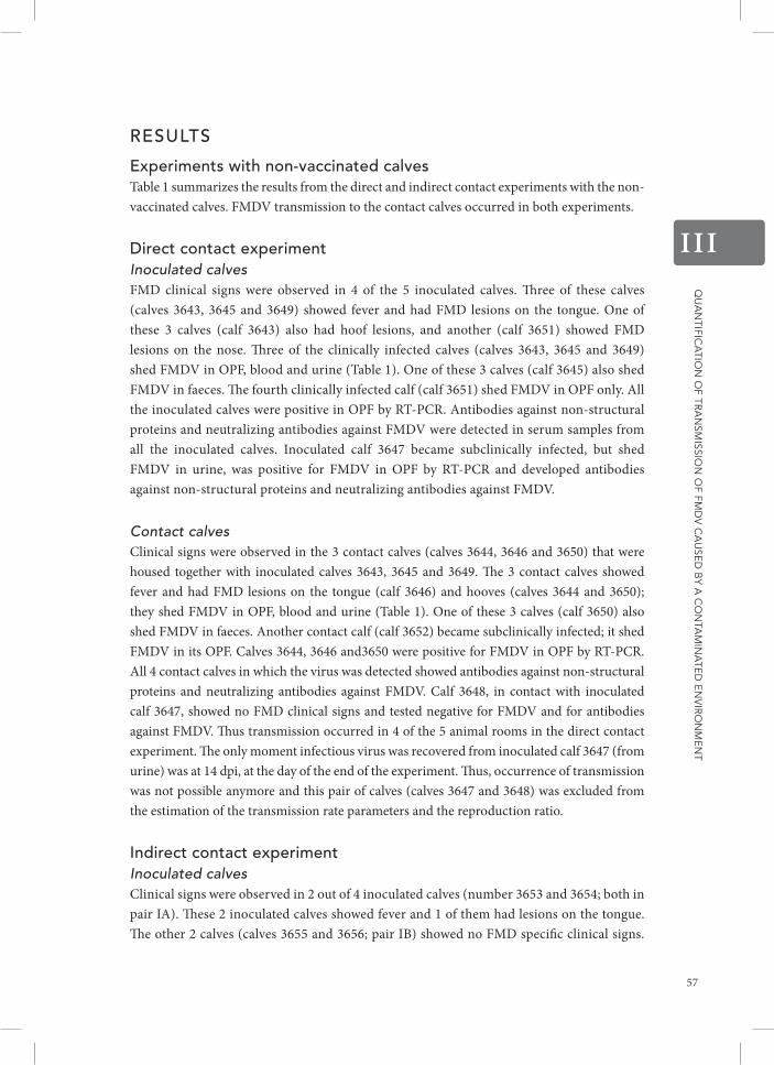
Qu
an
tifica
tion
of tr
an
smissio
n o
f fmD
V c
au
seD
by a
co
nta
min
ate
D e
nV
iro
nm
en
t
III
resuLts
experiments with non-vaccinated calves Table 1 summarizes the results from the direct and indirect contact experiments with the non-vaccinated calves. FMDV transmission to the contact calves occurred in both experiments.
direct contact experiment Inoculated calvesFMD clinical signs were observed in 4 of the 5 inoculated calves. Three of these calves (calves 3643, 3645 and 3649) showed fever and had FMD lesions on the tongue. One of these 3 calves (calf 3643) also had hoof lesions, and another (calf 3651) showed FMD lesions on the nose. Three of the clinically infected calves (calves 3643, 3645 and 3649) shed FMDV in OPF, blood and urine (Table 1). One of these 3 calves (calf 3645) also shed FMDV in faeces. The fourth clinically infected calf (calf 3651) shed FMDV in OPF only. All the inoculated calves were positive in OPF by RT-PCR. Antibodies against non-structural proteins and neutralizing antibodies against FMDV were detected in serum samples from all the inoculated calves. Inoculated calf 3647 became subclinically infected, but shed FMDV in urine, was positive for FMDV in OPF by RT-PCR and developed antibodies against non-structural proteins and neutralizing antibodies against FMDV.
Contact calves Clinical signs were observed in the 3 contact calves (calves 3644, 3646 and 3650) that were housed together with inoculated calves 3643, 3645 and 3649. The 3 contact calves showed fever and had FMD lesions on the tongue (calf 3646) and hooves (calves 3644 and 3650); they shed FMDV in OPF, blood and urine (Table 1). One of these 3 calves (calf 3650) also shed FMDV in faeces. Another contact calf (calf 3652) became subclinically infected; it shed FMDV in its OPF. Calves 3644, 3646 and3650 were positive for FMDV in OPF by RT-PCR. All 4 contact calves in which the virus was detected showed antibodies against non-structural proteins and neutralizing antibodies against FMDV. Calf 3648, in contact with inoculated calf 3647, showed no FMD clinical signs and tested negative for FMDV and for antibodies against FMDV. Thus transmission occurred in 4 of the 5 animal rooms in the direct contact experiment. The only moment infectious virus was recovered from inoculated calf 3647 (from urine) was at 14 dpi, at the day of the end of the experiment. Thus, occurrence of transmission was not possible anymore and this pair of calves (calves 3647 and 3648) was excluded from the estimation of the transmission rate parameters and the reproduction ratio.
indirect contact experiment Inoculated calves Clinical signs were observed in 2 out of 4 inoculated calves (number 3653 and 3654; both in pair IA). These 2 inoculated calves showed fever and 1 of them had lesions on the tongue. The other 2 calves (calves 3655 and 3656; pair IB) showed no FMD specific clinical signs.
57

Qu
an
tifica
tion
of tr
an
smissio
n o
f fmD
V c
au
seD
by a
co
nta
min
ate
D e
nV
iro
nm
en
t
III
Table 1 Results of virus isolation, RT-PCR (OPF swabs only), antibody detection and detection of FMD clinical signs
Expe
rim
ent
Cal
f ID
I: In
ocul
ated
C
: Con
tact
Gro
up
FMDV detection by virus isolation in OPF swabs (in log10 titres), blood, urine and faeces samples and by RT-PCR (OPF swabs only, shaded in grey) days post
infection of the inoculated calves
FMDV detection by virus isolation in OPF swabs (in log10 titres), blood, urine and faeces samples and by RT-PCR (OPF swabs only, shaded in grey)
days post infection of the inoculated calvesAntibody detection
Clin
ical
sign
s
Infe
ctio
us
1 2 3 4 5 6 7 8 9 10 11 12 13 14 15 16 17 18 19 20NS-
ELISA VNT
Dire
ct C
onta
ct
3643 I ≡e 2.2f ≡,vg 2.6,v,uh 3.8,v,u 2.0,u - - - - - - - - + + Yes Yes
3644 C - - - - 1.5 -,v,u 2.1,v 1.4,v ≡ 1.7,u - - 1.2 ≡ + + Yes Yes
3645 I - - ≡,v 2.5,v,fi 2.6,v 0.9,u 1.2,u 0.7 0.7 - - - - - + + Yes Yes
3646 C - - - - - ≡ 3.8 4.3,v 3.0,v,u 3.0,u -,u 0.4 - - + + Yes Yes
3647 I - ≡ - - - - - - - - - - - -,u + + No No
3648 C - - - - - - - - - - - - - - - - No No
3649 I - - 0.7,v 2.3,v,u 3.4,v 0.9,u -,u -,u - - - - - - + + Yes Yes
3650 C - - - - - 2.3,v,u 1.6,v,u,f 0.4,u,f ≡,u 0.9 0.4 -,u - - + + Yes Yes
3651 I - - 1.4 ≡ 4.2 1.7 0.4 - - - - - - - + + Yes Yes
3652 C - - - - - - - 0.4 - - - - - - + + No Yes
Indi
rect
Con
tact
3653 I A - - n.tj 1.7,v,u 3.4,v 2.1,u - - Yes Yes
3654 I A - ≡ 6.0,v 4.6,v 3.7,v,f 1.7 - - Yes Yes
3657 C1 A - - - - - - - - - - - - - - - - - - - - No No
3658 C1 A - - - - - - - - - - - - - - - - - - - - No No
3661 C2 A - - - - - 3.7,v ≡,v,u ≡,v,u 2.4 ≡,u ≡ ≡ - - - + + Yes Yes
3662 C2 A - - - - 2.0 ≡ ≡ ≡,v 1.3,v 3.8,v,u 3.5,v,u 3.6,u ≡ - ≡ + + Yes Yes
3655 I B - - 2.1 5.2,v 4.9,v,u 2.6,u - - No Yes
3656 I B - 0.4 ≡,v 3.2,v,u 5.0,v,u,f 3.5,u - - No Yes
3659 C1 B - - 1.9 2.0 3.6,v,f 4.9,v,u 4.1,v,u 3.4,u - 1.2 - - - - - - - - + + Yes Yes
3660 C1 B - - - - 0.9 1.3,u 0.7 1.0 - - - - - - - - - - - - Yes Yes
3663 C2 B - - - - - - - 2.4 0.7 1.6 3.3,u 2.3,u ≡ - - + + Yes Yes
3664 C2 B - 0.4 1.0 1.9 3.0,v 5.2,v ≡,v 2.6 0.4 ≡ - - - - - + + Yes Yes
aExp=experiment: DC=direct contact, IC=indirect contact ; bI=inoculated, C=contact animal ; cClin=clinical signs ; dInf=infectious ; eresults of virus isolation (VI) and RT-PCR of oral swab sample: - = VI and RT-PCR negative, ≡ = VI negative and RT-PCR positive;
foral swab sample scored positive for FMDV by VI (log10 pfu/mL), RT-PCR positive samples are indicated in bold ; gv=viraemia: blood sample scored positive for FMDV by VI ; hu=urine sample scored positive for FMDV by VI ; if=faeces sample scored positive for FMDV by VI ; jn.t.=not tested
In all 4 inoculated calves, virus was detected in the OPF (IA and IB). All four secreted and excreted FMDV in their blood, urine and/or faeces (Table 1). They all were positive for FMDV in OPF by RT-PCR. Thus, inoculated calves 3655 and 3656 were subclinically infected. Serum samples from all 4 inoculated calves were obtained only at 0 dpi and 3 dpi; in these samples neither antibodies against non-structural proteins nor neutralizing antibodies against FMDV were detected as expected.
58

Qu
an
tifica
tion
of tr
an
smissio
n o
f fmD
V c
au
seD
by a
co
nta
min
ate
D e
nV
iro
nm
en
t
III
Table 1 Results of virus isolation, RT-PCR (OPF swabs only), antibody detection and detection of FMD clinical signs
Expe
rim
ent
Cal
f ID
I: In
ocul
ated
C
: Con
tact
Gro
up
FMDV detection by virus isolation in OPF swabs (in log10 titres), blood, urine and faeces samples and by RT-PCR (OPF swabs only, shaded in grey) days post
infection of the inoculated calves
FMDV detection by virus isolation in OPF swabs (in log10 titres), blood, urine and faeces samples and by RT-PCR (OPF swabs only, shaded in grey)
days post infection of the inoculated calvesAntibody detection
Clin
ical
sign
s
Infe
ctio
us
1 2 3 4 5 6 7 8 9 10 11 12 13 14 15 16 17 18 19 20NS-
ELISA VNT
Dire
ct C
onta
ct
3643 I ≡e 2.2f ≡,vg 2.6,v,uh 3.8,v,u 2.0,u - - - - - - - - + + Yes Yes
3644 C - - - - 1.5 -,v,u 2.1,v 1.4,v ≡ 1.7,u - - 1.2 ≡ + + Yes Yes
3645 I - - ≡,v 2.5,v,fi 2.6,v 0.9,u 1.2,u 0.7 0.7 - - - - - + + Yes Yes
3646 C - - - - - ≡ 3.8 4.3,v 3.0,v,u 3.0,u -,u 0.4 - - + + Yes Yes
3647 I - ≡ - - - - - - - - - - - -,u + + No No
3648 C - - - - - - - - - - - - - - - - No No
3649 I - - 0.7,v 2.3,v,u 3.4,v 0.9,u -,u -,u - - - - - - + + Yes Yes
3650 C - - - - - 2.3,v,u 1.6,v,u,f 0.4,u,f ≡,u 0.9 0.4 -,u - - + + Yes Yes
3651 I - - 1.4 ≡ 4.2 1.7 0.4 - - - - - - - + + Yes Yes
3652 C - - - - - - - 0.4 - - - - - - + + No Yes
Indi
rect
Con
tact
3653 I A - - n.tj 1.7,v,u 3.4,v 2.1,u - - Yes Yes
3654 I A - ≡ 6.0,v 4.6,v 3.7,v,f 1.7 - - Yes Yes
3657 C1 A - - - - - - - - - - - - - - - - - - - - No No
3658 C1 A - - - - - - - - - - - - - - - - - - - - No No
3661 C2 A - - - - - 3.7,v ≡,v,u ≡,v,u 2.4 ≡,u ≡ ≡ - - - + + Yes Yes
3662 C2 A - - - - 2.0 ≡ ≡ ≡,v 1.3,v 3.8,v,u 3.5,v,u 3.6,u ≡ - ≡ + + Yes Yes
3655 I B - - 2.1 5.2,v 4.9,v,u 2.6,u - - No Yes
3656 I B - 0.4 ≡,v 3.2,v,u 5.0,v,u,f 3.5,u - - No Yes
3659 C1 B - - 1.9 2.0 3.6,v,f 4.9,v,u 4.1,v,u 3.4,u - 1.2 - - - - - - - - + + Yes Yes
3660 C1 B - - - - 0.9 1.3,u 0.7 1.0 - - - - - - - - - - - - Yes Yes
3663 C2 B - - - - - - - 2.4 0.7 1.6 3.3,u 2.3,u ≡ - - + + Yes Yes
3664 C2 B - 0.4 1.0 1.9 3.0,v 5.2,v ≡,v 2.6 0.4 ≡ - - - - - + + Yes Yes
aExp=experiment: DC=direct contact, IC=indirect contact ; bI=inoculated, C=contact animal ; cClin=clinical signs ; dInf=infectious ; eresults of virus isolation (VI) and RT-PCR of oral swab sample: - = VI and RT-PCR negative, ≡ = VI negative and RT-PCR positive;
foral swab sample scored positive for FMDV by VI (log10 pfu/mL), RT-PCR positive samples are indicated in bold ; gv=viraemia: blood sample scored positive for FMDV by VI ; hu=urine sample scored positive for FMDV by VI ; if=faeces sample scored positive for FMDV by VI ; jn.t.=not tested
Contact calves C1 Contact calves C1 were exposed to the animal rooms that were contaminated by the inoculated calves from 0 to 3 dpi. The contact calves of group C1A (calves 3657 and 3658) did not get infected; no FMD specific clinical signs were seen and both calves tested negative by virus isolation, by RT-PCR and, for antibodies against FMDV. The contact calves of group C1B (calves 3659 and 3660) showed fever and one had FMD lesions on the mouth, tongue, nose and hooves. Both C1B calves had virus detected in their OPF; one of them secreted and excreted
59

Qu
an
tifica
tion
of tr
an
smissio
n o
f fmD
V c
au
seD
by a
co
nta
min
ate
D e
nV
iro
nm
en
t
III
virus in blood, urine and faeces, the other one excreted virus in urine. They tested positive for FMDV in OPF by RT-PCR. One C1B calf showed antibodies against non-structural proteins in serum (calf 3660). Both C1B calves showed neutralizing antibodies in serum.
Contact calves C2 Contact calves C2 were exposed to the animal rooms that were contaminated by the inoculated calves from 3 to 6 dpi. All the contact calves of groups C2A and C2B showed clinical signs. Three of them showed fever, and all of them showed FMD lesions on the nose and in the mouth. In all 4 calves, virus was detected in their OPF (Table 1); the calves secreted and/or excreted FMDV in the blood (calves 3661, 3662 and 3664) and in urine (calves 3661, 3662 and 3663). They all were positive for FMDV in OPF by RT-PCR. All developed antibodies against non-structural proteins as well as neutralising antibodies. Thus transmission occurred in the indirect contact experiment in 1 of the 2 animal rooms that were contaminated from 0 to 3 dpi and, in both of the animal rooms that were contaminated from 3 to 6 dpi.
statistical analysis of virus secretion and excretionThe mean values for the AUC’s, peak of virus shedding and duration of virus shedding (and their ranges) for OPF swabs, urine samples, faeces samples and blood samples for the inoculated group, the direct contact group and the indirect contact groups C1 and C2 are shown in Additional file 3.
No significant difference in log AUC could be determined between the different experimental groups i.e. inoculated, direct contacts, indirect contacts C1 and indirect contacts C2, neither for OPF swabs nor for urine nor for faeces (p > 0.05). No significant difference in the maximum FMDV log titres was found between the different experimental groups neither for OPF swabs nor for urine nor for faeces (p > 0.05). No significant difference in the duration of FMDV secretion and excretion could be determined between the different experimental groups (p> 0.05) (Additional file 3).
experiments with vaccinated calvesAt day of challenge (0 dpi, 7 days post vaccination), the average virus neutralisation test (VNT) titre against the vaccine strain FMDV Asia-1 Shamir for all the vaccinated calves (including the vaccine controls) was 2.210log. The average virus neutralisation test (VNT) titre against the challenge strain FMDV Asia-1 TUR/11/2000 was 1.2 10log.
direct contact experimentAfter challenge, neither the vaccinated inoculated calves nor the vaccinated contact calves showed clinical signs of FMD and all calves tested negative by virus isolation and RT-PCR. Only 2 inoculated calves (calves 3972 and 3976) developed antibodies against non-structural proteins.
60

Qu
an
tifica
tion
of tr
an
smissio
n o
f fmD
V c
au
seD
by a
co
nta
min
ate
D e
nV
iro
nm
en
t
III
indirect contact experiment After challenge, neither the vaccinated inoculated calves nor the non-vaccinated contact calves showed clinical signs of FMD. All calves tested negative by virus isolation and RT-PCR. Neither the vaccinated inoculated nor the non-vaccinated contact calves showed detectable antibodies against non-structural protein.
Fmdv survival rate (σ)From the combined published and own experimental data, it was estimated that at 20°C a 10-fold reduction in FMDV titres occurs in 2.4 days (95% CI: 1.7, 3.3). We calculated the FMDV survival rate (σ) using the lowest (in spiked urine), middle (in spiked faeces) and highest (in spiked buffered solution) estimates obtained at 20°C. An additional file shows these estimates inside a dashed pointed rectangle (Additional file 4). The estimated time needed for 10-fold reduction in FMDV titres in spiked urine (lowest value) was 0.5 days, indicating an FMDV survival rate (σ) of 0.014 day−1. The estimated time needed for 10-fold reduction in FMDV titres in spiked faecal material (middle value) was 2.8 day indicating an FMDV survival rate (σ) of 0.44 day−1. The estimated time needed for 10-fold reduction in FMDV titres in spiked buffered solution (highest value) was 8.2 days, indicating an FMDV survival rate (σ) of 0.75 day−1. For the quantification of FMDV transmission, we used the middle estimate i.e. σ = 0.44 day−1.
Quantification of Fmdv transmission Results of the 1R-SIR model The transmission rate parameter β̂ was 0.67 per day (95% CI: 0.26, 1.8). The average infectious period from the inoculated calves T̂ was 5.5 days (95% CI: 4.5, 6.7). Therefore the estimated reproduction ratio R̂0
1R was 3.7 (95% CI: 1.3, 10.), significantly above 1.
Results of the 2R- SIR model The regression coefficient of fe, the extra infectivity contributed by the environment, was not significantly different from 0 which means that βcontact and βenvironment are not significantly different. Because βenvironment /βcontact equalled 1.4 (95% CI 0.14, 14), there is contribution of the environment. Using the most parsimonious model βcontact and βenvironment were estimated both to be 0.45 per day (95% CI: 0.24, 0.85). Because T̂ was 5.5 days (95% CI: 4.5, 6.7), R̂0
2Rcontact equalled 2.5 (95% CI: 1.3, 5.0). The average infectious period from the contaminated environment τ̂ was 4.3 days (95%
CI: 3.6, 5.2), which leads to a R̂02R
environment of 1.9 (95% CI: 1.007, 3.8). Combination of the two estimates (R̂0
2Rcontact + R̂0
2Renvironment) resulted in R̂0
2R equalled to 4.4 (95% CI: 1.5, 7.4), which is significantly above 1. R̂0
2R was not significantly different from R̂01R as can be seen from their
overlapping confidence intervals. The contribution of the environmental transmission to the total transmission of FMDV was 44% (R̂0
2Renvironment / R̂0
2R).The sensitivity analysis, i.e. multiplication of the new infections or cases (C) in Et
by either 0 or 0.5, resulted in the same contribution of the environmental transmission
61

Qu
an
tifica
tion
of tr
an
smissio
n o
f fmD
V c
au
seD
by a
co
nta
min
ate
D e
nV
iro
nm
en
t
III
(44%). When the lowest and the highest values of σ were used, the contribution of the environmental transmission to the total transmission was estimated to be 31% (when σ=0.014 day−1) and 75% (when σ = 0.75 day). The sensitivity analysis in which we included a latent period of 1, 2 or 3 days, resulted in higher estimates for β (Additional file 5) and R0 (Additional file 6) for the models with a latent period, but the estimated contribution of the environment stayed the same (Additional file 6).
Results of the final size model The R̂0
FS equalled 14 (95% CI: 1.3, infinite), which is significantly above 1. Based on the comparison of the confidence intervals, R̂0
FS seems to be not significantly different from R̂01R nor from R̂0
2R.
experiments with vaccinated calvesAfter challenge, none of the inoculated or contact calves became infectious; therefore transmission parameters could not be estimated.
disCussion
In this study, we quantified the contribution of a contaminated environment to the transmission of FMDV and analysed whether vaccination one week prior to inoculation of the calves could block FMDV transmission. We show that using a 2R-SIR model allows FMDV transmission to be quantified in two parts: the direct contact component and the indirect i.e. via the environment component. Our results show that roughly 44% of the transmission of FMDV occurs via the environment, in the days after the calves started secreting and excreting the virus. The contribution of the environment to the transmission of FMDV depends on the FMDV survival rate; if the survival rate is high, the contribution of the environment is higher. An environment that has previously housed infectious animals can contain FMDV if it is not properly disinfected after the removal of the infectious animals [5] and our study shows that this virus accumulation can cause new infections.
As we show, environmental transmission of FMDV plays a role in the total transmission of FMDV also in groups of animals that do have direct contact. Transmission of FMDV has been quantified before in several studies by using a 1R- SIR model [14,18,30-35]. We believe that in all of these studies, transmission occurred through both routes: via direct contact to an infected animal and via indirect contact to a contaminated environment. However within the experimental design of those studies, the role of the environment could not be separated from the role of direct contact on the transmission of FMDV. By using both direct and indirect contact experiments we could employ a 2R-SIR model (that included accumulation of FMDV in the environment) to quantify the contribution of the environment (R̂0
2Renvironment)
to the total transmission of FMDV. As expected, the estimated R̂01R, R̂0
2R and R̂0FS are very
similar to each other and moreover, they are similar to the R̂0 (by using a 1R-SIR model) estimated in other direct contact experiments with cattle infected with FMDV O/NET/2001
62

Qu
an
tifica
tion
of tr
an
smissio
n o
f fmD
V c
au
seD
by a
co
nta
min
ate
D e
nV
iro
nm
en
t
III
[18,35]. The consistency of these results indicates that our 2R-SIR model is valid for the estimation of the reproduction ratio and that it is very useful to separate both components i.e. the environment and direct contact transmission, for the quantification of their separate contribution to the transmission of FMDV. Moreover based on the statistical analysis of virus secretion and excretion, the results obtained with the 2R-SIR model are not biased by the route of infection i.e. inoculated and contact infected calves.
In our models, we used an SIR model and we did not incorporate a latent period (then we would have a SEIR i.e. susceptible, exposed, infectious, recovered model), although the data from the virus excretion of the inoculated animals suggest that for this group there is a latent period of approximately 2 days. The main reason why we did not incorporate a latent period in our study is because we did not want to introduce more complexity in the model. Also, incorporation of a latent period affects the estimates for the direct and indirect transmission more or less equally and thus the estimation of the role of the environment (the main interest of this research) was not been affected.
Our sensitivity analysis showed that, when a latent period is incorporated in the models, the estimates of the transmission parameters are still “equal” i.e. not significantly different (Additional files 5 and 6). The transmission parameters we provide in Additional files 5 and 6, where a latent period was used, could be useful when the transmission parameters are applied for modelling disease outbreaks and the effect of control measures.
The temporal separation used in our indirect contact experiment allowed us to observe the occurrence of transmission through the environment by taking into consideration virus accumulation in 2 different periods i.e. 0–3 and 3–6 dpi. Temporal separation was also used by Charleston et al. [36] to study FMDV transmission, although they exposed “donor” calves to “recipient” calves by direct contact for 8 hours in separate environments that had been previously disinfected, and thus with no accumulation of virus in the environment. This would, based on our results, reduce transmission of FMDV. They conclude in their study that the occurrence of FMDV transmission is correlated with the presence of clinical signs. However, it has been previously shown that FMDV transmission also can occur before clinical signs are seen [33]. In our study as well, transmission through the environment was caused by one group of calves that contaminated the environment from 0 to 3 dpi but showed no clinical disease. This supports the conclusion that the correlation of FMDV transmission with the presence of clinical signs cannot be generalized to populations, if animals have direct contact to each other for a longer period and/or are present where accumulation of FMDV in the environment is plausible. FMDV transmission may not occur, however, when animals are separated by fences or wooden walls (in pigs [37]; in calves: Charleston et al. (personal communication), [36]), indicating that either exposure to virus secreting and/or excreting animals or exposure to virus contaminated surfaces is important for the occurrence of transmission. Vaccination can be used as a tool to reduce transmission of FMDV [17]. In our study the calves vaccinated one week prior to inoculation with FMDV did not shed virus. Previously, vaccinating animals 2 weeks prior inoculation with FMDV was reported [18] to reduce FMDV transmission; our results indicate that vaccination reduces FMDV
63

Qu
an
tifica
tion
of tr
an
smissio
n o
f fmD
V c
au
seD
by a
co
nta
min
ate
D e
nV
iro
nm
en
t
III
transmission even earlier. As others have demonstrated, vaccination rapidly protects cattle from clinical disease, and reduces virus shedding by infected cattle [38-40]. As our results indicate, vaccination as early as one week before challenge cannot only protect calves against infection but also, can avoid contamination of the environment and so prevent new infections.
In summary, our study shows that the environment is a relevant mechanism in the transmission of FMDV. The quantification of the magnitude of the contribution of transmission via the environment emphasizes again that hygiene is an extremely important control measure for FMDV. And that, as already recommended by veterinary authorities, good disinfection of e.g. vehicles, walls and floors previously contaminated by infected animals is necessary to reduce the accumulation of the virus in the environment and therefore FMDV transmission. Also, the data from our experiment give some insight in which secretions and excretions contain FMDV at different times post infection and also this knowledge could be to improve control measures. The accumulation of FMDV in the environment should be taken into account when studying FMDV transmission. Further, the environmental aspect in the transmission of FMDV should be considered during the planning and implementation of measures to control FMD during an outbreak.
additionaL FiLes
Additional file 1: On the FMDV survival rate σ. Detailed calculation of the FMDV survival rate σ, which was calculated using published data on FMDV thermal inactivation combined with own laboratory data.
on the Fmdv survival rate σPublished data on FMDV titres of serum samples [42], medium [7, 42], buffer [43], milk [44, 45], slurry [7], bone marrow [46], lymph nodes [46] and hemal nodes [46] after being exposed to different temperatures, were collected and put in a database.
Further, in a laboratory experiment, we contaminated water, Eagle’s Minimum Essential Medium (EMEM), faeces (from calves) and urine (from calves) samples at the starting time (0h) with an inoculum that contained 10 6.8 pfu/ml of FMDV Asia-1 TUR/11/2000 (all in Duplo). All samples were exposed to 4 ºC and 20 ºC for 0 h, 8 h, 24 h, 48 h, 96 h, 168 h, 336 h, or 504 h. Water and urine, mixed previously with 10% ABII (by volume, v/v), were contaminated with 10% of the FMDV inoculum (v/v). After the required incubation, 800 µl of water or urine was mixed with 200 µl of a 50% FCS, 40% ABII solution and stored at -70 °C until virus isolation analysis. EMEM (containing 10% ABII and 2% FCS) was mixed with 10% ABII (v/v) and contaminated with 10% of the FMDV inoculum (v/v). After the required incubation, samples were stored at -70 °C until virus isolation analysis. Using a sonicator, we mixed and homogenized 50% of faeces with 50% of the FMDV inoculum (w/v). 10% ABII (v/v) was added to the mix. After the required incubation, the faecal mix was suspended 1:10 (by weight, w/v) in EMEM containing 10% FCS, and vortexed with glass beads. After 20 minutes of incubation at environmental temperature, the suspension was vortexed and
64
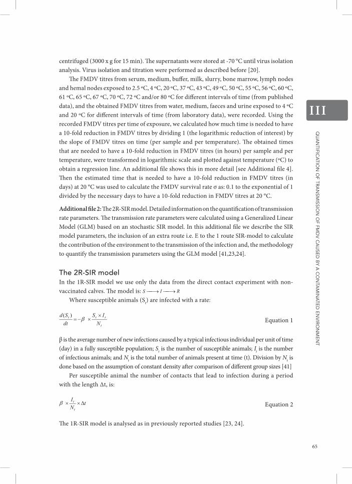
Qu
an
tifica
tion
of tr
an
smissio
n o
f fmD
V c
au
seD
by a
co
nta
min
ate
D e
nV
iro
nm
en
t
III
centrifuged (3000 x g for 15 min). The supernatants were stored at -70 °C until virus isolation analysis. Virus isolation and titration were performed as described before [20].
The FMDV titres from serum, medium, buffer, milk, slurry, bone marrow, lymph nodes and hemal nodes exposed to 2.5 ºC, 4 ºC, 20 ºC, 37 ºC, 43 ºC, 49 ºC, 50 ºC, 55 ºC, 56 ºC, 60 ºC, 61 ºC, 65 ºC, 67 ºC, 70 ºC, 72 ºC and/or 80 ºC for different intervals of time (from published data), and the obtained FMDV titres from water, medium, faeces and urine exposed to 4 ºC and 20 ºC for different intervals of time (from laboratory data), were recorded. Using the recorded FMDV titres per time of exposure, we calculated how much time is needed to have a 10-fold reduction in FMDV titres by dividing 1 (the logarithmic reduction of interest) by the slope of FMDV titres on time (per sample and per temperature). The obtained times that are needed to have a 10-fold reduction in FMDV titres (in hours) per sample and per temperature, were transformed in logarithmic scale and plotted against temperature (ºC) to obtain a regression line. An additional file shows this in more detail [see Additional file 4]. Then the estimated time that is needed to have a 10-fold reduction in FMDV titres (in days) at 20 °C was used to calculate the FMDV survival rate σ as: 0.1 to the exponential of 1 divided by the necessary days to have a 10-fold reduction in FMDV titres at 20 °C.
Additional file 2: The 2R-SIR model. Detailed information on the quantification of transmission rate parameters. The transmission rate parameters were calculated using a Generalized Linear Model (GLM) based on an stochastic SIR model. In this additional file we describe the SIR model parameters, the inclusion of an extra route i.e. E to the 1 route SIR-model to calculate the contribution of the environment to the transmission of the infection and, the methodology to quantify the transmission parameters using the GLM model [41,23,24].
the 2r-sir modelIn the 1R-SIR model we use only the data from the direct contact experiment with non-vaccinated calves. The model is:
Where susceptible animals (St) are infected with a rate:
Equation 1
β is the average number of new infections caused by a typical infectious individual per unit of time (day) in a fully susceptible population; St is the number of susceptible animals; It is the number of infectious animals; and Nt is the total number of animals present at time (t). Division by Nt is done based on the assumption of constant density after comparison of different group sizes [41]
Per susceptible animal the number of contacts that lead to infection during a period with the length ∆t, is:
Equation 2
The 1R-SIR model is analysed as in previously reported studies [23, 24].
65

Qu
an
tifica
tion
of tr
an
smissio
n o
f fmD
V c
au
seD
by a
co
nta
min
ate
D e
nV
iro
nm
en
t
III
In the 2R-SIR model we use data from both the direct contact experiment and the indirect contact experiment. In this model we included an extra route to the 1R-SIR model: E. The model is:
In this case calves are exposed to both infectious animals (It) and/or to virus coming from infectious animals via the environment (Et) (see Figure 2 in manuscript). Et is based on the secretion and excretion of FMDV by the infectious animals on previous days as well as on the remaining virus in the environment. We therefore include the FMDV survival rate (σ), described in Additional file 2, to correct for the decrease of FMDV in time, thus:
Equation 3
Thus the rate of infection per susceptible individual during a period with the length becomes:
Equation 4
βcontact+environment is a combined transmission rate parameter for contact exposure to an infected animal and for contact exposure to a contaminated environment (for its calculation we used data from both direct contact and indirect contact experiments).
By replacing βcontact+environment by this can be rewritten as:
Equation 5
Where fe is the fraction of transmission by the environment and its regression coefficient measures the extra infectivity contributed by the environment. If the contribution of the environment is zero, then fe becomes zero (because Et is zero) and βcontact+environment = βcontact equal to eco. If there are no infectious animals present, then fe is 1 and βcontact+environment = βenvironment equal to eco+c1.
The probability that a single susceptible animal becomes infected is then binomial distributed with:
Equation 6
The data are analysed with a Generalised Linear Model (GLM) with a complementary log-log link, thus we take the log(-log(1-p)). The expected value of C/S when applying the link function is:
66

Qu
an
tifica
tion
of tr
an
smissio
n o
f fmD
V c
au
seD
by a
co
nta
min
ate
D e
nV
iro
nm
en
t
III
Equation 7
So this is a GLM with offset:
Equation 8
With S as binomial total, a binomial error function, and with explanatory variable fe (the infectivity contributed by the environment):
Additional file 3: Mean values (plus range) and the Kruskal-Wallis statistics of virus present in secretions, excretions and blood samples, for the inoculated, direct contact and indirect contact groups. H is the Kruskal-Wallis test statistic and df the degrees of freedom.
Measured variables
Mean values (range)
Kruskal-Wallis statisticsInoculated group
Direct contact group
Indirect contact groups
C1 C2 H df P-value
AUC (log10 titres)
OPF swabs 8.8 (0, 15) 5.7 (0, 15) 6.2 (0, 21) 6.1 (2.6, 14) 2.32 3 0.51
Urine 2.3 (0, 5.1) 2.1 (0, 6.2) 1.8 (0, 5.3) 1.0 (0, 4.0) 1.60 3 0.66
Faeces 0.3 (0, 1.2) 0.4 (0, 2.1) 0.2 (0, 0.2) 0 1.54 3 0.67
Blood 5.8 (0,9.5) 4.0 (0, 8.7) 2.1 (0, 8.3) 4.6 (0, 9.8) 2.35 3 0.50
Maximum FMDV excretion (log10 titres)
OPF swabs 3.7 (0, 6.0) 1.8 (0, 4.3) 1.6 (0, 4.9) 4.0 (3.3, 5.2) 5.53 3 0.14
Urine 1.2 (0, 2.6) 0.7 (0, 1.9) 1.1 (0, 2.7) 1.7 (0, 2.5) 1.81 3 0.61
Faeces 0.3 (0, 1.2) 0.3 (0, 1.7) 0.2 (0, 0.9) 0 1.54 3 0.67
Blood 2.5 (0, 3.6) 1.9 (0, 3.3) 0.9 (0, 3.7) 2.2 (0, 3.4) 1.65 3 0.65
Duration of FMDV excretion (days)
OPF swabs 4.0 (0, 6) 4.4 (0, 9) 3.0 (0, 8) 6.3 (4, 8) 3.01 3 0.39
Urine 2.1 (0, 5) 3.0 (0, 7) 1.0 (0, 3) 2.3 (0, 4) 1.68 3 0.64
Faeces 0.3 (0, 1) 0.4 (0, 2) 0.3 (0, 1) 0 1.52 3 0.68
Blood 2.1 (0, 3) 1.4 (0, 3) 0.8 (0, 3) 2.5 (0, 4) 4.03 3 0.26
Additional file 4: Plotted linear regression estimates of the log time (hours) needed for a 10-fold reduction in FMDV titres. In this additional file we show the obtained times (log hours) that are needed to have a 10-fold reduction in FMDV titres per sample and per temperature. Light blue points correspond to estimates from water; green from buffers; grey from hemal and lymph nodes and bone marrow; black from faeces; red from urine; pink
67

Qu
an
tifica
tion
of tr
an
smissio
n o
f fmD
V c
au
seD
by a
co
nta
min
ate
D e
nV
iro
nm
en
t
III
from milk; blue from slurry. Inside the dashed pointed rectangles, only obtained estimates at 20 °C. Red dashed lines, regression lines at 95% CI.
Latent period (days) βcontact βenvironment Ratio1 βoverall
2 N3
Number of contact infections for each number of latent days
0 1 2 3
0 0.373 0.523 1.404 0.453 47 10
1 0.459 0.753 1.640 0.625 40 1 9
2 0.868 0.928 1.071 0.909 33 1 1 8
3 1.280 1.258 0.983 1.265 29 1 1 3 5
1Ratio = βenvironment/βcontact2 This is the beta estimate for contact and environment when the two are not significantly different which is the case here for all latent periods.3 N = number of rows in the dataset used in the GLM analysis
Additional file 5: Sensitivity analysis considering latent periods. In this additional file we show results of the estimation of transmission parameters for latent periods of 0 (as used in the paper), 1, 2 and 3 days. We show the effect of different latent periods on transmission parameters βcontact and βenvironment from the model assuming that both are not the same and, on the βoverall, the overall estimate for both, under the assumption that both are the same.
Additional file 6: Sensitivity analysis considering latent periods. In this additional file we show results of the estimation of R0’s for latent periods of 0 (as used in the paper), 1, 2 and 3 days. We show the effect of different latent periods on the different estimates for R0 (including 95% confidence interval) and on the estimated contribution of the environment
68

Qu
an
tifica
tion
of tr
an
smissio
n o
f fmD
V c
au
seD
by a
co
nta
min
ate
D e
nV
iro
nm
en
t
III
Latent period (days) R̂0
1R R̂02R
contact R̂02R
environment R̂02R
Environmental contribution to
transmission (%)
0 3.7 <1.3, 10> 2.5 <1.3, 4.8> 1.9 <1.0, 3.8> 4.4 <1.5, 7.4> 44
1 5.6 <2.0, 15> 3.4 <1.8, 6.7> 2.7 <1.4, 5.2> 6.1 <3.2, 9.1> 44
2 12 <4.2, 34> 5.0 <2.5, 9.8> 3.9 <2.0, 7.7> 8.9 <5.9, 12> 44
3 17 <5.8, 53> 6.9 <3.5, 14> 5.4 <2.8, 11> 5.4 <2.8, 11> 44
List of RefeRences
1 Thomson GR: Foot and Mouth Disease. In: Coetzer JAW, Thomson GR, Tustin RC (ed) 2004. Infectious diseases of livestock with special reference to Southern Africa. Volume 1. Oxford University Press, Capetown, pp 825–852.
2 Woolhouse M, Chase-Topping M, Haydon D, Friar J, Matthews L, Hughes G, Shaw D, Wilesmith J, Donaldson A, Cornell S, Keeling M, Grenfell B: Foot-and-mouth disease under control in the UK. Nature 2001, 411:258–259.
3 Bravo de Rueda C, Dekker A, Eblé PL, de Jong MCM: Identification of factors associated with increased excretion of Foot-and-Mouth Disease Virus. Preventive Veterinary Medicine 2014, 113:23–33.
4 Parker J: Presence and inactivation of foot-and-mouth disease virus in animal faeces. Veterinary Records 1971, 88:659–662.
5 Sellers RF, Herniman KA, Donaldson AI: The effects of killing or removal of animals affected with foot-and-mouth disease on the amounts of airborne virus present in looseboxes. British Veterinary Journal 1971, 127:358–365.
6 Botner A, Belsham GJ: Virus survival in slurry: analysis of the stability of foot-and-mouth disease, classical swine fever, bovine viral diarrhoea and swine influenza viruses. Veterinary Microbiology 2012, 157:41–49.
7 Turner C, Williams SM, Cumby TR: The inactivation of foot and mouth disease, Aujeszky’s disease and classical swine fever viruses in pig slurry. Journal of Applied Microbiology 2000, 89:760–767.
8 van Bunnik BA, Hagenaars TJ, Bolder NM, Nodelijk G, de Jong MCM: Interaction effects between sender and receiver processes in indirect transmission of Campylobacter jejuni between broilers. BMC Veterinary Research 2012, 8:123.
9 van Bunnik BA, Katsma WE, Wagenaar JA, Jacobs-Reitsma WF, de Jong MCM: Acidification of drinking water inhibits indirect transmission,
but not direct transmission of Campylobacter between broilers. Preventive Veterinary Medicine 2012, 105:315–319.
10 Breban R, Drake JM, Stallknecht DE, Rohani P: The role of environmental transmission in recurrent avian influenza epidemics. PLoS Computational Biology 2009, 5:e1000346.
11 Rohani P, Breban R, Stallknecht DE, Drake JM: Environmental transmission of low pathogenicity avian influenza viruses and its implications for pathogen invasion. Proccedings National Academy of Sciences USA 2009, 106:10365–10369.
12 Velkers FC, Bouma A, Stegeman JA, de Jong MCM: Oocyst output and transmission rates during successive infections with Eimeria acervulina in experimental broiler flocks. Veterinary Parasitology 2012, 187:63–71.
13 Eblé PL, de Koeijer AA, de Jong MCM, Engel B, Dekker A: A meta-analysis quantifying transmission parameters of FMDV strain O Taiwan among non-vaccinated and vaccinated pigs. Preventive Veterinary Medicine 2008, 83:98–106.
14 Orsel K, Dekker A, Bouma A, Stegeman JA, de Jong MCM: Quantification of foot and mouth disease virus excretion and transmission within groups of lambs with and without vaccination. Vaccine 2007, 25:2673–2679.
15 Kermack WO, McKendrick AG: A Contribution to the mathematical theory of epidemics. Proceedings Royal Society of London 1927, 115:700–721.
16 Velthuis AGJ, de Jong MCM, De Bree J, Nodelijk G, Van Boven M: Quantification of transmission in one-to-one experiments. Epidemiology and Infection 2002, 128:193–204.
17 Cox SJ, Barnett PV: Experimental evaluation of foot-and-mouth disease vaccines for emergency use in ruminants and pigs: a review. Veterinary Research 2009, 40:13.
69

Qu
an
tifica
tion
of tr
an
smissio
n o
f fmD
V c
au
seD
by a
co
nta
min
ate
D e
nV
iro
nm
en
t
III
18 Orsel K, Dekker A, Bouma A, Stegeman JA, de Jong MCM: Vaccination against foot and mouth disease reduces virus transmission in groups of calves. Vaccine 2005, 23:4887–4894.
19 Hajer R, Hendrikse J, Rutgers LJE, van Oldruitenborgh-Oosterbaan MMS, van der Weyden GC: Het klinisch onderzoek bij grote huisdieren. Utrecht 1985.
20 Bouma A, Dekker A, de Jong MCM: No foot-and-mouth disease virus transmission between individually housed calves. Veterinary Microbiology 2004, 98:29–36.
21 Moonen P, Boonstra J, van der Honing RH, Leendertse CB, Jacobs L, Dekker A: Validation of a LightCycler-based reverse transcription polymerase chain reaction for the detection of foot-and-mouth disease virus. Journal of Virology Methods 2003, 113:35–41.
22 Dekker A, Terpstra C: Prevalence of foot-and-mouth disease antibodies in dairy herds in The Netherlands, four years after vaccination. Research in Veterinary Sciences 1996, 61:89–91.
23 Velthuis AG, de Jong MCM, Kamp EM, Stockhofe N, Verheijden JH: Design and analysis of an Actinobacillus pleuropneumoniae transmission experiment. Preventive Veterinary Medicine 2003, 60:53–68.
24 McCullagh P, Nelder JA: Generalized Linear Models. Boca Raton, Chapman & Hall/CRC 1989.
25 R Development Core Team: R: A Language and Environment for Statistical Computing. The R Foundation for Statistical Computing, Vienna 2012.
26 Diekmann O, Heesterbeek JA, Metz JA: On the definition and the computation of the basic reproduction ratio R0 in models for infectious diseases in heterogeneous populations. Journal of Mathematical Biology 1990, 28:365–382.
27 de Jong MCM, Kimman TG: Experimental quantification of vaccine induced reduction in virus transmission. Vaccine 1994, 8:761–766.
28 Velthuis AG, de Jong MCM, Stockhofe N, Vermeulen TM, Kamp EM: Transmission of Actinobacillus pleuropneumoniae in pigs is characterized by variation in infectivity. Epidemiology and Infection 2002, 129:203–214.
29 Velthuis AG, de Jong MCM, De Bree J: Comparing methods to quantify experimental transmission of infectious agents. Mathematical Biosciences 2007, 210:157–176.
30 Eblé PL, de Koeijer A, Bouma A, Stegeman A, Dekker A: Quantification of within- and between-
pen transmission of Foot-and-Mouth disease virus in pigs. Veterinary Research 2006, 37:647–654.
31 Eblé PL, Bouma A, de Bruin MG, van Hemert-Kluitenberg F, van Oirschot JT, Dekker A: Vaccination of pigs two weeks before infection significantly reduces transmission of foot-and-mouth disease virus. Vaccine 2004, 22:1372–1378.
32 Goris NE, Eblé PL, de Jong MCM, De Clercq K: Quantification of foot- and-mouth disease virus transmission rates using published data. Altex 2009, 26:52–54.
33 Orsel K, Bouma A, Dekker A, Stegeman JA, de Jong MCM: Foot and mouth disease virus transmission during the incubation period of the disease in piglets, lambs, calves, and dairy cows. Preventive Veterinary Medicine 2009, 88:158–163.
34 Orsel K, de Jong MCM, Bouma A, Stegeman JA, Dekker A: Foot and mouth disease virus transmission among vaccinated pigs after exposure to virus shedding pigs. Vaccine 2007, 25:6381–6391.
35 Orsel K, de Jong MCM, Bouma A, Stegeman JA, Dekker A: The effect of vaccination on foot and mouth disease virus transmission among dairy cows. Vaccine 2007, 25:327–335.
36 Charleston B, Bankowski BM, Gubbins S, Chase-Topping ME, Schley D, Howey R, Barnett PV, Gibson D, Juleff ND, Woolhouse MEJ: Relationship between clinical signs and transmission of an infectious disease and the implications for control. Science 2011, 332:726–729.
37 van Roermund HJ, Eblé PL, de Jong MCM, Dekker A: No between-pen transmission of foot-and-mouth disease virus in vaccinated pigs. Vaccine 2010, 28:4452–4461.
38 Golde WT, Pacheco JM, Duque H, Doel T, Penfold B, Ferman GS, Gregg DR, Rodriguez LL: Vaccination against foot-and-mouth disease virus confers complete clinical protection in 7 days and partial protection in 4 days: Use in emergency outbreak response. Vaccine 2005, 23:5775–5782.
39 Doel TR, Williams L, Barnett PV: Emergency vaccination against foot- and-mouth disease: rate of development of immunity and its implications for the carrier state. Vaccine 1994, 12:592–600.
40 Cox SJ, Voyce C, Parida S, Reid SM, Hamblin PA, Paton DJ, Barnett PV: Protection against direct-contact challenge following emergency FMD vaccination of cattle and the effect on virus excretion from the oropharynx. Vaccine 2005, 23:1106–1113.
70

Qu
an
tifica
tion
of tr
an
smissio
n o
f fmD
V c
au
seD
by a
co
nta
min
ate
D e
nV
iro
nm
en
t
III
41 Bouma A, de Jong MCM, Kimman TG: Transmission of pseudorabies virus within pig-populations is independent of the size of the population. Preventive Veterinary Medicine1995, 23:163–172.
42 Dekker A: Inactivation of foot-and-mouth disease virus by heat, formaldehyde, ethylene oxide and gamma radiation. Veterinary Records 1998, 143:168–169.
43 Bachrach HL, Breese SS, Callis JJ, Hess WR, Patty RE: Inactivation of foot-and-mouth disease virus by pH and temperature changes and by formaldehyde. Proccedings of the Society for Experimental Biology and Medicine 1957, 95:147–152.
44 Blackwell JH, Hyde JL: Effect of heat on foot-and-mouth disease virus (FMDV) in the components of milk from FMDV-infected cows. Journal of Hygiene 1976, 77:77–83.
45 Hyde JL, Blackwell JH, Callis JJ: Effect of pasteurization and evaporation on foot-and-mouth disease virus in whole milk from infected cows. Canadian Journal of Comparative Medicine 1975, 39:305–309.
46 Cottral GE: Persistence of foot-and-mouth disease virus in animals, their products and the environment. Bulletin Office International des Epizooties 1969, 70:549–568.
71


IVestimation of the transmission of foot-and-mouth disease virus
from infected sheep to cattle
Carla Bravo de Rueda, Mart C.M. de Jong, Phaedra L. Eblé, Aldo Dekker
Veterinary Research 2014; 45(1): 58

Estim
atio
n o
f thE
tra
nsm
ission
of fo
ot-a
nd
-mo
uth
disE
asE
vir
us fr
om
infE
ctE
d sh
EE
p to c
attlE
IV
aBstraCt
The quantitative role of sheep in the transmission of foot-and-mouth disease virus (FMDV) is not well known. To estimate the role of sheep in the transmission of FMDV, a direct contact transmission experiment with 10 groups of animals each consisting of 2 infected lambs and 1 contact calf was performed. Secretions and excretions (oral swabs, blood, urine, faeces and probang samples) from all animals were tested for the presence of FMDV by virus isolation (VI) and/or RT-PCR. Serum was tested for the presence of antibodies against FMDV. To estimate FMDV transmission, the VI, RT-PCR and serology results were used. The partial reproduction ratio R0
p i.e. the average number of new infections caused by one infected sheep introduced into a population of susceptible cattle, was estimated using either data of the whole infection chain of the experimental epidemics (the transient state method) or the final sizes of the experimental epidemics (the final size method). Using the transient state method, R0
p was estimated as 1.0 (95% CI 0.2 - 6.0) using virus isolation results and 1.4 (95% CI 0.3 - 8.0) using RT-PCR results. Using the final size method, R0
p was estimated as 0.9 (95% CI 0.2 - 3.0). Finally, R0
p was compared to the R0’s obtained in previous transmission studies with sheep or cattle only. This comparison showed that the infectivity of sheep is lower than that of cattle and that sheep and cattle are similarly susceptible to FMD. These results indicate that in a mixed population of sheep and cattle, sheep play a more limited role in the transmission of FMDV than cattle.
74

Estim
atio
n o
f thE
tra
nsm
ission
of fo
ot-a
nd
-mo
uth
disE
asE
vir
us fr
om
infE
ctE
d sh
EE
p to c
attlE
IV
introduCtion
Foot-and-mouth disease (FMD) is a contagious viral disease in cloven-hoofed animals caused by foot-and-mouth disease virus (FMDV). Clinical signs of FMD in sheep are frequently mild or not apparent [1]. But while sheep may not manifest clear clinical signs of FMD, they can secrete and excrete considerable amounts of FMDV [2-4] and therefore may play a significant role in FMDV transmission. Transmission of FMDV between sheep [5-8] and between cattle [9-11] has been studied previously. Transmission of FMDV from sheep to cattle may have occurred during the 1994 type O epidemic in Greece [12], during the 1999 type O epidemics in Morocco [13] and during the 2001 type O epidemics in UK [14]. However, transmission of FMDV from sheep to cattle has not yet been quantified.
In epidemiology, the reproduction ratio (R0) is an important quantitative parameter of transmission. R0 is defined as the average number of new infections caused by one typical infectious individual, during its entire infectious period, introduced into a population made up entirely of susceptible individuals [15]. Major outbreaks of FMDV can occur only if R0 is above 1. In the previously mentioned studies, R0 was estimated within species i.e. intraspecies transmission either in sheep or in cattle. When different species are mixed, the R0 for a mixed population of cattle and sheep not only depends on the occurrence of intraspecies (cattle-to-cattle and sheep-to-sheep) transmission but also on the occurrence of interspecies (sheep-to-cattle and cattle-to-sheep) transmission. To estimate R0 for a mixed population of cattle and sheep, all 4 (2 intraspecies and 2 interspecies) transmission parameters have to be known. The 2 interspecies transmission parameters will be called partial R0’s to emphasise that these parameters are strictly speaking not reproduction ratios. On the interspecies transmission of FMDV between sheep and cattle no quantitative information is available yet.
Moreover, with estimates for the intraspecies and interspecies (partial) R0’s, relative infectivity and susceptibility of sheep and cattle can be determined. Because for FMDV, relative infectivity and susceptibility have not extensively been quantified, modellers have had to rely on educated guesses about the relative infectivity and susceptibility of cattle, sheep and pigs herds [16]. Knowledge on relative infectivity and susceptibility of different species would improve modelling of FMDV transmission and more importantly could be used to implement tailored control measures in accordance to the animal species.
This study fills part of the gap on quantitative information on interspecies transmission of FMD. We estimated interspecies transmission of FMDV from infected sheep to contact cattle by estimating a partial R0 (R0
p) for sheep to cattle transmission. Further, comparison of our results to those obtained in intraspecific transmission studies allowed us to define the relative infectivity and susceptibility of sheep and cattle.
75

Estim
atio
n o
f thE
tra
nsm
ission
of fo
ot-a
nd
-mo
uth
disE
asE
vir
us fr
om
infE
ctE
d sh
EE
p to c
attlE
IV
materiaLs and methods
experimental designTwenty conventionally reared lambs (crossbred Texelaar-Noordhollander) aged between 6 and 7 months and 10 conventionally reared calves (pure- or crossbred (87%) Holstein-Frisian) aged between 6 and 8 months were used in this study. The study was performed in 10 separate animal rooms within the biosecurity facilities of the Central Veterinary Institute (CVI, Lelystad, The Netherlands). Each animal room was between 9 and 11 m2 in size. In each animal room, 2 infected lambs and 1 contact calf were housed together for 31 days. The study received ethical approval from the animal experiment committee of the CVI in accordance with Dutch law.
On the day of infection (0 days post infection (dpi)), all the lambs were moved to a separate animal room and inoculated with FMDV. Eight hours after inoculation, the lambs were reunited with their original roommates. The lambs were inoculated with FMDV strain Asia-1 TUR/11/2000 by intranasal instillation. The virus was obtained from the World Reference Laboratory for Foot-and-Mouth Disease (Pirbright, United Kingdom); it was passaged once in cattle before its use. The inoculum contained 105.8 plaque forming units (pfu)/mL (tested on primary lamb kidney cells). Each lamb received 1.5 mL of inoculum per nostril.
sampling proceduresDuring animal inspection and/or sampling, animal caretakers changed coveralls and gloves between animal rooms. All the animals were inspected daily for clinical signs of FMD. In these inspections, rectal temperature above 39.5 °C in calves and above 40 °C in lambs was considered fever [17], and the animals were checked for the presence of vesicles and/or lameness. Oral swab samples were collected daily from each animal from 0 dpi until the end of the experiment (31 dpi). They were collected and processed as described previously [11], with the exception that we used medium containing 2% foetal bovine serum. The oral swab samples were stored at −70 °C until analysis by virus isolation (VI) and real time RT-PCR. Probang samples were collected from each animal at 29, 30 and 31 dpi. These were stored at -70 °C until analysis by real time RT-PCR. Heparinized blood samples were collected daily from each animal from 0 dpi until 11 dpi. The heparinized blood samples were centrifuged at 2500 RPM for 15 min; plasma was stored at −70 °C until analysis by VI. Samples for serum (clotted blood) were collected twice per week from 0 dpi till the end of the experiment (31 dpi). Serum was stored at −20 °C until serological analysis.
From the calves urine samples were collected daily during the first two weeks of the experiment and then twice per week until the end of the experiment. Urine samples were collected, as calves were stimulated to urinate spontaneously by rubbing the skin next to the vulva. In the laboratory, 800 μL of urine was mixed with 100 μL of foetal bovine serum and 100 μL of antibiotics (1000 U/mL of penicillin, 1 mg/mL of streptomycin, 20 μg/mL of amphotericin B, 500 μg/mL of polymixin B, and 10 mg/mL of kanamycin). Urine samples were stored at −70 °C until analysis by VI. From both animal species, faeces samples were collected from the rectum daily during the first two weeks of the experiment and then
76

Estim
atio
n o
f thE
tra
nsm
ission
of fo
ot-a
nd
-mo
uth
disE
asE
vir
us fr
om
infE
ctE
d sh
EE
p to c
attlE
IV
twice per week until the end of the experiment. Faeces samples were processed as described previously [18] with the exception that the samples were centrifuged at 3000 RPM for 15 min. The supernatants were stored at −70 °C until analysis by VI.
virus detectionAll oral swab, heparinised blood, urine, and faeces samples were tested for the presence of FMDV as described previously [11], using plaque titration on monolayers of secondary lamb kidney cells (VI, i.e. detection of infectious virus particles). In addition all oral swab and probang samples were tested for the presence of FMDV using real time RT-PCR because in these samples neutralising antibodies, that could influence the virus isolation results, were expected to be present. RNA isolation was performed using the Magna Pure LC total Nucleid Acid Isolation kit (03 038 505) in the MagNa Pure 96 system (Roche®, Mannheim, Germany). Isolated RNA was tested as described previously [19] using a LightCycler 480 Real-Time PCR System (Roche®) with the exception that we used a Quantifast Probe RT-PCR Kit (Qiagen®, Venlo, The Netherlands).
serological analysisThe serum samples were tested for the presence of antibodies against both non-structural and structural proteins of FMDV. To detect antibodies against non-structural proteins, a PrioCHECK FMDV NS ELISA (Prionics®, Lelystad, The Netherlands) was performed in accordance to the manufacturers’ instructions. To detect antibodies against structural proteins, a virus neutralisation test (VNT) was performed as described previously [20], using the FMDV isolate Asia-1 TUR/11/2000 and Baby Hamster Kidney cells (BHK-21). Samples were considered to be positive when the VNT titres were above 100.6 (VNT cut-off).
estimation of transmission parametersInterspecies transmission rateTo estimate the transmission rate parameter β, which is the average number of new infections in a fully susceptible population caused by one typical infectious individual per unit of time [21], i.e. in our case the number of cattle (in a population of only cattle) that will become infected from one infectious lamb per day, we used a generalized linear model (GLM) [22]. The GLM was based on a stochastic SIR model [23] (in which infection dynamics are described by the change in number of susceptible (S), infectious (I), recovered (R) and total number (N) of animals). The GLM uses the number of new cases (of cattle in this case) as dependent variable and the total number of cattle as binomial total. The analysis is done with a complementary log-log (cloglog) link function, a binomial error term, and an offset as explained below [24].
The expression for the GLM is:
cloglogE(Ct/St) = ln(β) + ln(ItΔt /Nt)
where ln(β) is the regression coefficient and ln(ItΔt /Nt) is the offset variable.
77

Estim
atio
n o
f thE
tra
nsm
ission
of fo
ot-a
nd
-mo
uth
disE
asE
vir
us fr
om
infE
ctE
d sh
EE
p to c
attlE
IV
E(Ct/St) = the expected number of cases (Ct) during the interval (t, t + Δt) divided by the number of susceptible individuals (St) at the start of the time interval (i.e. at t).β = the transmission rate parameter.It = the number of infectious animals at the start of time interval (t).Δt = the duration of the time interval.Nt = the total number of animals at the start of the time interval (t).
Note that because of the experimental design i.e. with all sheep infectious and all susceptible animals being cattle, the estimated β is an interspecies transmission rate parameter of sheep to cattle.
We assumed that the lambs were infectious from the first day until the last day FMDV was detected in their oral swab samples (by either VI or RT-PCR). Calves were considered infected if FMDV or antibodies against FMDV were detected in their samples. Because no virus was detected in 2 of the 4 contact calves that seroconverted, we assumed that both calves became infected 7 days before they scored positive in the VNT (which corresponded to the results from the calves that tested positive in VI and/or RT-PCR).
The data were analysed using the statistical program R [25]. The 95% confidence intervals (CI) of the estimated interspecies β were calculated using the standard error of the mean of log β.
Infectious period: TWe calculated the infectious period (T) based on the presence of virus in the oral swab samples from the individual lambs. Also for this purpose, both VI and RT-PCR results were used separately. The first moment at which an individual lamb tested positive in virus detection was considered as day 1 of its infectious period. The last day on which an individual lamb tested positive in virus detection (even if at one or more days in between no virus was detected), was considered as the last day of its infectious period.
Because some lambs still scored positive in virus detection at the end of the experiment, the mean duration of the infectious period T was calculated using a parametric (exponential) survival analysis [26]. To that end the time series of the lambs that scored positive in virus detection at the last day of the observational period were treated as censored data. The survival analysis was performed using the statistical program R [25] with the package “survival” [27]. The 95% confidence intervals (CI) of the estimated infectious period T were calculated using the standard error of the mean of log T.
Partial reproduction ratio: R0 p
The partial reproduction ratio R0p i.e. the average number of new infections caused by one
infectious sheep, during its entire infectious period, when introduced into a population of susceptible cattle, was estimated using two different methods.
78

Estim
atio
n o
f thE
tra
nsm
ission
of fo
ot-a
nd
-mo
uth
disE
asE
vir
us fr
om
infE
ctE
d sh
EE
p to c
attlE
IV
the transient state methodThe transient state method takes the time course of the epidemic process into account [21]. We estimated the R0
p by multiplying interspecies β with the mean infectious period T, both estimated using VI and RT-PCR results. The 95% confidence intervals (CI) of the estimated reproduction ratio were calculated using exp (log β + log T ± 1.96 · √ (var log β + var log T) based on the assumption that the log transformed parameters follow a normal distribution and are independent.
the final size methodThe final size method is based on the total number of infected calves at the end of the direct contact experiment, under the assumption that the epidemic process has ended before the experiment is stopped [21]. Even though some sheep (in contact to calves that did not become infected) were still shedding virus at the end of the experiment, we assumed that the epidemic process had ended at the end of the experiment. This assumption was based on the fact that FMDV transmission, leading to virus detection in the contact calves, occurred during the first week of the experiment (calf nr 5457 and calf nr 5463) at the moment when virus titres in oral swabs of sheep were high.
In a one-to-one experimental transmission design, the maximum likelihood estimate (MLE) of R0 (RMLE) can be derived analytically [21,28]. Because we used a two-to-one experimental transmission design, we derived the maximum likelihood estimate of
where p is the total number of infection events divided by the number of independent replications. In the Additional files 1 and 2 the derivation of RMLE is shown in more detail. The confidence intervals for p were derived from the binomial distribution. Consequently the confidence intervals for the final size R0
p could be calculated.
relative infectivities and susceptibilities of sheep and cattleThe relative infectivities and susceptibilities of sheep and cattle were determined by comparing the final size R0
p estimate obtained in this interspecies transmission study with the final size R0 estimates obtained in intraspecies transmission studies performed previously. The (intraspecies) final size R0 estimates used were: R0 sheep-to-sheep = 1.1 [5, 6] and R0 cattle-to-cattle = ∞ [9], 2.52 [10], 14 (Bravo de Rueda et al., in press). By comparing R0
p sheep-to-cattle with R0 sheep-to-sheep, we could determine the relative susceptibility of sheep and cattle. By comparing R0
p sheep-to-cattle with R0 cattle-to-cattle, we could determine the relative infectivity of sheep and cattle.
79

Estim
atio
n o
f thE
tra
nsm
ission
of fo
ot-a
nd
-mo
uth
disE
asE
vir
us fr
om
infE
ctE
d sh
EE
p to c
attlE
IV
resuLts
Fmd clinical signsIn total 15 of the 20 inoculated lambs developed clinical signs of FMD (fever, vesicles and/or lameness). In lambs, fever (n = 13) was most frequently observed followed by vesicle formation (n = 11) and lameness (n = 10) (Table 1). Only one of the 10 contact calves (nr 5457) developed fever and had vesicles on the feet; the other 9 calves did not show clinical signs of FMD.
vi and rt-PCrAll the lambs tested positive for FMDV in oral swabs by VI. FMDV was first detected at 1–3 dpi. Higher levels of FMDV in oral swabs were detected in the first week after infection (Table 2). At the end of the experiment, oral swabs of 3 lambs (nr 5452, 5456 and 5458) still contained the virus. In total, 16 lambs tested positive by VI in their blood. Only 1 lamb (nr 5458) tested VI positive in its faecal sample. Only 1 calf (nr 5457) tested positive for FMDV in its oral swabs by VI (at 7–11 dpi). Virus was also isolated from blood and urine samples of this calf. No virus was isolated from faeces samples from any of the calves.
All the lambs tested positive for FMDV RNA in oral swabs by means of RT-PCR (Table 3). FMDV RNA in oral swabs was first detected at 1–2 dpi. At the end of the experiment, 8 lambs (nr 5446, 5447, 5452, 5455, 5456, 5458, 5461 and 5464) still tested positive for FMDV RNA in oral swabs. In total 9 lambs tested positive for FMDV RNA in their probang samples. Two of the 10 contact calves (nr 5457 and nr 5463) tested positive for FMDV RNA in oral swabs. Another contact calf (nr 5442) tested positive for FMDV RNA in one of its probang samples.
serological resultsNeutralising antibodies (by VNT) (Figure 1) were developed by all lambs, as were antibodies against non-structural proteins (by NS-ELISA) (Table 1). Neutralising antibodies were developed by four of the ten contact calves (Figure 1), these four calves also developed antibodies against non-structural proteins (Table 1) (calves nr 5442, 5457, 5463 and 5466). Calf 5457 became VNT positive at 14 dpi; 7 days after becoming positive in VI and RT-PCR. Calf 5463 became VNT positive at 17 dpi; 7 days after becoming positive in RT-PCR. Calf 5442 became VNT positive at 10 dpi and calf 5466 became VNT positive at 17 dpi. Figure 1 shows the averages of the VNT titres from the VNT positive lambs, the averages of the VNT titres from the VNT negative calves and the individual VNT titres from the 4 VNT positive contact calves.
estimation of transmission parametersFMDV transmission occurred in 4 of the 10 groups. Calves 5457 and 5463 were detected infectious at 7 dpi and at 10 dpi respectively. Calves 5442 and 5466 did not test positive in any of the virus detection methods but they developed neutralizing antibodies at 10 and 17 dpi respectively. For the estimation of the transmission parameters, these calves were assumed becoming infected at 3 dpi and at 10 dpi respectively, 7 days prior to the detection of neutralizing
80
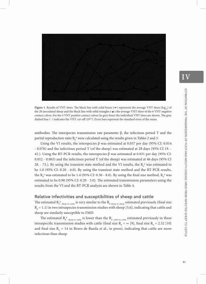
Estim
atio
n o
f thE
tra
nsm
ission
of fo
ot-a
nd
-mo
uth
disE
asE
vir
us fr
om
infE
ctE
d sh
EE
p to c
attlE
IV
Figure 1. Results of VNT titres. The black line with solid boxes ( ■ ) represents the average VNT titres (log10) of the 20 inoculated sheep and the black line with solid triangles ( ▲ ) the average VNT titres of the 6 VNT negative contact calves. For the 4 VNT positive contact calves (in grey lines) the individual VNT titres are shown. The grey dashed line (--) indicates the VNT cut-off (100.6). Error bars represent the standard error of the mean.
antibodies. The interspecies transmission rate parameter β, the infectious period T and the partial reproduction ratio R0
p were calculated using the results given in Tables 2 and 3.Using the VI results, the interspecies β was estimated at 0.037 per day (95% CI: 0.014
- 0.076) and the infectious period T (of the sheep) was estimated at 28 days (95% CI 19. - 42.). Using the RT-PCR results, the interspecies β was estimated at 0.031 per day (95% CI: 0.012 - 0.065) and the infectious period T (of the sheep) was estimated at 46 days (95% CI 28. - 73.). By using the transient state method and the VI results, the R0
p was estimated to be 1.0 (95% CI: 0.20 - 6.0). By using the transient state method and the RT-PCR results, the R0
p was estimated to be 1.4 (95% CI: 0.30 - 8.0). By using the final size method, R0p was
estimated to be 0.90 (95% CI: 0.20 - 3.0). The estimated transmission parameters using the results from the VI and the RT-PCR analysis are shown in Table 4.
relative infectivities and susceptibilities of sheep and cattleThe estimated R0
p sheep-to-cattle is very similar to the R0 sheep-to-sheep estimated previously (final size R0 = 1.1) in two intraspecies transmission studies with sheep [5,6], indicating that cattle and sheep are similarly susceptible to FMD.
The estimated R0p sheep-to-cattle is lower than the R0 cattle-to-cattle estimated previously in three
intraspecific transmission studies with cattle (final size R0 = ∞ [9], final size R0 = 2.52 [10] and final size R0 = 14 in Bravo de Rueda et al., in press), indicating that cattle are more infectious than sheep.
81

Estim
atio
n o
f thE
tra
nsm
ission
of fo
ot-a
nd
-mo
uth
disE
asE
vir
us fr
om
infE
ctE
d sh
EE
p to c
attlE
IV
Table 1 Results of the virus isolation, RT-PCR, serology and clinical inspection.
Animal Virus isolation RT-PCR Serology FMD clinical signs
Contact infectionRoom Species Nr Oral swabs Blood Oral swabs Probang NS- ELISA VNT Fevera Vesicles Lameness
1 Calf 5439 - - - - - - - - - No
Lamb 5440 + + + + + + + + +
Lamb 5441 + + + - + + + + +
2 Calf 5442 - - - + + + - - - Yes
Lamb 5443 + + + - + + + - -
Lamb 5444 + - + + + + - - -
3 Calf 5445 - - - - - - - - - No
Lamb 5446 + - + - + + - - -
Lamb 5447 + - + - + + - - -
4 Calf 5448 - - - - - - - - - No
Lamb 5449 + + + + + + + + -
Lamb 5450 + + + - + + + - -
5 Calf 5451 - - - - - - - - - No
Lamb 5452 + + + + + + - - -
Lamb 5453 + + + + + + - + -
6 Calf 5454 - - - - - - - - - No
Lamb 5455 + + + - + + + + +
Lamb 5456 + + + + + + + + +
7 Calf 5457 + + + - + + + + - Yes
Lamb 5458 + + + - + + + + +
Lamb 5459 + + + - + + - + +
8 Calf 5460 - - - - - - - - - No
Lamb 5461 + + + - + + + - +
Lamb 5462 + + + - + + + - +
9 Calf 5463 - - + - + + - - - Yes
Lamb 5464 + + + + + + + + -
Lamb 5465 + - + + + + - - -
10 Calf 5466 - - - - + + - - - Yes
Lamb 5467 + + + - + + + + +
Lamb 5468 + + + + + + + + +
+/−, positive /negative in one or more of the tested samples. a fever in sheep: body temperature above 40 °C; fever in cattle: body temperature above 39.5 °C.
82

Estim
atio
n o
f thE
tra
nsm
ission
of fo
ot-a
nd
-mo
uth
disE
asE
vir
us fr
om
infE
ctE
d sh
EE
p to c
attlE
IV
Table 1 Results of the virus isolation, RT-PCR, serology and clinical inspection.
Animal Virus isolation RT-PCR Serology FMD clinical signs
Contact infectionRoom Species Nr Oral swabs Blood Oral swabs Probang NS- ELISA VNT Fevera Vesicles Lameness
1 Calf 5439 - - - - - - - - - No
Lamb 5440 + + + + + + + + +
Lamb 5441 + + + - + + + + +
2 Calf 5442 - - - + + + - - - Yes
Lamb 5443 + + + - + + + - -
Lamb 5444 + - + + + + - - -
3 Calf 5445 - - - - - - - - - No
Lamb 5446 + - + - + + - - -
Lamb 5447 + - + - + + - - -
4 Calf 5448 - - - - - - - - - No
Lamb 5449 + + + + + + + + -
Lamb 5450 + + + - + + + - -
5 Calf 5451 - - - - - - - - - No
Lamb 5452 + + + + + + - - -
Lamb 5453 + + + + + + - + -
6 Calf 5454 - - - - - - - - - No
Lamb 5455 + + + - + + + + +
Lamb 5456 + + + + + + + + +
7 Calf 5457 + + + - + + + + - Yes
Lamb 5458 + + + - + + + + +
Lamb 5459 + + + - + + - + +
8 Calf 5460 - - - - - - - - - No
Lamb 5461 + + + - + + + - +
Lamb 5462 + + + - + + + - +
9 Calf 5463 - - + - + + - - - Yes
Lamb 5464 + + + + + + + + -
Lamb 5465 + - + + + + - - -
10 Calf 5466 - - - - + + - - - Yes
Lamb 5467 + + + - + + + + +
Lamb 5468 + + + + + + + + +
+/−, positive /negative in one or more of the tested samples. a fever in sheep: body temperature above 40 °C; fever in cattle: body temperature above 39.5 °C.
83

Estim
atio
n o
f thE
tra
nsm
ission
of fo
ot-a
nd
-mo
uth
disE
asE
vir
us fr
om
infE
ctE
d sh
EE
p to c
attlE
IV
Table 2 FMDV virus titres in oral swab, blood, urine and faeces samples.
Animal Nr
Days post infection Days post infection
0 1 2 3 4 5 6 7 8 9 10 11 12 13 14 15 16 17 18 19 20 21 22 23 24 25 26 27 28 29 30 31
Calf 5439 -a - toxb - - - - - - - - N.A.c - - - - - - - - - - - - - - - - - - - -
Lamb 5440 - 2.6d tox 3.8/Vd
1.6 - 0.9 - - - 0.7 N.A. - - - 1.1 - - 1.7 1.1 0.7 - 1.0 - - 1.2 - 1.4 - - - -
Lamb 5441 - 1.3 tox 2.1/V 1.3 1.4 - - - - - N.A. - - - - - - - - - - - - - - - - - - - -
Calf 5442 - - tox -*e - - - - - - - N.A. - - - - - - - - - - - - - - - - - - - -
Lamb 5443 - - tox/V 2.8/V -/V 1.0 0.4 - - - - N.A. - - - - - - - - 0.4 - - - 1.2 - 0.4 - - - - -
Lamb 5444 - 2.2 tox - 0.4 1.0 0.9 - - - 1.0 N.A. 1.5 - 0.4 1.8 - 1.5 2.1 1.2 0.9 - 1.1 - 1.7 - 1.0 0.4 - - - -
Calf 5445 - - tox - - - - - - - - N.A. - - - - - - - - - - - - - - - - - - - -
Lamb 5446 - 0.4 tox 4.0 3.3 2.2 0.4 - 0.4 - - N.A. - - 1.0 1.1 - 2.5 N.A. 0.4 - - - - - - 1.3 - - - - -
Lamb 5447 - 2.7 tox 0.4 2.4 2.4 - - - - - N.A. - - 2.1 1.3 0.7 0.4 - 0.7 0.4 - - - - - 0.4 - - - - -
Calf 5448 - - tox - - - - - - - - N.A. - - - - - - - - - - - - - - - - - - - -
Lamb 5449 - 2.1/V tox/V 4.2/V - 0.9 - - - - - N.A. - - - - - - - - - - - - - - - - - - - -
Lamb 5450 - 3.7 tox/V 2.3/V 1.4 1.5 - - - - - N.A. - - - 1.7 - 1.2 0.4 - 1.7 - - - 2.1 - 1.0 - - - - -
Calf 5451 - - tox - - - - - - - - N.A. - - - - - - - - - - - - - - - - - - - -
Lamb 5452 - -/V tox/V 4.0 1.6 - 0.4 - - - - N.A. - - - - - 2.2 2.0 1.7 0.4 1.8 1.9 - - 0.9 1.3 1.3 1.6 - - 1.0
Lamb 5453 - 0.4/V tox/V 2.3/V 1.9 1.4 1.1 - - - 0.7 N.A. - - 1.9 1.5 0.7 0.4 - - - - - - - - 1.5 - - - - -
Calf 5454 - - tox - - - - - - - - N.A. - - - - - - - - - - - - - - - - - - - -
Lamb 5455 - - tox 0.7/V 0.7/V 1.9 0.4 - - - - N.A. - - 1.4 0.9 1.4 1.5 1.4 - - - 0.9 - - - - 1.4 - - 1.2 -
Lamb 5456 - 1.6 tox 2.7/V 2.1/V 1.0 0.4 - - - - N.A. - - 1.0 0.4 - 0.9 1.4 1.5 - - 0.4 - - - 0.9 - - 1.7 - 0.7
Calf 5457 - - tox - - - - 3.1/V* 3.4 4.3/Vf 4.3f N.A. - - - - - - - - - - - - - - - - - - - -
Lamb 5458 - 0.7 tox 3.9/V 0.9 0.9 1.2 - - -g - N.A. - - 1.3 1.9 2.5 2.8 2.5 0.4 2.1 2.6 2.4 1.5 1.6 - 1.7 - 2.3 - 0.4 2.4
Lamb 5459 - 1.6 tox 2.8/V 2.2 0.4 - - - - 2.3 N.A. - 0.9 2.6 - 1.2 1.2 1.8 1.9 - 1.5 2.3 - 1.2 1.7 1.4 - 0.7 - 1.6 -
Calf 5460 - - tox - - - - - - - - N.A. - - - - - - - - - - - - - - - - - - - -
Lamb 5461 - 0.4/V tox/V 2.7/V 1.2 0.7 - - - - - N.A. - - 0.4 1.4 - 0.9 - - - 1.2 1.7 - - - - - - - - -
Lamb 5462 - 2.1/V tox/V 4.0/V 1.7 1.9 - - - - - N.A. - - - - - - 0.9 - - - 1.1 - - - - - 1.6 - - -
Calf 5463 - - - - - - - - - - -* N.A. - - - - - - - - - - - - - - - - - - - -
Lamb 5464 - 1.7 2.5 3.7 2.1/V 0.9/V - - - - - N.A. - 1.7 1.5 1.2 2.6 0.9 1.2 2.3 - - 1.8 - 1.2 2.2 2.5 1.7 0.4 - - -
Lamb 5465 - - 2.1 - 0.4 2.2 - - - 1.5 - N.A. - 2.1 1.1 1.3 2.7 1.2 2.0 1.2 - - 1.2 - - - 0.7 - - - - -
Calf 5466 - - - - - - - - - - -* N.A. - - - - - - - - - - - - - - - - - - - -
Lamb 5467 - -/V 3.0/V 2.7/V 2.8 1.8 - - - - - N.A. - - - - - - - - - - - - - - - - - - - -
Lamb 5468 - 4.0/V 3.0/V -/V 1.4/V 1.0 - - - - - N.A. - - 1.0 0.9 0.4 1.2 0.4 - - 1.8 2.0 2.1 - - - - - - - -
aoral swab sample that scored positive for FMDV by virus isolation (VI) (log10 pfu/mL); −: no virus was detected.btox: toxic oral swab sample, no VI result available. cN.A.: results not-available for oral swab samples.
dV = viraemia: blood sample that scored positive for FMDV by VI.e* indicates the (estimated) day of infection of the contact calves 5442, 5457, 5463 and 5466.furine sample that scored positive for FMDV by VI.gfaeces sample that scored positive for FMDV by VI.
84

Estim
atio
n o
f thE
tra
nsm
ission
of fo
ot-a
nd
-mo
uth
disE
asE
vir
us fr
om
infE
ctE
d sh
EE
p to c
attlE
IV
Table 2 FMDV virus titres in oral swab, blood, urine and faeces samples.
Animal Nr
Days post infection Days post infection
0 1 2 3 4 5 6 7 8 9 10 11 12 13 14 15 16 17 18 19 20 21 22 23 24 25 26 27 28 29 30 31
Calf 5439 -a - toxb - - - - - - - - N.A.c - - - - - - - - - - - - - - - - - - - -
Lamb 5440 - 2.6d tox 3.8/Vd
1.6 - 0.9 - - - 0.7 N.A. - - - 1.1 - - 1.7 1.1 0.7 - 1.0 - - 1.2 - 1.4 - - - -
Lamb 5441 - 1.3 tox 2.1/V 1.3 1.4 - - - - - N.A. - - - - - - - - - - - - - - - - - - - -
Calf 5442 - - tox -*e - - - - - - - N.A. - - - - - - - - - - - - - - - - - - - -
Lamb 5443 - - tox/V 2.8/V -/V 1.0 0.4 - - - - N.A. - - - - - - - - 0.4 - - - 1.2 - 0.4 - - - - -
Lamb 5444 - 2.2 tox - 0.4 1.0 0.9 - - - 1.0 N.A. 1.5 - 0.4 1.8 - 1.5 2.1 1.2 0.9 - 1.1 - 1.7 - 1.0 0.4 - - - -
Calf 5445 - - tox - - - - - - - - N.A. - - - - - - - - - - - - - - - - - - - -
Lamb 5446 - 0.4 tox 4.0 3.3 2.2 0.4 - 0.4 - - N.A. - - 1.0 1.1 - 2.5 N.A. 0.4 - - - - - - 1.3 - - - - -
Lamb 5447 - 2.7 tox 0.4 2.4 2.4 - - - - - N.A. - - 2.1 1.3 0.7 0.4 - 0.7 0.4 - - - - - 0.4 - - - - -
Calf 5448 - - tox - - - - - - - - N.A. - - - - - - - - - - - - - - - - - - - -
Lamb 5449 - 2.1/V tox/V 4.2/V - 0.9 - - - - - N.A. - - - - - - - - - - - - - - - - - - - -
Lamb 5450 - 3.7 tox/V 2.3/V 1.4 1.5 - - - - - N.A. - - - 1.7 - 1.2 0.4 - 1.7 - - - 2.1 - 1.0 - - - - -
Calf 5451 - - tox - - - - - - - - N.A. - - - - - - - - - - - - - - - - - - - -
Lamb 5452 - -/V tox/V 4.0 1.6 - 0.4 - - - - N.A. - - - - - 2.2 2.0 1.7 0.4 1.8 1.9 - - 0.9 1.3 1.3 1.6 - - 1.0
Lamb 5453 - 0.4/V tox/V 2.3/V 1.9 1.4 1.1 - - - 0.7 N.A. - - 1.9 1.5 0.7 0.4 - - - - - - - - 1.5 - - - - -
Calf 5454 - - tox - - - - - - - - N.A. - - - - - - - - - - - - - - - - - - - -
Lamb 5455 - - tox 0.7/V 0.7/V 1.9 0.4 - - - - N.A. - - 1.4 0.9 1.4 1.5 1.4 - - - 0.9 - - - - 1.4 - - 1.2 -
Lamb 5456 - 1.6 tox 2.7/V 2.1/V 1.0 0.4 - - - - N.A. - - 1.0 0.4 - 0.9 1.4 1.5 - - 0.4 - - - 0.9 - - 1.7 - 0.7
Calf 5457 - - tox - - - - 3.1/V* 3.4 4.3/Vf 4.3f N.A. - - - - - - - - - - - - - - - - - - - -
Lamb 5458 - 0.7 tox 3.9/V 0.9 0.9 1.2 - - -g - N.A. - - 1.3 1.9 2.5 2.8 2.5 0.4 2.1 2.6 2.4 1.5 1.6 - 1.7 - 2.3 - 0.4 2.4
Lamb 5459 - 1.6 tox 2.8/V 2.2 0.4 - - - - 2.3 N.A. - 0.9 2.6 - 1.2 1.2 1.8 1.9 - 1.5 2.3 - 1.2 1.7 1.4 - 0.7 - 1.6 -
Calf 5460 - - tox - - - - - - - - N.A. - - - - - - - - - - - - - - - - - - - -
Lamb 5461 - 0.4/V tox/V 2.7/V 1.2 0.7 - - - - - N.A. - - 0.4 1.4 - 0.9 - - - 1.2 1.7 - - - - - - - - -
Lamb 5462 - 2.1/V tox/V 4.0/V 1.7 1.9 - - - - - N.A. - - - - - - 0.9 - - - 1.1 - - - - - 1.6 - - -
Calf 5463 - - - - - - - - - - -* N.A. - - - - - - - - - - - - - - - - - - - -
Lamb 5464 - 1.7 2.5 3.7 2.1/V 0.9/V - - - - - N.A. - 1.7 1.5 1.2 2.6 0.9 1.2 2.3 - - 1.8 - 1.2 2.2 2.5 1.7 0.4 - - -
Lamb 5465 - - 2.1 - 0.4 2.2 - - - 1.5 - N.A. - 2.1 1.1 1.3 2.7 1.2 2.0 1.2 - - 1.2 - - - 0.7 - - - - -
Calf 5466 - - - - - - - - - - -* N.A. - - - - - - - - - - - - - - - - - - - -
Lamb 5467 - -/V 3.0/V 2.7/V 2.8 1.8 - - - - - N.A. - - - - - - - - - - - - - - - - - - - -
Lamb 5468 - 4.0/V 3.0/V -/V 1.4/V 1.0 - - - - - N.A. - - 1.0 0.9 0.4 1.2 0.4 - - 1.8 2.0 2.1 - - - - - - - -
aoral swab sample that scored positive for FMDV by virus isolation (VI) (log10 pfu/mL); −: no virus was detected.btox: toxic oral swab sample, no VI result available. cN.A.: results not-available for oral swab samples.
dV = viraemia: blood sample that scored positive for FMDV by VI.e* indicates the (estimated) day of infection of the contact calves 5442, 5457, 5463 and 5466.furine sample that scored positive for FMDV by VI.gfaeces sample that scored positive for FMDV by VI.
85

Estim
atio
n o
f thE
tra
nsm
ission
of fo
ot-a
nd
-mo
uth
disE
asE
vir
us fr
om
infE
ctE
d sh
EE
p to c
attlE
IV
Table 3 FMDV RT-PCR results in oral swab samples.
Days post infection Days post infection
Animal Nr 0 1 2 3 4 5 6 7 8 9 10 11 12 13 14 15 16 17 18 19 20 21 22 23 24 25 26 27 28 29 30 31
Calf 5439 -a - - - - - - - - - - N.A.b - - - - - - - - - - - - - - - - - - - -
Lamb 5440 - + + + + + + - - - - N.A. - + + + + + + + + + + + + + + + - - + -
Lamb 5441 - + - + + + + - - - - N.A. - - - - - - - - - - - - - - - - - - - -
Calf 5442 - - - - - - - - - - - N.A. - - - - - - - - - - - - - - - - - - - -
Lamb 5443 - + - + + + - - - + + N.A. - + - + - - - - + - - + + - + - - - + -
Lamb 5444 - + + - + + + - - + + N.A. + - + + - + + + + + + + + - + + + - - -
Calf 5445 - - - - - - - - - - - N.A. - - - - - - - - - - - - - - - - - - - -
Lamb 5446 - + - + + + + + + + + N.A. + - + + + + N.A. + + + + + + - + + + - - +
Lamb 5447 - + + - + + + - - - - N.A. - - + + + + + + + + - + - - - - - - - +
Calf 5448 - - - - - - - - - - - N.A. - - - - - - - - - - - - - - - - - - - -
Lamb 5449 - + + + + + + - - - - N.A. - - - - - + - + + + + - + + + - - - - -
Lamb 5450 - + - + + + + + - - - N.A. + - + + - + + - + + + - + + + - - - - -
Calf 5451 - - - - - - - - - - - N.A. - - - - - - - - - - - - - - - - - - - -
Lamb 5452 - - + + + - + - - - - N.A. - + + + + + + + + + + + + + + + + - + +
Lamb 5453 - - + + + + + + - + + N.A. + + + + + + + - + - + + + + + + + - + -
Calf 5454 - - - - - - - - - - - N.A. - - - - - - - - - - - - - - - - - - - -
Lamb 5455 - + + + + + - - - - - N.A. - + + + + + + + + + + + + - + + - - + +
Lamb 5456 - + + + + + + + - + + N.A. + + + + + + + + - + + + + + + - + + - +
Calf 5457 - - - - - - - + + + + N.A. + + - - - - - - - - - - - - - - - - - -
Lamb 5458 - + - + + + + - - - + N.A. - + + + + + + + + + + + + - + - + - + +
Lamb 5459 - + + + + + + - - + + N.A. + + + + + + + + + + + + + + + - + - + -
Calf 5460 - - - - - - - - - - - N.A. - - - - - - - - - - - - - - - - - - - -
Lamb 5461 - + - + + + - - - - - N.A. - + + + - + + + - + + + - + + + - + + +
Lamb 5462 - + + + + + - - - - - N.A. - - + - + + + - - - - - + - - - + - - -
Calf 5463 - - - - - - - - - - + N.A. + - - - - - - - - - - - - - - - - - - -
Lamb 5464 - + + + + + - - - + + N.A. + + + + + + + + + + + + + + + + + + + +
Lamb 5465 - + + - + + - - - + - N.A. + + + + + + + + + - + + + - - - - - + -
Calf 5466 - - - - - - - - - - - N.A. - - - - - - - - - - - - - - - - - - - -
Lamb 5467 - + + + + + - - - - - N.A. - - - - - - - - - - - - - - - - - - - -
Lamb 5468 - + + + + + + + + + + N.A. + + + + + + + + + + + + + + + - + + + -
aoral swab sample that scored positive for FMDV by RT-PCR; −: no virus was detected and +: virus detected. bN.A.: results not-available for oral swab samples.
86

Estim
atio
n o
f thE
tra
nsm
ission
of fo
ot-a
nd
-mo
uth
disE
asE
vir
us fr
om
infE
ctE
d sh
EE
p to c
attlE
IV
Table 3 FMDV RT-PCR results in oral swab samples.
Days post infection Days post infection
Animal Nr 0 1 2 3 4 5 6 7 8 9 10 11 12 13 14 15 16 17 18 19 20 21 22 23 24 25 26 27 28 29 30 31
Calf 5439 -a - - - - - - - - - - N.A.b - - - - - - - - - - - - - - - - - - - -
Lamb 5440 - + + + + + + - - - - N.A. - + + + + + + + + + + + + + + + - - + -
Lamb 5441 - + - + + + + - - - - N.A. - - - - - - - - - - - - - - - - - - - -
Calf 5442 - - - - - - - - - - - N.A. - - - - - - - - - - - - - - - - - - - -
Lamb 5443 - + - + + + - - - + + N.A. - + - + - - - - + - - + + - + - - - + -
Lamb 5444 - + + - + + + - - + + N.A. + - + + - + + + + + + + + - + + + - - -
Calf 5445 - - - - - - - - - - - N.A. - - - - - - - - - - - - - - - - - - - -
Lamb 5446 - + - + + + + + + + + N.A. + - + + + + N.A. + + + + + + - + + + - - +
Lamb 5447 - + + - + + + - - - - N.A. - - + + + + + + + + - + - - - - - - - +
Calf 5448 - - - - - - - - - - - N.A. - - - - - - - - - - - - - - - - - - - -
Lamb 5449 - + + + + + + - - - - N.A. - - - - - + - + + + + - + + + - - - - -
Lamb 5450 - + - + + + + + - - - N.A. + - + + - + + - + + + - + + + - - - - -
Calf 5451 - - - - - - - - - - - N.A. - - - - - - - - - - - - - - - - - - - -
Lamb 5452 - - + + + - + - - - - N.A. - + + + + + + + + + + + + + + + + - + +
Lamb 5453 - - + + + + + + - + + N.A. + + + + + + + - + - + + + + + + + - + -
Calf 5454 - - - - - - - - - - - N.A. - - - - - - - - - - - - - - - - - - - -
Lamb 5455 - + + + + + - - - - - N.A. - + + + + + + + + + + + + - + + - - + +
Lamb 5456 - + + + + + + + - + + N.A. + + + + + + + + - + + + + + + - + + - +
Calf 5457 - - - - - - - + + + + N.A. + + - - - - - - - - - - - - - - - - - -
Lamb 5458 - + - + + + + - - - + N.A. - + + + + + + + + + + + + - + - + - + +
Lamb 5459 - + + + + + + - - + + N.A. + + + + + + + + + + + + + + + - + - + -
Calf 5460 - - - - - - - - - - - N.A. - - - - - - - - - - - - - - - - - - - -
Lamb 5461 - + - + + + - - - - - N.A. - + + + - + + + - + + + - + + + - + + +
Lamb 5462 - + + + + + - - - - - N.A. - - + - + + + - - - - - + - - - + - - -
Calf 5463 - - - - - - - - - - + N.A. + - - - - - - - - - - - - - - - - - - -
Lamb 5464 - + + + + + - - - + + N.A. + + + + + + + + + + + + + + + + + + + +
Lamb 5465 - + + - + + - - - + - N.A. + + + + + + + + + - + + + - - - - - + -
Calf 5466 - - - - - - - - - - - N.A. - - - - - - - - - - - - - - - - - - - -
Lamb 5467 - + + + + + - - - - - N.A. - - - - - - - - - - - - - - - - - - - -
Lamb 5468 - + + + + + + + + + + N.A. + + + + + + + + + + + + + + + - + + + -
aoral swab sample that scored positive for FMDV by RT-PCR; −: no virus was detected and +: virus detected. bN.A.: results not-available for oral swab samples.
87

Estim
atio
n o
f thE
tra
nsm
ission
of fo
ot-a
nd
-mo
uth
disE
asE
vir
us fr
om
infE
ctE
d sh
EE
p to c
attlE
IV
disCussion
The purpose of this study was to estimate transmission of FMDV from infected sheep to contact cattle and, together with results from previous studies, to identify differences in either susceptibility to FMD or infectivity of FMD infected sheep and cattle. Our study shows that FMDV transmission from sheep to cattle occurs, but the estimated partial reproduction ratio (R0
p) indicates that the expected number of secondary cases in calves, caused by infected lambs, is relatively low. Moreover, the susceptibility of sheep to FMD seems to be similar to the susceptibility of cattle to FMD. This finding is supported by French et al. [29] who found overlapping distributions when analysing dose–response relationships in cattle and sheep exposed to FMDV in aerosols. The fact that cattle and sheep have a similar susceptibility to FMD and the fact that the transmission (R0) from cattle to cattle is higher than the transmission (R0
p) from sheep to cattle, indicate that cattle are more infectious than sheep. Thus, cattle play the major role in the transmission of FMDV in a mixed population with sheep and cattle. These relative infectivities and susceptibilities are useful for modelling FMD spread such as for example in Backer et al. [16]. In their model they assumed that the susceptibility of cattle herds is twice the susceptibility of sheep herds. Our results can be used to update such FMD spread models, and more importantly, could be a reason to implement different control strategies for both animal species.
We estimated a partial reproduction ratio for sheep-to-cattle transmission. This estimate alone does not reflect transmission for an entire mixed population consisting of sheep and cattle. In such a population, cattle-to-cattle, sheep-to-sheep, sheep-to-cattle and cattle-to-sheep transmission can take place. For the estimation of transmission in a mixed population, more information and/or other mathematical techniques are required [15]. Even though sheep play a more limited role in transmitting FMDV as compared to cattle, the reproduction ratio in a mixed population of sheep and cattle can still be larger than 1, meaning that major outbreaks can occur. Probably, the R0 for a mixed population of cattle and sheep will be higher if a higher proportion of cattle are present.
Previously, we studied transmission of FMDV between cattle [9-11] and between sheep [6] using FMDV strain O/NET/2001. However, different strains of FMDV might affect different species and might have different transmission characteristics. In more recent studies, we therefore
Table 4 Estimated transmission parameters using the results from the Virus isolation (VI) and the RT-PCR analysis.
Transmission rate parameter (interspecies β) Infectious period (T)
Partial reproduction ratio (R0p)
Transient state method Final size method
β (day−1) 95% CI T (days) 95% CI R0p 95% CI R0
p 95%CI
VI 0.037 0.014 - 0.076 28. 19. - 42. 1.0 0.20 - 6.0
RT-PCR 0.031 0.012 - 0.065 46. 28. - 73. 1.4 0.30 - 8.0
0.90 0.20 -3.0
88

Estim
atio
n o
f thE
tra
nsm
ission
of fo
ot-a
nd
-mo
uth
disE
asE
vir
us fr
om
infE
ctE
d sh
EE
p to c
attlE
IV
used another serotype of FMDV to study transmission of FMDV. We chose FMDV Asia-1 because this serotype spread towards mainland Europe [30,31]. We observed transmission of FMDV Asia-1 between sheep [5] and between cattle (Bravo de Rueda et al., in press), and now studied transmission between sheep and cattle. The R0 values obtained in the studies using serotype O and Asia-1 are not significantly different. Still, differences might exist for other serotypes.
In this study, we investigated within pen transmission. The animals in this study were in close proximity. Extrapolation of experimental data to field conditions should always be done with care. However, the relative infectivity and susceptibility will not change under field conditions. In field conditions, the estimated R0
p will probably be lower because it is known that between-pen transmission is lower than within-pen transmission [32-34]. Additionally, between-herd transmission will most likely be even lower [35].
The relative low R0p in the transmission of FMDV from sheep to cattle can have
implications for control measures implemented during an outbreak, e.g. whether or not to use vaccination in sheep, given the fact that vaccination against FMD is very effective in cattle [9,10]. If all cattle were vaccinated and thus became less infectious, then vaccination of sheep would not have an additional contribution to FMD control, especially when other control measures are implemented e.g. movement restrictions.
The observed relatively low infectivity of sheep is remarkable if we take into consideration that the duration of the secretion and excretion of FMDV in sheep (specifically in oral swabs) is much longer than in cattle. The mean duration of secretion and excretion of FMDV in sheep, in this study, was 28 days (VI results from oral swab samples). A similarly long period was shown by Eblé et al. [5], who showed that sheep secrete and excrete FMDV for longer than 30 days. In contrast, calves infected with the same strain of FMDV, secrete and excrete FMDV for on average 5.0 days (VI results from oral swab samples in Bravo de Rueda et al., in press). It was already known that sheep are long- term secretors and excretors of FMDV [3,36]. Nevertheless the results reported here show that this long-term secretion and excretion of FMDV in sheep does not enhance transmission of the infection from sheep to cattle. In our study, transmission events took place mainly during the first week after infection. This is in accordance with what others have observed in sheep [5-8] and in cattle [9,10].
In our study as well as in the above-mentioned studies, it was observed that FMDV is secreted and excreted in higher quantities during the first week post infection. Previous research showed that virus titres in upper respiratory tract samples from sheep are lower than those in cattle [4]. The ability of cattle to shed more virus than sheep could (partially) explain the observed difference in the infectivity of sheep and cattle. Moreover, in FMDV infected cattle, profuse salivation and nasal discharge are often observed [37]. Compared to cattle, salivation and nasal discharge after FMDV infection in sheep is less profuse i.e. sheep show less severe clinical signs [1,38,39]. Therefore it can be assumed that profuse secretion and excretion of the virus contributes to a higher contamination of the environment with FMDV. A recent analysis showed that FMDV transmission occurs for a large part through the environment (Bravo de Rueda et al., in press), and thus more new cases of FMD will take place if animals would shed more infected secretions and excretions.
89

Estim
atio
n o
f thE
tra
nsm
ission
of fo
ot-a
nd
-mo
uth
disE
asE
vir
us fr
om
infE
ctE
d sh
EE
p to c
attlE
IV
We conclude that despite the ability of sheep to secrete and excrete FMDV for a relatively long period of time, sheep are less infectious than cattle. The observed differences in the relative susceptibility and infectivity of sheep and cattle could be a reason to implement different control strategies for both animal species.
additionaL FiLes
Additional file 1 Calculating reproduction ratio R0 in two-to-one transmission experiments. This additional file shows the analytical derivation of the maximum likelihood estimate (MLE) of R0 (RMLE) in a two-to-one experimental transmission design.
introductionIn disease transmission, quantifying the average number of newly infected individuals caused by an infectious individual during its entire infectious period (Reproduction ratio, R0) is important. When several infectious and susceptible individuals are mixed in an experiment, both infectious and susceptible individuals should have a similar status e.g. both be vaccinated, or e.g. be the same species. To avoid this problem transmission experiments can also be performed in a one-to-one transmission experiment set up [21,24]. In one-to-one transmission experiments, the expression for R0 has been derived analytically, R0 = 2 p/(1-p) where p is the total number of infection events divided by the number of independent replications. Given the rates of infection and recovery (see Additional file 2), R0 is β times the infectious period (1/α) [21,28]. Similarly to a one-to-one transmission experiment, in this paper we derive R0 for a two-to-one transmission experiment.
maximum Likelihood estimate for r0 in a transmission experiment using 2 infectious and 1 susceptible individualsThe two-to-one transmission experiment can graphically be represented as an SI (susceptible-infected) plane. An additional file shows this graphical SI plane (see Additional file 2).
The probability that both infected individuals recover and the susceptible individual escapes infection (St = 1 and Nt = 3) is:
So the probability that the susceptible individual becomes infected is:
and .
90

Estim
atio
n o
f thE
tra
nsm
ission
of fo
ot-a
nd
-mo
uth
disE
asE
vir
us fr
om
infE
ctE
d sh
EE
p to c
attlE
IV
Where p̂ is the MLE of p, the total number of infection events divided by the number of independent replications and R̂0 is the MLE of R0.
Note that in this case the estimated R0 is a partial reproduction ratio (R0p) as infectious
animals are all sheep and susceptible animals are all cattle.
Additional file 2 The two-to-one transmission experiment is graphically represented as an SI (susceptible-infected) plane. This additional file is part of Additional file 1. This graph shows how a two-to-one transmission experiment can be represented using an infectious-susceptible plane. β is the transmission rate parameter, St is the number of susceptible animals, It is the number of infectious animals, Nt is the total number of animals and, α the recovery rate.
List oF reFerenCes
1 Kitching RP, Hughes GJ: Clinical variation in foot and mouth disease: sheep and goats. Revue Scientifique Technique 2002, 21:505–512.
2 Burrows R: The persistence of foot-and-mouth disease virus in sheep. Journal of Hygiene (London) 1968, 66:633–640.
3 Donaldson AI, Sellers RF: Foot-and-mouth disease. In Diseases of Sheep. 3rd edition. Edited by Martin WB, Aitken ID. Oxford: Blackwell science; 2000:254–258.
4 Bravo de Rueda C, Dekker A, Eblé PL, de Jong MCM: Identification of factors associated with increased excretion of foot-and-mouth disease virus. Preventive Veterinary Medicine 2014, 113:23–33.
5 Eblé PL, Orsel K, Dekker A: FMDV infection in vaccinated and non-vaccinated sheep: transmission to contact animals and diagnostic aspects. In Session of the Research Group of the Standing Technical Committee of EuFMD; 29–31
91

Estim
atio
n o
f thE
tra
nsm
ission
of fo
ot-a
nd
-mo
uth
disE
asE
vir
us fr
om
infE
ctE
d sh
EE
p to c
attlE
IV
October 2012. Jerez de la Frontera: FAO of the United Nations; 2012.
6 Orsel K, Dekker A, Bouma A, Stegeman JA, de Jong MCM: Quantification of foot and mouth disease virus excretion and transmission within groups of lambs with and without vaccination. Vaccine 2007, 25:2673–2679.
7 Cox SJ, Barnett PV, Dani P, Salt JS: Emergency vaccination of sheep against foot-and-mouth disease: protection against disease and reduction in contact transmission. Vaccine 1999, 17:1858–1868.
8 Parida S, Fleming L, Oh Y, Mahapatra M, Hamblin P, Gloster J, Paton DJ: Emergency vaccination of sheep against foot-and-mouth disease: significance and detection of subsequent sub-clinical infection. Vaccine 2008, 26:3469–3479.
9 Orsel K, de Jong MCM, Bouma A, Stegeman JA, Dekker A: The effect of vaccination on foot and mouth disease virus transmission among dairy cows. Vaccine 2007, 25:327–335.
10 Orsel K, Dekker A, Bouma A, Stegeman JA, de Jong MCM: Vaccination against foot and mouth disease reduces virus transmission in groups of calves. Vaccine 2005, 23:4887–4894.
11 Bouma A, Dekker A, de Jong MCM: No foot-and-mouth disease virus transmission between individually housed calves. Veterinary Microbiology 2004, 98:29–36.
12 Donaldson A: The role of sheep in the epidemiology of foot-and-mouth disease and proposasls for control and eradication in animal populations with a high density of sheep. In Session of the Research Group of the Standing Technical Committee of EuFMD 5–8 September 2000. Borovets: FAO of the United Nations; 2000.
13 Blanco E, Romero LJ, El Harrach M, Sanchez-Vizcaino JM: Serological evidence of FMD subclinical infection in sheep population during the 1999 epidemic in Morocco. Veterinary Microbiology 2002, 85:13–21.
14 Gibbens JC, Sharpe CE, Wilesmith JW, Mansley LM, Michalopoulou E, Ryan JB, Hudson M: Descriptive epidemiology of the 2001 foot-and-mouth disease epidemic in Great Britain: the first five months. Veterinary Records 2001, 149:729–743.
15 Diekmann O, Heesterbeek JA, Metz JA: On the definition and the computation of the basic reproduction ratio R0 in models for infectious diseases in heterogeneous populations. Journal Mathematical Biology 1990, 28:365–382.
16 Backer JA, Hagenaars TJ, Nodelijk G, van Roermund HJ: Vaccination against foot-and-
mouth disease I: epidemiological consequences. Preventive Veterinary Medicine 2012, 107:27–40.
17 Hajer R, Hendrikse J, Rutgers LJE, Sloet van Oldruitenborgh-Oosterbaan MM, van der Weyden GC: Het klinisch onderzoek bij grote huisdieren. Bunge: Utrecht; 1985.
18 Weesendorp E, Stegeman A, Loeffen W: Dynamics of virus excretion via different routes in pigs experimentally infected with classical swine fever virus strains of high, moderate or low virulence. Veterinary Microbiology 2009, 133:9–22.
19 Moonen P, Boonstra J, van der Honing RH, Leendertse CB, Jacobs L, Dekker A: Validation of a LightCycler-based reverse transcription polymerase chain reaction for the detection of foot-and-mouth disease virus. Journal of Virological Methods 2003, 113:35–41.
20 Dekker A, Terpstra C: Prevalence of foot-and-mouth disease antibodies in dairy herds in The Netherlands, four years after vaccination. Research in Veterinary Sciences 1996, 61:89–91.
21 Velthuis AG, De Jong MCM, de Bree J, Nodelijk G, van Boven M: Quantification of transmission in one-to-one experiments. Epidemiology and Infection 2002, 128:193–204.
22 McCullagh P, Nelder JA: Generalized Linear Models. 2nd edition. Boca Raton: Chapman and Hall/CRC; 1989.
23 Kermack WO, McKendrick AG: A contribution to the mathematical theory of epidemics. Procceedings Royal Society of London A 1927, 115:700–721.
24 Velthuis AG, De Jong MCM, Stockhofe N, Vermeulen TM, Kamp EM: Transmission of Actinobacillus pleuropneumoniae in pigs is characterized by variation in infectivity. Epidemiology and Infection 2002, 129:203–214.
25 R Development Core Team: R: A Language and Environment for Statistical Computing. Vienna: The R Foundation for Statistical Computing; 2012.
26 Klein JP, Moeschberger ML: Survival Analysis: Techniques for Censored and Truncated Data. New York: Springer; 2003.
27 Therneau TM: A Package for Survival Analysis in S. R package version 2.36-14 [http://CRAN.R-project.org/package=survival<http://cran.r-project.org/package=survival>]
28 Kroese AH, de Jong MCM: Design and analysis of transmission experiments. In Society for Veterinary Epidemiology and Preventive Medicine; 28–30 March. Edited by Menzies FD. Noordwijkerhout: Reid SWJ; 2001:21–36.
29 French NP, Kelly L, Jones R, Clancy D: Dose–response relationships for foot and mouth
92

Estim
atio
n o
f thE
tra
nsm
ission
of fo
ot-a
nd
-mo
uth
disE
asE
vir
us fr
om
infE
ctE
d sh
EE
p to c
attlE
IV
disease in cattle and sheep. Epidemiology and Infection 2002, 128:325–332.
30 Lee KN, Oem JK, Park JH, Kim SM, Lee SY, Tserendorj S, Sodnomdarjaa R, Joo YS, Kim H: Evidence of recombination in a new isolate of foot-and-mouth disease virus serotype Asia 1. Virus Research 2009, 139:117–121.
31 Valarcher JF, Knowles NJ, Ferris NP, Paton DJ, Zakharov V, Sherbakov A, Shang YJ, Liu ZX, Liu X, Sanyal A, Hemadri D, Tosh C, Rasool TJ: Recent spread of FMD virus serotype Asia 1. Veterinary Records 2005, 157:30.
32 Klinkenberg D, de Bree J, Laevens H, de Jong MCM: Within- and between-pen transmission of Classical Swine Fever Virus: a new method to estimate the basic reproduction ratio from transmission experiments. Epidemiology and Infection 2002, 128:293–299.
33 van Roermund HJ, Eblé PL, de Jong MCM, Dekker A: No between-pen transmission of foot-and-mouth disease virus in vaccinated pigs. Vaccine 2010, 28:4452–4461.
34 Eblé PL, de Koeijer A, Bouma A, Stegeman A, Dekker A: Quantification of within- and
between-pen transmission of Foot-and-Mouth disease virus in pigs. Veterinary Research 2006, 37:647–654.
35 Van Nes A, De Jong MCM, Buijtels JA, Verheijden JH: Implications derived from a mathematical model for eradication of pseudorabies virus. Preventive Veterinary Medicine 1998, 33:39–58.
36 Geering WA: Foot and mouth disease in sheep. Australian Veterinary Journal 1967, 43:485–489.
37 Thomson GR: Foot and Mouth Disease. In Infectious Diseases of Livestock with special reference to Southern Africa. Volume 2. 1st edition. Edited by Coetzer JAW, Thomson GR, Tustin RC. Capetown: Oxford University Press Southern Africa; 1994:825–851.
38 Gibson CF, Donaldson AI, Ferris NP: Response of sheep vaccinated with large doses of vaccine to challenge by airborne foot and mouth disease virus. Vaccine 1984, 2:157–161.
39 Hughes GJ, Mioulet V, Haydon DT, Kitching RP, Donaldson AI, Woolhouse ME: Serial passage of foot-and-mouth disease virus in sheep reveals declining levels of viraemia over time. Journal of General Virology 2002, 83:1907–1914.
93


Vvaccination of cattle only is sufficient to stop Fmdv transmission in mixed
populations of sheep and cattle
Carla Bravo de Rueda, Aldo Dekker, Phaedra L. Eblé, Mart C.M. de Jong
Epidemiology and Infection 2014 Dec 3:1-8

Vac
cin
atio
n o
f ca
ttle o
nly is su
fficie
nt to
stop fM
DV
tra
nsM
ission
V
aBstraCt
We quantify the transmission of foot-and-mouth disease virus in mixed cattle-sheep populations and the effect of different vaccination strategies. The (partial) reproduction ratios (R) in groups of non-vaccinated and vaccinated cattle and/or sheep were estimated from (published) transmission experiments. A 4 × 4 next-generation matrix (NGM) was constructed using these estimates. The dominant eigenvalue of the NGM, the R for a mixed population, was determined for populations with different proportions of cattle and sheep and for three different vaccination strategies. The higher the proportion of cattle in a mixed cattle-sheep population, the higher the R for the mixed population. Therefore the impact of vaccination of the cattle is higher. After vaccination of all animals R = 0.1 independent of population composition. In mixed cattle-sheep populations with at least 14% of cattle, vaccination of cattle only is sufficient to reduce R to < 1.
96

Vac
cin
atio
n o
f ca
ttle o
nly is su
fficie
nt to
stop fM
DV
tra
nsM
ission
V
introduCtion
Foot-and-mouth disease (FMD) is a viral disease in cloven-hoofed animals caused by foot-and-mouth disease virus (FMDV). Transmission of FMDV is difficult to control. The magnitude of transmission of any infection is assessed using the reproduction ratio R [1, 2]. R is defined as the average number of new cases arising from a typical infected individual during its whole infectious period in a fully susceptible population. An infectious agent is able to cause major outbreaks only if R is > 1 [3]. For FMDV, R has been quantified using field data [4] and experimental data [5–12]. Using experimental data, R has been quantified for both vaccinated and non-vaccinated sheep-to-sheep transmission [7, 11] and for vaccinated and non-vaccinated cattle-to-cattle transmission [9, 10] (and in Bravo de Rueda et al., in press). In addition, a partial R for non-vaccinated sheep to non-vaccinated cattle [13] has been quantified; however, this estimate alone is not sufficient to assess the magnitude of transmission of FMDV in a mixed population of cattle and sheep. In order to understand the transmission of FMDV in field conditions where different species co-exist, it is necessary to quantify R for heterogeneous populations (i.e. consisting of sheep and cattle).
Vaccination against FMDV has been recognized as an important tool for the control of FMDV. Vaccination against FMDV can prevent transmission of the virus both in field conditions [14–16] and experimentally [5, 7, 9–11, 17, 18]. In mainland Europe, FMDV was eradicated by prophylactic vaccination of cattle only [15]. In parts of South America, FMDV was successfully eradicated by vaccination of cattle only [16]. For example, in Uruguay, where cattle and sheep, mingle freely and where (before 2002) the proportion of sheep in the population was slightly higher than that of cattle, vaccination of cattle only was sufficient to eradicate FMDV [16, 19]. In the European Union, emergency vaccination of (all) susceptible species is an option during an FMDV outbreak (EU directive 2003/85). The Netherlands used emergency vaccination in the 2001 outbreak with vaccination of all FMDV susceptible animals. However, it is unclear whether emergency vaccination of all susceptible species is necessary to control an epidemic or if targeting vaccination to certain species (e.g. only cattle) could be sufficient.
In the current study we developed a method to determine R for mixed populations consisting of cattle and sheep by using experimental transmission data and a technique known as the next-generation matrix (NGM) [20]. The method allows analysis of different vaccination strategies in different mixed populations consisting of cattle and sheep.
methods
data source from experimental studies Data available from direct contact transmission studies [7, 9–11, 13] (and in Bravo de Rueda et al. in press) were used. These data were collected from three cattle-to-cattle transmission studies (26 experimental groups), two sheep-to-sheep transmission studies (24 experimental groups) and one sheep-to-cattle transmission study (10 experimental groups). These transmission studies were selected because the raw data on the number of susceptible,
97

Vac
cin
atio
n o
f ca
ttle o
nly is su
fficie
nt to
stop fM
DV
tra
nsM
ission
V
infectious, and recovered animals were readily available, and because comparable methods were used in the experiments. The donors in all these studies were inoculated via the intranasal route using either FMDV O/NET/2001 or Asia-1 TUR/11/2000. Five of these six studies also contained data on transmission of FMDV after vaccination, using either FMDV O Manisa or FMDV Asia-1 Shamir as vaccine strains.
Quantification of (partial) r values by using experimental data The SIR model [21] was used for the quantification of R cattle to cattle (Rc–c), sheep to sheep (Rs–s), and partial R sheep to cattle (partial Rs–c) for non- vaccinated animals and of Rc-c
vac and Rs-s
vac for vaccinated animals. The animals from the direct contact transmission studies [7, 9–11, 13] (and in Bravo de Rueda et al., in press) were classified as susceptible, infectious, or recovered (S-I-R, respectively), at the start (S0, I0) and at the end (St, Rt) of the experiment. It was assumed that the animals were infectious if they tested positive by virus isolation (on secondary lamb kidney cells) or if they developed infection specific antibodies (detected by NS-ELISA). Contact animals were considered infected if they tested positive for FMDV or FMDV specific antibodies during the experiment. Animals that were infectious during the experiment were considered as recovered at the end of the experiment (Rt). Data originating from experiments with the same donor species and same contact animal species were pooled for the calculation of the reproduction ratio R. The recorded data as S0, I0, St and Rt and the frequencies at which they occurred (see Tables 1 and 2), were used to estimate the reproduction ratio R [22] for non-vaccinated and/or vaccinated groups by using the final size method [23, 24]. The Rc–c, Rc-c
vac, Rs–s, Rs-svac and partial Rs–c, and their 95% confidence intervals (CI) were
calculated from the final sizes using the maximum likelihood estimation and exact confidence bounds [25] in Mathematica® (http://www.wolfram.com/mathematica/).
The null hypothesis that no difference existed between the estimates of Rc–c and Rs–s, Rs–s and partial Rs–c, and Rc–c and partial Rs–c was tested by using a two-sided test with a significance level of 0.05 [25].
estimation of relative infectivities, susceptibilities and the unknown (partial) r values by using the separable mixing assumption We built a 4 × 4 table using the (partial) R’s between non-vaccinated/ vaccinated cattle and non-vaccinated/ vaccinated sheep. In this 4 × 4 table only five out of the possible 16 values were quantified using the experimental data. By assuming separable mixing, i.e. assuming that the (partial) R’s are the product of a relative infectivity fi [where i is either non-vaccinated cattle (nc), vaccinated cattle (vc), non-vaccinated sheep (ns), or vaccinated sheep (vs)] and a relative susceptibility gi [where i is either non-vaccinated cattle (nc), vaccinated cattle (vc), non-vaccinated sheep (ns), or vaccinated sheep (vs)], we calculated the missing values in the table. Without loss of generality we chose non-vaccinated cattle to have a susceptibility of 1. Further, we assumed the relative susceptibility of vaccinated animals also to be 1, the same as the relative susceptibility of non-vaccinated animals. This assumption might seem counterintuitive, but local virus replication is often detected in vaccinated animals after challenge [26], indicating
98

Vac
cin
atio
n o
f ca
ttle o
nly is su
fficie
nt to
stop fM
DV
tra
nsM
ission
V
Table 1 Final outcome from the transmission experiments with non-vaccinated animals.
ExperimentFMDV
serotype AnimalsNo. of
animals/group S0 I0 St Rt Frequency Reference
a Asia-1 Calves 2 1 1 0 2 4 (Bravo de Rueda et al., in press)
a Asia-1 Calves 2 2 0 2 0 1 (Bravo de Rueda et al., in press)
b O Calves 4 2 2 0 4 4 (10)
b O Calves 4 2 2 1 3 2 (10)
c O Cows 10 5 5 0 10 2 (9)
d Asia-1 Lambs 4 2 2 0 4 2 (7)
d Asia-1 Lambs 4 2 2 1 3 1 (7)
d Asia-1 Lambs 4 2 2 2 2 3 (7)
e O Lambs 4 2 2 0 4 2 (11)
e O Lambs 4 2 2 1 3 1 (11)
e O Lambs 4 2 2 2 2 3 (11)
f Asia-1 Lambs -calf 3 1 2 0 3 4 (13)
f Asia-1 Lambs -calf 3 1 2 1 2 6 (13)
S0 and St represent the number of susceptible animals at the start and at the end of the experiment; I0 represents the number of infectious animals at the start of the experiment; and Rt represents the number of recovered animals at the end of the experiment. Frequency represents the number of experimental groups with the same outcome. Dashed lines separate the experimental groups of animals.
that vaccinated animals are still susceptible. In our calculations, the value is only necessary for filling the table. It does not influence the results on the diagonal, which are the only numbers that will be used (as will be explained below) in the calculation of the R values for the different strategies. Note that the reduction in transmission due to vaccination is now assumed to be due to the lower infectivity of the vaccinated and then infected animals (Table 3).
Construction of a nGm A NGM allows the analysis of the effect of different categories of individuals on the overall transmission, i.e. in a mixed population [27]. In our case, R for a mixed population of cattle and sheep depends on the proportion of each animal species in the population. In the matrix pc is the proportion of cattle (i.e. the total number of cattle divided by the total number of cattle and sheep in a population) and 1 – pc is the proportion of sheep in the same population. In the matrix the proportion of vaccinated animals per species are indicated by pvc and pvs, where pvc and pvs represent the proportion of vaccinated cattle and the proportion of vaccinated sheep, respectively. The relative infectivity fi and relative susceptibility gi from the above 4 × 4 table were added to the NGM. Thus, the elements of our matrix are functions of the relative infectivity (fi), relative susceptibility (gi), the proportion of cattle (pc), and the proportion of vaccinated cattle and that of sheep (pvc or pvs).
99

Vac
cin
atio
n o
f ca
ttle o
nly is su
fficie
nt to
stop fM
DV
tra
nsM
ission
V
evaluation of the influence of different proportions of cattle (pc) and sheep (1 – pc) We studied the influence of different proportions of cattle and sheep on the transmission of FMDV in our NGM. To illustrate this we used five different populations: (1) a population consisting of cattle only, (2) a population with a higher number of cattle than sheep, (3) a population with a relatively similar number of cattle and sheep, (4) a population with a higher number of sheep than cattle, and (5) a population consisting of sheep only. For defining the different mixed populations consisting of cattle and sheep we used proportions of known livestock populations from the FAOSTAT database [28]. In 2011 these pc values were: 0.78 in The Netherlands (for population 2), 0.61 in Uruguay (for population 3), and 0.24 in New Zealand (for population 4). The proportions of the population of cattle (pc per population) were included in the NGM. Finally, the dominant eigenvalue of the NGM, i.e. the reproduction ratio for the mixed populations, was determined for all five populations.
evaluation of the effect of different vaccination strategies We used the five above-mentioned populations to evaluate the effect of three different vaccination strategies for the control of FMD transmission. These strategies were: (1) vaccinating both species equally, thus pvc = pvs, (2) vaccinating all cattle with additional vaccination of sheep (pvc = 1 and pvs ≠ 0) and, (3) vaccinating all sheep with additional
Table 2 Final outcome from the transmission experiments with vaccinated animals.
ExperimentFMDV
serotype Animals No. of
animals/group S0 I0 St Rt Frequency Reference
a Asia-1 Calves 2 2 0 2 0 5 (Bravo de Rueda et al., in press)
b O Calves 4 2 2 2 2 4 (10)
b O Calves 4 2 2 1 3 1 (10)
b O Calves 4 3 1 3 1 1 (10)
c O Cows 10 7 3 7 3 1 (9)
c O Cows 10 10 0 10 0 1 (9)
d Asia-1 Lambs 4 2 2 2 2 4 (7)
d Asia-1 Lambs 4 3 1 3 1 2 (7)
e O Lambs 4 2 2 2 2 2 (11)
e O Lambs 4 3 1 2 2 1 (11)
e O Lambs 4 3 1 3 1 2 (11)
e O Lambs 4 4 0 4 0 1 (11)
S0 and St represent the number of susceptible animals at the start and at the end of the experiment; I0 represents the number of infectious animals at the start of the experiment; and Rt represents the number of recovered animals at the end of the experiment. Frequency represents the number of experimental groups with the same outcome. Dashed lines separate the experimental groups of animals.
100

Vac
cin
atio
n o
f ca
ttle o
nly is su
fficie
nt to
stop fM
DV
tra
nsM
ission
V
vaccination of cattle (pvc ≠ 0 and pvs = 1). The obtained results were plotted for each strategy. Because R depends on pc, pvc and pvs, we calculated the proportion of animals that has to be vaccinated (or has to be present in a population) at which R reached the value of 1.
Results
Quantification of (partial) R values by using experimental data In groups where no vaccination was applied, Rc–c was estimated as 5.3 (95% CI 3.0–42) and Rs–s was estimated as 1.1 (95% CI 0.44–2.4). The partial Rs–c was estimated as 0.87 (95% CI 0.20–2.9) (bold values in Table 3). Rc–c was found to be significantly higher than Rs–s (P = 0.002). Moreover, Rc–c was significantly higher than partial Rs–c (P = 0.005). Rs–s was not significantly different from partial Rs–c (P = 0.56) and therefore based on these results the susceptibility of cattle and sheep are considered similar.
In groups where vaccination was applied, the Rvacc–c was estimated as 0.13 (95% CI
0.0032–0.83) and Rvac s–s was estimated as 0.098 (95% CI 0.0026–0.65). The estimated relative
infectivities (fi), relative susceptibilities (gi), and the (partial) R’s are shown in Table 3.
Construction of the NGM Equation (1) shows the 4 × 4 NGM in which the proportions of cattle and sheep and the proportion of vaccinated animals are included. In our matrix, because of the assumption of separable mixing, the dominant eigenvalue equals the sum of the elements on the diagonal (from top left to bottom right) which is called the trace of the matrix [27]. This dominant eigenvalue is the R for the mixed population described by the NGM [20, 27]. Thus R(pc,pvc,pvs) = pc((1-pvc) fcgc+ pvcfvcgvc) + (1-pc)((1-pvs)fsgs + pvsfvsgvs).
Table 3 (Partial) R values as estimated from infected non-vaccinated (NV) or vaccinated (V) animals to non-vaccinated (NV) or vaccinated (V) contact animals
From
NV cattle(fnc =5.3)
NV sheep(fns =0.87)
V cattle(fvc =0.13)
V sheep(fvs=0.075)
To
NV cattle (gnc=1) 5.3 0.87 0.13 0.075
NV sheep (gns =1.3) 6.9 1.1 0.17 0.10
V cattle (gvc =1) 5.3 0.87 0.13 0.075
V sheep (gvs =1.3) 6.9 1.1 0.17 0.10
The values in bold were estimated from experimental data using the final size method. The other R values were based on the product of the relative infectivity (fi) and relative susceptibility (gj). We assumed that the relative susceptibility of both non-vaccinated and vaccinated cattle and sheep are equal. Without any loss of generality we took non-vaccinated cattle to have susceptibility equal to 1. Note: this table is not yet the NGM as the proportion of the different animal species and the proportion of vaccinated animals are still missing.
101
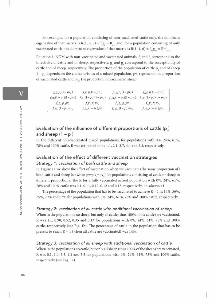
Vac
cin
atio
n o
f ca
ttle o
nly is su
fficie
nt to
stop fM
DV
tra
nsM
ission
V
For example, for a population consisting of non-vaccinated cattle only, the dominant eigenvalue of that matrix is R(1, 0, 0) = fcgc = Rc–c and, for a population consisting of only vaccinated cattle, the dominant eigenvalue of that matrix is R(1, 1, 0) = fvcgvc = Rvac
c–c.
Equation 1: NGM with non-vaccinated and vaccinated animals. fc and fs correspond to the infectivity of cattle and of sheep, respectively. gc and gs correspond to the susceptibility of cattle and of sheep, respectively. The proportion of the population of cattle pc and of sheep 1 – pc depends on the characteristics of a mixed population. pvc represents the proportion of vaccinated cattle and pvs, the proportion of vaccinated sheep:
evaluation of the influence of different proportions of cattle (pc) and sheep (1 – pc) In the different non-vaccinated mixed populations, for populations with 0%, 24%, 61%, 78% and 100% cattle, R was estimated to be 1.1, 2.1, 3.7, 4.4 and 5.3, respectively.
evaluation of the effect of different vaccination strategies Strategy 1: vaccination of both cattle and sheep In Figure 1a we show the effect of vaccination when we vaccinate (the same proportion of) both cattle and sheep (so when pv=pvc=pvs) for populations consisting of cattle or sheep in different proportions. The R for a fully vaccinated mixed population with 0%, 24%, 61%, 78% and 100% cattle was 0.1, 0.11, 0.12, 0.12 and 0.13, respectively, i.e. always <1.
The percentage of the population that has to be vaccinated to achieve R = 1 is: 14%, 56%, 75%, 79% and 83% for populations with 0%, 24%, 61%, 78% and 100% cattle, respectively.
Strategy 2: vaccination of all cattle with additional vaccination of sheep When in the populations no sheep, but only all cattle (thus 100% of the cattle) are vaccinated, R was 1.1, 0.90, 0.52, 0.35 and 0.13 for populations with 0%, 24%, 61%, 78% and 100% cattle, respectively (see Fig. 1b). The percentage of cattle in the population that has to be present to reach R = 1 (when all cattle are vaccinated) was 14%.
Strategy 3: vaccination of all sheep with additional vaccination of cattle When in the populations no cattle, but only all sheep (thus 100% of the sheep) are vaccinated, R was 0.1, 1.4, 3.3, 4.1 and 5.3 for populations with 0%, 24%, 61%, 78% and 100% cattle, respectively (see Fig. 1c).
102

Vac
cin
atio
n o
f ca
ttle o
nly is su
fficie
nt to
stop fM
DV
tra
nsM
ission
V
Fig. 1. The effect of different vaccination strategies on the reduction of R in mixed populations. (a) The effect of vaccination of both cattle and sheep (in equal proportions) on the reduction of R in different mixed populations with cattle and sheep. (b) The effect of vaccination of all cattle and additional vaccination of sheep on the reduction of R in different mixed populations with cattle and sheep. (c) The effect of vaccination of all sheep and additional vaccination of cattle on the reduction of R in different mixed populations with cattle and sheep. pc represents the proportion of cattle of the mixed population. The threshold value of R = 1 is indicated by a grey line. The percentage of the population of (a) cattle and sheep, (b) sheep, or (c) cattle that needs to be (additionally) vaccinated to reach the threshold value of 1 is indicated.
The additional percentage of the cattle population that has to be vaccinated to reach R = 1 was 0%, 29%, 72% and 78% and 83%, respectively, for populations with 0%, 24%, 61%, 78%, and 100% cattle, respectively.
disCussion
In the current study we quantified the transmission of FMDV in mixed cattle-sheep populations and evaluated the effect of different vaccination strategies. The evaluation of different vaccination strategies was based on the transmission estimates from experimental transmission studies. The higher the proportion of cattle in a mixed cattle-sheep population, the higher the R for the mixed population is. Thus, the impact of vaccination of the cattle is higher. When the whole population is vaccinated, R < 1 regardless of the population composition. In mixed cattle-sheep populations with at least 14% of cattle, vaccination of cattle only is sufficient to reduce R to
103

Vac
cin
atio
n o
f ca
ttle o
nly is su
fficie
nt to
stop fM
DV
tra
nsM
ission
V
<1. The strategy of vaccinating cattle only for eradication purposes has been used in the past in continental Europe [15] and South America [16, 19] with success. Previous studies using mathematical modelling also predicted that for emergency vaccination, targeting cattle only is much more efficient than using other vaccination strategies [29]. Therefore, this strategy will be more cost-effective in countries with mixed populations of cattle and sheep where prophylactic vaccination is applied [30], as it would mean a reduction in the number of vaccine doses needed and in required manpower. Moreover, when using it as an emergency vaccination strategy, it would also mean a reduction in the time needed to immunize all the animals.
While our conclusions are valid for mixed cattle- sheep populations, different results might be expected for mixed populations where other FMDV-susceptible species are present. For instance, in The Netherlands, where routine annual vaccination of cattle only was used from 1953 to 1991, FMD outbreaks occurred be- tween 1961 and 1967 in mixed cattle and pig farms. At that time, additional vaccination of pig herds was used effectively to control the outbreaks [31]. Additionally, in Asian countries, where the Asian buffalo (Bubalus bubalis) is a host of epidemiological importance [32], a vaccination strategy that includes (additional) vaccination of the Asian buffalo is probably advisable. Thus depending on the different species and percentages of these species in a population, different vaccination strategies might be needed. When quantitative data of transmission of FMDV for other animal species are known, this could be included in the NGM and then similar analyses can be performed for other heterogeneous populations.
In our analysis, we used data from transmission studies in which good quality vaccines, containing >6 PD50 per dose, were used. Experience in South America [33] shows that the strategy of vaccinating cattle only is only effective when good quality vaccines are used. The use of good quality vaccines is of course a prerequisite when using vaccination to control a disease. We used data from within-pen transmission studies in which cattle and/or sheep were mingling in one animal room, thus within-pen transmission occurred. However, in many situations, cattle and sheep within a population will have less intensive contact. Other studies show that transmission between pens is in general lower than within a pen [12, 34] and that between-herd transmission will be even lower [35]. Thus, the effect of targeting vaccination towards cattle will probably be even better under these circumstances than predicted in the current study. In the current study, we used a mathematical approach to calculate which vaccination strategies would be effective. In field situations, other aspects, e.g. vaccine quality, might influence the results. However, even if the quality of the vaccine batch is low, it is probably better to use it in cattle only than spread the available capacity over both species. Although we did use different serotypes in the current study, which produced similar results, there might be a different outcome for other virus strains. Moreover, our approach looks only at the scenario where eradication of FMDV is the goal, there may be an interest to consider scenarios where intermediate situations (FMDV still endemic) have also to be considered, but this has not been studied here.
We developed an NGM that can be used to evaluate the transmission of FMDV for mixed populations of cattle and sheep and we analysed the effect of a targeted vaccination strategy. We conclude that vaccination of cattle only in mixed populations consisting of sheep and cattle will in most cases be sufficient for controlling FMDV epidemics.
104

Vac
cin
atio
n o
f ca
ttle o
nly is su
fficie
nt to
stop fM
DV
tra
nsM
ission
V
List oF reFerenCes
1 Anderson RM, May RM: The invasion, persistence and spread of infectious diseases within animal and plant communities. Philosophical Transactions of the Royal Society of London 1986, 314: 533–570.
2 Heffernan JM, Smith RJ, Wahl LM: Perspectives on the basic reproductive ratio. Journal of the Royal Society of London: Interface 2005, 2: 281–293.
3 Kermack WO, McKendrick AG: A Contribution to the Mathematical Theory of Epidemics. Proceedings of the Royal Society of London, Series A: Mathematical, Physical and Engineering Sciences 1927, 115: 700–721.
4 Hagenaars TJ, Dekker A, de Jong MCM, Eblé PL: Estimation of foot and mouth disease transmission parameters, using outbreak data and transmission experiments. Revue Scientifique et Technique (International Office of Epizootics) 2011, 30: 467–77.
5 Cox SJ, Barnett P V, Dani P, Salt JS: Emergency vaccination of sheep against foot-and-mouth disease: protection against disease and reduction in contact transmission. Vaccine 1999, 17: 1858–1868.
6 Eblé PL, Bouma A, de Bruin MG, van Hemert-Kluitenberg F, van Oirschot JT, Dekker A: Vaccination of pigs two weeks before infection significantly reduces transmission of foot-and- mouth disease virus. Vaccine 2004, 22: 1372–1378.
7 Eblé PL, Orsel K, Dekker A. FMDV infection in vaccinated and non-vaccinated sheep: transmission to con- tact animals and diagnostic aspects. In: Session of the Research Group of the Standing Technical Committee of EuFMD. Jerez de la Frontera, Spain, 29–31 October 2012.
8 Goris NE, Eblé PL, de Jong MCM, De Clercq K, Eblé PL: Quantification of foot-and-mouth disease virus transmission rates using published data. ALTEX 2009, 26: 52–54.
9 Orsel K, Dekker A, Bouma A, Stegeman JA, de Jong MCM: The effect of vaccination on foot and mouth disease virus transmission among dairy cows. Vaccine 2007, 25: 327–335.
10 Orsel K, Dekker A, Bouma A, Stegeman JA, de Jong MCM: Vaccination against foot and mouth disease reduces virus transmission in groups of calves. Vaccine 2005, 23: 4887–4894.
11 Orsel K, Dekker A, Bouma A, Stegeman JA, de Jong MCM: Quantification of foot and mouth disease virus excretion and transmission within groups of lambs with and without vaccination. Vaccine 2007, 25: 2673– 2679.
12 Van Roermund HJ, Eblé PL, de Jong MCM, Dekker A: No between-pen transmission of foot-and-mouth disease virus in vaccinated pigs. Vaccine 2010, 28: 4452–4461.
13 Bravo de Rueda C, de Jong MCM, Eblé PL, Dekker A: Estimation of the transmission of foot-and-mouth disease virus from infected sheep to cattle. Veterinary Research 2014, 45: 58.
14 Leforban Y. How predictable were the outbreaks of foot and mouth disease in Europe in 2001 and is vaccination the answer? Revue Scientifique et Technique (International Office of Epizootics) 2002, 21: 549–556, 539–547.
15 Leforban Y. Review of the status of foot and mouth disease and approach to control/eradication in Europe and Central Asia. Revue Scientifique et Technique (International Office of Epizootics) 2002, 21: 477–492.
16 Sutmoller P, Barteling SS, Olascoaga RC, Sumption KJ: Control and eradication of foot-and-mouth disease. Virus Research 2003, 91: 101– 144.
17 Gibson CF, Donaldson AI: Exposure of sheep to natural aerosols of foot-and-mouth disease virus. Research in Veterinary Science 1986, 41: 45–49.
18 Parida S, Fleming L, Oh Y, Mahapatra M, Hamblin P, Gloster J, Paton DJ: Emergency vaccination of sheep against foot-and-mouth disease: significance and detection of subsequent sub-clinical infection. Vaccine 2008, 26: 3469–3479.
19 Donaldson A: The role of sheep in the epidemiology of foot-and-mouth disease and proposals for control and eradication in animal populations with a high density of sheep. In: Session of the Research Group of the Standing Technical Committee of EuFMD. Borovets, Bulgaria, 5–8 September 2000.
20 Diekmann O, Heesterbeek JA, Metz JA: On the definition and the computation of the basic reproduction ratio R0 in models for infectious diseases in heterogeneous populations. Journal of Mathematical Biology 1990, 28: 365–382.
21 Becker NG: Analysis of Infectious Disease Data. London: Chapman and Hall Ltd, 1989.
22 Anderson RM, May RM: Infectious Diseases of Humans: Dynamics and Control. Oxford: Oxford University Press, 1991.
23 Velthuis AG, Bouma A, Katsma WEA, Nodelijk G, De Jong MC: Design and analysis of small-scale transmission experiments with animals. Epidemiology and Infection 2007, 135: 202–217.
105

Vac
cin
atio
n o
f ca
ttle o
nly is su
fficie
nt to
stop fM
DV
tra
nsM
ission
V
24 De Jong MCM, Kimman TG: Experimental quantification of vaccine-induced reduction in virus transmission. Vaccine 1994, 8: 761–766.
25 Kroese AH, De Jong MC: Design and analysis of transmission experiments. In: Proceedings of the Annual Meeting of the Society for Veterinary Epidemiology and Preventive Medicine. Noordwijkerhout, 2001, pp. 21–36.
26 Cox SJ, Parida S, Voyce C, Reid SM, Hamblin PA, Hutchings G, Paton DJ, Barnett PV: Further evaluation of higher potency vaccines for early protection of cattle against FMDV direct contact challenge. Vaccine 2007, 25: 7687–7695.
27 Diekmann O, Heesterbeek JA, Roberts MG: The construction of next-generation matrices for compartmental epidemic models. Journal of the Royal Society of London: Interface 2010, 7: 873–885.
28 Food and Agriculture Organization of the United Nations (FAO) database. FAOSTAT (http://faostat.fao.org/site/ 569/default.aspx#ancor). Accessed 9 October 2013.
29 Keeling MJ, Keeling MJ, Woolhouse ME, May RM, Davies G, Grenfell BT: Modelling vaccination strategies against foot-and-mouth disease. Nature 2003, 421: 136–142.
30 Yadin H, Brenner J, Chai D, Oved Z, Hadany Y, Kusak A, Haimovich M: The NSP immune
response of vaccinated animals after in-field exposure to FMDV. Vaccine 2007, 25: 8298–8305.
31 Van Bekkum J, Bool PH, Vermeulen CJ: Experience with the vaccination of pigs for the control of foot-and-mouth disease in the Netherlands. Tijdschrift voor Diergeneeskunde 1967, 92: 87–97.
32 Maroudam V, Nagendrakumar SB, Madhanmohan M, Santhakumar P, Thiagarajan D, Srinivasan VA: Experimental transmission of foot-and-mouth disease among Indian buffalo (Bubalus bubalis) and from buffalo to cattle. Journal of Comparative Pathology 2008, 139: 81–85.
33 La Torre J: Integrated procedures to assess FMD vaccine quality and herd immunity in Argentina. In: Session of the Research Group of the Standing Technical Committee of EuFMD. Vienna, Austria, 27 September–1 October 2010.
34 Klinkenberg D, de Bree J, Laevens H, de Jong MCM: Within- and between-pen transmission of classical swine fever virus: a new method to estimate the basic reproduction ratio from transmission experiments. Epidemiology and Infection 2002, 128: 293–299.
35 Van Nes A, De Jong MCM, Buijtels JA, Verheijden JH: Implications derived from a mathematical model for eradication of pseudorabies virus. Preventive Veterinary Medicine 1998, 33: 39–58.
106


VIGeneral discussion


Ge
ne
ra
l disc
ussio
n
VI
Part i: the roLe oF inFeCtion routes in the transmission oF Fmdv
In part 1 of this thesis (chapters 2 and 3), we analysed the role of infectious secretions and excretions in the transmission of FMDV and correlated it to the indirect route of transmission of FMDV. First (in chapter 2), those factors that contribute to higher secretion and excretion of FMDV by the infected animals were identified. Various FMDV infected secretions and excretions contribute to the transmission of the virus between susceptible animals. It is therefore important to think how these infected secretions and excretions can reach non-infected susceptible animals either within or between farms. Within a farm, it is likely that the infected secretions and excretions are deposited in the environment and the neighbor animals become infected by inhaling air shed by the infected animals, inhaling re-aerosolized infected material, or by getting infected secretions and excretions in abrasions or small wounds. Ruminants are known to be reasonably sensitive to airborne infection [1] but pigs are relatively resistant to airborne infection [2]. Therefore, it is likely that one of the natural routes of infection in ruminants is via aerosols that spread to the upper respiratory tract. However, in pigs, small wounds could comparatively be one of the most important routes of infection.
After infection, FMDV replicates in the epithelial tissues and in the internal organs of the animals and is shed in a variety of secretions and excretions. Consequently, it was of interest to quantify the role of the environment, contaminated with infected secretions and excretions, in the transmission of FMDV. As it was demonstrated in chapter 2 of this thesis, cattle contaminate the environment with large amounts of FMDV especially via the respiratory route (i.e. upper respiratory tract excretions). This can be linked with the fact that most of the replication of FMDV occurs in the respiratory tissues from this species [3]. And the fact that even larger amounts of FMDV can be found in these secretions and excretions during the clinical stage of the disease (as it was also demonstrated in chapter 2) can be associated to the excessive production of oral (saliva) and respiratory (nasal discharge) secretions and excretions during this stage of the disease, in proximity to the replication sites. But still, large amounts of FMDV can be found early after infection, before the appearance of clinical signs e.g. in saliva [4] or in milk [5]. Thus worryingly, the asymptomatic animals could pass undetected and contaminate the environment with their infectious secretions and excretions resulting in new FMD cases. Early detection of FMDV infected animals is essential for the control of the disease. In field conditions, early detection could be a challenge specially when small ruminants are involved because the lack of apparent clinical signs [6].
Certainly direct transmission of FMDV will occur when FMDV-susceptible animals e.g. cattle or sheep are in close vicinity to infectious animals; the infected animals will shed the virus onto the environment and the susceptible animals will pick it up [7] preceding virus replication and later virus secretions and excretion. As described in chapter 3, even when the livestock are removed from the premises, transmission of FMDV will occur. Accordingly, in the outbreak of 2001, Irish cattle became infected after being exposed to an environment contaminated previously by infected British sheep on their way to The
111

Ge
ne
ra
l disc
ussio
n
VI
Netherlands [8] leading to outbreaks of FMDV in the Dutch territory. Thus then, the role of indirect routes (i.e. by contaminated environments with FMDV) was already notorious in the transmission of the infection. Transmission of the infection to other animals via contaminated environments with FMDV is feasible either inside the same premise (e.g. holding FMDV infected and susceptible animals in a same pen, feeding FMDV-susceptible animals with infectious milk from the same farm [9], or during artificial insemination of animals with infectious semen from the same farm [10]), or between-farms (e.g. when moving infected animals or objects from an infected farm to another farm or neighbour premises). Supportively, it has been suggested that movement of contaminated persons and objects were responsible for the between-farm transmission of FMDV in Denmark in 1982 and in Italy in 1993 [11], in United Kingdom in 2007 [12] and in Japan in 2010 [13]. Thus again physical interaction of infected animals (direct contact) is not a prerequisite for the occurrence of transmission of FMDV as shown in chapter 3 contradicting previous studies that suggest that direct contact transmission occurs only after 0.5 days post clinical detection in the animals [14]. Between-farm transmission of FMDV will depend on the amount of virus that is transported from location A to location B. The accumulation of virus in the environment will be determined by the amount of virus that is released in the environment, and as others have determined, also by the time needed to reach a location and the decay of the virus [15]. Indeed, in chapter 3, a decay of FMDV in the environment (depending on temperature) was taken into consideration for the quantification of the contribution of the environment in the transmission of FMDV. In this study an average temperature (20 °C) was considered in the 2 routes stochastic susceptible-infectious-recovered (or removed) (SIR) model but other temperatures can be used accordingly. Simple SIR models [16] have been employed for the quantification of transmission in between cattle [14, 17–20], between sheep [21–24], between pigs [25–29], and between buffaloes [30, 31] studies. The so far used SIR models enabled estimation of intraspecies transmission but no quantification of the contribution of the environment was performed. Knowing that SIR models can be updated by using extra routes [32], an SIR model for FMDV with the environment as an extra route was developed in chapter 3. Because it is of importance that models include an accurate set of transmission parameters to give better predictions [33], this model used a survival rate of FMDV for giving more precise transmission estimates. The results from using the 2 routes SIR model demonstrate that approximately 44% of the transmission of FMDV occurs via the environment when temperatures are in average 20 °C. This contribution is relative high if we consider that it relates to the presence of infectious secretions and excretions in an environment without the presence of livestock. This and the fact that FMDV may survive [34] and persist in a contaminated environment for up to 1 month, depending on the temperature and pH conditions [35], raise awareness on the importance of biosecurity measures for the control of FMDV (see below “Targeting control measures”).
112

Ge
ne
ra
l disc
ussio
n
VI
Part ii: the roLe oF sPeCies diFFerenCes in the transmission oF Fmdv and vaCCination strateGies towards the sPeCies
We have seen that cattle are shedders of high quantities of virus followed by swine and by small ruminants i.e. sheep and goats (in chapter 2). Although cattle are major producers of infected secretions and excretions (specially upper respiratory tract excretions as shown in chapter 2), swine are major producers of infected airborne excretions. Thus both animal species, when in close proximity, can together contaminate the environment enormously and have a major effect in the transmission of FMDV. Even though the airborne route has been considered to introduce FMDV in livestock populations [36–38], we have seen (in chapter 3) that approximately 44% of the transmission of FMDV occurs via other types of infectious secretions and excretions that are either on the floor or in air droplets inside an environment (e.g. saliva from cattle that contains high quantities of FMDV, in chapter 2). Furthermore, transmission of FMDV via the airborne route will depend on atmospheric conditions [36]. Because when in close proximity, cattle and pigs can together contaminate the environment enormously, high transmission of FMDV can be expected in mixed cattle-pigs populations. While in pigs the risk of infection with FMDV could rely on e.g. the ingestion of contaminated feedstuffs or ground [39] with infectious secretions and excretions from cattle, in cattle the inspiration of suspended viral particles could lead to infection with FMDV. Supportively, it has been shown that in populations where cattle and pigs are in close vicinity, transmission of FMDV is very high [40, 41].
On the other hand, small ruminants shed lower levels of FMDV in their secretions and excretions than cattle and swine do (in chapter 2). It was already known that small ruminants contaminate the environment with infected secretions and excretions limitedly [42] but their contribution in the transmission of FMDV in mixed populations was until now not clear. Even though cattle and sheep have been reported to be similarly susceptible to airborne infection [1], others claim that the small ruminants are the least susceptible animal species [43]. Additionally, a decline in the transmission of FMDV in this species (when the virus is passaged in a population composed of sheep only) has been previously observed [24]. Accordingly, others have observed a decline in the transmission of the virus via this species in field conditions [6]. However the role of sheep in the epidemiology of FMD has been noted as important as this species show clinical signs of the disease only vaguely and their infectious status could pass without notice [6, 44]. Because the latter and the need to understand more about the transmission of FMDV between different species, in chapter 4 the quantitative role of sheep in transmitting FMDV to susceptible cattle was investigated. In a study, using a statistical model, reducing the number of sheep in a mixed population with cattle did not have a significant effect on reducing transmission of FMDV and thus cattle were blamed to have driven the UK 2001 epidemic [45]. The results from chapters 4 and 5 confirm this observation; cattle are the most important epidemiological species as compared to sheep. Only 40% of the cattle in the transmission experiment with
113

Ge
ne
ra
l disc
ussio
n
VI
infected sheep in chapter 4 got infected with the virus and only 10% of it became clinically infected and shed virus to the environment (partial R0= 0.9). In chapter 4, it is shown that cattle and sheep are similarly susceptible to FMDV but that the relative infectivity of cattle is higher than that of sheep. In chapter 4, by using a SIR model, only a part of the transmission of FMDV in mixed sheep-cattle populations could be quantified. Thus, to determine total transmission of FMDV for a mixed population consisting of cattle and sheep, in chapter 5 another technique was employed: a next generation matrix (NGM). The NGM has been previously used for other diseases [46] and used limitedly to determine transmission of FMDV in mixed populations [47, 48]. In chapter 5 this method was useful for assessing transmission of FMDV in different mixed populations (with different animal proportions) and for evaluating targeted vaccination. In accordance with chapters 2, 3 and 4 of the thesis, chapter 5 shows the importance of cattle in the epidemiology of FMDV. In chapter 5, it is shown that mixed cattle-sheep populations containing a larger proportion of cattle might run a higher risk of transmission of FMDV (i.e. higher transmission rates will be expected). In the same chapter, it is indicated that vaccination of cattle only is sufficient to reduce transmission of FMDV (see below “Targeting control measures”).
tarGetinG ControL measures to reduCe the transmission oF the inFeCtion
According to this thesis, control measures to reduce transmission of FMDV should be focused on early detection of new infections, prevention of physical interaction between infected and susceptible animals, biosecurity (e.g. disinfection) and targeted vaccination.
early detection and the reduction of the transmission of FmdvEfforts to control FMD should be directed at early detection of infection and rapid intervention e.g. by emergency vaccination of the animals and by applying movement restrictions. As seen in chapter 2 of the thesis, as early as 0-3 dpi, large amounts of the virus can be found in secretions and excretions from cattle 4.8 10log TCID50/ml (1.0, 8.5) and swine 5.6 10log TCID50/ml (4.4, 8.6). This indicates that high contamination of the environment can occur fast after the infection of animals and thus transmission of FMDV will occur. For the purpose of early detection of FMDV, there are highly specific tests for detecting NSPs to differentiate non-infected from infected vaccinated animals and to even detect silent infections in populations of either cattle or sheep. The latter might be of consideration in populations of sheep where sometimes the detection of clinical signs is impossible. Early detection of FMDV infected animals aids to the prompt use of control interventions which will result in the reduction of the transmission parameter β, of the number of new infections caused by a typical infected individual per unit of time. As early the detection of infections, as early the biosecurity measures can be applied as well as the emergency vaccinations can be started.
114

Ge
ne
ra
l disc
ussio
n
VI
Prevention of physical interaction between infected and susceptible animalsBy reducing the amount of FMDV that is left in the environment (e.g. by virus inactivation), contact to infectious surfaces and thus new infections can be avoided. But also, it is necessary to avoid contact of the animals to contaminated environments and to restrict movement between farms.
During an outbreak, transmission of FMDV can be controlled by stamping-out of all susceptible affected animals [49–52] reducing the average number of infections caused by a single typical infectious individual during its entire infectious period (R0). One other control strategy to reduce transmission of FMDV is movement restriction of the animals and/or of farm related equipment [51]. If during an outbreak no movement restriction is applied, modelers predict that transmission of FMDV may persist [53, 54] and especially when large number of cattle are involved [55]. This is supported by the findings in chapter 2, where cattle are shown to be the major contributors of contamination of the environment with FMDV. Previously, it has been observed that even when movement restriction of animals is applied outbreaks still occur [49]. Movement of machinery, agriculture equipment, or, milk bulk tanks between farms can be responsible for the transmission of the disease between farms [49, 56]. This thesis shows that aside of reducing contact of animals to contaminated environments, restricting transport and/or disinfecting all materials that enter the farms (see below for details on disinfection) should be taken as a basic rule to avoid new infections.
Biosecurity and the reduction of the transmission of FmdvDue to that most of the secretions and excretions from animals infected with FMDV contain virus [42] as corroborated in the chapter 2 of this thesis, there exist concerns about the role of infectious secretions and excretions, and infectious animal products (e.g. milk and meat) in the transmission of FMDV [49, 50]. Thus import restrictions are implemented as a strategy to prevent introductions of FMDV in free areas.
In chapter 3, transmission of FMDV after the exposure of calves to a contaminated environment occurred in time intervals from 0-3 days and from 4-6 days post infection of the donors. This showed that the virus might be transmitted to other animals even after 3 days post infection of animals that were housed in the same environment. Hence in order to control transmission of FMDV via a contaminated environment with infectious secretions and excretions, disinfection of the premises should be applied as soon as possible. Thorough cleaning and disinfection of the environment will prevent indirect transmission of FMDV and thus R0 will be reduced. Disinfection of the environment can be performed using thermal and chemical inactivation methods. Thermal inactivation of FMDV has been studied extensively [57–60], especially for the control of transmission via contaminated animal products [35, 61–63]. FMDV is progressively inactivated by temperatures above 50°C [64] with effective inactivating temperature at approximately 60°C [65]. Thus in most open-air conditions, the virus is not inactivated very quickly.
115

Ge
ne
ra
l disc
ussio
n
VI
FMDV is reported to be resistant to inactivation between pH 7 and 7.5 but rapid inactivation is seen in more acid or more basic conditions [59]. Several scientists have evaluated the effect of different chemicals on the inactivation of FMDV [57, 66, 67]. And the OIE recommends the use of sodium hydroxide (2%), sodium carbonate (4%), citric acid (0.2%), acetic acid (2%), sodium hypochlorite (3%), potassium peroxymonosulfate/sodium chloride (1%), or chlorine dioxide for the disinfection of premises and all infected material, such as implements, cars, and clothes [64]. In this thesis it is shown that if no proper disinfection of the environment is performed, transmission of FMDV will occur.
vaccination of the animals as a tool for Fmd controlIn chapter 5, it is shown that vaccinating cattle only in mixed populations with sheep is in a lot of cases (when at least 14% of the population is cattle) sufficient to control transmission of FMDV (R0<1).
Vaccination is used as a tool to reduce the number of susceptible animals and to reduce infectivity and thus virus shedding [68], and consequently contamination of the environment. Unlikely to conventional vaccines, emergency vaccines are sometimes of higher quality (≥6 PD50) [69]. Conventional vaccines are used during annual vaccination campaigns in countries where FMD is still endemic [70] e.g. in Israel [71]. And emergency vaccines are used during outbreaks for control [51, 72].
Previously, it has been shown that the average number of new infections caused by one typical infectious individual (the reproduction ratio, R) can be reduced after vaccination of the susceptible animals [18, 21, 26, 27, 45, 73, 74]. And in field conditions, eradication of FMD has been achieved after vaccination of susceptible animals (mostly cattle only) in different regions [49, 75, 76]. In mixed populations, vaccination of all susceptible animal species is impractical and very uneconomical. Models have predicted that vaccination of a heterogeneous population composed by cattle, sheep and pigs reduce transmission of FMDV [77]. In that study [52], excluding pigs from vaccination increased slightly the transmission rates but halved the number of animals that needed to be vaccinated. Even though vaccination targeted towards cattle in a heterogeneous population composed by cattle and sheep reduces transmission of FMDV significantly [49], vaccinating cattle only either in annual vaccination campaigns or during outbreaks is not globally practiced. In this thesis, it is shown that targeted vaccination is a good alternative to reduce transmission of FMDV in mixed cattle-sheep populations and thus it should be considered during the planning of control strategies against FMD.
ConCLudinG remarks
During the past years, an effort to understand the epidemiology of FMDV and its transmission dynamics has been performed. Quantification of transmission parameters have been performed by using animal experiments and by using data from published animal experiments. And mathematical models have been used to understand and predict the transmission dynamic of the
116

Ge
ne
ra
l disc
ussio
n
VI
virus. In this work, efforts have been made to understand a little bit more on how the virus can be transmitted through the environment and which animal species should be targeted primarily during the control of an outbreak. We showed that cattle infected with FMDV contaminate the environment with huge amounts of infected secretions and excretions than other species (in chapter 2). And thus the risk of transmission of FMDV, via the environment (in chapter 3), by this animal species is high (except when vaccination is applied). Thus according to chapters 2 and 3, transmission of FMDV depends on several determinants: the amount of secretions and excretions that are in a determined environment, the affected animal species which determines higher or lower contamination of the environment, the clinical status of the animal which determines higher contaminations, the vaccination status of the animals, and the temperature in which the virus is exposed in the environment.
In mixed populations consisting of cattle and sheep, cattle are of higher risk for transmission of FMDV as compared to sheep (in chapter 4). In these mixed populations, transmission of FMDV can be controlled after vaccination of cattle only when at least 14% of cattle are present in these populations (in chapter 5). Thus according to chapters 4 and 5, transmission of FMDV depends as well on the type of animal population and its proportion inside a population, and on the vaccination status of the animals. And in general when observing the results from chapters 2, 3, 4 and 5, the presence of cattle inside a population contributes to a higher transmission of the infection, being even higher when pigs are present.
In order to develop global strategies for FMDV transmission control, a multidisciplinary effort of virologists, modelers, veterinarians and authorities will be necessary in order to update contingency plans. Efforts should be made on strengthen biosecurity measures (i.e. disinfection of premises and equipment, and controlling in and outs), segregating animal species or using physical barriers when possible, and targeting cattle to vaccination during an epidemic.
imPLiCations and Further researCh
The results from this thesis could be used to improve the control of FMDV by e.g improving contingency plans and update vaccination strategies. This thesis raises the importance of possible silent transmission of FMDV within and between farms. The conclusions of this study highlight the importance of a contaminated environment in the transmission of the disease even when the infected animals are not present anymore.
There is still little known on how much infectious virus could be transported from farm to farm and which is the route of infection that determines major transmission of the virus when animal transport is not allowed i.e. illegal transport of infectious milk or of contaminated feedstuff. Quantification studies on the amounts of virus that can be transported between farms and its contribution in the transmission of FMDV (by the estimation of infection rates from this contaminated material) are recommended.
Additionally, this work identifies cattle as the species that can infect larger number of animals during an outbreak. Therefore cattle should be targeted primarily during vaccination campaigns. However, in populations where pigs coexist with cattle and sheep,
117

Ge
ne
ra
l disc
ussio
n
VI
1 French NP, Kelly L, Jones R, Clancy D: Dose-response relationships for foot and mouth disease in cattle and sheep. Epidemiology and Infection 2002, 128:325–32.
2 Alexandersen S, Donaldson AI: Further studies to quantify the dose of natural aerosols of foot-and-mouth disease virus for pigs. Epidemiology and Infection 2002, 128:313–323.
3 Arzt J, Pacheco JM, Rodriguez LL: The early pathogenesis of foot-and-mouth disease in cattle after aerosol inoculation. Identification of the nasopharynx as the primary site of infection. Veterinary Pathology 2010, 47:1048–1063.
4 Hyslop NS: Secretion of Foot-and-Mouth disease virus and antibody in the saliva of infected and immunized cattle. Journal of Comparative Pathology 1965, 75:111–7.
5 Burrows R: Excretion of foot and mouth disease virus prior to the development of lesions. Veterinary Record 1968, 82:387.
6 Donaldson A: The role of sheep in the epidemiology of foot-and-mouth disease and proposasls for control and eradication in animal populations with a high density of sheep. In Session of the Research Group of the Standing Technical Committee of EuFMD. Borovets: FAO of the United Nations; 2000.
7 Alexandersen S, Zhang Z, Donaldson A., Garland AJ.: The Pathogenesis and Diagnosis of Foot-and-Mouth Disease. Journal of Comparative Pathology 2003, 129:1–36.
8 Bouma A, Elbers ARW, Dekker A, De Koeijer A, Bartels C, Vellema P, Van Der Wal P, Van Rooij EMA, Pluimers FH, De Jong MCM: The foot-and-mouth disease epidemic in The Netherlands in 2001. Preventive Veterinary Medicine 2003, 57:155–166.
9 Sellers RF, Daggupaty SM: The epidemic of foot-and-mouth disease in Saskatchewan, Canada, 1951-1952. Canadian journal of veterinary research = Revue canadienne de recherche veterinaire 1990, 54:457–464.
10 Cottral GE, Gailiunas P, Cox BF: Foot-and-mouth disease virus in semen of bulls and its transmission by artificial insemination. Archiv für die gesamte Virusforschung 1968, 23:362–77.
11 Mahy BWJ: Foot-and-Mouth Disease Virus (Google eBook). Springer; 2006:188.
12 Cottam EM, Wadsworth J, Shaw AE, Rowlands RJ, Goatley L, Maan S, Maan NS, Mertens PPC, Ebert K, Li Y, Ryan ED, Juleff N, Ferris NP, Wilesmith JW, Haydon DT, King DP, Paton DJ, Knowles NJ: Transmission pathways of foot-and-mouth disease virus in the United Kingdom in 2007. PLoS Pathogens 2008, 4:e1000050.
13 Muroga N, Kobayashi S, Nishida T, Hayama Y, Kawano T, Yamamoto T, Tsutsui T: Risk factors for the transmission of foot-and-mouth disease during the 2010 outbreak in Japan: a case-control study. BMC Veterinary Research 2013, 9:150.
14 Charleston B, Bankowski BM, Gubbins S, Chase-Topping ME, Schley D, Howey R, Barnett P V, Gibson D, Juleff ND, Woolhouse MEJ: Relationship Between Clinical Signs and Transmission of an Infectious Disease and the Implications for Control. Science 2011, 332:726–729.
15 Bunnik BAD van, Ssematimba A, Hagenaars TJ, Nodelijk G, Haverkate MR, Bonten MJM, Hayden MK, Weinstein RA, Bootsma MCJ, Jong MCM De: Small distances can keep bacteria at bay for days. Proceedings of the National Academy of Sciences 2014, 111:3556–3560.
16 Becker NG: Analysis of Infectious Disease Data. Volume 1. London: Chapman and Hall Ltd; 1989.
17 Orsel K, Dekker A, Stegeman JA, De Jong MCM, Bouma A: Different infection parameters between dairy cows and calves after an infection with foot-and-mouth disease virus. Veterinary Journal 2010, 186:116–118.
18 Orsel K, Dekker A, Bouma A, Stegeman JA, de Jong MCM: Vaccination against foot and mouth disease reduces virus transmission in groups of calves. Vaccine 2005, 23:4887–4894.
19 Bouma A, Dekker A, de Jong MCM: No foot-and-mouth disease virus transmission between individually housed calves. Veterinary Microbiology 2004, 98:29–36.
20 Orsel K, Bouma A, Dekker A, Stegeman JA, de Jong MCM: Foot and mouth disease virus transmission during the incubation period of the
vaccination of pigs might be considered. The developed NGM tool in this study should be adapted using pigs as well as cattle and sheep, as we expect higher transmission rates in populations where cattle, sheep and pigs are in close vicinity to evaluate which vaccination strategy is more convenient to use.
List oF reFerenCes
118

Ge
ne
ra
l disc
ussio
n
VI
disease in piglets, lambs, calves, and dairy cows. Preventive Veterinary Medicine 2009, 88:158–163.
21 Eblé PL, Orsel K, Dekker A: FMDV infection in vaccinated and non-vaccinated sheep: transmission to contact animals and diagnostic aspects. Session of the research group of the European commission for the control of foot-and-mouth disease 2012.
22 Valarcher JF, Gloster J, Doel CA, Bankowski B, Gibson D: Foot-and-mouth disease virus (O/UKG/2001) is poorly transmitted between sheep by the airborne route. Veterinary Journal 2008, 177:425–428.
23 Orsel K, Dekker A, Bouma A, Stegeman JA, de Jong MCM: Quantification of foot and mouth disease virus excretion and transmission within groups of lambs with and without vaccination. Vaccine 2007, 25:2673–2679.
24 Hughes GJ, Mioulet V, Haydon DT, Kitching RP, Donaldson AI, Woolhouse ME: Serial passage of foot-and-mouth disease virus in sheep reveals declining levels of viraemia over time. Journal of General Virology 2002, 83(Pt 8):1907–1914.
25 Eblé PL, de Koeijer A, Bouma A, Stegeman A, Dekker A: Quantification of within- and between-pen transmission of Foot-and-Mouth disease virus in pigs. Veterinary Research 2006, 37:647–654.
26 Eblé PL, Bouma A, de Bruin MG, van Hemert-Kluitenberg F, van Oirschot JT, Dekker A: Vaccination of pigs two weeks before infection significantly reduces transmission of foot-and-mouth disease virus. Vaccine 2004, 22:1372–1378.
27 Van Roermund HJ, Eblé PL, de Jong MCM, Dekker A: No between-pen transmission of foot-and-mouth disease virus in vaccinated pigs. Vaccine 2010, 28:4452–4461.
28 Orsel K, de Jong MCM, Bouma A, Stegeman JA, Dekker A: Foot and mouth disease virus transmission among vaccinated pigs after exposure to virus shedding pigs. Vaccine 2007, 25:6381–6391.
29 Pacheco JM, Mason PW: Evaluation of infectivity and transmission of different Asian foot-and-mouth disease viruses in swine. Journal of Veterinary Science 2010, 11:133–142.
30 Maroudam V, Nagendrakumar SB, Madhanmohan M, Santhakumar P, Thiagarajan D, Srinivasan VA: Experimental transmission of foot-and-mouth disease among Indian buffalo (Bubalus bubalis) and from buffalo to cattle. Journal of Comparative Pathology 2008, 139:81–5.
31 Vosloo W, Bastos AD, Kirkbride E, Esterhuysen JJ, van Rensburg DJ, Bengis RG, Keet DW, Thomson GR: Persistent infection of African buffalo (Syncerus caffer) with SAT-type foot-and-mouth disease viruses: rate of fixation of mutations, antigenic change and interspecies transmission. Journal of General Virology 1996, 77:1457–1467.
32 Velthuis AG, de Jong MCM, Kamp EM, Stockhofe N, Verheijden JH: Design and analysis of an Actinobacillus pleuropneumoniae transmission experiment. Preventive Veterinary Medicine 2003, 60:53–68.
33 Hagenaars TJ, Dekker A, de Jong MCM, Eblé PL: Estimation of foot and mouth disease transmission parameters, using outbreak data and transmission experiments. Revue scientifique et technique (International Office of Epizootics) 2011, 30:467–77.
34 Sellers RF, Herniman KA, Donaldson AI: The effects of killing or removal of animals affected with foot-and-mouth disease on the amounts of airborne virus present in looseboxes. British Veterinary Journal 1971, 127:358–365.
35 Cottral GE: Persistence of foot-and-mouth disease virus in animals, their products and the environment. Bulletinn International Office of Epizootics 1969, 70:549–568.
36 Gloster J: Risk of airborne spread of foot-and-mouth disease from the continent to England. Veterinary Record 1982, 111:290–295.
37 Gloster J, Champion HJ, Sorensen JH, Mikkelsen T, Ryall DB, Astrup P, Alexandersen S, Donaldson AI: Airborne transmission of foot-and-mouth disease virus from Burnside Farm, Heddon-on-the-Wall, Northumberland, during the 2001 epidemic in the United Kingdom. Veterinary Record 2003, 152:525–533.
38 Daggupaty SM, Sellers RF: Airborne spread of foot-and-mouth disease in Saskatchewan, Canada, 1951-1952. Canadian journal of veterinary research = Revue canadienne de recherche veterinaire 1990, 54:465–468.
39 Alexandersen S, Mowat N: Foot-and-mouth disease: host range and pathogenesis. Current Topics in Microbiology and Immunology 2005, 288:9–42.
40 Donaldson AI, Alexandersen S, Sørensen JH, Mikkelsen T: Relative risks of the uncontrollable (airborne) spread of FMD by different species. The Veterinary Record 2001, 148:602–4.
41 Donaldson AI, Alexandersen S: Predicting the spread of foot and mouth disease by airborne virus. Revue scientifique et technique (International Office of Epizootics) 2002, 21:569–75.
119

Ge
ne
ra
l disc
ussio
n
VI
42 Thomson GR: Foot-and-mouth disease. In Infectious Diseases of Livestock with special reference to Southern Africa. Volume Vol. 2. Edited by Coetzer J, Thomson G, Tustin R. Capetown: Oxford University Press Southern Africa; 1994:825–851.
43 Kitching RP, Hughes GJ: Clinical variation in foot and mouth disease: sheep and goats. Revue scientifique et technique (International Office of Epizootics) 2002, 21:505–512.
44 Geering WA: Foot and mouth disease in sheep. Australian Veterinary Journal 1967, 43:485–489.
45 Bessell PR, Shaw DJ, Savill NJ, Woolhouse MEJ: Estimating risk factors for farm-level transmission of disease: Foot and mouth disease during the 2001 epidemic in Great Britain. Epidemics 2010, 2:109–115.
46 Gubbins S, Carpenter S, Baylis M, Wood JL, Mellor PS: Assessing the risk of bluetongue to UK livestock: uncertainty and sensitivity analyses of a temperature-dependent model for the basic reproduction number. Journal Royal Society Interface 2008, 5:363–371.
47 Cox SJ, de Jong MCM, Barnett P V, Eblé PL: Reusing published data to quantify foot-and-mouth disease transmission parameter β. In Foot and Mouth Disease International Symposium and Workshop. Melbourne; 2010.
48 De Jong MCM, Hagenaars TJ, Eblé PL: Transmission of FMDV within and between species: Quantification and comparisons. In 13th International Symposium on Veterinary Epidemiology and Economics. Maastricht: Wageningen Academic Publishers; 2012.
49 Sutmoller P, Barteling SS, Olascoaga RC, Sumption KJ: Control and eradication of foot-and-mouth disease. Virus Research 2003, 91:101–44.
50 Sanson RL: The epidemiology of foot-and-mouth disease: implications for New Zealand. New Zealand Veterinary Journal 1994, 42:41–53.
51 FAO: Preparation of Foot-and-Mouth Disease Contingency Plans. Volume 16. Rome: FAO; 2002:100.
52 Muroga N, Hayama Y, Yamamoto T, Kurogi A, Tsuda T, Tsutsui T: The 2010 Foot-and-Mouth Disease Epidemic in Japan. Journal of Veterinary Medical Science 2012, 399–404.
53 Velthuis AGJ, Mourits MCM: Effectiveness of movement-prevention regulations to reduce the spread of foot-and-mouth disease in The Netherlands. Preventive Veterinary Medicine 2007, 82:262–281.
54 Rweyemamu M, Roeder P, Mackay D, Sumption K, Brownlie J, Leforban Y, Valarcher J-F, Knowles
NJ, Saraiva V: Epidemiological patterns of foot-and-mouth disease worldwide. Transboundary and Emerging Diseases 2008, 55:57–72.
55 Green DM, Kiss IZ, Kao RR: Modelling the initial spread of foot-and-mouth disease through animal movements. Proceedings Biological sciences / The Royal Society 2006, 273:2729–35.
56 Donaldson A l. I: Risks of spreading foot and mouth disease through milk and dairy products. Revue scientifique et technique (International Office of Epizootics)1997, 16:117–124.
57 Dekker A: Inactivation of foot-and-mouth disease virus by heat, formaldehyde, ethylene oxide and gamma radiation. The Veterinary Record 1998, 143:168–169.
58 Walker JS, de Leeuw PW, Callis JJ, van Bekkum JG: The thermal death time curve for foot-and-mouth disease virus contained in primarily infected milk. Journal of Biological Standardization 1984, 12:185–9.
59 Bachrach HLL, Breese Jr. SS, Callis JJJ, Hess WRR, Patty REE, Breese SS: Inactivation of foot- and-mouth disease virus by pH and temperature changes and by formaldehyde. Proccedings of the Society for Experimental Biology and Medicine 1957, 95:147–152.
60 Doel TR, Baccarini PJ: Thermal-Stability of Foot-and-Mouth-Disease Virus. Archives of Virology 1981, 70:21–32.
61 Tomasula PM, Konstance RP: The survival of foot-and-mouth disease virus in raw and pasteurized milk and milk products. Journal of Dairy Science 2004, 87:1115–1121.
62 Tomasula PM, Kozempel MF, Konstance RP, Gregg D, Boettcher S, Baxt B, Rodriguez LL: Thermal inactivation of foot-and-mouth disease virus in milk using high-temperature, short-time pasteurization. Journal of Dairy Science 2007, 90:3202–3211.
63 Hyde JL, Blackwell JH, Callis JJ: Effect of pasteurization and evaporation on foot-and-mouth disease virus in whole milk from infected cows. Canadian Veterinary Medical Association 1975, 39:305–309.
64 Foot-and-Mouth Disease [http://www.oie.int/fileadmin/home/eng/Animal_Health_in_the_world/docs/pdf/foot_and_mouth_disease_final.pdf]
65 Kamolsiripichaiporn S, Subharat S, Udon R, Thongtha P, Nuanualsuwan S: Thermal inactivation of foot-and-mouth disease viruses in suspension. Applied and Environmental Microbiology 2007, 73:7177–7184.
120

Ge
ne
ra
l disc
ussio
n
VI
66 Cunliffe HR: Inactivation of foot-and-mouth disease virus with ethylenimine. Applied Microbiology 1973, 26:747–750.
67 Krug PW, Lee LJ, Eslami AC, Larson CR, Rodriguez L: Chemical disinfection of high-consequence transboundary animal disease viruses on nonporous surfaces. Biologicals 2011, 39:231–235.
68 Cox SJ, Barnett P V: Experimental evaluation of foot-and-mouth disease vaccines for emergency use in ruminants and pigs: a review. Veterinary Research 2009, 40:13.
69 Barnetta PV, Carabin H: A review of emergency foot-and-mouth disease (FMD) vaccines. Vaccine 2002, 20:1505–1514.
70 Doel T.: FMD vaccines. Virus Research 2003, 91:81–99.
71 Yadin H, Brenner J, Chai D, Oved Z, Hadany Y, Kusak A, Haimovich M: The NSP immune response of vaccinated animals after in-field exposure to FMDV. Vaccine 2007, 25:8298–305.
72 Foot and mouth disease. [http://www.oie.int/en/animal-health-in-the-world/oie-listed-diseases-2014/]
73 Parida S, Fleming L, Oh Y, Mahapatra M, Hamblin P, Gloster J, Paton DJ: Emergency vaccination of sheep against foot-and-mouth disease: significance and detection of subsequent sub-clinical infection. Vaccine 2008, 26:3469–79.
74 Cox SJ, Voyce C, Parida S, Reid SM, Hamblin PA, Paton DJ, Barnett P V: Protection against direct-contact challenge following emergency FMD vaccination of cattle and the effect on virus excretion from the oropharynx. Vaccine 2005, 23:1106–1113.
75 Leforban Y: Review of the status of foot and mouth disease and approach to control/eradication in Europe and Central Asia. Revue scientifique et technique (International Office of Epizootics) 2002, 21:477–492.
76 Grubman MJ, Baxt B: Foot-and-mouth disease. Clinical Microbiology Reviews 2004, 17:465–493.
77 Backer JA, Hagenaars TJ, Nodelijk G, van Roermund HJ: Vaccination against foot-and-mouth disease I: epidemiological consequences. Preventive Veterinary Medicine 2012, 107:27–40.
121


VIIsummary & samenvatting


Sum
ma
ry & Sa
me
nva
tting
VII
summary
Foot-and-mouth disease (FMD) continues to be a major worldwide veterinary problem causing serious disease resulting in production losses and trade restrictions, therefore FMD is an OIE-Listed disease [1]. Control strategies for FMD are available [2] however the disease is until now not globally controlled. Because there were still gaps in the understanding of the transmission of FMDV this thesis aimed to gain more knowledge about underlying mechanisms in the transmission of FMDV, which might contribute to improved control of the infection. The thesis is divided in two parts: Part 1: The role of infection routes in the transmission of FMDV (Chapters 2 and 3), and Part 2: The role of species differences in the transmission of FMDV and the effect of targeted vaccination strategies (Chapters 4 and 5). It is generally accepted that the most common route of transmission of FMDV is via contact exposure to an infectious individual (the direct route) [3]. However, transmission of FMDV occurs even when the animals are not exposed to infectious individuals (the indirect route) [4–6]. Some of the current control strategies against FMDV involve movement restrictions [2] but even when this control measure is applied, transmission between farms still occurs (e.g. during the UK epidemic and Dutch epidemic in 2001). For understanding how transmission could occur via the indirect route, chapters 2 and 3 aimed to analyse the role of infected secretions and excretions in the transmission of FMDV. For this purpose, in chapter 2 the factors that are associated with the amount of virus shed by the infected animals were identified. Information from 32 published scientific articles in which FMDV infection experiments were described, were recorded in a database and analysed using multivariate regression models with and without random effects. The identified factors were: type of secretions and excretions (se-excretions), animal species, stage of the disease (clinical or non-clinical) and days post infection. Cattle were shown to contribute mainly in the contamination of the environment via their respiratory se-excretions early after infection when they showed clinical signs. In chapter 3, the contribution of an environment that was contaminated with se-excretions of infected cattle to the transmission of FMDV, was quantified. For this purpose, calves that were not vaccinated and calves that were vaccinated (1 week prior to inoculation with FMDV) were used in direct and indirect transmission experiments. By using the results from both experiments, FMDV transmission was quantified using a generalized linear model based on a 2 route (2R, i.e. direct contact and environment) SIR model that included information on FMDV survival in the environment. Roughly 44% of the transmission of FMDV occurred via environmental contamination with infectious se-excretions from the affected animals. Vaccination of the calves 1 week prior to inoculation with the virus, completely prevented transmission of FMDV in both direct and indirect transmission experiments and therefore no transmission parameters could be estimated.
It has been described that cattle can contaminate the environment with larger quantities of FMDV than sheep can do [5, 7] and that they are more infectious than sheep [8]. However, because the difficulties of detecting FMD clinical signs in sheep [9], the epidemiological role of sheep in the transmission of FMDV is sometimes considered important [10].
125

Sum
ma
ry & Sa
me
nva
tting
VII
Even though transmission of FMDV occurs in mixed cattle-sheep populations [11], there were until now no quantified transmission parameter for this type of population. For understanding how species differences could have an effect on the transmission of FMDV, chapters 4 and 5 aimed to quantify transmission of FMDV between cattle and sheep and to evaluate which vaccination strategies could stop an epidemic in mixed populations of cattle and sheep. In chapter 4, an animal experiment is described using infected sheep, which were kept in contact to cattle to quantify the interspecies sheep-to-cattle transmission parameter (a partial reproduction ratio, R0
p). Limited transmission of FMDV from infected sheep to in-contact cattle was observed (R0
p = 0.87). R0p was compared to the R0’s obtained
in previous transmission studies with sheep or cattle only allowing the determination of the relative infectivity and relative susceptibility of both species. Cattle were found to be more infectious than sheep but similarly susceptible. Due to that R0
p is not sufficient to quantify the transmission of FMDV in populations consisting of cattle and sheep, we implemented in chapter 5 a method to estimate total transmission of FMDV in mixed cattle-sheep populations. Using this method, transmission of FMDV (R0) in mixed cattle-sheep populations (having different proportions of cattle and different proportions of vaccinated) was estimated and the effects of the different (targeted) vaccination strategies against FMDV were analysed. The method consisted of a 4 by 4 next generation matrix (NGM) that included infectivity and susceptibility of cattle and sheep, population proportions of cattle and sheep, and their vaccination status. In mixed populations of cattle and sheep where cattle are predominant, transmission of FMDV is higher and targeting vaccination for cattle only can be sufficient to control FMDV (R0 < 1).
This thesis covers some gaps regarding the dynamics of the transmission of FMDV. The results of this thesis can be used to improve the measures to prevent and control FMDV by e.g. implementing better biosecurity measures based on the material that is most likely infected (e.g. extra effort in the disinfection of premises and farm equipment that have had contact with se- and excretions of cattle) or updating vaccination plans (i.e. vaccinating cattle only).
126

Sum
ma
ry & Sa
me
nva
tting
VII
samenvattinG
Mond-en-klauwzeer (MKZ) is nog steeds een belangrijk veterinair probleem in grote delen van de wereld. MKZ veroorzaakt ernstige ziekteverschijnselen hetgeen resulteert in verlies van productiviteit (melkgift, verminderde groei) en handelsbeperkingen en MKZ staat dan ook op de OIE lijst met aangifteplichtige dierziektes [1]. Ondanks de beschikbaarheid van bestrijdingsmaatregelen voor MKZ [2] is de ziekte nog steeds niet uitgeroeid. Ook zijn er nog steeds lacunes in de kennis over transmissie van het mond-en-klauwzeer virus (MKZV). In het onderzoek dat wordt beschreven in dit proefschrift werd geprobeerd meer kennis te verkrijgen over onderliggende mechanismes die belangrijk zijn voor de transmissie van MKZV. Deze kennis kan gebruikt worden om verbeterde bestrijdingsmaatregelen te ontwikkelen voor MKZ.
Het proefschrift is verdeeld in twee delen. In het eerste deel werd onderzoek gedaan naar de rol die verschillende infectieroutes spelen in de transmissie van MKZV (Hoofdstukken 2 en 3). In het tweede deel werd onderzoek gedaan naar verschillen tussen MKZ gevoelige diersoorten die een rol kunnen spelen bij transmissie van MKZV en het effect van zgn. gerichte vaccinatie, dat wil zeggen een vaccinatie strategie waarbij niet alle dieren maar alleen een bepaalde doelgroep wordt gevaccineerd (Hoofdstukken 4 en 5). In het algemeen wordt aangenomen dat contact met een MKZ geïnfecteerd dier de meest gangbare route is voor transmissie van MKZV (de directe route) [3]. Maar ook dieren die geen direct contact hebben gehad met geïnfecteerde dieren kunnen besmet worden met MKZV (indirecte route) [4–6]. In huidige bestrijdingsmaatregelen worden vervoersverboden toegepast [2] maar zelfs in periodes met een vervoersverbod treedt transmissie tussen bedrijven op (b.v. tijdens de MKZ epidemieën in Groot-Brittannië en in Nederland in 2001). Om inzicht te krijgen in hoe transmissie via de indirecte route plaatsvindt, werd in hoofdstuk 2 en 3 geprobeerd om te analyseren welke rol verschillende se- en excreta (van MKZV geïnfecteerde dieren) spelen in de transmissie van MKZV. In hoofdstuk 2 werden factoren geïdentificeerd die gerelateerd zijn aan de hoeveelheid virus die wordt uitgescheiden door geïnfecteerde dieren.
Hiervoor werd gebruik gemaakt van informatie uit 32 gepubliceerde wetenschappelijke artikelen waarin gegevens van experimentele MKZV infecties werden gerapporteerd. De gegevens van deze experimenten werden verzameld in een database. Vervolgens werd met behulp van statistisch analyse ( multivariaat regressie model; met en zonder random effect) bepaald welke factoren de virusuitscheiding beïnvloedden. Factoren die invloed hadden op de virusuitscheiding waren: het type se- / excreta, de diersoort, het ziektestadium (klinisch ziek versus niet klinisch ziek) en het aantal dagen na infectie. Runderen scheidden het meeste virus uit in de omgeving, vooral via respiratoire se- /excreta, kort na infectie en wanneer ze klinisch ziek waren. In hoofdstuk 3 werd gekwantificeerd welke rol een besmette (met se- / excreta van geïnfecteerde dieren) omgeving speelt in de transmissie van MKZV. Voor dit doel werden eerst niet gevaccineerde en later gevaccineerde (1 week voor inoculatie met MKZV) kalveren gebruikt, beiden in zowel directe als indirecte transmissie experimenten. Met behulp van de gecombineerde resultaten van de directe en indirecte transmissie experimenten, kon het deel van de MKZV transmissie dat plaatsvond via de omgeving worden gekwantificeerd.
127

Sum
ma
ry & Sa
me
nva
tting
VII
Hiervoor werd gebruik gemaakt van een gegeneraliseerd lineair model (GLM), dat was gebaseerd op een zgn. 2-route (2R, d.w.z. direct contact en omgeving) SIR model. Informatie over overleving van MKZV in de omgeving (zgn. ‘survival’) werd in het model gebruikt. Bij de niet-gevaccineerde dieren, vond ruwweg 44% van de transmissie van MKZV plaats via een omgeving die besmet was met infectieuze se- / excreta van besmette dieren. Vaccinatie van kalveren een week voor inoculatie met MKZ virus, voorkwam dat er transmissie van MKZV plaatsvond, zowel in de directe als de indirecte transmissie experimenten en daardoor konden geen transmissie parameters worden geschat.
Er is al eerder beschreven dat runderen de omgeving met grotere hoeveelheden MKZV kunnen contamineren dan schapen [5, 7] en dat ze infectieuzer zijn dan schapen [8]. Maar omdat het soms erg moeilijk is om klinische MKZ verschijnselen bij schapen te detecteren [9] wordt de epidemiologische rol van schapen in de transmissie van MKZV toch belangrijk geacht [10]. Ondanks dat er transmissie van MKZV plaatsvindt in gemengde populaties van runderen en schapen [11], waren er tot nu toe geen gekwantificeerde transmissie parameters bekend voor dit type populaties.
Om inzicht te krijgen in hoe verschillende diersoorten de transmissie van MKZV beïnvloeden, werd in hoofdstukken 4 en 5 beoogd om de transmissie van MKZV tussen runderen en schapen te kwantificeren en werd geëvalueerd welke vaccinatie strategieën een epidemie in gemengde populaties van runderen en schapen tot staan kunnen brengen.
In hoofdstuk 4 wordt een dierexperiment beschreven waarin geïnfecteerde schapen samen werden gehuisvest met contact runderen, om zo de interspecies (tussen diersoort) schaap-naar-rund transmissie parameter te kwantificeren (deze transmissie parameter is een zgn. partiële reproductie ratio, R0
p). Er werd slechts beperkte transmissie van de geïnfecteerde schapen naar de contact runderen waargenomen (R0
p = 0.87). Deze R0p werd vergeleken met de R0’s
die in eerder uitgevoerde transmissie experimenten met of alleen schapen of alleen runderen waren verkregen. Op deze manier kon de relatieve infectiviteit (besmettelijkheid) en relatieve vatbaarheid van beide diersoorten worden bepaald. Runderen bleken infectieuzer te zijn dan schapen, maar de vatbaarheid van runderen en schapen was vergelijkbaar. Met alleen de partiële transmissieparameter die transmissie van schaap naar rund beschrijft, kan de transmissie in een gemengde schaap-rund populatie niet worden beschreven. Daarom werd in hoofdstuk 5 een techniek geïmplementeerd waarmee de transmissie in gemengde populaties (in dit geval runderen en schapen) kan worden geschat. Met deze methode werd de transmissie van MKZV in gemengde schaap-rund populaties (met verschillende verhoudingen van runderen en schapen) geschat (R0 waardes) en werd bovendien het effect van verschillende vaccinatie strategieën, (met verschillende verhoudingen niet-gevaccineerde en gevaccineerde dieren, waaronder zgn. gerichte vaccinatie) geanalyseerd. Voor deze analyses werd gebruik gemaakt van een zgn. 4x4 volgende generatie matrix (next generation matrix, NGM), waarin de infectiviteit en vatbaarheid van runderen en schapen, proportie van runderen en schapen in een populatie en de vaccinatie status waren opgenomen. Hoe meer runderen er aanwezig zijn in een gemengde schaap-rund populatie, des te hoger is R0. In populaties met minstens 14% runderen kan vaccinatie van alleen de runderen voldoende zijn om een uitbraak van MKZ te stoppen (R0 < 1).
128

Sum
ma
ry & Sa
me
nva
tting
VII
In dit proefschrift wordt onderzoek beschreven dat antwoord geeft op sommige leemtes in kennis over de transmissie van MKZV. De resultaten die beschreven worden in dit proefschrift, kunnen worden gebruikt om maatregelen die ingesteld worden om uitbraken van MKZ te voorkomen of onder controle te brengen of te verbeteren, zoals bijvoorbeeld implementatie van nog betere biosecurity maatregelen (hygiëne en afscherming), die gebaseerd zijn op kennis over met MKZV geïnfecteerd materiaal (bv extra aandacht voor desinfectie van bedrijven en materialen die contact hebben gehad met se- / excreta van runderen) of het updaten van vaccinatie-strategieën (onder bepaalde omstandigheden toepassen van gerichte vaccinatie).
List of references (summary and samenvatting)
1 Foot and mouth disease. [http://www.oie.int/en/animal-health-in-the-world/oie-listed-diseases-2014/]
2 FAO: Preparation of Foot-and-Mouth Disease Contingency Plans. Volume 16. Rome: FAO; 2002:100.
3 Chase-Topping ME, Handel I, Bankowski BM, Juleff ND, Gibson D, Cox SJ, Windsor M a, Reid E, Doel C, Howey R, Barnett P V, Woolhouse ME, Charleston B: Understanding foot-and-mouth disease virus transmission biology: identification of the indicators of infectiousness. Veterinary Research 2013, 44:46.
4 Pharo HJ: Foot-and-mouth disease: an assessment of the risks facing New Zealand. New Zealand Veterinary Journal 2002, 50:46–55.
5 Thomson GR: Foot and Mouth Disease. Volume Vol. 2. Capetown: Oxford University Press Southern Africa.
6 Sellers RF: Quantitative aspects of the spread of foot-and-mouth disease. Veterinary Bulletin 1971, 41:431–439.
7 Sutmoller P, Barteling SS, Olascoaga RC, Sumption KJ: Control and eradication of foot-and-mouth disease. Virus Research 2003, 91:101–44.
8 Backer JA, Hagenaars TJ, Nodelijk G, van Roermund HJ: Vaccination against foot-and-mouth disease I: epidemiological consequences. Preventive Veterinary Medicine 2012, 107:27–40.
9 Kitching RP, Hughes GJ: Clinical variation in foot and mouth disease: sheep and goats. Revue scientifique et technique (International Office of Epizootics) 2002, 21:505–512.
10 Barnett P V, Cox SJ: The role of small ruminants in the epidemiology and transmission of foot-and-mouth disease. Veterinary Journal 1999, 158:6–13.
11 Donaldson A: The role of sheep in the epidemiology of foot-and-mouth disease and proposasls for control and eradication in animal populations with a high density of sheep. In Session of the Research Group of the Standing Technical Committee of EuFMD. Borovets: FAO of the United Nations; 2000.
129


&
AB
OU
T THE
AU
THO
R
ABOUT THE AUTHOR
Carla Bravo de Rueda Cabrera was born in Lima, Peru. In 2006, she graduated from the Cayetano Heredia University (Peru) as a Doctor in Veterinary Medicine. Her thesis was titled “Molecular characterization of the genes stx1, stx2 and eae of Escherichia coli in neo-natal alpacas with diarrhea”. Immediately after she graduated as a DVM, she departed to Europe to pursue a Masters degree in Biomolecular Sciences at the University of Utrecht (The Netherlands), from which she graduated in 2008. Her thesis was titled “Using cell technology in the development of Avian Influenza vaccines”. After one year working as a pre-clinical manager for a contracted research organization, she started her PhD at the Department of Quantitative Veterinary Epidemiology of Wageningen University and was stationed throughout her PhD at the department of Virology of the Central Veterinary Institute part of Wageningen UR in Lelystad (The Netherlands). Her project involved combining animal experimental data with epidemiological tools to quantify the contribution of the environment in the transmission of FMDV and to evaluate the role of sheep in the transmission of the virus. At present, Carla is working for the U.S. Department of Agriculture (USDA) at the Plum Island Animal Disease Center (PIADC) in New York (USA), as a post-doctoral collaborator in the area of Molecular Epidemiology. She focuses on exploring epidemiological factors that contribute to the endemic status of FMDV in central Africa. She is particularly interested in the control of Foot-and-mouth disease within international cooperative projects.
131


&
TRA
inin
g A
nD
SUPE
Rv
iSiOn
PLAn
traininG and suPervision PLan
wageningen institute of animal sciences (wias)
Education and Training Year Year
The basic package (3 ECTS)
WIAS Introduction Course 2010
Course on philosophy of science and/or ethics 2010
Scientific Exposure (16 ECTS)
International conferences
European Commission for the Control of Foot-and-Mouth Disease (EuFMD), Vienna 2010
European Commission for the Control of Foot-and-Mouth Disease (EuFMD), Spain 2012
Fifth annual meeting Network of Excellence for Epizootic Disease Diagnosis and Control (EPIZONE), The Netherlands
2011
Sixth annual meeting Network of Excellence for Epizootic Disease Diagnosis and Control (EPIZONE), United Kingdom
2012
XIIIth International Symposium on Veterinary Epidemiology and Economics (ISVEE), The Netherlands
2012
Seminars and workshops
WIAS Science Day, Wageningen, 2010 2010
FMD - DISCONVAC. 8-10 December 2009 in Tervuren, Belgium 2009
FMDV workshop: Early pathogenesis and transmission, IAH, UK 2010
FMD - DISCONVAC. 14-16 May 2013 in Lyon, France 2013
Presentations
European Commission for the Control of Foot-and-Mouth Disease (EuFMD), Vienna. Poster
2010
European Commission for the Control of Foot-and-Mouth Disease (EuFMD), Spain. Oral
2012
Fifth annual meeting Network of Excellence for Epizootic Disease Diagnosis and Control (EPIZONE), The Netherlands. Poster
2011
Sixth annual meeting Network of Excellence for Epizootic Disease Diagnosis and Control (EPIZONE), United Kingdom. Oral
2012
XIIIth International Symposium on Veterinary Epidemiology and Economics (ISVEE), The Netherlands. Oral
2012
Oral presentations at monthly scientific meeting QVERA/QVE (CVI, Lelystad) 2010-2013
Oral presentation at Cees Wensing Lectures (CVI, Lelystad) 2013
In-Depth Studies (16 ECTS)
Disciplinary and interdisciplinary courses
Course on the Design and Analysis of Transmission Experiments 2009
Advanced statistics courses
Modern statistics for the life sciences 2010
Advanced Statistics course: Design of Experiments 2011
133

&
TRA
inin
g A
nD
SUPE
Rv
iSiOn
PLAn
PhD students’ discussion groups
Monthly scientific meeting QVERA/QVE 2009-2013
Reading group on Immuno Biology 2009-2010
MSc level courses
Course for the Quantitative Researcher - Basic Statistics & introduction to R 2009
Statutory Courses (3 ECTS)
Use of Laboratory Animals (mandatory when working with animals) 2008
Professional Skills Support Courses (4 ECTS)
Techniques for writing and presenting a scientific paper (Writing for Academic Publication) 2011
Project and Time Management (WU Business school) 2012
Presentation Workshop-Poster Workshop-Career Workshop 2012
Education and Training Total 42 ECTS
134

&
LiST Of PU
BLiC
ATiO
nS
List oF PuBLiCations
Bravo de Rueda C., Dekker A., Eblé PL, de Jong MCM. 2015. Quantification of transmission of foot-and-mouth disease virus caused by an environment contaminated with secretions and excretions from infected calves. Veterinary Research. In press.
Bravo de Rueda C., Dekker A., Eblé PL, de Jong MCM. 2014. Estimation of the transmission of foot-and-mouth disease virus from infected sheep to cattle. Veterinary Research 45(1): 58
Bravo de Rueda C., Dekker A., Eblé PL, de Jong MCM. 2014. Vaccination of cattle only is sufficient to stop FMDV transmission in mixed populations of sheep and cattle. Epidemiology and Infection 3:1-8.
Bravo de Rueda C., Dekker A., Eblé PL, de Jong MCM. 2014. Identification of factors associated with increased excretion of Foot-and-Mouth Disease Virus. Preventive Veterinary Medicine 113(1):23-33.
Bravo de Rueda C., Dekker A., Eblé PL, van Hemert-Kluitenberg, F., de Jong MCM. Limited transmission of Foot-and-Mouth disease virus from infected sheep to naïve calves. 2012. EU FMD 2012, Jerez de la Frontera. Oral presentation.
Bravo de Rueda C., Dekker A., Eblé PL, de Jong MCM. 2012b. Contribution of Foot-and-Mouth Disease virus contaminated environment to the transmission of the disease in calves. XIIIth International Symposium on Veterinary Epidemiology and Economics (ISVEE), Maastricht, The Netherlands, August 20-24th 2012. Oral presentation.
Bravo de Rueda C., Dekker A., Eblé PL, de Jong MCM. 2012a. Contribution of Foot-and Mouth Disease virus contaminated environment to the transmission of the disease in calves. Sixth annual meeting Network of Excellence for Epizootic Disease Diagnosis and Control (EPIZONE), Brighton, UK, June 12-14th 2012. Oral presentation.
Bravo de Rueda C., Dekker A., Eblé PL, van Hemert-Kluitenberg, F., de Jong MCM. 2011. No Foot-and-mouth disease virus infection in one week vaccinated calves. Fifth annual meeting Network of Excellence for Epizootic Disease Diagnosis and Control (EPIZONE), Arnhem, NL. Poster.
Bravo de Rueda C., Dekker A., Eblé PL, de Jong MCM. 2010. Secretion and excretion of Foot-and-Mouth-Disease Virus: A preliminary quantitative risk assessment. EU FMD 2010, Vienna. Oral presentation.
Hung, A., Bravo de Rueda, C., Martínez, C., Medrano, G., Rubio, N., Arias, N. 2005. Immunological evaluation of a vaccine against Sarcocystis in alpacas. Revista Médica Herediana 16: 29-30
135


&
AC
Kn
OW
LED
gE
ME
nT
aCknowLedGement
This dissertation would not have been possible without the guidance and the exhaustive work of Dr. De Jong, Dr. Dekker and Dr. Eblé. I highly value your dedication and professionalism.I would also like to acknowledge several individuals from around the world who contributed in the preparation and completion of this study. Many of you contributed in a personal and/or professional level and I would like to say to all of you…
thank yougraciasgràciesdank ugrazie
köszönömmercidankie
faleminderitshukran
Շնորհակալություն hvala
благодаря M̀h’gōi hvala děkuji
tak tänan kiitos danke
ευχαριστώ הדות.
dhanyavād takk
terima kasiharigatô
감사합니다
paldies choukrane
ačiū Благодарамterima kasih
grazzi Xièxiè
Баярлалаа dziękuję
mulţumesc спасибо
xвала Ďakujem
hvala tack
nandrikop khun
teşekkür ederimДякую diolch
ngiyabonga
137

Colophon
The research described in this thesis was financially supported by the European Community’s Seventh Framework Programme (FP7/2007-2013) under grant agreement n° 226556 (FMD-DISCONVAC) and the Dutch Ministry of Economic affairs (WOT-01-003-11).
Printing of this thesis was financially supported by the Central Veterinary Institute (CVI) part of Wageningen UR, Lelystad, The Netherlands.
Printed by: Off Page (Amsterdam, The Netherlands).
Cover design: Carla Bravo de Rueda & Daniela Muente.