Reproductive seasonality in captive wild ruminants - Zurich ...

ORIGINAL ARTICLE
Prevalence Estimates of Antibodies TowardsFoot-and-Mouth Disease Virus in Small Ruminants inUgandaS. N. Balinda1, K. Tjørnehøj2, V. B. Muwanika1, A. K. Sangula1, F. N. Mwiine3, C. Ayebazibwe3,C. Masembe1, H. R. Siegismund4 and S. Alexandersen2,*
1 Molecular Biology Laboratory Institute of Environment and Natural Resources, Makerere University, Kampala, Uganda2 National Veterinary Institute, Technical University of Denmark, Lindholm, Denmark3 Ministry of Agriculture Animal Industry and Fisheries, Entebbe, Uganda4 Department of Biology, Universitetsparken 15, Copenhagen Ø, Denmark
Introduction
Foot-and-mouth disease (FMD) is one of the most
important livestock diseases worldwide because of its eco-
nomic impact (James and Rushton, 2002; Chenard et al.,
2003). It affects many species including cattle, sheep,
goats and pigs, and results in reduced productivity (Alex-
andersen and Mowat, 2005). This is mainly through
reduced milk yields, loss of weight, abortions and delayed
conception, perinatal mortality and lameness in draught
animals (James and Rushton, 2002). The severity of the
disease depends on the virus strain and the type of animal
affected (Geering, 1967; Dunn and Donaldson, 1997; Kit-
ching and Hughes, 2002). Usually cattle and pigs are
more severely affected while the disease has less impact in
goats and sheep (Donaldson and Sellers, 2000), however,
Keywords:
foot-and-mouth disease virus; small
ruminants; antibodies; husbandry practice
Correspondence:
K. Tjørnehøj, National Veterinary Institute,
Technical University of Denmark, Lindholm,
DK-4771 Kalvehave, Denmark.
Tel.: +45 35 88 78 57; Fax: +45 35 88 79 01;
E-mail: [email protected]
*Current address: National Centre for Foreign
Animal Disease, 1015 Arlington Street,
Winnipeg MB R3E 3M4, Canada.
Received for publication June 23, 2009
doi:10.1111/j.1865-1682.2009.01094.x
Summary
Foot-and-mouth disease (FMD) is endemic in Uganda with control strategies
focusing on vaccination of cattle, while small ruminants are largely ignored. In
order for Uganda to establish effective control strategies, it is crucial that the
epidemiology of the disease is fully understood. This study summarizes results
of serological investigations of sheep and goats for antibodies to FMDV from
four districts in 2006 following an FMD outbreak in the region and from an
attempted comprehensive random sampling in two districts in 2007. Antibodies
were quantified and serotyped using competitive ELISA for antibodies towards
non-structural proteins (NSP) and structural proteins towards serotype O, and
blocking ELISA for antibodies towards the seven serotypes of FMD virus
(FMDV). In 2006, sheep and goats in Bushenyi and Isingiro districts were free
from antibodies towards FMDV, while herds in Kasese and Mbarara districts
excluding Kahendero village were all positive for antibodies towards NSP and
SP-O. In 2007, mean prevalence estimates of antibodies towards FMDV NSP
was 14% in goats and 22% in sheep in Kasese district, while Bushenyi was still
free. The difference between these two districts probably reflects different levels
of FMDV challenge attributed to the variation in exposure rates which again in
part may be as a result of the differing husbandry practices. Contrary to 2006,
with clear antibodies towards serotype O, the serotype-specificity of the anti-
bodies was less clear in 2007, as antibodies towards both serotype O and SAT
serotypes were identified. Our results show that goats and sheep are infected
during FMD outbreaks, and that they may be useful for determining the
serotype of FMD outbreaks in Uganda, if they are sampled shortly after an
outbreak.
Transboundary and Emerging Diseases
362 ª 2009 Blackwell Verlag GmbH • Transboundary and Emerging Diseases. 56 (2009) 362–371
some strains can cause severe disease in small ruminants
(Alexandersen and Mowat, 2005; Barnett and Cox, 1999;
Kitching et al., 2005).
In addition to the loss of income to farmers as a result
of restrictions on animal movement, local purchase and
sale, the gross domestic income of the affected countries
is greatly reduced because of loss of revenue attributed to
heavy restrictions on trade of animals and animal prod-
ucts with FMD-free countries (Thompson et al., 2002).
This can ultimately result in restrictions on export of
other products such as vegetables and fresh fruit (James
and Rushton, 2002; Kock et al., 2002). In sub-Saharan
Africa, FMD is endemic and the constant heavy restric-
tions on exports undermine poverty alleviation programs
put in place to improve the livelihood of the population
(Kock et al., 2002).
In an effort to reduce and/or eradicate the disease with
the objective of participating in animal product export,
FMD endemic countries have embarked on several con-
trol measures. Within Africa, South Africa adopted the
zoning system which comprises of a control zone, a buffer
zone and a free zone (Bruckner et al., 2002). Similarly,
Zimbabwe employed the zoning system with success in
the 1970s until the 1990s, and this resulted in exports to
the international market including the European Union
(Anderson et al., 1993; Rweyemamu et al., 2008).
In Uganda, vaccination of cattle coupled with zoosani-
tary measures such as animal movement restrictions,
quarantine and restriction in animal product trade during
FMD outbreaks are carried out but with some limited
success. FMD is still a problem with 25–38 outbreaks
reported annually between 2000 and 2006 as opposed to
1–15 between 1996 and 1999 (Anonymous, 2006). As in
most countries in the sub-Saharan region, in Uganda,
small ruminants are not included in FMD virus (FMDV)
control strategies/measures. Currently, only cattle are vac-
cinated to limit the spread of FMD outbreaks. This is
mainly because the disease is perceived as not affecting
small ruminants because of the subclinical nature of the
disease in sheep and goats. However, the role of small
ruminants in maintaining FMD epidemics has over time
attracted more attention, and it has been established that
sheep and goats can also become carriers for 9 and
4 months respectively (Zhang and Kitching, 2001; Blanco
et al., 2002; Kitching, 2002).
In Uganda, FMD outbreaks have mainly been reported
in cattle and the major serotypes involved have been O
and SAT 2. Other serotypes recorded in the past have
included A, C, SAT 1 and SAT 3. Type C was last
recorded in the 1970s while SAT 3 has been isolated only
in the African Buffalo sampled in Queen Elizabeth
National Park (QENP) in 1970 and 2005 (Vosloo et al.,
2002; Kalema-Zikusoka et al., 2005).
The keeping of livestock is a key agricultural activity in
Uganda with the majority of the animals being grazed
under agro pastoral systems which involve extensive live-
stock intermingling through communal grazing resulting
in easy disease spread. These areas also harbour large
FMD susceptible wildlife populations. However, in some
districts modern agriculture practices that involve fencing
have been adopted thereby limiting animal mixing which
could have a positive impact in reducing disease spread.
This study was aimed at determining the prevalence of
antibodies against FMD in small ruminants in an area
that experienced FMD outbreaks in cattle and attempts to
assess the influence of husbandry practices on the distri-
bution of FMD in Uganda.
Materials and Methods
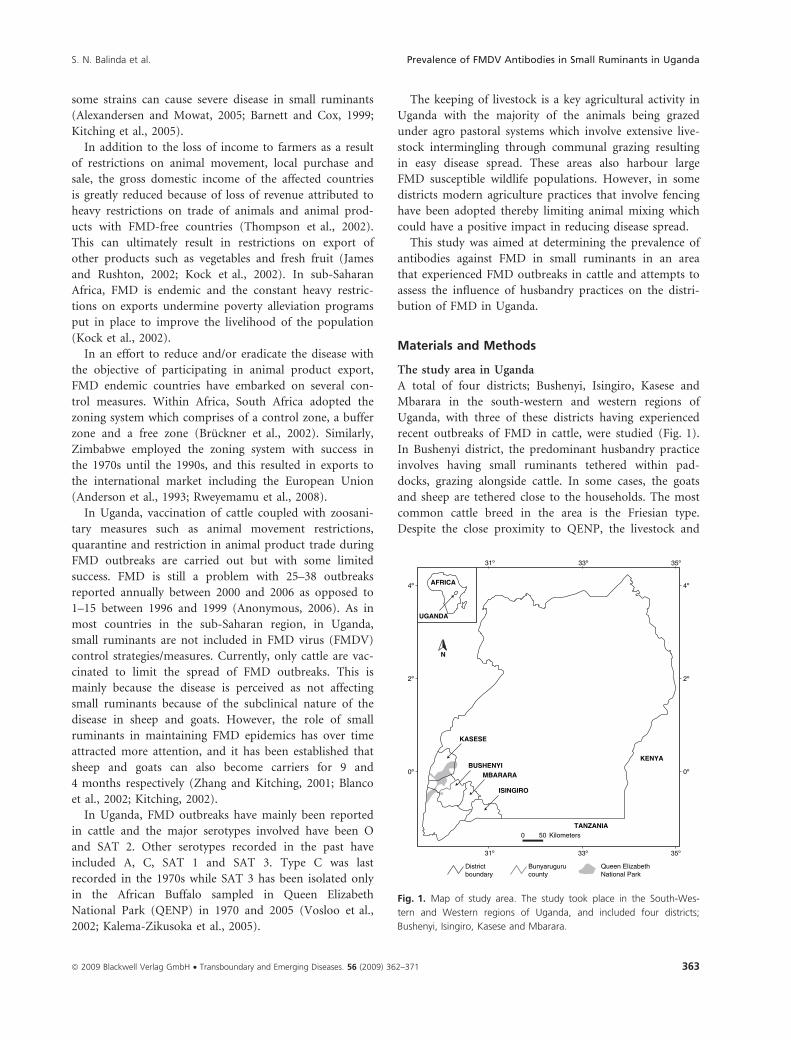
The study area in Uganda
A total of four districts; Bushenyi, Isingiro, Kasese and
Mbarara in the south-western and western regions of
Uganda, with three of these districts having experienced
recent outbreaks of FMD in cattle, were studied (Fig. 1).
In Bushenyi district, the predominant husbandry practice
involves having small ruminants tethered within pad-
docks, grazing alongside cattle. In some cases, the goats
and sheep are tethered close to the households. The most
common cattle breed in the area is the Friesian type.
Despite the close proximity to QENP, the livestock and
Districtboundary
Bunyarugurucounty
Queen ElizabethNational Park
AFRICA
UGANDA
KASESE
KENYABUSHENYI
MBARARA
ISINGIRO
TANZANIA
31º 33º
0 50 Kilometers
4º
N
35º
31º 33º 35º
2º
0º
4º
2º
0º
Fig. 1. Map of study area. The study took place in the South-Wes-
tern and Western regions of Uganda, and included four districts;
Bushenyi, Isingiro, Kasese and Mbarara.
S. N. Balinda et al. Prevalence of FMDV Antibodies in Small Ruminants in Uganda
ª 2009 Blackwell Verlag GmbH • Transboundary and Emerging Diseases. 56 (2009) 362–371 363
wildlife populations generally do not mix. This is
achieved through a buffer area (Bunyaruguru) inhabited
by crop cultivators.
On the other hand, communal grazing is predominant
in Kasese district with almost all the livestock (cattle,
goats and sheep) allowed to graze in QENP in close prox-
imity to the African buffalo, wild antelope and other
FMD susceptible animal species. Local breeds of cattle like
the Ankole comprise the majority of the stock. A few
fenced farms exist in the district.
In addition, Isingiro and Mbarara districts, which are
part of Greater Mbarara, were also studied. Fenced farm-
ing is the principal farming type in Mbarara district while
communal grazing is predominant in Isingiro district,
particularly close to the Tanzanian border. Friesian and
Ankole breeds comprise the animal stock in this region.
The livestock populations (Anonymous, 2005) in the
respective districts are summarized in Table 1.
The sampling strategy
In 2006, 26 serum samples were obtained from asymp-
tomatic goats and sheep in Kasese and Mbarara following
FMD outbreaks in cattle in both districts, and five goat
serum samples were obtained from Isingiro district dur-
ing an active FMD outbreak with the majority of the cat-
tle on the farms clinically affected, while the goats were
asymptomatic. An additional 15 serum samples were
obtained from goats in Bushenyi district because of close
proximity to affected districts.
In 2007, a simple comprehensive random study was
attempted in Bushenyi and Kasese districts. Survey
Toolbox program (Cameron, 1999) was used to compute
the numbers required for a two stage random sampling
of the respective districts, however, because of limited
number of animals, particularly the sheep, together with
weather and time restraints, it was not possible to sample
the calculated number of villages and animals during the
actual field sampling. Instead the sampling included 1–11
goats on 26 farms in 15 villages distributed in Ryeru, Ki-
chwamba, Bumbaire, Kyeizoba, Mutara, Mitooma, Kagan-
go and Kitagata sub-counties of Bushenyi and 2–20 goats
on 14 farms in seven villages distributed in Karusandara,
Kisinga, Lake Katwe, Muhokya, Mukunyu and Nya-
kiyumbu sub-counties in Kasese. Similarly for sheep, in
Bushenyi the sampling comprised 1–4 sheep on seven
farms in four villages distributed in Ryeru, Bumbaire,
Kyeizoba and Kitagata sub-counties and 2–15 sheep on
seven farms in five villages distributed in Bukunyu, Karu-
sandara, Kisinga, Lake Katwe and Muhokya sub-counties
of Kasese. Altogether, 346 sera were collected from about
31% of the desired number of animals on about 15% of
the expected farms in about 73% of the targeted villages.
At the time of sampling, there was no FMD outbreak and
all animals sampled were not clinically affected. None of the
sampled sheep and goats had previously been vaccinated
against FMD. With the exception of two farms out of 26
(7%) in Bushenyi and three farms out of 14 (21%) in Kas-
ese, the farms visited in the two districts also had cattle.
Unlike the small ruminants, the majority of the cattle in
Kasese area are vaccinated against FMD as a control measure
following frequent outbreaks of FMD. The vaccines used
during 2005–2007 include serotypes O, SAT 1 and SAT 2.
Laboratory Methods
Measurement of antibodies against the non-structural
proteins (NSP) of FMDV by NSP ELISA
This assay was performed using a commercially available
kit, Cedi� FMDV NS ELISA kit (Prionics, Zurich,
Switzerland ) as described by the manufacturer. Briefly, 96
microtitre plates pre-coated with recombinant 3ABC pro-
tein were set up with test sera in single wells, and duplicates
of negative control serum and two positive control sera, all
at a 1 : 5 dilution. After overnight incubation at room tem-
perature (20–25�C), the plates were washed, and conjugate
comprising MabL74D5 linked to horseradish peroxidase
was added and incubated for 1 h at room temperature.
After washing, substrate [tetramethyl benzidine (TMB) and
H2O2] was added and the reaction stopped after 15 min
with 0.5 m H2SO4. Optical density (OD) was measured at
450 and 620 nm after 15 min on a spectrophotometer
(Thermo Electron Corporation/Thermo Fisher Electron,
Waltham, MA, USA ), and results were expressed as a per-
centage of the mean negative control; [(OD450 ) OD620
sample)/ODmean negative control] · 100. Sera with ODP £ 50%
were scored as positive (Sorensen et al., 1998, 2005).
Measurement of antibodies against the structural
proteins of FMDV serotype O (SP-O) by SP-O ELISA
The Cedi� FMDV type O ELISA kit (SP-O ELISA) is a
ready to use commercial kit currently from Prionics and
the manufacturer’s instructions were followed. In brief,
reference sera 1–4 (provided with the kit and used in
Table 1. Livestock populations in the study area population size of
the different livestock species estimated by the Ministry of Agriculture
Animal Industry and Fisheries in 2005 (Anonymous, 2005)
District
Animal species
Cattle Goats Sheep
Bushenyi 160 000 60 000 23 000
Kasese 55 000 25 000 2000
Mbarara 807 000 573 000 47 000
Prevalence of FMDV Antibodies in Small Ruminants in Uganda S. N. Balinda et al.
364 ª 2009 Blackwell Verlag GmbH • Transboundary and Emerging Diseases. 56 (2009) 362–371

duplicate) and samples (dilution 1 : 5) were added to 96
well microtitre plates pre-coated with inactivated FMDV
O1 Manisa (approximately 80 ng/well) and incubated for
1 h at room temperature (20–25�C). After washing, the
plates were incubated with horseradish peroxidase-conju-
gated monoclonal antibody (MAb99), which recognizes
an epitope on VP1-protein of FMDV type O, for 1 h at
room temperature. Then the plates were washed and
incubated with substrate (TMB and H2O2) for 15 min.
Finally the reaction was stopped by adding 0.5 m H2SO4.
The plates were read on a spectrophotometer (Thermo
Electron Corporation) at 450 and 620 nm, and the results
were expressed as a percentage of the OD of the mean
negative control as follows;
ððOD450 �OD650sample)/ODmean negative controlÞ � 100
Sera with ODP £ 50% were positive (Chenard et al., 2003).
Measurement of antibodies by serotype-specific solid
phase blocking ELISA
Sera positive in either the NSP ELISA or SP-O ELISA kits
were subsequently screened in serotype-specific solid
phase blocking ELISA developed at the Department of
Virology of the National Veterinary Institute of the Tech-
nical University of Denmark (Lindholm).
The FMD virus strains O Manisa, A Iraq 96, C Noville,
Asia 1 Shamir, SAT 1 (BOT 1/68), SAT 2 (ZIM 5/8) and
SAT 3 (ZIM 4/81) were kindly provided by WRL, Pirbright,
UK, and propagated in primary or secondary calf kidney
(CK) cells or baby hamster kidney (BHK) cells at Lind-
holm. Virus harvests were inactivated using ethylenimine
and used as antigens.
Guinea pig and rabbit immune sera towards O Manisa,
A Iraq 96, C-Turup/Denmark/61 (a Danish FMDV strain)
and Asia 1 Shamir were produced at Lindholm, while
guinea pig and rabbit sera towards SAT 1 (BOT1/68),
SAT 2 (ZIM 5/8) and SAT 3 (ZIM 4/81) were purchased
from WRL, Pirbright, UK.
All incubations were at room temperature (20–25�C),
and plates were washed three times between steps using
ELISA buffer (0.015 m Na2 HPO4, 0.0025 m KH2 PO4,
0.5 m NaCl, 0.05% Tween-20), except the last wash step
which consisted of five rinses. Serotypes were run on sep-
arate plates, and plates were coated with serotype-specific
guinea pig sera diluted at an optimum dilution in coating
buffer (0.035 m NaHCO3, 0.015 m Na2CO3Æ10H2O) for
1 h and subsequently reacted with the corresponding
antigen diluted in ELISA buffer for 1 h.
Each plate included four wells with 10% negative calf
serum, two wells with a weak positive control serum and
two wells with a strong positive control serum from ani-
mals vaccinated with monovalent vaccines.
For serotype screening, test and control sera were added
to the plates in 1/5 dilution in ELISA buffer with 10% nor-
mal calf serum (NCS) and 0.05% NaN3. Following an
Fig. 3. Result of 2006 sampling in Bushenyi, Isingiro, Kasese and
Mbarara districts. Number of small ruminants positive and negative in
NSP and SP-O ELISA at farm and village level.
KASESEKAMWENGE
IBANDA
KIRUHURA
BUSHENYI MBARARA
ISINGIRO
NTUNGAMORUKUNGIRI
KANUNGU
30º00’ 30º30’
30º00’ 30º30’
0 10 Kilometers
0º30
’0º
00’ 0º00’
0º30’
FMDV STATUS
2006Negative
Positive
Negative
Positive
2007District boundary
N
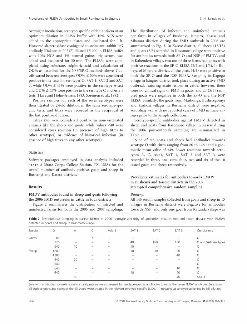
Fig. 2. Location of farms sampled during the sampling in 2006 and
the attempted comprehensive random sampling in 2007. The sam-
pling in 2006 (triangles) included Bushenyi, Isingiro, Kasese and Mbar-
ara districts, while the attempted comprehensive random sampling in
2007 (squares) included Kasese and Bushenyi Districts. Location of
infected farms (dots) and uninfected farms (plain) are depicted.
S. N. Balinda et al. Prevalence of FMDV Antibodies in Small Ruminants in Uganda
ª 2009 Blackwell Verlag GmbH • Transboundary and Emerging Diseases. 56 (2009) 362–371 365
overnight incubation, serotype-specific rabbit antisera at an
optimum dilution in ELISA buffer with 10% NCS were
added to the appropriate plates and incubated for 1 h.
Horseradish-peroxidase conjugated to swine anti-rabbit IgG
antibody (Dakopatts P0217) diluted 1/1000 in ELISA buffer
with 10% NCS and 1% normal guinea pig serum, was
added and incubated for 30 min. The ELISAs were com-
pleted using substrate, sulphuric acid and calculation of
OD% as described for the NSP/SP-O methods above. Cut-
offs varied between serotypes: OD% £ 50% were considered
positive in the tests for serotypes O, SAT 1, SAT 2 and SAT
3, while OD% £ 45% were positive in the serotype A-test
and OD% £ 35% were positive in the serotype C and Asia 1
tests (Have and Holm-Jensen, 1983; Sorensen et al., 1992).
Positive samples for each of the seven serotypes were
then titrated by 2-fold dilution in the same serotype-spe-
cific tests, and titres were recorded as the reciprocal of
the last positive dilution.
Titres ‡40 were considered positive in non-vaccinated
animals like the sheep and goats, while values <40 were
considered cross reaction (in presence of high titres to
other serotypes) or evidence of historical infection (in
absence of high titres to any other serotypes).
Statistics
Software packages employed in data analysis included
stata 8 (State Corp., College Station, TX, USA) for the
overall number of antibody-positive goats and sheep in
Bushenyi and Kasese districts.
Results
FMDV antibodies found in sheep and goats following
the 2006 FMD outbreaks in cattle in four districts
Figure 2 summarizes the distribution of infected and
uninfected farms for both the 2006 and 2007 samplings.
The distribution of infected and uninfected animals
per farm in villages of Bushenyi, Isingiro, Kasese and
Mbarara districts during the FMD outbreak in 2006 are
summarized in Fig. 3. In Kasese district, all sheep (13/13)
and goats (3/3) sampled in Kasomoro village were positive
for antibodies towards both SP-O and NSP of FMDV, and
in Kahendero village, two out of three farms had goats with
positive reactions in the SP-O ELISA (2/2 and 1/3). In Ru-
baya of Mbarara district, all the goats (4/4) were positive in
both the SP-O and the NSP ELISA. Sampling in Kagogo
village in Isingiro district took place during an active FMD
outbreak featuring acute lesions in cattle, however, there
were no clinical signs of FMD in goats, and all (5/5) sam-
pled goats were negative in both the SP-O and the NSP
ELISA. Similarly, the goats from Mashonga, Busheregyenyi
and Kashozi villages in Bushenyi district were negative,
according well with no reported cases of FMD in these vil-
lages prior to the sample collection.
Serotype-specific antibodies against FMDV detected in
sheep and goats from Kasomoro village in Kasese during
the 2006 post-outbreak sampling are summarized in
Table 2.
Nine of ten goats and sheep had antibodies towards
serotype O with titres ranging from 80 to 1280 and a geo-
metric mean value of 549. Lower reactions towards sero-
types A, C, Asia1, SAT 1, SAT 2 and SAT 3 were
recorded in three, one, zero, four, two and six of the 10
tested goats and sheep respectively.
Prevalence estimates for antibodies towards FMDV
in Bushenyi and Kasese districts in the 2007
attempted comprehensive random sampling
Bushenyi
All 146 serum samples collected from goats and sheep in 15
villages in Bushenyi district were negative for antibodies
towards NSP, and only one goat from Katunda village was
Table 2. Post-outbreak sampling in Kasese District in 2006: serotype-specificity of antibodies towards foot-and-mouth disease virus (FMDV)
detected in goats and sheep in Kasomoro village
Species O A C Asia 1 SAT 1 SAT 2 SAT 3 Conclusions
Goats 80 ) 5 ) ) ) ) O
320 ) ) ) 40 160 160 O and SAT-serotypes
640 10 ) ) 10 ) ) O
Sheep 1280 ) ) ) 10 10 20 O
1280 ) ) ) ) ) 40 O
640 20 ) ) ) ) ) O
640 ) ) ) ) ) ) O
640 ) ) ) ) ) 10 O
640 ) ) ) 10 ) 40 O
) 10 ) ) ) ) 40 SAT 3
Sera with antibodies towards non-structural proteins were screened for serotype-specific antibodies towards the seven FMDV serotypes. Sera from
all positive goats and seven of the 13 sheep were titrated in the relevant serotype-specific ELISA. ()) negative at serotype screening in 1/5 dilution.
Prevalence of FMDV Antibodies in Small Ruminants in Uganda S. N. Balinda et al.
366 ª 2009 Blackwell Verlag GmbH • Transboundary and Emerging Diseases. 56 (2009) 362–371

found positive for antibodies towards SP-O (data not pre-
sented). Screening and titration of this sample as described
above identified antibodies to SAT 1 with a titre of 160.
Kasese
Estimates of prevalences of antibodies towards NSP in
Kasese was 14% [95% confidence interval (CI): 10–18%]
in 143 serum samples from goats and 22% (95% CI: 12–
32%) in 56 serum samples from sheep. At farm level,
prevalence estimates of the goats and sheep were as sum-
marized in Table 3.
Animals belonging to three fenced farms in three Kas-
ese villages (Busunga, Rwentutu and Rwembyo) were neg-
ative for antibodies towards NSP. For comparison, in
Busunga, a high proportion of communally reared goats
and sheep were positive in the NSP ELISA (6/10 goats
and 2/2 sheep). However, two Kasese farmers practicing
communal grazing also had sero-negative animals (Kisasa:
0/3 goats; Kayanja: 0/20 goats) despite sharing grazing
with neighbours whose animals were sero-positive
(Kisasa: 4/6 goats, 3/8 goats and 6/10 sheep; Kayanja:
1/11 goats and 1/12 goats). Two fenced farms in Kabaka
had animals with antibodies towards NSP (3/11 goats, 2/13
goats and 3/13 sheep), probably because of the public
watering of the cattle on these farms in River Mobuku.
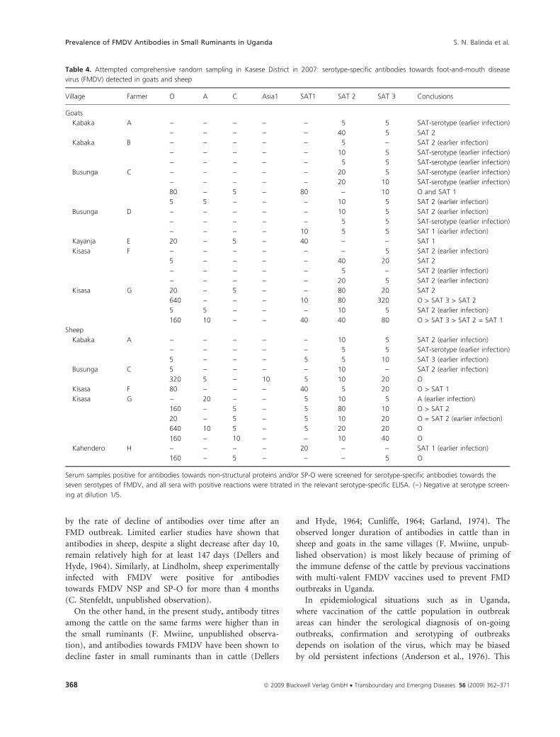
The results of screening sera with antibodies towards
NSP and/or SP-O in tests for antibodies towards all seven
FMDV serotypes, and subsequent titration of sera
screened positive, are summarized in Table 4.
Only a few goats reacted in the screening tests for anti-
bodies towards serotypes A (6/40), C (8/40) and Asia 1
(1/40). Titration of these sera showed that nearly all anti-
body reactions towards serotypes A, C and Asia 1 were
cross-reactions, since the titres were below 40 and all but
one serum had higher titres towards other serotypes. This
serum had a titre of 20 for antibodies towards serotype A
and even lower titres towards the SAT serotypes.
One goat and one sheep in Busunga had serological
evidence of exposure to serotype O. Similarly, sheep and
goats in Kisasa except those belonging to farmer F
showed serological evidence of exposure to serotypes O,
SAT 1, SAT 2 and SAT 3 with the highest titres registered
towards serotype O. In contrast, in Kabaka and Kayanja,
goats and sheep did not have serological evidence of
exposure to serotype O, while there was scattered evi-
dence of exposure to SAT 1 (one goat in Kayanja) and
SAT 2 (one goat in Kabaka). Besides this, there were low-
level titres towards other serotypes.
Discussion
Goat and sheep samples collected after FMD outbreaks
in cattle in four Ugandan districts showed estimated
prevalences of antibodies towards NSP approximating
100%. Attempted comprehensive random sampling of
sheep and goats in 2007 in one of these, Kasese district,
gave much lower estimated prevalences of antibodies
towards NSP (14% and 22% respectively) as well as
towards SP-O. The lower estimates of antibody-preva-
lences in 2007 than in 2006 probably reflects the differ-
ent sampling methods, and is most likely also a result of
time, since the last outbreak of FMD in cattle (2 months
in 2006 and 5–11 months in 2007). However, the differ-
ence in sample sizes may also be partly responsible for
the variation.
Similar studies in Morocco following an FMD outbreak
in 1999 identified a prevalence of 13% among sheep
(Blanco et al., 2002). In contrast, an earlier Kenyan study
showed high antibody prevalences of 89% (type O) and
56% (SAT 2) in small ruminants (Anderson et al., 1976).
Based on the observations in the present study, it may be
argued that these differences could have been explained
Table 3. Attempted comprehensive random sampling in Kasese Dis-
trict in 2007: prevalence estimates of antibodies towards foot-and-
mouth disease virus (FMDV) non-structural proteins (NSP) and FMDV
SP-O
Villages Farm type No. sera
tested
No. NSP (+)
sera (% positive)
No. SP (+) sera
(% positive)
Goats
Kahendero Communal 3 0 (0) 0 (0)
Busunga Fenced 11 0 (0) 0 (0)
Communal 5 3 (60) 1 (20)
Communal 5 3 (60) 0 (0)
Rwentutu Fenced 19 0 (0) 0 (0)
Kabaka Fenceda 11 3 (27) 0 (0)
Fenceda 13 2 (15) 0 (0)
Kayanja Communal 20 0 (0) 0 (0)
Communal 11 1 (9) 1 (9)
Communal 12 1 (8) 0 (0)
Kisasa Communal 6 4 (67) 3 (50)
Communal 3 0 (0) 0 (0)
Communal 8 3 (38) 4 (50)
Rwembyo Fenced 16 0 (0) 0 (0)
Total/average 143 20 (14) 9 (6)
Sheep
Kahendero Communal 5 1 (20) 2 (40)
Busunga Fenced 6 0 (0) 0 (0)
Communal 2 2 (100) 1 (50)
Kabaka Fenceda 13 3 (23) 10 (23)
Kisasa Communal 10 6 (60) 4 (40)
Fenced 4 0 (0) 1 (25)
Rwembyo Fenced 15 0 (0) 0 (0)
Total/average 55 12 (22) 18 (33)
All sera from the district were screened for antibodies towards both
NSP and SP-O using commercial kits supplied by CEDI Diagnostics. (+)
Positive sera on respective method.aFenced, but watering the cattle at the same farms in the river.
S. N. Balinda et al. Prevalence of FMDV Antibodies in Small Ruminants in Uganda
ª 2009 Blackwell Verlag GmbH • Transboundary and Emerging Diseases. 56 (2009) 362–371 367
by the rate of decline of antibodies over time after an
FMD outbreak. Limited earlier studies have shown that
antibodies in sheep, despite a slight decrease after day 10,
remain relatively high for at least 147 days (Dellers and
Hyde, 1964). Similarly, at Lindholm, sheep experimentally
infected with FMDV were positive for antibodies
towards FMDV NSP and SP-O for more than 4 months
(C. Stenfeldt, unpublished observation).
On the other hand, in the present study, antibody titres
among the cattle on the same farms were higher than in
the small ruminants (F. Mwiine, unpublished observa-
tion), and antibodies towards FMDV have been shown to
decline faster in small ruminants than in cattle (Dellers
and Hyde, 1964; Cunliffe, 1964; Garland, 1974). The
observed longer duration of antibodies in cattle than in
sheep and goats in the same villages (F. Mwiine, unpub-
lished observation) is most likely because of priming of
the immune defense of the cattle by previous vaccinations
with multi-valent FMDV vaccines used to prevent FMD
outbreaks in Uganda.
In epidemiological situations such as in Uganda,
where vaccination of the cattle population in outbreak
areas can hinder the serological diagnosis of on-going
outbreaks, confirmation and serotyping of outbreaks
depends on isolation of the virus, which may be biased
by old persistent infections (Anderson et al., 1976). This
Table 4. Attempted comprehensive random sampling in Kasese District in 2007: serotype-specific antibodies towards foot-and-mouth disease
virus (FMDV) detected in goats and sheep
Village Farmer O A C Asia1 SAT1 SAT 2 SAT 3 Conclusions
Goats
Kabaka A ) ) ) ) ) 5 5 SAT-serotype (earlier infection)
) ) ) ) ) 40 5 SAT 2
Kabaka B ) ) ) ) ) 5 ) SAT 2 (earlier infection)
) ) ) ) ) 10 5 SAT-serotype (earlier infection)
) ) ) ) ) 5 5 SAT-serotype (earlier infection)
Busunga C ) ) ) ) ) 20 5 SAT-serotype (earlier infection)
) ) ) ) ) 20 10 SAT-serotype (earlier infection)
80 ) 5 ) 80 ) 10 O and SAT 1
5 5 ) ) ) 10 5 SAT 2 (earlier infection)
Busunga D ) ) ) ) ) 10 5 SAT 2 (earlier infection)
) ) ) ) ) 5 5 SAT-serotype (earlier infection)
) ) ) ) 10 5 5 SAT 1 (earlier infection)
Kayanja E 20 ) 5 ) 40 ) ) SAT 1
Kisasa F ) ) ) ) ) ) 5 SAT 2 (earlier infection)
5 ) ) ) ) 40 20 SAT 2
) ) ) ) ) 5 ) SAT 2 (earlier infection)
) ) ) ) ) 20 5 SAT 2 (earlier infection)
Kisasa G 20 ) 5 ) ) 80 20 SAT 2
640 ) ) ) 10 80 320 O > SAT 3 > SAT 2
5 5 ) ) ) 10 5 SAT 2 (earlier infection)
160 10 ) ) 40 40 80 O > SAT 3 > SAT 2 = SAT 1
Sheep
Kabaka A ) ) ) ) ) 10 5 SAT 2 (earlier infection)
) ) ) ) ) 5 5 SAT-serotype (earlier infection)
5 ) ) ) 5 5 10 SAT 3 (earlier infection)
Busunga C 5 ) ) ) ) 10 ) SAT 2 (earlier infection)
320 5 ) 10 5 10 20 O
Kisasa F 80 ) ) ) 40 5 20 O > SAT 1
Kisasa G ) 20 ) ) 5 10 5 A (earlier infection)
160 ) 5 ) 5 80 10 O > SAT 2
20 ) 5 ) 5 10 20 O = SAT 2 (earlier infection)
640 10 5 ) 5 20 20 O
160 ) 10 ) ) 10 40 O
Kahendero H ) ) ) ) 20 ) ) SAT 1 (earlier infection)
160 ) 5 ) ) ) 5 O
Serum samples positive for antibodies towards non-structural proteins and/or SP-O were screened for serotype-specific antibodies towards the
seven serotypes of FMDV, and all sera with positive reactions were titrated in the relevant serotype-specific ELISA. ()) Negative at serotype screen-
ing at dilution 1/5.
Prevalence of FMDV Antibodies in Small Ruminants in Uganda S. N. Balinda et al.
368 ª 2009 Blackwell Verlag GmbH • Transboundary and Emerging Diseases. 56 (2009) 362–371
again depends on probang or blood sampling immedi-
ately after outbreak reporting. In such areas, unvacci-
nated sheep and goats can be used as tracer animals to
determine the serotype of on-going outbreaks of FMD.
This method can be seen as an alternative to virus isola-
tion and sequencing where facilities and expertise for
such techniques are not available. In this study, the test-
ing in Kasese and Bushenyi districts in 2007 resulted in
very different prevalence estimates of antibodies towards
NSP and SP-O, with Bushenyi almost free and the over-
all prevalence estimates of antibodies towards NSP of
14% in goats and 22% in sheep in Kasese. The single
seropositive animal in Bushenyi was probably purchased
and introduced on the farm. This difference in preva-
lence estimate levels most likely reflects the different
level of FMDV exposure in the two areas, since Kasese
district was experiencing frequent outbreaks, while Bush-
enyi district had only had one isolated incident of FMD
(2006) during the last 10 years.
This variation cannot be directly attributed to distance
from a national park, as this is about the same for the
two districts. However, Bushenyi cattle are separated from
the national park by Banyaruguru County, which is lar-
gely inhabited by crop cultivators, while such a buffer
zone does not exist in Kasese district. Consequently, Kas-
ese livestock are routinely grazed in the park area within
close proximity to wildlife, including African buffalo,
which are known to harbour FMDV as a persistent infec-
tion (Hedger, 1972; Condy et al., 1985; Thomson et al.,
2003).
Another important factor could be the differences in
farm management system in place in the two districts. In
Bushenyi, a paddocked animal management system based
on rearing cross breeds or entirely exotic (Friesian) cattle
has been in existence for over 20 years. Such animals are
highly susceptible to tropical animal diseases, and out-
breaks of FMD and other infectious diseases are greatly
felt by the dairy farmers, who as a consequence are excep-
tionally well organized into communities with a high level
of knowledge about infectious diseases which might affect
their dairy industry.
In contrast, in Kasese, local breeds of cattle comprise
the majority of the stock, with communal grazing as the
main husbandry practice, and farmers experience less
adverse effect of FMD outbreaks because of inherently less
severe clinical FMD in the local cattle breeds.
In Kasese, some farmers that practice fencing in high-
risk areas (Rwembyo, Rwentutu and Busunga) had man-
aged to keep their sheep and goats free from exposure to
FMDV, however, so had some farmers practicing com-
munal grazing (Kisasa, Kayanja and Kahendero). During
sampling it was observed that some animals were teth-
ered around homesteads. This may explain the absence
of antibodies towards FMDV on some farms in the com-
munities practicing communal grazing. This survey has
found that fencing of farms may help protect against
FMDV infection but this may not be sufficient in high-
risk areas.
During the 2007 sampling, the numbers of ruminants,
particularly of sheep, found on the majority of the farms
were lower than expected and many of the farms were
inaccessible because of heavy rains. So we were not suc-
cessful in collecting the comprehensive number of sam-
ples calculated by the Survey Toolbox. However, these
experiences will be valuable for planning of future sam-
pling trips to the area.
Despite the low number of small ruminant samples
collected from the 2006 outbreak sampling, investigation
of serotype-specificity clearly showed that this massive
outbreak was caused by a serotype O virus. One sheep
without O antibodies had a low level of antibodies
towards SAT 3, and a number of other animals showed
low levels of antibodies towards the SAT serotypes.
These reactions may be evidence of previous infections
or simply cross reactions. The overall results from post-
outbreak sampling and testing of cattle in 2006 con-
firmed that this outbreak was caused by a serotype O
FMD virus, but also identified some SAT 1 activity in
some villages (F. Mwiine, unpublished observation). The
SAT 1 reactivity was not evident in the small ruminants
in this area, and it is thus likely that these SAT 1 anti-
bodies in cattle were derived from vaccinations or previ-
ous outbreaks.
The result of the attempted comprehensive random
sampling in Kasese in 2007 gave a less clear result than
expected, since there was evidence of exposure to at least
two different serotypes in many goats and sheep; e.g. a
Kisasa goat with antibody titres of 640 towards serotype
O and of 320 towards serotype SAT 3. In such a case,
possible recent exposure to both serotypes O and SAT 3
may be likely. With regard to low titre levels (5–40),
such titres in a serum with high titres to other serotypes
suggest cross reactions, while similar values in the
absence of antibodies towards other serotypes may sug-
gest old infections (Hedger et al., 1982). The attempted
comprehensive random sampling was carried out
between 5 and 12 months after the 2006 FMD outbreak
which was serologically typed as O by Ministry of Agri-
culture Animal Industry and Fisheries, and while the
small ruminants belonging to some of the farmers still
had convincing serological evidence of this outbreak, sera
from other farms were negative for antibodies towards
this serotype.
The many cross-reactions recorded in the 2006 out-
break, as well as the very mixed picture in the 2007 sera
with low levels of antibodies towards one or several
S. N. Balinda et al. Prevalence of FMDV Antibodies in Small Ruminants in Uganda
ª 2009 Blackwell Verlag GmbH • Transboundary and Emerging Diseases. 56 (2009) 362–371 369

serotypes, may be a result of waning antibodies from
previous outbreaks, but may also signal imperfect speci-
ficity of the serotype-specific ELISAs in the face of
repeated infections.
Conclusions
Sheep and goats are also infected with FMD virus during
FMD outbreaks in cattle in Uganda. In endemic situa-
tions where serological diagnosis may be affected by fre-
quent vaccination of cattle, unvaccinated sheep and goats
can be used as tracer animals to serotype outbreaks. For
definite results, the sampling should be carried out imme-
diately following an outbreak.
Bushenyi district is confirmed free from FMDV which
may be as a result of management practices, but is more
likely because of decreased risk of exposure (challenge)
brought about by a buffer zone and the highly organized
farming industry in the district.
Husbandry practices involving less movement and
mixing of animals such as paddocking and fencing may
protect against exposure to FMDV even in high-risk
areas like Kasese district, but do not prevent all cases of
FMD infection. The data from sheep and goats tested
after the massive FMD outbreak in 2006 indicated that
the outbreak was caused by serotype O. The current
serotype-specific ELISA may lose specificity in areas with
repeated FMDV infections and vaccinations.
Recommendations
Further investigation into the dynamics of FMDV anti-
bodies in sheep and goats in Uganda should be carried
out. In addition, more work to validate the serotype-spe-
cific ELISA coupled with efforts targeted at increasing
their specificity is required.
Acknowledgements
The authors thank Jane Borch, Jani Christiansen, Eugene
Arinaitwe and Zac Duluga for excellent technical assis-
tance, and Graham Belsham and Gavin Thomson for use-
ful commentary. This work was supported by Danish
International Development Agency (DANIDA) under the
Livestock Wildlife Diseases in East Africa project.
References
Alexandersen, S., and N. Mowat, 2005: Foot-and-mouth disease:
host range and pathogenesis. Curr. Top. Microbiol. 288, 9–42.
Anderson, E.C., W.J. Doughty, and J. Anderson, 1976: The role
of sheep and goats in the epizootiology of foot-and-mouth
disease in Kenya. J. Hyg Cambridge 76, 395–402.
Anderson, E.C., C. Foggin, M. Atkinson, K.J. Sorensen, R.L.
Madekurozva, and J. Nqindi, 1993: The role of wild animals,
other than buffalo, in the current epidemiology of foot-and-
mouth disease in Zimbabwe. Epidemiol. Infect. 111, 559–563.
Anonymous, 2005: Uganda Districts Information Handbook;
Expanded Edition 2005–2006. Fountain Publishers, Kampala.
Anonymous, 2006: Annual Performance Monitoring and
Evaluation Report for Financial Year July 2005–June 2006
(Unpublished Data). Ministry of Agriculture Animal
Industry and Fisheries, Kampala.
Barnett, P.V., and S.J. Cox, 1999: The role of small ruminants
in the epidemiology and transmission of foot and mouth
disease. Vet. J. 158, 6–13.
Blanco, E., L.J. Romero, M.E. Harrachb, and J.M. Sanchez-
Vizcaino, 2002: Serological evidence of FMD subclinical
infection in sheep population during the 1999 epidemic in
Morocco. Vet. Microbiol. 85, 13–21.
Bruckner, G.K., W. Vosloo, B.J.A. Du Plessis, P.E.L.G. Kloeck,
L. Connoway, M.D. Ekron, D.B. Weaver, C.J. Dickason, F.J.
Schreuder, T. Marais, and M.E. Mogajane, 2002: Foot and
mouth disease: the experience of South Africa. Rev. Sci.
Tech. Off. Int. Epiz. 21, 751–764.
Cameron, A. 1999. A practical manual and software
package for active surveillance of livestock disease in
developing countries. Available at http://www.aciar.gov.au/
publication/MN054. (accessed on 16 October 2009).
Chenard, G., K. Miedema, P. Moonen, R.S. Schrijver, and A.
Dekker, 2003: A solid-phase blocking ELISA for detection of
type O foot-and-mouth disease virus antibodies suitable for
mass serology. J. Virol. Methods 107, 89–98.
Condy, J.B., R.S. Hedger, C. Hamblin, and I.T.R. Barnett,
1985: The duration of foot-and-mouth disease carrier
state in African buffalo i) in the individual animal and
ii) in a free-living herd. Comp. Immunol. Microbiol. 8,
259–265.
Cunliffe, H.R., 1964: Observations on the duration of
immunity in cattle after experimental infection with
foot-and-mouth disease. Cornell Vet. 54, 501–510.
Dellers, R.W., and J.L. Hyde, 1964: Response of sheep
to experimental infection with foot-and-mouth disease
virus. Am. J. Vet. Res. 25, 469–473.
Donaldson, A.I., and R.F. Sellers, 2000: Foot-and-mouth
disease. In: Aitken, I.D. (ed.), Diseases of Sheep, pp. 282–287.
Blackwell Science, Oxford.
Dunn, C.S., and A.I. Donaldson, 1997: Natural adaption to
pigs of a Taiwanese isolate of foot-and-mouth disease virus.
Vet. Rec. 141, 174–175.
Garland, A.J.M., 1974: Inhibitory activity of secretions in cattle
against foot-and-mouth disease virus. PhD thesis, University
of London, London, UK.
Geering, W.A., 1967: Foot and mouth disease in Sheep. Aus.
Vet. J. 43, 485–489.
Have, P. and M., Holm-Jensen, 1983: Detection of Antibodies
to Foot-and-Mouth Disease Virus Type 01 by Enzyme
Linked Immunosorbent Assay (ELISA). Report of the
Prevalence of FMDV Antibodies in Small Ruminants in Uganda S. N. Balinda et al.
370 ª 2009 Blackwell Verlag GmbH • Transboundary and Emerging Diseases. 56 (2009) 362–371
Session of the Research Group of the Standing Technical
Committee of the European Commission for the Control of
Foot-and-Mouth Disease. Lelystad, Netherlands. Appendix
VIII: pp. 45–51.
Hedger, R.S., 1972: Foot-and-mouth disease in wildlife with
particular reference to the African buffalo (Syncerus caffer).
J. Comp. Path. 82, 19–28.
Hedger, R.S., I.T. R Barnett, D.V. Gradwell, and P. Travassos
Dias., 1982: Serological tests for foot-and-mouth disease in
bovine serum samples. problems of interpretation. Rev. Sci.
Tech. Off. Int. Epiz. 1, 387–393.
James, A.D., and J. Rushton, 2002: The economics of foot and
mouth disease. Rev. Sci. Tech. Off. Int. Epiz. 21, 637–644.
Kalema-Zikusoka, G., R.G. Bengis, A.l. Michel, and M.H.
Woodford, 2005: A preliminary investigation of tuberculosis
and other diseases in African buffalo (Syncerus caffer) in
Queen Elizabeth National Park, Uganda. Onderstepoort J.
Vet. Res. 2, 145–151.
Kitching, R.P., 2002: Identification of foot and mouth disease
virus carrier and subclinically infected animals and differen-
tiation from vaccinated animals. Rev. Sci. Tech. Off. Int.
Epiz. 21, 531–538.
Kitching, R.P., and G.J. Hughes, 2002: Clinical variation in
foot and mouth disease: sheep and goats. Rev. Sci. Tech. Off.
Int. Epiz. 21, 505–512.
Kitching, R.P., A.M. Hutber, and M.V. Thrusfield, 2005: A
review of foot-and-mouth disease with special consider-
ation for the clinical and epidemiological factors relevant
to predictive modelling of the disease. Vet. J. 169, 197–
209.
Kock, R., B. Kebkiba, R. Heinonen, and B. Bedane, 2002:
Wildlife and pastoral society shifting paradigms in disease
control. Ann. N. Y. Acad. Sci. 969, 24–33.
Rweyemamu, M., P. Roeder, D.J. Mackay, K. Sumption,
J. Brownlie, Y. Leforban, J.F. Valarcher, N.J. Knowles,
and V. Saraiva, 2008: Epidemiological patterns of foot-
and-mouth disease worldwide. Transbound. Emerg. Dis. 55,
57–72.
Sorensen, K.J., R.L. Madekurozwa, and P. Dawe, 1992: Foot-
and-mouth disease: detection of antibodies in cattle sera by
blocking ELISA. Vet. Microbiol. 32, 253–265.
Sorensen, K.J., K.G. Madsen, E.S. Madsen, J.S. Salt, J. Nqindi,
and D.J. Mackay, 1998: Diffrentiation of infection from
vaccination in foot-and-mouth disease by the detection of
antibodies to the non-structural proteins 3D, 3AB and
3ABC in ELISA using antigens expressed in baculovirus.
Arch. Virol. 14, 1461–1476.
Sorensen, K.J., K.d. Stricker, K.C. Dyrting, S. Grazioli, and B.
Haas, 2005: Differentiation of foot-and-mouth disease virus
infected animals from vaccinated animals using a blocking
ELISA based on baculovirus expressed FMDV 3ABC antigen
and a 3ABC monoclonal antibody. Arch. Virol. 150, 805–814.
Thompson, D., P. Muriel, D. Russell, P. Osborne, A. Bromley,
M. Rowland, S. Creigh-Tyte, and C. Brown, 2002: Economic
costs of the foot and mouth disease outbreak in the United
Kingdom in 2001. Rev. Sci. Tech. Off. Int. Epiz. 21, 675–687.
Thomson, G.R., W. Vosloo, and A.D.S. Bastos, 2003: Foot-
and-mouth disease in wildlife. Virus Res. 91, 145–161.
Vosloo, W., A.D.S. Bastos, O. Sangare, S.K. Hargreaves, and
G.R. Thomson, 2002: Review of the status ad control of
foot-and-outh disease in sub-saharan Africa. Rev. Sci. Tech.
Off. Int. Epiz. 21, 437–449.
Zhang, Z.D., and R.P. Kitching, 2001: The localisation of
persistent foot and mouth disease virus in the epithelial
cells of the soft palate and pharynx. J. Comp. Pathol. 124,
89–94.
S. N. Balinda et al. Prevalence of FMDV Antibodies in Small Ruminants in Uganda
ª 2009 Blackwell Verlag GmbH • Transboundary and Emerging Diseases. 56 (2009) 362–371 371
Copyright © 2022 FDOKUMEN