Early Hemodynamic and Biochemical Changes in Overloaded Swine Ventricle
Upload
independentCategory
view
4download
0
Immunosuppression during Acute Infection with Foot-and-Mouth Disease Virus in Swine Is Mediated by IL-10Fayna Dıaz-San Segundo1¤., Teresa Rodrıguez-Calvo1., Ana de Avila2, Noemı Sevilla1,2*
1 Centro de Investigacion en Sanidad Animal, INIA,Valdeolmos, Madrid, Spain, 2 Centro de Biologıa Molecular Severo Ochoa, Cantoblanco, Madrid, Spain
Abstract
Foot-and-mouth disease virus (FMDV) is one of the most contagious animal viruses, causing a devastating disease in cloven-hoofed animals with enormous economic consequences. Identification of the different parameters involved in the immuneresponse elicited against FMDV remains unclear, and it is fundamental the understanding of such parameters beforeeffective control measures can be put in place. In the present study, we show that interleukin-10 (IL-10) production bydendritic cells (DCs) is drastically increased during acute infection with FMDV in swine. In vitro blockade of IL-10 with aneutralizing antibody against porcine IL-10 restores T cell activation by DCs. Additionally, we describe that FMDV infects DCprecursors and interferes with DC maturation and antigen presentation capacity. Thus, we propose a new mechanism ofvirus immunity in which a non-persistent virus, FMDV, induces immunosuppression by an increment in the production of IL-10, which in turn, reduces T cell function. This reduction of T cell activity may result in a more potent induction ofneutralizing antibody responses, clearing the viral infection.
Citation: Dıaz-San Segundo F, Rodrıguez-Calvo T, de Avila A, Sevilla N (2009) Immunosuppression during Acute Infection with Foot-and-Mouth Disease Virus inSwine Is Mediated by IL-10. PLoS ONE 4(5): e5659. doi:10.1371/journal.pone.0005659
Editor: Mario A. Ostrowski, University of Toronto, Canada
Received February 4, 2009; Accepted April 27, 2009; Published May 21, 2009
Copyright: � 2009 Dıaz-San Segundo et al. This is an open-access article distributed under the terms of the Creative Commons Attribution License, whichpermits unrestricted use, distribution, and reproduction in any medium, provided the original author and source are credited.
Funding: This research was supported by grant AGL2004-0049, AGL2007-61374 and CSD2006-07 from Ministerio de Educacion y Ciencia, Spain and EU, Netwokof Excellence, EPIZONE (Contract # FOOD-CT-2006-016236). F.D.-S. was a fellowship of INIA. T.R. was supported by a contract from Comunidad Autonoma deMadrid. The funders had no role in study design, data collection and analysis, decision to publish or preparation of the manuscript.
Competing Interests: The authors have declared that no competing interests exist.
* E-mail: [email protected]
¤ Current address: Plum Island Animal Disease Center, North Atlantic Area, Agricultural Research Service, U.S. Department of Agriculture, Greenport, New York,United States of America
. These authors contributed equally to this work.
Introduction
Viruses use a variety of strategies to avoid recognition by the
host immune system [1,2,3]. The active induction of immune
suppression is one mechanism by which viruses escape clearance
[4]. Several viruses are known to target dendritic cells (DCs) and
impair antiviral T cell responses [5,6,7,8,9,10]. DCs are the most
potent of the professional antigen-presenting cells (APCs), with the
unique capacity to prime naive T cell reponses and to maintain
immunity acting as sentinels of the immune system [11,12]. It has
been demonstrated that DCs can directly indentify and recognize
pathogens via Toll-like receptors (TLRs), resulting in the
subsequent up-regulation of expression of co-stimulatory mole-
cules, proinflammatory cytokines, chemokines and enhancement
of antigen presentation [13,14]. These processes allow these cells
to mature, and be capable of eliciting adaptive immunity [15]. The
status of DCs, mature or immature, affects their ability to uptake,
process, and migrate to the draining lymph nodes, where they
present the antigen to effector T cells, ultimately mounting an
effective cell-mediated response [16]. It has been recently reported
that interleukin 10 (IL-10) has an important role in the induction
of immunosuppression in vivo [17,18]. IL-10 inhibits a broad
spectrum of cellular responses. Among them, it suppresses the
function of APCs and T cells by inhibiting co-stimulation, MHC
class II expression, and chemokine secretion [19,20]. IL-10 also
has anti-inflammatory activity and stimulates cellular proliferation
of B cells [19,21].
Foot-and-mouth disease (FMD) is a highly contagious disease
with high morbidity in cloven-hoofed animals, including impor-
tant livestock species such as cattle and swine. FMD is caused by
FMD virus (FMDV), the only member of the Picornaviridae family,
genus Aphtovirus [22]. The virus genome consists of a positive
stranded RNA molecule of about 8,500 nucleotides, which
encodes four structural proteins (VP1, VP2, VP3 and VP4) and
nine non-structural proteins (L/L9, 2A, 2B, 2C, 3A, 3B, 3C and
3D) [23]. FMDV infection in susceptible animals results in viremia
1–3 days post-infection, followed by a rapid onset of clinical
disease, with an incubation period of 2–10 days. Protection against
FMDV is often associated with the induction of high levels of
circulating neutralizing antibodies in serum, and these neutralizing
antibodies can be found as early as 4 days post-infection, although
this response does not ensure clinical protection, and animals with
low levels of neutralizing antibodies may nevertheless be protected
[24]. This indicates that cell-mediated immunity may play a role
in the elimination of the virus. Thus, given that exposure to the
virus results in the production of T-cell dependent neutralizing
IgG class antibodies, and subsequent T-cell dependent memory, it
is clear that CD4+ cells must be stimulated [25,26,27,28].
During acute infection with FMDV in swine, we and others
have reported an immunosuppressive stage, characterized by T
cell unresponsiveness [29,30]. Throughout this stage, a severe but
transient lymphopenia affecting all T cell subsets correlates with
PLoS ONE | www.plosone.org 1 May 2009 | Volume 4 | Issue 5 | e5659

the appearance of viremia. However, when viremia is cleared,
both, the number of lymphocytes and the altered T cell function,
start to recuperate normal levels. These effects on the early host
immune response provide the perfect conditions for viral spread
through the organism, and subsequent shedding to the environ-
ment. The mechanism by which the virus induces immunosupres-
sion is not completely understood. We have described infection of
lymphocytes by FMDV serotype C in vivo [29] as a possible cause
of T cell functional impairment. However, the implication of other
cells and/or other mechanisms of the immune system deserve to
be deeply analyzed. It is of great interest to study the interaction of
FMDV with APCs, and more precisely, DCs. The interaction of
FMDV with DCs has been previously described by others who
have reported an absence of detectable FMDV replication in
mature DCs in vitro [30,31,32]. Nevertheless, it is important to
determine the interaction of FMDV with DC progenitors, and
how these cells function during acute infection with FMDV. In this
report, we trace the events following infection of DCs and its
progenitors by FMDV serotype C. First, we show that FMDV
infects DCs progenitors CD172+ cells, but not differentiated DCs
in vitro. This infection leads to inhibition of maturation and antigen
presentation of DCs. Second, we show that antigen presentation of
DCs isolated ex vivo from FMDV infected swine is impaired.
Finally, we reveal that production of IL-10 by DCs plays a critical
role in impairing activation of T cells during acute infection with
FMDV in swine.
Results
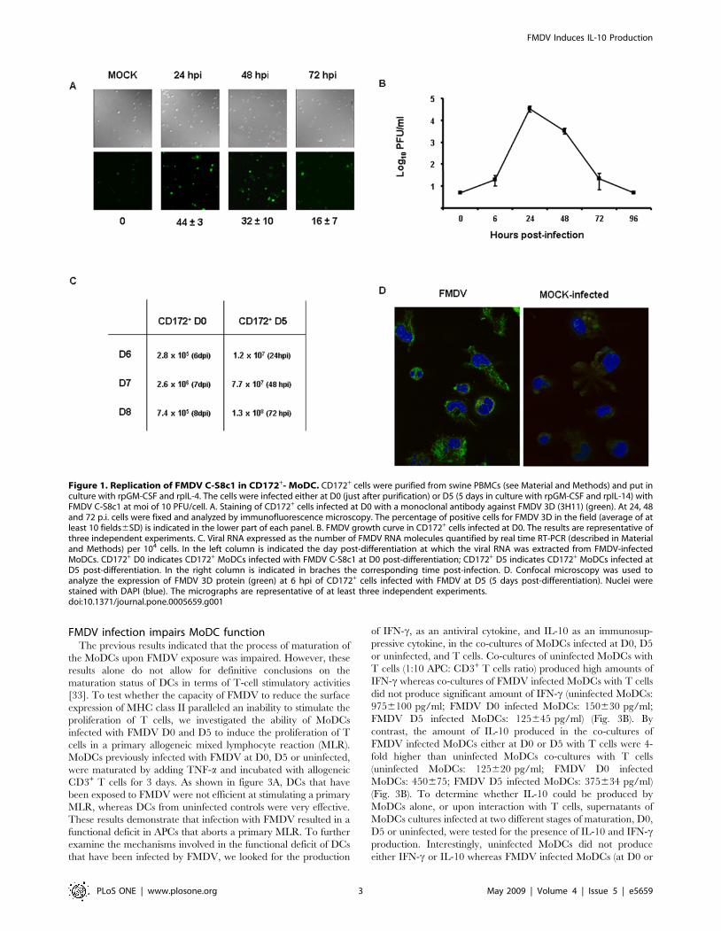
FMDV infects immature MoDCsTo gain a more comprehensive understanding of the mecha-
nism(s) by which FMDV infected swine are immunosuppressed
during acute infection [29,30], we studied the susceptibility of
porcine monocyte-derived DCs (MoDCs) to FMDV infection in
vitro. CD172+ cells were generated from peripheral blood
mononuclear cells (PBMCs) and were cultured in the presence
of rpGM-CSF and rpIL-4. In the first set of experiments, CD172+
cells were infected with FMDV C-S8c1 at MOI of 10 at day 0 (D0)
of development, washed with phosphate buffer pH 6.0 to
eliminate any virus bound to cell membranes that may account
for infectivity, incubate, in the presence of rpGM-CSF and rpIL-4
and, at different time-points, the supernatants of the cells were
harvested and the cells were fixed. By 24 hours post infection (hpi)
a high percentage of cells (4463) were seen to express FMDV non-
structural protein 3D by immunoflourescence (Fig. 1A). The
presence of positive cells was maintained by 48 hpi (32610) and by
72 hpi the number of cells expressing FMDV 3D started to decline
(1667) (Fig. 1A), suggesting a productive infection of MoDCs.
This result was confirmed by the observation of a 4–5 log10-fold
increase in the virus titer determined by plaque assay in susceptible
BHK-21 cells at 24 to 48 hpi, but clearance by 72 hpi (Fig. 1B).
Neither positive cells for FMDV 3D nor virus were detected by 96
hpi. Nevertheless, we use real time RT PCR at later times post-
infection to determine if by a more sensitive technique we could
detect any viral RNA. The amount of FMDV RNA detected at
day 6, 7 and 8 post-infection did not increase, indicating that
active viral replication might be hampered (Fig. 1C) when DCs
were infected at D0. Thus, these results indicate that FMDV is
able to infect CD172+ cells but viral replication may be aborted
along with cell differentiation.
To clarify whether FMDV could infect at multiple stages along
a development pathway of MoDCs, we repeated the same earlier
experimental designed but using CD172+ cells previously cultured
with rpGM-CSF and rpIL-4 during 5 days (D5). Viral production
by plaque assay on BHK-21 cells was not detected even at the
earliest time post-infection tested (6 and 24 hpi) (data not shown).
However, by confocal microscopy cells were expressing non-
structural viral proteins (Fig. 1D), and by real time RT PCR viral
RNA was detected at 24, 48 and 72 hpi, showing an increased in
the amount of viral RNA (Fig. 1C). This suggests that the virus was
able to replicate but there was not viral particles produced that
could be detected by plaque assay. To determine if virus was
produced even though no virus was detected by plaque assay on
BKH-21 cells, supernatants of D5 CD172+ cells infected with
FMDV were harvested at different time points (6, 24 and 48 hpi)
and were subjected to two blind passages in BHK-21 cells.
Cytopathic effect was detected after two passages in BHK-21 of
supernatants only from cultures at 6 hpi, but at no at other times
post-infection, indicating that CD172+ cells at later time post-
differentiation (D5 post-culture) are not productively infected by
FMDV, therefore an abortive infection is taking place.
FMDV interferes with MoDC development in vitroIn order to determine if the viral infection had any effect on cell
differentiation or in the expression of cell surface proteins
important in T-cell stimulation, we next analyzed the effect(s) of
viral infection on cell differentiation and expression of co-
stimulatory molecules (CD80/86) and MHC class II. CD172+
cells infected with FMDV at D0 or D5 were cultured in the
presence of rpGM-CSF and rpIL-4 until day 7 (immature
phenotype), and then TNF-a was added to the culture for an
additional 24 h to induce complete maturation. We initially
studied by flow cytometry the profile of forward scatter (FSC) and
side scatter (SSC) in order to determine cell size and granularity
evolution in the process of maturation. Uninfected cultures
underwent a dramatic increase in size and granularity after
TNF-a treatment, according to mature MoDC phenotype
(Fig. 2A). In stark contrast, when cells were previously infected
with FMDV, either at D0 or D5, the FACS analyses did not show
an increase in size and granularity after adding TNF-a (Fig. 2A).
Interestingly, CD172+ cells matured in the presence of rpGM-CSF
and rpIL-4 according to a normal pattern until FMDV was added
to the culture (compare Fig. 2A, panel uninfected and FMDV-D5
without TNF-a). Due to the fact that FMDV is a highly cytopathic
virus in several lines, we evaluated apoptosis and viability of
mature MoDCs (after TNF-a treatment) by annexin-V staining
and 7AAD exclusion. Infection of MoDCs by FMDV did not
decrease cell viability after complete maturation compared with
uninfected or BEI-inactivated FMDV MoDCs (Fig. 2A). We
therefore analyzed the expression of co-stimulatory molecules and
MHC class II on FMDV infected at different stages of maturation
(D0 and D5), and uninfected CD172+ cells. After incubating with
TNF-a, the mean fluorescence intensity of CD80/86 was
increased in FMDV infected cells to similar levels than uninfected
cells (Fig. 2B). However, MHC class II expression did not increase
on FMDV infected cells upon TNF-a treatment (Fig. 2B), making
this difference statistically significant (p,0.05) as compared with
the uninfected MoDCs after adding TNF-a. Together, the data
show that FMDV may disrupt MoDCs at multiple stages along a
development pathway, even when they showed only abortive
infection (D5 infected MoDCs). To determine if viral replication is
required to interfere with MoDCs development, BEI-inactivated
FMDV was added to MoDCs at D0 and D5 post-differentiation.
These cultures underwent an increase in size and granularity as
well as up-regulation of CD80/86 and MHC-II similar to
uninfected controls (Fi. 2A, B), indicating that virus replication is
needed to trigger impair MoDCs development.
FMDV Induces IL-10 Production
PLoS ONE | www.plosone.org 2 May 2009 | Volume 4 | Issue 5 | e5659
FMDV infection impairs MoDC functionThe previous results indicated that the process of maturation of
the MoDCs upon FMDV exposure was impaired. However, these
results alone do not allow for definitive conclusions on the
maturation status of DCs in terms of T-cell stimulatory activities
[33]. To test whether the capacity of FMDV to reduce the surface
expression of MHC class II paralleled an inability to stimulate the
proliferation of T cells, we investigated the ability of MoDCs
infected with FMDV D0 and D5 to induce the proliferation of T
cells in a primary allogeneic mixed lymphocyte reaction (MLR).
MoDCs previously infected with FMDV at D0, D5 or uninfected,
were maturated by adding TNF-a and incubated with allogeneic
CD3+ T cells for 3 days. As shown in figure 3A, DCs that have
been exposed to FMDV were not efficient at stimulating a primary
MLR, whereas DCs from uninfected controls were very effective.
These results demonstrate that infection with FMDV resulted in a
functional deficit in APCs that aborts a primary MLR. To further
examine the mechanisms involved in the functional deficit of DCs
that have been infected by FMDV, we looked for the production
of IFN-c, as an antiviral cytokine, and IL-10 as an immunosup-
pressive cytokine, in the co-cultures of MoDCs infected at D0, D5
or uninfected, and T cells. Co-cultures of uninfected MoDCs with
T cells (1:10 APC: CD3+ T cells ratio) produced high amounts of
IFN-c whereas co-cultures of FMDV infected MoDCs with T cells
did not produce significant amount of IFN-c (uninfected MoDCs:
9756100 pg/ml; FMDV D0 infected MoDCs: 150630 pg/ml;
FMDV D5 infected MoDCs: 125645 pg/ml) (Fig. 3B). By
contrast, the amount of IL-10 produced in the co-cultures of
FMDV infected MoDCs either at D0 or D5 with T cells were 4-
fold higher than uninfected MoDCs co-cultures with T cells
(uninfected MoDCs: 125620 pg/ml; FMDV D0 infected
MoDCs: 450675; FMDV D5 infected MoDCs: 375634 pg/ml)
(Fig. 3B). To determine whether IL-10 could be produced by
MoDCs alone, or upon interaction with T cells, supernatants of
MoDCs cultures infected at two different stages of maturation, D0,
D5 or uninfected, were tested for the presence of IL-10 and IFN-cproduction. Interestingly, uninfected MoDCs did not produce
either IFN-c or IL-10 whereas FMDV infected MoDCs (at D0 or
Figure 1. Replication of FMDV C-S8c1 in CD172+- MoDC. CD172+ cells were purified from swine PBMCs (see Material and Methods) and put inculture with rpGM-CSF and rpIL-4. The cells were infected either at D0 (just after purification) or D5 (5 days in culture with rpGM-CSF and rpIL-14) withFMDV C-S8c1 at moi of 10 PFU/cell. A. Staining of CD172+ cells infected at D0 with a monoclonal antibody against FMDV 3D (3H11) (green). At 24, 48and 72 p.i. cells were fixed and analyzed by immunofluorescence microscopy. The percentage of positive cells for FMDV 3D in the field (average of atleast 10 fields6SD) is indicated in the lower part of each panel. B. FMDV growth curve in CD172+ cells infected at D0. The results are representative ofthree independent experiments. C. Viral RNA expressed as the number of FMDV RNA molecules quantified by real time RT-PCR (described in Materialand Methods) per 104 cells. In the left column is indicated the day post-differentiation at which the viral RNA was extracted from FMDV-infectedMoDCs. CD172+ D0 indicates CD172+ MoDCs infected with FMDV C-S8c1 at D0 post-differentiation; CD172+ D5 indicates CD172+ MoDCs infected atD5 post-differentiation. In the right column is indicated in braches the corresponding time post-infection. D. Confocal microscopy was used toanalyze the expression of FMDV 3D protein (green) at 6 hpi of CD172+ cells infected with FMDV at D5 (5 days post-differentiation). Nuclei werestained with DAPI (blue). The micrographs are representative of at least three independent experiments.doi:10.1371/journal.pone.0005659.g001
FMDV Induces IL-10 Production
PLoS ONE | www.plosone.org 3 May 2009 | Volume 4 | Issue 5 | e5659

Figure 2. FMDV interferes with MoDC development in vitro. A. Dot plots show forward scatter (FSC) and side scatter (SSC) for uninfected,FMDV-infected at D0 and FMDV-infected at D5 before TNF-a stimulation (2TNF-a) and upon TNF-a treatment (+TNF-a). Note the lack of sizeincreased in FMDV-infected MoDC after TNF-a at either D0 or D5 compared with uninfected control cells. It is shown the population with MoDCsphenotype in a circle and the percentage of this population is indicated. In the right panel, the viability and apoptosis of MoDCs analyzed by annexin-V and 7AAD staining is shown. This is the results from a representative experiment (n = 4) B. The mean fluorescence intensity (MFI) of surfacemolecules expressed on MoDCs. Each bar represents the MFI of a given surface molecule (CD80/86 or MHC class II) before and after TNF-a addition.
FMDV Induces IL-10 Production
PLoS ONE | www.plosone.org 4 May 2009 | Volume 4 | Issue 5 | e5659

D5) produced substantial amount of IL-10 but no IFN-c (Fig. 3B).
These results indicate that FMDV induces production of IL-10 in
DCs, and more likely, IL-10 inhibits activation of T cells by DCs.
DCs ex vivo from FMDV infected pigs do not activate Tcells
Based on our initial in vitro findings that FMDV-infected
MoDCs do not stimulate T cells, we hypothesized that the loss of
T cell function observed during acute FMDV infection in pigs
would be mediated by MoDCs. We therefore examined MoDCs
obtained ex vivo from FMDV infected pigs. Twenty-four Large
White6Landrace pigs were inoculated with 105 PFU of FMDV C-
S8c1 and four pigs were used as uninfected controls (see Materials
and Methods). All the inoculated animals developed clinical signs
including fever and vesicles in tongue, feet and snout and severe
viremia, with the peak between day 2 and 3 post-infection that was
cleared by day 10 pi (data not shown). PBMCs were purified at
different days (1, 3, 5, 7, 10 and 14 pi) and CD172+ cells were
sorted to be cultured with rpGM-CSF and rpIL-4. At day 7 of
culture, TNF-a was added and kept for additional 24 hours. First,
we determined the immunophenotype of MoDCs by quantifying
the expression of co-stimulatory molecules and MHC class II. We
found that, in contrast to the in vitro data, MoDCs obtained ex vivo
from infected swine do not up-regulate CD80/86 after TNF-atreatment (Fig. 4A). At day 1 pi, MoDCs still responded to the
stimuli but at day 2 pi the MoDCs were not able to up-regulate co-
stimulatory molecules. Although the viremia was cleared by day 10
pi the MoDCs started to recover by day 14 pi. Interestingly, MHC
class II was up-regulated on ex vivo MoDCs from FMDV infected
swine to values reached by naıve MoDCs (data not shown). Based
on these results, we assessed the functionality of MoDCs in
primary MLR. We discovered that ex vivo MoDCs from FMDV
infected pigs at day 3 pi were not able to stimulate T cells, and this
lack of MoDC function lasted until day 17 pi (the latest time
observed in this report) (Fig. 4B). This data suggests that the
immunosuppression described in FMDV C-S8c1 infected swine
[29] may be mediated partially by the lack of MoDC function.
Figure 3. FMDV infection impairs T cell function. A. The allostimulatory capacity of uninfected MoDCs (triangles), FMDV C-S8c1-infected MoDCsat D0 (squares) and FMDV C-S8c1-infected MoDCs at D5 (circles) is indicated at different CD3+ T cells: MoDC ratios. Uninfected or infected MoDCswere irradiated and used as stimulators cells for allogeneic CD3+ T cells. Proliferation was measured in cpm (average cpm6SD) after [3H]Thymidineincorporation and are representative of three independent experiments. B. 72 hours after the onset of the co-cultures MoDCs and CD3+ T cells (ratio1:10), or MoDCs, supernatants were collected and analyzed for IFN-c and IL-10 production by quantitative ELISA. The figure shows meanconcentration values (pg/ml). Black bars: FMDV-infected MoDC at D0; white bars: FMDV-infected MoDCs at D5; gray bars: uninfected controls.doi:10.1371/journal.pone.0005659.g003
Black bars: FMDV-infected MoDC at D0; white bars: FMDV-infected MoDC at D5; gray bars: uninfected cells. Data are average of four independentexperiments6SD. Asterisks denote a statistically significant reduction in FMDV-infected MoDC compared with uninfected controls (student t test,p,0.05).doi:10.1371/journal.pone.0005659.g002
FMDV Induces IL-10 Production
PLoS ONE | www.plosone.org 5 May 2009 | Volume 4 | Issue 5 | e5659

A main question to be addressed was if CD172+ cells isolated
from infected animals, and used as a source for the generation of
MoDCs, were infected by FMDV. To test this, we took
supernatants of CD172+ cells at different times post-differentia-
tion. We were not able to detect the virus replication, neither by
plaque assay on BHK-21 cells nor in blind passages in BHK-21
cells, (data not shown). This suggests that FMDV does not infect
productively MoDCs in vivo, at least for the methods used to detect
viral infection production.
IL-10 suppresses T cell proliferationOne possible factor inducing suppression of T cell proliferation
by ex vivo MoDCs is IL-10, mainly because we have shown that this
cytokine is highly produced by in vitro FMDV infected MoDCs,
Figure 4. MoDCs from FMDV-infected swine do not up-regulate CD80/86 and do not stimulate T cells. A. Flow cytometric analyses wereperformed to measure the expression of CD80/86 on MoDCs differentiated from PBMCs isolated from FMDV C-S8c1-infected swine. It is indicated themean fluorescence intensity (MFI) of CD80/86 on MoDCs from naıve animals (N) and MoDCs from FMDV C-S8c1-infected pigs at different times post-inoculation (indicated as D1, D3, D5, D10 and D17) before treatment with TNF-a (black bars) and after treatment with TNF-a (white bars). B. Theallostimulatory capacity of MoDCs isolated from FMDV-infected swine either treated with TNF-a (diamonds and dashed line) or untreated (squares) atdifferent times post-infection (indicated in each graph). MoDCs were irradiated and used as stimulator cells for allogeneic CD3+ T cells at the T cell/DCratio indicated. Proliferation was measured in cpm (average cpm6SD) after [3H]Thymidine incorporation. Data is representative of three independentexperiments.doi:10.1371/journal.pone.0005659.g004
FMDV Induces IL-10 Production
PLoS ONE | www.plosone.org 6 May 2009 | Volume 4 | Issue 5 | e5659

and due to the immunosuppressive role on T cell proliferation of
this cytokine [34]. Therefore, the amount of IL-10 being produced
in co-cultures of T cells and MoDCs isolated ex vivo from FMDV
infected swine at different times post-infection was evaluated. We
found that IL-10 was highly produced in co-cultures of MoDCs
isolated ex vivo between days 3 and 17 pi and T cells of a naıve
animal (average value of 720 pg/ml in FMDV infected pigs versus
250 pg/ml in naıve animals) (Fig. 5B), when the maximum
inhibition of T cell proliferation was observed (see figure 4B).
Thus, all the data pointed out to a role of IL-10 in T cell
suppression. To confirm a possible role of IL-10, we next
determined whether antibody blockade of IL-10 could restore T
cell activation. T cells and MoDCs were cocultured in the
presence of anti-porcine IL-10 monoclonal antibody for a period
of 3 days. Interestingly, T cell activation was almost restored to the
level shown by naıve animals in all the co-cultures that were
previously inhibited (Fig. 5A). Surprisingly, although by day 10
and 17 we were able to observe a certain level of restoration of T
cell stimulation, it never achieved the levels of the controls
(Fig. 5A). Therefore, IL-10 seems to be causing T cell inhibition.
One possibility may be that MoDCs were producing IL-10, as we
have previously shown with in vitro FMDV infected MoDCs, or
that they induce the secretion of this cytokine upon an interaction
with T cells. To gain insight into the cell subtype responsible for
IL-10 secretion, supernatants of MoDCs produced from CD172+
cells isolated from FMDV infected pigs and, the amount of IL-10
secreted was determined. Notably, MoDCs were producing a high
amount of IL-10 (Fig. 5C), indicating that IL-10 is produced by
MoDCs in vivo. Actually, the amount of IL-10 in serum of FMDV
infected swine is significantly high compared with naıve animals
(Fig. 5D). An important cytokine that has been implicated in
mediating immunosuppression in acute viral infections is IFN-a.
To determine any role of this cytokine in the immunosuppressive
stage of FMDV-infected swine, we have looked at the level of IFN-
a in serum of FMDV-infected pigs at different times post-
inoculation by quantitative ELISA. The amount of IFN-a in
infected swine is similar or even below the levels found in naıve
animals (Fig. 5 E), indicating that IFN-a is not playing a relevant
role in immunossuppresion during FMDV infection. All these data
together suggest that IL-10 is contributing to the immunossup-
pression observed in FMDV infected swine during the peak of the
viremia but not IFN-a.
Discussion
In this study, we report for the first time that IL-10 signaling in
vivo leads to immunosuppression in FMDV infected swine. Our
previous data demonstrated that upon challenge of swine with
FMDV C-S8c1, a state of immunosuppression associated with
generalized lymphopenia is imposed rapidly after infection [29],
being the animals recovered at later times post-infection. IL-10, a
interleukin that inhibits a broad spectrum of cellular responses, has
been reported to have a role in inducing immunosuppression in
vivo [17,18]. In this paper, we show that the early immunosup-
pression observed in FMDV infected pigs is associated with
impaired MoDC function and high production of IL-10. The
implication of IL-10 in the induction and maintenance of
immunosuppression is demonstrated by the reversion of MoDC
function upon blockade of IL10-IL-10R signaling. Furthermore,
we show that exposure of MoDCs to FMDV in vitro impairs
MoDC function and causes the production of high amounts of IL-
10, although no viral replication is required. Indeed, after FMDV
infection in pigs, viral replication in MoDCs was not found. Thus,
it is possible that the generalized state of T cell unresponsiveness
shown by FMDV-infected swine at early times post-infection is
mediated by IL-10 produced by MoDCs. We speculate that IL-10
modulates MoDC function early after infection and, as a result,
dictates the nature of the cytokine response induced early during
antiviral immunity, favoring a Th2 cell/cytokine-like environment
inducing FMDV specific neutralizing antibodies.
Our data indicates that FMDV C-S8c1 is able to infect
productively MoDC-CD172+ progenitors cells in vitro, interfering
with the development of mature MoDCs and mount an efficient
immune response. In addition, CD172+ cells at later time post-
maturation (immature MoDCs, D5) can be infected by FMDV but
no viral production was detected and results in an abortive
replication. In our hands, and according to previous reports
[31,32,35,36], mature MoDCs are not infected by FMDV (data
not shown). The inhibitory effect that FMDV has on MoDC
progenitor cells appears to be associated with its ability to infect
progenitor or immature CD172+ cells. The way by which FMDV
directly disrupt cell differentiation may be by different mecha-
nisms. One mechanism could be by selectively inhibiting the
transcription of specific genes. In this sense, the virus can abort the
differentiation (luxury) function of a cell without affecting its vital
‘‘housekeeping’’ functions or inducing cell death [37,38,39].
Another mechanism by which FMDV may interfere with
progenitor cells maturation is through the indirect induction of
an inhibitory factor (or factors). In support of this second
mechanism, induction of IFNa/b in mice by lymphocytic
choriomeningitis virus (LCMV) infection has been reported to
interfere with DC differentiation in vivo [40]. However, we have
looked at IFN-a production in FMDV-infected MoDC cultures
and the levels of IFN-a are very low or below the detection limit of
the technique used (data not shown). Actually, this data agrees
with previous report in which MoDCs exposed to FMDV do not
produce IFN-a (Nfon….).
Although MoDC differentiation is accompanied by an increased
expression of several surface molecules that facilitates T cell
stimulation, the regulation of MHC class II molecule transport
plays a central role in developmentally restricting antigen
presentation [42,43]. CD172+ cells at both early and late times
in vitro infected by FMDV showed a marker reduction in the
expression of MHC class II molecules (Fig. 2B). Importantly,
MoDCs isolated from FMDV-infected swine did not show down-
regulation of MHC class II molecules. The fact that we have not
detected any viral replication in MoDCs ex vivo, may indicate that
down-regulation of MHC class II molecules requires virus
replication or at least certain level of viral replication. However,
we cannot discard that FMDV replicates in MoDCs in vivo but
under the limit of our detection method or using a more sensitive
method to detect virus. This observation indicates that FMDV
infection interferes either the transcription or translation of MHC
class II molecules. The resultant lack of MHC class II molecules
ultimately contributes to the inability of MoDCs to function
properly. It is worth noting that interference with MHC class II
surface expression has also been reported for HIV, cytomegalo-
virus and LCMV [40,44,45].
The failure of FMDV-infected MoDCs in vitro and MoDCs ex
vivo from FMDV-infected swine to stimulate T cell proliferation is
associated with the production of IL-10 by MoDCs. MoDCs
exhibit the most potent substantial increase in IL-10 production
during persistent infections and may contribute to immunosupres-
sion. Increased IL-10 production by APCs is also observed during
HIV and hepatitis virus C infections and has been shown to
specifically down-regulate T-cell responses [46,47,48,49,50].
Because IL-10 can induce T-cell unresponsiveness when present
during T-cell activation [51], the ongoing interaction of
FMDV Induces IL-10 Production
PLoS ONE | www.plosone.org 7 May 2009 | Volume 4 | Issue 5 | e5659

immunosuppressive IL-10 producing MoDCs with T cells
probably leads to the loss of T-cell responsiveness. A very clear
example is the persistent infection of mice with LCMV,
characterized by an inactivation of antiviral T cells, which show
a significant up-regulation of IL-10. In vivo blockade of IL-10
receptor resulted in rapid resolution of the persistent infection
[17,18]. Thus, our data show that FMDV infection induces IL-10
production by MoDCs in vitro and in vivo, and therefore inhibits T-
cell dependent responses. One of the most noteworthy findings of
this study is that in vitro antibody blockade of IL-10 completely or
Figure 5. Anti-IL-10 restores T cell activation by MoDCs. A. The allostimulatory capacity of MoDCs from FMDV-infected swine (D1 to D17 pi)and naıve swine is evaluated in the presence of anti-IL-10 monoclonal Ab (gray squares) or an irrelevant antibody (white squares) (see material andMethods). MoDCs were irradiated and used as stimulator cells for allogeneic CD3+ T cells at the T cell/DC ratio indicated. Proliferation was measuredin cpm after [3H]Thymidine incorporation. Data is representative of three independent experiments. B. IL-10 production by co-cultures of MoDC fromFMDV-infected swine and T cells from a naıve pig. 72 h after the onset of the co-culture (MoDC: CD3+ T cell ratio of 1:10), supernatants were collectedand analyzed for IL-10 production by quantitative ELISA. The figure shows mean concentration values (pg/ml). C. IL-10 production by MoDCs fromFMDV-infected swine. IL-10 production by cultures of MoDCs was determined by quantitative ELISA. It is expressed in pg/ml6SD. D. IL-10 producedin sera from FMDV-infected swine at different times post-inoculation. IL-10 was detected by ELISA. Each bar corresponds to one animal and it isexpressed as pg/ml6SD. E. IFN-a produced in sera from FMDV-infected swine at days 1, 3, 5, 10 and 17 post-inoculation by ELISA. N, indicates naıveanimals. Each bar corresponds to one animal and it is expressed as pg/ml6SD.doi:10.1371/journal.pone.0005659.g005
FMDV Induces IL-10 Production
PLoS ONE | www.plosone.org 8 May 2009 | Volume 4 | Issue 5 | e5659

partially restored T cell activation, implying an important role of
IL-10 in T cell suppression. These data are consistent with
previous reports in a mouse model system that demonstrated that
FMDV infection in mice induces production of IL-10 in vivo
[36,52]. Although the in vivo role of IL-10 is generally
immunosuppressive, this cytokine plays an important stimulatory
role in the function of B-lymphocytes and the production of
antibodies by B1 lymphocytes during the development of an
immune response against antigens from pathogens. In agreement
to that, some researchers have reported the IL-10 B-cell
stimulatory role in mice immunized with glutamate dehydroge-
nase from Trypanosoma cruzi [53].
In conclusion, we have shown for the first time a role of IL-10 in
pathogenesis of FMDV in the natural host. During acute infection
of swine with FMDV, a viremic phase occurs followed by a rapid
neutralizing antibody response that eliminates the infection. In
acute infections, a strong virus-specific CD4+ T cell activation may
result in heightened early polyclonal B cell activation, which
competed with virus-specific B cell activation and the formation of
neutralizing antibodies. A reduction in CD4+ T cell function and/
or in CD4+ T cell numbers decreases polyclonal B cell stimulation,
apparently by concentration of specific T cell help onto virus-
specific B cells [54], stimulating the production of neutralizing
antibodies. Such a scenario may be the case for FMDV infection.
Based on our data, we propose that IL-10 produced upon FMDV
infection suppresses CD4+ T cell activity, as we have previously
shown [29] during acute infection, promoting the activation of
virus-specific B cells to produce neutralizing antibodies, clearing
the viral infection.
Materials and Methods
Animals and virusTwenty Large White6Landrace female pigs of 8–9 weeks old
were used in this study. The animals were housed in isolation at
the Centro de Investigacion en Sanidad Animal (CISA-INIA) in
Spain. Sixteen animals were inoculated by intradermal route in
the coronary band of the right front limb with 105 PFU of
FMDV C-S8c1 in 0.5 ml of PBS. The FMDV C-S8c1 is a
plaque-purified derivative of the natural isolate C1-Sta Pau-
Spain 70, a representative of the European subtype C1 FMDV
[55]. The preparation of gradient-purified binary-ethylenimine
(BEI)-inactivated FMDV C-S8c1 has been done as previously
described {Bahnemann, 1975 #2026}. The animals were
euthanized in batches of two animals at 1, 2, 3, 5, 7, 10, 14
and 17 days post-inoculation (dpi). Four pigs were used as
uninfected controls, housed in different boxes, and killed at the
end of the experiment: 2 were non-inoculated controls (NI) and
2 received an injection of 0.5 ml sterile PBS in the coronary
band of the right front limb. All experiments involving animals
were approved by the ethical review committee at the Centro de
Investigacion en Sanidad Animal (CISA-INIA), following
guidelines set forth the European Union (Directive 86/609/
EEC).
Generation of porcine monocyte-derived DCsPeripheral blood mononuclear cells (PBMCs) were prepared
from whole blood of FMDV C-S8c1 infected and uninfected
control pigs as previously described [29]. Briefly, PBMCs were
obtained from heparanized blood of specific pigs by density
gradient centrifugation at 1,0006g for 25 min, over Ficoll-Plaque
(GE Healthcare, Madrid, Spain). Cells expressing CD172+ were
purified from total PBMCs (with .97% purity) using an anti-
CD172 porcine pan-myeloid cell marker (monoclonal antibody
BL1H7, provided by J. Dominguez, INIA, Madrid, Spain) by
magnetic cell sorting using a MACS system (Miltenyi Biotec,
Bergisch Gladbach, Germany) and positive selection LS column.
These cells were cultured for 6 days in RPMI medium
supplemented with 10% (v/v) fetal calf serum and the recombi-
nants porcine cytokines granulocyte-macrophage colony-stimulat-
ing factor (rpGM-CSF) at 50 ng/ml (Invitrogen, Carlsbad, CA,
USA) and recombinant porcine interleukin-4 (rpIL-4) (50 ng/ml,
Biosource, Nivelles,Belgium) to allow differentiation of DCs. Fresh
cytokines were added every 2 days. To drive DC to complete
maturation, non-adherent cells were collected at day 6 of
incubation and resuspended in culture medium containing
rpGM-CSF, rpIL-4 and Tumor Necrosis Factor-alpha (TNF-a)
(5 ng/ml, Sigma, St Louis, MO, USA), and incubated for another
24 hours.
Cell infectionTotal PBMCs were infected before CD172+ cells selection.
CD172+ cells were either infected after magnetic cell separation
(D0) or after 5 days of culture in rpGM-CSF and rpIL-4 (D5). In
either case, cells were infected in six-well plates at an MOI of 10
PFU/cell. Viral attachment was performed during one hour at
37uC, and subsequent washing with phosphate buffer pH 6.0 to
eliminate any virus bound to the cellular membrane that could
account for the infectivity. Supernatants of FMDV infected DC
were titrated by plating serial dilutions on BHK-21 cells
monolayers. After 1 h adsorption at 37uC in 5% CO2, the cells
were overlaid with DMEM containing 2% fetal calf serum, 0.5%
agar and DEAE-dextran (0.045 mg/ml) [56]. Plaques were
visualized 24 h post-infection (hpi) by crystal violet staining of
formaldehyde (7% vol/vol) fixed cells. For infection of confluent
BHK-21 cell monolayers with supernatants of FMDV-infected
DCs in liquid medium, the cell culture medium was removed and
the supernatant added onto the cell monolayer. Virus was
adsorbed to cells for 1 h at 37uC in 5% CO2 with gentle rocking
every 15 min; then the cells were overlaid with DMEM containing
2% fetal calf serum. Infections were allowed to proceed until
cytopathology was complete, or in case of lack of cytopathology,
the supernatant of this infection was used to infect fresh BHK-21
monolayers.
Viral RNA quantificationRNA was extracted from FMDV infected DCs by treatment
with Trizol (Invitrogen) according to the instructions of the
manufacturer [57]. FMDV RNA quantification was performed by
real time RT-PCR using the LightCycler instrument (Roche,
Indianapolis, IN, USA) and the RNA Master SYBR green I kit
(Roche), as specified by the manufacturer. Quantification was
relative to a standard curve obtained with known amounts of
FMDV C-S8c1 RNA, using a procedure that has been described
previously described [58,59].
Cytokine ELISACytokine concentrations were determined in cell culture
supernatants and serum from infected animals. IL-10, IFN-cand IFN-a was determined by sandwich enzyme-linked
immunosorbent assay (ELISA) according to the manufacturer’s
directions (Biosource). It was developed with 3, 39, 5, 59,
tetramethylbenzidine (TMB) from Sigma. The absorbance at
450 nm was measured in an ELISA reader (VersaMax,
Molecular Devices, Sunnyvale, CA, USA). Cytokine concentra-
tions were calculated based on the optical densities obtained
with the standards.
FMDV Induces IL-10 Production
PLoS ONE | www.plosone.org 9 May 2009 | Volume 4 | Issue 5 | e5659

Flow cytometric analysisCells were pelleted by centrifugation and resuspended in
staining buffer (PBS containing 2% [vol/vol] fetal calf serum
and 0.2% [wt/vol] NaN3) for flow cytometry. To analyze the
expression of cell surface molecules we used monospecific
antibodies, fluorochrome dyes and flow cytometry. The primary
antibodies used were mouse-anti SLA-IIDR-biotin (clone 1F12,
provided by J. Domınguez, INIA, Madrid, Spain) and human
CD152 (CTLA-4) murine immunoglobulin/Biotin fusion protein
(IG2a), that binds swine CD80/86, purchased from Ancell. In a
second step, streptavidin conjugated to APC or PE (BD
Pharmingen) was used. After staining, cells were fixed in PBS/
1% fetal bovine serum/4% PFA (wt/vol). Cells were acquired
using a FACSCalibur flow cytometer (Becton Dickinson, Franklin
Lakes, NJ, USA). Dead cells were excluded on the basis of forward
and side light scatter. Data were analysed with CellQuest software
(Becton Dickinson).
Determination of cell viability and apoptosisViability and apoptosis in MoDCs cultures were determined
using the Annexin V-PE apoptosis detection kit from Pharmingen
(BD Pharmingen) according to the manufacturer’s instructions.
Briefly, 16105 cells were centrifugated and resuspend in 100 ml of
binding buffer in the presence of 5 ml of Annexin-V-PE and 5 ml of
7-AAD. After 15 min incubation at room temperature, 400 ml of
binding buffer was added, and cells were analyzed by flow
cytometry using a FACSCalibur flow cytometer (Becton Dick-
inson, Franklin Lakes, NJ, USA).
Measurement of T cell stimulationThe ability of DCs to act as accessory cells for T cell stimulation
in a one-way mix lymphocyte reaction (MLR) was compared in a
[3H]thymidine incorporation assay as described [29]. Briefly, the
responding cells were CD3+ T cells purified by magnetic sorting
from total PBMCs using MACS system (Miltenyi Biotec) and
mouse-anti swine-CD3 monoclonal antibody (provided by J.
Domınguez, INIA, Madrid, Spain). Accessory cells were DCs
uninfected or infected with FMDV at D0 or D5. Also, DCs
isolated from FMDV infected swine were used. DCs were placed
in suspension and c-irradiated (2,000 rads) after which 26103 cells
were mixed with different number of responder cells in a total
volume of 200 ml. The cells were then incubated at 37uC for 3
days. During the final 18 hours of culture, cells were pulsed with
1 mCi of [3H]-thymidine added to each well and then harvested.
The amount of radioactivity incorporated was determined by
using a liquid scintillation beta counter (Beckman Instruments,
Inc.Madrid, Spain). Results shown are the mean [3H]-thymidine
incorporation of triplicate wells.
Blocking of induced IL-10Co-cultures of CD3+ T cells and DCs were cultured for three
days in the presence of neutralizing IL-10 antibody, used at a final
concentration of 3 mg/ml (clone 148801, R & D Systems,
Minneapolis, MN, USA). The co-cultures were incubated with
an irrelevant antibody (mouse anti-porcine IgG2) as a negative
control. During the last 18 hours of culture, cells were pulsed with
1 mCi of [3H]-thymidine added to each well and then harvested.
The amount of radioactivity incorporated was determined by
using a liquid scintillation beta counter (Beckman Instruments,
Inc.). Results shown are the mean [3H]-thymidine incorporation of
triplicate wells.
Immunohistochemistry and confocal microscopyFor immunohistochemical studies, cells were added to poly-
lysine coated chamber slides (Lab-Tek) and fixed with 4%
paraformaldehide (PFA) for 10 min., permeabilized with 0.1%
Triton X-100 in PBS for 10 min and blocked with avidin/biotin
blocking solution (Vectorlabs, Burlingame, CA, USA). Mouse anti-
FMDV 3D (MAb 3H11, gently donated by E. Brocchi, IZS
Brescia, Italy) (1:100) was added and incubated at room
temperature for 1 hour. As secondary antibodies, anti-mouse
IgG conjugated to Alexa-594 (Invitrogen) was used at a 1:100
dilution. Stained cells were visualized using a LSM 510 META
confocal microscope (Zeiss) and 636 oil objective. DAPI staining
was used to visualize nuclei.
Statistical analysesData handling, analyses and graphic representation was
performed using Prism 2.01 (GraphPad Software Inc. SanDiego,
CA, USA). Statistic differences were determined using a Student t
test or a one-way ANOVA (p,0.05).
Acknowledgments
We thank J. Domınguez and E. Brocchi for supplying several antibodies
used in this work.
Author Contributions
Conceived and designed the experiments: FDSS TRC NS. Performed the
experiments: FDSS TRC AdA. Analyzed the data: FDSS TRC NS. Wrote
the paper: FDSS NS.
References
1. Orange JS, Fassett MS, Koopman LA, Boyson JE, Strominger JL (2002) Viral
evasion of natural killer cells. Nat Immunol 3: 1006–1012.
2. Benedict CA, Norris PS, Ware CF (2002) To kill or be killed: viral evasion ofapoptosis. Nat Immunol 3: 1013–1018.
3. Yewdell JW, Hill AB (2002) Viral interference with antigen presentation. Nat
Immunol 3: 1019–1025.
4. Rouse BT, Horohov DW (1986) Immunosuppression in viral infections. RevInfect Dis 8: 850–873.
5. Knight SC, Patterson S (1997) Bone marrow-derived dendritic cells, infection
with human immunodeficiency virus, and immunopathology. Annu RevImmunol 15: 593–615.
6. Fugier-Vivier I, Servet-Delprat C, Rivailler P, Rissoan MC, Liu YJ, et al. (1997)
Measles virus suppresses cell-mediated immunity by interfering with the survivaland functions of dendritic and T cells. J Exp Med 186: 813–823.
7. Andrews DM, Andoniou CE, Granucci F, Ricciardi-Castagnoli P, Degli-
Esposti MA (2001) Infection of dendritic cells by murine cytomegalovirusinduces functional paralysis. Nat Immunol 2: 1077–1084.
8. Sevilla N, Kunz S, Holz A, Lewicki H, Homann D, et al. (2000)
Immunosuppression and resultant viral persistence by specific viral targeting
of dendritic cells. J Exp Med 192: 1249–1260.
9. Kruse M, Rosorius O, Kratzer F, Stelz G, Kuhnt C, et al. (2000) Mature
dendritic cells infected with herpes simplex virus type 1 exhibit inhibited T-cellstimulatory capacity. J Virol 74: 7127–7136.
10. Ho LJ, Wang JJ, Shaio MF, Kao CL, Chang DM, et al. (2001) Infection ofhuman dendritic cells by dengue virus causes cell maturation and cytokine
production. J Immunol 166: 1499–1506.
11. Banchereau J, Briere F, Caux C, Davoust J, Lebecque S, et al. (2000)
Immunobiology of dendritic cells. Annu Rev Immunol 18: 767–811.
12. Steinman RM (1991) The dendritic cell system and its role in immunogenicity.Annu Rev Immunol 9: 271–296.
13. Iwasaki A, Medzhitov R (2004) Toll-like receptor control of the adaptive
immune responses. Nat Immunol 5: 987–995.
14. Fujii S, Liu K, Smith C, Bonito AJ, Steinman RM (2004) The linkage of innate
to adaptive immunity via maturing dendritic cells in vivo requires CD40 ligation
in addition to antigen presentation and CD80/86 costimulation. J Exp Med 199:1607–1618.
15. Munz C, Steinman RM, Fujii S (2005) Dendritic cell maturation by innate
lymphocytes: coordinated stimulation of innate and adaptive immunity. J Exp
Med 202: 203–207.
FMDV Induces IL-10 Production
PLoS ONE | www.plosone.org 10 May 2009 | Volume 4 | Issue 5 | e5659

16. Weigel BJ, Nath N, Taylor PA, Panoskaltsis-Mortari A, Chen W, et al. (2002)
Comparative analysis of murine marrow-derived dendritic cells generated byFlt3L or GM-CSF/IL-4 and matured with immune stimulatory agents on the in
vivo induction of antileukemia responses. Blood 100: 4169–4176.
17. Ejrnaes M, von Herrath MG, Christen U (2006) Cure of chronic viral infectionand virus-induced type 1 diabetes by neutralizing antibodies. Clin Dev Immunol
13: 337–347.18. Brooks DG, Trifilo MJ, Edelmann KH, Teyton L, McGavern DB, et al. (2006)
Interleukin-10 determines viral clearance or persistence in vivo. Nat Med 12:
1301–1309.19. Moore KW, O’Garra A, de Waal Malefyt R, Vieira P, Mosmann TR (1993)
Interleukin-10. Annu Rev Immunol 11: 165–190.20. Pestka S, Krause CD, Sarkar D, Walter MR, Shi Y, et al. (2004) Interleukin-10
and related cytokines and receptors. Annu Rev Immunol 22: 929–979.21. Balabanian K, Foussat A, Bouchet-Delbos L, Couderc J, Krzysiek R, et al.
(2002) Interleukin-10 modulates the sensitivity of peritoneal B lymphocytes to
chemokines with opposite effects on stromal cell-derived factor-1 and B-lymphocyte chemoattractant. Blood 99: 427–436.
22. Grubman MJ, Baxt B (2004) Foot-and-mouth disease. Clin Microbiol Rev 17:465–493.
23. Rueckert RR, Wimmer E (1984) Systematic nomenclature of picornavirus
proteins. J Virol 50: 957–959.24. Sobrino F, Saiz M, Jimenez-Clavero MA, Nunez JI, Rosas MF, et al. (2001)
Foot-and-mouth disease virus: a long known virus, but a current threat. Vet Res32: 1–30.
25. Collen T, Baron J, Childerstone A, Corteyn A, Doel TR, et al. (1998)Heterotypic recognition of recombinant FMDV proteins by bovine T-cells: the
polymerase (P3Dpol) as an immunodominant T-cell immunogen. Virus Res 56:
125–133.26. Collen T, Pullen L, Doel TR (1989) T cell-dependent induction of antibody
against foot-and-mouth disease virus in a mouse model. J Gen Virol 70 (Pt 2):395–403.
27. van Lierop MJ, van Maanen K, Meloen RH, Rutten VP, de Jong MA, et al.
(1992) Proliferative lymphocyte responses to foot-and-mouth disease virus andthree FMDV peptides after vaccination or immunization with these peptides in
cattle. Immunology 75: 406–413.28. Glass EJ, Millar P (1994) Induction of effective cross-reactive immunity by
FMDV peptides is critically dependent upon specific MHC-peptide-T cellinteractions. Immunology 82: 1–8.
29. Diaz-San Segundo F, Salguero FJ, de Avila A, de Marco MM, Sanchez-
Martin MA, et al. (2006) Selective lymphocyte depletion during the early stage ofthe immune response to foot-and-mouth disease virus infection in swine. J Virol
80: 2369–2379.30. Bautista EM, Ferman GS, Golde WT (2003) Induction of lymphopenia and
inhibition of T cell function during acute infection of swine with foot and mouth
disease virus (FMDV). Vet Immunol Immunopathol 92: 61–73.31. Guzylack-Piriou L, Bergamin F, Gerber M, McCullough KC, Summerfield A
(2006) Plasmacytoid dendritic cell activation by foot-and-mouth disease virusrequires immune complexes. Eur J Immunol 36: 1674–1683.
32. Harwood LJ, Gerber H, Sobrino F, Summerfield A, McCullough KC (2008)Dendritic cell internalization of foot-and-mouth disease virus: influence of
heparan sulfate binding on virus uptake and induction of the immune response.
J Virol 82: 6379–6394.33. Reis e Sousa C (2006) Dendritic cells in a mature age. Nat Rev Immunol 6:
476–483.34. Taylor A, Verhagen J, Blaser K, Akdis M, Akdis CA (2006) Mechanisms of
immune suppression by interleukin-10 and transforming growth factor-b: the
role of T regulatory cells. Immunology 117: 433–442.35. Bautista EM, Ferman GS, Gregg D, Brum MC, Grubman MJ, et al. (2005)
Constitutive expression of alpha interferon by skin dendritic cells confersresistance to infection by foot-and-mouth disease virus. J Virol 79: 4838–4847.
36. Ostrowski M, Vermeulen M, Zabal O, Geffner JR, Sadir AM, et al. (2005)
Impairment of thymus-dependent responses by murine dendritic cells infectedwith foot-and-mouth disease virus. J Immunol 175: 3971–3979.
37. Oldstone MB (1989) Viruses can cause disease in the absence of morphologicalevidence of cell injury: implication for uncovering new diseases in the future.
J Infect Dis 159: 384–389.38. Oldstone MB (2002) Biology and pathogenesis of lymphocytic choriomeningitis
virus infection. Curr Top Microbiol Immunol 263: 83–117.
39. de la Torre JC, Oldstone MB (1996) Anatomy of viral persistence: mechanismsof persistence and associated disease. Adv Virus Res 46: 311–343.
40. Sevilla N, McGavern DB, Teng C, Kunz S, Oldstone MBA (2004) Viraltargeting of hematopoietic progenitors and inhibition of DC maturation as a
dual strategy for immune subversion. J Clin Invest 113: 737–745.
41. Grubman MJ, Moraes MP, Diaz-San Segundo F, Pena L, de los Santos T (2008)
Evading the host immune response: how foot-and-mouth disease virus hasbecome an effective pathogen. FEMS Immunol Med Microbiol 53: 8–17.
42. Pierre P, Turley SJ, Gatti E, Hull M, Meltzer J, et al. (1997) Developmentalregulation of MHC class II transport in mouse dendritic cells. Nature 388:
787–792.
43. Cella M, Engering A, Pinet V, Pieters J, Lanzavecchia A (1997) Inflammatorystimuli induce accumulation of MHC class II complexes on dendritic cells.
Nature 388: 782–787.
44. Stumptner-Cuvelette P, Morchoisne S, Dugast M, Le Gall S, Raposo G, et al.
(2001) HIV-1 Nef impairs MHC class II antigen presentation and surfaceexpression. Proc Natl Acad Sci U S A 98: 12144–12149.
45. Cebulla CM, Miller DM, Zhang Y, Rahill BM, Zimmerman P, et al. (2002)Human cytomegalovirus disrupts constitutive MHC class II expression.
J Immunol 169: 167–176.
46. Brady MT, MacDonald AJ, Rowan AG, Mills KH (2003) Hepatitis C virus non-
structural protein 4 suppresses Th1 responses by stimulating IL-10 productionfrom monocytes. Eur J Immunol 33: 3448–3457.
47. Marın-Serrano E, Rodrıguez-Ramos C, Diaz F, Martın-Herrera L, Giron-
Gonzalez JA (2006) Modulation of the anti-inflammatory interleukin 10 and ofproapoptotic IL-18 in patients with chronic hepatitis C treated with interferon
alpha and ribavirin. J Viral Hepat 13: 230–234.
48. Carbonneil C, Donkova-Petrini V, Aouba A, Weiss L (2004) Defective dendritic
cell function in HIV-infected patients receiving effective highly activeantiretroviral therapy: neutralization of IL-10 production and depletion of
CD4+CD25+ T cells restore high levels of HIV-specific CD4+ T cell responsesinduced by dendritic cells generated in the presence of IFN-alpha. J Immunol
172: 7832–7840.
49. Granelli-Piperno A, Golebiowska A, Trumpfheller C, Siegal FP, Steinman RM
(2004) HIV-1-infected monocyte-derived dendritic cells do not undergo
maturation but can elicit IL-10 production and T cell regulation. Proc NatlAcad Sci U S A 101: 7669–7674.
50. Ji J, Sahu GK, Braciale VL, Cloyd MW (2005) HIV-1 induces IL-10 productionin human monocytes via a CD4-independent pathway. Int Immunol 17:
729–736.
51. Groux H, Bigler M, de Vries JE, Roncarolo MG (1996) Interleukin-10 induces a
long-term antigen-specific anergic state in human CD4+ T cells. J Exp Med 184:19–29.
52. Ostrowski M, Vermeulen M, Zabal O, Zamorano PI, Sadir AM, et al. (2007)The early protective thymus-independent antibody response to foot-and-mouth
disease virus is mediated by splenic CD9+ B lymphocytes. J Virol 81:
9357–9367.
53. Montes CL, Acosta-Rodrıguez EV, Mucci J, Zuniga EI, Campetella O, et al.
(2006) A Trypanosoma cruzi antigen signals CD11b+ cells to secrete cytokinesthat promote polyclonal B cell proliferation and differentiation into antibody-
secreting cells. Eur J Immunol 36: 6.
54. Recher M, Lang KS, Hunziker L, Freigang S, Eschli B, et al. (2004) Deliberate
removal of T cell help improves virus-neutralizing antibody production. NatImmunol 5: 934–942.
55. Sobrino F, Davila M, Ortin J, Domingo E (1983) Multiple genetic variants arisein the course of replication of foot-and-mouth disease virus in cell culture.
Virology 128: 310–318.
56. de la Torre JC, Martinez-Salas E, Diez J, Villaverde A, Gebauer F, et al. (1988)
Coevolution of cells and viruses in a persistent infection of foot-and-mouth
disease virus in cell culture. J Virol 62: 2050–2058.
57. Sierra S, Davila M, Lowenstein PR, Domingo E (2000) Response of foot-and-
mouth disease virus to increased mutagenesis: influence of viral load and fitnessin loss of infectivity. J Virol 74: 8316–8323.
58. Garcia-Arriaza J, Manrubia SC, Toja M, Domingo E, Escarmis C (2004)Evolutionary transition toward defective RNAs that are infectious by
complementation. J Virol 78: 11678–11685.
59. Gonzalez-Lopez C, Arias A, Pariente N, Gomez-Mariano G, Domingo E (2004)
Preextinction viral RNA can interfere with infectivity. J Virol 78: 3319–3324.
FMDV Induces IL-10 Production
PLoS ONE | www.plosone.org 11 May 2009 | Volume 4 | Issue 5 | e5659
Copyright © 2022 FDOKUMEN