Molecular epidemiology of African swine fever virus studied by ...

www.elsevier.com/locate/yviro
Virology 322 (2004) 264–275
Immunogenicity and T cell recognition in swine of foot-and-mouth disease
virus polymerase 3D
Marıa M. Garcıa-Briones,a,b,1 Esther Blanco,a Cristina Chiva,c David Andreu,c
Victoria Ley,a and Francisco Sobrinoa,b,*
aCentro de Investigacion en Sanidad Animal, INIA, Valdeolmos, 28130 Madrid, SpainbCentro de Biologıa Molecular ‘‘Severo Ochoa’’ (CSIC-UAM), 28049 Madrid, Spain
cDepartament de Ciencies Experimentals i de la Salut, Universitat Pompeu Fabra, 08003 Barcelona, Spain
Received 17 November 2003; returned to author for revision 17 December 2003; accepted 23 January 2004
Abstract
Immunization of domestic pigs with a vaccinia virus (VV) recombinant expressing foot-and-mouth disease virus (FMDV) 3D protein
conferred partial protection against challenge with infectious virus. The severity reduction of the clinical symptoms developed by the
challenged animals occurred in the absence of significant levels of anti-3D circulating antibodies. This observation suggested that the partial
protection observed was mediated by the induction of a 3D-specific cellular immune response. To gain information on the T cell recognition
of FMDV 3D protein, we conducted in vitro proliferative assays using lymphocytes from outbred pigs experimentally infected with FMDV
and 90 overlapping peptides spanning the complete 3D sequence. The use of pools of two to three peptides allowed the identification of T cell
epitopes that were efficiently recognized by lymphocytes from at least four of the five animals analyzed. This recognition was heterotypic
because anti-peptide responses increased upon reinfection of animals with a FMDV isolate from a different serotype. The results obtained
with individual peptides confirmed the antigenicity observed with peptide pools. Detection of cytokine mRNAs by RT-PCR in lymphocytes
stimulated in vitro by individual 3D peptides revealed that IFN-g mRNAwas the most consistently induced, suggesting that the activated T
cells belong to the Th 1 subset. These results indicate that 3D protein contains epitopes that can be efficiently recognized by porcine T
lymphocytes from different infected animals, both upon primary and secondary (heterotypic) FMDV infection. These epitopes can extend the
repertoire of viral T cell epitopes to be included in subunit and synthetic FMD vaccines.
D 2004 Elsevier Inc. All rights reserved.
Keywords: FMDV; 3D protein; Peptides
Introduction a positive-strand RNA molecule of about 8500 nucleotides,
Foot-and-mouth disease virus (FMDV) belongs to the
genus aphthovirus of the family Picornaviridae and causes a
highly contagious vesicular disease (FMD) of cloven-
hoofed farm animals (reviewed in Pereira, 1981; Sobrino
et al., 2001). FMD is considered the most important animal
disease, and its devastating consequences have been dra-
matically illustrated by the epizootics that recently occurred
in Taiwan and the United Kingdom (Knowles et al., 2001;
Sobrino and Domingo, 2001). The FMDV particle contains
0042-6822/$ - see front matter D 2004 Elsevier Inc. All rights reserved.
doi:10.1016/j.virol.2004.01.027
* Corresponding author. CBMSO, Cantoblanco 28049, Madrid 28130,
Spain. Fax: +34-91-4978087.
E-mail address: [email protected] (F. Sobrino).1 Present address: Departamento de Biotecnologıa, INIA. Carretera de
la Coruna s/n. Madrid, Spain.
enclosed within an icosahedral capsid comprising 60 copies
each of four virus proteins VP1–4 (reviewed in Bachrach,
1977). The FMDV RNA encodes a unique polyprotein from
which the different viral polypeptides are cleaved by viral
proteases (Belsham, 1993) to render capsid proteins as well
as nine different mature nonstructural proteins (NSP), the
latter involved in functions that are relevant to the virus life
cycle in infected cells (Mason et al., 2003; Porter, 1993).
FMDV shows a high genetic and antigenic variability,
reflected in the seven serotypes and the numerous variants
described to date (Domingo et al., 1990).
FMD control is mainly implemented by using chemically
inactivated whole virus vaccines (Barteling and Vreeswijk,
1991). Viral infection and immunization with conventional
vaccines usually elicit high levels of circulating neutralizing
antibodies, which correlate with protection against the
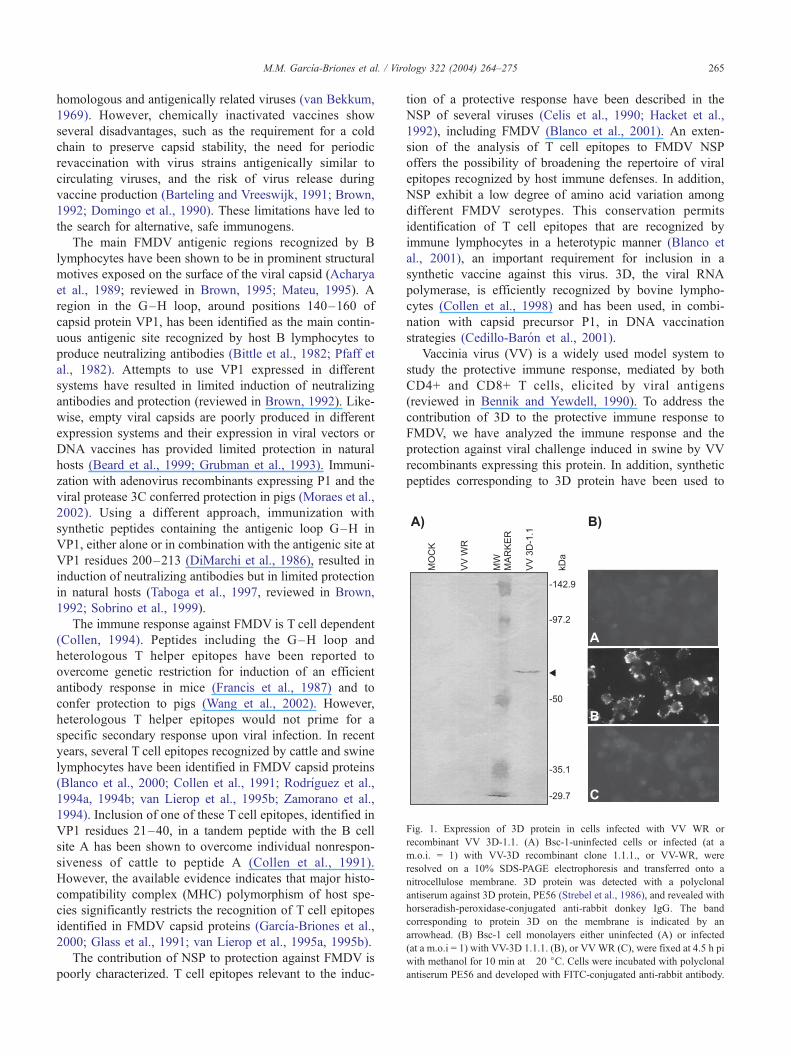
Fig. 1. Expression of 3D protein in cells infected with VV WR or
recombinant VV 3D-1.1. (A) Bsc-1-uninfected cells or infected (at a
m.o.i. = 1) with VV-3D recombinant clone 1.1.1., or VV-WR, were
resolved on a 10% SDS-PAGE electrophoresis and transferred onto a
nitrocellulose membrane. 3D protein was detected with a polyclonal
antiserum against 3D protein, PE56 (Strebel et al., 1986), and revealed with
horseradish-peroxidase-conjugated anti-rabbit donkey IgG. The band
corresponding to protein 3D on the membrane is indicated by an
arrowhead. (B) Bsc-1 cell monolayers either uninfected (A) or infected
(at a m.o.i = 1) with VV-3D 1.1.1. (B), or VVWR (C), were fixed at 4.5 h pi
with methanol for 10 min at �20 jC. Cells were incubated with polyclonal
antiserum PE56 and developed with FITC-conjugated anti-rabbit antibody.
M.M. Garcıa-Briones et al. / Virology 322 (2004) 264–275 265
homologous and antigenically related viruses (van Bekkum,
1969). However, chemically inactivated vaccines show
several disadvantages, such as the requirement for a cold
chain to preserve capsid stability, the need for periodic
revaccination with virus strains antigenically similar to
circulating viruses, and the risk of virus release during
vaccine production (Barteling and Vreeswijk, 1991; Brown,
1992; Domingo et al., 1990). These limitations have led to
the search for alternative, safe immunogens.
The main FMDV antigenic regions recognized by B
lymphocytes have been shown to be in prominent structural
motives exposed on the surface of the viral capsid (Acharya
et al., 1989; reviewed in Brown, 1995; Mateu, 1995). A
region in the G–H loop, around positions 140–160 of
capsid protein VP1, has been identified as the main contin-
uous antigenic site recognized by host B lymphocytes to
produce neutralizing antibodies (Bittle et al., 1982; Pfaff et
al., 1982). Attempts to use VP1 expressed in different
systems have resulted in limited induction of neutralizing
antibodies and protection (reviewed in Brown, 1992). Like-
wise, empty viral capsids are poorly produced in different
expression systems and their expression in viral vectors or
DNA vaccines has provided limited protection in natural
hosts (Beard et al., 1999; Grubman et al., 1993). Immuni-
zation with adenovirus recombinants expressing P1 and the
viral protease 3C conferred protection in pigs (Moraes et al.,
2002). Using a different approach, immunization with
synthetic peptides containing the antigenic loop G–H in
VP1, either alone or in combination with the antigenic site at
VP1 residues 200–213 (DiMarchi et al., 1986), resulted in
induction of neutralizing antibodies but in limited protection
in natural hosts (Taboga et al., 1997, reviewed in Brown,
1992; Sobrino et al., 1999).
The immune response against FMDV is T cell dependent
(Collen, 1994). Peptides including the G–H loop and
heterologous T helper epitopes have been reported to
overcome genetic restriction for induction of an efficient
antibody response in mice (Francis et al., 1987) and to
confer protection to pigs (Wang et al., 2002). However,
heterologous T helper epitopes would not prime for a
specific secondary response upon viral infection. In recent
years, several T cell epitopes recognized by cattle and swine
lymphocytes have been identified in FMDV capsid proteins
(Blanco et al., 2000; Collen et al., 1991; Rodrıguez et al.,
1994a, 1994b; van Lierop et al., 1995b; Zamorano et al.,
1994). Inclusion of one of these T cell epitopes, identified in
VP1 residues 21–40, in a tandem peptide with the B cell
site A has been shown to overcome individual nonrespon-
siveness of cattle to peptide A (Collen et al., 1991).
However, the available evidence indicates that major histo-
compatibility complex (MHC) polymorphism of host spe-
cies significantly restricts the recognition of T cell epitopes
identified in FMDV capsid proteins (Garcıa-Briones et al.,
2000; Glass et al., 1991; van Lierop et al., 1995a, 1995b).
The contribution of NSP to protection against FMDV is
poorly characterized. T cell epitopes relevant to the induc-
tion of a protective response have been described in the
NSP of several viruses (Celis et al., 1990; Hacket et al.,
1992), including FMDV (Blanco et al., 2001). An exten-
sion of the analysis of T cell epitopes to FMDV NSP
offers the possibility of broadening the repertoire of viral
epitopes recognized by host immune defenses. In addition,
NSP exhibit a low degree of amino acid variation among
different FMDV serotypes. This conservation permits
identification of T cell epitopes that are recognized by
immune lymphocytes in a heterotypic manner (Blanco et
al., 2001), an important requirement for inclusion in a
synthetic vaccine against this virus. 3D, the viral RNA
polymerase, is efficiently recognized by bovine lympho-
cytes (Collen et al., 1998) and has been used, in combi-
nation with capsid precursor P1, in DNA vaccination
strategies (Cedillo-Baron et al., 2001).
Vaccinia virus (VV) is a widely used model system to
study the protective immune response, mediated by both
CD4+ and CD8+ T cells, elicited by viral antigens
(reviewed in Bennik and Yewdell, 1990). To address the
contribution of 3D to the protective immune response to
FMDV, we have analyzed the immune response and the
protection against viral challenge induced in swine by VV
recombinants expressing this protein. In addition, synthetic
peptides corresponding to 3D protein have been used to

Table 1
Pyrexia and clinical symptoms developed in pigs after FMDV challenge
M.M. Garcıa-Briones et al. / Virology 322 (2004) 264–275266
identify T cell epitopes in this protein consistently recog-
nized by lymphocytes from infected animals.
Inoculum Animal Pyrexiaa LesionsbEmergence Duration Snoutc Feetd
VV 3D A1 3 3 1 (4) 1,2,0 (4)
A2 3 4 1 (4) 1,1,1 (4)
A3 3 3 none 0,1,1 (4)
VV WR B1 2 5 3 (3) 3,2,3 (3)
B2 1 7 3 (3) 2,3,2 (2)
E-MEM C1 1 7 2 (3) 2,3,2 (2)
C2 3 5 3 (3) 3,3,3 (2)
a Estimated as rectal temperature >39 jC. The day of emergence and the
duration (in days) are indicated.b The day of emergence of the lesions is indicated in parenthesis.c Approximate size (diameter) of the secondary lesions in the snout: 1 (<5
mm), 2 (5–10 mm), 3 (>10 mm).d The severity of the secondary lesions in the feet was estimated from 0
Results
Construction of recombinant VV expressing FMDV 3D
We first studied the immune response elicited by the
FMDV polymerase 3D expressed by a replicating vector. To
this end, recombinant VV harboring a copy of 3D gene were
obtained as described in Materials and methods. A recom-
binant clone, VV 3D-1.1, was selected to infect Bsc-1 cells
that were analyzed for 3D protein expression. A product of
the expected size (56 kDa) was detected by Western blotting
in extracts of infected cells (Fig. 1A). Control cells, either
Fig. 2. Specific antibody responses in pigs inoculated with VV WR or VV
3D-1.1. (A) Ratio of ELISA titers (OD) against a FMDV 3D recombinant
protein between serum collected at days 30 post re-inoculation (black bars)
or 15 after viral challenge (stripped bars) and serum obtained before the first
immunization (day 0). (B) ELISA titers against VV WR in sera from
animals at day 0 (white bars) and at day 30 post re-inoculation (black bars).
Data are expressed as the inverse of the serum dilution that gave 50% of the
maximum OD obtained for each serum. n.d., not determined. The ELISAs
used are detailed in Materials and methods.
(none) to 3, based on the average size and number of the vesicles observed
at the left fore, right hind, and left hind foot, respectively.
uninfected or infected with VV WR, did not show 3D
expression. The expression of this protein was also evalu-
ated by indirect immunofluorescence. As shown in Fig. 1B,
cells infected with VV 3D-1.1 exhibited a high intensity of
3D-specific fluorescence that appears in the periphery of
infected cells. In FMDV infected cells, 3D expression is in
the cytosol (data not shown). Thus, further experiments are
required to study the possible association of 3D with
membrane structures in the context of its expression in
VV. Expression of 3D in infected cells was also confirmed
by dot blot (data not shown).
Immune response elicited by recombinant VV 3D in pigs
To study the immune response and the protection in-
duced by VV recombinants expressing 3D, three pigs (group
A) were inoculated, oronasally and subcutaneously, with
recombinant VV 3D-1.1, as detailed in Materials and
methods. As controls, two (group B) and three (group C)
animals were inoculated with VV WR or E-MEM, respec-
tively. Animals of groups A and B received two equal doses
of virus (109PFU) at days 1 and 30.
The induction of specific 3D antibodies in immunized
pigs was estimated from serum samples obtained at days 0
(before infection), 30 post re-inoculation, and 15 days after
challenge. As shown in Fig. 2A, the three animals inocu-
lated with VV 3D-1.1 showed at day 30 post re-inoculation
ELISA values (OD), relative to those observed at day 0,
slightly higher than those found in pigs inoculated with VV
WR. This difference was in the limit of significance and
could be due to experimental variation. After challenge, all
the animals showed similar OD ratios that were greater than
those observed before challenge. The three pigs immunized
with VV 3D-1.1 showed detectable antibody titers against
VV at day 30 post re-inoculation as estimated by an ELISA
using VV WR (Fig. 2B). In addition, no detectable neutral-
izing activity against the homologous FMDV C-S8 was

M.M. Garcıa-Briones et al. / Virology 322 (2004) 264–275 267
found in a parallel analysis of sera from day 30 post re-
inoculation using a plaque reduction assay (data not shown).
Protection of immunized pigs against FMDV challenge
Forty-five days post re-inoculation, pigs from groups A,
B, and C were challenged by intradermal injection of
homologous FMDV C-S8. Clinical symptoms were moni-
tored daily by measuring rectal temperatures and following
the appearance of lesions in mouth and feet. The criteria
considered as indicative of protection were the delay,
Table 2
Synthetic peptides used in this studya
Amino acid sequence Peptideb
GLIVDTRDVEERVHV 1 (1–15)
TRDVEERVHVMRKTK 2 (6–20)
ERVHVMRKTKLAPTV 3 (11–25)
MRKTKLAPTVAHGVF 4 (16–30)
LAPTVAHGVFNPEFG 5 (21–35)
AHGVFNPEFGPAALS 6 (26–40)
NPEFGPAALSNKDPR 7 (31–45)
PAALSNKDPRLNEGV 8 (36–50)
NKDPRLNEGVVLDEV 9 (41–55)
LNEGVVLDEVIFSKH 10 (46–60)
VLDEVIFSKHKGDTK 11 (51–65)
IFSKHRGDTKMSAED 12 (56–70)
RGDTKMSAEDKALFR 13 (61–75)
MSAEDKALFRRCAAD 14 (66–80)
KALFRRCAADYASRL 15 (71–85)
RCAADYASRLHSVLG 16 (76–90)
YASRLHSVLGTANAP 17 (81–95)
HSVLGTANAPLSIYE 18 (86–100)
TANAPLSIYEAIKGV 19 (91–105)
LSIYEAIKGVDGLDA 20 (96–110)
AIKGVDGLDAMEPDT 21 (101–115)
DGLDAMEPDTAPGLP 22 (106–120)*
MEPDTAPGLPWALQG 23 (111–125)
APGLPWALQGKRRGA 24 (116–130)
WALQGKRRGALIDFE 25 (121–135)
KRRGALIDFENGTVG 26 (126–140)
LIDFENGTVGPEVEA 27 (131–145)
NGTVGPEVEAALKLM 28 (136–150)
PEVEAALKLMEKREY 29 (141–155)
ALKLMEKREYKFACQ 30 (146–160)
EKREYKFACQTFLKD 31 (151–165)
KFACQTFLKDEIRPM 32 (156–170)
TFLKDEIRPMEKVRA 33 (161–175)
EIRPMEKVRAGKTRI 34 (166–180)
EKVRAGKTRIVDVLP 35 (171–185)
GKTRIVDVLPVEHIL 36 (176–190)
VDVLPVEHILYTRMM 37 (181–195)
VEHILYTRMMIGRFC 38 (186–200)
YTRMMIGRFCAQMHS 39 (191–205)
IGRFCAQMHSNNGPQ 40 (196–210)
AQMHSNNGPQIGSAV 41 (201–215)
NNGPQIGSAVGCNPD 42 (206–220)
IGSAVGCNPDVDWQR 43 (211–225)
GCNPDVDWQRFGTHF 44 (216–230)
VDWQRFGTHFAQYRN 45 (221–235)
FGTHFAQYRNVWDVD 46 (226–240)
a Amino acid sequence (as in Toja et al., 1999) and numbering are indicated forb The yield of the chemical synthesis of peptides 22 and 59 (denoted by an aster
reduction or absence of epithelial lesions and pyrexia (rectal
temperature >39 jC). As shown in Table 1, a significant
reduction in the clinical symptoms developed upon chal-
lenge was noticed in animals immunized with VV 3D
(group A). Control animals (groups B and C) developed
severe clinical signs: large vesicles in feet and snout,
anorexia and lameness, and showed pyrexia from days 1
to 3 that lasted between 5 and 7 days post challenge.
Conversely, pigs from group A showed a delay of 1–2
days in the onset of pyrexia and lesions (vesicles). In
addition, only two of the three animals of this group
Amino acid sequence Peptideb
AQYRNVWDVDYSAFD 47 (231–145)
VWDVDYSAFDANHCS 48 (236–250)
YSAFDANHCSDAMNI 49 (141–255)
ANHCSDAMNIMFEEV 50 (246–260)
DAMNIMFEEVFRTEF 51 (251–265)
MFEEVFRTEFGFHPN 52 (256–270)
FRTEFGFHPNAEWYL 53 (261–275)
GFHPNAEWYLKTLVN 54 (266–280)
AEWYLKTLVNTEHAY 55 (271–285)
KTLVNTEHAYENKRI 56 (276–290)
TEHAYENKRITVEGG 57 (281–295)
ENKRITVEGGMPSGC 58 (286–300)
TVEGGMPSGCSATSI 59 (291–305)*
MPSGCSATSIINTIL 60 (296–310)
SATSIINTILNNIYV 61 (301–315)
INTILNNIYVLYALR 62 (306–320)
NNIYVLYALRRAYEG 63 (311–325)
LYALRRAYEGVELDT 64 (316–330)
RAYEGVELDTYTMIS 65 (321–335)
VELDTYTMISYGDDI 66 (326–340)
YTMISYGDDIVVASD 67 (331–345)
YGDDIVVASDYDLDF 68 (336–350)
VVASDYDLDFEALKP 69 (341–355)
YDLDFEALKPHFKSL 70 (346–360)
EALKPHFKSLGQTYT 71 (351–365)
HFKSLGQTYTPADKS 72 (356–370)
GQTYTPADKSDKGFV 73 (361–375)
PADKSDKGFVLGHSI 74 (366–380)
DKGFVLGHSITDVTF 75 (371–385)
LGHSITDVTFLKRHF 76 (376–390)
TDVTFLKRHFHMDYG 77 (381–395)
LKRHFHMDYGTGFYK 78 (386–400)
HMDYGTGFYKPVMAS 79 (391–405)
TGFYKPVMASKTLEA 80 (396–410)
PVMASKTLEAILSFA 81 (401–415)
KTLEAILSFARRGTI 82 (406–420)
ILSFARRGTIQEKLI 83 (411–425)
RRGTIQEKLISVAGL 84 (416–430)
QEKLISVAGLAVHSG 85 (421–435)
SVAGLAVHSGPDEYR 86 (426–440)
AVHSGPDEYRRLFEP 87 (431–445)
PDEYRRLFEPFQGLF 88 (436–450)
RLFEPFQGLFEIPSY 89 (441–455)
FQGLFEIPSYRSLYL 90 (446–460)
EIPSYRSLYLRWVNA 91 (451–465)
RSLYLRWVNAVCGDA 92 (456–470)
each peptide.
isk) was low and did not allow its use in the proliferation experiments.
M.M. Garcıa-Briones et al. / Virology 322 (2004) 264–275268
developed lesions at the snout and these lesions were of a
small size (Table 1).
These results indicate that immunization of pigs with VV
3D-1.1 induced partial protection to viral challenge in the
absence of induction of detectable neutralizing antibodies
against FMDV. These results suggest that expression of 3D
in pigs might elicit T cell responses that could play a role in
the delay and reduction of the symptoms observed. Identi-
fication of T cell epitopes is an informative approach to
understand the T immune mechanisms elicited by FMDV
proteins (Blanco et al., 2000, 2001; van Lierop et al., 1995a,
1995b) and can allow identification of epitopic components
relevant for the design of subunit vaccines. Thus, to gain
insight on the contribution of 3D to the viral immune
response, we conducted a new experiment to assess the
antigenic specificity of the T cell recognition of 3D protein
in infected pigs.
Identification of T cell epitopes recognized on 3D by pigs
infected with FMDV
A new group of five pigs (D1–D5, group D) that was
infected with FMDV C-S8 was used to study the antigenic
specificity of the T cell response elicited by 3D protein. To
this end, 92 overlapping (15-mer) peptides covering the
amino acid sequence of 3D were synthesized (see Materials
and methods and Table 2 for details) and used in lympho-
proliferation assays. Due to the high number of peptides to
be tested, assays were performed with pools of two to three
overlapping peptides. These peptide pools were employed
to stimulate in vitro lymphocytes obtained at days 0, 10, 21,
28, 35, 42, and 63 p.i. None of the peptide pools induced
positive SI in PBMC collected before infection. Peptides
inducing positive responses (SI z 2.5) in at least four of the
five animals studied are shown in Fig. 3, in which the higher
SI observed at the different days p.i. analyzed are indicated.
Responses against peptides and whole virus were detectable
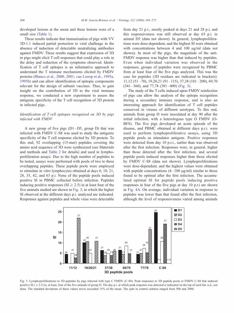
Fig. 3. Lymphoproliferations to 3D peptides by pigs infected with type C FMDV
positive SI (z 2.5) in, at least, four of the five animals of group D. The day p.i. at w
done. The standard deviations of these values never exceeded 15% of the mean.
from day 21 p.i., mostly peaked at days 21 and 28 p.i, and
this responsiveness was still observed at day 63 p.i. in
animal D5 (data not shown). In general, lymphoprolifera-
tions were dose-dependent, and the highest SI were obtained
with concentrations between 4 and 100 Ag/ml (data not
shown). In most of the pigs, the magnitude of the anti-
FMDV response was higher than that induced by peptides.
Even when individual variation was observed in the
responses, groups of peptides were recognized by PBMC
from at least four of the five pigs analyzed. This was the
case for peptides (3D residues are indicated in brackets):
11,12 (51–70), 19,20,21 (91–115), 37,38 (181–200), 69,70
(341–360), and 77,78 (381–400) (Fig. 3).
The study of the T cells induced upon FMDV reinfection
of pigs can allow the analysis of the epitope recognition
during a secondary immune response, and is also an
interesting approach for identification of T cell peptides
conserved in viruses of different serotypes. To this end,
animals from group D were inoculated at day 90 after the
initial infection, with a heterologous type O FMDV (O-
BFS). The five pigs developed an acute episode of the
disease, and PBMC obtained at different days p.r.i. were
used to perform lymphoproliferative assays, using 3D
peptide pools as stimulator antigens. Positive responses
were detected from day 10 p.r.i., earlier than was observed
after the first infection. Responses were, in general, higher
than those detected after the first infection, and several
peptide pools induced responses higher than those elicited
by FMDV C-S8 (data not shown). Lymphoproliferations
were dose-dependent, and the highest values were obtained
with peptide concentrations (4–100 Ag/ml) similar to those
found to be optimal after the first infection. The accumu-
lated optimal SI for peptide pools inducing positive
responses in four of the five pigs at day 10 p.r.i are shown
in Fig. 4A. On average, individual variation in response to
peptides was lower than that found after the first infection,
although the level of responsiveness varied among animals
(C-S8). Peak responses to 3D peptide pools or FMDV C-S8 that induced
hich peak response was detected is indicated on the top of each bar. n.d., not
The cpm in control cultures ranged from 500 and 2000.
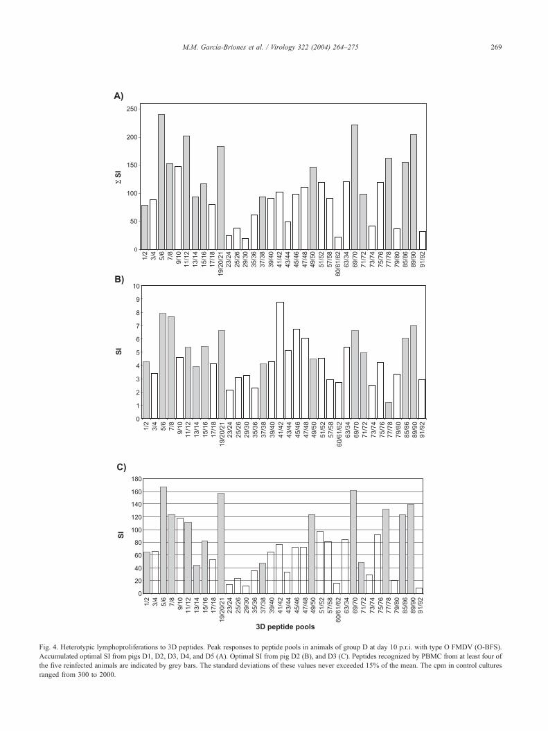
Fig. 4. Heterotypic lymphoproliferations to 3D peptides. Peak responses to peptide pools in animals of group D at day 10 p.r.i. with type O FMDV (O-BFS).
Accumulated optimal SI from pigs D1, D2, D3, D4, and D5 (A). Optimal SI from pig D2 (B), and D3 (C). Peptides recognized by PBMC from at least four of
the five reinfected animals are indicated by grey bars. The standard deviations of these values never exceeded 15% of the mean. The cpm in control cultures
ranged from 300 to 2000.
M.M. Garcıa-Briones et al. / Virology 322 (2004) 264–275 269

Fig. 5. Heterotypic lymphoproliferations to individual 3D peptides. Peak
responses to individual peptides in animals of group D at different days
p.r.i. with FMDV O-BFS. Peptide number is indicated on top of the bar.
SI corresponded to day 51 p.r.i. The standard deviations of these values
never exceeded 15% of the mean. The cpm in control cultures ranged
from 300 to 2000.
Table 3
Analysis of cytokine mRNA expression by RT-PCR
Peptidea Pigb G3PDHc IFN-gd IL-4d IL-10d
1 D2 + + � �11 D1 + + � �13 D1/D2 + �/+ � �15 D2 + + � �20 D1/D2 +/+ +/+ +/+ �/�72 D2 + + � �77 D2/D4 +/+ +/+ +/� �85 D2 + + � �89 D1 + + + �a Peptide used for PBMC stimulation.b PBMC were obtained at day 35 p.r.i., except for pig D2 whose
lymphocytes were collected at day 51 p.r.i.c Positive amplification was obtained in PBMC cultured in the absence of
peptide stimulation.d Negative amplification was obtained in PBMC cultured in the absence of
peptide stimulation.
M.M. Garcıa-Briones et al. / Virology 322 (2004) 264–275270
(Figs. 4B, C). This pattern of response was maintained at
days 21 and 35 p.r.i. (data not shown). The following 3D
peptides were efficiently recognized by at least four of the
five reinfected pigs: 1,2; 5–8; 11–16; 19–21; 37,38; 49,50;
69–72; 77,78; 85,86; and 89–90 (Fig. 4A), which included
peptides identified as antigenic after the first infection (Fig.
3). When individual peptides from stimulator pools (11,12;
19,20,21; 49,50; 77,78; and 85,86) were included in prolif-
eration assays, variations in the SI induced by individual
peptides were observed between animals. However, in all
cases, at least one of the overlapping peptides gave a high
level response (Fig. 5).
As summarized in Fig. 6, the results obtained indicate
that 3D protein regions contain T cell epitopes which are
efficiently recognized by porcine T lymphocytes from
different infected animals, both upon primary and secondary
(heterotypic) FMDV infection.
Cytokine profiles induced in PBMC by different peptides
To gain information on the functional implications of the
lymphoproliferative responses observed, antigenic peptides
were used to determine the cytokine profiles that they
Fig. 6. Schematic representation of 3D peptides pools that were efficiently recogn
reinfection with FMDV. Grey quadrangles denote sequences spanned by peptide
sequence stretches containing peptides recognized upon reinfection with FMDV
indicated in normal and italic characters, respectively.
induced upon in vitro incubation of PBMC from FMDV
reinfected pigs. To this end, mRNA from the stimulated
cells was extracted and amplified by RT-PCR using specific
primers as described in Materials and methods. Table 3
summarizes the RT-PCR amplification of IFN-g, IL-4, and
IL-10 mRNAs in peptide-stimulated cultures of different
pigs. For most of the peptides analyzed, positive amplifi-
cation of IFN-g mRNAs was detected. RT-PCR from
PBMC cultures incubated in the absence of peptides did
not render detectable amplification of IFN-g, IL-4, and IL-
10 mRNAs, although positive amplification was observed
for G3PDH mRNA. These results suggest that the lympho-
proliferations induced by 3D peptides are mediated by T
cells and suggests that a Th1 response is produced when T
cells from infected animals are stimulated with 3D FMDV
peptides.
Discussion
Partial protection in the absence of anti-FMDV neutral-
izing antibodies has been reported in cattle and pigs immu-
ized by lymphocytes from at least four of the five pigs upon infection and
s recognized upon infection with FMDV C-S8. Black quadrangles denote
O-BFS. Amino acid positions and peptide numbering (as in Table 2) are

M.M. Garcıa-Briones et al. / Virology 322 (2004) 264–275 271
nized with VV and adenovirus expressing capsid proteins
precursor P1 (Sanz-Parra et al., 1999a,1999b). These obser-
vations emphasize the role of the T cell response in the
protection of pigs against FMDV infection (Childerstone et
al., 1999; Collen, 1994). However, little is known on the
role of FMDV specific T cells in protection mechanisms
other than their contribution to induction of anti-FMDV
antibodies, particularly on the activation and functional
implications of FMDV CTL lymphocytes (Sobrino et al.,
2002).
In this report, we have shown that inoculation of pigs
with a VV recombinant expressing FMDV 3D protein
(group A) results in a delay in the emergence and a lower
severity of the lesions developed by immunized animals
upon viral challenge, relative to those observed in control
animals (groups B and C). This partial protection occurs in
the absence of induction of detectable anti-FMDV neutral-
izing antibodies, providing new evidence in favor of the
involvement of non-antibody-mediated responses in the
induction of protection against FMDV. The immune mech-
anisms underlying the partial protection observed in animals
immunized with VV 3D-1.1 remained unclear because the
use of a recombinant 3D protein expressed as a fusion in E.
coli did not allow detection of significant lymphoprolifer-
ative responses in infected animals (data not shown). This
lack of responsiveness was likely to be due to the poor
antigenicity of the 3D recombinant used (Blanco et al.,
2001). Also, the difficulties for optimizing FMDV CTL
assays with pig lymphocytes (Childerstone et al., 1999;
Rodrıguez et al., 1996) have impaired attempts to analyze
the contribution of this response to the partial protections
observed. However, these results emphasize the importance
of the characterization of the T cell responses elicited by
FMDV proteins.
Recognition of T cell epitopes by lymphocytes from
different host species and individuals is restricted by the
polymorphism of the MHC molecules, which are responsi-
ble for the presentation of foreign antigens by antigen
presenting cells (Germain, 1999). Advances have been
made in the last years in the analysis of the antigenic
specificity of FMDV specific T helper cells, which are
required for an efficient activation of specific B cells
(reviewed in Collen, 1994; Sobrino et al., 2002).
One of the aims of these studies is the identification of
FMDV T cell epitopes capable of inducing an effective
response, although being widely recognized by MHC alleles
frequent in natural populations of host species. Identifica-
tion of such T cell epitopes is relevant for the improvement
of new vaccines, particularly of those based on subunit
vaccines (Rowlands, 1994; Sobrino et al., 1999). 3D poly-
merase is one of the FMDV proteins showing a lower
degree of amino acid variation between isolates of different
serotypes (Domingo et al., 1990). Therefore, 3D is an
interesting candidate to contain conserved viral T cell
epitopes, as suggested by its heterotypic recognition by
bovine T cells (Collen, 1994) and the improvement of the
immune response elicited by P1-based DNA vaccines when
3D protein was co-expressed (Cedillo-Baron, 2001).
To gain information of the T cell immunogenicity of
FMDV 3D protein, we conducted an analysis of the anti-
genic specificity of the lymphocytes from a new group of
pigs (group D) infected with FMDV C-S8. As reported for
other FMDV proteins (Blanco et al., 2001; Rodrıguez et al.,
1994b), several peptides along 3D were efficiently recog-
nized by lymphocytes from infected pigs and this recogni-
tion showed individual animal variation. Five peptide pools
stimulated efficiently lymphocytes from four of the five
animals tested (Fig. 3A) as summarized in Fig. 6. These
peptides were consistently recognized by lymphocytes from
animals reinfected with a type O FMDV. In addition, four of
the five reinfected animals efficiently recognized eight new
peptide pools (Fig. 3B) as indicated in Fig. 6. Consistently
with induction of a secondary response, the proliferations
observed were, in general, higher than those developed after
the first infection. Proliferation to individual peptides con-
firmed that at least one of the peptides incorporated in the
pools induced high proliferation levels in reinfected pigs
(Fig. 5).
Although the number of animals used for this type of
study was not sufficient for statistical demonstration, we
think that the data are consistent with the identification of T
cell epitopes in 3D able to stimulate porcine T cells
following infection with viruses of different serotypes. This
heterotypic recognition of FMDV peptides correlates well
with the high level of 3D sequence conservation among
different FMDV serotypes (99.2% homology between
FMDV C-S8 and O-BFS) (Martınez-Salas et al., 1985).
Heterotypic recognition of peptides corresponding to con-
served FMDV proteins has been reported also for peptides
in VP4 and 3ABC (Blanco et al., 2000, 2001).
Activation of a Th 1 response has been associated with
efficient protection against FMDV (van Lierop et al., 1995a,
1995b). The cytokine profile in PBMC stimulated in vitro
by 3D peptides revealed that IFN-g mRNA synthesis was
induced in most of the cases. These results suggest that T
cells that become activated in response to 3D peptides
phenotypically belong to the Th 1 subset. Further charac-
terization of the responder cell populations is required to
identify their phenotype, whether they belong to the CD4+
or CD8+ subset, and the contribution of the cytokines
induced in FMDV immune response.
Taken together, the findings in this paper provide new
evidence of the presence on 3D protein of antigenic regions
efficiently recognized by porcine T lymphocytes. The re-
duced number of B and T cell epitopes included in subunit
and synthetic FMDV vaccines is considered as one of their
limitations for eliciting solid protective immune responses
(Cedillo-Baron et al., 2001; Collen, 1994; Rowlands, 1994;
Sobrino et al., 1999; van Lierop et al., 1995a, 1995b). The
results described here can contribute to improve the reper-
toire of viral T cell epitopes to be included in the design of
new FMDV vaccines.
M.M. Garcıa-Briones et al. / Virology 322 (2004) 264–275272
Materials and methods
Recombinant vaccinia virus expressing 3D polypeptide
The recombinant VV expressing the 3D protein was
constructed as follows: 3D gene sequence was amplified
by PCR from plasmid pUC-3D carrying the whole 3D
gene from FMDV C-S8 (J.C. Saiz, unpublished results),
using the following primers (Isogen, Bioscience. The
Netherlands): 3D1 (antisense), 5V-CCGAATTCTCTA-GAATGGGGTTGATCGGAT-3V; and 3D2 (sense), 3V-AAGCTAGCTCTAGATTATGCGCGTCCGCACAC-3V.These primers allowed amplification of the complete 3D
gene sequence flanked by the recognition sequences of
endonucleases EcoRI and NheI (at the 5V and the 3V end,respectively), as well as in-frame translational start and
stop codons. The PCR product and plasmid pRB21
(Blasco and Moss, 1995) were digested with endonu-
cleases EcoRI and NheI and ligated to obtain plasmid
pRB21-3D. The integrity of 3D gene, including the
initiation and stop codons, was confirmed by DNA
sequencing. Plasmid pRB21-3D was used to produce
recombinant VV using a plaque selection method (Blasco
and Moss, 1992,1995). Briefly, subconfluent CV-1 mono-
layers were infected with the non-plaquing RB12a4 vac-
cinia virus (Blasco and Moss, 1991,1995) and transfected
with 10 Ag of calcium phosphate-precipitated plasmid
pRB21-3D. After 48 h of incubation, cells were detached
from plastic and freeze-thawed three times to release the
virus. Recombinant clones (plaque forming) were selected
after three rounds of plaque purification on Bsc-1 cells.
The presence of guanosine phosphoribotransferase (GPT)
gene in the recombinants was confirmed as described
(Blasco and Moss, 1995).
3D expression was tested by immunoblotting, indirect
immunofluorescence (IIF), and dot-blotting of Bsc-1 cells
infected with the different recombinant clones, using stan-
dard protocols. VV Western Reserve (VV WR) and recom-
binant vaccinia virus expressing 3D were grown and tittered
by plaque assay in Bsc-1 cells.
Animal immunization, experimental infections, and virus
challenge
To analyze the immunogenicity of VV 3D-1.1, 7 pigs (2-
month-old Large White � Landrace), obtained from differ-
ent litters, were inoculated by both oronasal and subcutane-
ous routes with 109 PFU each of VV 3D-1.1.1 (three
animals, group A) or VV WR (two animals, group B).
Two control animals were inoculated with medium (E-
MEM) (group C). At day 30, animals were re-inoculated
with the same regime. Forty-five days later (day 75 post
inoculation), pigs were challenged intradermally, in the heel
bulbs of the right forefoot, with 4.5 � 105 PFU of type C
FMDV (C-S8). After challenge, rectal temperatures and
clinical signs were monitored daily for 10 days.
To analyze the T cell response induced by 3D protein,
five additional pigs (group D) were infected with FMDV C-
S8, as described above. To study the specificity of the
antigen recognition after a secondary response to FMDV,
animals were reinfected with 104 PFU of a type O FMDV
(O-BFS) at day 90 after infection with virus C-S8. In all
cases, blood samples collected at different days post infec-
tion (p.i.) or post reinfection (p.r.i) were used to perform
lymphoproliferative assays against different homologous
FMDV C-S8 antigens.
Peptide synthesis
A total of 92 pentadecapeptides corresponding to the
3D sequence of FMDV C-S8 isolate (Toja et al., 1999)
with a 10-residue overlap (sequences and numbering in
Table 2) were synthesized by solid phase methods (Merri-
field, 1963) using Fmoc chemistry as previously described
(Mateu et al., 1996). After cleavage, all peptides were
>80% pure by HPLC and had amino acid analyses and
MALDI-TOF mass spectra consistent with the expected
compositions.
Antibody determination
ELISA of anti-3D was performed using GST-3D fusion
protein as antigen diluted (2.6 Ag/ml) in carbonate buffer.
After blocking with 3% BSA, 0.05% Tween 20, serum
dilutions ranging from 1/10 to 1/400 were added and
incubated for 12 h at 4 jC. All serum samples were
preincubated with E. coli DH5-a strain extract, lysed by
sonication. Bound antibodies were detected by an anti-
swine immunoglobulin-horseradish peroxidase (Dako,
Denmark). The reaction was developed with 0.4 mg/ml
o-phenylenediamine (Sigma Co., St. Louis, MO) as chro-
mogenic substrate. Results are expressed as the ratio of the
OD at 492 nm determined for a 1/20 dilution of sera from
immunized animals and sera obtained at day 0 (before
immunization).
To evaluate the generation of anti-VV antibodies in
animals from groups A, B, and C, 106 PFU/well of VV
WR in carbonate buffer were used. The blocking step was
done with 1% BSA, 0.05% Tween 20. Sera were preincu-
bated 30 min at 37 jC with a Bsc-1 cell extract, and 2-fold
dilutions (from 1/20 to 1/2500) were incubated overnight
with the VV WR-coated plates at 4 jC. The secondary
antibody used was an anti-swine immunoglobulin-horserad-
ish peroxidase (Dako) and the reaction was developed with
0.4 mg/ml o-phenylenediamine (Sigma Co.) as chromogenic
substrate.
Lymphoproliferation assays
Proliferation assays of swine lymphocytes were per-
formed as described previously (Rodrıguez et al., 1994b).
Blood was collected in 5mM EDTA and used to obtain
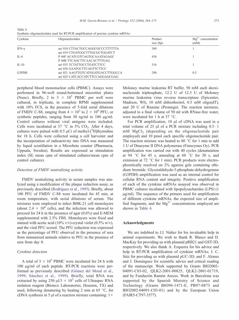
Table 4
Synthetic oligonucleotides used for RT-PCR amplification of porcine cytokine mRNAs
Cytokine Oligonucleotides Product
size (bp)
Mg2+ concentration
(mM)
IFN-g aei 918 CTTACTGCCAGGCGCCCTTTTTTA 389 1
aei 019 CTGATGGCTTTGCGCTGGATCT
IL-4 P 04F ACATCGTCAGTGCAAATAGAGC 438 1
P 04R TTCAGCTTCAACACTTTGAG
IL-10 aei 035 TCTATTGCCTGATCTTCC 516 1
aei 036 GAATGCTTCAGTTCTTCC
G3PDH aei 021 AAGTTGTCATGGATGACCTTGGCCA 298 0.5
aei 025 CATCACCATCTTCCAGGAGCGAG
M.M. Garcıa-Briones et al. / Virology 322 (2004) 264–275 273
peripheral blood mononuclear cells (PBMC). Assays were
performed in 96-well round-bottomed microtiter plates
(Nunc). Briefly, 2 to 3 � 105 PBMC per well were
cultured, in triplicate, in complete RPMI supplemented
with 10% FCS, in the presence of 5-fold serial dilutions
of FMDV C-S8, ranging from 4 � 103 to 2 � 106 PFU, or
synthetic peptides, ranging from 30 ng/ml to 100 Ag/ml.
Control cultures without viral antigens were included.
Cells were incubated at 37 jC in 5% CO2. After 4 days,
cultures were pulsed with 0.5 ACi of methyl-[3H]thymidine
for 18 h. Cells were collected using a cell harvester and
the incorporation of radioactivity into DNA was measured
by liquid scintillation in a Microbeta counter (Pharmacia,
Uppsala, Sweden). Results are expressed as stimulation
index (SI; mean cpm of stimulated cultures/mean cpm of
control cultures).
Detection of FMDV neutralizing activity
FMDV neutralizing activity in serum samples was ana-
lyzed using a modification of the plaque reduction assay, as
previously described (Rodrıguez et al., 1995). Briefly, about
100 PFU of FMDV C-S8 were incubated for 45 min, at
room temperature, with serial dilutions of serum. The
mixtures were employed to infect BHK-21 cell monolayers
(about 2.4 � 106 cells), and the infection was allowed to
proceed for 24 h in the presence of agar (0.6%) and E-MEM
supplemented with 2.5% FBS. Monolayers were fixed and
stained with acetic acid (10% v/v)-crystal violet (0.5% w/v),
and the viral PFU scored. The PFU reduction was expressed
as the percentage of PFU observed in the presence of sera
from immunized animals relative to PFU in the presence of
sera from day 0.
Cytokine detection
A total of 3 � 105 PBMC were incubated for 24 h with
100 Ag/ml of each peptide. RT-PCR reactions were per-
formed as previously described (Gomez del Moral et al.,
1999; Sanchez et al., 1999). Briefly, total RNA was
extracted by using 250 Al/3 � 105 cells of Ultraspec RNA
isolation reagent (Biotecx Laboratories, Houston, TX) and
used, following denaturing by heating 2 min at 65 jC, forcDNA synthesis in 5 Al of a reaction mixture containing: 1�
Moloney murine leukemia RT buffer, 50 mM each deoxi-
nucleoside triphosphate, 12.2 U of 12.5 U of Moloney
murine leukemia virus reverse transcriptase (Epicenter,
Madison, WI), 10 mM dithiothreitol, 0.5 mM oligo(dT),
and 20 U of Rnasine (Promega). The reaction mixtures,
adjusted to a final volume of 50 ml with RNase-free water,
were incubated for 1 h at 37 jC.For PCR amplification, 10 Al of cDNA was used in a
total volume of 25 Al of a PCR mixture including 0.5–1
mM MgCl2 (depending on the oligonucleotide pair
employed) and 10 pmol each specific oligonucleotide pair.
The reaction mixture was heated to 80 jC for 1 min to add
1 U of Dnazyme II DNA polymerase (Finnzymes Oy). PCR
amplification was carried out with 40 cycles (denaturation
at 94 jC for 45 s, annealing at 60 jC for 30 s, and
extension at 72 jC for 1 min). PCR products were electro-
phoretically resolved on 2% agarose gels containing ethi-
dium bromide. Glyceraldehyde-3-phosphate-dehydrogenase
(G3PDH) amplification was used as an internal control for
cellular RNA content and integrity. Positive amplification
of each of the cytokine mRNAs assayed was observed in
PBMC cultures incubated with lipopolysacharides (LPS) (1
Ag/ml). The sequence of the primers used for amplification
of different cytokine mRNAs, the expected size of ampli-
fied fragments, and the Mg2+ concentrations employed are
shown in Table 4.
Acknowledgments
We are indebted to J.I. Nunez for his invaluable help in
animal experiments. We wish to thank R. Blasco and D.
MacKay for providing us with plasmid pRB21 and GST-3D,
respectively. We also thank A. Ezquerra for his advice and
help in RT-PCR amplification of cytokine mRNAs; J. C.
Saiz for providing us with plasmid pUC-3D; and F. Alonso
and J. Domınguez for scientific advice and critical reading
of the manuscript. Work supported by Grants BIO2002-
04091-C03-02, QLK2-2001-00825, QLK2-2001-01719,
and by Fundacion Ramon Areces. Work in Barcelona was
supported by the Spanish Ministry of Science and
Technology (Grants BIO96-1471-E, PB97-0873 and
BIO2002-04091-C03-01) and by the European Union
(FAIR5-CT97-3577).
M.M. Garcıa-Briones et al. / Virology 322 (2004) 264–275274
References
Acharya, R., Fry, E., Stuart, D., Fox, G., Rowlands, D., Brown, F., 1989.
The three dimensional structure of foot-and-mouth disease virus at 2.9
A resolution. Nature 337, 709–716.
Bachrach, H.L., 1977. Foot-and-mouth disease virus, properties, molecular
biology and immunogenicity. In: Romberger, J.A. (Ed.), Beltsville Sym-
posia in Agricultural Research, I, Virology in Agriculture. Allanheld,
Osmun, Montclair, NJ.
Barteling, S.J., Vreeswijk, J., 1991. Development in foot-and-mouth dis-
ease vaccines. Vaccine 9, 75–88.
Beard, C., Ward, G., Rieder, E., Chinsangaram, J., Grubman, M.J., Mason,
P.W., 1999. Development of DNA vaccines for foot-and-mouth disease,
evaluation of vaccines encoding replicating and non-replicating nucleic
acids in swine. J. Biotechnol. 73, 243–249.
Belsham, G.J., 1993. Distinctive features of foot-and-mouth disease virus,
a member of the picornavirus family; aspects of virus protein synthe-
sis, protein processing and structure. Prog. Biophys. Mol. Biol. 60,
241–260.
Bennik, J.R., Yewdell, J.W., 1990. Recombinant vaccinia viruses as vectors
for studying T lymphocyte specificity and function. Curr. Top. Mibro-
biol. Immunol. 163, 153–183.
Bittle, J.L., Houghten, R.A., Alexander, H., Shinnick, T.M., Sutcliffe, J.G.,
Lerner, R.A., Rowlands, D.J., Brown, F., 1982. Protection against foot-
and-mouth disease by immunization with a chemically synthesized pep-
tide predicted from the viral nucleotide sequence. Nature 298, 30–33.
Blanco, E., McCullough, K., Summerfield, A., Fiorini, J., Andreu, D.,
Chiva, C., Borras, E., Barnett, P., Sobrino, F., 2000. Interspecies
MHC-restricted Th cell epitope on foot-and-mouth disease virus capsid
protein VP4. J. Virol. 74, 4902–4907.
Blanco, E., Garcıa-Briones, M.M., Sanz-Parra, A., Gomes, P., de Oliveira,
E., Valero, M.L., Andreu, D., Ley, V., Sobrino, F., 2001. Identification
of T-cell epitopes in nonstructural proteins of foot-and-mouth disease
virus. J. Virol. 35, 3164–3174.
Blasco, R., Moss, B., 1991. Extracellular vaccinia virus formation and cell-
to-cell virus transmission are prevented by deletion of the gene encod-
ing the 37000 Dalton outer envelope protein. J. Virol. 65, 5910–5920.
Blasco, R., Moss, B., 1992. Role of cell-associated enveloped vaccinia
virus in cell-to-cell spread. J. Virol. 66, 4170–4179.
Blasco, R., Moss, B., 1995. Selection of recombinants vaccinia viruses on
the basis of plaque formation. Gene 158, 157–162.
Brown, F., 1992. New approaches to vaccination against foot-and-mouth
disease. Vaccine 10, 1022–1026.
Brown, F., 1995. Antibody recognition and neutralization of foot-and-
mouth disease virus. Semin. Virol. 6, 243–248.
Cedillo-Baron, L., Foster-Cuevas, M., Belsham, G., Lefevre, F., Parkhouse,
M.E., 2001. Induction of a protective response in swine vaccinated with
DNA encoding foot-and-mouth disease virus empty capsid proteins and
the 3D RNA polymerase. J. Gen. Virol. 82, 1713–1724.
Celis, E., Larson Jr., J., Otvos, L., Wunner, W.H., 1990. Identification of a
rabies virus T cell epitope on the basis of its similarity with a hepatitis B
surface antigen peptide presented to T cells by the same MHC molecule
(HLA-DPw4). J. Immunol. 145, 305–310.
Childerstone, A.J., Cedillo-Baron, J., Foster-Cuevas, R., Parkhouse, M.E.,
1999. Demonstration of bovine CD8+ T-cell responses to foot-and-
mouth disease virus. J. Gen. Virol. 80, 663–669.
Collen, T., 1994. In: Goddeevis, B.M.L., Morrison, I. (Eds.), Cell Mediated
Immunity in Ruminants, Foot-and-mouth Disease (Aphthovirus): Viral
T Cell Epitopes. CRC Press, Boca Raton, FL.
Collen, T., DiMarchi, R., Doel, T.A., 1991. AT cell epitope in VP1 of foot-
and-mouth disease virus is immunodominant for vaccinated cattle. J.
Immunol. 146, 749–755.
Collen, T., Baron, J., Childerstone, A., Corteyn, A., Doel, T.R., Flint, M.,
Garcıa-Valcarcel, M., Parkhouse, R.M.E., Ryan, M.D., 1998. Hetero-
typic recognition of recombinant FMDV proteins by bovine T-cells: the
polymerase (P3Dpol) as an immunodominant T-cell immunogen. Virus
Res. 56, 125–133.
DiMarchi, R., Brooke, G., Gale, C., Cracknell, V., Doel, T., Mowat, N.,
1986. Protection of cattle against foot-and-mouth disease by a synthetic
peptide. Science 232, 639–641.
Domingo, E., Mateu, M.G., Martınez, M.A., Dopazo, J., Moya, A.,
Sobrino, F., 1990. Applied virology research. In: Kurstak, E., Marusyk,
R.G., Murphy, S.A., Van Regenmortel, M.H.V. (Eds.), Virus Variation
and Epidemiology, Genetic Variability and Antigenic Diversity of Foot-
and-Mouth Disease Virus, vol. 2. Plenum, New York, NY.
Francis, M.J., Hastings, G.Z., Syred, A.D., McGinn, B., Brown, F., Row-
lands, D.J., 1987. Non-responsiveness to a foot-and-mouth disease virus
peptide overcomed by addition of foreign helper T-cell determinants.
Nature 330, 168–170.
Garcıa-Briones, M.M., Russell, G.C., Oliver, R.A., Tami, C., Taboga, O.,
Carrillo, E., Palma, E.L., Sobrino, F., Glass, E.J., 2000. Association of
bovine DRB3 alleles with immune response to FMDV peptides and
protection against viral challenge. Vaccine 19, 1167–1171.
Germain, R.N., 1999. In: Paul, W.E. (Ed.), Fundamental Immunology.
Antigen Processing and Presentation. Lippincot-Raven, New York, NY.
Glass, E.J., Oliver, R.A., Collen, T., Doel, T.R., DiMarchi, R., Spooner,
R.L., 1991. MHC Class II restricted recognition of FMDV peptides by
bovine T cells. Immunology 74, 594–599.
Gomez del Moral, M., Ortuno, E., Fernandez-Zapatero, P., Alonso, F.,
Alonso, C., Ezquerra, A., Domınguez, J., 1999. African swine fever
virus infection induces tumor necrosis factor alpha production: impli-
cations in pathogenesis. J. Virol. 73, 2173–2180.
Grubman, M.J., Lewis, S.A., Morgan, D.O., 1993. Protection of swine
against foot-and-mouth disease with viral capsid proteins expressed in
heterologous systems. Vaccine 11, 825–829.
Hacket, C.J., Horowitz, D., Wysocka, M., Dillon, S.B., 1992. Influenza
virus infection elicits class II major histocompatibility complex-restrict-
ed T cells specific for an epitope identified in the NS1 non-structural
protein. J. Gen. Virol. 73, 1339–1343.
Knowles, N.J., Samuel, A.R., Davies, P.R., Kitching, R.P., Donaldson, A.I.,
2001. Outbreak of foot-and-mouth disease virus serotype O in the UK
caused by a pandemic strain. Vet. Rec. 148, 258–259.
Martınez-Salas, E., Ortın, J., Domingo, E., 1985. Sequence of the viral
replicase gene from foot-and-mouth disease virus C1-Santa Pau
(C-S8). Gene 35, 55–61.
Mason, P.W., Grubman, M.J., Baxt, B., 2003. Molecular basis of patho-
genesis of FMDV. Virus Res. 91, 9–32.
Mateu, M.G., 1995. Antibody recognition of picornaviruses and escape
from neutralization: a structural view. Virus Res. 38, 1–24.
Mateu, M.G., Valero, M.L., Andreu, D., Domingo, E., 1996. Systematic
replacement of amino acid residues within an Arg-Gly-Asp-containing
loop of foot-and-mouth disease virus: effect on cell recognition. J. Biol.
Chem. 271, 12814–12819.
Merrifield, R.B., 1963. Solid phase peptide synthesis: I. The synthesis of a
tetrapeptide. J. Am. Chem. Soc. 85, 5833–5887.
Moraes, M.P., Mayr, G.A., Mason, P.W., Grubman, M.J., 2002. Early pro-
tection against homologous challenge after a single dose of replication-
defective human adenovirus type 5 expressing capsid proteins of foot-
and-mouth disease virus (FMDV) strain A24. Vaccine 20, 1631–1639.
Pereira, H.G., 1981. Virus disease of food animals. In: Gibbs, E.P.J. (Ed.),
Foot-and-Mouth Disease, vol. 2. Academic Press.
Pfaff, E., Mussgay, M., Bohm, H.O., Schultz, G.E., Schaller, H., 1982.
Antibodies against a preselected peptide recognize and neutralize
foot-and-mouth disease virus. EMBO J. 1, 869–874.
Porter, A.G., 1993. Picornavirus nonstructural proteins: emerging roles in
virus replication and inhibition of host cell functions. J. Virol. 67,
6917–6921.
Rodrıguez, A., Dopazo, J., Saiz, J.C., Sobrino, F., 1994a. Immunogenicity
of nonstructural proteins of foot-and-mouth disease virus: differences
between infected and vaccinated swine. Arch. Virol. 136, 123–131.
Rodrıguez, A., Saiz, J.C., Nombela, I.S., Andreu, D., Sobrino, F., 1994b.
Antigenic specificity of porcine T cell response against foot-and-mouth
disease virus: identification of T helper epitopes in VP1. Virology 205,
24–33.
M.M. Garcıa-Briones et al. / Virology 322 (2004) 264–275 275
Rodrıguez, A., Saiz, J.C., Sobrino, F., 1995. In vitro synthesis of foot-and-
mouth disease virus specific antibodies by porcine leukocytes. Arch.
Virol. 140, 1645–1652.
Rodrıguez, A., Ley, V., Ortuno, E., Ezquerra, A., Saalmuller, A., Sobrino, F.,
Saiz, J.C., 1996. A porcine CD8+ T cell clone with heterotypic speci-
ficity to foot-and-mouth disease virus. J. Gen. Virol. 77, 2089–2096.
Rowlands, D.J., 1994. In: Nicholson, B.H. (Ed.), Synthetic Vaccines, Ex-
perimental Determination of Immunogenic Sites. Blackwell, Oxford.
Sanchez, C., Domenech, J., Vazquez, J., Alonso, N.F., Ezquerra, A., Dom-
ınguez, J., 1999. The porcine 2A10 antigen is homologous to human
CD163 and related to macrophage differentiation. J. Immunol. 162,
5230–5237.
Sanz-Parra, A., Jimenez-Clavero, M.A., Garcıa-Briones, M.M., Blanco, E.,
Sobrino, F., Ley, V., 1999a. Recombinant viruses expressing the foot-
and-mouth disease virus P1 precursor polypeptide induces cellular but
not humoral antiviral immunity and partial protection in pigs. Virology
259, 129–134.
Sanz-Parra, A., Vazquez, B., Sobrino, F., Cox, S., Ley, V., Salt, J.S., 1999b.
Evidence of partial protection against foot-and-mouth disease in cattle
immunised with recombinant adenovirus vector expressing the precur-
sor polypeptide of FMDV capsid protein. J. Gen. Virol. 80, 671–679.
Sobrino, F., Domingo, E., 2001. Foot-and-mouth disease in Europe. EMBO
Rep. 2, 459–461.
Sobrino, F., Blanco, E., Garcıa-Briones, M., Ley, V., 1999. Synthetic pep-
tide vaccines: foot-and-mouth disease virus as a model. Dev. Biol.
Stand. 101, 39–43.
Sobrino, F., Saiz, M., Jimenez-Clavero, M.A., Nunez, J.I., Rosas, M.F.,
Baranowski, E., Ley, V., 2001. Foot-and-mouth disease virus: a long
known virus, but a current threat. Vet. Res. 32, 1–30.
Sobrino, F., Blanco, E., Nunez, J.I., Jimenez-Clavero, M.A., Ley, V., 2002.
Cellular immune response to foot-and-mouth disease virus. Mod.
Aspects Immunobiol. 2, 166–168.
Strebel, K., Beck, E., Strohmaier, K., Schaller, H., 1986. Characterization
of foot-and-mouth disease virus gene products with antisera against
bacterially synthesized fusion proteins. J. Virol. 57, 983–991.
Taboga, O., Tami, C., Carrillo, E., Nunez, J.I., Rodrıguez, A., Saiz, J.C.,
Blanco, E., Valero, M.L., Roig, X., Camarero, J.A., Andreu, D., Mateu,
M.G., Giralt, E., Domingo, E., Sobrino, F., Palma, E.L., 1997. A large
scale evaluation of peptide vaccines against foot-and-mouth disease:
lack of solid protection in cattle and isolation of scape mutants. J. Virol.
71, 2606–2614.
Toja, M., Escarmıs, C., Domingo, E., 1999. Genomic nucleotide sequence
of a foot-and-mouth disease virus clone and its persistent derivatives.
Implications for the evolution of viral quasispecies during a persistent
infection. Virus Res. 64, 161–171.
van Bekkum, J.G., 1969. Session of Research Group of the standing Tech-
nical Committee off the European Commission for the Control of Foot
and Mouth Disease. Correlation between Serum Antibody Level and
Protection against Challenge with FMD, Brescia, Italy. FAO.
van Lierop, M.J.C., Nilsson, P.R., Wagenaar, J.P.A., van Noort, J.M.,
Campbell, J.D.M., Glass, E.J., Joosten, I., Hensen, E.J., 1995a. The
influence of MHC polymorphism on the selection of T cell determinants
of foot-and-mouth disease virus in cattle. Immunology 84, 79–85.
van Lierop, M.J.C., Wagenaar, J.P.A., van Noort, J.M., Hensen, E.J.,
1995b. Sequences derived from the highly antigenic VP1 region 140
to 160 of foot-and-mouth disease virus do not prime for a bovine T cell
response against intact virus. J. Virol. 69, 4511–4514.
Wang, C.Y., Chang, T.Y., Walfield, A.M., Ye, J., Shen, M., Chen, S.P., Li,
M.C., Lin, Y.L., Jong, P.C., Yang, P.C., Chyr, N., Kramer, E., Brown, F.,
2002. Effective synthetic peptide vaccine for foot-and-mouth disease in
swine. Vaccine 20, 2603–2610.
Zamorano, P., Wigdorovitz, A., Perez-Filguera, C., Fernandez, F.M., Car-
rillo, C., Marcovecchio, F.E., Sadir, M.A.M., Borca, V., 1994. Recog-
nition of B and T cell epitopes by cattle immunized with a synthetic
peptide containing the major immunogenic site of VP1 FMDV O1
Campos. Virology 201, 383–387.
Copyright © 2022 FDOKUMEN