The making of versatile peroxidase by directed evolution
27
1 Invited review for the Special Issue of Biocatalysis in Spain Submitted to Biocatalysis and Biotransformation ID: GBAB-17-1067 Reviewed version The making of versatile peroxidase by directed evolution David Gonzalez-Perez 1 & Miguel Alcalde 1* 1 Department of Biocatalysis, Institute of Catalysis, CSIC, 28049 Madrid, Spain * Corresponding author: [email protected] Running title: Directed VP evolution
-
Upload
khangminh22 -
Category
Documents
-
view
3 -
download
0
Transcript of The making of versatile peroxidase by directed evolution
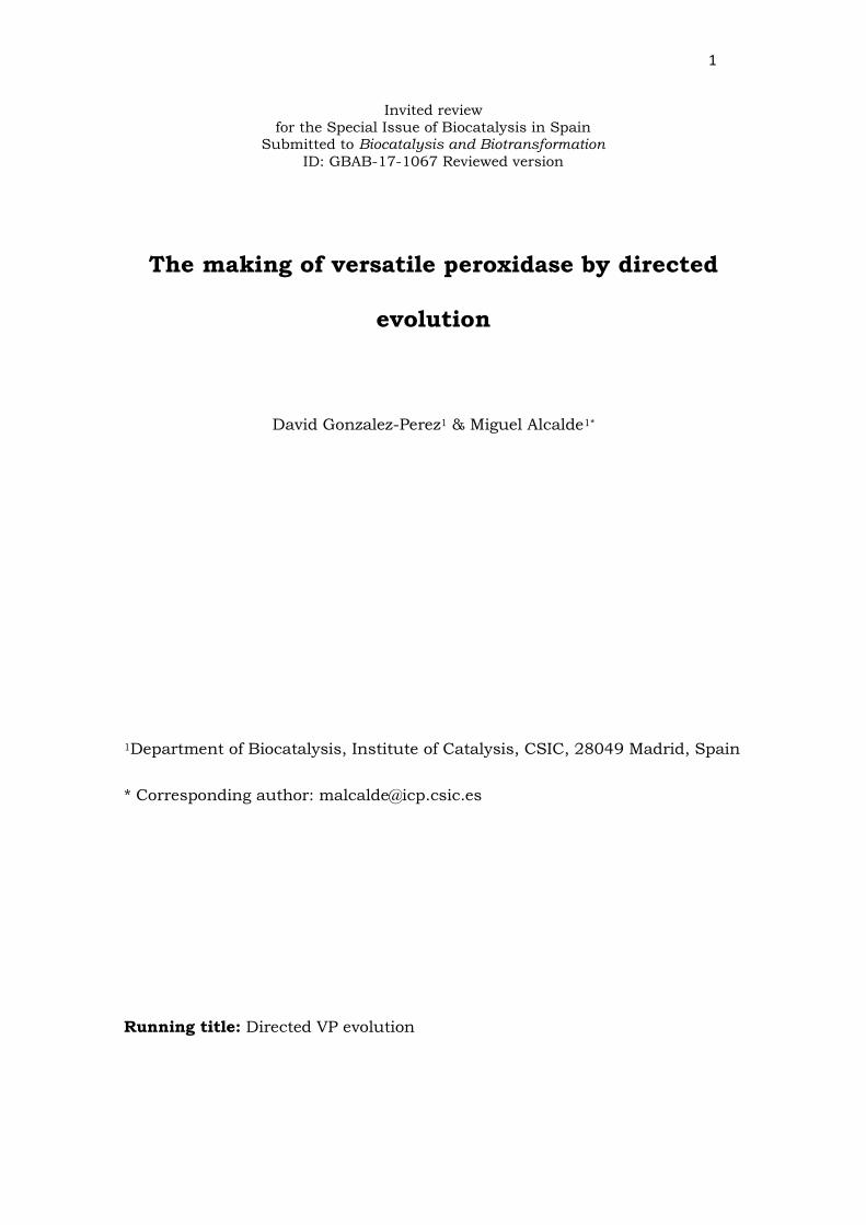
1
Invited review
for the Special Issue of Biocatalysis in Spain Submitted to Biocatalysis and Biotransformation
ID: GBAB-17-1067 Reviewed version
The making of versatile peroxidase by directed
evolution
David Gonzalez-Perez1 & Miguel Alcalde1*
1Department of Biocatalysis, Institute of Catalysis, CSIC, 28049 Madrid, Spain
* Corresponding author: [email protected]
Running title: Directed VP evolution
2
ABSTRACT
Versatile peroxidase (VP) secreted by white-rot fungi is involved in the
degradation of lignin within land ecosystems. With a broad substrate scope
and minor requirements, VP is an extremely attractive blueprint to be
designed by the directed evolution tool-box. In recent years, improved VP
variants have been generated that meet industrial requirements in terms of
improved heterologous functional expression and activity, as well as stability
at high temperatures or in the presence of strong inhibitors. This review
describes the making of VP by directed evolution, addressing the most
important findings and challenges that were faced, along with the future
prospects for this research field.
Keywords: Versatile peroxidase, directed evolution, Saccharomyces cerevisiae,
functional expression, stability, activity.

3
INTRODUCTION
Lignin is a highly persistent biopolymer that gives consistency to the
plant cell wall. Acting like a molecular mortar, lignin imbricates between
cellulose and hemicellulose fibers providing protection against several external
factors, including microbial attack (Martínez et al. 2005; Ruiz-Dueñas and
Martínez 2009; Wong 2009). Given that lignin represents 1/3 of the total
carbon content of the biosphere, its degradation is a crucial aspect of the
carbon cycle (Ragauskas et al. 2006, 2014). Among the organisms capable of
modifying lignin in nature, white-rot basidiomycetes have possibly developed
the most efficient oxidative system yet reported, a system based on the action
of extracellular oxidoreductases (ligninases) that complete the combustion of
lignin to CO2 and H2O (Martínez 2009; Sigoillot 2012). The ligninolytic armory
is formed mainly by Class II peroxidases (generic peroxidases, -GPs-; lignin
peroxidases, -LiPs-; manganese peroxidases, -MnPs-; versatile peroxidases,
VPs); decolorizing peroxidases -DyP- and unspecific peroxygenases -UPO-;
laccases; and H2O2-supplying enzymes. Together these enzymes interact in a
complex cascade, assisted by small diffusible metabolites derived from fungal
activity during wood decay (Alcalde 2015; Ruiz-Dueñas and Martínez 2009;
Martínez et al. 2005).
The deciphering of the whole process of lignin mineralization identified
VP (EC 1.11.1.16) as an important piece of the puzzle. As a result, it has now
become possible to resolve fundamental questions on the natural evolution of
lignin degrading fungi, the enzymes of which are thought to be linked to the
end of coal deposition on earth during the Jurassic period (Ayuso-Fernandez
et al. 2017; Floudas et al. 2012). VP was first discovered around 20 years ago
in the edible mushroom Pleurotus enryngii, and a few years later in
Bjerkandera species (Camarero et al. 1996, 1999; Mester and Field 1998;
4
Ruiz-Dueñas et al. 1999, 2001). Fueled by catalytic concentrations of H2O2, VP
can oxidize a wide variety of molecules, ranging from low- to high-redox
potential substrates including phenolic, non-phenolic and diazo compounds
(Ruiz-Dueñas et al. 2009a). The overall structure of VP involves 11 α-helices
that are sustained by four disulfide bridges and two structural calcium atoms
(Pérez-Boada et al. 2005) (Figure 1). In terms of catalytic sites, like GPs, VP
has an access channel that is open to the solvent and where low-redox
potential substrates are oxidized. In addition, VP has a superficial catalytic
tryptophan that, in its active state (Trp•+), oxidizes both low-redox and more
significantly, high-redox potential substrates through a long-range electron
transfer pathway to the heme, like LiP. Last but not least, in the sagittal plane
of the protein structure there is a small heme access channel where Mn2+ is
oxidized to Mn3+, the latter acting as a diffusible oxidizer as also occurs in MnP
(Ruiz-Dueñas et al. 2007, 2009a) (Figure 2).
From a biotechnological viewpoint, VP represents a wonderful template
for protein engineers. It is a relatively compact enzyme yet very promiscuous,
with three catalytic sites for the oxidation of a multitude of compounds.
Conversely, VP finds plenty of potential applications, such as in the design of
biosensors and analytic kits, in paper and pulp bio-bleaching, bioremediation,
textile industry, organic synthesis or biofuel production (Knop et al. 2015;
Martínez et al. 2009; Mendonça-Maciel et al. 2010; Pizzul et al. 2009; Yadav
and Yadav 2015). Despite this potential, VP must be subjected to further
engineering in order to meet industrial standards and to become a truely
robust industrial biocatalyst. In the last couple of decades, progress in
rational design has helped unveil the different oxidation sites and the
mechanisms by which VP acts (the reader is referred to other works and
reviews that comprehensively address these aspects (Bao et al. 2014; Morales

5
et al. 2012; Pérez-Boada et al. 2005; Pogni et al. 2005; Ruiz-Dueñas et al.
2007, 2008, 2009a, 2009b; Sáez-Jiménez 2015a, 2015b, 2015c, 2016). In the
current article we wish to summarize the journey we have taken in the past
decade to tackle crucial issues associated with VP engineering by directed
evolution, including: i) achieveing adequate functional expression in
heterologous hosts in which directed evolution can be perform; ii)
thermostability; iii) inactivation in the presence of H2O2; and iv) activity and
instability at neutral/alkaline pHs.
1. The kick-off: Directed evolution for functional expression in
Saccharomyces cerevisiae and for thermostability
Although heterologous functional expression of VP was reported in
Emericella nidulans, Aspergillus niger and Phanerochaete chrysosporium
(Coconi-Linares et al. 2015; Eibes et al. 2009; Lu-Chau et al. 2004), these may
not be the most suitable hosts for directed evolution enterprises. Indeed,
evolution platforms assisted by Saccharomyces cerevisiae or Escherichia coli
are the preferred choices due to their ease of manipulation and adaptability to
different high-throughput contexts (Pourmir and Johannes 2012).
Unfortunately, all attempts to express a functional VP in E. coli failed, the
enzyme ending up in inclusion bodies that required in vitro refolding and/or
the help of fusion partners to obtain the soluble protein (Bao et al. 2012;
Pérez-Boada et al. 2002). By contrast, S. cerevisiae is usually a good departure
point for library construction, functional expression (secretion) and the
screening of eukaryotic genes/proteins in directed evolution strategies, and
the case of VP is not an exception (Garcia-Ruiz et al. 2014; Gonzalez-Perez et
al. 2012; Mate et al. 2016). This yeast can perform the complex post-
translational modifications required by ligninases, including glycosylation, the
formation of disulfide bridges, or N- and C-terminal processing, while it
6
maintains an active secretory pathway. Moreover, a full set of episomal vectors
are available to rapidly recover the target gene. In addition, large mutant
libraries can be readily obtained due to the high transformation efficiency (106-
108 transformants/µg DNA). Yet undoubtedly, the most remarkable feature of
S. cerevisiae for synthetic biology and metabolic engineering experiments
stems from its high frequency of homologous DNA recombination, in
conjunction with an accurate proof-reading machinery (Krivoruchko et al.
2011; Nevoigt 2008). As such, the DNA double-strand break repair mechanism
used by S. cerevisiae does not involve restriction and ligation, and it is also
error-free. By harnessing this system, simple library creation methods can be
designed to shuffle different parental genes, to focus the molecular evolution
on given segments (as predicted by computational algorithms), or to splice
different blocks in order to construct chimaeras, to name just a few
possibilities. The rule of thumb for all these methods is to engineer
appropriate overlapping areas that flank each DNA fragment and foster the in
vivo splicing between these stretches (Gonzalez-Perez et al. 2012). Thus, a
gene can be fully reconstructed and cloned in vivo in the presence of the
linearized plasmid in a single transformation step. In S. cerevisiae, in vivo
homologous recombination is mediated by the Rad51 recombinase and
ancillary factors. In practical terms, a length of 40 bp for each overlapping
area is recommended to achieve a good compromise between transformation
efficiency and the number of crossover events (Alcalde 2010; Symington 2002).
As the starting block for directed evolution to achieve functional
expression in S. cerevisiae, we centered our interest on the allelic variant vpl2
from Pleurotus eryngii (Garcia-Ruiz et al. 2012). The failure to detect secretion
of native VP in yeast microcultures (96-well plates) hampered the use of high-
redox potential substrates like reactive black five or veratryl alcohol in a high-

7
throughput screening (HTS) assay of directed evolution. Instead, the
colorimetric substrate ABTS (2,2’-Azino-bis(3-ethylbenzothiazoline-6-sulfonic
acid) was used for the HTS so that activity against low redox potential
compounds could be preserved at both the catalytic Trp and the heme access
channel. The poor initial secretion was enhanced by switching the native
signal peptide of VP with the α-factor prepro-leader from S. cerevisiae, which is
commonly used for the heterologous expression of foreign proteins in yeast.
Through this approach, the α-VP fusion protein was incorporated into the
yeast secretory pathway, which facilitated: i) its processing during extrusion
into the endoplasmic reticulum (pre-leader cleaving by a signal peptidase), ii)
the action of the KEX2 and STE13 proteases in the Golgi compartment
(cleaving the pro-leader), and iii) the final packaging of mature VP into vesicles
for exocytosis. In addition, the production of VP in microplate format for HTS
was optimized by adjusting cultivation parameters like shaking, aeration,
heme source, Ca2+ supplement or ethanol concentration (the latter to enhance
cytoplasmic membrane permeability). In this way, it was possible to enhance
secretion to 0.4 mg/L.
To avoid incorporating neutral or deleterious mutations during the
evolution process, the mutagenic loads and the use of in vivo DNA shuffling
were strictly controlled to favor the accumulation of, on average, one beneficial
mutation per round of evolution. Thus, after four generations of random
mutation and shuffling, the final secretion mutant (R4) contained four
mutations in the protein scaffold. These four mutations were introduced in the
first and second round of evolution, and they were further recombined to
generate the mutated backbone for secretion E37K-V160A-T184M-Q202L
(Figure 3). By this strategy secretion was improved 129-fold, yielding 22 mg/L
of active, soluble and stable VP which can now be easily translated to Pichia

8
pastoris for overproduction in bioreactor (unpublished material). HTS for
thermostability was introduced from the fourth cycle of evolution onwards,
whereby the selective pressure was increased from 60ºC (4th generation) to
90ºC (6th generation) (Garcia-Ruiz et al. 2010, 2012). This approach allowed
three further stabilizing mutations (H39R-D213A-G330R) to be introduced
into the final mutant (2-1B), with an overall enhancement of 8ºC in kinetic
thermostability. In the breakdown of the improvements in terms of specific
activity and secretion of the R4 mutant, we found that while the specific
activity increased 2.5-fold, its expression was enhanced ~52-fold. However, the
enhanced thermostability of the 2-1B mutant was at the cost of reducing its
activity, an expected natural trade-off between these two properties. After the
biochemical characterization of both variants we detected some interesting
side effects. Although oxidative stability was not targeted in the evolution
experiment, we observed a 5-fold improved oxidative stability of the R4
secretion mutant accompanied by a noticeable increment in the Km for H2O2.
Likewise, the thermostability mutant 2-1B displayed remarkable stability at
alkaline pH (with a residual activity above 60% at pH 9 after 120 h of
incubation), which is rather unusual in fungal peroxidases. These results
allowed us to commence new directed evolution enterprises, aimed at i)
improving the oxidative stability of the R4 secretion mutant and ii) rising
activity at alkaline pH in the thermostable mutant 2-1B (Figure 3).
2. Directed evolution for oxidative stability
Although the inactivation of heme containing peroxidases by H2O2 is a
longstanding problem, it remains rather poorly understood and it still limits
the use of peroxidases in many biotechnological sectors (Martínez 2007;
Valderrama et al. 2002). At the ferric resting state, heme-peroxidases are first
activated by a single molecule of H2O2 to yield an oxidized catalytic
9
intermediate called compound I (oxoferryl IV porphyrin π-cation radical),
releasing one molecule of water as the only by-product. The enzyme then
catalyzes two consecutive one-electron oxidations of two reducing substrates,
regenerating the ground (reduced) state through a second catalytic
intermediate called compound II (oxoferryl) and with the concomitant
production of a second molecule of water (Figure 4).
It is well known that the main limiting step within this catalytic cycle is
the low conversion rate from compound II to the ground state, which allows
the former to accumulate and react with a new molecule of H2O2, either in the
presence (with an excess of H2O2) or absence (at catalytic concentrations of
H2O2) of the reducing substrate. In such scenarios compound II is diverted
from the main catalytic cycle, producing the highly reactive intermediate
compound III, a FeIII superoxide radical complex that irreversibly inactivates
the enzyme irrespective of the route followed (e.g. via heme bleaching or
through structural damage provoked by the interaction with the reactive
oxygen species -ROS- generated). This inhibition is known as suicide
inactivation, because paradoxically, H2O2 is both the co-substrate and an
inhibitor of the enzyme (Valderrama et al. 2002, 2010). Suicide inactivation of
VP is even more complex because of the presence of several catalytic sites and
intermediates (Figure 4), which to the best of our knowledge, makes VP the
most sensitive peroxidase to peroxides yet reported (Ruiz-Dueñas et al. 2009a).
Typically, the oxidative stability of peroxidases can be engineered by
replacing highly oxidizable amino acids (e.g. Met, Cys, Tyr, His, Trp) by lower-
redox potential counterparts (e.g. Ala, Val, Ile) (Kim et al. 2001; Ogola et al.
2010; Sáez-Jiménez et al. 2015a; Valderrama et al. 2002), and by means of
directed evolution (Cherry et al. 1999; Miyazaki-Imamura et al. 2003;
Morawsky et al. 2001). Given that suicide inactivation is mechanism-based,

10
we combined structure-guided experiments with directed evolution to tailor
oxidative stability in VP (Gonzalez-Perez et al. 2014a). Our R4 departure
mutant showed a 4-fold enhancement in the Km for H2O2, allowing it to work at
high efficiency in saturating H2O2 conditions (at 2 mM of H2O2 R4 has an
activity of 57000 ABTS-Units/L in culture supernatants) (Garcia-Ruiz et al.
2012). Rather than using a HTS based essentially on the incubation of mutant
libraries at high concentrations of H2O2, we developed an assay in which the
H2O2:enzyme molar ratio was carefully controlled. Through this approach, the
selection of mutants with distinct oxidative stabilities that depend on activity
and/or secretion was circumvented, while we progressively increased the
oxidative stress over the course of evolution (up to 0.6 mM of H2O2).
A focused evolution protocol named MORPHING (Mutagenic Organized
Recombination Process by Homologous In vivo Grouping) (Gonzalez-Perez et al
2014b) was one of the library creation methods developed for this study.
Through MORPHING, we targeted specific regions for random mutagenesis
and recombination (predicted by computational guidance) while keeping the
remaining protein structure protected from evolution. Thanks to the high
frequency of homologous recombination in S. cerevisiae and with the help of
the homologous overhangs flanking each MORPHING segment, functional
mutant libraries were easily constructed and screened for the desired
function. Indeed, MORPHING has become an ideal method for engineering not
only VP in yeast but also, for other ligninases like: UPO, to evolve its native
signal leader for secretion, to modify the peroxygenative:peroxidative activity
ratio, or to improve yields in the synthesis of human drug metabolites;
laccases, for the synthesis of heteropolymeric dyes, or to enhance
thermostability and activity against high redox mediators; and aryl-alcohol
oxidases to identify consensus/ancestor mutations or to oxidize secondary

11
alcohols (Gonzalez-Perez et al. 2014b; Mate et al. 2017; Molina-Espeja et al.
2014; Vicente et al. 2016; Viña-Gonzalez et al. 2015, 2016 and unpublished
results).
In engineering VP, we first applied MORPHING to three independent
segments that were selected after careful structural alignment with other
peroxidases with better oxidative stability. The regions targeted, from 26 to 69
amino acids in length, were mainly located in the heme cavity, including the
proximal His domain (Leu149-Ala174), the distal His domain (Leu28-Gly57)
and the Met environment (Ile199-Leu268). The improved mutants were found
in both the distal/proximal His domains but not in the Met environment that
contained three of the four oxidizable methionines (Met247, Met262 and
Met265). This result was confirmed after subjecting Met265 and Met262 to
combinatorial saturation mutagenesis (CSM), where the mutant library
landscape showed 95% of the clones to be inactive. Recently, improved
oxidative stability by mutating Met residues in VP was only achieved when all
four Met residues were simultaneously removed, albeit at the cost of its
activity, in accordance with our observations (Sáez-Jiménez et al. 2015a).
Given that the S. cerevisiae recombination machinery was inefficient in
recombining nearby mutations (e.g. E40K, T45A), to keep the evolutionary
process moving we performed in vitro recombination, in conjunction with new
rounds of MORPHING, DNA shuffling, error-prone PCR and IvAM (in vivo
assembly of mutant libraries). After four generations of directed evolution we
entered a dense scenario in which multiple improved VP variants with a
diverse trade-off between oxidative stability and activity were identified (i.e.
variants with notably improved half-life for H2O2 but decreased activity, or
variants with milder improvements in half-life but more sustained activity). To
achieve a reasonable compromise between oxidative stability and activity, we

12
developed another in vivo DNA recombination tool called DNApuzzle (Gonzalez-
Perez et al. 2014a). With this domain engineering method, we selected mutant
variants from the 2nd to 4th round of evolution in order to amplify up to 9
evolved sequence blocks from three independent segments of VP. As such, the
mutations in these sequences could be recombined in vivo and relocated in
other mutational environments in a drive to find new epistatic interactions.
Screening of the DNApuzzle mutant library flagged dozens of functional clones,
from which two mutants were finally chosen on the basis of their half-life and
activity. While the AT-AT variant (D22N-T45A-E83G-I103V-G107S-P141A-
F186L) resulted from the recombination of three sequence blocks, the SIth
mutant (N11D-G35K-E40K-T45A-S86R-P141A-F186L-T323I) was the product
of four evolved sequence blocks. In the presence of 3,000 equivalents of H2O2,
the half-life of these mutants extended from 4 min to 18 (AT-AT) and 33 min
(SIth) (Figure 3). Moreover, there was a 6ºC improvement in the kinetic
thermostability of SIth, whereas AT-AT maintained the same thermostability as
the parental type. In terms of kinetics, the catalytic efficiency of AT-AT and SIth
for ABTS was lower than the parental type, although these values were still
higher than those of the native α-VP expressed in S. cerevisiae. Activity at the
Mn2+ site was negatively affected, with slower kinetics for the AT-AT mutant
and almost complete suppression in SIth, probably related to the enhanced
thermostability of this mutant and the mutations introduced close this
catalytic site. Finally, there were minor differences in the catalytic efficiency
for the oxidation of high-redox potential substrates at the catalytic tryptophan,
whereas the kinetics for H2O2 were mostly conserved.
The mutations identified were analyzed through a comprehensive
structural alignment with other tolerant peroxidases in order to reveal key
structural determinants. In general terms, the regions and mutations mapped

13
were responsible for improvements in oxidative stability by making VP more
compact/robust, while modifying the different catalytic sites.
Directed evolution for activity at alkaline pH
One important drawback for the application of VP -and by extension to
all ligninolytic peroxidases- as an industrial biocatalyst in many processes
(ranging from pulp bleaching to organic synthesis) is its lack of
activity/stability at neutral/alkaline pH. The structural integrity of VP is
dependent on the correct coordination of two structural calcium atoms located
above (fairly accessible at the distal domain) and below (buried deeply at the
proximal domain) the plane of the heme (Pérez-Boada et al. 2005) (Figure 1A).
Indeed, 7 and 8 oxygen atoms from surrounding amino acids and water
molecules enter into the coordination spheres of such structural distal and
proximal calcium ions, respectively (Figure 1B). It is well known that
neutral/basic pH and/or high temperatures can modify Ca2+ coordination
spheres, provoking their release and therefore the loss of activity. Indeed, after
distal Ca2+ depletion, the catalytic distal His47 approaches the iron atom of
the heme, which in turn switches from a penta- to hexa-coordination state
(bis-histidyl heme iron complex in low spin state), collapsing the heme cavity
and inactivating the enzyme (George et al. 1999; Laberge et al. 2003; Lu-Chau
et al. 2004; Youngs et al. 2000).
Notably, the 2-1B mutant obtained through directed evolution aimed to
improve secretion and thermostability overcame such Ca2+ depletion under
alkaline conditions (Garcia-Ruiz et al. 2012). Recently, this effect was
attributed to the mutated E37K-H39R-G330R backbone that stabilized both
distal and proximal Ca2+, allowing them to be correctly coordinated at alkaline
pH and hence, preserving the pentacoordinated high-spin Fe3+ (Sáez-Jiménez

14
et al. 2016). Although 2-1B was stable at alkaline conditions, there was hardly
any activity at its three catalytic sites at basic pH. Accordingly, further
directed evolution to enhance the activity of this variant at neutral/alkaline
pH was undertaken, while conserving activity at acid pH (Gonzalez-Perez et al.
2016), implying a selection criterion during HTS based on the ratio of activity
at pH 4 to that at neutral/basic pH. Clones with >80% of the parental activity
at pH 4 and improved activity at alkaline pH were initially selected for further
analysis, and the process was further accelerated by increasing the selective
pressure from pH 6 to 8.5. After only three rounds of evolution with this
strategy, we obtained the BB-8 mutant (harboring the mutations E140G-
P182S-Q229P) that was active over an enhanced pH range and that displayed
strong hyperactivation after incubation at alkaline pH (with a 3-fold increase
in activity) (Figure 3). The active pH range for BB-8 was expanded
considerably for several substrates, including ABTS, sinapic acid and guaiacol.
Consequently, BB-8 was active in the acid range (pH 3-4) and remarkably, in
the pH interval from 5 to 9 in which the activity of the parental VP was
negligible.
The kinetic parameters measured for ABTS revealed enhanced catalytic
efficiency at acid pH as result of increased affinity, which permitted BB-8 to
remain active at basic pHs. This effect was mostly attributed to the E140G
mutation that enabled the mutant to work with similar catalytic efficiency at
pH 6 as the parental type at pH 3.5, due to the widening of the heme channel.
Whilst the activity against Mn2+ was diminished due to the P182S mutation
introduced close to this catalytic site, this mutation offered the first
experimental insight into the role of the Mn2+ site for the direct (non-mediated)
oxidation of ABTS at neutral/basic pH. Site-directed mutagenesis experiments
subsequently focused on blocking this catalytic site and molecular docking

15
studies supported this finding (Gonzalez-Perez et al. 2016). Alternatively, we
did not observe any activity at the catalytic Trp164 at basic pH due to the fact
that the reduction potential of the Trp164 radical decreases as the pH
increases, hindering the oxidation of high-redox potential substrates at
neutral/basic pH. Bearing in mind that the pKa of Trp•+ is ~4.3 (a value that
varies slightly in function of the microenvironment), it is fully uncharged and
possibly catalytically inactive at neutral or basic pH. For these reasons, the
long-range electron transfer pathway from Trp164 to the heme is permanently
cancelled out at pHs >5, thereby diverting the oxidative route for the oxidation
of low-redox potential substrates to the other two catalytic sites at the time
that the oxidation of high-redox potential compounds is supressed.
Conclusions and outlook
Almost 10 years ago we set out to perform directed evolution of VP
using only S. cerevisiae, both as main expression host and as a vital tool-box
for the creation of a DNA diversity. This long shot paid-off, since apart from
creating a reliable platform to efficiently perform directed evolution, we
showcased a broad portfolio of VP variants with improved traits. This not only
paved the way for the directed evolution of VP, enhancing its functional
expression and stabilization, but it also allowed us to understand how to
harness recombination in S. cerevisiae in order to design methods to create
libraries suitable for laboratory evolution. Such libraries have helped us
identify hidden properties of VP that bring this enzyme one step closer to
meeting industrial standards. Far from rejoicing, we consider these results
represent merely the beginning and we believe important future progress in VP
engineering will include the directed evolution of ancestral VP and the
engineering of an artificial ligninolytic secretome. The former will be aided by
recent data on the ancestral lineages of ligninolytic peroxidases, where several
16
nodes from the late Carboniferous period were computationally reconstructed
and expressed to situate VP as a potential generalist in the midst of the lignin
degrading enzyme route (Ayuso-Fernandez et al. 2017). The use of ancestral
VP and modern counterparts to recreate this evolutionary journey in vitro by
means of laboratory evolution will help answer key questions on the natural
evolution of VP, while providing a new collection of evolved variants with
unexpected properties (Alcalde 2015, 2017). In terms of engineering the first
artificial ligninolytic secretome, we recently co-expressed evolved versions of
VP and laccase in S. cerevisiae, overcoming several metabolic barriers and
bottlenecks associated with expression (Gonzalez-Perez and Alcalde 2014). In
the drive to design a prototype of the white-rot yeast (WRY), we have just
added new evolved versions of UPO and aryl-alcohol oxidases to the secretome
pool, which can be used for the production of biofuels and value added
products from lignocellulose. In the long term, the WRY could become an
invaluable vehicle to understand the complex process of lignin mineralization
in nature.
ACKNOWDLEGEMENTS
This work was supported by grants from the European Union [FP7-
KBBE-2013-7-613549-INDOX] the COST Action [CM1303 Systems
Biocatalysis] and the Spanish Government [BIO2016-79106-R-Lignolution].
REFERENCES
Alcalde M. 2010. Mutagenesis protocols in Saccharomyces cerevisiae by in vivo
overlap extension. In: Bramman J. Editor. In vitro Mutagenesis
Protocols, 3rd ed. Methods in Molecular Biology 634. New Jersey:
Springer-Humana Press. pp.3-15.
17
Alcalde M. 2015. Engineering the ligninolytic enzyme consortium. Trends
Biotechnol 33:155-162.
Alcalde M. 2017. When directed evolution met ancestral enzyme resurrection.
Microb Biotechnol 10:22-24.
Ayuso-Fernandez I, Martínez AT, Ruiz-Dueñas FJ. 2017. Experimental
recreation of the evolution of lignin degrading enzymes from the
Jurassic to date. Biotechnol Biofuels. In press.
Bao X, Liu A, Lu X, Li JJ. 2012. Direct over-expression, characterization and
H2O2 stability study of active Pleurotus eryngii versatile peroxidase in
Escherichia coli. Biotechnol Lett 34:1537-1543.
Bao X, Huang X, Lu X, Li JJ. 2014. Improvement of hydrogen peroxide
stability of Pleurotus eryngii versatile ligninolytic peroxidase by rational
protein engineering. Enzyme Microb Technol 54:51-58.
Camarero S, Bockle B, Martínez MJ, Martínez AT. 1996. Manganese-mediated
lignin degradation by Pleurotus pulmonarius. Appl Environ Microbiol
62:1070-1072.
Camarero S, Sarkar S, Ruiz-Dueñas FJ, Martínez MJ, Martínez AT. 1999.
Description of a versatile peroxidase involved in the natural degradation
of lignin that has both manganese peroxidase and lignin peroxidase
substrate interaction sites. J Biol Chem 274:10324-10330.
Cherry JR, Lamsa MH, Schneider P, Vind J, Svendsen A, Jones A, Pedersen
AH. 1999. Directed evolution of a fungal peroxidase. Nat Biotechnol
17:379-384.
Coconi-Linares N, Ortiz-Vázquez E, Fernández F, Loske AM, Gómez-Lim MA.
2015. Recombinant expression of four oxidoreductases in
Phanerochaete chrysosporium improves degradation of phenolic and
non-phenolic substrates. J Biotechnol 209:76-84.
18
Eibes GM, Lu‐Chau TA, Ruiz‐Dueñas FJ, Feijoo G, Martínez MJ, Martínez AT,
Lema, JM. 2009. Effect of culture temperature on the heterologous
expression of Pleurotus eryngii versatile peroxidase in Aspergillus hosts.
Bioproc Biosyst Eng 32:129‐134.
Floudas D, Binder M, Riley R, Barry K, Blanchette RA, Henrissat B, Martínez
AT, Otillar R, Spatafora JW, Yadav JS, et al. 2012. The Paleozoic origin
of enzymatic lignin decomposition reconstructed from 31 fungal
genomes. Science 336:1715-1719.
Garcia-Ruiz E, Mate D, Ballesteros A, Martínez AT, Alcalde M. 2010. Evolving
thermostability in mutant libraries of ligninolytic oxidoreductases
expressed in yeast. Microb Cell Fact 9:17.
Garcia-Ruiz E, Gonzalez-Perez D, Ruiz-Dueñas FJ, Martínez AT, Alcalde M.
2012. Directed evolution of a temperature, peroxide and alkaline pH
tolerant versatile peroxidase. Biochem J 441:487-498.
Garcia-Ruiz E, Mate DM, Gonzalez-Perez D, Molina-Espeja P, Camarero S,
Martínez AT, Ballesteros AO, Alcalde M. 2014. Directed evolution of
ligninolytic oxidoreductases: from functional expression to stabilization
and beyond. In: Sergio Riva, Wolf-Dieter Fessner, Editors. Cascade
Biocatalysis. Wiley-VCH Verlag GmbH & Co. KGaA. pp.1-22.
George SJ, Kvaratskhelia M, Dilworth MJ, Thorneley RN. 1999. Reversible
alkaline inactivation of lignin peroxidase involves the release of both the
distal and proximal site calcium ions and bishistidine co-ordination of
the haem. Biochem J 344:237-244.
Gonzalez-Perez D, Garcia-Ruiz E, Alcalde M. 2012. Saccharomyces cerevisiae
in directed evolution: An efficient tool to improve enzymes. Bioeng bugs
3:172-177.

19
Gonzalez-Perez D, Garcia-Ruiz E, Ruiz-Dueñas FJ, Martínez AT, Alcalde M.
2014a. Structural determinants of oxidative stabilization in an evolved
versatile peroxidase. ACS Catal 4:3891-3901.
Gonzalez-Perez D, Molina-Espeja P, Garcia-Ruiz E, Alcalde M. 2014b.
Mutagenic organized recombination process by homologous in vivo
grouping (MORPHING) for directed enzyme evolution. Plos one 9:
e90919.
Gonzalez-Perez D, Alcalde M. 2014. Assembly of evolved ligninolytic genes in
Saccharomyces cerevisiae. Bioengineered 5:254-263.
Gonzalez-Perez D, Mateljak I, Garcia-Ruiz E, Ruiz-Dueñas FJ, Martínez AT,
Alcalde A. 2016. Alkaline versatile peroxidase by directed evolution.
Catal Sci Technol 6:6625-6636.
Kim YH, Berry AH, Spencer DS, Stites WE. 2001. Comparing the effect on
protein stability of methionine oxidation versus mutagenesis: steps
toward engineering oxidative resistance in proteins. Protein Eng Des Sel
14:343-347.
Knop D, Yarden O, and Hadar Y. 2015. The ligninolytic peroxidases in the
genus Pleurotus: divergence in activities, expression, and potential
applications. Appl Microbiol Biotechnol 99:1025-1038.
Krivoruchko A, Siewers V, Nielsen J. 2011. Opportunities for yeast metabolic
engineering: Lessons from synthetic biology. Biotechnol J 6:262-276.
Laberge M, Huang Q, Schweitzer-Stenner R, Fidy J. 2003. The endogenous
calcium ions of horseradish peroxidase c are required to maintain the
functional nonplanarity of the heme. Biophys J 84:2542-2552.
Lu-Chau TA, Ruiz-Dueñas FJ, Camarero S, Feijoo G, Martínez MJ, Lema JM,
Martínez AT. 2004. Effect of pH on the stability of Pleurotus eryngii
20
versatile peroxidase during heterologous production in Emericella
nidulans. Bioproc Biosyst Eng 26:287-293.
Martínez AT, Speranza M, Ruiz-Dueñas FJ, Ferreira P, Camarero S, Guillen F,
Martinez MJ, Gutierrez A, del Rio JC. 2005. Biodegradation of
lignocellulosics: microbial, chemical, and enzymatic aspects of the
fungal attack of lignin. Int Microbiol 8:195-204.
Martínez AT. 2007. High redox potential peroxidases. In: Polaina J and
MacCabe, AP, Editors. Industrial Enzymes. Springer. pp.477-488.
Martínez AT, Ruiz-Dueñas FJ, Martínez MJ, del Rio JC, Gutierrez A. 2009.
Enzymatic delignification of plant cell wall: from nature to mill. Curr
Opin Biotechnol 20:348-357.
Mate DM, Gonzalez-Perez D, Mateljak I, Gomez de Santos P, Vicente AI,
Alcalde M. 2016. The pocket manual of directed evolution: Tips and
tricks. In: Brahmachari G, Demain AL, Adrio JL, Editors. Biotechnology
of microbial enzymes: Production, biocatalysis and industrial
applications. Amsterdam: Elsevier. pp.185-214.
Mate DM, Palomino MA, Molina-Espeja P, Martin-Diaz J, Alcalde M. 2017.
Modification of the peroxygenative:peroxidative activity ratio in the
unspecific peroxygenase from Agrocybe aegerita by structure-guided
evolution. Protein Eng Des Sel 1-8.
Mendonça-Maciel MJ, Castro e Silva A, Telles Ribeiro HC. 2010. Industrial and
biotechnological applications of ligninolytic enzymes of the
basidiomycota: a review. Electron J Biotech 13:14-15.
Mester T, Field JA. 1998. Characterization of a novel manganese peroxidase-
lignin peroxidase hybrid isozyme produced by Bjerkandera species
strain BOS55 in the absence of manganese. J Biol Chem 273:15412-
15417.

21
Miyazaki-Imamura C, Oohira K, Kitagawa R, Nakano H, Yamane T, Takahashi
H. 2003. Improvement of H2O2 stability of manganese peroxidase by
combinatorial mutagenesis and high-throughput screening using in
vitro expression with protein disulfide isomerase. Protein Eng Des Sel
16:423-428.
Molina-Espeja P, Garcia-Ruiz E, Gonzalez-Perez D, Ullrich R, Hofrichter M,
Alcalde M. 2014. Directed evolution of unspecific peroxygenase from
Agrocybe aegerita. Appl Environ Microbiol 80:3496-3507.
Morales M, Mate MJ, Romero A, Martínez MJ, Martínez AT, Ruiz-Dueñas FJ.
2012. Two oxidation sites for low redox-potential substrates: A directed
mutagenesis, kinetic and crystallographic study on Pleurotus eryngii
versatile peroxidase. J Biol Chem 287:41053-41067.
Morawsky B, Quan S, Arnold FH. 2001. Functional expression and
stabilization of horseradish peroxidase by directed evolution in
Saccharomyces cerevisiae. Biotechnol Bioeng 76:99-107.
Nevoigt E. 2008. Progress in metabolic engineering of Saccharomyces
cerevisiae. Microbiol Mol Biol Rev 72:379-412.
Ogola HJ, Hashimoto N, Miyabe S, Ashida H, Ishikawa T, Shibata H Sawa Y.
2010. Enhancement of hydrogen peroxide stability of a novel Anabaena
sp. DyP-type peroxidase by site-directed mutagenesis of methionine
residues. Appl Microbiol Biotechnol 87:1727-1736
Pérez-Boada M, Doyle WA, Ruiz-Dueñas FJ, Martınez MJ, Martınez AT, Smith
AT. 2002. Expression of Pleurotus eryngii versatile peroxidase in
Escherichia coli and optimisation of in vitro folding. Enzyme Microb
Technol 30:518-524.
Pérez-Boada M, Ruiz-Dueñas FJ, Pogni R, Basosi R, Choinowski T, Martínez
MJ, Piontek K, Martínez AT. 2005. Versatile peroxidase oxidation of
22
high redox potential aromatic compounds: Site-directed mutagenesis,
spectroscopic and crystallographic investigations of three long-range
electron transfer pathways. J Mol Biol 354:385-402.
Pizzul L, Castillo M del P, Stenstrom J. 2009. Degradation of glyphosate and
other pesticides by ligninolytic enzymes. Biodegradation 20:751-759.
Pogni R, Baratto MC, Giansanti S, Teutloff C, Verdin J, Valderrama B,
Lendzian F, Lubitz W, Vazquez-Duhalt R, Basosi R. 2005. Tryptophan-
based radical in the catalytic mechanism of versatile peroxidase from
Bjerkandera adusta. Biochemistry 44:4267-4274.
Pourmir A, Johannes TW. 2012. Directed evolution: selection of the host
organism. Comput Struct Biotechnol J 2:e201209012.
Ragauskas AJ, Williams CK, Davison BH, Britovsek G, Cairney J, Eckert CA,
Frederick WJ, Hallett JP, Leak DJ, Liotta CL, et al. 2006. The path
forward for biofuels and biomaterials. Science 311:484-489.
Ragauskas AJ, Beckham GT, Biddy MJ, Chandra R, Chen F, Davis MF,
Davison BH, Dixon RA, Gilna P, Keller M, et al. 2014. Lignin
valorization: improving lignin processing in the biorefinery. Science
344:1246843.
Ruiz-Dueñas FJ, Martínez MJ, Martínez AT. 1999. Molecular characterization
of a novel peroxidase isolated from the ligninolytic fungus Pleurotus
eryngii. Mol Microbiol 31:223-235.
Ruiz-Dueñas FJ, Camarero S, Pérez-Boada M, Martínez MJ, Martínez AT.
2001. A new versatile peroxidase from Pleurotus. Biochem Soc T 29:
116-122.
Ruiz-Dueñas FJ, Morales M, Pérez-Boada M, Choinowski T, Martínez MJ,
Piontek K, Martínez AT. 2007. Manganese oxidation site in Pleurotus

23
eryngii versatile peroxidase: A site-directed mutagenesis, kinetic and
crystallographic study. Biochemistry 46:66-77.
Ruiz-Dueñas FJ, Morales M, Mate MJ, Romero A, Martínez MJ, Smith AT,
Martínez AT. 2008. Site-directed mutagenesis of the catalytic
tryptophan environment in Pleurotus eryngii versatile peroxidase.
Biochemistry 47:1685-1695.
Ruiz-Dueñas, FJ, Morales M, García E, Miki Y, Martínez MJ, Martínez AT.
2009a. Substrate oxidation sites in versatile peroxidase and other
basidiomycete peroxidases. J Exp Bot 60:441-452.
Ruiz-Dueñas FJ, Pogni R, Morales M, Giansanti S, Mate MJ, Romero A,
Martínez MJ, Basosi R, Martínez AT. 2009b. Protein radicals in fungal
versatile peroxidase: Catalytic tryptophan radical in both Compound I
and Compound II and studies on W164Y, W164H and W164S variants.
J Biol Chem 284:7986-7994.
Ruiz-Dueñas FJ, Martinez AT. 2009. Microbial degradation of lignin: how a
bulky recalcitrant polymer is efficiently recycled in nature and how we
can take advantage of this. Microb Biotechnol 2:164-177.
Sáez-Jiménez V, Acebes S, Guallar V, Martinez AT, Ruiz-Dueñas FJ. 2015a.
Improving the oxidative stability of a high redox potential fungal
peroxidase by rational design. Plos one 10:e0124750.
Sáez-Jiménez V, Baratto MC, Pogni R, Rencoret J, Gutierrez A, Santos JI,
Martinez AT, Ruiz-Dueñas FJ. 2015b. Demonstration of lignin-to-
peroxidase direct electron transfer: a transient-state kinetics, directed
mutagenesis, EPR and NMR study. J Biol Chem 290:23201-23213.
Sáez-Jiménez V, Fernández-Fueyo E, Medrano FJ, Romero A, Martinez AT,
Ruiz-Dueñas FJ. 2015c. Improving the pH-stability of versatile

24
peroxidase by comparative structural analysis with a naturally-stable
manganese peroxidase. Plos one 10:e0140984.
Sáez-Jiménez V, Acebes S, García-Ruiz E, Romero A, Guallar V, Alcalde M,
Medrano FJ, Martínez AT, Ruiz-Dueñas FJ. 2016. Unveiling the basis of
alkaline stability of an evolved versatile peroxidase. Biochem J
473:1917-1928.
Sigoillot JC, Berrin JG, Bey M, Lesage-Meessen L, Levasseur A, Lomascolo A,
Record E, Uzan-Boukhrins. 2012. Fungal strategies for lignin
degradation. Adv Bot Res 61:263-308.
Symington LS. 2002. Role of RAD52 epistasis group genes in homologous
recombination and double-strand break repair. Microbiol Mol Biol Rev
66:630-670.
Valderrama B, Ayala M, Vázquez-Duhalt R. 2002. Suicide inactivation of
peroxidases and the challenge of engineering more robust enzymes.
Chem Biol 9:555-565.
Valderrama B. 2010. Deactivation of hemeperoxidases by hydrogen peroxide:
Focus on compound III. In: Torres E, Ayala M, Editors. Biocatalysis
based on heme peroxidases: peroxidases as potential industrial
biocatalysts. Heidelberg: Springer. pp.291-314.
Vicente AI, Viña-Gonzalez J, Santos-Moriano P, Marquez-Alvarez C,
Ballesteros O, Alcalde M. 2016. Evolved alkaline fungal laccase secreted
by Saccharomyces cerevisiae as useful tool for the synthesis of C-N
heteropolymeric dye. J Mol Catal B-Enzym 134:323-330.
Viña-Gonzalez J, Gonzalez-Perez D, Ferreira P, Martinez AT, Alcalde M. 2015.
Focused directed evolution of aryl-alcohol oxidase in Saccharomyces
cerevisiae by using chimeric signal peptides. Appl Environ Microbiol
81:6451-6462.

25
Viña-Gonzalez J, Gonzalez-Perez D, Alcalde M. 2016. Directed evolution
method in Saccharomyces cerevisiae: Mutant library creation and
screening. J Vis Exp 110:e53761.
Wong DW. 2009. Structure and action mechanism of ligninolytic enzymes.
Appl Biochem Biotechnol 157:174-209.
Yadav M, Yadav HS. 2015. Applications of ligninolytic enzymes to pollutants,
wastewater, dyes, soil, coal, paper and polymers. Environ Chem Lett
13:309-318.
Youngs HL, Moënne-Loccoz P, Loehr TM, Gold MH. 2000. Formation of a
bis(histidyl) heme iron complex in manganese peroxidase at high pH
and restoration of the native enzyme structure by calcium.
Biochemistry 39:9994-10000.

26
FIGURE LEGENDS
FIGURE 1. General structure of VP and Ca2+ attachment. (A) VP crystal
structure (PDB ID: 3FJW) highlighted in blue cartoon mode, with the heme
group shown as sticks in CPK colors and the disulfide bridges as yellow sticks.
White letters indicate the name of the secondary VP structures. (B) Distal and
proximal Ca2+ coordination spheres. Amino acid residues are indicated in CPK
ball and stick mode. Water molecules and Ca2+ ions are highlighted as red
and orange spheres, respectively.
FIGURE 2. VP oxidation sites. VP crystal structure shown in surface mode
with the relative electrostatic potentials. Heme groups are highlighted in CPK
sticks mode. The catalytic Trp164 and Leu165 are represented in yellow CPK
sticks mode. LRTE, Long-range electron transfer pathway. Glu36, Glu40 and
Asp175 at the Mn2+ binding site are shown as CPK sticks. The His47 and
Lys43 residues are involved in the heterolytic cleavage of the H2O2 molecule
while His169 residue acts as fifth ligand of Fe3+.
FIGURE 3. Different directed evolution campaigns followed for VP
engineering. Stars show the new mutations while the squares reflect the
accumulated mutations. Green and gray boxes indicate the α-prepro-leader.
T50, the temperature at which the enzyme losses 50 % of its total activity after
a 10 min incubation; H2O2 t1/2, half-life in the presence of 3,000 equivalents of
H2O2 at 25ºC pH 6.0.
FIGURE 4. Catalytic cycle of VP. VP in its ground state is activated by H2O2
giving rise to compound I (VP-IA), which can be diverted to compound VP-IB
corresponding to the tryptophanyl cation radical at the surface of the enzyme.
Compound I oxidizes a first reducing substrate switching to compound II (VP-
IIA) which can also be diverted to the corresponding compound VP-IIB. Finally,
27
compound II in any of its catalytic forms returns to the ground state after a
second oxidation of a new reducing substrate. VP-IA and VP-IIA catalyze the
oxidation of low-redox potential substrates (mostly phenolics) at the heme
channel as well as the oxidation of Mn2+ at the Mn2+ binding site. VP-IB and
VP-IIB catalyze the oxidation of both low- and high-redox potential compounds
(e.g. veratryl alcohol, VA) at the catalytic Trp at the surface of the protein
through a long range electron transfer route to the heme.