Lipidomics identifies cardiolipin oxidation as a mitochondrial target for redox therapy of brain...
20
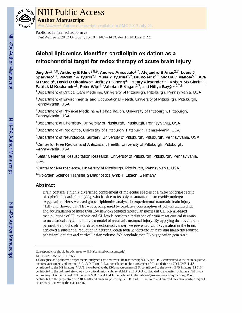
Global lipidomics identifies cardiolipin oxidation as a mitochondrial target for redox therapy of acute brain injury Jing Ji 1,2,7,8 , Anthony E Kline 3,8,9 , Andrew Amoscato 2,7 , Alejandro S Arias 2,7 , Louis J Sparvero 2,7 , Vladimir A Tyurin 2,7 , Yulia Y Tyurina 2,7 , Bruno Fink 10 , Mioara D Manole 5,8 , Ava M Puccio 6 , David O Okonkwo 6 , Jeffrey P Cheng 3,8 , Henry Alexander 1,8 , Robert SB Clark 1,8 , Patrick M Kochanek 1,8 , Peter Wipf 4 , Valerian E Kagan 2,7 , and Hülya Bayýr 1,2,7,8 1 Department of Critical Care Medicine, University of Pittsburgh, Pittsburgh, Pennsylvania, USA 2 Department of Environmental and Occupational Health, University of Pittsburgh, Pittsburgh, Pennsylvania, USA 3 Department of Physical Medicine & Rehabilitation, University of Pittsburgh, Pittsburgh, Pennsylvania, USA 4 Department of Chemistry, University of Pittsburgh, Pittsburgh, Pennsylvania, USA 5 Department of Pediatrics, University of Pittsburgh, Pittsburgh, Pennsylvania, USA 6 Department of Neurological Surgery, University of Pittsburgh, Pittsburgh, Pennsylvania, USA 7 Center for Free Radical and Antioxidant Health, University of Pittsburgh, Pittsburgh, Pennsylvania, USA 8 Safar Center for Resuscitation Research, University of Pittsburgh, Pittsburgh, Pennsylvania, USA 9 Center for Neuroscience, University of Pittsburgh, Pittsburgh, Pennsylvania, USA 10 Noxygen Science Transfer & Diagnostics GmbH, Elzach, Germany Abstract Brain contains a highly diversified complement of molecular species of a mitochondria-specific phospholipid, cardiolipin (CL), which - due to its polyunsaturation - can readily undergo oxygenation. Here, we used global lipidomics analysis in experimental traumatic brain injury (TBI) and showed that TBI was accompanied by oxidative consumption of polyunsaturated CL and accumulation of more than 150 new oxygenated molecular species in CL. RNAi-based manipulations of CL-synthase and CL levels conferred resistance of primary rat cortical neurons to mechanical stretch - an in vitro model of traumatic neuronal injury. By applying the novel brain permeable mitochondria-targeted electron-scavenger, we prevented CL oxygenation in the brain, achieved a substantial reduction in neuronal death both in vitro and in vivo, and markedly reduced behavioral deficits and cortical lesion volume. We conclude that CL oxygenation generates Correspondence should be addressed to H.B. ([email protected]). AUTHOR CONTRIBUTIONS J.J. designed and performed experiments, analyzed data and wrote the manuscript; A.E.K and J.P.C. contributed to the neurocognitive outcome assessment and writing; A.A. ,Y.Y.T and A.S.A. contributed to the assessment of CL oxidation by 2D-LCMS; L.J.S. contributed to the MS imaging; V.A.T. contributed to the EPR measurements; B.F. contributed to the in vivo EPR imaging; M.D.M. contributed to the unbiased stereology for cortical lesion volume. A.M.P. and D.O.O. contributed to evaluation of human TBI tissue and writing. H.A. performed CCI model; R.S.B.C. and P.M.K. contributed to the data analysis and manuscript writing; P.W. contributed to the preparation of XJB-5-131 and manuscript writing; V.E.K. and H.B. initiated and directed the entire study, designed experiments and wrote the manuscript. NIH Public Access Author Manuscript Nat Neurosci. Author manuscript; available in PMC 2013 July 01. Published in final edited form as: Nat Neurosci. 2012 October ; 15(10): 1407–1413. doi:10.1038/nn.3195. NIH-PA Author Manuscript NIH-PA Author Manuscript NIH-PA Author Manuscript
-
Upload
independent -
Category
Documents
-
view
0 -
download
0
Transcript of Lipidomics identifies cardiolipin oxidation as a mitochondrial target for redox therapy of brain...
Global lipidomics identifies cardiolipin oxidation as amitochondrial target for redox therapy of acute brain injury
Jing Ji1,2,7,8, Anthony E Kline3,8,9, Andrew Amoscato2,7, Alejandro S Arias2,7, Louis JSparvero2,7, Vladimir A Tyurin2,7, Yulia Y Tyurina2,7, Bruno Fink10, Mioara D Manole5,8, AvaM Puccio6, David O Okonkwo6, Jeffrey P Cheng3,8, Henry Alexander1,8, Robert SB Clark1,8,Patrick M Kochanek1,8, Peter Wipf4, Valerian E Kagan2,7, and Hülya Bayýr1,2,7,8
1Department of Critical Care Medicine, University of Pittsburgh, Pittsburgh, Pennsylvania, USA2Department of Environmental and Occupational Health, University of Pittsburgh, Pittsburgh,Pennsylvania, USA3Department of Physical Medicine & Rehabilitation, University of Pittsburgh, Pittsburgh,Pennsylvania, USA4Department of Chemistry, University of Pittsburgh, Pittsburgh, Pennsylvania, USA5Department of Pediatrics, University of Pittsburgh, Pittsburgh, Pennsylvania, USA6Department of Neurological Surgery, University of Pittsburgh, Pittsburgh, Pennsylvania, USA7Center for Free Radical and Antioxidant Health, University of Pittsburgh, Pittsburgh,Pennsylvania, USA8Safar Center for Resuscitation Research, University of Pittsburgh, Pittsburgh, Pennsylvania,USA9Center for Neuroscience, University of Pittsburgh, Pittsburgh, Pennsylvania, USA10Noxygen Science Transfer & Diagnostics GmbH, Elzach, Germany
AbstractBrain contains a highly diversified complement of molecular species of a mitochondria-specificphospholipid, cardiolipin (CL), which - due to its polyunsaturation - can readily undergooxygenation. Here, we used global lipidomics analysis in experimental traumatic brain injury(TBI) and showed that TBI was accompanied by oxidative consumption of polyunsaturated CLand accumulation of more than 150 new oxygenated molecular species in CL. RNAi-basedmanipulations of CL-synthase and CL levels conferred resistance of primary rat cortical neuronsto mechanical stretch - an in vitro model of traumatic neuronal injury. By applying the novel brainpermeable mitochondria-targeted electron-scavenger, we prevented CL oxygenation in the brain,achieved a substantial reduction in neuronal death both in vitro and in vivo, and markedly reducedbehavioral deficits and cortical lesion volume. We conclude that CL oxygenation generates
Correspondence should be addressed to H.B. ([email protected]).
AUTHOR CONTRIBUTIONSJ.J. designed and performed experiments, analyzed data and wrote the manuscript; A.E.K and J.P.C. contributed to the neurocognitiveoutcome assessment and writing; A.A. ,Y.Y.T and A.S.A. contributed to the assessment of CL oxidation by 2D-LCMS; L.J.S.contributed to the MS imaging; V.A.T. contributed to the EPR measurements; B.F. contributed to the in vivo EPR imaging; M.D.M.contributed to the unbiased stereology for cortical lesion volume. A.M.P. and D.O.O. contributed to evaluation of human TBI tissueand writing. H.A. performed CCI model; R.S.B.C. and P.M.K. contributed to the data analysis and manuscript writing; P.W.contributed to the preparation of XJB-5-131 and manuscript writing; V.E.K. and H.B. initiated and directed the entire study, designedexperiments and wrote the manuscript.
NIH Public AccessAuthor ManuscriptNat Neurosci. Author manuscript; available in PMC 2013 July 01.
Published in final edited form as:Nat Neurosci. 2012 October ; 15(10): 1407–1413. doi:10.1038/nn.3195.
NIH
-PA Author Manuscript
NIH
-PA Author Manuscript
NIH
-PA Author Manuscript

neuronal death signals and that its prevention by mitochondria-targeted small molecule inhibitorsrepresents a new target for neuro-drug discovery.
Every year in the United States alone, an estimated 1.7 million people sustain acute braininjury from trauma and of those, 52,000 die and 85,000 suffer from long term disabilities1,2.This includes injuries from many etiologies such as road traffic accidents, falls, assaults, andsports concussion. Also, the RAND Corporation estimates that over 400,000 US soldiershave suffered TBI in Operation Iraqi Freedom and Operation Enduring Freedom3. A specifictherapy for TBI does not yet exist and standard treatment remains supportive in nature. Likemany forms of acute brain injury, TBI involves primary injury that is felt to be refractory totreatment and secondary injury, characterized by a cascade of biochemical and cellularevents contributing to neuronal damage over time4. Secondary injury involves multiplepathways of neuronal death which represent viable therapeutic targets5,6.
The complexity of brain function requires sophisticated coordination of highly diversifiedoperational signals. Numerous small molecule-signals formed from a relatively limitednumber of metabolic precursors are accountable for the precision and effectiveness of thebrain’s operational language. Among these are polyunsaturated lipids capable of undergoingenzymatic oxidation and other metabolic conversions yielding oxygenated signaling entities- from eicosanoids, prostanoids and resolvins to cannabinoids and neuroprotectins. There issubstantial archaeological evidence that exploitation of abundant food resource enrichedwith polyunsaturated lipids was essential for sustaining the comparatively large size, theapparent unique complexity and high level of interconnectivity in the modern human brainand provided the advantage in multi-generational brain development, thus making possiblethe advent of H. sapiens7. The demand for diverse polyunsaturated lipid precursors is solarge that mitochondria-specific CL is represented in the brain—but not in other tissues—byhundreds of polyunsaturated individual molecular species8. Our previous work demonstratedthat in fatally injured cells, oxidation of polyunsaturated CL species by cytochrome c (cyt c)generates death signals essential for mitochondrial permeability transition and release ofdeath factors into the cytosol9.
RESULTSTBI induces selective oxidation of CL
While lipid peroxidation has been long associated with acute brain injury, its specific role inmediating damaging pathways and signaling cascades is not well understood10,11. Wereasoned that aberrant CL peroxidation may be an important pathogenic pathway in acutebrain injury. Using a newly developed 2-dimensional liquid chromatography massspectrometry (2D-LC-MS) protocol, we performed global lipidomics analysis ofphospholipids (Fig. 1a), which revealed almost 190 individual molecular species of CL innormal rat brain, of which only 10 were oxygenated. Notably, experimental TBI - controlledcortical impact (CCI) - induced oxidation of the majority of polyunsaturated molecularspecies of CL; the number of non-oxidized CL species decreased to ~100, while that ofoxygenated species increased to 166 (Fig. 1b). Quantitatively, the content of oxidized CLspecies increased 20-fold at the expense of decreased amounts of non-oxidized CL (Fig. 1c).Major CL species that underwent oxidation and oxidation products generated after TBI arepresented in Supplementary Table 1. This oxidation effect was specific to CL, as other, moreabundant polyunsaturated phospholipids, phosphatidylcholine (PC) andphosphatidylethanolamine (PE), remained non-oxidized (Supplementary Fig. 1). Clusters ofoxidized CL were also observed in the 2D-LC-MS lipidomics profile in a human brainsample after TBI (Supplementary Fig. 2). The scale of CCI-induced oxidative changes in CLfar exceeded the detectable but much less pronounced decreases of the levels of glutathione
Ji et al. Page 2
Nat Neurosci. Author manuscript; available in PMC 2013 July 01.
NIH
-PA Author Manuscript
NIH
-PA Author Manuscript
NIH
-PA Author Manuscript

(GSH) and protein sulfhydryls (PSH) in the rat brain (Supplementary Fig. 3a,b). Thedecrease in GSH levels after CCI could be associated with the responses to accumulation ofCL hydroperoxides by mitochondrial GSH peroxidase IV known to catalytically reduce CL-hydroperoxides at the expense of GSH12,13.
To gain further insight into the role of CL and its oxidation in CCI induced damage, weutilized an in vitro TBI model of mechanical stretch injury in rat primary cortical neurons.Mechanical stretch triggered robust and selective peroxidation of CL (Supplementary Fig.3c) but not PC or PE, which comprise 49% and 30%, respectively, of total phospholipids inprimary neurons. Stretch decreased neuronal viability assessed by: i) LDH, MTT andTrypan blue exclusion assays (Fig. 2a and Supplementary Fig. 4, 5a), ii) microtubule-associated protein 2 (MAP-2, neuronal cytoskeleton marker), and cleaved caspase 3immunostaining (Supplementary Fig. 5b,c), iii) and caspase 3/7 activity measurements (Fig.2b). Of note, exposure of primary neurons to oxidized CL (but not to non-oxidized CL)caused dose-dependent cell death (P < 0.01) (Fig. 3).
Knock down of CL synthase or cytochrome c attenuates mechanical stretch inducedneuronal death
We further utilized an RNAi approach to manipulate the content of CL in neurons byknocking-down CL-synthase (Supplementary Fig. 6a), the key-enzyme of de novo CLbiosynthesis, and assessed their responses to stretch. The CL content decreased to 44% of itslevel in parental cells (Supplementary Fig. 6b), whereas the ATP content did not change(Supplementary Fig. 6c). Notably, characteristic markers of stretch-induced cell death –elevated caspase 3/7 activity, release of cyt c from mitochondria into the cytosol, increasedannexin V-positivity, and release of LDH– were all attenuated and cell survival wasincreased in CL-deficient cells (Fig. 2a–d and Supplementary Fig. 5). Our previous workdemonstrated that cell death-associated CL oxidation was catalyzed by peroxidase activityof the cyt c/CL complex9. With this in mind, we generated cyt c-deficient primary neuronalcells (Supplementary Fig. 6d) and examined their vulnerability to stretch-induced damagecompared to control cells. Sensitivity of cyt c-deficient neurons to stretch was lower thanthat of parental cells as evidenced by caspase 3/7 activity, cyt c release, annexin Vpositivity, and cell viability (Fig. 2a–d and Supplementary Fig. 5). Taken together, these invitro results are compatible with our hypothesis that CL oxidation, possibly catalyzed by cytc, was involved in TBI induced damage of neurons.
Targeted electron scavenger XJB-5-131 is effectively delivered into neuronal mitochondriaand crosses blood brain barrier
Because mutation or suppression of CL synthesis and cyt c is therapeutically impractical, wehypothesized that inhibition of CL peroxidation could attenuate neuronal death and braininjury. This might be achieved by targeting electron scavengers which will preventformation of H2O2 - the fuel for cyt c/CL peroxidase - in the mitochondria. Therefore, wedesigned novel mitochondria-targeted nitroxide pay-loads conjugated to selectivetransporters into mitochondria14. We reasoned that these novel mito-nitroxides mayrepresent effective therapeutic agents for brain injury, if they i) accumulate in mitochondriaand prevent oxidative damage; ii) penetrate into the central nervous system (CNS) andaccumulate in the brain tissue, and iii) display low toxicity and high neuroprotectivepotential. To this end, we used XJB-5-131 (Supplementary Fig. 3d) – a conjugate of 4-amino TEMPO and a chemically modified segment of a bacterial membrane targetingantibiotic, Gramicidin S (GS) – that effectively delivered the nitroxide into mitochondria.The characteristic triplet electron paramagnetic resonance (EPR) signal of GS-nitroxide wasdetected almost exclusively in the mitochondrial fraction of primary cortical neurons treatedwith XJB-5-131 (Fig. 4a), without affecting cell viability (Supplementary Fig. 4).
Ji et al. Page 3
Nat Neurosci. Author manuscript; available in PMC 2013 July 01.
NIH
-PA Author Manuscript
NIH
-PA Author Manuscript
NIH
-PA Author Manuscript

To evaluate CNS penetration of XJB-5-131, we measured the drug concentration incerebrospinal fluid (CSF) by EPR spectroscopy. Typical nitroxide signals were detected inCSF when XJB-5-131 (10 mg/kg) was administered intravenously (i.v.) to naïve post-natalday 17 (PND17) rats (Fig. 4b). At 30 min after injection, total concentration of nitroxide andits reduced form (revealed in the presence of ferricyanide) in CSF was 1.4 ± 0.1 (mean ±s.d.) nM. By employing micro computerized tomography (CT) and L-Band in vivo EPRimaging, we documented a time- and dose-dependent distribution of XJB-5-131 in naïvePND17 rat brains after i.v. (Fig. 4c) injection. We further utilized mass spectrometricimaging and confirmed the presence of XJB-5-131 in the brain tissue after a single i.v. dose(Fig. 4d). Finally, direct quantification of XJB-5-131 in the brain tissue by LC-MS alsodemonstrated its accumulation to a level of 16.5 ± 4.3 pmole/gram of tissue (mean ± s.d.) at3 h after a single 10 mg/kg i.v. injection.
XJB-5-131 inhibits CL oxidation and improves behavioral outcome after TBIWe further examined whether XJB-5-131 would inhibit TBI-induced CL oxidation. Wefound that CL oxidation was almost completely blocked by XJB-5-131 administered 10 minafter TBI (Fig. 1). The number of non-oxygenated and oxygenated species was comparableto those in control brain. Quantitatively, the amounts of non-oxidized and oxidized CLsremained also similar to those in the non-traumatized brain (Fig. 1c). In addition, XJB-5-131protected brain thiols - GSH and PSH – oxidized by TBI15,16(Supplementary Fig. 3a).
Based on the ability of XJB-5-131 to protect against CL oxidation, we evaluated itsneuroprotective potential in TBI and performed neurobehavioral testing on PND 17 ratssubjected to CCI injury. We assessed motor function with the well-established beam-balanceand inclined platform tasks and evaluated cognitive performance using novel objectrecognition (NOR) and Morris water maze tests, which are sensitive to motor and cognitivefunction/dysfunction after TBI17–19. Balancing ability or inclined platform performance didnot differ among groups prior to surgery (Fig. 5a, b) suggesting that pre-training wasconsistent among all groups. However, after surgery, a significant improvement in motorperformance (with both tasks) was observed on days 3 to 5 in the sham and CCI +XJB-5-131 groups compared to the CCI + vehicle group (Fig. 5a, b). Although all groupsbegan at a similar level in the water maze, both the CCI + XJB-5-131 and sham groupsperformed progressively better vs. the CCI + vehicle group. This cognitive benefit wasobserved during both the acute and sub-chronic phases of training, which correspond topost-injury days 11–15 and 30–34, respectively (Fig. 5c and Supplementary Fig. 7a) (P <0.001). While the rats treated with XJB-5-131 found the platform significantly faster thanvehicle treated rats (P < 0.001, N=7-10 per group), the time spent searching in the targetquadrant during a single probe trial was not statistically different between the two groups (P= 0.09, N=7-10 per group). To further assess memory retention, we utilized the NORmemory test on post-operative day 29. XJB-5-131 treated rats spent significantly more timeexploring the novel object vs. the vehicle treated rats (P < 0.01, Fig. 5d). Swim speed did notdiffer among groups (P = 0.12), indicating that water maze or NOR test performance werenot influenced by differences in swimming or motor ability or motivational deficits.
XJB-51-131 attenuates TBI-induced apoptotic neuronal death and decreases cortical lesionvolume
To evaluate the potency of XJB-5-131 in preventing neuronal death, we employed Fluoro-Jade C staining (Fig. 5e), commonly used to detect degenerating neurons regardless of thespecific cell death pathway20. The number of Fluoro-Jade C positive cells increased at 24 hvs. 3 h after TBI and naïve/sham. Fluoro-Jade C positivity was observed predominantly inthe pericontusional area and was significantly attenuated by treatment with XJB-5-131.Furthermore, cortical lesion volume was significantly decreased at 5 weeks in the TBI group
Ji et al. Page 4
Nat Neurosci. Author manuscript; available in PMC 2013 July 01.
NIH
-PA Author Manuscript
NIH
-PA Author Manuscript
NIH
-PA Author Manuscript

treated with XJB -5-131 vs. the TBI group that received vehicle (P < 0.05, SupplementaryFig. 7b).
To better define possible cell death pathways responsible for neuronal degeneration and theeffect of XJB-5-131 in vivo, we examined caspase 3/7 activity. Ipsilateral corticalcaspase-3/7 activity was increased at 24 h (but not at 3 h) after CCI (Supplementary Fig. 8).This effect was attenuated by XJB-5-131, pointing to its anti-apoptotic mechanism of action.Similarly, treatment with XJB-5-131 (5, 10, and 25 μM) attenuated stretch-induced neuronaldeath (Supplementary Fig. 4 and 5) via suppression of mitochondrial superoxide production(Supplementary Fig. 9a) and stretch-induced cyt c release into the cytosol (SupplementaryFig. 9b). 4-Amino-TEMPO - the active nitroxide part of XJB-5-131 - without mitochondria-targeting hemigramicidin moiety, failed to protect against stretch-induced neuronal death(Fig. 6), suggesting that mitochondrial damage was involved in neuronal cell death.
XJB-5-131 acts as an electron scavengerThe two likely mechanisms of XJB-5-131’s action in the brain are either as a SOD-mimetic- catalyzing the dismutation of superoxide radicals into H2O2 - or as an electron scavenger -preventing one-electron reduction of molecular oxygen to superoxide. In the former case,XJB-5-131 would retain its radical state; while in the latter case, XJB-5-131 would bereduced to hydroxylamine. In different redox-environments, dynamic inter-conversionsbetween the nitroxide and hydroxylamine forms take place resulting in establishment ofequilibrium between the two. This equilibrium is readily detectable by EPR spectra of thenitroxides (Fig. 7). Nitroxide radicals elicit a characteristic EPR signal with hyperfinesplitting constants of 16.4 G (aN). In contrast, hydroxylamines are EPR-silent and produceno signals in the spectrum. A characteristic triplet EPR signal (Fig. 6aA) from nitroxideradicals disappeared and was substituted by a typical doublet signal of ascorbate radicals(with hyperfine splitting constants of 1.84 G) upon reduction of nitroxides (B, C). Thissignal of ascorbate radicals disappeared upon addition of ascorbate oxidase (D). As potentreductants, hydroxylamines are readily oxidized by metalloproteins in their higher valencystates (e.g. Fe(III)-cyt c) and even more so by their reactive intermediates (eg, oxo-ferrylsFe(IV)=O produced in the presence of H2O2). Indeed, addition of oxidized ferri-cytochromec (E) or cyt c/H2O2 (F) – to hydroxylamine (produced by the ascorbate-driven reduction ofthe nitroxide with subsequent elimination of excess ascorbate by ascorbate oxidase) resultedin one-electron oxidation of hydroxylamines and re-appearance of the characteristicnitroxide EPR signals.
We also observed these inter-conversions between hydroxylamines and nitroxides in brainhomogenates (Fig. 7b). Addition of the nitroxide to brain homogenate produced a typicalascorbate radical signal due to the reaction with endogenous ascorbate (A) 16. Afterpretreatment of the homogenate with ascorbate oxidase, the addition of the nitroxideimmediately yielded the characteristic triplet signal of the nitroxide radical (B). Notably,triggering NADH-dependent transport of electrons resulted in significant suppression of theEPR signal of nitroxides due to their reduction by electron transport chain (ETC) (C).Finally, the redox inter-conversion between the nitroxide (> N–O*) and hydroxylamine (>N–OH) functionalities is a H+-dependent process, whereby, in the pH range of 6 -7,deprotonation of the hydroxylamine (> N+H–OH) markedly accelerates its auto-oxidation tonitroxide21,22. This suggests that in basic environment of mitochondria (pH~8.0), targetedXJB-5-131 hydroxylamine will spontaneously undergo autooxidation to the respectivenitroxide (Fig. 7c). Furthermore utilizing LC/MS-based quantification, we found that nearlyall XJB-5-131 (91.7 ± 14.5%) was present in the hydroxylamine form and the nitroxiderepresented only a small fraction of total XJB-5-131. Given that the hemigramicidin S-derived alkene peptide isostere sequence in XJB-5-131 is responsible for its predominantlocalization in mitochondria, its action is likely through scavenging of electrons leaking
Ji et al. Page 5
Nat Neurosci. Author manuscript; available in PMC 2013 July 01.
NIH
-PA Author Manuscript
NIH
-PA Author Manuscript
NIH
-PA Author Manuscript

from discoordinated electron carriers, rather than through involvement of SOD-like activity(see also Fig.7).
DISCUSSIONThe central role of oxidative stress in acute brain injury has been implicated fordecades10,23,24 and prompted some of the earliest randomized controlled trials in the field ofTBI25. However, limitations with specificity, potency, blood-brain barrier and/or cellpenetration with conventional antioxidant approaches may have contributed to these pasttreatment failures26. This work identified selective peroxidation of CL as an importantpathogenic mechanism for TBI. The peroxidation process yields a high diversity ofoxygenated CL products that may be required to activate the neuronal death program.
Redistribution of CL from the inner mitochondrial membrane to the outer mitochondrialmembrane and subsequent accumulation of CL oxidation products, catalyzed by cyt c, arerequired stages in the execution of the intrinsic apoptotic program9,27–31. Oxidized CL (butnot CL) causes release of pro-apoptotic factors, including cyt c, from mitochondria into thecytosol and caspase activation32. The exact mechanisms of mitochondrial membranepermeabilization triggered by oxidized CL are currently under study and likely involvesinteractions with several outer membrane proteins (eg, Bax/Bak andVDAC) 33–35. It islikely that CL peroxidation is not an aftermath of cell death but rather is causative to it, thusoffering an opportunity for a targeted therapy. Indeed, we found that mitochondrial deliveryof small molecule inhibitors – electron scavengers – lead to prevention of both accumulationof a large number of CL oxidation products as well as brain damage. The use of themitochondria-targeted nitroxides offers considerable therapeutic advantages compared tostrategies used in the past, in terms of getting to the source of oxidative stress at an earlypoint with the highest specificity and the least toxicity, and possibly limiting deleteriouseffects on oxidative signaling in other brain lipids.
Stable nitroxide radicals combine electron and free radical scavenging actions withsuperoxide dismutase (SOD)-mimicking and recycling capacities. One of these nitroxides,Tempol, has been shown to improve motor function after experimental TBI36; however,high millimolar concentrations were required to attain this improvement. Dependent on theredox environment, nitroxides may be present in three major forms: (1) oxidized -oxoammonium cation, RN+=O, (2) semi-reduced - the nitroxide radical, RN-O•, and (3)reduced - the hydroxylamine, RN-OH 37. Accordingly, there are three major biochemicalmechanisms of action for nitroxides: i) Superoxide anion-radical-driven inter-conversionsbetween 1↔2 underlie the SOD-mimicking activity of nitroxides 38. This catalytic activityregenerates the nitroxide form of the compound; no reduced form, hydroxylamine, isproduced in this reaction. ii) Electron scavenging from other reductants, particularly reducedrespiratory complexes of mitochondria. This mechanism prevents leakage of electrons fromelectron transport chains to molecular oxygen hence the formation of superoxide radicalsand yields hydroxylamines via a one-electron reduction process. In extra-mitochondrialcompartments, ascorbate is the most significant low molecular weight reductant ofnitroxides whereas the reactivity of GSH and other thiols is much lower39. iii) Finally,interactions of hydroxylamines with oxidizing radicals, oxidized intermediates ofperoxidases or oxidized forms of transition metals or metalloproteins yield the nitroxide. Forexample, reduction of hydroxylamines by oxidizing radicals – peroxyl, alkoxyl, or ferri-cytochrome c, etc – will convert hydroxylamines back to their nitroxide forms22,40,41. Weperformed EPR spectroscopy to more clearly illustrate these inter-conversions of nitroxidesand hydroxylamines. Importantly, these inter-conversions between hydroxylamines andnitroxides can be observed in brain homogenates.
Ji et al. Page 6
Nat Neurosci. Author manuscript; available in PMC 2013 July 01.
NIH
-PA Author Manuscript
NIH
-PA Author Manuscript
NIH
-PA Author Manuscript

It is known that the immature brain has compromised antioxidant defenses compared toadults42, implying that the beneficial effects of XJB-5-131 could potentially be magnified inthe developing brain. Given the enormous burden of TBI in pediatrics, our use of adevelopmental TBI model may be of additional clinical relevance. Germane to our findings,considerable mitochondrial dysfunction with altered bioenergetics43, increased oxidativestress markers44, membrane permeabilization45 and release of proapoptotic factors46 havebeen demonstrated both in experimental models and in humans after severe TBI. There isaccumulating evidence in support of the role of TBI as a trigger for a number ofneurodegenerative diseases such as dementia47, Amyotrophic lateral sclerosis, andParkinson’s disease48. Given the role of oxidative stress across the field of brain injury, ourwork suggests the possibility that CL peroxidation may be characteristic of other types ofacute brain injury, such as that seen in stroke and cardiac arrest. Therefore, this mechanismand the linked new therapeutic approach are relevant to other forms CNS injury.
METHODSMethods and any associated references are available in the online version of the paper.
Supplementary MaterialRefer to Web version on PubMed Central for supplementary material.
AcknowledgmentsThis study was supported in part by grants from NIH (NS061817 (HB), NS060005 (AEK), HL70755 (VEK),ES020693 (YYT & VEK), U19AIO68021 (HB & VEK)); NIOSH (OH008282 (VEK)) and the US Army(W81XWH-09-0187 (PMK)). A.S.A. is a recipient of a research fellowship from La Junta de Extremadura y elFondo Social Europeo (2010063090). The authors would like to thank Jesse Lewis for the technical assistance ofunbiased stereology for cortical lesion volume, Dr. Jennifer Davoren for the preparation of XJB-5-131, and Dr.Yves-Michel Frapart (Laboratoire de Chimie Biochimie Pharmacologique et Toxicologique, Université ParisDescartes) for providing an L-band EPR spectrometer for in vivo imaging.
References1. Faul, M.; Xu, L.; Wald, MM.; Coronado, VG. Traumatic brain injury in the United States:
emergency department visits, hospitalizations, and deaths 2002-2006. Atlanta (GA): Centers forDisease Control and Prevention, National Center for Injury Prevention and Control; 2010.
2. Thurman DJ, Alverson C, Dunn KA, Guerrero J, Sniezek JE. Traumatic brain injury in the UnitedStates: A public health perspective. J Head Trauma Rehabil. 1999; 14:602–615. [PubMed:10671706]
3. RAND. RAND Invisible Wounds of War Study. Jun 26. 2011 www.rand.org/multi/military/veterans.html
4. Ghajar J. Traumatic brain injury. Lancet. 2000; 356:923–929. [PubMed: 11036909]
5. Maas AI, Roozenbeek B, Manley GT. Clinical trials in traumatic brain injury: past experience andcurrent developments. Neurotherapeutics. 2010; 7:115–126. [PubMed: 20129503]
6. Stoica BA, Faden AI. Cell death mechanisms and modulation in traumatic brain injury.Neurotherapeutics. 2010; 7:3–12. [PubMed: 20129492]
7. Broadhurst CL, et al. Brain-specific lipids from marine, lacustrine, or terrestrial food resources:potential impact on early African Homo sapiens. Comp Biochem Physiol B Biochem Mol Biol.2002; 131:653–673. [PubMed: 11923081]
8. Kiebish MA, et al. Lipidomic analysis and electron transport chain activities in C57BL/6J mousebrain mitochondria. J Neurochem. 2008; 106:299–312. [PubMed: 18373617]
9. Kagan VE, et al. Cytochrome c acts as a cardiolipin oxygenase required for release of proapoptoticfactors. Nat Chem Biol. 2005; 1:223–232. [PubMed: 16408039]
Ji et al. Page 7
Nat Neurosci. Author manuscript; available in PMC 2013 July 01.
NIH
-PA Author Manuscript
NIH
-PA Author Manuscript
NIH
-PA Author Manuscript

10. Chan PH, Fishman RA. Transient formation of superoxide radicals in polyunsaturated fatty acid-induced brain swelling. J Neurochem. 1980; 35:1004–1007. [PubMed: 6256498]
11. Verweij BH, et al. Impaired cerebral mitochondrial function after traumatic brain injury in humans.J Neurosurg. 2000; 93:815–820. [PubMed: 11059663]
12. Nomura K, Imai H, Koumura T, Kobayashi T, Nakagawa Y. Mitochondrial phospholipidhydroperoxide glutathione peroxidase inhibits the release of cytochrome c from mitochondria bysuppressing the peroxidation of cardiolipin in hypoglycaemia-induced apoptosis. Biochem J. 2000;351:183–193. [PubMed: 10998361]
13. Ran Q, et al. Transgenic mice overexpressing glutathione peroxidase 4 are protected againstoxidative stress-induced apoptosis. J Biol Chem. 2004; 279:55137–55146. [PubMed: 15496407]
14. Wipf P, et al. Mitochondrial targeting of selective electron scavengers: synthesis and biologicalanalysis of hemigramicidin-TEMPO conjugates. J Am Chem Soc. 2005; 127:12460–12461.[PubMed: 16144372]
15. Bayir H, et al. Selective early cardiolipin peroxidation after traumatic brain injury: an oxidativelipidomics analysis. Ann Neurol. 2007; 62:154–169. [PubMed: 17685468]
16. Bayir H, et al. Assessment of antioxidant reserves and oxidative stress in cerebrospinal fluid aftersevere traumatic brain injury in infants and children. Pediatr Res. 2002; 51:571–578. [PubMed:11978879]
17. Hamm RJ, et al. Cognitive deficits following traumatic brain injury produced by controlled corticalimpact. J Neurotrauma. 1992; 9:11–20. [PubMed: 1619672]
18. Kline AE, Massucci JL, Ma X, Zafonte RD, Dixon CE. Bromocriptine reduces lipid peroxidationand enhances spatial learning and hippocampal neuron survival in a rodent model of focal braintrauma. J Neurotrauma. 2004; 21:1712–1722. [PubMed: 15684763]
19. Scafidi S, Racz J, Hazelton J, McKenna MC, Fiskum G. Neuroprotection by acetyl-L-carnitineafter traumatic injury to the immature rat brain. Dev Neurosci. 2010; 32:480–487. [PubMed:21228558]
20. Schmued LC, Stowers CC, Scallet AC, Xu L. Fluoro-Jade C results in ultra high resolution andcontrast labeling of degenerating neurons. Brain Res. 2005; 1035:24–31. [PubMed: 15713273]
21. Stoyanovsky DA, Cederbaum AI. ESR and HPLC-EC analysis of ethanol oxidation to 1-hydroxyethyl radical: rapid reduction and quantification of POBN and PBN nitroxides. Free RadicBiol Med. 1998; 25:536–545. [PubMed: 9741590]
22. Zhang R, Goldstein S, Samuni A. Kinetics of superoxide-induced exchange among nitroxideantioxidants and their oxidized and reduced forms. Free Radic Biol Med. 1999; 26:1245–1252.[PubMed: 10381196]
23. Siesjo BK. Cell damage in the brain: a speculative synthesis. J Cereb Blood Flow Metab. 1981;1:155–185. [PubMed: 6276420]
24. Wei EP, Kontos HA, Dietrich WD, Povlishock JT, Ellis EF. Inhibition by free radical scavengersand by cyclooxygenase inhibitors of pial arteriolar abnormalities from concussive brain injury incats. Circ Res. 1981; 48:95–103. [PubMed: 6777069]
25. Muizelaar JP, et al. Improving the outcome of severe head injury with the oxygen radicalscavenger polyethylene glycol-conjugated superoxide dismutase: a phase II trial. J Neurosurg.1993; 78:375–382. [PubMed: 8433137]
26. Lo EH, Singhal AB, Torchilin VP, Abbott NJ. Drug delivery to damaged brain. Brain Res BrainRes Rev. 2001; 38:140–148. [PubMed: 11750930]
27. Sorice M, et al. Cardiolipin and its metabolites move from mitochondria to other cellularmembranes during death receptor-mediated apoptosis. Cell Death Differ. 2004; 11:1133–1145.[PubMed: 15181455]
28. Nakagawa Y. Initiation of apoptotic signal by the peroxidation of cardiolipin of mitochondria. AnnN Y Acad Sci. 2004; 1011:177–184. [PubMed: 15126295]
29. Ott M, Robertson JD, Gogvadze V, Zhivotovsky B, Orrenius S. Cytochrome c release frommitochondria proceeds by a two-step process. Proc Natl Acad Sci U S A. 2002; 99:1259–1263.[PubMed: 11818574]
Ji et al. Page 8
Nat Neurosci. Author manuscript; available in PMC 2013 July 01.
NIH
-PA Author Manuscript
NIH
-PA Author Manuscript
NIH
-PA Author Manuscript

30. Zhao Z, et al. Protection of pancreatic beta-cells by group VIA phospholipase A(2)-mediated repairof mitochondrial membrane peroxidation. Endocrinology. 2010; 151:3038–3048. [PubMed:20463052]
31. Petrosillo G, Moro N, Paradies V, Ruggiero FM, Paradies G. Increased susceptibility to Ca(2+)-induced permeability transition and to cytochrome c release in rat heart mitochondria with aging:effect of melatonin. J Pineal Res. 2010; 48:340–346. [PubMed: 20345745]
32. Petrosillo G, Casanova G, Matera M, Ruggiero FM, Paradies G. Interaction of peroxidizedcardiolipin with rat-heart mitochondrial membranes: induction of permeability transition andcytochrome c release. FEBS Lett. 2006; 580:6311–6316. [PubMed: 17083938]
33. Korytowski W, Basova LV, Pilat A, Kernstock RM, Girotti AW. Permeabilization of themitochondrial outer membrane by Bax/truncated Bid (tBid) proteins as sensitized by cardiolipinhydroperoxide translocation: mechanistic implications for the intrinsic pathway of oxidativeapoptosis. J Biol Chem. 2011; 286:26334–26343. [PubMed: 21642428]
34. Betaneli V, Petrov EP, Schwille P. The role of lipids in VDAC oligomerization. Biophys J. 2012;102:523–531. [PubMed: 22325275]
35. Rostovtseva TK, Kazemi N, Weinrich M, Bezrukov SM. Voltage gating of VDAC is regulated bynonlamellar lipids of mitochondrial membranes. J Biol Chem. 2006; 281:37496–37506. [PubMed:16990283]
36. Deng-Bryant Y, Singh IN, Carrico KM, Hall ED. Neuroprotective effects of tempol, a catalyticscavenger of peroxynitrite-derived free radicals, in a mouse traumatic brain injury model. J CerebBlood Flow Metab. 2008; 28:1114–1126. [PubMed: 18319733]
37. Wilcox CS, Pearlman A. Chemistry and antihypertensive effects of tempol and other nitroxides.Pharmacol Rev. 2008; 60:418–469. [PubMed: 19112152]
38. Mitchell JB, et al. Biologically active metal-independent superoxide dismutase mimics.Biochemistry. 1990; 29:2802–2807. [PubMed: 2161256]
39. Bobko AA, Kirilyuk IA, Grigor’ev IA, Zweier JL, Khramtsov VV. Reversible reduction ofnitroxides to hydroxylamines: roles for ascorbate and glutathione. Free Radic Biol Med. 2007;42:404–412. [PubMed: 17210453]
40. Soule BP, et al. The chemistry and biology of nitroxide compounds. Free Radic Biol Med. 2007;42:1632–1650. [PubMed: 17462532]
41. Trnka J, Blaikie FH, Smith RA, Murphy MP. A mitochondria-targeted nitroxide is reduced to itshydroxylamine by ubiquinol in mitochondria. Free Radic Biol Med. 2008; 44:1406–1419.[PubMed: 18206669]
42. Fan P, Yamauchi T, Noble LJ, Ferriero DM. Age-dependent differences in glutathione peroxidaseactivity after traumatic brain injury. J Neurotrauma. 2003; 20:437–445. [PubMed: 12803976]
43. Scheff SW, Sullivan PG. Cyclosporin A significantly ameliorates cortical damage followingexperimental traumatic brain injury in rodents. J Neurotrauma. 1999; 16:783–792. [PubMed:10521138]
44. Singh IN, Sullivan PG, Hall ED. Peroxynitrite-mediated oxidative damage to brain mitochondria:Protective effects of peroxynitrite scavengers. J Neurosci Res. 2007; 85:2216–2223. [PubMed:17510982]
45. Alessandri B, et al. Cyclosporin A improves brain tissue oxygen consumption and learning/memory performance after lateral fluid percussion injury in rats. J Neurotrauma. 2002; 19:829–841. [PubMed: 12184853]
46. Lewen A, et al. Oxidative stress-dependent release of mitochondrial cytochrome c after traumaticbrain injury. J Cereb Blood Flow Metab. 2001; 21:914–920. [PubMed: 11487726]
47. DeKosky ST, Ikonomovic MD, Gandy S. Traumatic brain injury--football, warfare, and long-termeffects. N Engl J Med. 2010; 363:1293–1296. [PubMed: 20879875]
48. McKee AC, et al. TDP-43 proteinopathy and motor neuron disease in chronic traumaticencephalopathy. J Neuropathol Exp Neurol. 2010; 69:918–929. [PubMed: 20720505]
Ji et al. Page 9
Nat Neurosci. Author manuscript; available in PMC 2013 July 01.
NIH
-PA Author Manuscript
NIH
-PA Author Manuscript
NIH
-PA Author Manuscript

Figure 1.Assessment of molecular species of CL and its oxidation products by 2D-LCMS after TBI.(a) Typical spectra of CL obtained from brain cortex. Upper panel shows the presence ofmultiple non-oxidized (8-10 major clusters of mass ions shown in blue) and few oxidized(shown in red, CLox) clusters of CL in naïve rat. Left upper insert: 1st dimensionchromatographic separation of phospholipids in lipid extracts of the ipsilateral cortex. CLeluted with the 10–12 min retention time window. Right inserts: 2nd dimensionchromatographic separation of non-oxidized and oxidized CL. The latter eluted during the5-6 min retention time window. Middle panel demonstrates non-oxidized (blue) and the
Ji et al. Page 10
Nat Neurosci. Author manuscript; available in PMC 2013 July 01.
NIH
-PA Author Manuscript
NIH
-PA Author Manuscript
NIH
-PA Author Manuscript

appearance of numerous oxidized (red) CL species after TBI. Lower panel illustrates theeffect of XJB-5-131 administration after TBI on the profile of non-oxidized (blue) andoxidized (red) CL species. (b) Evaluation of the number of non-oxidized and oxidizedmolecular species of CL in the brain.(c) Quantification of CL oxidation by 2D-LCMS.Increased content of CLox at 3 h after CCI and its attenuation by XJB-5-131. *P < 0.05 vs.naïve and XJB-5-131; error bars, standard deviation, n = 4 rats per group.
Ji et al. Page 11
Nat Neurosci. Author manuscript; available in PMC 2013 July 01.
NIH
-PA Author Manuscript
NIH
-PA Author Manuscript
NIH
-PA Author Manuscript

Figure 2.Response of CL or cyt c deficient neurons to in vitro TBI.Quantification of cytotoxicity (lactate dehydrogenase (LDH) release) relative to Tritonexposure (a), caspase 3/7 activity (b), cyt c release from mitochondria into cytosol (c),annexin V (d) and propidium iodide (PI) positivity (e) in rat cortical neurons transfectedwith Cardiolipin synthase (CS) or Cytochrome c (CYC) or scrambled control (SC) siRNAafter mechanical stretch. Rat cortical neurons were transfected 72 h before mechanicalstretch and measurements were obtained at 24 h after stretch injury. C: control normalneurons; N: non-transfected neurons.* P < 0.01 vs. N and SC; error bars, standard deviation;
Ji et al. Page 12
Nat Neurosci. Author manuscript; available in PMC 2013 July 01.
NIH
-PA Author Manuscript
NIH
-PA Author Manuscript
NIH
-PA Author Manuscript

n = 4 experiments. Stretch induced PI positivity did not change in CL and cyt c deficientneurons (P > 0.05).
Ji et al. Page 13
Nat Neurosci. Author manuscript; available in PMC 2013 July 01.
NIH
-PA Author Manuscript
NIH
-PA Author Manuscript
NIH
-PA Author Manuscript

Figure 3.Neuronal cell death in response to non-oxidized and oxidized cardiolipin.(a) Quantification of Annexin V and/or propidium iodide (PI) positivity, (b) cytotoxicity(lactate dehydrogenase (LDH) release) relative to Triton exposure, and (c) caspase 3/7activity in rat cortical neurons exposed to non-oxidized tetralinoleyl cardiolipin (TLCL) andoxidized TLCL (TLCLox) in the form of liposomes. At three tested concentrations (5, 10,25uM), TLCLox caused dose-dependent cell death in contrast to non-oxidized TLCL. * P <0.01 vs. 0 μM and TLCL; error bars, standard deviation; n = 3 experiments.
Ji et al. Page 14
Nat Neurosci. Author manuscript; available in PMC 2013 July 01.
NIH
-PA Author Manuscript
NIH
-PA Author Manuscript
NIH
-PA Author Manuscript

Figure 4.Analysis of XJB-5-131 distribution in neurons and brain.(a) XJB-5-131 (10 μM) partitions into mitochondria in primary cortical neurons. Recoverednitroxide radicals in whole cells, mitochondria, and cytosol fractions were suspended inphosphate buffered saline in the presence or absence of 1.5 mM ferricyanide (K3Fe(CN)6).Insert: representative EPR spectra of XJB-5-131 in different fractions in the presence offerricyanide. *P < 0.05 vs. without ferricyanide; error bars, standard deviation; n = 4experiments. (b) EPR-based analysis of XJB-5-131 in CSF in naïve rats. A typical ascorbateradical signal (top) is detected by EPR in the absence of ferricyanide. After addition of
Ji et al. Page 15
Nat Neurosci. Author manuscript; available in PMC 2013 July 01.
NIH
-PA Author Manuscript
NIH
-PA Author Manuscript
NIH
-PA Author Manuscript
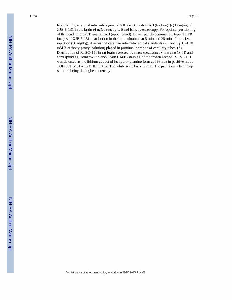
ferricyanide, a typical nitroxide signal of XJB-5-131 is detected (bottom). (c) Imaging ofXJB-5-131 in the brain of naïve rats by L-Band EPR spectroscopy. For optimal positioningof the head, micro-CT was utilized (upper panel). Lower panels demonstrate typical EPRimages of XJB-5-131 distribution in the brain obtained at 5 min and 25 min after its i.v.injection (50 mg/kg). Arrows indicate two nitroxide radical standards (2.5 and 5 μL of 10mM 3-carboxy-proxyl solution) placed in proximal portions of capillary tubes. (d)Distribution of XJB-5-131 in rat brain assessed by mass spectrometry imaging (MSI) andcorresponding Hematoxylin-and-Eosin (H&E) staining of the frozen section. XJB-5-131was detected as the lithium adduct of its hydroxylamine form at 966 m/z in positive modeTOF/TOF MSI with DHB matrix. The white scale bar is 2 mm. The pixels are a heat mapwith red being the highest intensity.
Ji et al. Page 16
Nat Neurosci. Author manuscript; available in PMC 2013 July 01.
NIH
-PA Author Manuscript
NIH
-PA Author Manuscript
NIH
-PA Author Manuscript

Figure 5.Assessments of neurobehavioral and histological outcome in PND 17 rats treated withXJB-5-131 after TBI.(a) Ability of rats to remain (seconds) on the beam balance apparatus before and after CCIor sham injury. A repeated measures ANOVA revealed a significant group (F2,15 = 14.452,P = 0.0003), day (F5,75 = 47.631, P < 0.0001), and group by day interaction effect (F10,75 =2.186, P = 0.028). Bonferroni post hoc analyses revealed that the CCI + XJB groupperformed significantly better than the CCI + vehicle group (*P = 0.001; error bars, standarderror; n = 7-10 rats per group). (b) Maximum angle (degrees) for rats to remain on an
Ji et al. Page 17
Nat Neurosci. Author manuscript; available in PMC 2013 July 01.
NIH
-PA Author Manuscript
NIH
-PA Author Manuscript
NIH
-PA Author Manuscript

inclined platform. A repeated measures ANOVA revealed significant group (F2,15 = 13.164,P = 0.0005), day (F5,75 = 18.085, P < 0.0001), and group by day interaction (F10,75 = 2.902,P = 0.004). Bonferroni post hoc analyses revealed that the TBI + XJB group performedsignificantly better than the TBI + vehicle group (*P < 0.001; error bars, standard error; n =7-10 rats per group). (c) Latency (seconds) for rats to locate a hidden (submerged) platformon post-TBI days 11-15. A repeated measures ANOVA revealed significant group (F2,15 =19.753, P < 0.0001), day (F4,120 = 22.126, P < 0.0001), and group by day interaction (F8,120= 2.437, P = 0.018). Bonferroni post hoc analyses revealed that the TBI + XJB groupperformed significantly better than the TBI + vehicle group (P < 0.001; error bars, standarderror; n = 7-10 rats per group) and did not differ from the sham controls (P = 0.08). (d) NORtask performance 29 days after sham or CCI injury. TBI + XJB rats exhibited a better 24-hour delay NOR task performance score compared to TBI + vehicle rats (P < 0.001,ANOVA, F2,27 = 14.736; error bars, standard error; n = 9-10 rats per group). (e) Assessmentof neurodegeneration by Fluoro-Jade C (FJC) staining. Neurodegeneration observed in thepericontusional area at 24 h after CCI was attenuated by XJB-5-131. The white scale bar is40 μm. *P < 0.05 vs. naïve and sham controls, CCI 3h + Vehicle, and CCI + XJB-5-131;error bars, standard deviation; n = 4 rats per group.
Ji et al. Page 18
Nat Neurosci. Author manuscript; available in PMC 2013 July 01.
NIH
-PA Author Manuscript
NIH
-PA Author Manuscript
NIH
-PA Author Manuscript

Figure 6.Response of neurons treated with 4-Amino-TEMPO (4-AT) to in vitro TBI.(a) Percent cytotoxicity (lactate dehydrogenase (LDH) release relative to Triton exposure(corrected for background LDH). (b) 3-(4,5- dimethylthiazol-2-yl)-2,5-diphenyltetrazoliumbromide (MTT) as a percentage of normal control conditions. Vehicle or 4-AT was added tothe medium 10 min before stretch. Mechanical stretch induced approximately 35% neuronaldeath assessed by MTT and LDH at 24 hours. *P < 0.01 vs. stretch neurons, error bars,standard deviation; n = 3 experiments.
Ji et al. Page 19
Nat Neurosci. Author manuscript; available in PMC 2013 July 01.
NIH
-PA Author Manuscript
NIH
-PA Author Manuscript
NIH
-PA Author Manuscript

Figure 7.Electron paramagnetic resonance (EPR) spectroscopy assessment of inter-conversions ofnitroxides and hydroxylamines.(a) Effect of Ascorbate, Ascorbate oxidase and Cyt c/H2O2 on the EPR signal of XJB5-131. A: XJB-5-131 alone in 35 μL of PBS plus 35 μL of DMSO. B: A plus 250 μMAscorbate, 1 min. C: A plus 250 μM Ascorbate, 5 min. D: A plus 250 μM Ascorbate, 5 minplus ascorbate oxidase (1U, 15 min). E: D plus10 μM Cyt c. F: D plus10 μM Cyt c/200 μMH2O2. (b) Effect of ascorbate, ascorbate oxidase, and NADH on the EPR signal ofXJB-5-131 in brain homogenates. A: 35 μL of Brain Homogenate (350 μg protein) in PBSplus 7 μM XJB-5-131 plus 35μL of DMSO. B: A plus Ascorbate Oxidase (2U), 30 min. C:B plus NADH (200 μM), 30 min. D: C plus K3Fe(CN)6 (100 μM). (c) Effect of pH on theEPR signal of XJB-5-131-OH. 50 μL of 7 μM XJB-5-131-OH in DMSO were mixed with50 μL of 50 μM HEPES pH 7.45 (open bars) or with 50 μL of 50 μM HEPES pH 8.25(closed bars). Error bars, standard deviation.
Ji et al. Page 20
Nat Neurosci. Author manuscript; available in PMC 2013 July 01.
NIH
-PA Author Manuscript
NIH
-PA Author Manuscript
NIH
-PA Author Manuscript