Mitochondrial redox metabolism: Aging, longevity and dietary effects
11
Review Mitochondrial redox metabolism: Aging, longevity and dietary effects Melissa M. Page, Ellen L. Robb, Kurtis D. Salway, Jeffrey Alan Stuart * Department of Biological Sciences, Brock University, St. Catharines, ON, Canada L2S 3A1 1. Introduction For several decades, mitochondrial redox metabolism has attracted great interest for its potential role(s) in aging and longevity. Three lines of evidence implicate mitochondrial redox metabolism in aging. Firstly, mitochondria are the primary source of reactive oxygen species (ROS) in most cell types, and cumulative oxidative damage to cellular macromolecules may underlie the cellular dysfunction observed in aging animals. Secondly, signal- ling proteins that mediate apoptotic cell death are in the mitochondrial compartment and are redox sensitive. Tissue degeneration due to cumulative cell loss is a common observation in aging animals. Alterations of mitochondrial redox metabolism that influence the propensity to release apoptotic factors may therefore modulate the rate of tissue degeneration and thus aging. Finally, mitogenic signals originating from mitochondria modulate rates of cell division, which may be significant in cancer and the gradual decline of tissue progenitor cell populations in adults. Dietary factors, including restriction of caloric intake, restric- tion of protein or methionine intake, or the ingestion of specific nutrients, have been shown to alter mitochondrial redox metabo- lism, cellular oxidative stress and animal lifespans. In this review, we consider the evidence that specific aspects of mitochondrial redox metabolism affect aging and longevity of animals. We then discuss dietary manipulations known to modulate these mito- chondrial properties, and examine evidence that they are linked to extension of lifespan. Due to the vast quantity of relevant data, we have confined our focus primarily to experimental results from mammals, and discuss results from other animals when mamma- lian data are limiting. In addition, while we are able to cover most aspects of mitochondrial redox metabolism that are relevant to aging and nutrition, we can provide only a brief overview of particular subfields. In these instances, the reader is referred to recent in-depth reviews. 2. Mitochondrial respiration and ROS production In the mitochondrial respiratory chain, electrons entering at complexes I and II are transferred to complex III, then IV where they are combined with molecular oxygen and hydrogen to form H 2 O. Redox reactions at respiratory complexes I, III and IV are coupled to the extrusion of protons from the mitochondrial matrix into the intermembrane space. The re-entry of protons into the matrix is coupled to the synthesis of ATP from ADP and P i . This oxidative phosphorylation is responsible for the vast majority of ATP production and oxygen consumption in most types of animal cells (Rolfe and Brown, 1997). In many types of cells, including the highly oxidative cell types that are vulnerable to the degenerative effects of aging, mitochondrial respiration is also the source of most intracellular ROS production (reviewed in Lambert and Brand, 2009). Mitochondrial ROS have been considered primarily for their pathological consequences, as they are capable of oxidizing and destabilizing most mitochondrial macromolecules. However, ROS also play key roles in intracellular signalling events Mechanisms of Ageing and Development 131 (2010) 242–252 ARTICLE INFO Article history: Available online 26 February 2010 Keywords: Mitochondria Reactive oxygen species Superoxide Hydrogen peroxide MnSOD Antioxidant Caloric restriction Oxidative stress Aging Lifespan ABSTRACT Mitochondrial redox metabolism has long been considered to play important roles in mammalian aging and the development of age-related pathologies in the major oxidative organs. Both genetic and dietary manipulations of mitochondrial redox metabolism have been associated with the extension of lifespan. Here we provide a broad overview of the circumstantial evidence showing associations between mitochondrial reactive oxygen species (ROS) metabolism, aging and longevity. We address most aspects of mitochondrial ROS metabolism, from superoxide production, to ROS detoxification and the repair/ removal of ROS-mediated macromolecular damage. Finally, we discuss the effects of dietary manipulations (e.g. caloric restriction, methionine restriction), dietary deficiencies (e.g. folate) and dietary supplementation (e.g. resveratrol) on mitochondrial ROS metabolism and lifespan. ß 2010 Elsevier Ireland Ltd. All rights reserved. * Corresponding author. Tel.: +1 905 688 5550x4814; fax: +1 905 688 1855. E-mail address: [email protected] (J.A. Stuart). Contents lists available at ScienceDirect Mechanisms of Ageing and Development journal homepage: www.elsevier.com/locate/mechagedev 0047-6374/$ – see front matter ß 2010 Elsevier Ireland Ltd. All rights reserved. doi:10.1016/j.mad.2010.02.005
Transcript of Mitochondrial redox metabolism: Aging, longevity and dietary effects

Mechanisms of Ageing and Development 131 (2010) 242–252
Review
Mitochondrial redox metabolism: Aging, longevity and dietary effects
Melissa M. Page, Ellen L. Robb, Kurtis D. Salway, Jeffrey Alan Stuart *
Department of Biological Sciences, Brock University, St. Catharines, ON, Canada L2S 3A1
A R T I C L E I N F O
Article history:
Available online 26 February 2010
Keywords:
Mitochondria
Reactive oxygen species
Superoxide
Hydrogen peroxide
MnSOD
Antioxidant
Caloric restriction
Oxidative stress
Aging
Lifespan
A B S T R A C T
Mitochondrial redox metabolism has long been considered to play important roles in mammalian aging
and the development of age-related pathologies in the major oxidative organs. Both genetic and dietary
manipulations of mitochondrial redox metabolism have been associated with the extension of lifespan.
Here we provide a broad overview of the circumstantial evidence showing associations between
mitochondrial reactive oxygen species (ROS) metabolism, aging and longevity. We address most aspects
of mitochondrial ROS metabolism, from superoxide production, to ROS detoxification and the repair/
removal of ROS-mediated macromolecular damage. Finally, we discuss the effects of dietary
manipulations (e.g. caloric restriction, methionine restriction), dietary deficiencies (e.g. folate) and
dietary supplementation (e.g. resveratrol) on mitochondrial ROS metabolism and lifespan.
� 2010 Elsevier Ireland Ltd. All rights reserved.
Contents lists available at ScienceDirect
Mechanisms of Ageing and Development
journa l homepage: www.e lsev ier .com/ locate /mechagedev
1. Introduction
For several decades, mitochondrial redox metabolism hasattracted great interest for its potential role(s) in aging andlongevity. Three lines of evidence implicate mitochondrial redoxmetabolism in aging. Firstly, mitochondria are the primary sourceof reactive oxygen species (ROS) in most cell types, and cumulativeoxidative damage to cellular macromolecules may underlie thecellular dysfunction observed in aging animals. Secondly, signal-ling proteins that mediate apoptotic cell death are in themitochondrial compartment and are redox sensitive. Tissuedegeneration due to cumulative cell loss is a common observationin aging animals. Alterations of mitochondrial redox metabolismthat influence the propensity to release apoptotic factors maytherefore modulate the rate of tissue degeneration and thus aging.Finally, mitogenic signals originating from mitochondria modulaterates of cell division, which may be significant in cancer and thegradual decline of tissue progenitor cell populations in adults.
Dietary factors, including restriction of caloric intake, restric-tion of protein or methionine intake, or the ingestion of specificnutrients, have been shown to alter mitochondrial redox metabo-lism, cellular oxidative stress and animal lifespans. In this review,we consider the evidence that specific aspects of mitochondrialredox metabolism affect aging and longevity of animals. We thendiscuss dietary manipulations known to modulate these mito-
* Corresponding author. Tel.: +1 905 688 5550x4814; fax: +1 905 688 1855.
E-mail address: [email protected] (J.A. Stuart).
0047-6374/$ – see front matter � 2010 Elsevier Ireland Ltd. All rights reserved.
doi:10.1016/j.mad.2010.02.005
chondrial properties, and examine evidence that they are linked toextension of lifespan. Due to the vast quantity of relevant data, wehave confined our focus primarily to experimental results frommammals, and discuss results from other animals when mamma-lian data are limiting. In addition, while we are able to cover mostaspects of mitochondrial redox metabolism that are relevant toaging and nutrition, we can provide only a brief overview ofparticular subfields. In these instances, the reader is referred torecent in-depth reviews.
2. Mitochondrial respiration and ROS production
In the mitochondrial respiratory chain, electrons entering atcomplexes I and II are transferred to complex III, then IV wherethey are combined with molecular oxygen and hydrogen to formH2O. Redox reactions at respiratory complexes I, III and IV arecoupled to the extrusion of protons from the mitochondrial matrixinto the intermembrane space. The re-entry of protons into thematrix is coupled to the synthesis of ATP from ADP and Pi. Thisoxidative phosphorylation is responsible for the vast majority ofATP production and oxygen consumption in most types of animalcells (Rolfe and Brown, 1997). In many types of cells, including thehighly oxidative cell types that are vulnerable to the degenerativeeffects of aging, mitochondrial respiration is also the source ofmost intracellular ROS production (reviewed in Lambert andBrand, 2009). Mitochondrial ROS have been considered primarilyfor their pathological consequences, as they are capable ofoxidizing and destabilizing most mitochondrial macromolecules.However, ROS also play key roles in intracellular signalling events

M.M. Page et al. / Mechanisms of Ageing and Development 131 (2010) 242–252 243
that are implicated in cell death, growth and transformation. Bothaspects of mitochondrial ROS may be relevant to aging.
3. Mitochondrial ROS production and lifespan
Electron transfer between respiratory complexes is favouredboth energetically and perhaps also due to the proximity ofrespiratory complexes to each other within respiratory super-complexes (Acın-Perez et al., 2008). Nonetheless, under conditionsin which complexes I and III are highly reduced, a significantproportion of electrons are diverted from them directly tomolecular oxygen, thus forming the radical superoxide (O2
��)(reviewed in Lambert and Brand, 2009). In addition to thesereactions, it recently has been shown that p66shc, a protein presentin the intermembrane space (IMS), can also catalyse superoxideproduction via electron transfer from reduced cytochrome c tomolecular oxygen (reviewed in Pinton and Rizzuto, 2008).Superoxide production occurs on both the matrix and IMS facesof the mitochondrial inner membrane (IMM), and is thus activeboth within mitochondria and within the cytosol.
Superoxide participates directly in a variety of reactions,including: (1) spontaneous or enzyme-catalysed dismutation tohydrogen peroxide. In the presence of ferrous iron, hydrogenperoxide can give rise to highly oxidative hydroxyl radicals; (2)reaction with nitric oxide (NO) to produce peroxynitrite; (3) directoxidation of proteins, such as the TCA cycle enzyme aconitase.These reactions can be problematic if they produce sufficientlyhigh quantities of their products. Hydroxyl radicals indiscrimi-nately damage proteins, lipids and DNA. Similarly, peroxynitrite ishighly oxidative, with a wide range of potential molecular targetsthat includes both cytochrome c (Jang and Han, 2006) andcytochrome c oxidase (Cooper and Davies, 2000).
A number of studies have found an inverse correlation betweenthe rate of respiratory ROS production and lifespan. The data inthese studies were collected using assays that measure hydrogenperoxide diffusion out of respiring isolated mitochondria. Long-lived bats have lower rates of hydrogen peroxide production byheart, brain and kidney mitochondria compared to a short-livedshrew species (Brunet-Rossinni, 2004). Similarly, long-lived birdsproduce less heart mitochondrial hydrogen peroxide than do rats(Ku and Sohal, 1993; Herrero and Barja, 1998). Also, mitochondrialhydrogen peroxide production is lower in cells of the exceptionallylong-lived naked mole rat compared to Fischer 344 rats (Csiszar etal., 2007). Barja’s group reported a negative correlation betweenROS production of isolated heart mitochondria and MLSP inmammals and birds (reviewed in Barja, 2002), a result that waslater confirmed and extended in 14 mammal and bird species byLambert et al. (2007).
Taken together, these studies provide substantial evidence for anegative correlation between mitochondrial ROS production andspecies longevity. However, thisconclusion istemperedbyexamplesin which the opposite trend has been found. Vascular endothelialcells of long-lived Ames dwarf mice produce more hydrogenperoxide than those of normal littermates (Csiszar et al., 2008).Similarly, no relationship between mitochondrial ROS productionand longevity is evident in Drosophila melanogaster (Miwa et al.,2004). Finally, a weakness of virtually all of these studies is the non-physiological conditions under which the measurements wereperformed (see Robb et al., 2009 for review). Nonetheless, the weightof the evidence at this time suggests that mitochondrial ROSproduction is generally lower in longer lived species.
4. Dietary effects on mitochondrial ROS production
Mitochondrial ROS production is particularly amenable todietary interventions that restrict intake of total calories, protein,
or the amino acid methionine. Caloric restriction of rodents, whichconfers increased longevity, is also associated with reducedmitochondrial hydrogen peroxide production. Hydrogen peroxideproduction by mitochondria isolated from liver (Hagopian et al.,2005), skeletal muscle (Bevilacqua et al., 2004) and brain (Sanz etal., 2005) is reduced in calorie-restricted rats. Mitochondrialhydrogen peroxide production is similarly reduced in mice fed adlibitum every other day (Caro et al., 2008a), or consuming dietswith reduced protein or methionine content (Sanz et al., 2006a;Caro et al., 2008b). All of these diets also confer increasedlongevity in mice (reviewed in Lopez-Torres and Barja, 2008; Sunet al., 2009). In contrast, restricting the dietary intake of lipids(Sanz et al., 2006b) or carbohydrates (Sanz et al., 2006c) in miceneither alters mitochondrial ROS production nor extends longev-ity. These results suggest an inverse correlation betweenmitochondrial ROS production and longevity in rodents. However,they do not indicate mitochondrial ROS production as adeterminant of longevity. Some evidence suggesting that thismay be the case is discussed below.
The mechanism(s) by which the rate of mitochondrial ROSproduction is reduced in long-lived species and by caloricrestriction has not been fully elucidated. However three studieshave shown a relationship between the rate of ROS production andrespiratory complex I content. As respiratory complex I is a majorsource of superoxide, the relative abundance of complex I inmitochondria could directly affect the rate of its production. Pigeonheart mitochondria have lower levels of complex I than rat heartmitochondria, based on relative levels of flavine nucleotides (St.Pierre et al., 2002; Lambert et al., 2010). Similarly, Ayala et al.(2007) found by western blot that complex I levels were reduced inliver mitochondria of calorie-restricted rats. In contrast, Valle et al.(2007) found that the activity of complex I in liver mitochondriaisolated from the same rat strain was unaffected by a similarduration of caloric restriction. Thus, while modulation of complex Ilevels may represent one mechanism by which mitochondrial ROSproduction is reduced in parallel with extended longevity, morework will be required to confirm this.
5. Mitochondrial cardiolipin peroxidation and lifespan
As the redox centres of the respiratory complexes are within theinner membrane bilayer, most superoxide produced at complexes Iand III likely originates within the lipid interior of the bilayer.Oxygen solubility is up to 8-fold greater in this milieu than in theaqueous matrix or cytosol (Smotkin et al., 1991), and this relativelyhigh concentration of oxygen favours superoxide production.Products of superoxide readily undergo peroxidative reactionswith membrane phospholipids. Membrane lipid peroxidation inturn alters the biophysical properties of the bilayer (e.g. Megli andSabatini, 2003, 2004), affecting the lateral mobility of proteins. Theproducts of lipid peroxidation also form adducts on innermembrane proteins, impacting specific enzyme catalysed activi-ties (Chen et al., 1998a,b), and may thus directly affect aspects ofoxidative phosphorylation.
In addition to these non-specific effects, peroxidation of specificphospholipids may be critical. Recently, evidence has accumulatedthat peroxidation of cardiolipin is a key step in mitochondriapermeability transition and apoptosis. Cardiolipin is a uniquephospholipid that is found almost exclusively in the mitochondrialinner membrane. Most respiratory complexes as well as associatedenzymes such as the adenine nucleotide translocator requirecardiolipin for maximal activity (Fry and Green, 1981; reviewed inSchlame et al., 2000 and Bogdanov et al., 2008), and thisphospholipid may also be essential for the formation of respiratory‘supercomplexes’ (Pfeiffer et al., 2003; Zhang et al., 2005).Peroxidation of cardiolipin fatty acyl chains inhibits the activities

M.M. Page et al. / Mechanisms of Ageing and Development 131 (2010) 242–252244
of respiratory complexes (reviewed in Pope et al., 2008), and thusimpacts mitochondrial redox metabolism directly.
In addition to being a target of non-specific peroxidation due toits proximity to sites of ROS formation, cardiolipin appears to bedirectly peroxidized by cytochrome c (see Kagan et al., 2009 forreview). Although cardiolipin is localized primarily to the matrixface of the IMM, a portion of it is found on the IMS face of the IMM,associated with contact sites. This IMS-facing cardiolipin isinvolved in electrostatic interactions with cytochrome c, whichmay be important in mediating the association of the latter withrespiratory complexes. However, an early step in mitochondrial-mediated apoptosis is the redistribution of cardiolipin from thematrix face of the IMM to the IMS face, facilitating interactionsbetween cardiolipin and cytochrome c. Under these conditions,conformational changes in cytochrome c induced by cardiolipinbinding stimulate a cytochrome c peroxidize activity thatspecifically targets cardiolipin. This peroxidation of cardiolipin isin turn associated with a number of key steps in the apoptoticdeath cascade: (1) loss of the cytochrome c association; (2)permeabilization of the outer membrane, possibly via interactionswith other apoptotic proteins; (3) stimulation of mitochondrialpermeability transition pore opening, thus depolarizing the innermembrane and facilitating mitochondrial destruction and pro-moting cell death.
Mammalian mitochondria contain an isoform of glutathioneperoxidase, GPx4 (or mitochondrial phospholipid hydroperoxideglutathione peroxidase) that has the unique ability to catalyse thereduction of peroxidized acyl groups in phospholipids (Ursini et al.,1985). GPx4 has been hypothesized to prevent stress-inducedapoptosis by protecting against cardiolipin oxidation (reviewed inImai and Nakagawa, 2003). Deletion of the GPx4 gene in mice isembryonic lethal (Yant et al., 2003). In contrast, overexpression ofGPx4 protects mouse embryonic fibroblasts from cell deathinduced by potent oxidants, such as t-butylhydroperoxide and,in vivo, protects against liver damage caused by diquat (Ran et al.,2003, 2004). GPx4 prevents a decrease in electron transport chaincomplex activity, and protects cardiac function following ische-mia/reperfusion in mice (Dabkowski et al., 2008), perhaps due toits ability to prevent cardiolipin peroxidation. Recently, Liang et al.(2009) reported that transgenic overexpression of GPx4 attenuatescardiolipin peroxidation and the release of mitochondrial apopto-tic factors following oxidative stress. Surprisingly, despite theseobservations of GPx4s protective functions, GPx4+/� mice whichhave only about 50% of normal GPx4 protein levels in brain, liver,kidney and spleen have a reduced incidence of cancer and longerlifespan than wild type mice (Ran et al., 2007). A possibleexplanation for this result is that reduced GPx4 activity predis-poses cancerous cells to apoptotic death. However, it should benoted that in these experiments levels of GPx4 in subcellularcompartments other than mitochondria were also affected, makingit impossible to attribute all observed effects to mitochondrialfunctions. Also, the implications of GPx4 underexpression in otherspecies with a lower propensity of developing cancer are unknown.
6. Dietary effects on cardiolipin fatty acyl composition
Given the central role that cardiolipin appears to play in themitochondrial apoptotic pathway, it is important to ask whetherthis phospholipid might be a useful target for modulating the agingprocess (see Pepe, 2005 for review). Cardiolipin is particularlysusceptible to peroxidation because of its high content ofunsaturated fatty acyl chains. In heart mitochondria, 80–90% ofcardiolipin fatty acyl chains are linoleic acid (18:2n � 6). Lee et al.(2006) reported that the fatty acyl composition of heartmitochondrial cardiolipin is altered with age: 24-month-old ratshave almost 35% less linoleic acid, and higher levels of the highly
unsaturated arachadonic acid (20:4n � 6) and docosahexaenoicacid (22:6n � 3). These more highly unsaturated fatty acids aremore susceptible to peroxidation. Cardiolipin linoleic acid contentis amenable to dietary manipulation (for example, see Dannen-berger et al., 2007). Dietary supplementation with linoleic acid canattenuate the age-related depletion of linoleic acid from rat heartmitochondrial cardiolipin, while improving overall heart functionand resistance to oxidative challenges (Chicco et al., 2008). Theseresults indicate that dietary supplementation is capable ofpromoting the incorporation of linoleic acid into cardiolipin,which may in turn be significant in maintaining mitochondrialfunction with age and thus perhaps promote longevity. Moreresearch into this possibility is therefore warranted.
7. Uncoupling of oxidative phosphorylation, mitochondrialROS production and longevity
Most vertebrate tissue mitochondria contain uncouplingproteins (UCPs; summarized in Stuart et al., 2001), though levelsof expression of this family of proteins is generally low with theexception of UCP1 in brown adipose tissue. UCPs are IM-spanningproteins that catalyse proton conductance from the IMS to matrix.UCP2 is widely expressed, at low levels, in many mammalian celltypes; UCP3 expression is highest in skeletal muscle. One functionof UCPs may be suppressing superoxide formation via a reductionof membrane potential that increases the rate of electron transferand oxygen consumption while maintaining respiratory com-plexes in a more oxidized state (Echtay et al., 2002). As a result, theincreased rate of oxygen consumption reduces local PO2, while thedecreased proportion of respiratory complexes I and III in areduced state decreases the likelihood of electron donation tooxygen. The proton conductance activity of UCPs is stimulated bylipid peroxidation products such as 4-hydroxynonenal (Echtay etal., 2005). Thus, phenomena that promote membrane phospholipidperoxidation stimulate UCP activity, possibly as part of a negativefeedback circuit that reduces superoxide formation to defend thestructure and function of the IM. Evidence for this includesobservations that mice deficient in UCPs have higher levels ofmembrane lipid peroxidative damage (Brand et al., 2002). Alsoconsistent with this role, transgenic UCP2 overexpression canpartially rescue the early neonatal lethality of mitochondrialsuperoxide dismutase (MnSOD) deficiency (Andrews and Horvath,2009), and confers neuroprotection in adult mice under conditionsrelated to oxidative stress (Mattiasson et al., 2003; Deierborg et al.,2008). Andrews and Horvath (2009) report that UCP2 genedeletion dramatically reduces maximum lifespan in mice;however, simultaneous overexpression of UCP2 and UCP3 hasminimal effect on longevity (McDonald et al., 2008). Evidence frominvertebrate experiments shows no clear outcome with respect tolifespan (Fridell et al., 2005; Iser et al., 2005; Sanchez-Blanco et al.,2006; Humphrey et al., 2009).
Although UCP overexpression is not consistently associatedwith increased longevity, this could be related to the requirementof these proteins for effector molecules. Maximal activity of UCPsrequires stimulation with fatty acids, including endproducts offatty acid peroxidation. UCP activities are also inhibited by adeninenucleotides. Thus, increasing UCP expression without modifyinglevels of stimulators and inhibitors of their activities may not affectmembrane potential under normal conditions.
8. Dietary interventions affecting mitochondrial coupling
UCP levels in various tissues are affected by dietary manipula-tions including caloric restriction (Xiao et al., 2004), short-termstarvation (Jikumaru et al., 2007) and dietary fat intake (Samec etal., 1999). However, these changes have not always been

M.M. Page et al. / Mechanisms of Ageing and Development 131 (2010) 242–252 245
associated with altered proton leak, and their significance is thusnot well understood. Therefore there is little compelling evidenceto suggest that dietary modulation of UCP activities can be usefullyapplied to modulate lifespan.
Caloric restriction has been reported to induce a marginalincrease in liver mitochondrial proton leak that is accompanied byreduced substrate oxidation activity, thus lowering the protonmotive force (Lambert and Merry, 2004). Liver mitochondria lackUCPs, indicating that this difference cannot be due to differentialexpression of UCPs. However, other IMM proteins contribute toproton leak and these may be altered by caloric restriction.
The uncoupling of oxidative phosphorylation can also berealized by dietary supplementation with chemical uncouplers,such as dinitrophenol (DNP). Low doses of DNP (or other chemicaluncouplers) that provide a marginal reduction in the mitochon-drial inner membrane potential reduce ROS production whilemaintaining ATP synthesis (Korshunov et al., 1997). DNP confersneuroprotection against oxidative stress (reviewed de Felice andFerreira, 2006). A subtle uncoupling of oxidative phosphorylation,via chronic dietary intake of DNP, reduced rates of ROS productionand levels of oxidative damage in brain, heart and liver tissues ofrats (Caldeira da Silva et al., 2008). Concomitantly, this treatmentprovided a marginal increase in median and mean lifespan. Whilethese results are consistent with a direct role for mitochondrialuncoupling in reducing oxidative and extending longevity, it isequally possible that chronic DNP treatment of rodents elicitstissue-specific responses that could invoke a caloric restrictiontype of response.
9. Mitochondrial superoxide dismutase in aging and longevity
Attenuating respiratory superoxide production at its source isan effective strategy for reducing mitochondrial ROS productionand oxidative stress. However, many of the same outcomes can beachieved by upregulating mitochondrial antioxidant enzymes. Inaddition, antioxidant enzyme activities can alter the relative levelsof different ROS, such as superoxide and hydrogen peroxide, whichis of regulatory significance. Matrix superoxide is dismutated tohydrogen peroxide by the mitochondrial superoxide dismutase(MnSOD). MnSOD is localized exclusively within the matrix, and aportion of the mitochondrial MnSOD pool is associated with thematrix face of the IM (Okado-Matsumoto and Fridovich, 2001).MnSOD may thus be physically close to sites of respiratorysuperoxide production. Manipulation of MnSOD expression hasbeen attempted in a variety of species and experimental contexts,and the majority of reported results suggest beneficial effects onage-related disease, if not on aging per se (Chen et al., 1998a,b;Jones et al., 2003; Dumont et al., 2009).
In vitro, MnSOD overexpression provides protection againststress-induced cell death in a wide range of cell types (forexamples see Keller et al., 1998; Epperly et al., 2003; Lee et al.,2004; Cruthirds et al., 2005; Silva et al., 2005; Hu et al., 2007).MnSOD levels in dermal fibroblasts correlate positively withcellular stress resistance and mammalian species lifespan (seeKapahi et al., 1999; Brown and Stuart, 2007). In vivo evidence isclearly supportive of a protective role for MnSOD against apoptoticcell death. In mice, deletion of the MnSOD gene causes severedefects from birth including cardiomyopathy and neurodegenera-tion, limiting lifespan to several weeks (Li et al., 1995; Lebovitz etal., 1996). Mitochondria from MnSOD+/� mice have reducedmitochondrial respiratory control ratios (RCR) and increasedpropensity of isolated mitochondria to undergo permeabilitytransition (Williams et al., 1998; Van Remmen et al., 2003).MnSOD+/� mice show increased incidence of apoptotic cell deathinduced by oxidative stress (Van Remmen et al., 2003; Loch et al.,2009). Overexpression of MnSOD in mice provides protection from
stress-induced cell death. Transgenic mice that overexpresshuman MnSOD are protected from oxidative damage and neuronloss induced by ischemia (Keller et al., 1998). MnSOD over-expression confers resistance to a wide variety of degenerativedisorders that are common in advanced age (Dumont et al., 2009;Chen et al., 1998a,b; Jones et al., 2003). MnSOD activity in brain,standardized to mitochondrial content using citrate synthaseactivity, is positively correlated with maximum species lifespan in14 species of mammals and birds (Page et al., in press).
Given the results outlined above, it is somewhat surprising thatMnSOD overexpression in mice fails to extend lifespan (Jang et al.,2009; Perez et al., 2009), though a contradictory report suggests itmay (Hu et al., 2007; see Jang et al., 2009 for discussion of thisdiscrepancy). Equally surprising is the report that MnSOD+/� micedo not have significantly shorter lifespans than their wild typecounterparts (Van Remmen et al., 2003). These results areobviously significant to our understanding of relationshipsbetween mitochondrial redox metabolism, stress resistance andlongevity, and challenge the basic hypothesis that stress resistantmitochondria that produce less ROS promote extended longevity.It nonetheless appears to be the case that higher levels of MnSODexpression are associated with increased resistance to neurode-generative disorders and reduced rates of cancer cell growth,suggesting health benefits to the elderly if similar results could beachieved in humans.
MnSOD overexpression is significant also for its role in alteringthe relative levels of superoxide and hydrogen peroxide (Beuttneret al., 2006). ROS are hypothesized to act as intracellular signallingmolecules in cell replication (for review see Sauer et al., 2001). Theconversion of superoxide anion, thought to act as a mitogen, tohydrogen peroxide by SOD may provide a signal to slow replicativegrowth. The change in redox state that accompanies differences inmitochondrial MnSOD activity may therefore be important in theregulation of cell proliferation. Also, because of its high reactivitywith NO, by reducing steady state superoxide levels, MnSODprobably mediates reduced rates of reaction with NO to produceperoxynitrite. Some of the effects of manipulating the MnSODexpression level are thus undoubtedly due to effects on signallingpathways modulated by ROS and NO/RNS (reactive nitrogenspecies).
Cullen et al. (2003) correlated MnSOD activity to the growth ofhuman pancreas and human pancreatic cancer cell lines and foundsignificantly lower MnSOD activity in rapidly replicating pancre-atic cancer cell lines compared to normal pancreatic cell lines.Transgenic MnSOD overexpression has also been shown to slowthe replicative growth rates of human pancreatic cancer cells invitro, and result in slower tumour growth in vivo (Ough et al.,2004; Weydert et al., 2003). Decreased cellular replication rate isalso observed following MnSOD overexpression in the transformedmouse fibroblast cell line NIH/3T3 (Li et al., 1998). Although themajority of experiments concerning cellular replication rate andSOD have involved MnSOD, Weydert et al. (2006) found thatadenoviral MnSOD or adenoviral CuZnSOD overexpression weresimilarly able to inhibit the growth rate of three human breastcancer cell lines in vitro. The absence of a MnSOD specific effectmay suggest that the observed change in replicative growth rate isnot dependent on a mitochondrial, but rather an overall change inintracellular redox state.
The ability of MnSOD to slow replicative growth appears to berelated to a role in regulating progression through the cell cycle.Sarsour et al. (2008) reported that manipulation of MnSOD activityin mouse embryonic fibroblasts influenced the ability of thesefibroblasts to move through the cell cycle. An increase in MnSODactivity favoured a transition into quiescence. In contrast,fibroblasts deficient in MnSOD were unable to successfully exitthe cell cycle (Sarsour et al., 2008). In vitro, transgenic MnSOD
M.M. Page et al. / Mechanisms of Ageing and Development 131 (2010) 242–252246
overexpression in mice protects the replicative capacity of mousemyoblasts (Lee et al., 2009), suggesting that progenitor cellturnover is slowed by MnSOD in vivo. MnSOD’s ability to protectreplicative capacity in vivo may be especially important in thecontext of stem cell depletion and aging, as a slowed rate ofdivision may extend the replicative capacity of these cells. Thiswould also suggest a role for MnSOD in aging and longevity. Takentogether, the data regarding MnSOD’s role in cell proliferation aresufficiently strong to support the targeting of this enzyme tomodulate cell growth rates in vivo.
10. Dietary interventions and nutrient signalling pathwaysaffecting mitochondrial superoxide dismutase
Mitochondrial antioxidant enzymes are targets of nutrientsensing pathways involved in the regulation of longevity. This hasbeen studied thoroughly for MnSOD, though there is evidence thatmitochondrial glutathione peroxidase activity (Valle et al., 2007)and mitochondrial peroxiredoxin III (Chiribau et al., 2008; seebelow for discussion of peroxiredoxin III) are similarly affected. Inmice, disruption of insulin/insulin-like growth factor-1 signalling(IIS) extends maximum lifespan in a variety of genetic back-grounds, though the magnitude of the effect is almost alwaysgreater in females than males (Baba et al., 2005; Holzenbergeret al., 2003; Selman et al., 2008). The FOXO family of transcriptionfactors mediate some of the effects of IIS signalling (reviewed byGross et al., 2009). One downstream target of FOXO3a is MnSOD,whose expression is upregulated by activated FOXO3a (Kops et al.,2002). This MnSOD upregulation is evident in heterozygous insulinreceptor knockout (IR+/�) mice (Baba et al., 2005). FOXO3a activityis stimulated by deacetylation during caloric restriction (Wang etal., 2007), thus providing a link between this dietary interventionand mitochondrial MnSOD. The klotho gene, whose proteinproducts include a hormone that inhibits IIS similarly extends
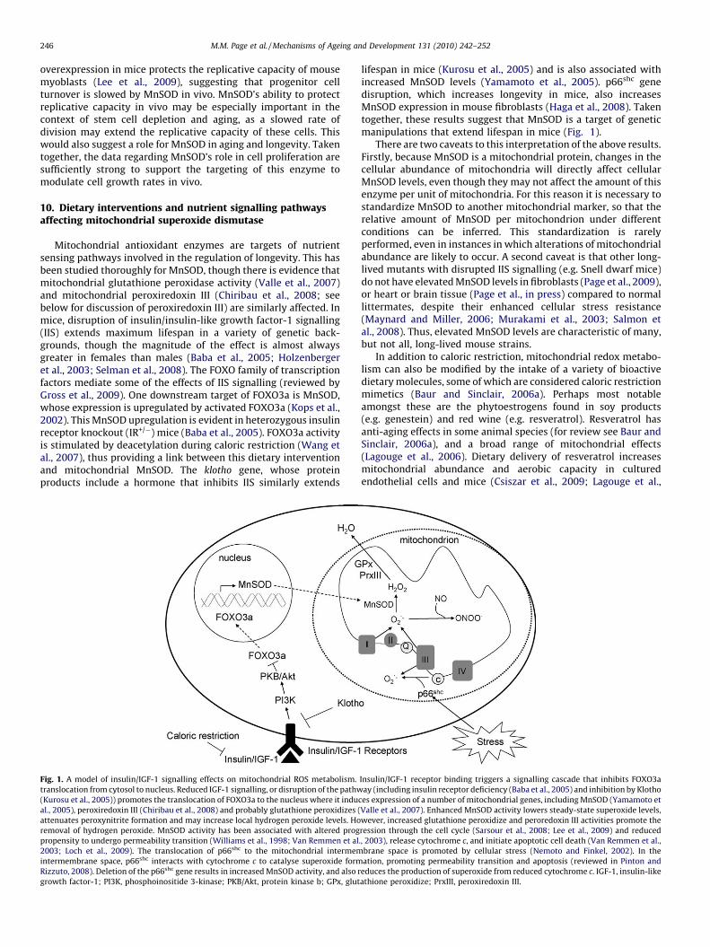
Fig. 1. A model of insulin/IGF-1 signalling effects on mitochondrial ROS metabolism.
translocation from cytosol to nucleus. Reduced IGF-1 signalling, or disruption of the pathw
(Kurosu et al., 2005)) promotes the translocation of FOXO3a to the nucleus where it indu
al., 2005), peroxiredoxin III (Chiribau et al., 2008) and probably glutathione peroxidizes (
attenuates peroxynitrite formation and may increase local hydrogen peroxide levels. Ho
removal of hydrogen peroxide. MnSOD activity has been associated with altered prog
propensity to undergo permeability transition (Williams et al., 1998; Van Remmen et al
2003; Loch et al., 2009). The translocation of p66shc to the mitochondrial intermem
intermembrane space, p66shc interacts with cytochrome c to catalyse superoxide form
Rizzuto, 2008). Deletion of the p66shc gene results in increased MnSOD activity, and also r
growth factor-1; PI3K, phosphoinositide 3-kinase; PKB/Akt, protein kinase b; GPx, glut
lifespan in mice (Kurosu et al., 2005) and is also associated withincreased MnSOD levels (Yamamoto et al., 2005). p66shc genedisruption, which increases longevity in mice, also increasesMnSOD expression in mouse fibroblasts (Haga et al., 2008). Takentogether, these results suggest that MnSOD is a target of geneticmanipulations that extend lifespan in mice (Fig. 1).
There are two caveats to this interpretation of the above results.Firstly, because MnSOD is a mitochondrial protein, changes in thecellular abundance of mitochondria will directly affect cellularMnSOD levels, even though they may not affect the amount of thisenzyme per unit of mitochondria. For this reason it is necessary tostandardize MnSOD to another mitochondrial marker, so that therelative amount of MnSOD per mitochondrion under differentconditions can be inferred. This standardization is rarelyperformed, even in instances in which alterations of mitochondrialabundance are likely to occur. A second caveat is that other long-lived mutants with disrupted IIS signalling (e.g. Snell dwarf mice)do not have elevated MnSOD levels in fibroblasts (Page et al., 2009),or heart or brain tissue (Page et al., in press) compared to normallittermates, despite their enhanced cellular stress resistance(Maynard and Miller, 2006; Murakami et al., 2003; Salmon etal., 2008). Thus, elevated MnSOD levels are characteristic of many,but not all, long-lived mouse strains.
In addition to caloric restriction, mitochondrial redox metabo-lism can also be modified by the intake of a variety of bioactivedietary molecules, some of which are considered caloric restrictionmimetics (Baur and Sinclair, 2006a). Perhaps most notableamongst these are the phytoestrogens found in soy products(e.g. genestein) and red wine (e.g. resveratrol). Resveratrol hasanti-aging effects in some animal species (for review see Baur andSinclair, 2006a), and a broad range of mitochondrial effects(Lagouge et al., 2006). Dietary delivery of resveratrol increasesmitochondrial abundance and aerobic capacity in culturedendothelial cells and mice (Csiszar et al., 2009; Lagouge et al.,
Insulin/IGF-1 receptor binding triggers a signalling cascade that inhibits FOXO3a
ay (including insulin receptor deficiency (Baba et al., 2005) and inhibition by Klotho
ces expression of a number of mitochondrial genes, including MnSOD (Yamamoto et
Valle et al., 2007). Enhanced MnSOD activity lowers steady-state superoxide levels,
wever, increased glutathione peroxidize and peroredoxin III activities promote the
ression through the cell cycle (Sarsour et al., 2008; Lee et al., 2009) and reduced
., 2003), release cytochrome c, and initiate apoptotic cell death (Van Remmen et al.,
brane space is promoted by cellular stress (Nemoto and Finkel, 2002). In the
ation, promoting permeability transition and apoptosis (reviewed in Pinton and
educes the production of superoxide from reduced cytochrome c. IGF-1, insulin-like
athione peroxidize; PrxIII, peroxiredoxin III.

M.M. Page et al. / Mechanisms of Ageing and Development 131 (2010) 242–252 247
2006; Baur et al., 2006b). Direct interactions between resveratroland various components of the mitochondrial electron transportchain, including the ATP synthase are also observed in vitro. Atmicromolar concentrations, resveratrol is able to bind and inhibitthe activity of complex III in isolated mitochondria (Zini et al.,1999). Similarly, a resveratrol binding site on the F1-ATPase hasbeen identified (Gledhill and Walker, 2005). The importance ofthese observations in terms of mitochondrial membrane potentialand rates of ROS production is currently unknown.
Both genestein (Borras et al., 2006) and resveratrol (Robb et al.,2008b) stimulate MnSOD expression. Chronic exposure of humancells in culture to resveratrol stimulates an approximately 6-foldincrease in MnSOD protein levels and activity (Robb et al., 2008b).When fed to mice as part of a high fat diet, resveratrolsupplementation for six weeks stimulates an approximately 1.5-fold increase in brain MnSOD (Robb et al., 2008a). Resveratrol-induced upregulation of MnSOD is concomitant with increasedcellular stress resistance and reduced rates of cell proliferation inreplicating cells in vitro (Robb and Stuart, 2010). In cardiomyo-cytes, resveratrol pretreatment has been shown to protect againstROS production by the chemotherapeutic drug doxorubicin and tomaintain mitochondria membrane potential, observations theauthors attribute to an increase in MnSOD (Danz et al., 2009).Similarly, resveratrol pretreatment provides protection againstneurotoxic compounds associated with oxidative damage both invitro and in vivo (Alvira et al., 2007; Okawara et al., 2007; Blanchetet al., 2008), a result that can be achieved through geneticmanipulation of MnSOD. Recently, a vegetarian diet has beenshown to induce MnSOD expression in human buccal mucosa cellsby decreasing DNA methylation of the MnSOD promoter region(Thaler et al., 2008). These exciting results highlight the impor-tance of further research into how diet and specific dietarymolecules, including resveratrol, can be used to manipulatemitochondrial redox metabolism in humans as a means ofprotection against the degenerative diseases of aging.
11. Mitochondrial protein oxidative damage and longevity
Mitochondrial ROS interact directly with a wide variety ofmitochondrial proteins, and the resultant modifications are both
Table 1Enzymes involved in maintaining protein methionine and cysteine residues within the m
stress resistance.
Enzyme Activity Experim
manipu
Glutaredoxin 2 Thioltransferase, catalyses reduction
and formation of protein-glutathione
mixed disulphides
Overexp
Thioredoxin 2 Maintains protein thiols in reduced
state
Overexp
Heteroz
Peroxiredoxin 3 Thioredoxin peroxidase, uses
thioredoxin 2 as electron donor to
eliminate H2O2
Overexp
siRNA s
Methionine sulphoxide
reductase B2
Catalyses reduction of oxidized
methionine sulphoxides to methionine
Overexp
siRNA s
regulatory and pathological (Bulteau et al., 2006). For example,oxidation of methionine and cysteine residues constitutes ahomeostatic regulatory mechanism for some mitochondrialproteins, but excessive redox imbalance may compromise thefunctions of other proteins. Moosmann and Behl (2008) suggestthat detrimental aspects of cysteine oxidation in mitochondria aresufficient to have driven the replacement of this amino acid duringthe evolution of longevity in mammalian species. Cysteine contentof mitochondrial DNA (mtDNA) encoded proteins is inverselycorrelated with species longevity, or with adoption of an aerobicmetabolism (Moosmann and Behl, 2008). Mitochondria alsocontain isoforms of several protein redoxins that function torepair oxidized methionine and cysteine residues. These activitiesmay also be subject to evolutionary pressures associated withincreased lifespan, though they have not been studied in thiscontext. Examples of mitochondrial protein redoxins includethioredoxin 2, peroxiredoxin 3, and thioredoxin reductase 2 (Table1). All of these are localized to the mitochondrial matrix, wherethey function by reducing disulphide bridges that form followingoxidation by hydrogen peroxide. A fourth mitochondrial enzyme,glutaredoxin 2, catalyses S-glutathionylation and deglutathionyla-tion of proteins to protect thiol groups from oxidation and restorethe reactivity of active thiols. Finally, an isoform of methioninesulphoxide reductase (MsrB2), which catalyses the reduction ofoxidized methionine sulphoxides to methionine, is also localizedto mitochondria. Most of these ‘protein repair’ enzymes have beenstudied by gene knockout and overexpression. Similarly to otherantioxidant enzymes, these enzymes are found to play a key role incellular stress resistance (Table 1).
Only a limited subset of amino acid residues is amenable toreductive repair following oxidation, while the majority of proteinswhose function has been impaired by oxidation must be removedvia proteolytic degradation. Within the matrix, this degradation iscatalysed by the Lon protease, an enzyme that appears to fulfilsome similar functions to the 20S/26S proteasome, which is notpresent in mitochondria. The Lon protease catalyses the ATP-dependent degradation of damaged or oxidized proteins, prevent-ing their accumulation and aggregation (Ngo and Davies, 2007).The recognition system utilized by the mammalian Lon protease ispoorly understood. However, it has been shown in Escherichia coli
itochondrial compartment: effects on mitochondrial ROS metabolism and cellular
ental
lation
Effects
ression Attenuates doxorubicin- or 2-deoxy-D-glucose-induced
respiratory dysfunction and cytochrome c release
(Enoksson et al., 2004)
Prevents cardiomyocyte death during ischemia
(Nagy et al., 2007)
ression Attenuates effects of tert-butylhydroperoxide and inhibits
apoptosis (Chen et al., 2002)
ygous Trx2+/� Increases cytochrome c release and procaspase 3 cleavage
(Nonn et al., 2003)
Increases H2O2 production and oxidative damage
ression Protects heart function and reduces oxidative damage
following myocardial infarction (Matsushima et al., 2006)
ilencing In human neuroblastomas, causes increase in oxidative
damage and increase in apoptosis with exposure to MPP+
(de Simoni et al., 2008)
ression Reduces ROS formation, maintains inner membrane
potential and prevents cell death following H2O2
treatment (Cabreiro et al., 2008)
ilencing No effect on total cellular methionine sulphoxide
reductase activity or H2O2 toxicity (Cabreiro et al., 2008)

M.M. Page et al. / Mechanisms of Ageing and Development 131 (2010) 242–252248
Lon that specific sequences rich in aromatic residues that areexposed but hidden in the protein’s native state are imperative torecognition (Gur and Sauer, 2008).
12. Effects of aging and diet on mitochondrial protein repairand degradation
While non-mitochondrial protein redoxins have been generallywell studied in the context of aging and nutritional intervention,much less is known about the mitochondrial redoxins in thiscontext. Thioredoxin reductase 2 levels are reduced with aging,coincident with increased sensitivity to oxidative stress, andcaloric restriction prevents this age-related deterioration (Rohr-back et al., 2006). Thioredoxin-2 gene deletion in Drosophila resultsin sensitivity to oxidative stressors and reduced lifespan (Svenssonand Larsson, 2007). However, the effect of thioredoxin-2 genedeletion on lifespan in mammals is not yet known. Similarly,effects of aging and caloric restriction on activities of othermitochondrial protein redoxins have not yet been studied in detail.
Lon protease expression is stress-responsive. Following expo-sure of cultured human Rhabdomyosarcoma cells to hydrogenperoxide, Lon protease is dramatically upregulated, and siRNAsilencing to prevent this upregulation results in the persistence ofhigh levels of protein carbonyls (Ngo and Davies, 2009) and cellsenescence in culture (Bota et al., 2005). Data describing the effectsof aging on Lon protease expression and/or activity generallyindicate an age-associated decline in a variety of mouse and rattissues (Lee et al., 1999; Bota et al., 2002; Bakala et al., 2003;Delaval et al., 2004). Similarly, RNAi knockdown of Lon proteaselevels is associated with mitochondrial dysfunction and structuralirregularities reminiscent of those seen in aging (Ngo and Davies,2007). Interestingly, caloric restriction has been shown to preventthe age-related decline in Lon protease expression (Lee et al.,1999), suggesting that manipulation of this activity might favourincreased longevity.
13. Age-related mitochondrial DNA mutations and dietarystrategies for their prevention
Two laboratories have demonstrated reduced lifespans in micegenetically manipulated to accumulate mtDNA mutations at highrates (Kujoth et al., 2005; Trifunovic et al., 2004, 2005). Thissuggests that mtDNA mutations may cause some of thephenotypes associated with aging, though the specific mechanismslinking mtDNA oxidative damage, mutation, dysfunction and agingremain to be determined. This topic has been reviewed in depthrecently (e.g. Terzioglu and Larsson, 2007; Reeve et al., 2008) andwill therefore be only briefly considered here.
Mitochondrial ROS may initiate the damage leading to mtDNAmutation. mtDNA is localized primarily at the matrix face of theinner membrane, putting it in close proximity to the sites ofsuperoxide production and therefore vulnerable to attack fromROS (reviewed in Stuart and Brown, 2006) and phospholipidperoxidation products that form adducts on DNA (Blair, 2008).Some forms of mtDNA damage, such as 8-oxodeoxyguanine (8-oxodG), can be repaired by enzymes of the base excision repair(BER) pathway, which are associated with the inner membrane(Stuart et al., 2005a). Mitochondrial BER activities are stimulatedby oxidative stress (Stuart et al., 2004a), and are thus notsurprisingly downregulated by caloric restriction (Stuart et al.,2004b), a nutritional intervention that decreases mitochondrialROS production. There have been many observations of reducedmtDNA damage and mutation in calorically restricted (e.g.Hamilton et al., 2001; Lezz et al., 2008) and every other day fed(e.g. Caro et al., 2008a,b) animals, indicating that these dietaryinterventions are effective in reducing the age-related accumula-
tion of mtDNA mutations. This may be explained simply by theability of these interventions to reduce rates of ROS production andtherefore the incidence of damage. Indeed, the relevance ofmitochondrial BER to aging and longevity remains to be proven, asvery high levels of BER substrates such as 8-oxodG can be toleratedwithout apparent effect on mitochondrial function (see de Souza-Pinto et al., 2001; Stuart et al., 2005b) or lifespan. In any case,further work will be required to characterize the mechanism(s) bywhich caloric restriction protects mtDNA integrity.
Dietary vitamin deficiencies also have been associated with theaccumulation of mtDNA mutations and subsequent mitochondrialdysfunction. Folate, or vitamin B9, deficiency has been linked to themajor diseases of aging: cancer, heart disease and neurodegenera-tion (Li et al., 2003). Folate deficiency causes the accumulation ofmtDNA deletions in a variety of tissues, concomitantly withmitochondrial oxidative stress and dysfunction (Crott et al., 2005;Chang et al., 2007; Chou et al., 2007). Folate deficiency increasesthe susceptibility of brain tissue to stress associated with ischemicevents (Endres et al., 2005). Thus, current evidence indicates thatmaintenance of dietary sufficiency of folate throughout thelifespan is important for preserving mtDNA integrity and thusmitochondrial function.
Recently, the nuclear DNA maintenance enzyme telomerase hasbeen implicated in the protection of mitochondria from oxidativestress (Passos et al., 2007; Ahmed et al., 2008). Oxidative stresscauses exclusion of telomerase from the nucleus and colocalizationwith mitochondria. Whether telomerase interacts directly withmtDNA has not been established. However, telomerase over-expression reduces the incidence of oxidative stress-inducedmtDNA damage in MRC-5 human lung fibroblasts (Ahmed et al.,2008; however see also Santos et al., 2004). Ornish et al. (2008)demonstrated the ability of diet and stress reduction to increasetelomerase activity in some cell types. It is therefore possible thatsuch lifestyle modifications could have positive effects onmitochondrial function via the uncharacterized interaction oftelomerase with mitochondria. Future research should address thispossibility.
14. Mitochondrial autophagy in aging and caloric restriction
In addition to the repair and removal of oxidatively damagedindividual mitochondrial macromolecules, degradation of mito-chondria and other organelles via autophagy has been implicatedin aging and longevity. Autophagy results in the degradation ofmitochondria (mitophagy), typically in concert with mitochon-drial biogenesis, thus promoting the maintenance of a fullyfunctional population of organelles within cells (reviewed inLemasters, 2005). Aging has been associated with reducedautophagic activity, and the resultant accumulation of damagedmitochondria. Caloric restriction and other dietary manipulationscan promote autophagic activity in advanced age and thusmaintain mitochondrial function. Several recent reviews havesuggested a role for mitochondrial ROS in the stimulation ofautophagy (Scherz-Shouval and Elazar, 2007; Yen and Klionsky,2008). However, given the findings that caloric restrictiongenerally reduces mitochondrial ROS production, it is unclearhow this might work. Generally, a wealth of data supports the ideathat autophagy becomes limited with advanced age and thisphenomenon can be attenuated by caloric restriction. Recently,Harrison et al. (2009) demonstrated that dietary delivery ofrapamycin, which stimulates some of the downstream effectors ofcaloric restriction including autophagy, extends lifespan in mice.This result provides hope that a molecule deliverable orally mightbe capable of stimulating autophagy in humans, with similaroutcome with respect to longevity. However, much work remainsto determine whether this is possible.
M.M. Page et al. / Mechanisms of Ageing and Development 131 (2010) 242–252 249
15. Conclusions
A vast body of data has accumulated linking mitochondrialredox metabolism to the aging process. Similarly, a growingnumber of dietary interventions have been demonstrated tomodulate mitochondrial ROS production, detoxification andoxidative damage repair. Many (but not all) of these dietaryinterventions are associated with lifespan extension, or protectionagainst age-related disease, in mammals. Emerging nutraceuticalssuch as resveratrol are showing promise as modulators ofmitochondrial redox metabolism capable of eliciting beneficialoutcomes that are similar to those of caloric restriction. If the goalof dietary interventions impacting mitochondrial redox metabo-lism is to promote the health of aging populations, rather than toextend lifespan per se, then these approaches hold significantpromise. Evidence of their positive effects on cardiovascular andbrain function (Sanz et al., 2005; Chicco et al., 2008; Caldeira daSilva et al., 2008), and cancer growth (Ran et al., 2007; Ough et al.,2004; Weydert et al., 2003) is extensive. Generally, significantadvances over the past several decades in the identification of keyprocesses and target molecules associated with mitochondrial ROSmetabolism have greatly enhanced the prospect of beneficiallymodulating these phenomena to promote healthy longevity.
References
Acın-Perez, R., Fernandez-Silva, P., Peleato, M.L., Perez-Martos, A., Enriquez, J.A.,2008. Respiratory active mitochondrial supercomplexes. Mol. Cell 32, 529–539.
Ahmed, S., Passos, J.F., Birket, M.J., Beckmann, T., Brings, S., Peters, H., Birch-Machin,M.A., von Zglinicki, T., Saretzki, G., 2008. Telomerase does not counteracttelomere shortening but protects mitochondrial function under oxidativestress. J. Cell Sci. 121, 1046–1053.
Alvira, D., Yeste-Velasco, M., Folch, J., Verdaguer, E., Canudas, A.M., Pallas, M.,Camins, A., 2007. Comparative analysis of the effects of resveratrol in twoapoptotic models: inhibition of complex I and potassium deprivation in cere-bellar neurons. Neuroscience 147, 746–756.
Andrews, Z.B., Horvath, T.L., 2009. Uncoupling protein-2 regulates lifespan in mice.Am. J. Physiol. Endocrinol. Metab. 296, E621–E627.
Ayala, V., Naudı, A., Sanz, A., Caro, P., Portero-Otin, M., Barja, G., Pamplona, R., 2007.Dietary protein restriction decreases oxidative protein damage, peroxidizabilityindex, and mitochondrial complex I content in rat liver. J. Gerontol. A Biol. Sci.Med. Sci. 62, 352–360.
Baba, T., Shimizu, T., Suzuki, Y., Ogawara, M., Isono, K., Koseki, H., Kurosawa, H.,Shirasawa, T., 2005. Estrogen, insulin, and dietary signals cooperatively regulatelongevity signals to enhance resistance to oxidative stress in mice. J. Biol. Chem.280, 16417–16426.
Bakala, H., Delaval, E., Hamelin, M., Bismuth, J., Borot-Laloi, C., Corman, B., Friguet, B.,2003. Changes in rat liver mitochondria with aging. Lon protease-like reactivityand N(epsilon)-carboxymethyllysine accumulation in the matrix. Eur. J. Bio-chem. 270, 2295–2302.
Barja, G., 2002. Rate of generation of oxidative stress-related damage and animallongevity. Free Radic. Biol. Med. 33, 1167–1172.
Baur, J.A., Sinclair, D.A., 2006a. Therapeutic potential of resveratrol: the in vivoevidence. Nat. Rev. Drug Discov. 5, 493–506.
Baur, J.A., Pearson, K.J., Price, N.L., Jamieson, H.A., Lerin, C., Kalra, A., Prabhu, V.V.,Allard, J.S., Lopez-Lluch, G., Lewis, K., Pistell, P.J., Poosala, S., Becker, K.G., Boss, O.,Gwinn, D., Wang, M., Ramaswamy, S., Fishbein, K.W., Spencer, R.G., Lakatta, E.G.,Le Couteur, D., Shaw, R.J., Navas, P., Puigserver, P., Ingram, D.K., de Cabo, R.,Sinclair, D.A., 2006b. Resveratrol improves health and survival of mice on ahigh-calorie diet. Nature 444, 337–342.
Beuttner, G.R., Ng, C.F., Wang, M., Rodgers, V.G., Schafer, F.Q., 2006. A new paradigm:manganese superoxide dismutase influences the production of H2O2 in cellsand thereby their biological state. Free Radic. Biol. Med. 41, 1338–1350.
Bevilacqua, L., Ramsey, J.J., Hagopian, K., Weindruch, R., Harper, M.E., 2004. Effects ofshort- and medium-term calorie restriction on muscle mitochondrial protonleak and reactive oxygen species production. Am. J. Physiol. Endocrinol. Metab.286, E852–E861.
Blair, I.A., 2008. DNA adducts with lipid peroxidation products. J. Biol. Chem. 283,15545–15549.
Blanchet, J., Longpre, F., Bureau, G., Morissette, M., DiPaolo, T., Bronchti, G., Marti-noli, M.G., 2008. Resveratrol, a red wine polyphenol, protects dopaminergicneurons in MPTP-treated mice. Prog. Neuropsychopharmacol. Biol. Psychiatry32, 1243–1250.
Bogdanov, M., Mileykovskaya, E., Dowhan, W., 2008. Lipids in the assembly ofmembrane proteins and organization of protein supercomplexes: implicationsfor lipid-linked disorders. Subcell Biochem. 49, 197–239.
Borras, C., Gambini, J., Gomez-Cabrera, M.C., Sastre, J., Pallardo, F.V., Mann, G.E.,Vina, J., 2006. Genistein, a soy isoflavone, up-regulates expression of
antioxidant genes: involvement of estrogen receptors, ERK1/2, and NFkappaB.FASEB J. 20, 2136–2138.
Bota, D.A., Van Remmen, H., Davies, K.J., 2002. Modulation of Lon protease activityand aconitase turnover during aging and oxidative stress. FEBS Lett. 532, 103–106.
Bota, D.A., Ngo, J.K., Davies, K.J., 2005. Downregulation of the human Lon proteaseimpairs mitochondrial structure and function and causes cell death. Free Radic.Biol. Med. 38, 665–677.
Brand, M.D., Pamplona, R., Portero-Otın, M., Requena, J.R., Roebuck, S.J., Bucking-ham, J.A., Clapham, J.C., Cadenas, S., 2002. Oxidative damage and phospholipidfatty acyl composition in skeletal muscle mitochondria from mice underex-pressing or overexpressing uncoupling protein 3. Biochem. J. 368, 597–603.
Brown, M.F., Stuart, J.A., 2007. Correlation of mitochondrial superoxide dismutaseand DNA polymerase beta in mammalian dermal fibroblasts with speciesmaximal lifespan. Mech. Ageing Dev. 128, 696–705.
Brunet-Rossinni, A.K., 2004. Reduced free-radical production and extreme longevityin the little brown bat (Myotis lucifugus) versus two non-flying mammals. Mech.Ageing Dev. 125, 11–20.
Bulteau, A.L., Szweda, L.I., Friguet, B., 2006. Mitochondrial protein oxidation anddegradation in response to oxidative stress and aging. Exp. Gerontol. 41, 653–657.
Cabreiro, F., Picot, C., Perichon, M., Castel, J., Friguet, B., Petropoulos, I., 2008.Overexpression of mitochondrial methionine sulfoxide reductase B2 protectsleukemia cells from oxidative stress-induced cell death and protein damage. J.Biol. Chem. 283, 16673–16681.
Caldeira da Silva, C.C., Cerqueira, F.M., Barbosa, L.F., Medeiros, M.H., Kowaltowski,A.J., 2008. Mild mitochondrial uncoupling in mice affects energy metabolism,redox balance and longevity. Aging Cell 7, 552–560.
Caro, P., Gomez, J., Lopez-Torres, M., Sanchez, I., Naudi, A., Portero-Otın, M.,Pamplona, R., Barja, G., 2008a. Effect of every other day feeding on mitochon-drial free radical production and oxidative stress in mouse liver. RejuvenationRes. 11, 621–629.
Caro, P., Gomez, J., Lopez-Torres, M., Sanchez, I., Naudı, A., Jove, M., Pamplona, R.,Barja, G., 2008b. Forty percent and eighty percent methionine restrictiondecrease mitochondrial ROS generation and oxidative stress in rat liver. Bio-gerontology 9, 183–196.
Chang, C.-M., Yu, C.-C., Lu, H.-T., Chou, Y.-F., Huang, R-F.S., 2007. Folate deprivationpromotes mitochondrial oxidative decay: DNA large deletions, cytochrome coxidase dysfunction, membrane depolarization and superoxide overproductionin rat liver. Br. J. Nutr. 97, 855–863.
Chen, J., Schenker, S., Frosto, T.A., Henderson, G.I., 1998a. Inhibition of cytochrome coxidase activity by 4-hydroxynonenal (HNE). Role of HNE adduct formationwith the enzyme subunits. Biochim. Biophys. Acta 1380, 336–344.
Chen, Y., Cai, J., Murphy, T.J., Jones, D.P., 2002. Overexpressed human mitochondrialthioredoxin confers resistance to oxidant-induced apoptosis in human osteo-sarcoma cells. J. Biol. Chem. 277, 33242–33248.
Chen, Z., Siu, B., Ho, Y.S., Vincent, R., Chua, C.C., Hamdy, R.C., Chua, B.H., 1998b.Overexpression of MnSOD protects against myocardial ischemia/reperfusioninjury in transgenic mice. J. Mol. Cell. Cardiol. 30, 2281–2289.
Chicco, A.J., Sparagna, G.C., McCune, S.A., Johnson, C.A., Murphy, R.C., Bolden, D.A.,Rees, M.L., Gardner, R.T., Moore, R.L., 2008. Linoleate-rich high-fat dietdecreases mortality in hypertensive heart failure rats compared with lardand low-fat diets. Hypertension 52, 549–555.
Chiribau, C.B., Cheng, L., Cucoranu, I.C., Yu, Y.-S., Clempus, R.E., Sorescu, D., 2008.FOXO3a regulates peroxiredoxin III expression in human cardiac fibroblasts. J.Biol. Chem. 283, 8211–8217.
Chou, Y.-F., Yu, C.-C., Huang, R-F.S., 2007. Changes in mitochondrial DNA deletion,content, and biogenesis in folate-deficient tissues of young rats depend onmitochondrial folate and oxidative DNA injuries. J. Nutr. 139, 2036–2042.
Cooper, C.E., Davies, N.A., 2000. Effects of nitric oxide and peroxynitrite on thecytochrome oxidase K(m) for oxygen: implications for mitochondrial patholo-gy. Biochim. Biophys. Acta 1459, 390–396.
Crott, J.W., Choi, S.-W., Branda, R.F., Mason, J.B., 2005. Accumulation of mitochon-drial DNA deletions is age, tissue and folate-dependent in rats. Mutat. Res. 570,63–70.
Cruthirds, D.L., Saba, H., MacMillan-Crow, L.A., 2005. Overexpression of manganesesuperoxide dismutase protects against ATP depletion-mediated cell death ofproximal tubule cells. Arch. Biochem. Biophys. 437, 96–105.
Csiszar, A., Labinskyy, N., Orosz, Z., Xiangmin, Z., Buffenstein, R., Ungvari, Z., 2007.Vascular aging in the longest-living rodent, the naked mole rat. Am. J. Physiol.Heart Circ. Physiol. 293, H919–H927.
Csiszar, A., Labinskyy, N., Perez, V., Recchia, F.A., Podlutsky, A., Mukhopadhyay, P.,Losonczy, G., Pacher, P., Austad, S.N., Bartke, A., Ungvari, Z., 2008. Endothelialfunction and vascular oxidative stress in long-lived GH/IGF-deficient Amesdwarf mice. Am. J. Physiol. Heart Circ. Physiol. 295, H1882–H1894.
Csiszar, A., Labinskyy, N., Pinto, J.T., Ballabh, P., Zhang, H., Losonczy, G., Pearson, K.,de Cabo, R., Pacher, P., Zhang, C., Ungvari, Z., 2009. Resveratrol induces mito-chondrial biogenesis in endothelial cells. Am. J. Physiol. Heart Circ. Physiol. 297,H13–H20.
Cullen, J.J., Weydert, C., Hinkhouse, M.M., Ritchie, J., Domann, F.E., Spitz, D., Oberley,L.W., 2003. The role of manganese superoxide dismutase in the growth ofpancreatic adenocarcinoma. Cancer Res. 63, 1297–1303.
Dabkowski, E.R., Williamson, C.L., Hollander, J.M., 2008. Mitochondria-specifictransgenic overexpression of phospholipid hydroperoxide glutathione peroxi-dase (GPx4) attenuates ischemia/reperfusion-associated cardiac dysfunction.Free Radic. Biol. Med. 45, 855–865.

M.M. Page et al. / Mechanisms of Ageing and Development 131 (2010) 242–252250
Dannenberger, D., Nuernberg, G., Scollan, N., Ender, K., Nuernberg, K., 2007. Dietalters the fatty acid composition of individual phospholipid classes in beefmuscle. J. Agric. Food Chem. 55, 452–460.
Danz, E.D., Skramsted, J., Henry, N., Bennett, J.A., Keller, R.S., 2009. Resveratrolprevents doxorubicin cardiotoxicity through mitochondrial stabilization andthe Sirt1 pathway. Free Radic. Biol. Med. 46, 1589–1597.
Delaval, E., Perichon, M., Friguet, B., 2004. Age-related impairment of mitochondrialmatrix aconitase and ATP-stimulated protease in rat liver and heart. Eur. J.Biochem. 271, 4559–4564.
Deierborg, T., Wieloch, T., Diano, S., Warden, C.H., Horvath, T.L., Mattiasson, G., 2008.Overexpression of UCP2 protects thalamic neurons following global ischemia inthe mouse. J Cereb. Blood Flow Metab. 28, 1186–1195.
de Felice, F.G., Ferreira, S.T., 2006. Novel neuroprotective, neuritogenic and anti-amyloidogenic properties of 2,4-dinitrophenol: the gentle face of Janus. IUBMBLife 58, 185–191.
de Simoni, S., Goemaere, J., Knoops, B., 2008. Silencing of peroxiredoxin 3 andperoxiredoxin 5 reveals the role of mitochondrial peroxiredoxins in the pro-tection of human neuroblastoma SH-SY5Y cells toward MPP+. Neurosci. Lett.433, 219–224.
de Souza-Pinto, N.C., Hogue, B.A., Bohr, V.A., 2001. DNA repair and aging in mouseliver: 8-oxodG glycosylase activity increase in mitochondrial but not in nuclearextracts. Free Radic. Biol. Med. 30, 916–923.
Dumont, M., Wille, E., Stack, C., Calingasan, N.Y., Beal, M.F., Lin, M.T., 2009. Reduc-tion of oxidative stress, amyloid deposition, and memory deficit by manganesesuperoxide dismutase overexpression in a transgenic mouse model of Alzhei-mer’s disease. FASEB J. 23, 2459–2466.
Echtay, K.S., Roussel, D., St-Pierre, J., Jekabsons, M.B., Cadenas, S., Stuart, J.A., Harper,J.A., Roebuck, S.J., Morrison, A., Pickering, S., Clapham, J.C., Brand, M.D., 2002.Superoxide activates mitochondrial uncoupling proteins. Nature 415, 96–99.
Echtay, K.S., Pakay, J.L., Esteves, T.C., Brand, M.D., 2005. Hydroxynonenal anduncoupling proteins: a model for protection against oxidative damage. Biofac-tors 24, 119–130.
Endres, M., Ahmadi, M., Kruman, I., Biniszkiewicz, D., Meisel, A., Gertz, K., 2005.Folate deficiency increases postishemic brain injury. Stroke 36, 321–325.
Enoksson, M., Fernandes, A.P., Prast, S., Lillig, C., Holmgren, A., Orrenius, S., 2004.Overexpression of glutaredoxin 2 attenuates apoptosis by preventing cyto-chrome c release. Biochem. Biophys. Res. Commun. 327, 774–779.
Epperly, M.W., Gretton, J.E., Sikora, C.A., Jefferson, M., Bernarding, M., Nie, S.,Greenberger, J.S., 2003. Mitochondrial localization of superoxide dismutaseis required for decreasing radiation-induced cellular damage. Radiat. Res.160, 568–578.
Fridell, Y.C., Sanchez-Blanco, A., Silvia, B.A., Helfand, S.L., 2005. Targeted expressionof the human uncoupling protein 2 (hUCP2) to adult neurons extends life spanin the fly. Cell Metab. 1, 145–152.
Fry, M., Green, D.E., 1981. Cardiolipin requirement for electron transfer in complex Iand III of the mitochondrial respiratory chain. J. Biol. Chem. 256, 1874–1880.
Gledhill, J.R., Walker, J.E., 2005. Inhibition sites in F1-ATPase from bovine heartmitochondria. Biochem. J. 386, 591–598.
Gross, D.N., Wan, M., Birnbaum, M.J., 2009. The role of FOXO in the regulation ofmetabolism. Curr. Diab. Rep. 9, 208–214.
Gur, E., Sauer, R., 2008. Recognition of misfolded proteins by Lon, a AAA+ protease.Genes Dev. 22, 2267–2277.
Haga, S., Terui, K., Fukai, M., Oikawa, Y., Irani, K., Furukawa, H., Todo, S., Ozaki, M.,2008. Preventing hypoxia/reoxygenation damage to hepatocytes by p66shc
ablation: up-regulation of anti-oxidant and anti-apoptotic proteins. J. Hepatol.48, 422–432.
Hagopian, K., Harper, M.E., Ram, J.J., Humble, S.J., Weindruch, R., Ramsey, J.J., 2005.Long-term calorie restriction reduces proton leak and hydrogen peroxideproduction in liver mitochondria. Am. J. Physiol. Endocrinol. Metab. 288,E674–E684.
Hamilton, M.L., Van Remmen, H., Drake, J.A., Yang, H., Guo, Z.M., Kewitt, K., Walter,C.A., Richardson, A., 2001. Does oxidative damage to DNA increase with age?Proc. Natl. Acad. Sci. U.S.A. 98, 10469–10474.
Harrison, D.E., Strong, R., Sharp, Z.D., Nelson, J.F., Astle, C.M., Flurkey, K., Nadon, N.L.,Wilkinson, J.E., Frenkel, K., Carter, C.S., Pahor, M., Javors, M.A., Fernandez, E.,Miller, R.A., 2009. Rapamycin fed late in life extends lifespan in geneticallyheterogeneous mice. Nature 460, 392–395.
Herrero, A., Barja, G., 1998. H2O2 production of heart mitochondria and aging rateare slower in canaries and parakeets than in mice: sites of free radical genera-tion and mechanisms involved. Mech. Ageing Dev. 103, 133–146.
Holzenberger, M., Dupont, J., Ducos, B., Leneuve, P., Geloen, A., Even, P.C., Cerverak,P., Le Bouc, Y., 2003. IGF-1 receptor regulates lifespan and resistance to oxida-tive stress in mice. Nature 421, 182–187.
Hu, D., Cao, P., Thiels, E., Chu, C.T., Wu, G., Oury, T.D., Klann, E., 2007. Hippocampallong-term potentiation, memory, and longevity in mice that overexpress mi-tochondrial superoxide dismutase. Neurobiol. Learn. Mem. 87, 372–384.
Humphrey, D.M., Toivonen, J.M., Giannakou, M., Partridge, L., Brand, M.D., 2009.Expression of human uncoupling protein-3 in Drosophila insulin-producingcells increases insulin-like peptide (DILP) levels and shortens lifespan. Exp.Gerontol. 44, 316–327.
Imai, H., Nakagawa, Y., 2003. Biological significance of phospholipid hydroperoxideglutathione peroxidase (PHGPx, GPx4) in mammalian cells. Free Radic. Biol.Med. 34, 145–169.
Iser, W.B., Kim, D., Bachman, E., Wolkow, C., 2005. Examination of the requirementfor ucp-4, a putative homolog of mammalian uncoupling proteins, for stresstolerance and longevity in C. elegans. Mech. Ageing Dev. 126, 1090–1096.
Jang, B., Han, S., 2006. Biochemical properties of cytochrome c nitrated by perox-ynitrite. Biochimie 88, 53–58.
Jang, Y.C., Perez, V.I., Song, W., Lustgarten, M.S., Salmon, A.B., Mele, J., Qi, W., Liu, Y.,Liang, H., Chaudhuri, A., Ikeno, Y., Epstein, C.J., Van Remmen, H., Richardson, A.,2009. Overexpression of Mn superoxide dismutase does not increase life span inmice. J. Gerontol. A Biol. Sci. Med. Sci. (Epub ahead of print).
Jikumaru, M., Hiramoto, K., Honma, T., Sato, E.F., Sekiyama, A., Inoue, M., 2007. Effectof starvation on the survival of male and female mice. Physiol. Chem. Phys. Med.NMR 39, 247–257.
Jones, S.P., Hoffmeyer, M.R., Sharp, B.R., Ho, Y.S., Lefer, D.J., 2003. Role of intracellularantioxidant enzymes after in vivo myocardial ischemia and reperfusion. Am. J.Physiol. Heart Circ. Physiol. 284, H277–H282.
Kagan, V.E., Bayir, H.A., Belikova, N.A., Kapralov, O., Tyurina, Y.Y., Tyurin, V.A., Jiang,J., Stoyanovsky, D.A., Wipf, P., Kochanek, P.M., Greenberger, J.S., Pitt, B., Shve-dova, A.A., Borisenko, G., 2009. Cytochrome c/cardiolipin relationships inmitochondria: a kiss of death. Free Radic. Biol. Med. 46, 1439–1453.
Kapahi, P., Boulton, M.E., Kirkwood, T.B., 1999. Positive correlation betweenmammalian life span and cellular resistance to stress. Free Radic. Biol.Med. 26, 495–500.
Keller, J.N., Kindy, M.S., Holtsberg, F.W., St. Clair, D.K., Yen, H.C., Germeyer, A.,Steiner, S.M., Bruce-Keller, A.J., Hutchins, J.B., Mattson, M.P., 1998. Mitochon-drial manganese superoxide dismutase prevents neural apoptosis and reducesischemic brain injury: suppression of peroxynitrite production, lipid peroxida-tion, and mitochondrial dysfunction. J. Neurosci. 18, 687–697.
Kops, G.J., Dansen, T.B., Polderman, P.E., Saarloos, I., Wirtz, K.W., Coffer, P.J.,Huang, T.T., Bos, J.L., Medema, R.H., Burgering, B.M., 2002. Forkhead tran-scription factor FOXO3a protects quiescent cells from oxidative stress. Nature419, 316–321.
Korshunov, S.S., Skulachev, V.P., Starkov, A.A., 1997. High protonic potentialactuates a mechanism of production of reactive oxygen species in mitochon-dria. FEBS Lett. 416, 15–18.
Ku, H.H., Sohal, R.S., 1993. Comparison of mitochondrial pro-oxidant generation andanti-oxidant defenses between rat and pigeon: possible basis of variation inlongevity and metabolic potential. Mech. Ageing Dev. 72, 67–76.
Kujoth, G.C., Hiona, A., Pugh, T.D., Someya, S., Panzer, K., Wohlgemuth, S.E., Hofer, T.,Seo, A.Y., Sullivan, R., Jobling, W.A., Morrow, J.D., Van Remmen, H., Sedivy, J.M.,Yamasoba, T., Tanokura, M., Weindruch, R., Leeuwenburgh, C., Prolla, T.A., 2005.Mitochondrial DNA mutations, oxidative stress, and apoptosis in mammalianaging. Science 309, 481–484.
Kurosu, H., Yamamoto, M., Clark, J.D., Pastor, J.V., Nandi, A., Gurnani, P., McGuinness,O.P., Chikuda, H., Yamaguchi, M., Kawaguchi, H., Shimomura, I., Takayama, Y.,Herz, J., Kahn, C.R., Rosenblatt, K.P., Kuro-o, M., 2005. Suppression of aging inmice by the hormone Klotho. Science 309, 1829–1833.
Lagouge, M., Argmann, C., Gerhart-Hines, Z., Meziane, H., Lerin, C., Daussin, F.,Messadeq, N., Milne, J., Lambert, P., Elliott, P., Geny, B., Laakso, M., Puigserver, P.,Auwerx, J., 2006. Resveratrol improves mitochondrial function and protectsagainst metabolic disease by activating SIRT1 and PGC-1alpha. Cell 127, 1109–1122.
Lambert, A.J., Merry, B.J., 2004. Effect of caloric restriction on mitochondrial reactiveoxygen species production and bioenergetics: reversal by insulin. Am. J. Physiol.Regul. Integr. Comp. Physiol. 286, R71–R79.
Lambert, A.J., Boysen, H.M., Buckingham, J.A., Yang, T., Podlutsky, A., Austad, S.N.,Kunz, T.H., Buffenstein, R., Brand, M.D., 2007. Low rates of hydrogen peroxideproduction by isolated heart mitochondria associate with long maximum life-span in vertebrate homeotherms. Aging Cell 6, 607–618.
Lambert, A.J., Brand, M.D., 2009. Reactive oxygen species production by mitochon-dria. Methods Mol. Biol. 554, 165–181.
Lambert, A.J., Buckingham, J.A., Boysen, H.M., Brand, M.D., 2010. Low complex Icontent explains the low hydrogen peroxide production rate of heartmitochondria from the long-lived pigeon, Columba livia. Aging Cell 9,78–91.
Lebovitz, R.M., Zhang, H., Vogel, H., Cartwright Jr., J., Dionne, L., Lu, N., Huang, S.,Matzuk, M.M., 1996. Neurodegeneration, myocardial injury, and perinataldeath in mitochondrial superoxide dismutase-deficient mice. Proc. Natl. Acad.Sci. U.S.A. 93, 9782–9787.
Lee, C.K., Klopp, R.G., Weindruch, R., Prolla, T.A., 1999. Gene expression profile ofaging and its retardation by caloric restriction. Science 285, 1390–1393.
Lee, H.J., Mayette, J., Rapoport, S.I., Bazinet, R.P., 2006. Selective remodeling ofcardiolipin fatty acids in the aged rat heart. Lipids Health Dis. 5, 2.
Lee, S., Van Remmen, H., Csete, M., 2009. Sod2 overexpression preserves myoblastmitochondrial mass and function, but not muscle mass with aging. Aging Cell 8,296–310.
Lee, Y.J., Cho, H.N., Jeoung, D.I., Soh, J.W., Cho, C.K., Bae, S., Chung, H.Y., Lee, S.J., Lee,Y.S., 2004. HSP25 overexpression attenuates oxidative stress-induced apopto-sis: roles of ERK1/2 signaling and manganese superoxide dismutase. Free Radic.Biol. Med. 36, 429–444.
Lemasters, J.J., 2005. Dying a thousand deaths: redundant pathways from differentorganelles to apoptosis and necrosis. Gastroenterology 129, 351–360.
Lezz, A.M.S., Fallacara, F.P., Pesce, V., Leeuwenburgh, C., Cantatore, P., Gadaleta,M.N., 2008. Localization of abasic sites and single-strand breaks in mitochon-drial DNA from brain of aged rate, treated or not with caloric restriction diet.Neurochem. Res. 33, 2609–2614.
Li, Y., Huang, T.-T., Carlson, E.J., Melov, S., Ursell, P.C., Olson, J.L., Noble, L.J.,Yoshimura, M.P., Berger, C., Chan, P.H., Wallace, D.C., Epstein, C.J., 1995. Dilatedcardiomyopathy and neonatal lethality in mutant mice lacking manganesesuperoxide dismutase. Nat. Genet. 11, 376–381.

M.M. Page et al. / Mechanisms of Ageing and Development 131 (2010) 242–252 251
Li, N., Oberley, T.D., Oberley, L.W., Zhong, W., 1998. Inhibition of cell growth in NIH/3T3 fibroblasts by overexpression of manganese superoxide dismutase: mech-anistic studies. J. Cell Physiol. 175, 359–369.
Li, G.M., Presnell, S.R., Gu, L., 2003. Folate deficiency, mismatch repair-dependentapoptosis, and human disease. J. Nutr. Biochem. 14, 568–575.
Liang, H., Ran, Q., Jang, Y.C., Holstein, D., Lechleiter, J., McDonald-Marsh, T., Musatov,A., Song, W., Van Remmen, H., Richardson, A., 2009. Glutathione peroxidase 4differentially regulates the release of apoptogenic proteins from mitochondria.Free Radic. Biol. Med. 47, 312–320.
Loch, T., Vakhrusheva, O., Piotrowska, I., Ziolkowski, W., Ebelt, H., Braun, T., Bober, E.,2009. Different extent of cardiac malfunction and resistance to oxidative stressin heterozygous and homozygous manganese-dependent superoxide dismu-tase-mutant mice. Cardiovasc. Res. 82, 448–457.
Lopez-Torres, M., Barja, G., 2008. Lowered methionine ingestion as responsible forthe decrease in rodent mitochondrial oxidative stress in protein and dietaryrestriction possible implications for humans. Biochim. Biophys. Acta 1780,1337–1347.
Matsushima, S., Ide, T., Yamato, M., Matsusaka, H., Hattori, F., Ikeuchi, M., Kubota, T.,Sunagawa, K., Hasegawa, Y., Kurihara, T., Oikawa, S., Kinugawa, S., Tsutsui, H.,2006. Overexpression of mitochondrial peroxiredoxin-3 prevents left ventric-ular remodelling and failure after myocardial infarction in mice. Circulation113, 1779–1786.
Mattiasson, G., Shamloo, M., Gido, G., Mathi, K., Tomasevic, G., Yi, S., Warden, C.H.,Castilho, R.F., Melcher, T., Gonzalez-Zulueta, M., Nikolich, K., Wieloch, T., 2003.Uncoupling protein-2 prevents neuronal death and diminishes brain dysfunc-tion after stroke and brain trauma. Nat. Med. 9, 1062–1068.
Maynard, S.P., Miller, R.A., 2006. Fibroblasts from long-lived Snell dwarf mice areresistance to oxygen-induced in vitro growth arrest. Aging Cell 5, 89–96.
McDonald, R.B., Walker, K.M., Warman, D.B., Griffey, S.M., Warden, C.H., Ramsey,J.J., Horwitz, B.A., 2008. Characterization of survival and phenotype through-out the life span in UCP2/UCP3 genetically altered mice. Exp. Gerontol. 43,1061–1068.
Megli, F.M., Sabatini, K., 2003. Respiration state IV-generated ROS destroy themitochondrial bilayer packing order in vitro. An EPR study. FEBS Lett. 550,185–189.
Megli, F.M., Sabatini, K., 2004. Mitochondrial phospholipid bilayer structure isruined after liver oxidative injury in vivo. FEBS Lett. 573, 68–72.
Miwa, S., Riyahi, K., Partridge, L., Brand, M.D., 2004. Lack of correlation betweenmitochondrial reactive oxygen species production and life span in Drosophila.Ann. N.Y. Acad. Sci. 1019, 388–391.
Moosmann, B., Behl, C., 2008. Mitochondrially encoded cysteine predicts animallifespan. Aging Cell 7, 32–46.
Murakami, S., Salmon, A., Miller, R.A., 2003. Multiplex stress resistance in cells fromlong-lived dwarf mice. FASEB J. 17, 1565–1566.
Nagy, N., Malik, G., Tosaki, A., Ho, Y.S., Maulik, N., Das, D.K., 2007. Overexpression ofglutaredoxin-2 reduces myocardial cell death by preventing both apoptosis andnecrosis. J. Mol. Cell Cardiol. 44, 252–260.
Nemoto, S., Finkel, T., 2002. Redox regulation of forkhead proteins through a p66shcdependent signalling pathway. Science 295, 2450–2452.
Ngo, J.K., Davies, K.J., 2007. Importance of the lon protease in mitochondrialmaintenance and the significance of declining lon in aging. Ann. N. Y. Acad.Sci. 1119, 78–87.
Ngo, J.K., Davies, K.J., 2009. Mitochondrial Lon protease is a human stress protein.Free Radic. Biol. Med. 46, 1042–1048.
Nonn, L., Williams, R.R., Erickson, R.P., Powis, G., 2003. The absence of mitochondrialthioredoxin 2 causes massive apoptosis, exencephaly, and early embryoniclethality in homozygous mice. Mol. Cell. Biol. 23, 916–922.
Okado-Matsumoto, A., Fridovich, I., 2001. Subcellular distribution of superoxidedismutases (SOD) in rat liver. J. Biol. Chem. 276, 38388–38393.
Okawara, M., Katsuki, H., Kurimoto, E., Shibata, H., Kume, T., Akaike, A., 2007.Resveratrol protects dopaminergic neurons in midbrain slice culture frommultiple insults. Biochem. Pharmacol. 73, 550–560.
Ough, M., Lewis, A., Zhang, Y., Hinkhouse, M.M., Ritchie, J.M., Oberley, L.W., Cullen,J.J., 2004. Inhibition of cell growth by overexpression of manganese superoxidedismutase (MnSOD) in human pancreatic carcinoma. Free Radic. Res. 38, 1223–1233.
Ornish, D., Lin, J., Daubenmier, J., Weidner, G., Epel, E., Kemp, C., Magbanua, M.J.,Marlin, R., Yglecias, L., Carroll, P.R., Blackburn, E.H., 2008. Increased telomeraseactivity and comprehensive lifestyle changes: a pilot study. Lancet Oncol. 9,1048–1057.
Page, M.M., Salmon, A.B., Leiser, S.F., Robb, E.L., Brown, M.F., Miller, R.A., Stuart, J.A.,2009. Mechanisms of stress resistance in Snell dwarf mouse fibroblasts: en-hanced antioxidant and DNA base excision repair capacity, but no differences inmitochondrial metabolism. Free Radic. Biol. Med. 46, 1109–1118.
Page, M.M., Richardson, J., Wiens, B.E., Tiedtke, E., Peters, C.W., Faure, P.A., Burness,G., Stuart, J.A. Antioxidant enzyme activities are not broadly correlated withlongevity in 14 vertebrate endotherm species. AGE, in press.
Passos, J.F., Saretzki, G., Ahmed, S., Nelson, G., Richter, T., Peters, H., Wappler, I.,Birket, M.J., Harold, G., Schaeuble, K., Birch-Machin, M.A., Kirkwood, T.B., vonZglinicki, T., 2007. Mitochondrial dysfunction accounts for the stochastic het-erogeneity in telomere-dependent senescence. PLoS Biol. 5, e110.
Pepe, S., 2005. Effect of dietary polyunsaturated fatty acids on age-related changesin cardiac mitochondrial membranes. Exp. Gerontol. 40, 369–376.
Perez, V.I., Van Remmen, H., Bokov, A., Epstein, C.J., Vijg, J., Richardson, A., 2009. Theoverexpression of major antioxidant enzymes does not extend the lifespan ofmice. Aging Cell 8, 73–75.
Pfeiffer, K., Gohil, V., Stuart, R.A., Hunte, C., Brandt, U., Greenberg, M.L., Schagger, H.,2003. Cardiolipin stabilizes respiratory chain supercomplexes. J. Biol. Chem.278, 52873–52880.
Pinton, P., Rizzuto, R., 2008. p66Shc, oxidative stress and aging: importing a lifespandeterminant into mitochondria. Cell Cycle 7, 304–308.
Pope, S., Land, J.M., Heales, S.J., 2008. Oxidative stress and mitochondrial dysfunc-tion in neurodegeneration; cardiolipin a critical target? Biochim. Biophys. Acta1777, 794–799.
Ran, Q., Van Remmen, H., Gu, M., Qi, W., Roberts 2nd, L.J., Prolla, T., Richardson, A.,2003. Embryonic fibroblasts from Gpx4+/�mice: a novel model for studying therole of membrane peroxidation in biological processes. Free Radic. Biol. Med. 35,1101–1109.
Ran, Q., Liang, H., Gu, M., Qi, W., Walter, C.A., Roberts 2nd, L.J., Herman, B.,Richardson, A., Van Remmen, H., 2004. Transgenic mice overexpressing gluta-thione peroxidase 4 are protected against oxidative stress-induced apoptosis. J.Biol. Chem. 279, 55137–55146.
Ran, Q., Liang, H., Ikeno, Y., Qi, W., Prolla, T.A., Roberts 2nd, L.J., Wolf, N., VanRemmen, H., Richardson, A., 2007. Reduction in glutathione peroxidase 4increases life span through increased sensitivity to apoptosis. J. Gerontol. ABiol. Sci. Med. Sci. 62, 932–942.
Reeve, A.K., Krishnan, K.J., Turnbull, D., 2008. Mitochondrial DNA mutations indisease, aging, and neurodegeneration. Ann. N. Y. Acad. Sci. 1147, 21–29.
Robb, E.L., Winkelmolen, L., Visanji, N., Brotchie, J., Stuart, J.A., 2008a. Dietaryresveratrol administration increases MnSOD expression and activity in mousebrain. Biochem. Biophys. Res. Commun. 372, 254–259.
Robb, E.L., Page, M.M., Wiens, B.E., Stuart, J.A., 2008b. Molecular mechanisms ofoxidative stress resistance induced by resveratrol: Specific and progressiveinduction of MnSOD. Biochem. Biophys. Res. Commun. 367, 406–412.
Robb, E.L., Page, M.M., Stuart, J.A., 2009. Mitochondria, cellular stress resistance,somatic cell depletion and lifespan. Curr. Aging Sci. 2, 12–27.
Robb, E.L., Stuart, J.A., 2010. trans-Resveratrol as a neuroprotectant. Molecules 15,1196–1212.
Rohrback, S., Gruenler, S., Teschner, M., Holtz, J., 2006. The thioredoxin system inaging muscle: key role of mitochondrial thioredoxin reductase in the protectiveeffects of caloric restriction? Am. J. Physiol. Regul. Integr. Comp. Physiol. 291,R927–R935.
Rolfe, D.F., Brown, G.C., 1997. Cellular energy utilization and molecular origin ofstandard metabolic rate in mammals. Physiol. Rev. 77, 731–758.
Salmon, A.B., Ljungman, M., Miller, R.A., 2008. Cells from long-lived mutant miceexhibit enhanced repair of ultraviolet lesions. J. Gerontol. Biol. Sci. 63A, 219–231.
Samec, S., Seydoux, J., Dulloo, A.G., 1999. Post-starvation gene expression of skeletalmuscle uncoupling protein 2 and uncoupling protein 3 in response to dietary fatlevels and fatty acid composition: a link with insulin resistance. Diabetes 48,436–441.
Sanchez-Blanco, A., Fridell, Y.C., Helfand, S.L., 2006. Involvement of drosophilauncoupling protein 5 in metabolism and aging. Genetics 172, 1699–1710.
Santos, J.H., Meyer, J.N., Skorvaga, M., Annab, L.A., Van Houten, B., 2004. Mitochon-drial hTERT exacerbates free-radical-mediated mtDNA damage. Aging Cell 3,399–411.
Sanz, A., Caro, P., Ibanez, J., Gomez, J., Gredilla, R., Barja, G., 2005. Dietaryrestriction at old age lowers mitochondrial oxygen radical production andleak at complex I and oxidative DNA damage in rat brain. J. Bioenerg.Biomembr. 37, 83–90.
Sanz, A., Caro, P., Ayala, V., Portero-Otin, M., Pamplona, R., Barja, G., 2006a.Methionine restriction decreases mitochondrial oxygen radical generationand leak as well as oxidative damage to mitochondrial DNA and proteins.FASEB J. 20, 1064–1073.
Sanz, A., Gomez, J., Caro, P., Barja, G., 2006b. Carbohydrate restriction does notchange mitochondrial free radical generation and oxidative DNA damage. J.Bioenerg. Biomembr. 38, 327–333.
Sanz, A., Caro, P., Sanchez, J.G., Barja, G., 2006c. Effect of lipid restriction onmitochondrial free radical production and oxidative DNA damage. Ann. N. Y.Acad. Sci. 1067, 200–209.
Sauer, H., Wartenberg, M., Hescheler, J., 2001. Reactive oxygen species as intracel-lular messengers during cell growth and differentiation. Cell Physiol. Biochem.11, 173–186.
Sarsour, E.H., Venkataraman, S., Kalen, A.L., Oberley, L.W., Goswami, P.C., 2008.Manganese superoxide dismutase activity regulates transitions between qui-escent and proliferative growth. Aging Cell 7, 405–417.
Scherz-Shouval, R., Elazar, Z., 2007. ROS, mitochondria and the regulation ofautophagy. Trends Cell Biol. 17, 422–427.
Schlame, M., Rua, D., Greenberg, M.L., 2000. The biosynthesis and functional role ofcardiolipin. Prog. Lipid Res. 39, 257–288.
Selman, C., Lingard, S., Choudhury, A.I., Batterham, R.L., Claret, M., Clements, M.,Ramadani, F., Okkenhaug, K., Schuster, E., Blanc, E., Piper, M.D., Al-Qassab, H.,Speakman, J.R., Carmignac, D., Robinson, I.C.A., Thornton, J.M., Gems, D., Par-tridge, L., Withers, D.J., 2008. Evidence for lifespan extension and delayed age-related biomarkers in insulin receptor substrate 1 null mice. FASEB J. 22, 807–818.
Silva, J.P., Shabalina, I.G., Dufour, E., Petrovic, N., Backlund, E.C., Hultenby, K.,Wibom, R., Nedergaard, J., Cannon, B., Larsson, N.G., 2005. SOD2 overexpression:enhanced mitochondrial tolerance but absence of effect on UCP activity. EMBOJ. 24, 4061–4070.
Smotkin, E.S., Moy, F.T., Plachy, W.Z., 1991. Dioxygen solubility in aqueous phos-phatidylcholine dispersions. Biochim. Biophys. Acta 1061, 33–38.
M.M. Page et al. / Mechanisms of Ageing and Development 131 (2010) 242–252252
St. Pierre, J., Buckingham, J.A., Roebuck, S.J., Brand, M.D., 2002. Topology of super-oxide production from different sites in the mitochondrial electron transportchain. J. Biol. Chem. 277, 44784–44790.
Stuart, J.A., Cadenas, S., Jekabsons, M.B., Roussel, D., Brand, M.D., 2001. Mitochon-drial proton leak and the uncoupling protein 1 homologues. Biochim. Biophys.Acta 1504, 144–158.
Stuart, J.A., Hashiguchi, K., Wilson 3rd, D.M., Copeland, W.C., Souza-Pinto, N.C., Bohr,V.A., 2004a. DNA base excision repair activities and pathway function inmitochondrial and cellular lysates from cells lacking mitochondrial DNA.Nucleic Acids Res. 32, 2181–2192.
Stuart, J.A., Karahalil, B., Hogue, B.A., Souza-Pinto, N.C., Bohr, V.A., 2004b. Mito-chondrial and nuclear DNA base excision repair are affected differently bycaloric restriction. FASEB J. 18, 595–597.
Stuart, J.A., Mayard, S., Hashiguchi, K., Souza-Pinto, N.C., Bohr, V.A., 2005a.Localization of mitochondrial DNA base excision repair to an innermembrane-associated particulate fraction. Nucleic Acids Res. 33, 3722–3732.
Stuart, J.A., Bourque, B.M., de Souza-Pinto, N.C., Bohr, V.A., 2005b. No evidence ofmitochondrial respiratory dysfunction in OGG1-null mice deficient in removalof 8-oxodeoxyguanine from mitochondrial DNA. Free Radic. Biol. Med. 38, 737–745.
Stuart, J.A., Brown, M.F., 2006. Mitochondrial DNA maintenance and bioenergetics.Biochim. Biophys. Acta 1757, 79–89.
Sun, L., Sadighi Akha, A.A., Miller, R.A., Harper, J.M., 2009. Life-span extension inmice by preweaning food restriction and by methionine restriction in middleage. J. Gerontol. A. Biol. Sci. Med. Sci. 64, 711–722.
Svensson, M.J., Larsson, J., 2007. Thioredoxin-2 affects lifespan and oxidative stressin Drosophila. Hereditas 144, 25–32.
Terzioglu, M., Larsson, N.G., 2007. Mitochondrial dysfunction in mammalian ageing.Novartis Found Symp. 287, 197–208 (discussion 208-13).
Thaler, R., Karlic, H., Rust, P., Haslberger, A.G., 2008. Epigenetic regulation of humanbuccal mucosa mitochondrial superoxide dismutase gene expression by diet.Br. J. Nutr. 101, 743–749.
Trifunovic, A., Wredenberg, A., Falkenberg, M., Spelbrink, J.N., Rovio, A.T., Bruder,C.E., Bohlooly-Y, M., Gidlof, S., Oldfors, A., Wibom, R., Tornell, J., Jacobs, H.T.,Larsson, N.G., 2004. Premature ageing in mice expressing defective mitochon-drial DNA polymerase. Nature 429, 417–423.
Trifunovic, A., Hansson, A., Wredenberg, A., Rovio, A.T., Dufour, E., Khvorostov, I.,Spelbrink, J.N., Wibom, R., Jacobs, H.T., Larsson, N.G., 2005. Somatic mtDNAmutations cause aging phenotypes without affecting reactive oxygen speciesproduction. Proc. Natl. Acad. Sci. U.S.A. 102, 17993–17998.
Ursini, F., Maiorino, M., Gregolin, C., 1985. The selenoenzyme phospholipid hydro-peroxide glutathione peroxidase. Biochim. Biophys. Acta 839, 62–70.
Valle, A., Guevara, R., Garcıa-Palmer, F.J., Roca, P., Oliver, J., 2007. Sexual dimorphismin liver mitochondrial oxidative capacity is conserved under caloric restrictionconditions. Am. J. Physiol. Cell Physiol. 293, C1302–C1308.
Van Remmen, H., Ikeno, Y., Hamilton, M., Pahlavani, M., Wolf, N., Thorpe, S.R.,Alderson, N.L., Baynes, J.W., Epstein, C.J., Huang, T.-T., Nelson, J., Strong, R.,Richardson, A., 2003. Life-long reduction in MnSOD activity results in increasedDNA damage and higher incidence of cancer but does not accelerate aging.Physiol. Genomics 16, 29–37.
Wang, F., Nguyen, M., Qin, F.X., Tong, Q., 2007. SIRT2 deacetylates FOXO3a inresponse to oxidative stress and caloric restriction. Aging Cell 6, 505–514.
Weydert, C., Roling, B., Liu, J., Hinkhouse, M.M., Ritchie, J.M., Oberley, L.W., Cullen,J.J., 2003. Suppression of the malignant phenotype in human pancreatic cancercells by the overexpression of manganese superoxide dismutase. Mol. CancerTher. 2, 361–369.
Weydert, C.J., Waugh, T.A., Ritchie, J.M., Iyer, K.S., Smith, J.L., Li, L., Spitz, D.R.,Oberley, L.W., 2006. Overexpression of manganese or copper–zinc superoxidedismutase inhibits breast cancer growth. Free Radic. Biol. Med. 41, 226–237.
Williams, M.D., Van Remmen, H., Conrad, C.C., Huang, T.T., Epstein, C.J., Richardson,A., 1998. Increased oxidative damage is correlated to altered mitochondrialfunction in heterozygous manganese superoxide dismutase knockout mice. J.Biol. Chem. 273, 28510–28515.
Xiao, H., Massaro, D., Massaro, G.D., Clerch, L.B., 2004. Expression of lung uncouplingprotein-2 mRNA is modulated developmentally and by caloric intake. Exp. Biol.Med. (Maywood) 229, 479–485.
Yamamoto, M., Clark, J.D., Pastor, J.V., Gurnani, P., Nandi, A., Kurosu, H., Miyoshi, M.,Ogawa, Y., Castrillon, D.H., Rosenblatt, K.P., Kuro-o, M., 2005. Regulation ofoxidative stress by the anti-aging hormone Klotho. J. Biol. Chem. 280, 38029–38034.
Yant, L.J., Ran, Q., Rao, L., Van Remmen, H., Shibatani, T., Belter, J.G., Motta, L.,Richardson, A., Prolla, T.A., 2003. The selenoprotein GPX4 is essential for mousedevelopment and protects from radiation and oxidative damage insults. FreeRadic. Biol. Med. 34, 496–502.
Yen, W.L., Klionsky, D.J., 2008. How to live long and prosper: autophagy, mitochon-dria, and aging. Physiology (Bethesda) 23, 248–262.
Zhang, M., Mileykovskaya, E., Dowhan, W., 2005. Cardiolipin is essential for orga-nization of complexes III and IV into a supercomplex in intact yeast mitochon-dria. J. Biol. Chem. 280, 29403–29408.
Zini, R., Morin, C., Bertelli, A., Bertelli, A.A., Tillement, J.P., 1999. Effects of resveratrolon the rat brain respiratory chain. Drugs Exp. Clin. Res. 25, 87–97.