Cobalt binding in the photosynthetic bacterium R. sphaeroides by X-ray absorption spectroscopy
11
Cobalt binding in the photosynthetic bacterium R. sphaeroides by X-ray absorption spectroscopy Benny D. Belviso • Francesca Italiano • Rocco Caliandro • Benedetta Carrozzini • Alessandra Costanza • Massimo Trotta Received: 3 April 2013 / Accepted: 2 June 2013 / Published online: 9 June 2013 Ó Springer Science+Business Media New York 2013 Abstract Cobalt is an important oligoelement required for bacteria; if present in high concentration, exhibits toxic effects that, depending on the microor- ganism under investigation, may even result in growth inhibition. The photosynthetic bacterium Rhodobacter (R.) sphaeroides tolerates high cobalt concentration and bioaccumulates Co 2? ion, mostly on the cellular surface. Very little is known on the chemical fate of the bioaccumulated cobalt, thus an X-ray absorp- tion spectroscopy investigation was conducted on R. sphaeroides cells to gain structural insights into the Co 2? binding to cellular components. X-ray absorp- tion near-edge spectroscopy and extended X-ray absorption fine structure measurements were per- formed on R. sphaeroides samples containing whole cells and cell-free fractions obtained from cultures exposed to 5 mM Co 2? . An octahedral coordination geometry was found for the cobalt ion, with six oxygen-ligand atoms in the first shell. In the soluble portion of the cell, cobalt was found bound to carboxylate groups, while a mixed pattern containing equivalent amount of two sulfur and two carbon atoms was found in the cell envelope fraction, suggesting the presence of carboxylate and sulfonate metal-binding functional groups, the latter arising from sulfolipids of the cell envelope. Keywords Cobalt coordination Membrane Sulfolipids Rhodobacter sphaeroides EXAFS Introduction The increasing anthropogenic release of heavy metals in the environment raises strong concerns in the public opinion. Concerns are largely justified since heavy metal ions are harmful to health and may enter in the human metabolism via food chain (Albering et al. 1999; Forstner and Wittmann 1983; Nevin 2000). Among heavy metals, cobalt belongs to the class of oligonutrients for living organisms, together with nickel, copper, iron and few others. It is involved in various metabolic functions (Okamoto and Eltis 2011). Cobalt is an essential cofactor in vitamin B12-dependent enzymes and in few other proteins (Kobayashi and Shimizu 1999). It has a low abun- dance in natural environments, but mining and smelt- ing activities, as well as industrial wastes, may lead to high cobalt contamination. At high concentration, cobalt is toxic to living cells: it increases the oxidative B. D. Belviso R. Caliandro B. Carrozzini Istituto di Cristallografia, Consiglio Nazionale delle Ricerche, Via Amendola 122/O, 70126 Bari, Italy F. Italiano (&) M. Trotta Istituto per i Processi Chimico Fisici, Consiglio Nazionale delle Ricerche, Via Orabona 4, 70126 Bari, Italy e-mail: [email protected] A. Costanza Dipartimento di Biologia, Universita ` Aldo Moro di Bari, Via Orabona 4, 70126 Bari, Italy 123 Biometals (2013) 26:693–703 DOI 10.1007/s10534-013-9641-3
-
Upload
independent -
Category
Documents
-
view
4 -
download
0
Transcript of Cobalt binding in the photosynthetic bacterium R. sphaeroides by X-ray absorption spectroscopy

Cobalt binding in the photosynthetic bacteriumR. sphaeroides by X-ray absorption spectroscopy
Benny D. Belviso • Francesca Italiano •
Rocco Caliandro • Benedetta Carrozzini •
Alessandra Costanza • Massimo Trotta
Received: 3 April 2013 / Accepted: 2 June 2013 / Published online: 9 June 2013
� Springer Science+Business Media New York 2013
Abstract Cobalt is an important oligoelement
required for bacteria; if present in high concentration,
exhibits toxic effects that, depending on the microor-
ganism under investigation, may even result in growth
inhibition. The photosynthetic bacterium Rhodobacter
(R.) sphaeroides tolerates high cobalt concentration
and bioaccumulates Co2? ion, mostly on the cellular
surface. Very little is known on the chemical fate of
the bioaccumulated cobalt, thus an X-ray absorp-
tion spectroscopy investigation was conducted on
R. sphaeroides cells to gain structural insights into the
Co2? binding to cellular components. X-ray absorp-
tion near-edge spectroscopy and extended X-ray
absorption fine structure measurements were per-
formed on R. sphaeroides samples containing whole
cells and cell-free fractions obtained from cultures
exposed to 5 mM Co2?. An octahedral coordination
geometry was found for the cobalt ion, with six
oxygen-ligand atoms in the first shell. In the soluble
portion of the cell, cobalt was found bound to
carboxylate groups, while a mixed pattern containing
equivalent amount of two sulfur and two carbon atoms
was found in the cell envelope fraction, suggesting the
presence of carboxylate and sulfonate metal-binding
functional groups, the latter arising from sulfolipids of
the cell envelope.
Keywords Cobalt coordination � Membrane �Sulfolipids � Rhodobacter sphaeroides � EXAFS
Introduction
The increasing anthropogenic release of heavy metals
in the environment raises strong concerns in the public
opinion. Concerns are largely justified since heavy
metal ions are harmful to health and may enter in the
human metabolism via food chain (Albering et al.
1999; Forstner and Wittmann 1983; Nevin 2000).
Among heavy metals, cobalt belongs to the class of
oligonutrients for living organisms, together with
nickel, copper, iron and few others. It is involved in
various metabolic functions (Okamoto and Eltis
2011). Cobalt is an essential cofactor in vitamin
B12-dependent enzymes and in few other proteins
(Kobayashi and Shimizu 1999). It has a low abun-
dance in natural environments, but mining and smelt-
ing activities, as well as industrial wastes, may lead to
high cobalt contamination. At high concentration,
cobalt is toxic to living cells: it increases the oxidative
B. D. Belviso � R. Caliandro � B. Carrozzini
Istituto di Cristallografia, Consiglio Nazionale delle
Ricerche, Via Amendola 122/O, 70126 Bari, Italy
F. Italiano (&) � M. Trotta
Istituto per i Processi Chimico Fisici, Consiglio Nazionale
delle Ricerche, Via Orabona 4, 70126 Bari, Italy
e-mail: [email protected]
A. Costanza
Dipartimento di Biologia, Universita Aldo Moro di Bari,
Via Orabona 4, 70126 Bari, Italy
123
Biometals (2013) 26:693–703
DOI 10.1007/s10534-013-9641-3

stress by catalyzing the generation of reactive oxygen
species (Kasprzak 1991; Leonard et al. 1998; Gault
et al. 2010), it competes with other essential metal ions
(i.e. Fe2?, Mg2?, Ca2?), and may hinder correct
macromolecular functions, modifying the ion trans-
port (Giotta et al. 2007, 2008) and the enzyme
structure (Jennette 1981) or binding to the sulfhydryl
groups of some sensitive enzymes (Bruins et al. 2000).
In humans, cobalt toxicity is associated with several
diseases such as contact dermatitis, thyroid or neuro-
logical disorders, pneumonia, allergic asthmas and
even lung cancer (Barceloux 1999).
The need for mitigation of heavy metal contami-
nation is spurring the research for biologically based
remediation technologies (bioremediation), which are
considered environmental friendly (Head 1998; Hebes
and Schwall 1978; Valls and de Lorenzo 2002). The
understanding of the metabolic mechanisms involved
in the interaction between a biological entity and a
metal ion is of paramount relevance for bioremedia-
tion development, hence many efforts are being made
in this direction (Guengerich 2012; Mishra and Malik
2013).
Photosynthetic organisms are ideal candidates for
bioremediation, since use solar radiation as unique
energy source. Among them, the photosynthetic
bacterium Rhodobacter (R.) sphaeroides is a very
interesting model, being able to adapt and grow under
a wide range of environmental and nutritional condi-
tions (Kiley and Kaplan 1988; Martinezluque et al.
1991; Schultz and Weaver 1982; Sardaro et al. 2013),
as well as to tolerate and sequester metals and to
reduce oxyanions (Bebien et al. 2001; Moore and
Kaplan 1992; Myllykallio et al. 1999; Italiano et al.
2012). Specifically, the R. sphaeroides strain R26, a
carotenoidless mutant highly susceptible to photoox-
idative stress, is able to grow in environments
contaminated by high concentrations of heavy metal
ions (Giotta et al. 2006; Buccolieri et al. 2006),
showing a marked tolerance to cobalt ions (Italiano
et al. 2009). The metabolic response of R26 mutant to
cobalt stress affects the photosynthetic apparatus of
the bacterium, resulting in a double effect: the down
expression of porphobilinogendeaminase (PBGD), a
key enzyme in the chlorophylls and heme metabolic
pathway and the up-regulation of several proteins and
DNA degradation enzymes, suggesting that part of the
catabolic reaction products may retrieve energy and
rescue bacterial growth in photosynthetically impaired
cells (Italiano et al. 2007, 2008, 2011; Pisani et al.
2009; Losurdo et al. 2011).
X-ray absorption spectroscopy (XAS) is a very
informative technique, which has been used in
obtaining structural information on the interaction of
metal ions and bacteria (Kantar et al. 2011; Boyanov
et al. 2003; Mishra et al. 2009). This article investi-
gates the fate of cobalt ions in R. sphaeroides R26
exposed to high Co2? concentration by XAS, gaining
structural insights into the cobalt coordination in three
different cases, namely the whole cells, the cell
envelope and the soluble portion.
Materials and methods
Sample preparation
Cells of the blue-green strain R. sphaeroides R26 were
grown in light under anaerobic conditions by using
the medium 27 of the German Collection of Microor-
ganisms and Cell Cultures (http://www.dsmz.de),
containing sub-millimolar Co2? concentration. The
photosynthetic growth was performed into glass vials
containing the initial inoculum of the starting culture in
exponential growing phase and completely filled with
liquid medium, sealed, kept in the dark for 4–6 h for
allowing the residual oxygen to be consumed and then
exposed to light. The cultures illumination was achieved
by 100 W tungsten filament light bulbs placed at 25 cm
from the vessels (Buccolieri et al. 2006). Bacteria grown
under these conditions are used as control samples.
Cobalt effect was investigated on bacteria grown in the
above medium containing a final 5 mM concentration
of CoCl2 9 6H2O (Buccolieri et al. 2006).
XAS spectra were recorded on: (a) whole cells for
control sample (WCC), (b) whole cells (WCCo), cell
envelope (CECo) fraction, and soluble fraction (SCo) for
cobalt exposed cells. Samples for XAS analysis were
prepared as follows: one liter of cell suspension, in
exponential growing phase, was centrifuged at
14,0009g at 4 �C for 15 min. Harvested cells from
either control (WCC) or 5 mM cobalt (WCCo) condi-
tions were washed in distilled water for at least three times
and re-suspended in water. Samples CECo and SCo were
obtained by adding to the washed cell, DNase I to a
final 100 lg mL-1 and then lysed in a French pressure
cell at 1.38 9 108 Pa. The resulting homogenate was
centrifuged at 16,0009g at 4 �C for 15 min to remove
694 Biometals (2013) 26:693–703
123
debris and unbroken cells. The supernatant was centri-
fuged at 150,0009g for 2 h to separate the cell envelope
fraction from the soluble fraction.
Three standard compounds were prepared in dis-
tilled water: (i) Mod1, by direct dissolution of CoCl2 9
6H2O (final concentration 50 mM); (ii) Mod2, by direct
dissolution of 50 mM cobalt acetate (Co(CH3COO)2);
(iii) Mod3, by direct dissolution of vitamin B12 up to its
saturation. Mod2 was chosen to model the cobalt ion-
carboxylate compounds coordination, presumably the
major interaction involved in the metal binding on the
bacterial surface of R. sphaeroides (Giotta et al. 2011),
while Mod3 was chosen as model of interaction
between corrin ring and cobalt ion in B12 vitamin,
the main Co-based compound in bacteria.
XAS measurements
The XAS spectra were collected on SUL-X beamline
of the ANKA synchrotron at the Karlsruhe Institute of
Technology. Sample solutions were mounted on
aluminum holders covered by a Kapton thin foil and
the measurements were performed in fluorescence
mode at room temperature. Energy calibration and
alignment were made with elemental cobalt foil
measured in transmission and fluorescence modes.
Scans were acquired at the Co–K absorption edge
(E0 = 7,709 eV), spanning the range from E = 7,500
to 8,700 eV. The XANES, pre-edge (-150 eV \ E
- E0 \ -30 eV) and near-edge regions (-30 eV \E - E0 \ ?30 eV), were acquired with 0.25 eV
energy increment. The energy step was set to 2.5 eV
in the extended (EXAFS) region (up to E - E0 \?900 eV). The exposure time was about 23 min for
each scan.
Possible cobalt photodegradation was tested on
5 mM CoCl2, by acquiring 50 consecutive XAS spec-
tra of 4 min each with a limited energy range
(-100 eV\ E - E0 \ ?200 eV) and an energy step
of 1 eV. No evidence of cobalt photodamage was
detected.
Analysis of XAS spectra
XAS spectra were processed in ATHENA (Ravel and
Newville 2005). Signal-to-noise ratio was improved
by merging four to eight scans of the same sample.
Small energy shifts between scans were corrected with
the ATHENA’s aligning procedure.
For the WCC control sample, XAS spectrum
exhibits very low signal intensity (ten times lower
than other R. sphaeroides samples), thus the EXAFS
analysis was limited to the first shell.
For each energy value, the fluorescence intensity
was divided by the X-ray beam intensity and the
obtained spectra were normalized by applying a pre-
and post-edge (?300 eV \ E - E0 \ ?800 eV) fit-
ting procedure. Finally, the background was removed
using AUTOBK algorithm (Newville et al. 1993). The
normalized background-subtracted spectra, denoted as
l(E), were converted in EXAFS signals v(k) by using
the first inflection point of the absorption edge as E0.
The distributions of the radial distances from the
photo-absorbing atom |v(R)| were then obtained by
applying a k-weighted Fourier transform to v(k). For
each sample a proper range of k was used to overcome
the poor background removal at low k values (between
2.1 and 2.6 A-1) and the low signal/noise ratio at high
k values.
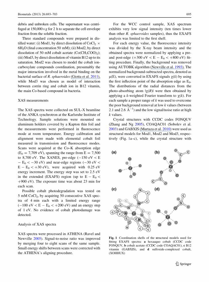
Crystal structures with CCDC codes FONQUV
(Zhang and Ng 2005), COAQAC01 (Sobolev et al.
2003) and GABXIS (Murtaza et al. 2010) were used as
structural models for Mod1, Mod2 and Mod3, respec-
tively (Fig. 1a–c), while the crystal structure with
Fig. 1 Coordination shells of the structural models used for
fitting EXAFS spectra: a hexaaquo cobalt (CCDC code
FONQUV, b cobalt acetate (CCDC code COAQAC01), c B12
vitamin (GABXIS), and d sulfoxide-complexed cobalt,
(XOHHUX)
Biometals (2013) 26:693–703 695
123

CCDC code XOHHUX (Barton et al. 2002) was used
to model the cobalt ion–sulfonic ligand interaction in
R. sphaeroides samples (Fig. 1d). In addition, an ideal
structural model containing a cobalt ion bound to six
oxygen atoms (at distance of 2 A) in octahedral
coordination around the metal center was generated by
using the tool ‘‘quick first-shell theory’’, present in the
ARTEMIS (Ravel and Newville 2005), and used to
calculate the contribution of multiple scattering paths
in the first coordination shell.
The structural models were decomposed in terms of
photoelectron scattering paths and their phase shifts,
together with back-scattering amplitudes required
during the fitting procedure, were calculated by FEFF
6 (Zabinsky et al. 1995). The EXAFS fitting of the
normalized XAS spectra was performed by using
ARTEMIS, with k2-weighted experimental data
(except Mod3, for which k1k2k3-weight was used).
Results and discussion
Qualitative analysis of XAS spectra
The normalized, derived, and Fourier transformed
spectra of each sample are shown in Fig. 2.
XANES may be conveniently used to infer the
coordination symmetry of the cobalt inner-shell by
using pre-edge features: the intensity of the 7.710 eV
pre-edge peak, associated to the quadrupole 1s–3d
transition, depends on the symmetry of the first shell
and on the 3d shell occupancy extent (Jacobs et al.
2002; Juhin et al. 2010). It appears intense in
tetrahedral coordination while vanishes in octahedral
coordination. Distortions of these two extreme cases
modify the intensity of the peak (Moen et al. 1997).
All recorded spectra show a rather small pre-edge
peak at about 7.710 eV (Fig. 2a), indicating a slightly
distorted octahedral first coordination shell, consisting
of six light atoms (possibly oxygen and/or nitrogen).
Among the observed samples, Mod3 shows a slightly
higher pre-edge peak, suggesting greater distortions of
the cobalt octahedral coordination symmetry for this
compound, which are also evidenced by the following
features: (i) all other samples show a sharp peak
(Fig. 2b) in the range -25 eV \ E - E0 \ 25 eV of
their l(E) first derivative functions (Frenkel and Kor-
shin 1999), while in Mod3 two small peaks are present,
(ii) the unique 2 eV energy increase in the Co K-edge
found in Mod3 points to increased charge on the cobalt
center with respect to the hexaaquo coordinated ion, (iii)
the radial distributions (Fig. 2c) show a peak centered
around 1.6 A for all samples excluded Mod3, where is
down-shifted to 1.5 A. This finding agrees with the
shorter distances of four out of the six ligand atoms in the
first shell, imposed by the presence of the corrin ring
around the cobalt ion (Brink et al. 1954).
The radial distributions of Mod2, WCCo, and SCo
beyond 2 A are comparable, indicating similar second
coordination shells, while significant differences
emerge for Mod3, where a clear peak occurs at
2.4 A. This Mod3 feature can be attributed to the
Fig. 2 Normalized
l(E) (a), first derivative of
normalized l(E) (b), and
modulus of the Fourier
transforms of k2-weighted
v(k) (c) for aqueous solution
of cobalt chloride (Mod1),
cobalt acetate (Mod2), B12
vitamin (Mod3) and for R.
sphaeroides samples grown
in CoCl2 5 mM: cell
envelope (CECo), soluble
fraction (SCo), and whole
cells (WCCo). Fourier
transforms are shown not
taking into account phase
correction
696 Biometals (2013) 26:693–703
123
complex interaction of the cobalt ion with the corrin
and imidazole rings of the B12 vitamin structural
model (Fig. 1c). A small specific feature at 2.6 A may
be observed in the radial distribution of CECo,
indicating a different second shell coordination.
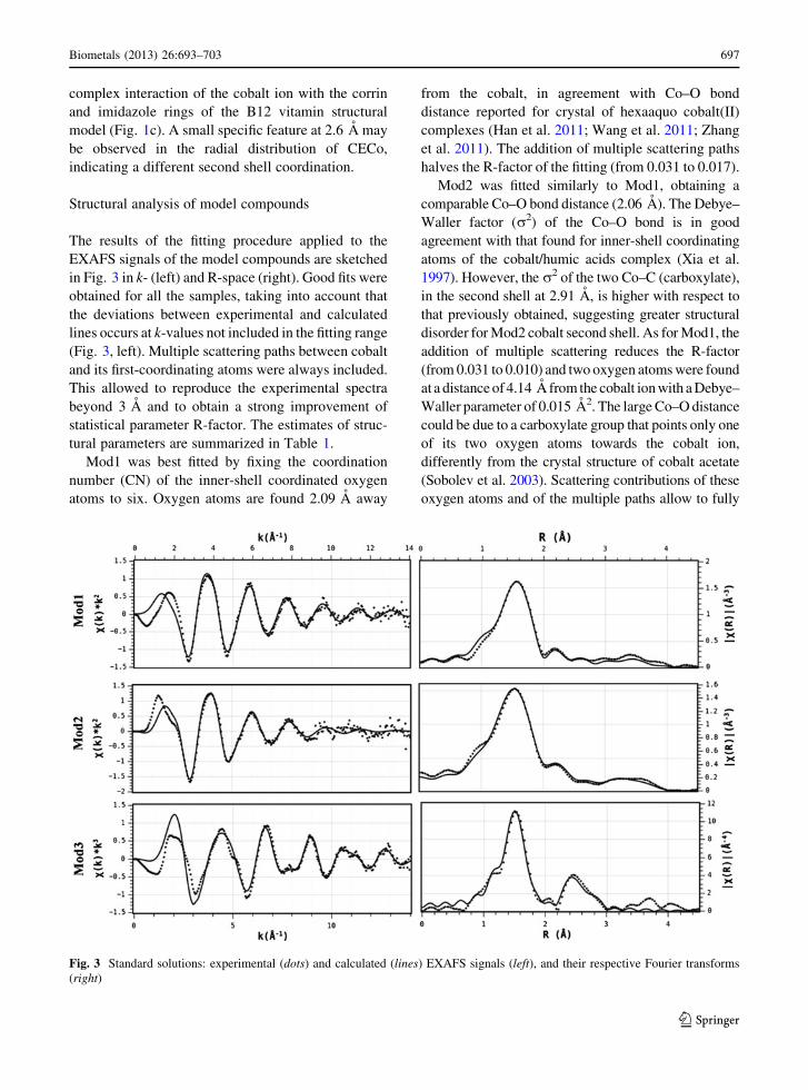
Structural analysis of model compounds
The results of the fitting procedure applied to the
EXAFS signals of the model compounds are sketched
in Fig. 3 in k- (left) and R-space (right). Good fits were
obtained for all the samples, taking into account that
the deviations between experimental and calculated
lines occurs at k-values not included in the fitting range
(Fig. 3, left). Multiple scattering paths between cobalt
and its first-coordinating atoms were always included.
This allowed to reproduce the experimental spectra
beyond 3 A and to obtain a strong improvement of
statistical parameter R-factor. The estimates of struc-
tural parameters are summarized in Table 1.
Mod1 was best fitted by fixing the coordination
number (CN) of the inner-shell coordinated oxygen
atoms to six. Oxygen atoms are found 2.09 A away
from the cobalt, in agreement with Co–O bond
distance reported for crystal of hexaaquo cobalt(II)
complexes (Han et al. 2011; Wang et al. 2011; Zhang
et al. 2011). The addition of multiple scattering paths
halves the R-factor of the fitting (from 0.031 to 0.017).
Mod2 was fitted similarly to Mod1, obtaining a
comparable Co–O bond distance (2.06 A). The Debye–
Waller factor (r2) of the Co–O bond is in good
agreement with that found for inner-shell coordinating
atoms of the cobalt/humic acids complex (Xia et al.
1997). However, the r2 of the two Co–C (carboxylate),
in the second shell at 2.91 A, is higher with respect to
that previously obtained, suggesting greater structural
disorder for Mod2 cobalt second shell. As for Mod1, the
addition of multiple scattering reduces the R-factor
(from 0.031 to 0.010) and two oxygen atoms were found
at a distance of 4.14 A from the cobalt ion with a Debye–
Waller parameter of 0.015 A2. The large Co–O distance
could be due to a carboxylate group that points only one
of its two oxygen atoms towards the cobalt ion,
differently from the crystal structure of cobalt acetate
(Sobolev et al. 2003). Scattering contributions of these
oxygen atoms and of the multiple paths allow to fully
Fig. 3 Standard solutions: experimental (dots) and calculated (lines) EXAFS signals (left), and their respective Fourier transforms
(right)
Biometals (2013) 26:693–703 697
123
reproduce the Fourier transform between 3 and 4 A,
despite a previous study on crystalline tetraaquo cobalt
acetate which ascribes this feature to an artifact on the
measured spectrum (Ghabbour et al. 2007).
The first shell of the cobalt ion in the Mod3 sample
is also composed by six light atoms, similarly to Mod1
and Mod2, and accordingly to the B12 vitamin
structure (Brink et al. 1954) contains five nitrogen
(four nitrogen from the corrin ring and one from the
imidazole group) and one carbon (from the cyanide
group) atoms. Two carbon atoms distant 2.6 A from
the cobalt, together with other eight carbon atoms at
2.88 A, of the corrin ring, complete the second
coordination shell. In addition, a nitrogen distant
3.10 A from the cobalt ion and belong to the imidazole
ring allows a further improvement of the fit result.
Structural analysis of R. sphaeroides samples
EXAFS fitting result for R. sphaeroides samples grown
in the presence of Co2? are summarized in Table 2 and
shown in Fig. 4 in k- (left) and R-space (right).
The first coordination shell of CECo sample
consists of six oxygen atoms (fixed CN) with Co–O
distance and Debye–Waller parameter (Table 2) com-
parable to Mod1 and Mod2 (Table 1). Two different
second cobalt coordination shells are proposed to fit
CECo: (i) two carbon atoms at 3.30 A or (ii) two sulfur
atoms at 2.92 A can be used without significant
difference of the R-factor statistical parameter. The
phase shift for Co–S was obtained by using the mono-
sulfonated complex XOHHUX (Fig. 1d). The addition
of multiple scattering paths significantly reduces the
R-factor in both the cases, found smaller in the Co–S
case. Neither 6O–2C nor 6O–2S can fully represent
the data, suggesting that the second shell can be a
mixture of the two configurations. Both motifs were
hence included in the fitting procedure, with the
fraction of 6O–2C configuration represented by a free
parameter (x). The fitting was carried out by neglect-
ing multiple scattering paths (to avoid over fitting),
fixing the structural parameters to their previously
obtained best values and leaving the global parame-
ters, E0, S20, and x free. The shell fraction containing
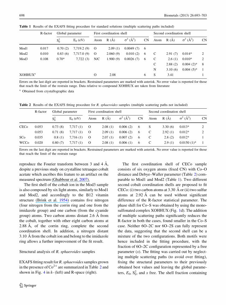
Table 1 Results of the EXAFS fitting procedure for standard solutions (multiple scattering paths included)
R-factor Global parameter First coordination shell Second coordination shell
S20
E0 (eV) Atom R (A) r2 (A2) CN Atom R (A) r2 (A2) CN
Mod1 0.017 0.70 (2) 7,719.2 (9) O 2.09 (1) 0.0049 (7) 6
Mod2 0.010 0.83 (6) 7,717.0 (9) O 2.060 (9) 0.010 (2) 6 C 2.91 (7) 0.014* 2
Mod3 0.108 0.70* 7,722 (3) N/C 1.900 (9) 0.0026 (7) 6 C 2.6 (1) 0.010* 2
C 2.88 (2) 0.004 (2)* 8
N 3.10 (6) 0.004 (5)* 1
XOHHUXa O 2.08 6 S 3.41 1
Errors on the last digit are reported in brackets. Restrained parameters are marked with asterisk. No error value is reported for those
that reach the limit of the restrain range. Data relative to compound XOHHUX are taken from literaturea Obtained from crystallographic data
Table 2 Results of the EXAFS fitting procedure for R. sphaeroides samples (multiple scattering paths not included)
R-factor Global parameter First coordination shell Second coordination shell
S20
E0 (eV) Atom R (A) r2 (A2) CN Atom R (A) r2 (A2) CN
CECo 0.053 0.73 (8) 7,717 (1) O 2.08 (1) 0.006 (2) 6 S 3.30 (6) 0.013* 2
0.053 0.71 (8) 7,717 (1) O 2.09 (1) 0.006 (2) 6 C 2.92 (1) 0.012* 2
SCo 0.035 0.8 (1) 7,716 (1) O 2.07 (1) 0.007 (2) 6 C 2.8 (2) 0.012* 1
WCCo 0.020 0.80 (7) 7,717 (1) O 2.08 (1) 0.006 (1) 6 C 2.9 (1) 0.0150 (1)* 1
Errors on the last digit are reported in brackets. Restrained parameters are marked with asterisk. No error value is reported for those
that reach the limit of the restrain range
698 Biometals (2013) 26:693–703
123

carbon atom was estimated as 0.494(9), leading to an
R-factor of 0.051.
SCo was fitted unambiguously by using six-fixed
oxygen atoms for the first shell (Fig. 4). A Co–O
distance of 2.07 A and a Debye–Waller parameter of
0.007 A2 was found, in good agreement with the
structural parameters obtained from Mod1 and Mod2.
The fitting improved by adding a single carbon atom in
the second shell with a distance of 2.8 A, comparable
to a carbon belonging to carboxylate group binding by
mono-dentate mode. (Coucouvanis et al. 1995). The
Co–C path shows a Debye–Waller factor which
increases up to the limit value imposed to the fitting,
confirming that only one carbon atom is sufficient to
explain the second shell. The addition of the multiple
scattering paths strongly reduces the R-factor (from
Fig. 4 Rhodobacter sphaeroides samples grown in 5 mM CoCl2: experimental (dots), and calculated with (dotted lines) and without
(full lines) multiple scattering paths EXAFS signals (left), and their respective Fourier transforms (right)
Biometals (2013) 26:693–703 699
123

0.035 to 0.014), indicating a slightly-distorted octa-
hedral coordination, in agreement with XANES and
l(E) first-derivate analysis (Fig. 2a, b). No fitting
improvement was obtained by using the second
oxygen atom of the carboxylate, as for Mod2.
The fitting procedure for WCCo shows first and
second coordination shells similar to those in SCo
(Table 2). For the second shell, the fit is also
compatible with the presence of a sulfur atom at a
distance of 3.2 A, but the special fit procedure carried
out by using mixed second shell structural hypotheses
resulted in a value of x [ 0.95, showing that in the
soluble fraction the second shell contribution (6O–C)
is the most populated one.
The EXAFS fits of the model compounds reproduce
the features of the experimental patterns, up to 3 A
from the cobalt ion. For Mod1 and Mod2, the
agreement was achieved up to the cobalt-center
second shell. In view of this result, we carried out
first and second-shell structural investigations on the
R. sphaeroides samples containing cobalt ion. The
fitting results indicate that six oxygen atoms are
sufficient to explain the first strong peak of their
Fourier transform (Fig. 4) with Co–O distances and
Debye–Waller parameters in agreement with Mod1
and Mod2. The six-oxygen coordination explains the
almost fully octahedral geometry of the cobalt coor-
dination inferred by XANES analysis (Fig. 2a, b). The
inclusion of the Co–C path was mandatory to fit the
second shell of each R. sphaeroides sample. In all
cases, the Co–C distances, obtained with no restrains,
were found compatible with a mono-dentate carboxyl
group, and in agreement with those obtained in cobalt
ion/humic acid by Xia et al. (1997) and Ghabbour et al.
(2007).
The EXAFS data of the cell-free fraction containing
the cell envelope (CECo) could be fitted assuming a
mixed population of the second shell, formed by a
50 % fraction of two carboxylic moieties and by a
50 % of two sulfur containing moieties. The sulfur
contribution to the EXAFS data in the cell-free soluble
fraction (SCo) was not detected and was found
negligible in the whole cell samples (WCCo), indi-
cating that this coordination pattern involves the cell
envelope and does not appear in the soluble portion.
The distance of the sulfur atom from cobalt (3.30 A)
together with the six oxygen atoms found in the first
shell, indicates the presence of oxygen donor ligands
having two oxygen atoms bound to a sulfur atom, as in
sulfoxide or sulfonate moiety, as suggested by the
structural information of compound XOHHUX.
The cell envelope of phototrophic bacteria com-
prises the cell wall, formed by the peptoglycane and
the outer cellular membrane, and the cytoplasmic
membrane (Weckesser et al. 1995), which in
R. sphaeroides invaginates in the so-called intra-
cytoplasmic membrane (ICM) when grown photosyn-
thetically (Drews and Golcki 1995; D’Amici et al.
2010). The cell envelope sample CECo is a mixture of
the peptoglycane, the outer membrane and cytoplas-
mic and intra-cytoplasmic membranes. In particular
the lipid composition of cytoplasmic and intra-
cytoplasmic membranes of R. sphaeroides, grown under
photosynthetic conditions, consists of phosphatidyletha-
nolamine, phosphatidylglycerol, phosphatidylcholine,
cardiolipin, phosphatidic acid and sulfoquinovosyldiacyl
glycerol (SQ) (Imhoff and Bias-Imhoff 1995; Fang
et al. 2000; Benning et al. 1993). This latter class of
lipids is characterized by a fatty acid bound to the
sulfoquinovosyl, a monosaccharide containing a neg-
atively charged sulfonate group (see Fig. 5). This
membrane composition is very influenced by chemical
stresses, among which the heavy metal ones (Italiano
et al. 2012). The presence of a robust amount of cobalt
bound to Co–O–S moiety (50 % of the total),
presumably belonging to the SQ lipids, suggests that
in the membrane exposed to Co2?, the amount of SQ is
much higher than the average 2 % found in
R. sphaeroides under physiological condition (Ben-
ning et al. 1993). This agrees with the preliminary
finding in our laboratory that in Co-exposed
R. sphaeroides cells, sulfolipids are overexpressed.
Fig. 5 Structure of the polar head of sulfolipids, the sulfoqui-
novosyl monosaccharide
700 Biometals (2013) 26:693–703
123

In Fig. 6 are shown two possible membrane binding
sites for cobalt in the case of two OS-containing ligands
(Fig. 6a) or mono-dentate carboxylate (Fig. 6b).
Differently from CECo sample, the second shell of
SCo contains only a carbon atom (6O-C) evidencing
that only a carboxylate group is sufficient to serve as
cobalt binding site for the soluble proteome extracted
from R. sphaeroides. In this case, the cobalt environ-
ment is made of pentaaquo ligands in addition to the
mono-dentate carboxylate, excluding any other ligand
in the inner-shell. Notwithstanding the different cobalt
coordination for CECo and SCo samples, no mixed
second shell was detected for WCCo (carboxylate
fraction [95 %). This should not come as a surprise
since the largest portion of a whole cells is its soluble
portion, and hence the membrane represents a very
small portion of the WCCo, and the Co–O–S path
originating from SQ accounts for less than 5 %.
Conclusion
XAS measurements may be conveniently exploited to
understand the coordination features of a metal center
in a rather complex matrix such as bacterial cells. In
this work, XAS was used to characterize the coordi-
nation of the cobalt ion in the presence of
R. sphaeroides, a bacterium with high metal tolerance
and considered a potential target for bioremediation
process, grown in solution medium containing very
high cobalt concentration. XANES measurements
have shown an octahedral coordination for cobalt
bound to the cell, with six oxygen atoms in the first
coordination shell. EXAFS data indicates that cobalt
binds to carboxylate in the soluble portion of the cell,
while on the photosynthetic membrane it also binds to
sulfolipids in large quantity, suggesting that this type
of lipids are present in Co-exposed cells in larger
quantity than control conditions, a typical response
mechanism of the photosynthetic membrane of
R. sphaeroides to abiotic stress condition.
Acknowledgments Ralph Steininger and Joerg Goettlicher at
SUL-X beamline at ANKA are greatly acknowledged (Project
ENV-219). Support for this work was obtained by the Italian
Ministry of Research Education and Education (PRIN 2009) and
by COST Action CM0902 Molecular machinery for ion
translocation across the membrane.
References
Albering H, van Leusen S, Moonen E, Hoogewerff J, Kleinjans J
(1999) Human health risk assessment: a case study
involving heavy metal soil contamination after the flooding
of the river Meuse during the winter of 1993–1994. Envi-
ron Health Perspect 107(1):37–43
Barceloux DG (1999) Cobalt. J Toxicol Clin Toxicol
37(2):201–206
Barton MR, Zhang Y, Atwood JD (2002) Mono-sulfonated
derivatives of triphenylphosphine, [NH4]TPPMS and M
(TPPMS)2 (TPPMS = P(Ph)2(m-C6H4SO3-); M = Mn2?,
Fe2?, Co2? and Ni2?). Crystal structure determinations for
[NH4]TPPMS*� H2O, [Fe(H2O)5(TPPMS)TPPMS], [Co
(H2O)5TPPMS]TPPMS and [Ni(H2O)6](TPPMS)4*H2O.
J Coord Chem 55(8):969–983
Bebien M, Chauvin JP, Adriano JM, Grosse S, Vermeglio A
(2001) Effect of selenite on growth and protein synthesis in
the phototrophic bacterium Rhodobacter sphaeroides.
Appl Environ Microbiol 67(10):4440–4447
Benning C, Beatty JT, Prince RC, Somerville CR (1993) The
sulfolipid sulfoquinovosyldiacylglycerol is not required for
photosynthetic electron transport in Rhodobacter sph-
aeroides but enhances growth under phosphate limitation.
Proc Natl Acad Sci USA 90(4):1561–1565
Boyanov MI, Kelly SD, Kemner KM, Bunker BA, Fein JB,
Fowle DA (2003) Adsorption of cadmium to Bacillus
subtilis bacterial cell walls: a pH-dependent X-ray
absorption fine structure spectroscopy study. Geochimica
Fig. 6 Cobalt binding sites for Co. a Cobalt ion coordinated to
four water molecules and two sulfonic groups, and b cobalt ion
coordinated to four water molecules and two carboxylic groups
Biometals (2013) 26:693–703 701
123

et Cosmochimica Acta 67(18):3299–3311. doi:
http://dx.doi.org/10.1016/S0016-7037(02)01343-1
Brink C, Hodgkin DC, Lindsey J, Pickworth J, Robertson JH,
White JG (1954) Structure of vitamin B12: X-ray crystal-
lographic evidence on the structure of vitamin B12. Nature
174(4443):1169–1171
Bruins MR, Kapil S, Oehme FW (2000) Microbial resistance to
metals in the environment. Ecotoxicol Environ Saf 45(3):
198–207
Buccolieri A, Italiano F, Dell’Atti A, Buccolieri G, Giotta L,
Agostiano A, Milano F, Trotta M (2006) Testing the pho-
tosynthetic bacterium Rhodobacter sphaeroides as heavy
metal removal tool. Ann Chim 96(3–4):195–203
Coucouvanis D, Reynolds RA, Dunham WR (1995) Synthesis
and characterization of a new class of asymmetric aqua-
acetate bridged dimers. Solid state molecular structures of
the [M2(.mu.-H2O)(.mu.-OAc)2(OAc)3(Py)2]-anions
(M = Mn(II), Fe(II), Co(II)). A structural model for the
Fe2 site in methane monooxygenase. J Am Chem Soc
117(28):7570–7571. doi:10.1021/ja00133a041
D’Amici GM, Rinalducci S, Murgiano L, Italiano F, Zolla L
(2010) Oligomeric characterization of the photosynthetic
apparatus of Rhodobacter sphaeroides R26.1 by nonde-
naturing electrophoresis methods. J Proteome Res 9(1):
192–203
Drews G, Golcki JR (1995) Structure, molecular organization
and biosynthesis of membranes of purple bacteria. In:
Blankenship RE, Madiga MT, Bauer CE (eds) Anoxygenic
photosyntetic bacteria. Advances in photosynthesis, vol 2.
Kluwer, Dordrecht, pp 231–257
Fang J, Barcelona MJ, Semrau JD (2000) Characterization of
methanotrophic bacteria on the basis of intact phospholipid
profiles. FEMS Microbiol Lett 189(1):67–72
Forstner U, Wittmann GTW (1983) Metal pollution in the
aquatic environment. Springer, Berlin
Frenkel AI, Korshin GV (1999) A study of non-uniformity of
metal binding sites in humic substances by X-ray absorp-
tion spectroscopy. Royal Society of Chemistry, Cambridge
Gault N, Sandre C, Poncy JL, Moulin C, Lefaix JL, Bresson C
(2010) Cobalt toxicity: chemical and radiological com-
bined effects on HaCaT keratinocyte cell line. Toxicol
In Vitro 24(1):92–98. doi:10.1016/j.tiv.2009.08.027
Ghabbour EA, Scheinost AC, Davies G (2007) XAFS studies of
cobalt(II) binding by solid peat and soil-derived humic
acids and plant-derived humic acid-like substances. Che-
mosphere 67(2):285–291. doi:http://dx.doi.org/10.1016/j.
chemosphere.2006.09.094
Giotta L, Agostiano A, Italiano F, Milano F, Trotta M (2006)
Heavy metal ion influence on the photosynthetic growth of
Rhodobacter sphaeroides. Chemosphere 62(9):1490–1499
Giotta L, Italiano F, Pisani F, Ceci LLR, De Leo F (2007) Cobalt
effect on the bacteriochlorophyll biosynthesis pathway and
magnesium metabolism in Rhodobacter sphaeroides strain
R26.1. Photosynth Res 91(2–3):302–303
Giotta L, Italiano F, Buccolieri A, Agostiano A, Milano F,
Trotta M (2008) Magnesium chemical rescue to cobalt-
poisoned cells from Rhodobacter sphaeroides. In: Allen
JF, Gantt E, Golbeck JH, Osmond B (eds) Photosynthesis.
Energy from the sun: 14th international congress on pho-
tosynthesis, vol 1. Springer, Dordrecht, pp 1455–1458
Giotta L, Mastrogiacomo D, Italiano F, Milano F, Agostiano A,
Nagy K, Valli L, Trotta M (2011) Reversible binding of
metal ions onto bacterial layers revealed by protonation-
induced ATR–FTIR difference spectroscopy. Langmuir
27(7):3762–3773. doi:10.1021/la104868m
Guengerich FP (2012) Thematic Minireview series: metals in
biology 2012. J Biol Chem 287(17):13508–13509. doi:
10.1074/jbc.R112.355933
Han L-J, Yang S-P, Fu L–L, Gao H-L (2011) Hexaaquacobalt(II)
bis(5-acetyl-2-hydroxybenzoate) dihydrate. Acta Crystal-
logr E 67(12):m1733. doi:10.1107/S1600536811046678
Head IM (1998) Bioremediation: towards a credible technology.
Microbiology 144:599–608
Hebes SE, Schwall IR (1978) Microbial degradation of polycyclic
aromatic hydrocarbons in pristine and petroleum contami-
nated sediments. Appl Environ Microb 35:306–316
Imhoff JF, Bias-Imhoff U (1995) Lipids, quinines and fatty acids
of anoxygenic phototropic bacteria. In: Blankenship RE,
Bauer CE (eds) Anoxygenic photosynthetic bacteria.
Kluwer, Dordrecht, pp 179–205
Italiano F, De Leo F, Pisani F, Ceci L, Gallerani R, Zolla L,
Rinalducci S, Gio L (2007) Effect of cobalt ions on the
soluble proteome of Rhodobacter sphaeroides carote-
noidless mutant. Photosynth Res 91(2–3):303
Italiano F, Pisani F, De Leo F, Ceci L, Gallerani R, Zolla L,
Rinalducci S, Giotta L, Milano F, Agostiano A, Trotta M
(2008) Effect of cobalt ions on the soluble proteome of a
Rhodobacter sphaeroides carotenoidless mutant. In: Allen
JF, Gantt E, Goldbeck J, Osmond B (eds) Photosynthesis.
Energy from the sun: 14th international congress on pho-
tosynthesis, vol 1. Springer, Dordrecht, pp 1479–1484
Italiano F, Buccolieri A, Giotta L, Agostiano A, Valli L, Milano
F, Trotta M (2009) Response of the carotenoidless mutant
Rhodobacter sphaeroides growing cells to cobalt and
nickel exposure. Int Biodeterior Biodegrad 63:948–957
Italiano F, D’Amici GM, Rinalducci S, De Leo F, Zolla L,
Gallerani R, Trotta M, Ceci LR (2011) The photosynthetic
membrane proteome of Rhodobacter sphaeroides R-26.1
exposed to cobalt. Res Microbiol 162(5):520–527
Italiano F, Rinalducci S, Agostiano A, Zolla L, De Leo F, Ceci
LR, Trotta M (2012) Changes in morphology, cell wall
composition and soluble proteome in Rhodobacter sph-
aeroides cells exposed to chromate. Biometals 25(5):
939–949. doi:10.1007/s10534-012-9561-7
Jacobs G, Patterson PM, Zhang Y, Das T, Li J, Davis BH (2002)
Fischer–Tropsch synthesis: deactivation of noble metal-
promoted Co/Al2O3 catalysts. Appl Catal A 233(1–2):
215–226. doi:http://dx.doi.org/10.1016/S0926-860X(02)
00147-3
Jennette KW (1981) The role of metals in carcinogenesis: bio-
chemistry and metabolism. Environ Health Perspect 40:
233–252
Juhin A, de Groot F, Vanko G, Calandra M, Brouder C (2010)
Angular dependence of core hole screening in LiCoO2: a
DFT?U calculation of the oxygen and cobalt K-edge
X-ray absorption spectra. Phys Rev B 81(11):115115
Kantar C, Demiray H, Dogan NM, Dodge CJ (2011) Role of
microbial exopolymeric substances (EPS) on chromium
sorption and transport in heterogeneous subsurface soils: I.
Cr(III) complexation with EPS in aqueous solution.
702 Biometals (2013) 26:693–703
123

Chemosphere 82(10):1489–1495. doi:http://dx.doi.org/
10.1016/j.chemosphere.2011.01.009
Kasprzak K (1991) The role of oxidative damage in metal car-
cinogenicity. Chem Res Toxicol 4(6):604–615
Kiley PJ, Kaplan S (1988) Molecular genetics of photosynthetic
membrane biosynthesis in Rhodobacter sphaeroides.
Microbiol Rev 52(1):50–69
Kobayashi M, Shimizu S (1999) Cobalt proteins. Eur J Biochem
261(1):1–9
Leonard SM, Gannett P, Rojanasakul Y, Schwegler-Berry D,
Castranova V, Vallyathan V, Shi X (1998) Cobalt-medi-
ated generation of reactive oxygen species and its possible
mechanism. J Inorg Biochem 70(3–4):239–244
Losurdo L, Italiano F, Trotta M, Gallerani R, Luigi RC, De Leo
F (2011) Assessment of an internal reference gene in
Rhodobacter sphaeroides grown under cobalt exposure.
J Basic Microbiol 50(3):302–305
Martinezluque M, Dobao MM, Castillo F (1991) Characteriza-
tion of the assimilatory and dissimilatory nitrate-reducing
systems in Rhodobacter: a comparative-study. FEMS
Microbiol Lett 83(3):329–334. doi:10.1111/j.1574-6968.
1991.tb04485.x
Mishra A, Malik A (2013) Recent advances in microbial metal
bioaccumulation. Crit Rev Environ Sci Technol
43(11):1162–1222. doi:10.1080/10934529.2011.627044
Mishra B, Boyanov MI, Bunker BA, Kelly SD, Kemner KM,
Nerenberg R, Read-Daily BL, Fein JB (2009) An X-ray
absorption spectroscopy study of Cd binding onto bacterial
consortia. Geochim Cosmochim Acta 73(15):4311–4325.
doi:http://dx.doi.org/10.1016/j.gca.2008.11.032
Moen A, Nicholson DG, Rnning M, Lamble GM, Lee J-F,
Emerich H (1997) X-Ray absorption spectroscopic study at
the cobalt K-edge on the calcination and reduction of the
microporous cobalt silicoaluminophosphate catalyst Co-
SAPO-34. J Chem Soc Faraday Trans 93(22):4071–4077
Moore MD, Kaplan S (1992) Identification of intrinsic high-
level resistance to rare-earth oxides and oxyanions in
members of the class Proteobacteria: characterization of
tellurite, selenite, and rhodium sesquioxide reduction in
Rhodobacter sphaeroides. J Bacteriol 174(5):1505–1514
Murtaza S, Ruetz M, Gruber K, Krautler B (2010) Isovitamin
B12: a vitamin B12 derivative that flips its tail. Chem Euro
J 16(36):10984–10988. doi:10.1002/chem.201001616
Myllykallio H, Zannoni D, Daldal F (1999) The membrane-
attached electron carrier cytochrome c(y) from Rhodob-
acter sphaeroides is functional in respiratory but not in
photosynthetic electron transfer. Proc Natl Acad Sci USA
96(8):4348–4353. doi:10.1073/pnas.96.8.4348
Nevin R (2000) How lead exposure relates to temporal changes
in IQ, violent crime, and unwed pregnancy. Environ Res
83(1):1–22. doi:http://dx.doi.org/10.1006/enrs.1999.4045
Newville M, Lıvins P, Yacoby Y, Rehr JJ, Stern EA (1993)
Near-edge X-ray-absorption fine structure of Pb: a
comparison of theory and experiment. Phys Rev B
47(21):14126–14131
Okamoto S, Eltis LD (2011) The biological occurrence and
trafficking of cobalt. Metallomics 3(10):963–970. doi:
10.1039/c1mt00056j
Pisani F, Italiano F, de Leo F, Gallerani R, Rinalducci S, Zolla L,
Agostiano A, Ceci LR, Trotta M (2009) Soluble proteome
investigation of cobalt effect on the carotenoidless mutant of
Rhodobacter sphaeroides. J Appl Microbiol 106(1):338–349
Ravel B, Newville M (2005) ATHENA, ARTEMIS,
HEPHAESTUS: data analysis for X-ray absorption spec-
troscopy using IFEFFIT. J Synchrotron Radiat
12(4):537–541. doi:10.1107/S0909049505012719
Sardaro A, Castagnolo M, Trotta M, Italiano F, Milano F,
Cosma P, Agostiano A, Fini P (2013) Isothermal micro-
calorimetry of the metabolically versatile bacterium Rho-
dobacter sphaeroides. J Therm Anal Calorim
112(1):505–511. doi:10.1007/s10973-012-2895-0
Schultz JE, Weaver PF (1982) Fermentation and anaerobic
respiration by Rhodospirillum rubrum and Rhodopseudo-
monas capsulata. J Bacteriol 149(1):181–190
Sobolev AN, Miminoshvili EB, Miminoshvili KE, Sakvarelidze
TN (2003) Cobalt diacetate tetrahydrate. Acta Crystallogr
E 59(10):m836–m837. doi:10.1107/S1600536803019093
Valls M, de Lorenzo V (2002) Exploiting the genetic and bio-
chemical capacities of bacteria for the remediation of
heavy metal pollution. FEMS Microbiol Rev 26(4):
327–338
Wang H, Gao S, Ng SW (2011) Hexaaquacobalt(II) bis(2,20-sulfanediyldiacetato-[kappa]3O, S, O’)cobaltate(II) tetra-
hydrate. Acta Crystallogr E 67(11):m1521. doi:
10.1107/S1600536811040979
Weckesser J, Mayer H, Schultz G (1995) Anoxygenic photo-
trophic bacteria: model organisms for studies on cell wall
macromolecules. In: Blankenship RE, Madiga MT, Bauer
CE (eds) Anoxygenic photosyntetic bacteria. Advances in
photosynthesis, vol 2. Kluwer, Dordrecht, pp 207–230
Xia K, Bleam W, Helmke PA (1997) Studies of the nature of
binding sites of first row transition elements bound to
aquatic and soil humic substances using X-ray absorption
spectroscopy. Geochim Cosmochim Acta 61(11):
2223–2235. doi:http://dx.doi.org/10.1016/S0016-7037(97)
00080-X
Zabinsky SI, Rehr JJ, Ankudinov A, Albers RC, Eller MJ (1995)
Multiple-scattering calculations of X-ray-absorption
spectra. Phys Rev B 52(4):2995–3009
Zhang X-L, Ng SW (2005) Hexaaquacobalt(II) bis(6-hydro-
xypyridine-3-carboxylate). Acta Crystallogr E
61(6):m1140–m1141. doi:10.1107/S1600536805014911
Zhang L-W, Gao S, Ng SW (2011) Hexaaquacobalt(II) bis[4-
(pyridin-2-ylmethoxy)benzoate] dihydrate. Acta Crystal-
logr E 67(11):m1519. doi:10.1107/S1600536811040931
Biometals (2013) 26:693–703 703
123