Three-dimensional structure of the Rhodobacter sphaeroides RC-LH1-PufX complex: dimerization and...
11
Three-Dimensional Structure of the Rhodobacter sphaeroides RC- LH1-PufX Complex: Dimerization and Quinone Channels Promoted by PufX Pu Qian, †,∥ Miroslav Z. Papiz, ‡,∥ Philip J. Jackson, †,§ Amanda A. Brindley, † Irene W. Ng, † John D. Olsen, † Mark J. Dickman, § Per A. Bullough, † and C. Neil Hunter* ,† † Department of Molecular Biology and Biotechnology, University of Sheffield, Western Bank, Firth Court, Sheffield S10 2TN, United Kingdom ‡ Institute of Integrative Biology, Biosciences Building, University of Liverpool, Crown Street, Liverpool L69 7ZB, United Kingdom § ChELSI Institute, Department of Chemical and Biological Engineering, University of Sheffield, Mappin Street, Sheffield, S1 3JD, United Kingdom * S Supporting Information ABSTRACT: Reaction center-light harvesting 1 (RC-LH1) complexes are the fundamental units of bacterial photosyn- thesis, which use solar energy to power the reduction of quinone to quinol prior to the formation of the proton gradient that drives ATP synthesis. The dimeric RC-LH1-PufX complex of Rhodobacter sphaeroides is composed of 64 polypeptides and 128 cofactors, including 56 LH1 bacterio- chlorophyll a (BChl a) molecules that surround and donate energy to the two RCs. The 3D structure was determined to 8 Å by X-ray crystallography, and a model was built with constraints provided by electron microscopy (EM), nuclear magnetic resonance (NMR), mass spectrometry (MS), and site-directed mutagenesis. Each half of the dimer complex consists of a RC surrounded by an array of 14 LH1 αβ subunits, with two BChls sandwiched between each αβ pair of transmembrane helices. The N- and C-terminal extrinsic domains of PufX promote dimerization by interacting with the corresponding domains of an LH1 β polypeptide from the other half of the RC-LH1-PufX complex. Close contacts between PufX, an LH1 αβ subunit, and the cytoplasmic domain of the RC-H subunit prevent the LH1 complex from encircling the RC and create a channel connecting the RC Q B site to an opening in the LH1 ring, allowing Q/QH 2 exchange with the external quinone pool. We also identified a channel that connects the two halves of the dimer, potentially forming a long-range pathway for quinone migration along rows of RC-LH1-PufX complexes in the membrane. The structure of the RC-LH1-PufX complex explains the crucial role played by PufX in dimer formation, and it shows how quinone traffic traverses the LH1 complex as it shuttles between the RC and the cytochrome bc 1 complex. P hotosynthesis, the ultimate source of all food and most bioenergy resources on Earth, begins with the absorption of solar energy by an array of antenna pigment molecules. The transfer of absorbed energy to specialized pigment−protein complexes, the reaction centers (RCs), initiates a series of electron-transfer reactions that trap the solar energy prior to its ultimate conversion to ATP, which powers the metabolism of the cell. In phototrophic bacteria, the conversion of solar energy to a chemical form, quinol, is accomplished by RC-LH1 complexes. 1,2 The largest of these pigment−proteins is the dimeric RC-LH1-PufX complex in which 28 LH1 αβBChl 2 subunits form a near-continuous palisade of 56 transmembrane helices enclosing two RCs. 3 A two-way traffic of outgoing quinols and incoming quinones must traverse this LH1 barrier to allow the turnover of RC photochemistry. Once inside the confines of the encircling LH1 complex, a quinone binds to the RC Q B site, picks up two electrons from RC photochemistry and two protons from the cytoplasm, and then exits the complex as a quinol to be replaced by a quinone from the membrane pool. The quinol migrates through the membrane bilayer to the cytochrome bc 1 complex, where a proton-motive force is generated. ATP synthase converts the proton gradient into ATP, the energy currency of the cell. Early gene deletion experiments in the phototrophic bacteria Rhodobacter (Rba.) sphaeroides and Rba. capsulatus showed that the transmembrane PufX polypeptide facilitates quinone/ quinol (Q/QH 2 ) exchange across the LH1 barrier. 4,5 Subsequently, other roles emerged for PufX: it was shown that dimerization of the RC-LH1-PufX complex from Rba. sphaeroides depends on the cytoplasmically exposed N-terminal domain of this transmembrane polypeptide. 6,7 The RC-LH1- PufX dimer adopts a bent configuration that imposes curvature on the membrane bilayer, 8 with far-reaching effects that include the partitioning of the membrane into dimer-rich regions, 9 Received: August 29, 2013 Revised: September 23, 2013 Article pubs.acs.org/biochemistry © XXXX American Chemical Society A dx.doi.org/10.1021/bi4011946 | Biochemistry XXXX, XXX, XXX−XXX
Transcript of Three-dimensional structure of the Rhodobacter sphaeroides RC-LH1-PufX complex: dimerization and...
Three-Dimensional Structure of the Rhodobacter sphaeroides RC-LH1-PufX Complex: Dimerization and Quinone Channels Promotedby PufXPu Qian,†,∥ Miroslav Z. Papiz,‡,∥ Philip J. Jackson,†,§ Amanda A. Brindley,† Irene W. Ng,† John D. Olsen,†
Mark J. Dickman,§ Per A. Bullough,† and C. Neil Hunter*,†
†Department of Molecular Biology and Biotechnology, University of Sheffield, Western Bank, Firth Court, Sheffield S10 2TN, UnitedKingdom‡Institute of Integrative Biology, Biosciences Building, University of Liverpool, Crown Street, Liverpool L69 7ZB, United Kingdom§ChELSI Institute, Department of Chemical and Biological Engineering, University of Sheffield, Mappin Street, Sheffield, S1 3JD,United Kingdom
*S Supporting Information
ABSTRACT: Reaction center-light harvesting 1 (RC-LH1)complexes are the fundamental units of bacterial photosyn-thesis, which use solar energy to power the reduction ofquinone to quinol prior to the formation of the protongradient that drives ATP synthesis. The dimeric RC-LH1-PufXcomplex of Rhodobacter sphaeroides is composed of 64polypeptides and 128 cofactors, including 56 LH1 bacterio-chlorophyll a (BChl a) molecules that surround and donate energy to the two RCs. The 3D structure was determined to 8 Å byX-ray crystallography, and a model was built with constraints provided by electron microscopy (EM), nuclear magneticresonance (NMR), mass spectrometry (MS), and site-directed mutagenesis. Each half of the dimer complex consists of a RCsurrounded by an array of 14 LH1 αβ subunits, with two BChls sandwiched between each αβ pair of transmembrane helices. TheN- and C-terminal extrinsic domains of PufX promote dimerization by interacting with the corresponding domains of an LH1 βpolypeptide from the other half of the RC-LH1-PufX complex. Close contacts between PufX, an LH1 αβ subunit, and thecytoplasmic domain of the RC-H subunit prevent the LH1 complex from encircling the RC and create a channel connecting theRC QB site to an opening in the LH1 ring, allowing Q/QH2 exchange with the external quinone pool. We also identified achannel that connects the two halves of the dimer, potentially forming a long-range pathway for quinone migration along rows ofRC-LH1-PufX complexes in the membrane. The structure of the RC-LH1-PufX complex explains the crucial role played by PufXin dimer formation, and it shows how quinone traffic traverses the LH1 complex as it shuttles between the RC and thecytochrome bc1 complex.
Photosynthesis, the ultimate source of all food and mostbioenergy resources on Earth, begins with the absorption
of solar energy by an array of antenna pigment molecules. Thetransfer of absorbed energy to specialized pigment−proteincomplexes, the reaction centers (RCs), initiates a series ofelectron-transfer reactions that trap the solar energy prior to itsultimate conversion to ATP, which powers the metabolism ofthe cell. In phototrophic bacteria, the conversion of solarenergy to a chemical form, quinol, is accomplished by RC-LH1complexes.1,2 The largest of these pigment−proteins is thedimeric RC-LH1-PufX complex in which 28 LH1 αβBChl2subunits form a near-continuous palisade of 56 transmembranehelices enclosing two RCs.3 A two-way traffic of outgoingquinols and incoming quinones must traverse this LH1 barrierto allow the turnover of RC photochemistry. Once inside theconfines of the encircling LH1 complex, a quinone binds to theRC QB site, picks up two electrons from RC photochemistryand two protons from the cytoplasm, and then exits thecomplex as a quinol to be replaced by a quinone from the
membrane pool. The quinol migrates through the membranebilayer to the cytochrome bc1 complex, where a proton-motiveforce is generated. ATP synthase converts the proton gradientinto ATP, the energy currency of the cell.Early gene deletion experiments in the phototrophic bacteria
Rhodobacter (Rba.) sphaeroides and Rba. capsulatus showed thatthe transmembrane PufX polypeptide facilitates quinone/quinol (Q/QH2) exchange across the LH1 barrier.4,5
Subsequently, other roles emerged for PufX: it was shownthat dimerization of the RC-LH1-PufX complex from Rba.sphaeroides depends on the cytoplasmically exposed N-terminaldomain of this transmembrane polypeptide.6,7 The RC-LH1-PufX dimer adopts a bent configuration that imposes curvatureon the membrane bilayer,8 with far-reaching effects that includethe partitioning of the membrane into dimer-rich regions,9
Received: August 29, 2013Revised: September 23, 2013
Article
pubs.acs.org/biochemistry
© XXXX American Chemical Society A dx.doi.org/10.1021/bi4011946 | Biochemistry XXXX, XXX, XXX−XXX

creation of a favorable environment for stable insertion of thelight-harvesting LH2 complex,10 and, ultimately, the formationof spherical intracytoplasmic membrane vesicles that contain allof the machinery for converting solar energy into ATP. It hasbeen proposed that these 50 nm diameter structures arebacterial organelles,11 and an atomic-level 3D map of a vesiclehas been calculated on the basis of the known membranelocations of the RC-LH1-PufX and LH2 complexes fromatomic force microscopy (AFM) studies.12
All of the above can be traced back to PufX as a facilitator ofquinone/quinol exchange and a driver of dimerization andhence membrane curvature. Here, we report the structure ofthe dimeric RC-LH1-PufX complex of Rba. sphaeroidesdetermined to 8 Å by X-ray crystallography, which deepensour understanding of these roles of PufX. The structure showsthat each PufX is positioned adjacent to a RC QB site byattachment of its N-terminal region to the cytoplasmic extrinsicdomain of RC-H and that both the N- and C-terminal extrinsicregions of PufX promote dimerization by interacting with anLH1 β polypeptide from the other half of the complex. Thearchitecture of the complex creates a channel, allowing Q/QH2molecules to cross the LH1 barrier and to internally migratebetween the two halves of the dimer structure.
■ MATERIALS AND METHODSPurification of the RC-LH1-PufX Complex. The Rba.
sphaeroides strain DBCΩG,13 which has neurosporene,hydroxyneurosporene, and methoxyneurosporene as the onlycarotenoids and which lacks the LH2 complex, was used for thepurification of the dimeric RC-LH1-PufX core complexaccording to previously published methods.3
Crystallization and Data Collection. The core dimercomplex was concentrated to an optical density of ∼100 at 875nm and washed several times using a 0.5 mL Vivaspin spinconcentrator (100 kDa cutoff) in 9 mM (0.42%) n-nonyl-β-D-maltopyranoside (NM) to exchange the purification detergent,n-dodecyl-β-D-maltopyranoside (β-DDM). Protein with anabsorbance of ∼100 (1 cm path length, 875 nm) wascrystallized in a sitting-drop format using the Screenmaker 96+ 8 crystallization robot (Gilson). Crystals grew as plates (500× 150 × 70 μm) in 14.00% PEG400, 0.10 M N-cyclohexyl-3-aminopropanesulfonic acid (CAPS) pH 10.5, and 1.0%spermidine. Crystals were frozen at 100 K, and data werecollected at the Science and Technology Facilities CouncilSynchrotron Radiation Source, Daresbury, on beamline 9.6with the Area Detector Systems Corporation Quantum 4detector at a wavelength of 0.912 Å. The crystals diffractedanisotropically between 8.5 and 7.5 Å along the various crystal-axis directions. A data cutoff of 8.0 Å was chosen for thepurpose of structure determination, at which resolution I/σ was1.0, a value usually accepted as the limit of useful data. Datawere processed with MOSFLM14 and merged with SCALA.15
The crystal space group was P21, the cell dimensions were a =78.08 Å, b = 415.07 Å, c = 129.81 Å, and β = 105.75° with amosaicity of 1.8°, and a data set was processed to an Rsym of9.3% and resolution of 8.0 Å. One dimer was found in theasymmetric unit with the noncrystallographic dimeric axis ∼4°misaligned from the crystallographic a axis.Molecular Replacement. A molecular replacement (MR)
search model of the RC-LH1-PufX dimer complex was derivedfrom EM data.3,8 The model was composed of the high-resolution RC structure (1PCR) and transmembrane poly-alanine helices representing LH1 αβ and PufX. Initial MR
solutions produced poor electron density maps, especially atthe periphery of the complex. Low-resolution EM and AFMimages indicate that dimers form V-shaped structures; however,the V angle can vary and depends on the sample preparation orthe substrate on which it is placed.11 To optimize the MRstarting model, the relative orientations of monomers wereexplored by varying two angles (Figure S3A): a rotation angle θabout an axis in the membrane plane “twist” and angle ψ thatdefines the “bend” angle between monomers (i.e., the V angle).A log likelihood probability function (L) was used in PHASERto calculate the agreement between the observed and calculatedstructure amplitudes for each model.16 PHASER was runrepeatedly on several dimer models (θ, ψ), and the best Lsolution occurred at angles θ = 1.5° and ψ = −11.0° (FigureS3A, B). The value for θ is similar or slightly smaller and the ψangle is significantly lower than the −17.0° found in single-particle reconstruction EM images, which implies that in the X-ray structure the core complex is flattened by comparison.
Electron Density Improvement. Electron density mapswere improved by cyclical electron density modification (DM)using solvent flattening, histogram matching, and domainaveraging.17 To prevent model bias from being introduced, thestarting model phases were not recombined with DM phasesduring repeated cycles of the program DM; this allowed theDM phases to evolve independently of the starting modelphases. In addition, although the calculated solvent content is64%, a conservative choice of 40% was used to ensure thatstructural features were not inadvertently flattened. Limitationsarising from data resolution and data completeness can result innoise and termination ripple errors in electron density maps.Solvent flattening and noncrystallographic symmetry (NCS)averaging mitigated some of these effects. Three averagingmodels were investigated that assumed (1) dimeric NCS onlyand (2) an NCS domain ‘A’ comprising reaction centers and 28NCS ‘B’ domain repeats comprising LH1 αβ pairs withassociated BChl a pigments (Figure S4A). Finally, a thirdmodel was considered comprising domain A, a new NCSdomain ‘C’ composed of the two LH1 αβ1 + PufX units, anddomain B reassigned to the remaining 26 αβ LH1 subunits.The phytol chains were removed from BChl a moleculesbecause their positions vary between light-harvesting complexstructures, for example, in LH218 and LH3.19 The final DMelectron density maps have NCS-related correlation coefficientsof 0.91 for model 1, 0.9 and 0.70 for model 2 domains A and B,and 0.93, 0.71, and 0.87 for model 3 domains A, B, and C,respectively. The figure of merit increased from 0.31 for thestarting model to 0.81 for the final DM phases (Figure S4B).The correlation coefficient for domain B in model 3 suggeststhat the 26 subunits across both monomers differ slightlyaround the LH1 ring. Correlation coefficient (CC) statisticsbetween NCS repeats were found to be powerful indicators thatstructural features were correctly identified, as incorrectlyassigned NCS domains result in zero or negative CCs.
Model Fitting to Electron Density. A RC-LH1 dimer wasfitted to electron density aided by high-resolution structures ofRC (1PCR)20 and LH2 (1KZU and 1LGH);18,21 the latterLH2 αβ repeating units, with chromophores, share significantsequence homology with LH1 αβ so that BChl a pigments canbe located in the structure with the correct orientation eventhough the electron density here is of low resolution. FittingPufX to the electron density began with a straight α-helixspanning the TM region (30−53) and was thereafter guided bythe appearance of new electron density. The starting model of
Biochemistry Article
dx.doi.org/10.1021/bi4011946 | Biochemistry XXXX, XXX, XXX−XXXB
RC-LH1, used in fitting to electron density, was the MR model.In this model, the RC was mostly intact, whereas the LH1polypeptides were modeled as 29-residue TM poly alaninechains, and so they were only 40% complete.The first 2Fo−Fc map, based on MR phases, showed a strong
elongated peak next to LH1 α1, and the electron densityaround LH1 β1 was reduced and noisy. The provisionalinterpretation was that the elongated peak was PufX. The oldPufX in the starting model was now reassigned to LH1 α1, andthe old LH1 α1 was now made into LH1 β1. Side chains werethen added to PufX (residues 30−53), and subsequent DMmaps revealed new electron density, which was interpreted asPufX (residues 20−31). Additionally, the electron densityaround LH1 αβ1 improved, indicating that the interpretation ofthese peptides was a better fit. At this point, side chains wereadded to all LH1 αβ peptides with MODELLER22 using NMR-determined LH1 β23 and X-ray-determined LH2 polypep-tides18,21 as templates. New DM maps indicated stronger, moreconnected electron density along LH1 β chains, and, for thefirst time, turns were revealed at both ends of the α and βchains. Residues were fitted for LH1 α (8−49), LH1 β (1−48),and for PufX density that could accommodate additionalresidues 2−19, and 54−58 was also found. Refinement (seebelow) and further rounds of DM calculations resulted in theemergence of density that could be fitted with residues RC-M(302−304) and RC-H (251−260) in the model. LH1 Bchl apigments were repositioned to be consistent with histidines asligands and were also checked with the position of Bchl a in theLH2 αβ unit from Phaeospirillum molischianum superimposedonto an LH1 αβ unit. Finally, the modeled PufX conformationwas compared (see Figure S6B) to an independentlydetermined NMR solution structure of PufX.24
Refinement. Refinement was performed in REFMAC5using tight atomic geometry restraints through an automaticallydetermined matrix weighting between X-ray and geometricterms. Refinement of 38 198 atoms was made against 6308 X-ray measurements and 133 500 geometry restraints. Thegeometry restraints, which include bond distances, bond angles,torsion, planar, and chiral restraints, cause individual atomiccoordinates to be highly correlated locally and so the X-ray andgeometry terms are sufficient for stable refinement, albeitmainly on the basis of geometry. The refinement was thereforemostly a geometry regularization process, with the X-ray termsproviding statistics on the consistency of the geometryrefinement with the X-ray data through the agreement betweenthe X-ray structure factor amplitudes (Fo) and atomic modelamplitudes (Fc). Additional weights were applied to geometryrestraints to produce refined rms distance errors between 0.01and 0.02 Å and rms angle error between 1.0−3.0°. These valuesare typical of higher-resolution structures, particularly ofmembrane protein structures. It could be argued that forlower-resolution data this degree of accuracy overconstrains therefinement, but it was judged that the final model should bewithin the same geometric error levels as obtained for higher-resolution structures. The residues refined were 8−49 for LH1α chains, 1−48 for LH1 β chains, RC-H 11−260, RC-M 1−304, and RC-L 1−281. It has been shown that the 12 C-terminal residues of PufX are removed by post-translationalmodification25 and we found no electron density for residues59−69 probably because of a disordered conformation of thispart of the C-terminal domain. Nevertheless, residues 2−58represent the majority of the residues in the RC-LH1 dimer.For each NCS monomer, 28 LH1 Bchl a pigments, without
phytol chains, four RC BChl a, two bacteriopheophytin a, twoubiquinone, one spheroidene, one phosphate, and one Fe(III)molecule were included in the refinement.Refinement was performed against data between 20 and 8.0
Å because data below 20 Å suffer from bulk solventcontributions that are not modeled well in refinementprograms; this is particularly true of membrane proteins,which also have contributions from detergent micelles. Theselow-resolution data do not add much to the refinement andeven at higher resolution are generally excluded. The possibilityof crystal twinning disorder was statistically tested during dataprocessing, which suggested that there could be pseudomer-ohedral twinning but, because of data resolution andinterference from strong NCS symmetry, this was inconclusive.The possibility was reassessed during refinement using thetwinning option in REFMAC5, which deconvolutes the dataduring refinement and so uses corrected structure factoramplitudes for refinement. This refinement indicated atwinning factor α ≈ 0.12 and resulted in slightly improvedrefinement statistics compared to twinned data (Table 1). Thesubsequent electron density maps for twinned and detwinneddata were similar and suggest that the small amount of twinninghas no major effect on structure determination.The refinement statistics are comparable with those found
for higher-resolution structures. It should be recognized that inmost refinements the Rfactor remains flat and 22% is typical inthe resolution range 20−8.0 Å, providing that the model iscorrect. The good statistics for this structure implies that themodel is substantially correct, with the important proviso thatthe optical resolution is between 5 and 6 Å and that anystructural errors (e.g., in side-chain positions) smaller than thiswill not be seen in the statistics. NCS skews the data intensitydistributions toward a number of stronger reflections, whichmay also account for a low Rfactor.
Calculation of the 3D QB Channel in the Dimeric CoreComplex. Interior cavities of the dimer complex weregenerated using Hollow.26 Casting of the protein interiorcavity was restricted within a cylinder that encompasses the QBchannel in one-half of the dimeric RC-LH1-PufX complex (d =10 nm, h = 7 nm). The produced cavity was then sliced, and theedge of each slice was digitized with a spacing step of 1 Å. Areasnot belonging to the QB channel were removed manually. TheQB channel in the dimer was then built using Pymol.27
Mass Spectrometry-Based Quantification of the CoreComplex Subunits: Preparation of 15N-Labeled InternalStandard. Cells (1.5 L) were grown at 37 °C in M9 mediumcontaining (15NH4)2SO4 (99 atom %, Cambridge IsotopeLaboratories) to an OD of 0.7 at 600 nm and were thentransferred to 20 °C for 16 h with 0.4 mM IPTG. Cells wereharvested at 4000g for 30 min at 4 °C and then resuspended in20 mL of IMAC binding buffer (25 mM Tris/HCl pH 7.4, 300mM NaCl, 5 mM imidazole). The cells were broken bysonication (10 × 30 s) on ice, and the lysate was clarified bycentrifugation at 33 000g for 30 min at 4 °C. The pellet wasresuspended in binding buffer and repelleted at 33 000g for 30min at 4 °C. This pellet was resuspended in binding buffercontaining 8 M urea and sonicated for a further 10 × 30 s onice. The resultant solution was stirred for 1 h at 4 °C andsubjected to a final centrifugation step at 33 000g for 30 min at20 °C. The supernatant was applied to a 5 mL Ni2+-chargedchelating Sepharose column (GE Healthcare) equilibrated inbinding buffer containing 8 M urea. The column was washedwith 50 mL of binding buffer containing 8 M urea and 50 mL of
Biochemistry Article
dx.doi.org/10.1021/bi4011946 | Biochemistry XXXX, XXX, XXX−XXXC
binding buffer containing 50 mM imidazole and 8 M urea. The15N-(His6) protein standard was eluted from the column at 250mM imidazole and 8 M urea, and its concentration wasdetermined from the calculated molar extinction coefficient at280 nm (www.expasy.org/protparam/).Relative Quantification of RC and PufX Proteins. The
RC-LH1-PufX complex, prepared as described above in 20 mMHEPES pH 7.8, 100 mM NaCl, 0.03% (w/v) β-dodecylmalto-side was buffer-exchanged into digestion buffer (50 mMammonium bicarbonate (BioUltra, Sigma), 0.05% (w/v)ProteaseMax surfactant (Promega)) in a centrifugal ultra-filtration device (Amicon Ultra, 3 kDa NMWL, Millipore)according to the manufacturer’s instructions. Each assay,performed in duplicate, contained 2 μL of the RC-LH1-PufXcomplex in digestion buffer (10 μg of protein by Bradfordassay) and either 10, 20, or 30 pmol of 15N-labeled internalstandard protein in a total volume of 16 μL of digestion buffer.One microliter of dithiothreitol (BioUltra, Sigma, 100 mM indigestion buffer) was added followed by incubation at 56 °C for20 min. S-alkylation was carried out with the addition of 1 μLof iodoacetamide (BioUltra, Sigma, 200 mM in digestion
buffer) and incubation in the dark at room temperature for 15min. Two microliters of trypsin (porcine, dimethylated,proteomics grade, Sigma, 0.2 g/L in digestion buffer) wasadded, and the proteins were digested at 48 °C for 4 h. Afurther 2 μL of trypsin (freshly dissolved in digestion buffer)was added followed by incubation at 37 °C for 16 h. Todegrade the ProteaseMax surfactant, 2.2 μL of 5% (v/v)trifluoroacetic acid (TFA) was added followed by incubation atroom temperature for 5 min. The tryptic peptides were desaltedon a C18 SpinTip (Proteabio) according to the manufacturer’sinstructions, dried by vacuum centrifugation, and redissolved in20 μL of 0.1% (v/v) TFA, 3% (v/v) acetonitrile. Aliquots of 2μL were analyzed in duplicate by LC−MS/MS using anUltimate 3000 RSLCnano liquid-chromatography system(Dionex) with 5 mm × 300 μm trapping and 75 μm × 15cm analytical PepMap C18 reverse-phase columns. Trypticpeptide elution was by a 60 min linear gradient from 94%solvent A (0.1% (v/v) formic acid) to 40% solvent B (0.1% (v/v) formic acid, 80% (v/v) acetonitrile) at a flow rate of 300 nL/min.
Stoichiometry Determination of Bacteriochlorophylla and Carotenoid in the RC-LH1-PufX Complex fromRba. sphaeroides. The purified core complex (OD 140 at 875nm, 1 cm path length) was used for the extraction of pigments.Twenty-five microliters of protein solution was mixed with 200μL of acetone/methanol (7:2, v/v), and the mixture was spunin a desktop centrifuge at 13 000 rpm for 1 min. Thesupernatant was collected, and the pellet was re-extracted twicewith 100 μL of acetone/methanol solvent. All of the collectedsupernatants were pooled together.The solvent in the supernatant was removed by gently
passing nitrogen gas over the surface of the pooled super-natants, which were kept on ice in the dark. The extractedpigment components, BChl a, and the carotenoids neuro-sporene, hydroxyneurosporene, and methoxyneurosporenewere redissolved in 300 μL of methanol, and the absorptionspectrum was recorded immediately at room temperature withpure methanol as a baseline. The carotenoids in the methanolsolution were extracted using four sequential treatments with200 μL of hexane, ensuring there was no observable carotenoidin the final hexane layer. The collected hexane solutions werepooled and dried with nitrogen in the dark on ice. The driedcarotenoid mixture was dissolved in 300 μL of pure hexane, andits absorption spectrum was recorded at room temperature withpure hexane as a baseline.To determine if all of the carotenoid had been extracted from
the original methanol extract, the hexane extract of carotenoidwas dried under nitrogen gas in the dark on ice and thenredissolved in 300 μL of methanol. The carotenoid absorbanceintensity of this methanol extract was almost the same as thesolution before carotenoid extraction. Neurosporene, hydrox-yneurosporene, and methoxyneurosporene have the sameabsorption properties, so the extinction coefficient of neuro-sporene in hexane at 453 nm,28 159 mM−1 cm−1, was used forquantification. For BChl a in methanol, the extinctioncoefficient at 770 nm was 76 mM−1 cm−1;29 the BChl a/neurosporene stoichiometry for the RC-LH1-PufX dimercomplex was calculated as 1.06 ± 0.09, consistent with previousextractions of photosynthetic membranes comprising a mixtureof LH1 and LH2 complexes30 and with the extraction of theisolated LH1 complex.31
Table 1. Data Collection and Refinement Statistics for theRC-LH1-PufX Core Complex Dimer from Rba. sphaeroidesa
data collection
X-ray source SRS 9.6space group P21cell dimensionsa, b, c (Å) 78.08
415.07129.81
α, β, γ (degree) 90.00105.7590.00
wavelength (Å) 0.912resolution (Å) 60.8−8.0
(8.4−8.0)b
Rsym (%) 9.3 (90.5)I/σ 5.6 (1.0)number of reflections 6831data completeness (%) 82 (80)multiplicity of reflections 2.9 (2.9)Refinement Statisticsresolution range (Å) 20−8.0number of reflections 6308number of geometry restraints 133 500number of atoms 38 198
twinned detwinned
twinning factor NA 0.1174Rfactor/Rfree (%) 23.3/26.4 22.8/25.8overall correlation coefficient 0.89 0.90figure of merit 0.84 0.85bond rms (Å) 0.018 0.013angle rms (degree) 2.66 2.63chiral volume rms (Å3) 0.10 0.08
aRsym =∑|(I − ⟨I⟩)|/∑I, where ⟨I⟩ is the average intensity of multiplemeasurements. Rfactor and Rfree =∑||Fo| − |Fc||/∑|Fo|, where |Fo| and |Fc| are the observed and calculated structure factor amplitudes,respectively. Refinement statistics are given for twinned data anddetwinned refinement. Detwinned data were deconvoluted with atwinning factor of 0.1174. bHighest-resolution bin.
Biochemistry Article
dx.doi.org/10.1021/bi4011946 | Biochemistry XXXX, XXX, XXX−XXXD
■ RESULTS AND DISCUSSION
Structural Analysis of the RC-LH1-PufX Complex. TheRC-LH1-PufX complex was purified from membranes of aLH2-minus mutant of Rba. sphaeroides. SDS polyacrylamide gelelectrophoresis shows that the RC H, L, and M subunits arepresent along with the LH1 and PufX polypeptides (Figure S1).To quantify the amount of PufX, we used a MS approach using15N-labeled internal standards for the RC-M, RC-L, and PufXpolypeptides (see Table S1). Proteotypic tryptic peptidesderived from the RC-M and -L chains and PufX components ofthe purified RC-LH1-PufX complex were all in the molar ratiorange 0.91−1.22 (Table S2). Thus, the MS analysis confirmsthat there is one PufX per RC.The complex was crystallized as described in the Materials
and Methods. Figure S2 shows the crystal packing of the RC-LH1-PufX complexes, with neighboring rows of dimersadopting an ‘up−down−up’ configuration, as seen for 2Dcrystals of this complex.3 Thus, these 3D crystals can beregarded as ordered stacks of 2D crystals, with the protrudingRC-H subunits in one layer aligned with the slightly recessedperiplasmic surface of the next. One consequence of thispacking appears to be a reduced curvature of the complex;single-particle reconstruction showed that the two halves ofRC-LH1-PufX incline toward each other at an angle of ∼146°,consistent with the 72 nm diameter of dimer-only tubularmembranes.8 However, some flattening has occurred in the 3Dlattice, and the angle subtended by the periplasmic face of thecomplex increases to 158°, indicating some flexibility at themonomer−monomer interface; a similar flattening process was
observed when curved native membranes were adsorbed onto aflat mica surface for AFM imaging.11
The initial dimer model for molecular replacement wasderived from cryo-electron microscopy images of 2D crystals3
and single-particle reconstruction.8 This model was composedof the 2.65 Å structure of the RC20 and polyalaninetransmembrane helices for LH1 polypeptides, which wereindividually fitted into the EM images. The best MR solutionfor the X-ray data was found by varying two angles that definethe relative monomer orientations (Figure S3A,B). The modelphases were improved by solvent flattening, histogrammatching, and domain averaging (see Figure S4A). Themethod, described in the Materials and Methods, is identicalto the procedure employed for the structure determination ofviruses, where structural redundancy is used to overcomelimitations of data resolution and data completeness.32 Thishigh degree of electron density averaging produced goodquality electron density maps at 8 Å resolution. The overallquality of the data is reasonable, with an overall Rsym of 9% and<5% in the lowest-resolution range (Table 1). It should benoted that the low rms values for bond lengths and bond anglesare a reflection of the geometry constraints; they are notabsolute coordinate errors. These constraints were used toensure that the geometry of the model is sensible and thatgeometry has not been sacrificed to achieve a spuriously lowRfactor. Given the resolution of our map, the absolute coordinateerror is likely to be around 5 to 6 Å. The starting EM-derivedmodel was only 40% complete for LH1, comprising onlytransmembrane polyalanine chains. Adding side-chains con-tributed significantly to the electron density, helping to reveal
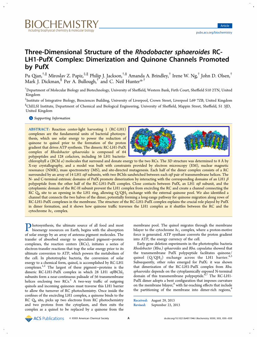
Figure 1. Density maps of selected parts of the RC-LH1-PufX dimer. (A) LH1 αβ subunits. LH1 α is in yellow, LH1 β, blue, and BChl a, brightgreen, and the electron density is represented by a gray mesh. All LH1 αβ BChl a subunits were fitted into the electron density individually. (B) RCcomplex. RC-H subunit is in cyan, RC-M, magenta, RC-L, orange, and electron density, gray. The RC complex is viewed from two angles rotated 90°about the z axis. (C) On the left, the arrangement of polypeptides at the dimer interface. For the sake of clarity, only the RC-H subunits, the twoPufX polypeptides in red, and the 1/1′ and 14/14′ LH1 αβ pairs are included. On the right, one-half of the dimer with the RC omitted, showing the14 LH1 αβ pairs together with a PufX polypeptide. All density maps were produced using Pymol with a contour level of 1.0σ based on the finalrefined electron density map of the dimer.
Biochemistry Article
dx.doi.org/10.1021/bi4011946 | Biochemistry XXXX, XXX, XXX−XXXE
new features such as N- and C-terminal turns for LH1 andimproved density for PufX (see the Materials and Methods fordetails). However, we emphasize that at this resolutionindividual side chains could not be resolved from each otherin the electron density map; subsequent discussion of the likelyinteractions between different amino acids relies therefore onthe input of various constraints on the final model.Construction and interpretation of this final model wasinformed not only by data from cryo-electron microscopy,which determined the 8.5 Å projection structure of thecomplex,3 but also from single-particle reconstruction,8 MSanalysis (this work), the solution structures of LH1 β andPufX,7,23,24 the 2.65 Å structure of the reaction center,20 site-directed-mutagenesis studies of the LH1 complex,33−35 and thestructures of LH2 complexes.18,21 The resultant Rfactor/Rfreevalues of 22.8/25.8%, can be compared with other structures atthis resolution such as the hexameric building block of the HIVcapsid (3GV2) (resolution 7.0 Å and Rfactor 28%)
36 and thestaphylococcal complement inhibitor in complex with humancomplement component C3b (3L5N) (resolution 7.5 Å andRfactor 26%).
37
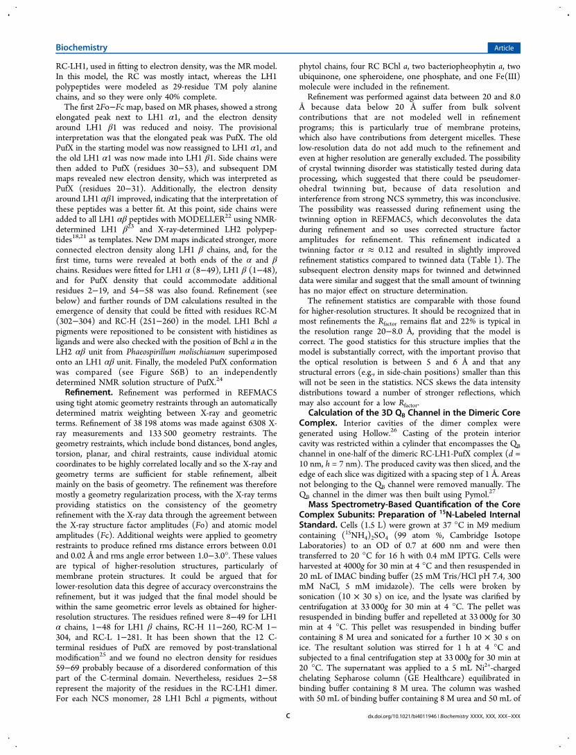
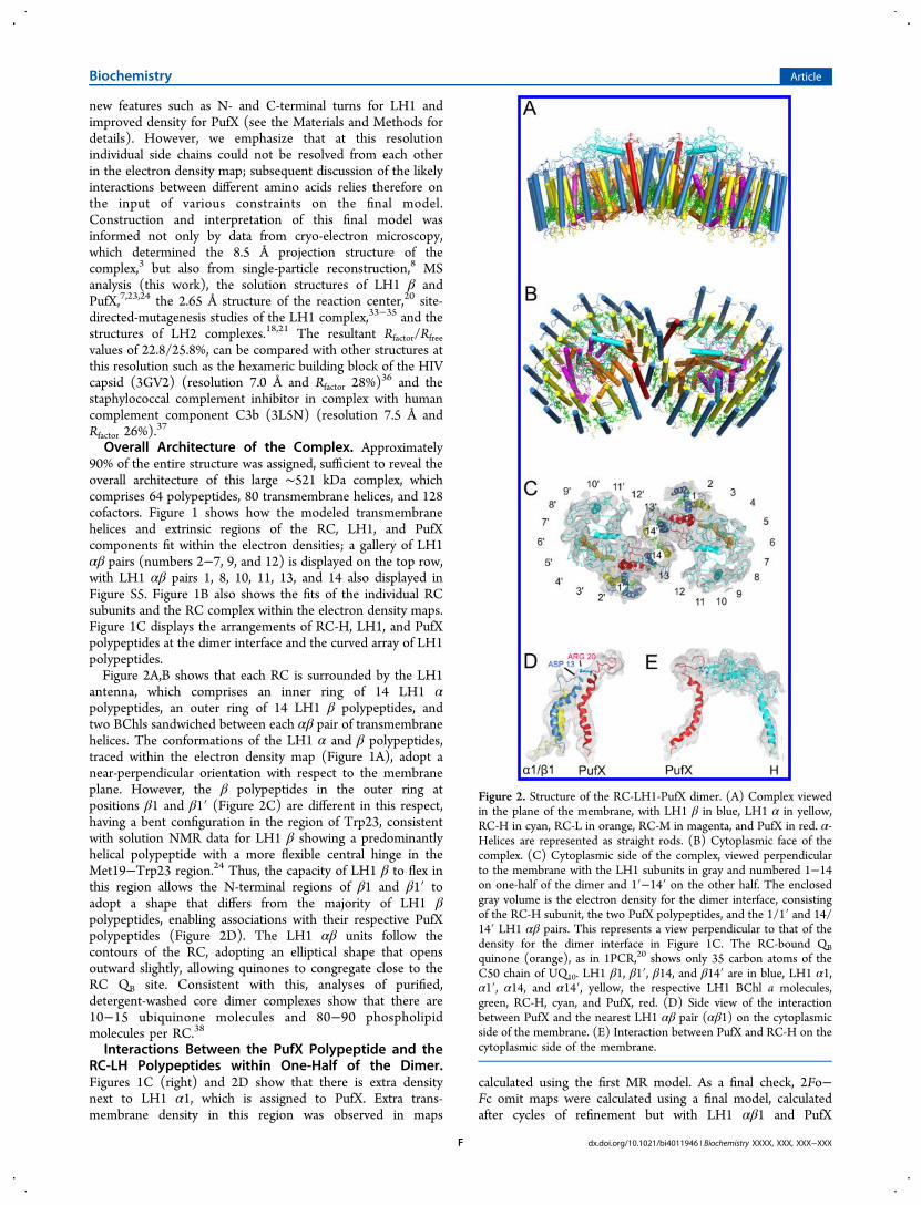
Overall Architecture of the Complex. Approximately90% of the entire structure was assigned, sufficient to reveal theoverall architecture of this large ∼521 kDa complex, whichcomprises 64 polypeptides, 80 transmembrane helices, and 128cofactors. Figure 1 shows how the modeled transmembranehelices and extrinsic regions of the RC, LH1, and PufXcomponents fit within the electron densities; a gallery of LH1αβ pairs (numbers 2−7, 9, and 12) is displayed on the top row,with LH1 αβ pairs 1, 8, 10, 11, 13, and 14 also displayed inFigure S5. Figure 1B also shows the fits of the individual RCsubunits and the RC complex within the electron density maps.Figure 1C displays the arrangements of RC-H, LH1, and PufXpolypeptides at the dimer interface and the curved array of LH1polypeptides.Figure 2A,B shows that each RC is surrounded by the LH1
antenna, which comprises an inner ring of 14 LH1 αpolypeptides, an outer ring of 14 LH1 β polypeptides, andtwo BChls sandwiched between each αβ pair of transmembranehelices. The conformations of the LH1 α and β polypeptides,traced within the electron density map (Figure 1A), adopt anear-perpendicular orientation with respect to the membraneplane. However, the β polypeptides in the outer ring atpositions β1 and β1′ (Figure 2C) are different in this respect,having a bent configuration in the region of Trp23, consistentwith solution NMR data for LH1 β showing a predominantlyhelical polypeptide with a more flexible central hinge in theMet19−Trp23 region.24 Thus, the capacity of LH1 β to flex inthis region allows the N-terminal regions of β1 and β1′ toadopt a shape that differs from the majority of LH1 βpolypeptides, enabling associations with their respective PufXpolypeptides (Figure 2D). The LH1 αβ units follow thecontours of the RC, adopting an elliptical shape that opensoutward slightly, allowing quinones to congregate close to theRC QB site. Consistent with this, analyses of purified,detergent-washed core dimer complexes show that there are10−15 ubiquinone molecules and 80−90 phospholipidmolecules per RC.38
Interactions Between the PufX Polypeptide and theRC-LH Polypeptides within One-Half of the Dimer.Figures 1C (right) and 2D show that there is extra densitynext to LH1 α1, which is assigned to PufX. Extra trans-membrane density in this region was observed in maps
calculated using the first MR model. As a final check, 2Fo−Fc omit maps were calculated using a final model, calculatedafter cycles of refinement but with LH1 αβ1 and PufX
Figure 2. Structure of the RC-LH1-PufX dimer. (A) Complex viewedin the plane of the membrane, with LH1 β in blue, LH1 α in yellow,RC-H in cyan, RC-L in orange, RC-M in magenta, and PufX in red. α-Helices are represented as straight rods. (B) Cytoplasmic face of thecomplex. (C) Cytoplasmic side of the complex, viewed perpendicularto the membrane with the LH1 subunits in gray and numbered 1−14on one-half of the dimer and 1′−14′ on the other half. The enclosedgray volume is the electron density for the dimer interface, consistingof the RC-H subunit, the two PufX polypeptides, and the 1/1′ and 14/14′ LH1 αβ pairs. This represents a view perpendicular to that of thedensity for the dimer interface in Figure 1C. The RC-bound QBquinone (orange), as in 1PCR,20 shows only 35 carbon atoms of theC50 chain of UQ10. LH1 β1, β1′, β14, and β14′ are in blue, LH1 α1,α1′, α14, and α14′, yellow, the respective LH1 BChl a molecules,green, RC-H, cyan, and PufX, red. (D) Side view of the interactionbetween PufX and the nearest LH1 αβ pair (αβ1) on the cytoplasmicside of the membrane. (E) Interaction between PufX and RC-H on thecytoplasmic side of the membrane.
Biochemistry Article
dx.doi.org/10.1021/bi4011946 | Biochemistry XXXX, XXX, XXX−XXXF
removed. Figure S6A shows the return of these polypeptides inthe electron density map, which is contoured at a 1σ level. Thefinal refined PufX polypeptide chain in our model can besuperimposed onto 2NRG, which is a minimized averagedsolution structure for PufX in solvent24 with an rms deviation of2.08 Å and a maximum Cα atom deviation of 3.75 Å (FigureS6B). The largest deviations are in the region 2−23, wherePufX is constrained by interactions with the N-terminal LH1 α-polypeptide and the RC-H chain, consistent with the absence ofsuch constraints in the NMR studies of purified PufX insolvent.7,24 Although the bend and the N-terminal loop forPufX both closely follow 2NRG (Figure S6B), they weredetermined completely independently of the NMR struc-tures.7,24 Furthermore, the position of PufX next to LH1α1 hasalso been determined independently by cryo-EM (3), althoughthe PufX transmembrane domain is now established as 17 Åfrom the original best estimate of its position in the EMprojection map.3
Figure 2C shows the position of PufX and RC-H in relationto the RC QB inferred from the RC structure (PDB 1PCR)incorporated into our dimer model. The location of the PufXtransmembrane domain is a consequence of the PufX-RC-Hinteraction and the association of PufX with the bent LH1 β1polypeptide. PufX prevents the LH1 array from completelysurrounding the RC, creating a channel (Figure 5B) that allowsthe traffic of quinones and quinols as they shuttle between theQB site, the external quinone pool, and the cytochrome bc1complex. A similar situation is envisaged for the RC-LH1-PufWcomplex of Rhodopseudomonas palustris;2 Figure S7A,B shows acomparison between the Rba. sphaeroides dimer and Rps.palustris monomer complexes, aligned using the Rba.sphaeroides RC that is modeled into both structures. FigureS7B shows the relative positions of PufX and W in relation tothe RC QB site; the minimum center-to-center distancebetween the transmembrane helices is ∼5 Å.Figure 2D shows that PufX is stabilized by close contacts
with the N-termini of LH1 α1 and β1 on the cytoplasmic sideof the membrane. PufX is also intimately associated with theextrinsic domain of the RC-H subunit (Figure 2E), with PufXresidues 21−30 running parallel with the RC-H C-terminalregion, which forms a bend at residues 244−246. The last 10residues of RC-H are usually missing in RC X-ray structures butare observed in 2J8D,39 where the residues 228−257 are α-helical with a bend at 244−245. Our structure is consistent withthis, but a greater bend has been modeled into RC-H in thisregion to follow the electron density and contours of PufX. Theabsence of PufX from all RC crystal structures may explain whythis otherwise flexible C-terminal region is not usually observedin electron density maps. The attachment of PufX to RC-Hthrough residues on the cytoplasmic faces of these proteins isconsistent with AFM nanodissection experiments whereremoval of RC-H was frequently seen in PufX-minus mutants.10
This association between RC-H and PufX forms a likely startsignal for the assembly of the RC-LH1-PufX complex byforming an arch with RC-H that prevents blockage of thepassage to the RC QB site and also provides an anchoring pointfor docking the first LH1 α1β1BChl2 subunit onto PufX.Encirclement of the RC could then proceed by the addition ofmore LH1 αβBChl2 units. This assembly sequence wasoriginally proposed on the basis of Western blotting experi-ments where the RC-H subunit was detectable first followed byPufX and then the LH1 α and β polypeptides.40
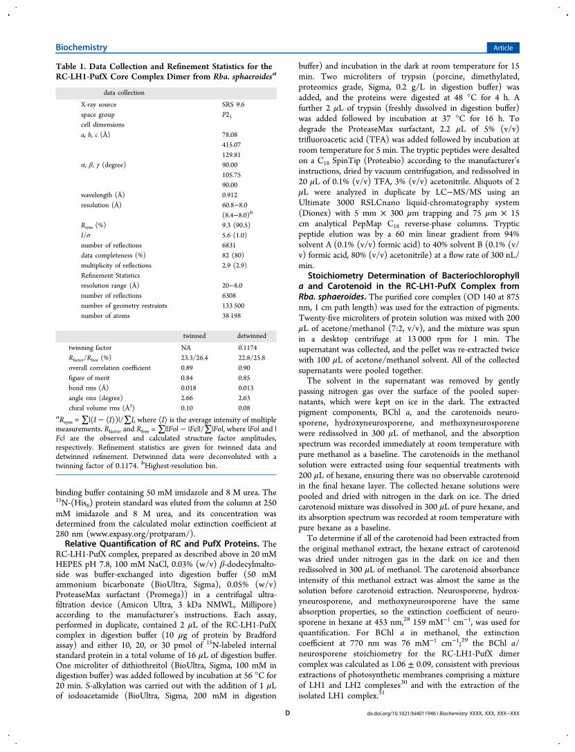
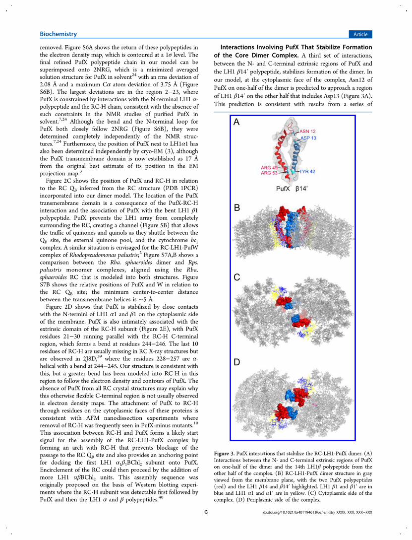
Interactions Involving PufX That Stabilize Formationof the Core Dimer Complex. A third set of interactions,between the N- and C-terminal extrinsic regions of PufX andthe LH1 β14′ polypeptide, stabilizes formation of the dimer. Inour model, at the cytoplasmic face of the complex, Asn12 ofPufX on one-half of the dimer is predicted to approach a regionof LH1 β14′ on the other half that includes Asp13 (Figure 3A).This prediction is consistent with results from a series of
Figure 3. PufX interactions that stabilize the RC-LH1-PufX dimer. (A)Interactions between the N- and C-terminal extrinsic regions of PufXon one-half of the dimer and the 14th LH1β polypeptide from theother half of the complex. (B) RC-LH1-PufX dimer structure in grayviewed from the membrane plane, with the two PufX polypeptides(red) and the LH1 β14 and β14′ highlighted. LH1 β1 and β1′ are inblue and LH1 α1 and α1′ are in yellow. (C) Cytoplasmic side of thecomplex. (D) Periplasmic side of the complex.
Biochemistry Article
dx.doi.org/10.1021/bi4011946 | Biochemistry XXXX, XXX, XXX−XXXG
deletions constructed at the N-terminus of PufX, wheretruncation of 12 or more residues from the N-terminusabolished dimerization of the complex.6,7 The structure alsoshows an association between the C-terminal regions of PufXand β14′, consistent with the effects of C-terminal truncation ofPufX.6 The resolution of the structure does not allow preciseassignments of contacts between polypeptides, but two PufXresidues, Arg49 and Arg53, are found in this C-terminal contactzone, and alteration of both of these residues to Leu abolishesdimer formation, leaving the levels of PufX unaffected.41 Theassociation between RC-LH1-PufX monomers is likely to berelatively weak because photosynthetic membranes containsome monomers.42 Figure 3B−D shows three views of thedimer structure with only the two PufX polypeptides (red) andthe LH1 β14 and β14′ highlighted, which emphasize theimportance of both the N- and C-terminal regions of PufX forpromoting dimerization of the complex.Pigments within the RC-LH1-PufX Dimer Complex.
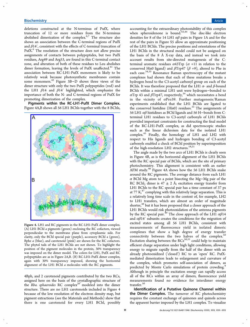
Figure 4A,B shows all 56 LH1 BChls together with the 8 BChls,
4Bph, and 2 carotenoid pigments contributed by the two RCs,assigned here on the basis of the crystallographic structure ofthe Rba. sphaeroides RC complex20 modeled into the dimerstructure. There are no LH1 carotenoids included in Figure 4because of the low resolution of the electron density map, butpigment extractions (see the Materials and Methods) show thatthere is one carotenoid for every LH1 BChl, possibly
accounting for the extraordinary photostability of this complexwhen spheroidenone is bound.43,44 The disc-like electrondensities for 8 of the 14 LH1 αβ pairs in Figure 1A and for therest of the pairs in Figure S5 allow an approximate positioningof the LH1 BChls. The precise positions and orientations of theLH1 BChls in the structural model could not be assigned onthe basis of the 8 Å X-ray data, and instead we took intoaccount results from site-directed mutagenesis of the C-terminal aromatic residues α43Trp (α +11 in relation to theconserved His0 ligand) and βTrp47 (β +9), altered to Phe ineach case.34,35 Resonance Raman spectroscopy of the mutantcomplexes had shown that each of these mutations breaks ahydrogen bond to the C3-acetyl carbonyl group on each of theBChls. It was therefore proposed that the LH1 α- and β-boundBChls within a minimal LH1 unit were hydrogen−bonded toαTrp 43 and βTrp47, respectively,34 which necessitates a bendin the vicinity of αPro39. Further mutagenesis/Ramanexperiments established that the LH1 BChls are ligated tothe conserved histidine (His0) residues.33 The assignments of56 LH1 αβ histidines as BChl ligands and 56 H−bonds from C-terminal LH1 residues to C3-acetyl carbonyls of LH1 BChlsprovided important constraints for constructing the final modelof the RC-LH1-PufX complex, as did spectroscopic studiessuch as the linear dichroism data for the isolated LH1complex.45 Finally, the homology of LH1 and LH2 withrespect to His ligands and hydrogen bonding of C3-acetylcarbonyls enabled a check of BChl position by superimpositionof the high-resolution LH2 structures.18,21
The angle made by the two arcs of LH1 BChls is clearly seenin Figure 4B, as is the horizontal alignment of the LH1 BChlswith the RC special pair of BChls, which are the site of primaryphotochemistry. This alignment is consistent with an earlierAFM study.46 Figure 4A shows how the 56 LH1 BChls snakearound the RC pigments. The average distance from each LH1α BChl Mg atom to a point bisecting the Mg−Mg pair of theRC BChl2 dimer is 47 ± 2 Å; excitation energy transfer fromLH1 BChls to the RC special pair has a time constant of 37 psat 77 K,47 complying with this relatively large separation. This isa relatively long time scale in the context of, for example, LH2to LH1 transfers, which are almost an order of magnitudeshorter,48 but it has been proposed that a closer approach of theLH1 BChls would risk photooxidation of the antenna pigmentsby the RC special pair.49 The close approach of the LH1 αβ14and αβ14′ subunits creates the conditions for the migration ofexcited states among all 56 LH1 BChls, consistent withmeasurements of fluorescence yield in isolated dimericcomplexes that show a high degree of energy transferconnectivity between the two halves of the complex.50
Excitation sharing between the RCs50,51 could help to maintainefficient charge separation under high light conditions, allowingenergy to migrate rapidly from the half of the dimer with analready photooxidized (‘closed’) RC to an ‘open’ RC. PufX-mediated dimerization leads to enlargement and curvature ofthe complex, which promotes self-association of dimers, aspredicted by Monte Carlo simulations of protein crowding.9
Although in principle the excitation energy can rapidly accessall of the RCs within an array of dimers, fluorescence yieldmeasurements found no evidence for interdimer energytransfer.50
Identification of a Putative Quinone Channel withinthe Dimer Complex. The turnover of RC photochemistryrequires the constant exchange of quinones and quinols acrossthe apparent barrier imposed by the LH1 complex. To visualize
Figure 4. LH1 and RC pigments in the RC-LH1-PufX dimer complex.(A) LH1 BChl a pigments (green) enclosing the RC cofactors, viewedperpendicular to the membrane plane from cytoplasmic side. Forclarity, only the BChl special pair (purple), accessory BChl a (green),Bphe a (blue), and carotenoid (pink) are shown for the RC cofactors.The phytol tails of the LH1 BChls are not shown. To highlight theposition of the pigment molecules in the protein, 30% transparencywas imposed on the dimer model. The colors for LH1, PufX and RCpolypeptides are as in Figure 2A,B. (B) RC-LH1-PufX dimer complex,again with 30% transparency imposed, showing the horizontalalignment of the LH1 BChl a molecules with the RC pigments.
Biochemistry Article
dx.doi.org/10.1021/bi4011946 | Biochemistry XXXX, XXX, XXX−XXXH
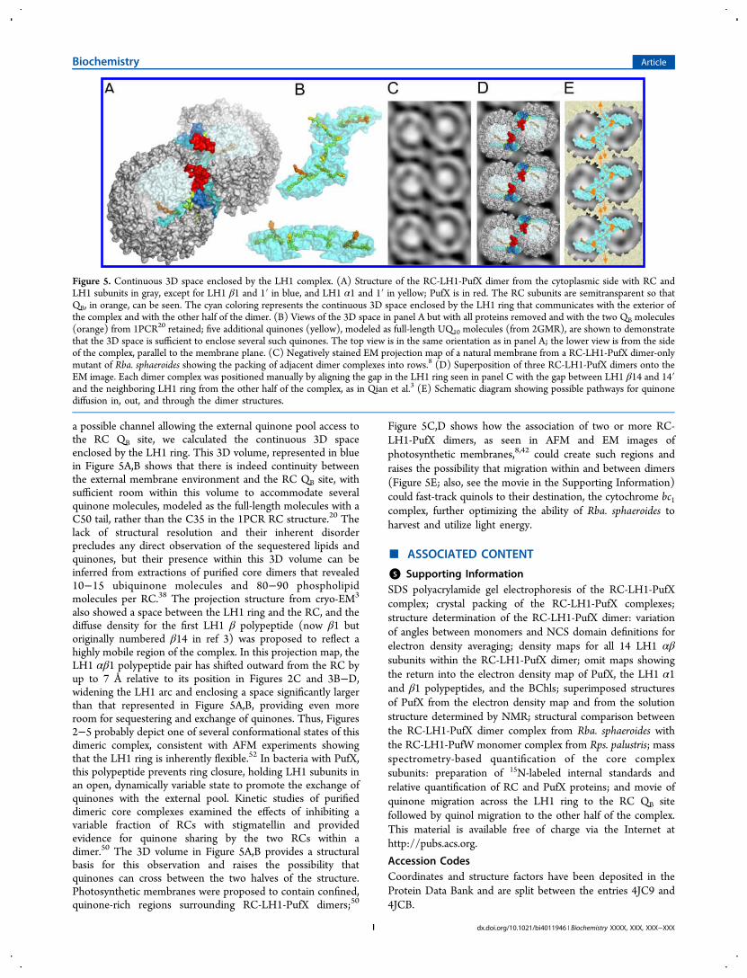
a possible channel allowing the external quinone pool access tothe RC QB site, we calculated the continuous 3D spaceenclosed by the LH1 ring. This 3D volume, represented in bluein Figure 5A,B shows that there is indeed continuity betweenthe external membrane environment and the RC QB site, withsufficient room within this volume to accommodate severalquinone molecules, modeled as the full-length molecules with aC50 tail, rather than the C35 in the 1PCR RC structure.20 Thelack of structural resolution and their inherent disorderprecludes any direct observation of the sequestered lipids andquinones, but their presence within this 3D volume can beinferred from extractions of purified core dimers that revealed10−15 ubiquinone molecules and 80−90 phospholipidmolecules per RC.38 The projection structure from cryo-EM3
also showed a space between the LH1 ring and the RC, and thediffuse density for the first LH1 β polypeptide (now β1 butoriginally numbered β14 in ref 3) was proposed to reflect ahighly mobile region of the complex. In this projection map, theLH1 αβ1 polypeptide pair has shifted outward from the RC byup to 7 Å relative to its position in Figures 2C and 3B−D,widening the LH1 arc and enclosing a space significantly largerthan that represented in Figure 5A,B, providing even moreroom for sequestering and exchange of quinones. Thus, Figures2−5 probably depict one of several conformational states of thisdimeric complex, consistent with AFM experiments showingthat the LH1 ring is inherently flexible.52 In bacteria with PufX,this polypeptide prevents ring closure, holding LH1 subunits inan open, dynamically variable state to promote the exchange ofquinones with the external pool. Kinetic studies of purifieddimeric core complexes examined the effects of inhibiting avariable fraction of RCs with stigmatellin and providedevidence for quinone sharing by the two RCs within adimer.50 The 3D volume in Figure 5A,B provides a structuralbasis for this observation and raises the possibility thatquinones can cross between the two halves of the structure.Photosynthetic membranes were proposed to contain confined,quinone-rich regions surrounding RC-LH1-PufX dimers;50
Figure 5C,D shows how the association of two or more RC-LH1-PufX dimers, as seen in AFM and EM images ofphotosynthetic membranes,8,42 could create such regions andraises the possibility that migration within and between dimers(Figure 5E; also, see the movie in the Supporting Information)could fast-track quinols to their destination, the cytochrome bc1complex, further optimizing the ability of Rba. sphaeroides toharvest and utilize light energy.
■ ASSOCIATED CONTENT
*S Supporting InformationSDS polyacrylamide gel electrophoresis of the RC-LH1-PufXcomplex; crystal packing of the RC-LH1-PufX complexes;structure determination of the RC-LH1-PufX dimer: variationof angles between monomers and NCS domain definitions forelectron density averaging; density maps for all 14 LH1 αβsubunits within the RC-LH1-PufX dimer; omit maps showingthe return into the electron density map of PufX, the LH1 α1and β1 polypeptides, and the BChls; superimposed structuresof PufX from the electron density map and from the solutionstructure determined by NMR; structural comparison betweenthe RC-LH1-PufX dimer complex from Rba. sphaeroides withthe RC-LH1-PufW monomer complex from Rps. palustris; massspectrometry-based quantification of the core complexsubunits: preparation of 15N-labeled internal standards andrelative quantification of RC and PufX proteins; and movie ofquinone migration across the LH1 ring to the RC QB sitefollowed by quinol migration to the other half of the complex.This material is available free of charge via the Internet athttp://pubs.acs.org.
Accession CodesCoordinates and structure factors have been deposited in theProtein Data Bank and are split between the entries 4JC9 and4JCB.
Figure 5. Continuous 3D space enclosed by the LH1 complex. (A) Structure of the RC-LH1-PufX dimer from the cytoplasmic side with RC andLH1 subunits in gray, except for LH1 β1 and 1′ in blue, and LH1 α1 and 1′ in yellow; PufX is in red. The RC subunits are semitransparent so thatQB, in orange, can be seen. The cyan coloring represents the continuous 3D space enclosed by the LH1 ring that communicates with the exterior ofthe complex and with the other half of the dimer. (B) Views of the 3D space in panel A but with all proteins removed and with the two QB molecules(orange) from 1PCR20 retained; five additional quinones (yellow), modeled as full-length UQ10 molecules (from 2GMR), are shown to demonstratethat the 3D space is sufficient to enclose several such quinones. The top view is in the same orientation as in panel A; the lower view is from the sideof the complex, parallel to the membrane plane. (C) Negatively stained EM projection map of a natural membrane from a RC-LH1-PufX dimer-onlymutant of Rba. sphaeroides showing the packing of adjacent dimer complexes into rows.8 (D) Superposition of three RC-LH1-PufX dimers onto theEM image. Each dimer complex was positioned manually by aligning the gap in the LH1 ring seen in panel C with the gap between LH1 β14 and 14′and the neighboring LH1 ring from the other half of the complex, as in Qian et al.3 (E) Schematic diagram showing possible pathways for quinonediffusion in, out, and through the dimer structures.
Biochemistry Article
dx.doi.org/10.1021/bi4011946 | Biochemistry XXXX, XXX, XXX−XXXI
■ AUTHOR INFORMATIONCorresponding Author*E-mail: [email protected]. Phone: +441142224191.Author Contributions∥These authors contributed equally.FundingP.Q., M.Z.P., P.J.J., A.A.B., J.D.O, M.J.D, P.A.B., and C.N.H.gratefully acknowledge funding from the Biotechnology andBiological Sciences Research Council (U.K.) and Science andTechnology Facilities Council. C.N.H. and A.A.B. were alsosupported as part of the Photosynthetic Antenna ResearchCenter (PARC), an Energy Frontier Research Center fundedby the U.S. Department of Energy, Office of Science, and Officeof Basic Energy Sciences under award no. DE-SC0001035.NotesThe authors declare no competing financial interest.
■ ABBREVIATIONS USEDAFM, atomic force microscopy; BChl a, bacteriochlorophyll a;CAPS, N-cyclohexyl-3-aminopropanesulfonic acid; DM, densitymodification; β-DDM, n-dodecyl-β-D-maltopyranoside; EM,electron microscopy; LH, light harvesting; MR, molecularreplacement; NM, n-nonyl-β-D-maltopyranoside; NMR, nuclearmagnetic resonance; RC, reaction center
■ REFERENCES(1) Bullough, P. A., Qian, P., and Hunter, C. N. (2008) Reactioncenter-light harvesting core complexes of purple bacteria, in The PurplePhototrophic Bacteria (Hunter, C. N., Daldal, F., Thurnauer, M. C., andBeatty, J. T., Eds.), pp 155−179, Springer, Dordrecht.(2) Roszak, A. W., Howard, T. D., Southall, J., Gardiner, A. T., Law,C. J., Isaacs, N. W., and Cogdell, R. J. (2003) Crystal structure of theRC-LH1 core complex from Rhodopseudomonas palustris. Science 302,1969−1972.(3) Qian, P., Hunter, C. N., and Bullough, P. A. (2005) The 8.5 Åprojection structure of the core RC-LH1-PufX dimer of Rhodobactersphaeroides. J. Mol. Biol. 349, 948−960.(4) Farchaus, J. W., Gruenberg, H., and Oesterhelt, D. (1990)Complementation of a reaction center-deficient Rhodobacter sphaer-oides puf LMX deletion strain in trans with puf BALM does not restorethe photosynthesis-positive phenotype. J. Bacteriol. 172, 977−985.(5) Lilburn, T. G., Haith, C. E., Prince, R. C., and Beatty, J. T. (1992)Pleiotropic effects of puf X gene deletion on the structure and functionof the photosynthetic apparatus of Rhodobacter capsulatus. Biochim.Biophys. Acta 1100, 160−170.(6) Francia, F., Wang, J., Zischka, H., Venturoli, G., and Oesterhelt,D. (2002) Role of the N- and C-terminal regions of the PufX proteinin the structural organization of the photosynthetic core complex ofRhodobacter sphaeroides. Eur. J. Biochem. 269, 1877−1885.(7) Ratcliffe, E. C., Tunnicliffe, R. B., Ng, I. W., Adams, P. G., Qian,P., Holden-Dye, K., Jones, M. R., Williamson, M. P., and Hunter, C. N.(2011) Experimental evidence that the membrane-spanning helix ofPufX adopts a bent conformation that facilitates dimerisation of theRhodobacter sphaeroides RC-LH1 complex through N-terminalinteractions. Biochim. Biophys. Acta 1807, 95−107.(8) Qian, P., Bullough, P., and Hunter, C. N. (2008) Three-dimensional reconstruction of a membrane-bending complex. J. Biol.Chem. 283, 14002−14011.(9) Frese, R. N., Pamies, J. C., Olsen, J. D., Bahatyrova, S., van derWeij-de Wit, C. D., Aartsma, T. J., Otto, C., Hunter, C. N., Frenkel, D.,and van Grondelle, R. (2008) Protein shape and crowding drivedomain formation and curvature in biological membranes. Biophys. J.94, 640−647.(10) Adams, P. G., Mothersole, D. J., Ng, I. W., Olsen, J. D., andHunter, C. N. (2011) Monomeric RC-LH1 core complexes retard
LH2 assembly and intracytoplasmic membrane formation in PufX-minus mutants of Rhodobacter sphaeroides. Biochim. Biophys. Acta 1807,1044−1055.(11) Tucker, J. D., Siebert, C. A., Escalante, M., Adams, P. G., Olsen,J. D., Otto, C., Stokes, D. L., and Hunter, C. N. (2010) Membraneinvagination in Rhodobacter sphaeroides is initiated at curved regions ofthe cytoplasmic membrane, then forms both budded and fullydetached spherical vesicles. Mol. Microbiol. 76, 833−847.(12) Sener, M. K., Olsen, J. D., Hunter, C. N., and Schulten, K.(2007) Atomic-level structural and functional model of a bacterialphotosynthetic membrane vesicle. Proc. Natl. Acad. Sci. U.S.A. 104,15723−15728.(13) Jones, M. R., Fowler, G. J. S., Gibson, L. C. D., Grief, G. G.,Olsen, J. D., Crielaard, W., and Hunter, C. N. (1992) Mutants ofRhodobacter sphaeroides lacking one or more pigment-proteincomplexes and complementation with reaction-centre, LH1, andLH2 genes. Mol. Microbiol. 6, 1173−1184.(14) Leslie, A. G. W. (1999) Integration of macromoleculardiffraction data. Acta Crystallogr., Sect. D 55, 1696−1702.(15) Winn, M. D., Ballard, C. C., Cowtan, K. D., Dodson, E. J.,Emsley, P., Evans, P. R., Krissinel, E. B., Leslie, A. G. W., McCoy, A.,McNicholas, S. J., Murshudov, G. N., Pannu, N. S., Potterton, E. A.,Powell, H. R., Read, R. J., Vagin, A., and Wilson, K. S. (2011)Overview of the CCP4 suite and current developments. ActaCrystallogr., Sect. D 67, 235−242.(16) McCoy, A. J., Grosse-Kunstleve, R. W., Adams, P. D., Winn, M.D., Storoni, L. C., and Read, R. J. (2007) Phaser crystallographicsoftware. J. Appl. Crystallogr. 40, 658−674.(17) Cowtan, K., and Main, P. (1998) Miscellaneous algorithms fordensity modification. Acta Crystallogr., Sect. D 54, 487−493.(18) McDermott, G., Prince, S. M., Freer, A. A., Hawthornthwaite-Lawless, A. M., Papiz, M. Z., Cogdell, R. J., and Isaacs, N. W. (1995)Crystal structure of an integral membrane light-harvesting complexfrom photosynthetic bacteria. Nature 374, 517−521.(19) McLuskey, K., Prince, S. M., Cogdell, R. J., and Isaacs, N. W.(2001) The crystallographic structure of the B800−820 LH3 light-harvesting complex from the purple bacteria Rhodopseudomonasacidophila strain 7050. Biochemistry 40, 8783−8789.(20) Ermler, U., Fritzsch, G., Buchanan, S. K., and Michel, H. (1994)Structure of the photosynthetic reaction centre from Rhodobactersphaeroides at 2.65 Å resolution: Cofactors and protein-cofactorinteractions. Structure 2, 925−936.(21) Koepke, J., Hu, X. C., Muenke, C., Schulten, K., and Michel, H.(1996) The crystal structure of the light-harvesting complex II (B800-B850) from Rhodospirillum molischanum. Structure 4, 581−597.(22) Sali, A., and Blundell, T. L. (1993) Comparative proteinmodeling by satisfaction of spatial restraints. J. Mol. Biol. 234, 779−815.(23) Conroy, M. J., Westerhuis, W. H., Parkes-Loach, P. S., Loach, P.A., Hunter, C. N., and Williamson, M. P. (2000) The solutionstructure of Rhodobacter sphaeroides LH1β reveals two helical domainsseparated by a more flexible region: Structural consequences for theLH1 complex. J. Mol. Biol. 298, 83−94.(24) Tunnicliffe, R. B., Ratcliffe, E. C., Hunter, C. N., andWilliamson, M. P. (2006) The solution structure of the PufXpolypeptide from Rhodobacter sphaeroides. FEBS Lett. 580, 6967−6971.(25) Parkes-Loach, P. S., Law, C. J., Recchia, P. A., Kehoe, J.,Nehrlich, S., Chen, J., and Loach, P. A. (2001) Role of the core regionof the PufX protein in inhibition of reconstitution of the core light-harvesting complexes of Rhodobacter sphaeroides and Rhodobactercapsulatus. Biochemistry 40, 5593−5601.(26) Ho, B. K., and Gruswitz, F. (2008) HOLLOW: Generatingaccurate representations of channel and interior surfaces in molecularstructures. BMC Struct. Biol 8, 49−54.(27) DeLano, W. L. (2002) The PyMOL Molecular Graphics System,DeLano Scientific Inc., San Carlos, CA.(28) Sashima, T., Koyama, Y., Yamada, T., and Hashimoto, H.(2000) The 1Bu
+, 1Bu−, and 2Ag
− energies of crystalline lycopene, β-carotene, and mini-9-β-carotene as determined by resonance-Raman
Biochemistry Article
dx.doi.org/10.1021/bi4011946 | Biochemistry XXXX, XXX, XXX−XXXJ
excitation profiles: Dependence of the 1Bu− state energy on the
conjugation length. J. Phys. Chem. B 104, 5011−5019.(29) Clayton, R. K. (1966) Spectroscopic analysis of bacteriochlor-ophylls in vitro and in vivo. Photochem. Photobiol. 5, 669−677.(30) Sistrom, W. R. (1978) Control of Antenna PigmentComponents, in The Photosynthetic Bacteria (Clayton, R. K. andSistrom, W. R., Eds.) pp 841−848, Plenum, New York.(31) Broglie, R. M., Hunter, C. N., Delepelaire, P., Niederman, R. A.,Chua, N.-H., and Clayton, R. K. (1980) Isolation and characterizationof the pigment-protein complexes of Rhodopseudomonas sphaeroides bylithium dodecyl sulfate polyacrylamide gel electrophoresis. Proc. Natl.Acad. Sci. U.S.A. 77, 87−91.(32) Grimes, M., Burroughs, J. N., Gouet, P., Diprose, J. M., Malby,R., Zientara, S., Mertens, P. P., and Stuart, D. I. (1998) The atomicstructure of the bluetongue virus core. Nature 395, 470−478.(33) Olsen, J. D., Sturgis, J. N., Westerhuis, W. H., Fowler, G. J. S.,Hunter, C. N., and Robert, B. (1997) Site-directed modification of theligands to the bacteriochlorophylls of the light-harvesting LH1 andLH2 complexes of Rhodobacter sphaeroides. Biochemistry 36, 12625−12632.(34) Sturgis, J. N., Olsen, J. D., Robert, B., and Hunter, C. N. (1997)Functions of conserved tryptophan residues of the core light-harvesting complex of Rhodobacter sphaeroides. Biochemistry 36,2772−2778.(35) Olsen, J. D., Sockalingum, G. D., Robert, B., and Hunter, C. N.(1994) Modification of a hydrogen bond to a bacteriochlorophyll amolecule in the light harvesting 1 antenna of Rhodobacter sphaeroides.Proc. Natl. Acad. Sci. U.S.A. 91, 7124−7128.(36) Pornillos, O., Ganser-Pornillos, B. K., Kelly, B. N., Hua, Y. Z.,Whitby, F. G., Stout, C. D., Sundquist, W. I., Hill, C. P., and Yeager, M.(2009) X-ray structures of the hexameric building block of the HIVcapsid. Cell 137, 1282−1292.(37) Garcia, B. L., Ramyar, K. X., Tzekou, A., Ricklin, D., McWhorter,W. J., Lambris, J. D., and Geisbrecht, B. V. (2010) Molecular basis forcomplement recognition and inhibition determined by crystallographicstudies of the staphylococcal complement inhibitor (SCIN) bound toC3c and C3b. J. Mol. Biol. 402, 17−29.(38) Dezi, M., Francia, F., Mallardi, A., Colafemmina, G., Palazzo, G.,and Venturoli, G. (2007) Stabilization of charge separation andcardiolipin confinement in antenna-reaction center complexes purifiedfrom Rhodobacter sphaeroides. Biochim. Biophys. Acta 1767, 1041−1056.(39) Koepke, J., Krammer, E. M., Klingen, A. R., Sebban, P., Ullmann,G. M., and Fritzsch, G. (2007) pH modulates the quinone position inthe photosynthetic reaction center from Rhodobacter sphaeroides in theneutral and charge separated states. J. Mol. Biol. 371, 396−409.(40) Pugh, R. J., McGlynn, P., Jones, M. R., and Hunter, C. N.(1998) The LH1-RC core complex of Rhodobacter sphaeroides:Interaction between components, time-dependent assembly, andtopology of the PufX protein. Biochim. Biophys. Acta 1366, 301−316.(41) Ng, I. W. (2008) A structural and functional study of the RC-LH1-PufX core complex from Rhodobacter sphaeroides, Ph.D. Thesis,University of Sheffield, Sheffield, United Kingdom.(42) Bahatyrova, S., Frese, R. N., Siebert, C. A., Olsen, J. D., van derWerf, K. O., van Grondelle, R., Niederman, R. A., Bullough, P. A.,Otto, C., and Hunter, C. N. (2004) The native architecture of aphotosynthetic membrane. Nature 430, 1058−1062.(43) Magis, G. J., Olsen, J. D., Reynolds, N. P., Leggett, G. J., Hunter,C. N., Aartsma, T. J., and Frese, R. N. (2011) Use of engineeredunique cysteine residues to facilitate oriented coupling of proteinsdirectly to a gold substrate. Photochem. Photobiol. 87, 1050−1057.(44) Slouf, V., Chabera, P., Olsen, J. D., Martin, E. C., Qian, P.,Hunter, C. N., and Polivka, T. (2012) Photoprotection in a purplephototrophic bacterium mediated by oxygen-dependent alteration ofcarotenoid excited-state properties. Proc. Natl. Acad. Sci. U.S.A. 109,8570−8575.(45) Kramer, H. J. M., Pennoyer, J. D., van Grondelle, R., Westerhuis,W. H. J., Niederman, R. A., and Amesz, J. (1984) Low-temperatureoptical properties and pigment organization of the B875 light-
harvesting bacteriochlorophyll-protein complex of purple photo-synthetic bacteria. Biochim. Biophys. Acta 767, 335−344.(46) Fotiadis, D., Qian, P., Philippsen, A., Bullough, P. A., Engel, A.,and Hunter, C. N. (2004) Structural analysis of the reaction centerlight-harvesting complex I photosynthetic core complex of Rhodospir-illum rubrum using atomic force microscopy. J. Biol. Chem. 279, 2063−2068.(47) Visscher, K. J., Bergstrom, H., Sundstrom, V., Hunter, C. N., andvan Grondelle, R. (1989) Temperature dependence of energy-transferfrom the long wavelength antenna BChl-896 to the reaction center inRhodospirillum rubrum, Rhodobacter sphaeroides (w.t. and M21 mutant)from 77 to 177K, studied by picosecond absorption spectroscopy.Photosynth. Res. 22, 211−217.(48) Hess, S., Chachisvilis, M., Timpmann, K., Jones, M. R., Fowler,G. J. S., Hunter, C. N., and Sundstrom, V. (1995) Temporally andspectrally resolved subpicosecond energy transfer within the peripheralantenna complex (LH2) and from LH2 to the core antenna complexin photosynthetic purple bacteria. Proc. Natl. Acad. Sci. U.S.A. 92,12333−12337.(49) Noy, D., Moser, C. C., and Dutton, P. L. (2006) Design andengineering of photosynthetic light-harvesting and electron transferusing length, time, and energy scales. Biochim. Biophys. Acta 1757, 90−105.(50) Comayras, R., Jungas, C., and Lavergne, J. (2005) Functionalconsequences of the organization of the photosynthetic apparatus inRhodobacter sphaeroides. I. Quinone domains and excitation transfer inchromatophores and reaction center center antenna complexes. J. Biol.Chem. 280, 11203−11213.(51) Sener, M., Hsin, J., Trabuco, L. G., Villa, E., Qian, P., Hunter, C.N., and Schulten, K. (2009) Structural model and excitonic propertiesof the dimeric RC-LH1-PufX complex from Rhodobacter sphaeroides.Chem. Phys. 357, 188−197.(52) Bahatyrova, S., Frese, R. N., van der Werf, K. O., Otto, C.,Hunter, C. N., and Olsen, J. D. (2004) Flexibility and sizeheterogeneity of the LH1 light harvesting complex revealed by atomicforce microscopy: functional significance for bacterial photosynthesis.J. Biol. Chem. 279, 21327−21333.
Biochemistry Article
dx.doi.org/10.1021/bi4011946 | Biochemistry XXXX, XXX, XXX−XXXK


![Structure of a [2Fe–2S] ferredoxin from Rhodobacter capsulatus likely involved in Fe–S cluster biogenesis and conformational changes observed upon reduction](https://static.fdokumen.com/doc/165x107/63363fcacd4bf2402c0b6cfb/structure-of-a-2fe2s-ferredoxin-from-rhodobacter-capsulatus-likely-involved.jpg)