Signaling by the Arc Two-Component System Provides a Link Between the Redox State of the Quinone...
16
INTRODUCTION L IFE ON EARTH EVOLVED virtually under anaerobic condi- tions for about 2 billion years that is close to half of the total period of biological evolution (5, 80). For that rea- son, it is commonly accepted that fermentative modes of metabolism preceded respiratory modes of metabolism and that the highly efficient aerobic respiration process became possible after the emergence of photosynthesis, which gen- erated O 2 . Fermentative modes of metabolism achieve redox balance by internal dismutation reactions and generate en- ergy principally by substrate-level phosphorylation. A seri- ous disadvantage is the wasteful sacrifice of some carbon and hydrogen compounds as excretion products. By con- trast, respiratory modes of metabolism exploit exogenous electron acceptors (oxidants) and generate energy princi- pally by proton extrusion, with the consequence of en- hanced anabolism (125). To maximize energy generation under different redox con- ditions, facultative anaerobes, such as Escherichia coli, have evolved multiple metabolic pathways, together with complex genetic regulatory networks. Aerobic respiration is preferred because O 2 has the highest oxidizing power (E° = +820 mV) among all utilizable electron acceptors. In E. coli the final electron transfer steps in this respiration are mediated in par- allel by cytochrome d (high O 2 affinity) and cytochrome o (high Vmax). When O 2 is unavailable, nitrate (E° = +420 mV) becomes the preferred electron acceptor in anaerobic respiration. If this choice is not available, the cell can resort to even less rewarding compounds, such as fumarate (E°= +31 mV). When no exogenous electron acceptor is accessi- ble, fermentative metabolism becomes the last resort. In aero- bic respiratory pathways, ubiquinone (E° = +100 mV) serves as the adapter between electron donor and electron disposal pathways. In anaerobic respiratory pathways, demethylme- naquionone (E° = +36 mV) and menaquinone (E°= 74 781 Departamento de Genética Molecular, Instituto de Fisiología Celular, Universidad Nacional Autónoma de México, México City, México. This article is dedicated to the memory of Edmund C.C. Lin. Signaling by the Arc Two-Component System Provides a Link Between the Redox State of the Quinone Pool and Gene Expression ROXANA MALPICA, GABRIELA R. PEÑA SANDOVAL, CLAUDIA RODRÍGUEZ, BERNARDO FRANCO, and DIMITRIS GEORGELLIS ABSTRACT The Arc two-component system is a complex signal transduction system that plays a key role in regulating en- ergy metabolism at the level of transcription in bacteria. This system comprises the ArcB protein, a tripartite membrane-associated sensor kinase, and the ArcA protein, a typical response regulator. Under anoxic growth conditions, ArcB autophosphorylates and transphosphorylates ArcA, which in turn represses or activates the expression of its target operons. Under aerobic conditions, ArcB acts as a phosphatase that catalyzes the de- phosphorylation of ArcA-P and thereby releasing its transcriptional regulation. The events for Arc signaling, including signal reception and kinase regulation, signal transmission, amplification, as well as signal output and decay are discussed. Antioxid. Redox Signal. 8, 781–795. Forum Review ANTIOXIDANTS & REDOX SIGNALING Volume 8, Numbers 5 & 6, 2006 © Mary Ann Liebert, Inc.
-
Upload
independent -
Category
Documents
-
view
2 -
download
0
Transcript of Signaling by the Arc Two-Component System Provides a Link Between the Redox State of the Quinone...

INTRODUCTION
LIFE ON EARTH EVOLVED virtually under anaerobic condi-tions for about 2 billion years that is close to half of
the total period of biological evolution (5, 80). For that rea-son, it is commonly accepted that fermentative modes ofmetabolism preceded respiratory modes of metabolism andthat the highly efficient aerobic respiration process becamepossible after the emergence of photosynthesis, which gen-erated O2. Fermentative modes of metabolism achieve redoxbalance by internal dismutation reactions and generate en-ergy principally by substrate-level phosphorylation. A seri-ous disadvantage is the wasteful sacrifice of some carbonand hydrogen compounds as excretion products. By con-trast, respiratory modes of metabolism exploit exogenouselectron acceptors (oxidants) and generate energy princi-pally by proton extrusion, with the consequence of en-hanced anabolism (125).
To maximize energy generation under different redox con-ditions, facultative anaerobes, such as Escherichia coli, haveevolved multiple metabolic pathways, together with complexgenetic regulatory networks. Aerobic respiration is preferredbecause O2 has the highest oxidizing power (E�° = +820 mV)among all utilizable electron acceptors. In E. coli the finalelectron transfer steps in this respiration are mediated in par-allel by cytochrome d (high O2 affinity) and cytochrome o(high Vmax). When O2 is unavailable, nitrate (E�° = +420mV) becomes the preferred electron acceptor in anaerobicrespiration. If this choice is not available, the cell can resortto even less rewarding compounds, such as fumarate (E�° =+31 mV). When no exogenous electron acceptor is accessi-ble, fermentative metabolism becomes the last resort. In aero-bic respiratory pathways, ubiquinone (E�° = +100 mV) servesas the adapter between electron donor and electron disposalpathways. In anaerobic respiratory pathways, demethylme-naquionone (E�° = +36 mV) and menaquinone (E�° = �74
781
Departamento de Genética Molecular, Instituto de Fisiología Celular, Universidad Nacional Autónoma de México, México City, México.This article is dedicated to the memory of Edmund C.C. Lin.
Signaling by the Arc Two-Component System Provides a Link Between the Redox State of the Quinone Pool
and Gene Expression
ROXANA MALPICA, GABRIELA R. PEÑA SANDOVAL, CLAUDIA RODRÍGUEZ, BERNARDO FRANCO, and DIMITRIS GEORGELLIS
ABSTRACT
The Arc two-component system is a complex signal transduction system that plays a key role in regulating en-ergy metabolism at the level of transcription in bacteria. This system comprises the ArcB protein, a tripartitemembrane-associated sensor kinase, and the ArcA protein, a typical response regulator. Under anoxic growthconditions, ArcB autophosphorylates and transphosphorylates ArcA, which in turn represses or activates theexpression of its target operons. Under aerobic conditions, ArcB acts as a phosphatase that catalyzes the de-phosphorylation of ArcA-P and thereby releasing its transcriptional regulation. The events for Arc signaling,including signal reception and kinase regulation, signal transmission, amplification, as well as signal outputand decay are discussed. Antioxid. Redox Signal. 8, 781–795.
Forum Review
ANTIOXIDANTS & REDOX SIGNALINGVolume 8, Numbers 5 & 6, 2006© Mary Ann Liebert, Inc.
14260c09.pgs 6/13/06 3:40 PM Page 781
782 MALPICA ET AL.
mV) serve as the adapters for electron transmission. In fer-mentation, NAD (E�° = �320 mV) plays the role of theadapter carrier between electron donor and acceptor path-ways, resulting in the excretion of compounds such as D-lactate, acetate, formate, succinate, ethanol, CO2, and H2 (13,118).
Setting a mode of energy metabolism at the level of tran-scriptional control frequently requires the cooperation of sev-eral global sensor-regulator proteins. Control of nitrate uti-lization, which is the preferred anaerobic electron acceptor,and its reduction product nitrite, is modulated by the NarX/L(106) and NarQ/P (89) two-component systems (105). Tran-scriptional regulation in response to O2 is mediated by the cy-tosolic global regulator Fnr (53) and the ArcA/B two compo-nent system (39, 42). Under anaerobic growth conditions Fnractivates the expression of genes that encode components ofalternative branches of the electron transport chain and it alsorepresses the expression of some genes with aerobic func-tions. Fnr consists of a carboxy-terminal DNA-binding do-main and an amino-terminal sensory domain that containsfour essential cysteine residues capable of binding a [4Fe–4S]2+ cluster, which functions as a direct oxygen sensor(117). Results from recent gene expression profiling experi-ments show that nearly two-thirds of the genes whose expres-sion is affected by Fnr are also affected by ArcA (95, 96). TheArcA/B two-component system is a global regulator of geneexpression under microaerobic and anaerobic growth condi-tions, and is the main topic in this review
DISCOVERY OF THE REGULATORYGENES ARCA AND ARCB
It has been known for many years that the activity levels ofnumerous enzymes associated with aerobic metabolism aresignificantly higher in aerobically grown cells than in anaero-bic grown cells (30, 31). Similarly, the activity levels of anumber of proteins that function in anaerobic electron trans-port were observed to be higher in anaerobic grown cells (30,31). The fact that the synthesis of many enzymes is dependenton only one variable, dioxygen tension, gave rise to the spec-ulation that a global control mechanism might exist. With theassumption that such a regulation would likely operate at thetranscriptional level, Iuchi and Lin designed an elegant ge-netic screen to search for mutants that highly expressed oper-ons of aerobic function under anaerobic conditions (42). Todo this, they constructed a chromosomal merodiploid harbor-ing both the sdh+ (encoding the succinate dehydrogenasecomplex) and a �(sdh-lacZ) operon fusion using a �lacstrain. This strain was grown anaerobically on MacConkeylactose agar for several days, and red papilla growing on thecolonies, indicating emergence of mutants with increased ex-pression of the reporter, were collected. The mutants werethen purified on the same selective agar and scored for ele-vated succinate dehydrogenase activity during anaerobiosis.Their approach resulted in the identification of two genes:arcA, located at min 0 (10), previously named dye gene (11,12), and arcB, located at min 69.5 (39). Because mutations ineither gene resulted in elevated anaerobic activity levels of
many enzymes involved in aerobic metabolism, they pro-posed the acronym for aerobic respiration control (arc) forthis pair of genes.
THE ARC MODULON AND MUTANT PHENOTYPES
Although arcA and arcB were first identified as trans act-ing regulatory genes affecting the transcriptional control ofthe sdh operon, subsequent studies revealed that mutations ineither arcA or arcB had pleiotropic effects on expression ofmany other aerobic function enzymes, including several TCAcycle and fatty acid degradation enzymes, some flavoproteindehydrogenases, and ubiquinone oxidase (42). The TCAcycle enzymes include citrate synthase, aconitase, isocitratedehydrogenase, �-ketoglutarate dehydrogenase, succinate de-hydrogenase, fumarase, and malate dehydrogenase. In addi-tion, the enzyme activities of several dehydrogenases, includ-ing L-lactate dehydrogenase, D-lactate dehydrogenase,D-amino acid dehydrogenase, and pyruvate dehydrogenase,involved in reactions supplying carbon precursors to the TCAcycle, were also abnormally elevated. In addition, enzymes inthe fatty acid degradation pathway (acyl-CoA dehydrogenaseand 3-hydroxyacyl-CoA dehydrogenase), and an enzyme inthe glyoxylate shunt (isocitrate lyase) were elevated in themutants. Also, expression of the ptsG encoded enzymeIICBGlc, which mediates the first step of glucose metabolism,was recently reported to be regulated by ArcA in response tothe redox conditions of growth (47). Because in the arc mu-tants most target operons exhibited increased anaerobic ex-pression, it was proposed that ArcA, in its active state, acts asa repressor. Later on, however, it was shown that in a fewcases ArcA also acts as activator. Examples of positive regu-lation are the control of the cydAB operon, encoding cy-tochrome d oxidase, and the pfl gene, encoding the pyruvateformate lyase enzyme involved in pyruvate cleavage duringfermentative conditions (98). Positive regulation of these twooperons is understandable, as their gene products play impor-tant roles when cells are deprived of oxygen (16, 40, 97).Over the years, genetic screens led to the identification ofabout 30 ArcA-controlled operons that are involved in redoxmetabolism (60, 61). Moreover, biochemical studies sug-gested a primary DNA sequence motif for ArcA binding, andsubsequent sequence alignments generated a consensus se-quence, further refined by bioinformatic methods (62, 70).Recently, a couple of comprehensive bioinformatic and array-based approaches predicted that the Arc system recruits some300 operons that mediate cellular adaptation to anaerobiosis(56, 96), a number by far more extensive than originallythought. In general, the Arc system mediates regulation ofoperons involved in respiratory metabolism. However, it hasbecome clear that its scope of control extends beyond redoxmetabolism to include functions such as F plasmid DNAtransfer (41, 100, 101), Xer site-specific recombination at psi(14), inhibition of chromosomal replication at oriC (55), andthe SOS response through the inhibition of uvrA that encodesthe nucleotide excision-repair protein UvrA (78). Also, theexpression of the general stress sigma factor �s (RpoS) was
14260c09.pgs 6/13/06 3:40 PM Page 782

SIGNALING BY THE ARC TWO-COMPONENT SYSTEM 783
recently found to be directly repressed by ArcA (72). Further-more, the recent microarray-based approaches predict thatgenes associated with functions such as flagella synthesis andflagellar motor switching (fliMN and fliE), cell division(ftsZ), stress-induced survival (surA), and nickel transport(nikABCDE) are part of the Arc regulon (56).
The arcA gene was previously known and sequenced asthe dye gene because null mutants were growth sensitive tothe redox dyes toluidine blue and methylene blue (22). Lateron, it was found that arcB null mutants are also dye sensitive(39, 44). Paradoxically, although this phenotype of arc mu-tants was the first to be reported, its molecular basis remainselusive. In recent years, the Arc system has also been impli-cated in the direct or indirect regulation of virulence in anumber of clinically important human pathogens. For in-stance, this system was recently shown to control resistanceto reactive oxygen and nitrogen species in Salmonella enter-ica serovar Enteritidis (58), and to modulate the expressionof membrane proteins implicated in serum resistance inHaemophilus influenzae (20) and of virulence factors in Vib-rio cholerae (99).
Subsequent analysis of the arcA and arcB gene products re-vealed that they belong to the large family of prokaryotic two-component signal transduction systems (42, 44, 75, 77, 93).
WHAT IS A TWO-COMPONENT SYSTEM?
Almost two decades ago the term “two-component sys-tem” was coined to describe a group of regulatory proteinsfound in bacteria (75, 77, 93). Today, hundreds of these sys-tems have been found in eubacteria, archaea, and some eu-karyotes such as plant and fungi (107). The prototypical two-component system comprises two protein components, asensor kinase and its cognate response regulator, that containtransmitter and receiver domains. Transmitter domains have aconserved kinase core with an invariant histidine residue,whereas receiver domains have an invariant aspartate residue.A typical sensor kinase is anchored to the cytoplasmic mem-brane with two transmembrane segments that separate aperiplasmic signal-sensing (or input) domain from a carboxy-terminal transmitter domain projecting into the cytoplasm,whereas a typical response regulator consists of an N-termi-nal receiver domain and a C-terminal output domain. Signalreception by the sensor kinase is believed to propagate con-formational changes in the protein that stimulate an ATP-dependent autophosphorylation at the conserved His residuein its cytosolic transmitter domain. The phosphokinase thendonates the phosphoryl group to the conserved Asp residue inthe receiver domain of the cognate response regulator,thereby rendering it functional, in general as a transcriptionalregulator. Upon cessation of signaling, both the cognate re-sponse regulator and the sensor kinase undergo dephosphory-lation, which results in silencing of the system. Many two-component systems, however, are more elaborate. Forinstance, signal transmission may involve hybrid sensor ki-nases that contain both transmitter and receiver domains, oradditional proteins as phosphorelay components (82, 83,108). It is noteworthy that most bacterial sensor kinasesposses only one transmitter domain, whereas the sensor ki-
nases present in eukaryotic microorganisms or plants are al-most exclusively multipartite kinases (126).
ARCHITECTURE OF THE ARCCOMPONENTS
The arcA gene and its product
As mentioned earlier, the arcA gene was first reported asthe dye gene (22). Its expression, as monitored by a �(arcA-lacZ) reporter fusion, was shown to increase about fourfoldduring anaerobiosis, an effect directly dependent on Fnr (15).DNA sequence analysis of arcA indicated that the gene en-codes a 29 kDa polypeptide of 238 amino acids (Fig. 1).Comparison with other bacterial proteins revealed that arcAencodes a typical response regulator consisting of ahelix–turn–helix DNA binding domain at the carboxy termi-nus and a receiver domain at the amino-terminus (42). In thereceiver domain, Glu10, Asp11 and Asp54 correspond to thethree conserved amino acid residues that form an acidicpocket in the crystal structure of the CheY protein, whichcomprises a single receiver domain (121), whereas Lys103corresponds to an invariant lysine residue found in thecarboxy-terminus of the receiver domain in all response regu-lator proteins. The conserved Asp54 has been demonstratedto be the phosphorylation site. It has been suggested that inits inactive state ArcA exists as a dimer and forms an octamerupon phosphorylation, with a 1:1 ratio of unphosphorylatedto phosphorylated protein (46). Recently the crystal structureof the ArcA N-terminal receiver domain was solved (113). Inthis study it was proposed that the DNA-binding domain ofArcA also participates in oligomerization. This was sup-ported by the fact that while the unphosphorylated receiverdomain of ArcA exists as a monomer in solution, intact ArcAhas some tendency for dimerization. Moreover, the observa-tion that the DNA-binding domain associates into oligomerslarger than tetramers, led to the suggestion that dimerizationof full-length ArcA may at least in part be mediated by theDNA-binding domain and that the unphosphorylated regula-tory domain may inhibit higher order oligomerization (113).
The arcB gene and its product
An early study using a �(arcB-lacZ) reporter fusion indi-cated that expression of ArcB is constitutive, and that there isno regulation of arcB at the transcriptional level (43). DNAsequence analysis of arcB revealed the identity of the secondmember of this two-component system (Fig. 1); the deducedprotein contains 778 amino acids (88 kDa), and is a trans-membrane protein as predicted by hydrophobic profile analy-sis (44). It has now been demonstrated that ArcB ismembrane-associated by two transmembrane segments (TM).TM1 corresponding to residues 23 to 41 and TM2 corre-sponding to residues 58 to 77, separate a periplasmic bridgeof about 16 amino acid residues (44, 52). A linker region,containing a putative leucine-zipper (25) and a PAS domain(112), connects TM2 with the catalytic domains of the sensor.ArcB possesses three cytosolic catalytic domains: a primarytransmitter domain (H1) at the N-terminus with a conserved
14260c09.pgs 6/13/06 3:40 PM Page 783

His residue at position 292, a central receiver domain (D1)with a conserved Asp residue at position 576, and a sec-ondary transmitter domain, or histidine-containing phospho-transfer domain, at the C-terminus (H2/HPt) with a conservedHis residue at position 717 (37, 43). Thus, ArcB belongs tothe subclass of hybrid sensor kinases that also includes BarA,BvgS, EvgS, LemA (GacS), RteA, and TorS (3, 35, 73, 102,104, 119).
STRUCTURE FUNCTION RELATIONSHIPS
The transmembrane domain
Most sensor kinases possess a periplasmic domain of sub-stantial size (approximately 150 amino acid residues) flankedby two transmembrane segments, believed to be involved insignal reception (108). In contrast, the periplasmic sequenceof ArcB is extremely small, having only 16 amino acidresidues, raising the question of whether this domain partici-pates directly in the reception of the signal. An early study in-volving treatment of cells with protonophores during growth,led to the suggestion that ArcB is activated by a decrease inproton motive force (PMF) across the cytoplasmic membrane(8). For ArcB to sense the proton motive force, at least oneamino acid residue on each side of the plasma membranewith pKa values within biological range would be required.Indeed, the short periplasmic bridge of ArcB contains a Hisresidue at position 47. However, substitution of His47 witheither Gln or Arg did not influence the in vivo activity ofArcB, rendering the PMF model unlikely (52). In the samestudy, Kwon and co-workers created a series of mutants inwhich the transmembrane segments and/or the periplasmicsequence of ArcB were replaced with the corresponding frag-ments of MalF, a subunit of maltose permease. MalF alsopossesses a short periplasmic bridge but lacks any sequence
784 MALPICA ET AL.
homology with ArcB. Surprisingly, all mutants were withoutsignificant impairment of the signal transduction process.Therefore, it was concluded that the transmembrane domainof ArcB does not participate directly in the reception of thesignal but rather serves as an anchor to keep the protein closeto the source of the signal. It was also proposed that tetheringArcB to the plasma membrane might promote the interactionbetween the cytoplasmic-portion of the sensor protein and anelectron transport element(s) (52).
The leucine zipper
The transmembrane domain of ArcB is immediately fol-lowed by an intriguing stretch of amino acids, which appearsto have a feature characteristic of the well-documentedleucine zipper motif (1, 10). The diagnostic feature of thismotif is an amphipathic helix with hydrophobic residues clus-tered on one face and hydrophilic residues on the oppositeface, and a leucine residue at the first position in each of fourcontiguous heptad-repeats (LeuX6LeuX6LeuX6Leu). It isclear from previous studies that such a motif is involved inhomo- or heterodimer formation through interaction of thehelices from two monomers, via their parallel hydrophobicfaces, to give a coiled coil dimeric structure. In general,leucine zippers are found in DNA-binding regulatory proteins(1, 10), but are also present in membrane proteins that do notbind to DNA (54, 131). Computer-aided analysis of the sec-ondary structure of ArcB revealed that the proposed proteinsection fulfills the characteristics of this well-documentedmotif, having the conserved leucine residues at positions 73,80, 87, and 94 (25). The functionality of this structure inArcB was explored by replacing each of the three Leuresidues (at positions 80, 87, and 94) to Ala. Substitution ofLeu80 and Leu94 did not alter the ability of ArcB signaling invivo nor its phosphorylating activity in vitro, whereas chang-ing Leu87 to Ala resulted in an arcB null phenotype in vivo
FIG. 1. Schematic representationof domain composition in ArcBand ArcA. ArcB is attached to theplasma membrane by TM1 corre-sponding to residues 23 to 41 andTM2 corresponding to residues 58 to77. The linker region contains a puta-tive leucine-zipper and a PAS do-main. Depicted, in the linker region,are also the cysteine residues 180and 241. The primary transmitter do-main (H1) contains the conservedHis292 and the catalytic determi-nants N, G1, and G2. The G1 and G2sequences typify nucleotide-bindingmotifs. The receiver domain (D1)contains the conserved Asp576, andthe histidine phosphotransfer domain(Hpt/H2) contains the conservedHis717. ArcA is shown with its N-terminal receiver domain containingthe conserved Asp54 and its C-termi-nal helix–turn–helix (HTH) domain.Adapted from Ref. (25) with somemodifications.
14260c09.pgs 6/13/06 3:40 PM Page 784
but conservation of the activity in vitro (67). Nevertheless,the authors concluded that Leu87 may be somehow impli-cated in signal-propagation, but they discarded the possibilitythat ArcB contains a functional leucine-zipper in strict sense.
The PAS domain
ArcB was proposed to contain a PAS domain in its linker,that is the region connecting the transmembrane to the cat-alytic domains (135). PAS is the acronym formed from thenames of the proteins in which imperfect repeat sequenceswere first recognized: the Drosophila period clock protein(PER), vertebrate aryl hydrocarbon receptor nuclear translo-cator (ARNT), and Drosophila single-minded protein (SIM)(74). The PAS motif has now been shown to be present in abroad family of proteins from all kingdoms of life, Bacteria,Archaea, and Eucarya (88, 135). In prokaryotes, many PAS-containing proteins are sensor kinases, and it has been dem-onstrated that they bind various small molecules such asheme, flavin, and a 4-hydroxycinnamyl chromophore to sensemolecular oxygen, redox potential, and light, respectively(112). Subsequently, it was suggested that the PAS domain inArcB is required for sensing the redox conditions (67). Thiswas based on the characterization of several E. coli ArcB mu-tant proteins. It was shown that a deletion spanning the puta-tive PAS domain of ArcB abrogated its ability to repress thesdh operon anaerobically. Moreover, one out of four testedsubstitutions of conserved amino acid residues within thePAS domain (the replacement of Asn181 with Ala) affected invivo signaling by ArcB (67). However, this mutant proteinwas also inactive in vitro, making it difficult to determinewhether the in vivo defect is at the level of signal reception oris due to a negative effect on the catalytic activity of ArcB.
In this context, it is important to point out that BLASTanalyses have identified ArcB homologues in different bacte-rial species including Salmonella typhimurium, Vibrio fis-cheri, Yersinia pestis, Erwinia carotovora, and Haemophilusinfluenzae (64). Interestingly, the ArcB homologue of H. in-fluenzae lacks almost the entire linker region, correspondingto amino acid residues 93 to 271 of E. coli ArcB (64), whichincludes the PAS domain. Despite this, the H. influenzaeArcB protein was able to complement E. coli strains contain-ing arcB null mutations, being capable to mediate responsessimilar to those of the E. coli ArcB protein, under a range ofredox conditions (28, 64). Therefore, these studies concludedthat the PAS-domain itself is nonessential for signaling, atleast in the H. influenzae protein, and it was proposed that thePAS domain in other ArcB proteins could play a sensory rolethat is critical only for cells that must adapt to a broader rangeof environmental conditions, such as growth outside of ahost, but is superfluous for an obligate parasite such as H. in-fluenzae (28, 64).
The transmitter and receiver catalytic domains
As mentioned earlier, ArcB is a hybrid sensor kinase pos-sessing three cytosolic catalytic domains: an N-terminaltransmitter domain (H1), a central receiver domain (D1), anda C-terminal phosphotransfer or secondary transmitter do-main (HPt/H2) (37, 43). As all histidine kinases, ArcB isidentified by the unique fingerprint sequences called H, N,
SIGNALING BY THE ARC TWO-COMPONENT SYSTEM 785
G1, and G2 boxes (83, 109), located in the primary transmit-ter domain. The H box contains an invariant histidine residueat position 292, which is the autophosphorylation site,whereas the following N, G1, and G2 boxes define thenucleotide-binding cavity. The central receiver domain, about120 amino acid residues in length, is homologous to the ArcAreceiver domain. Glu532, Asp533, and Asp576 correspond tothe three conserved amino acid residues that are expected tosubstitute the canonical acidic pocket, and Lys628 corre-sponds to the invariant downstream residue. The conservedaspartate residue at position 576 has been demonstrated to bethe phospho-accepting site in D1. Finally, the histidine-containing phosphotransfer domain was identified as astretch of approximately 125 amino acid residues containinga histidine residue at position 717 that participates in phos-phoryl-group transfer reactions (37). X-ray structure determi-nation revealed that this domain forms a four-helix bundlemotif, in which His717 is located on a solvent-exposed heli-cal face (48). It is of interest to note that this fold is alsoshared by the P1 domain of E. coli CheA, the B. subtilisSpoOB, and the S. cerevisae YPD1, although less than 10%sequence identity is shared between them (6, 120, 129).
Notably, each of the three ArcB domains has evolved suffi-cient autonomy to fold correctly into catalytically activeunits, with or without covalent linkage of the contiguous do-mains (29). This fortunate fact proved to be instrumental forsubsequent in vitro studies addressing various aspects of theArc signal transduction mechanisms.
SIGNAL TRANSMISSION: THEHIS–ASP–HIS–ASP PHOSPHORELAY
In contrast to the operation mode of the prototypical sys-tem, the Arc system follows a four-step reaction cascade (Fig.2), also known as phosphorelay (29, 50). It is thought thatanoxic conditions cause a shift in the conformation of ArcBthat favors activation of its kinase activity. The active kinasethen undergoes autophosphorylation at His292 by acceptingthe �-phosphate of ATP (43). Both in vitro and in vivo com-plementation studies of CheA, NtrB, and VirA showed thatautofosphorylation occurs intermolecularly between the sub-units of a homodimer, since the �-phosphoryl group of ATPbound by the conserved kinase site of one subunit must betransferred intermolecularly to the conserved His residue inthe other subunit (9, 76, 81, 110, 127, 133). By analogy au-tophosphorylation of ArcB is also expected to proceedthrough an intermolecular reaction within an ArcB homo-dimer. The phosphoryl group from His292 is then transferredto Asp576, which in turn is able to direct it reversibly toHis717 or irreversibly to H2O (29). Subsequently the phos-phoryl group from His717 is transferred to Asp54 of ArcA(29, 50). Interestingly, both in vitro and in vivo experimentsindicated that His292 also serves as a phosphoryl groupdonor for ArcA, albeit less effectively than His717 (29, 43,44, 115). In addition, it was proposed, on the basis of growthof arcB null strains bearing plasmids with different arcB mu-tant alleles, that the nature of the carbon source regulates theArcA phosphorylating activity of His292 and that the respira-
14260c09.pgs 6/13/06 3:40 PM Page 785
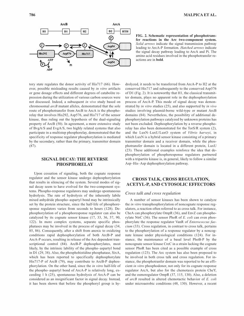
tory state regulates the donor activity of His717 (66). How-ever, possible misleading results caused by in vitro artifactsor gene dosage effects and different degrees of catabolite re-pression during the utilization of various carbon sources werenot discussed. Indeed, a subsequent in vivo study based onchromosomal arcB mutant alleles, demonstrated that the soleroute of phosphotransfer from ArcB to ArcA is the phospho-relay that involves His292, Asp576, and His717 of the sensorkinase, thus ruling out the hypothesis of the dual-signalingproperty of ArcB (50). In agreement, a more extensive studyof BvgA/S and EvgA/S, two highly related systems that alsoparticipate in a multistep phosphorelay, demonstrated that thespecificity of response regulator phosphorylation is mediatedby the secondary, rather than the primary, transmitter domain(87).
SIGNAL DECAY: THE REVERSEPHOSPHORELAY
Upon cessation of signaling, both the cognate responseregulator and the sensor kinase undergo dephosphorylationthat results in silencing of the system. Several modes of sig-nal decay seem to have evolved for the two-component sys-tems. Phospho-response regulators may undergo spontaneoushydrolysis. The rate of hydrolysis of the inherently labilemixed anhydride phospho–aspartyl bond may be intrinsicallyset by the protein structure, since the half-life of phosphore-sponse regulators varies from seconds to hours (128). De-phosphorylation of a phosphoresponse regulator can also becatalyzed by its cognate sensor kinase (17, 33, 36, 57, 90,122). In more complex systems, separate specific phos-phatases may be involved in the process of signal decay (34,85, 86). Consequently, after a shift from anoxic to oxidizingconditions rapid dephosphorylation of both ArcB-P andArcA-P occurs, resulting in release of the Arc dependent tran-scriptional control (84). ArcB-P dephosphorylates, mostlikely, by the intrinsic lability of the phospho–aspartyl bondin D1 (29, 38). Also, the phosphohistidine phosphatase, SixA,which has been reported to specifically dephosphorylateHis717-P of ArcB (79), may contribute to ArcB-P dephos-phorylation. On the other hand, since the in vitro half-life ofthe phospho–aspartyl bond of ArcA-P is relatively long, ex-ceeding 1 h (25), spontaneous hydrolysis of ArcA-P can beconsidered as an insignificant factor in signal decay. Instead,it has been shown that before the phosphoryl group is hy-
786 MALPICA ET AL.
drolyzed, it needs to be transferred from ArcA-P to H2 at theconserved His717 and subsequently to the conserved Asp576of D1 (Fig. 2). It is noteworthy that H1, the classical transmit-ter domain, plays no apparent role in the dephosphorylationprocess of ArcA-P. This mode of signal decay was demon-strated by in vitro studies (25), and also supported by in vivostudies involving plasmid-borne wild-type or mutant ArcBdomains (84). Nevertheless, the possibility of additional de-phosphorylation pathways catalyzed by unknown proteins hasnot been excluded. Dephosphorylation by a reverse phospho-relay has also been demonstrated for the TorS/R system (2),and the LuxN–LuxU/LuxO system of Vibrio harveyi, inwhich LuxN is a hybrid sensor kinase consisting of a primarytransmitter domain and a receiver domain, while the phos-photransfer domain is located in a different protein, LuxU(23). These additional examples reinforce the idea that de-phosphorylation of phosphoresponse regulators partneredwith a tripartite kinase is, in general, likely to follow a similarAsp–His–Asp dephosphorylation pathway.
CROSS TALK, CROSS REGULATION,ACETYL-P, AND CYTOSOLIC EFFECTORS
Cross talk and cross regulation
A number of sensor kinases has been shown to catalyzethe in vitro transphosphorylation of noncognate response reg-ulators, a reaction often referred to as cross talk. For instance,CheA can phosphorylate OmpR (36), and EnvZ can phospho-rylate NtrC (36). The sensor PhoR of E. coli can even phos-phorylate the response regulator VanR of Enterococcus fae-cium (33). Cross regulation, in contrast to cross talk, pertainsto the phosphorylation of a response regulator by a noncog-nate kinase under physiological conditions (124). For in-stance, the maintenance of a basal level PhoB-P by thenoncognate sensor kinase CreC in a strain lacking the cognatesensor PhoR has been cited as a possible example of crossregulation (123). The Arc system has also been proposed tobe involved in both cross talk and cross regulation. For in-stance, the phosphotranfer domain was reported to be an effi-cient in vitro phosphodonor, not only for its cognate responseregulator ArcA, but also for the chemotaxis protein CheY,and the osmoregulator OmpR (37, 115, 130). Also, a deletionof arcB resulted in altered chemotactic behavior of E. coliunder microaerobic conditions (48, 130). However, a recent
FIG. 2. Schematic representation of phosphotrans-fer reactions in the Arc two-component system.Solid arrows indicate the signal transmission pathwayleading to ArcA-P formation. Hatched arrows indicatethe signal decay pathway leading to ArcA and Pi. Theamino acid residues involved in the phosphotransfer re-actions are in bold.
14260c09.pgs 6/13/06 3:40 PM Page 786

in vitro study where the proteins of all two-component sys-tems were examined for cross talk demonstrated that ArcB isnot able to transphosphorylate CheY or OmpR (132). On theother hand, the same study demonstrated that ArcB doestransphosphorylate the orphan noncognate response regulatorRssB. Indeed, a recent study provided genetic and biochemi-cal evidence that ArcB is able to directly phosphorylate RssB,and thereby stimulate �s proteolysis (72). Yet another studysuggested that His717 received the phosphoryl group from anunknown sensor kinase (37). However, when the chromoso-mal arcB allele was replaced by either a full-length arcB mu-tated at H292 and D576 or when the phosphotranfer domain(arcB638–778) was expressed from a low-copy-number plasmidin a �arcB background, an arcB-null phenotype was found.In contrast, when the same plasmid (expressing arcB638–778)was tested in an arcB1–661 background (lacking the phospho-transfer domain), an arcB+ phenotype was obtained, indicat-ing that the phosphorylation of ArcA via His717 dependedsolely on the presence of His292 and Asp576 (50). These ap-parent contradicting observations emphasize the need ofextra caution when results from in vitro experiments usingpurified proteins, and in vivo complementation experimentsusing plasmid-borne alleles are interpreted.
Acetyl-P
ArcA has been found to catalyze its own phosphorylation,in vitro and in vivo, at the expense of the high-energy phos-phate compound acetyl phosphate (62, 84), like most otherresponse regulators (18, 19, 21, 59, 92). In all cases, the invivo evidence comes from studies using mutant strains lack-ing the sensor kinase. Also, it is of interest to note that theresidue of phosphorylation is the very same conserved Aspthat serves as the phosphoryl group acceptor in the transphos-phorylation reaction catalyzed by the sensor kinase. It hasbeen suggested that acetyl-P contributes to maintain and/oraugment the basal level of many response regulators in thephosphorylated state. But this pivotal intermediate, contain-ing carbon, phosphorus, and a high energy bond, may alsosignal global metabolic conditions (69). Such a hypothesis isattractive in view of the finding that acetyl-P levels in the cellcan vary from <0.04 mM (mid-exponential growth on glyc-erol) to 1.2 mM (mid-exponential growth on pyruvate) (68).However, this ArcA phosphorylation route does not seem tobe significant under physiological conditions, as it has beenclearly demonstrated that the phosphatase activity of ArcBwill nullify the acetyl-phosphate-dependent phosphorylationof ArcA. Therefore, the relative contribution of acetyl-Pmight be significantly overestimated in the absence of thesensor kinase.
Effectors
An advantage of the elaborate structure and complexphosphotransfer mechanism in tripartite sensor kinases maybe the availability of different check points that could allowvarious inputs to be integrated in a multilevel control mecha-nism that fine-tunes their signaling activity. ArcB may repre-sent an example for such a mechanism, as an early study sug-gested that the presence of the fermentative metabolitesD-lactate, acetate, and pyruvate enhance the in vitro level of
SIGNALING BY THE ARC TWO-COMPONENT SYSTEM 787
net ArcB phosphorylation, and impede the rate of Pi releasefrom the protein (38). Because the intracellular levels of thesecompounds are elevated during anoxia, it was suggested thatthey could serve as input signals by inhibiting the autophos-phatase activity of ArcB (38).
A more recent study with modular ArcB units, however,failed to confirm any inhibiting effect of these metabolites onthe various steps of the dephosphorylation pathway (26). Onthe other hand, the same study confirmed that D-lactate hasan enhancing effect on the in vitro rate of ArcB autophos-phorylation. Although the reaction mechanism by which D-lactate modulates the activity of ArcB remains unknown, it isworth mentioning that the ArcB receiver domain (D1), evenwhen it is catalytically inactive (e.g., Asp576 mutated to Ala)is indispensable for D-lactate to exert its effect in vitro (26).For that reason, it was suggested that either the effector bind-ing-site is located in D1 or the structural presence of D1 en-ables the effector to bind to the primary transmitter domainof ArcB (H1). A subsequent study provided further in vivoevidence that D-lactate plays a physiological role as an al-losteric effector that is able to accelerate, but not to activatethe kinase activity of ArcB (91). It was therefore proposedthat effector binding on ArcB might cause a conformationalchange that results in increasing the Vmax of the autophos-phorylation reaction (91). Most importantly, these resultsruled out the proposal that D-lactate might serve as the ac-tual signal through which ArcB senses anaerobic environ-ments in vivo (38).
IDENTIFICATION OF THE SIGNAL
It has been postulated that ArcB becomes progressively ac-tivated as a kinase during transition from aerobic to anaerobicgrowth (114). Consequently the concentration of phosphory-lated ArcA increases, resulting in activation or repression ofits target operons. Although it is apparent that the level of en-zymes controlled by the Arc system varies depending on theoxygen availability to the cell, two lines of evidence demon-strated that molecular oxygen is not the direct signal or stimu-lus for ArcB. First, it was shown that deletion of cyoABCDE(encoding the low oxygen affinity terminal oxidase), cydAB(encoding the high oxygen affinity terminal oxidase) or both,strongly repressed a �(cyo-lacZ) reporter fusion (cyo is re-pressed by ArcA-P) (40), indicating that the Arc system is op-erative in the presence of O2. Second, during anaerobic respi-ratory growth, the ArcA-P-dependent repression of the�(sdh-lacZ) reporter (sdh is repressed by ArcA-P) was liftedby supplementation of an electron acceptor, such as nitrate orfumarate. Interestingly, the degree of derepression was pro-portional to the oxidizing power (midpoint potential) of thesupplemented electron acceptor (42). Therefore, it was sug-gested that ArcB indirectly senses O2 tension by monitoringthe level of a metabolite that can exist physiologically in eitheran oxidized or reduced form, and the membrane-locatedquinone electron carriers were proposed as possible candi-dates (42). E. coli can synthesize three different quinones,ubiquinone (Q), menaquinone (MK), and demethyl-menaquinone (DMK), which function as adapters betweenvarious electron-donating and electron-accepting enzyme
14260c09.pgs 6/13/06 3:40 PM Page 787

complexes (32, 71, 116). Ubiquinone is predominantly usedduring aerobic respiration and during anaerobic respirationwith nitrate as the electron acceptor, while menaquinone anddemethylmenaquinone serve as the adaptors for anaerobic res-piration (24). Indeed, a recent study demonstrated that thequinones do signal the redox conditions to ArcB (27). The sur-prising finding, however, was that the quinones in their re-duced form did not convey a kinase activating signal but intheir oxidized form did convey a kinase inhibiting signal (Fig.3). This was demonstrated in vitro, by testing the effect of thesoluble analogs ubiquinone-0 (Q0) and menadione (MK3) onthe autophosphorylation rate of the purified ArcB cytoplasmicportion. In this regard, it has to be mentioned that the use ofthe soluble portion of ArcB was prompted by the previous dis-covery that the transmembrane domain of this sensor does notparticipate in signal reception (52). In vivo support for the in-volvement of quinones in Arc signaling was provided by theobservation that a �(cyd–lacZ) reporter (cydAB is activated byArcA-P) in an ubiCA mutant that is blocked in the synthesis ofthe Q precursor 3-octaprenyl-4-hydroxybenzoate (103) wasactivated during aerobic growth (27). This finding can readilyexplain the earlier observation that in a mutant lacking boththe cytochrome bo and cytochrome bd terminal oxidases, theArc system is operative during aerobiosis (40). In such a mu-tant, the pool of Q should be trapped in its reduced form de-spite the oxidizing growth conditions. Because reducedquinones are unable to inhibit the kinase activity of ArcB, thelevel of ArcA-P should rise and accordingly alter the expres-sion of the target operons. It is important to mention that Qand MK account for 60% and 3% of the total aerobic quinonesand the corresponding values when cells are grown anaerobi-cally on fumarate are 10% and 74%, respectively. The rest ofthe quinone pool is made up by demethylmenaquinone(DMK), whose effect on the activity of ArcB has not yet been
788 MALPICA ET AL.
tested. These facts together with the finding that the half-maximal inhibition of ArcB phosphorylation by Q0 occurredat about 5 µM, whereas that by MK3 occurred at about 50 µM(27) provide an explanation for the observation that duringanaerobic respiratory growth ArcB is a partially active kinaseand that its level of activity correlates with the midpoint po-tential of the alternative electron acceptor (28, 42). Sensingthe quinone pool is not restricted to the ArcB sensor, as the un-orthodox histidine kinases BvgS and EvgS of Bordetella per-tussis and E. coli, respectively, were shown to be responsive tooxidized ubiquinone-0 (Q-0) in vitro (7). Finally, it is worthmentioning that studies directed towards the discovery of thesignal may have been hampered by the fact that the ArcB sen-sor kinase is not activated but rather silenced by the actual sig-nal. Therefore, searches for an activating signal, which hasbeen at least our pursuit, were condemned to be fruitless.
SOLVING THE MECHANISM FOR ARCB SILENCING
Recently, the mechanism for quinone-dependent silencingof the ArcB kinase was clarified (63). Various observations inour laboratory provided a number of indications regardingthis mechanism. First, the purified ArcB78–778, lacking thetransmembrane domain, exhibited a kinase activity that wasinhibited by Q0, even when purified under denaturing condi-tions. Thus, no prosthetic groups could be present in thepreparation of ArcB under denaturing conditions, indicatingthat they are not necessary for the regulation of the kinase (B.Franco, unpublished data). Second, the kinase activity of thisprotein was inhibited not only by Q0 but also by chloramineT. Because the chemical structures of chloramine T and Q0
FIG. 3. A simplified model for ArcB inactivation. Upon a shift from anaerobic to aerobic conditions of growth the quinonepool shifts to its oxidized state. This allows the electron transfer from the Cys180 of ArcB to quinones leading to the formation ofan intermolecular disulfide-bond between the Cys180 of two monomers, which results in a significant reduction of the kinase ac-tivity of ArcB. As the electrons rapidly flow towards O2 via cytochrome bd or bo oxidase, the quinone pool maintains its oxidizedstate and induces the formation of a second disulfide bond between the two Cys241, resulting in the complete silencing of theArcB kinase activity. Adapted from Ref. (63).
14260c09.pgs 6/13/06 3:40 PM Page 788

are different, and chloramine T specifically oxidizes cysteineand methionine residues of proteins, it was postulated thatArcB is sensitive to oxidation rather than to the allostericbinding of Q0 (63). Third, the presence of the membrane-permeating reductants dithiothreitol (DTT) and ß-mercap-toethanol in the culture medium of aerobic grown cells didactivate ArcB, whereas glutathione (GSH), a reducing agentnot able to permeate the plasma membrane because of itslipophobic nature, did not. Because all the above and takinginto account that ArcB possesses a cytoplasm-located signalreception-site (52), and it has two unique cysteine-residues(Cys180 and Cys241) in the linker region, it was postulatedthat ArcB might respond to changes in redox conditionsthrough a thiol redox mechanism (63). Cysteine residues areuniquely suited to sensing a range of redox signals becausethe thiol side-chain can be oxidized to several different redoxstates, many of which are readily reversible. Indeed, both invivo and in vitro experiments demonstrated that both cysteineresidues are able to undergo oxidation and to form inter-molecular disulfide bonds between two ArcB monomers.Also it was shown that oxidation of Cys180 contributes to~85% of ArcB kinase inhibition, whereas oxidation of Cys241contributes to only ~15%. Therefore, it was concluded thatregulation of the ArcB kinase activity requires Cys180, and toa lesser extent Cys241 (Fig. 3). The evolution of such a mech-anism involving intermolecular disulfide bond formationwould require coadaptation of a signal receptor protein tofunction as a dimer to promote the proximity of the cysteineresidues. A possibility remains that the putative leucine zipperis in fact functional and serves as a dimerization domain.
Interestingly, in vivo and in vitro analyses indicated thatmolecular oxygen and hydrogen peroxide (H2O2) do not pro-mote disulfide bond formation in ArcB (63), despite that theirE�° values, +816 mV for O2 and +295 mV for H2O2, are muchhigher than the E�° values of Q0 (+162 mV) and MK3 (± 0mV). This, in turn, suggests that the specificity of the reactionis provided at the level of interaction between ArcB and theoxidizing molecule, whose structure seems to be of central im-portance for the reaction. Recently several quinone analoguescovering a broad range of redox potential values were testedfor their effect on the [�32P]ATP-dependent phosphorylationof ArcB (unpublished results of our laboratory). The mole-cules tested included chloramine T (E�° = +900 mV), and thequinone analogues Q0 (E�° = +162 mV), junglone (E�° = +30mV), MK3 (E�° = ± 0 mV), plumbagine (E�° = �39 mV) andlawsone (E�° = �152 mV). We observed that chloramine T,Q0, and junglone were very effective inhibitors of the kinaseactivity of ArcB, whereas MK3 was less effective in inhibitingArcB phosphorylation. In contrast, plumbagine and lawsonewere without effect. Therefore, it is important to point out thatthe aromatic ring is the only common feature in the molecularstructure of all inhibiting molecules. While chloramine T andQ0 consist of only one aromatic ring, junglone and MK3,whose structure is more reminiscent to plumbagine and law-sone, consist of two. Therefore, it appears that the redox po-tential of the inhibiting molecule is of chief importance, andalso that the quinone binding-pocket of ArcB is able to accom-modate both these molecular structures.
An interesting question is how do the membrane-embedded quinones communicate with the two cytosol-
SIGNALING BY THE ARC TWO-COMPONENT SYSTEM 789
located cysteine residues? Although, no answer has been pro-vided so far, it is of interest to note that quinones have beenshown to efficiently oxidize the periplasmic located cysteineresidues of DsbB, a membrane-bound protein that is a centralcomponent in thiol oxidation reactions (4). Also, these thiol-based redox sensing mechanisms raise the intriguing questionof how a stable disulfide can form in the reducing environ-ment of the cytoplasm. Presumably, ArcB is not in redoxequilibrium with the rest of the cytoplasm. Such a mechanismwould not be unique for ArcB, as it has been demonstratedthat RegB kinase inhibition during aerobiosis involves an in-termolecular disulfide bond formation that renders an inac-tive RegB tetramer (111). Also, a number of cytoplasmic pro-teins that are regulated by reversible disulfide bond formationare known, including the transcriptional factors Yap1p in Sac-charomyces cerevisiae, OxyR and the chaperone Hsp33 inEscherichia coli, and the CtrJ in Rhodobacter capsulatus (45,49, 65, 134).
Inhibition of the kinase activity of ArcB through disulfidebond formation could also provide a logical explanation tothe previous finding that regulation of the catalytic activity ofArcB is set by rotational movements that alter the orientationof the cytosolic portion of one monomer relative to the otherin the homodimer (51). This conclusion was based on the sig-naling activity of various Tar–ArcB chimeras in which thetransmembrane domain of the Tar sensor protein is fused tothe cytosolic portion of the ArcB sensor kinase. The trans-membrane domain of Tar forms a stable dimer in the cyto-plasmic membrane (94), and therefore the relative orientationbetween the two cytosolic ArcB monomers in a dimer was al-tered by fusing a set of Tar transmembrane domains with pro-gressively extended C-terminal region to a constant cytosolicportion of ArcB. Since the rigid helical structure of the sec-ond transmembrane segment of Tar protrudes into the cyto-plasm, each extra residue of Tar added at the Tar–ArcB junc-tion rotates the attached ArcB kinase by approximately 100°.The reported Tar–ArcB chimeras presented three differentphenotypes: a redox-regulated, a constitutively active, and acatalytically inactive ArcB kinase. The relative orientation ofthe kinase moieties in the redox regulated chimera, mostlikely represent that of the wild-type ArcB. In this conforma-tion it is expected that Cys180 and Cys241 are located on thesame side on the structure of the monomer and that they facethe ones of the other monomer. A 100° rotation of the ArcBmonomers relative to each other will place the cysteines al-most at the opposite sides, rendering disulfide bond forma-tion practically impossible, and therefore resulting in a con-stitutively active kinase. Rotation with additional 100° resultsin a catalytically inactive chimera, even though no disulfidebond formation is expected to be able to occur. A possible ex-planation may be that this conformation distorts the structureof the dimer in a way that one or more steps of the phosphore-lay from ArcB to ArcA is blocked.
It is of interest to point out that both Cys180 and Cys241are conserved in the ArcB homologues of different bacterialspecies except for the one of H. influenzae, which lacks al-most the entire linker region, and therefore the two conservedcysteine residues (28, 64). However, in contrast to most otherhomologues that possess only two cysteine residues, ArcB ofH. influenzae possesses five cysteine residues that are located
14260c09.pgs 6/13/06 3:40 PM Page 789

at positions 37, 268, 472, 574, and 596 of the protein. Moredetailed characterization of the H. influenzae ArcB sensor ki-nase might help to determine additional functional roles ofthe linker region, present in E. coli ArcB but not in H. in-fluenzae ArcB. Also, the H. influenzae ArcB may respond to asignal other than the oxidized forms of the quinones and/or itmight represent a different mechanism for redox regulation.
CONCLUDING REMARKS
Recent advances have contributed significantly to under-standing several aspects of the ArcA/B signal transduction,and a more complete picture of this system starts to emerge. Itis now known that the ArcB sensor kinase is kept inactive dur-ing aerobiosis through oxidation of two cytosol-located redox-active cysteine residues that participate in intermoleculardisulfide bond formation (63), a reaction in which thequinones provide the source of oxidative power (27). However,under these conditions, ArcB is active as a specific ArcA-Pphosphatase, in a reaction that occurs via an Asp54 → His717→ Asp576 → Pi phosphorelay (25, 84). Hence, this specificArcA-P phosphatase activity should eliminate the effect ofany inappropriate cross reactivity between ArcA and anoncognate sensor kinase, and also nullify the acetyl-phos-phate dependent autophosphorylation of ArcA. During anaer-obiosis, the inhibitory signals are absent and ArcB autophos-phorylates at the expense of ATP, a reaction that has beenshown to be enhanced by certain anaerobic metabolites suchas D-lactate, acetate, and pyruvate (26, 38, 91), and transphos-photylates its cognate response regulator ArcA via a His292→ Asp576 → His717 → Asp54 phosphorelay (29, 50). Phos-phorylated ArcA is thereby activated as a transcriptional regu-lator of some 300 target operons (42, 56, 96). Despite theprogress made, we are still far from understanding many keyaspects of this signal transduction pathway. For example, it iscurious that the conserved Asp576 and His717 residues partic-ipate in both the forward and reverse phosphorelay. Thus, itwould be useful to find out the implements that control the di-rection of the phosphoryl group transfers and/or influence theindividual transfer rates with regard to signal transmission andsignal decay. Furthermore, a pivotal link in the pathways forArc signaling that also remains elusive is how do the disulfidebonds become reduced for ArcB reactivation upon entry toanaerobiosis? Another challenge will be to solve the crystalstructure of ArcB as this will provide valuable information notonly for the steps of phosphoryl group transfer between thevarious modules of ArcB but most importantly for the mode ofcommunication of the cytosol-located cysteine residues of thesensor with the membrane embedded quinone electron carri-ers. These studies could provide further mechanistic insightinto the operation of this fascinating signal transduction sys-tem that might also facilitate the understanding of other signaltransduction systems involving tripartite sensor kinases.
ACKNOWLEDGMENTS
The authors thank J. Aguirre and B. Michel for criticallyreading the manuscript and R. Hengge for sharing data before
790 MALPICA ET AL.
public release. Research in our laboratory is funded byCONACyT and by DGAPA-PAPIIT, UNAM.
ABBREVIATIONS
Arc, anoxic redox control; DMK, demethylmenaquinone;DTT, dithiothreitol; E�°, standard reduction potential; GSH,glutathione; MK3, menadione; MK, menaquinone; NAD,nicotinamide adenine dinucleotide; PAS, acronym of periodclock protein (PER)–aryl hydrocarbon receptor nucleartranslocator (ARNT)–single-minded protein (SIM); PMF,proton motive force; TCA, tricarboxylic acid cycle; TM,transmembrane; Q, ubiquinone; Q0, ubiquinone-0.
REFERENCES
1. Abel T and Maniatis T. Gene regulation. Action of leucinezippers. Nature 341: 24–25, 1989.
2. Ansaldi M, Jourlin-Castelli C, Lepelletier M, Theraulaz L,and Mejean V. Rapid dephosphorylation of the TorR re-sponse regulator by the TorS unorthodox sensor in Es-cherichia coli. J Bacteriol 183: 2691–2695, 2001.
3. Arico B, Miller JF, Roy C, Stibitz S, Monack D, Falkow S,Gross R, and Rappuoli R. Sequences required for expres-sion of Bordetella pertussis virulence factors share homol-ogy with prokaryotic signal transduction proteins. ProcNatl Acad Sci USA 86: 6671–6675, 1989.
4. Bader M, Muse W, Ballou DP, Gassner C, and BardwellJC. Oxidative protein folding is driven by the electrontransport system. Cell 98: 217–227, 1999.
5. Barnabas J, Schwartz RM, and Dayhoff MO. Evolution ofmajor metabolic innovations in the precambrian. Orig Life12: 81–91, 1982.
6. Bilwes AM, Alex LA, Crane BR, and Simon MI. Structureof CheA, a signal-transducing histidine kinase. Cell 96:131–141, 1999.
7. Bock A and Gross R. The unorthodox histidine kinases BvgSand EvgS are responsive to the oxidation status of a quinoneelectron carrier. Eur J Biochem 269: 3479–3484, 2002.
8. Bogachev AV, Murtazina RA, and Skulachev VP. Cy-tochrome d induction in Escherichia coli growing underunfavorable conditions. FEBS Lett 336: 75–78, 1993.
9. Brencic A, Xia Q, and Winans SC. VirA of Agrobacteriumtumefaciens is an intradimer transphosphorylase and canactively block vir gene expression in the absence of pheno-lic signals. Mol Microbiol 52: 1349–1362, 2004.
10. Buckland R and Wild F. Leucine zipper motif extends. Na-ture 338: 547, 1989.
11. Buxton RS and Drury LS. Identification of the dye geneproduct, mutational loss of which alters envelope proteincomposition and also affects sex factor F expression in Es-cherichia coli K-12. Mol Gen Genet 194: 241–247, 1984
12. Buxton RS, Drury LS, and Curtis CA. Dye sensitivity cor-related with envelope protein changes in dye (sfrA) mu-tants of Escherichia coli K12 defective in the expression ofthe sex factor F. J Gen Microbiol 129: 3363–3370, 1983.
13. Böck A and Sawers G. Fermentation. In: Escherichia coliand Salmonella typhimurium: Cellular and Molecular Bi-
14260c09.pgs 6/13/06 3:40 PM Page 790
ology, Neidhardt FC, Ingraham JL, Lin EC, Low KB, Ma-gasanik B, Schaechter M, and Umbarger HE (eds.). Wash-ington, DC: ASM Press, 1996, pp. 262–282.
14. Colloms SD, Alen C, and Sherratt DJ. The ArcA/ArcBtwo-component regulatory system of Escherichia coli isessential for Xer site-specific recombination at psi. MolMicrobiol 28: 521–530, 1998.
15. Compan I and Touati D. Anaerobic activation of arcA tran-scription in Escherichia coli: roles of Fnr and ArcA. MolMicrobiol 11: 955–964, 1994.
16. Cotter PA, Chepuri V, Gennis RB, and Gunsalus RP. Cy-tochrome o (cyoABCDE) and d (cydAB) oxidase gene ex-pression in Escherichia coli is regulated by oxygen, pH, andthe fnr gene product. J Bacteriol 172: 6333–6338, 1990.
17. Dahl MK, Msadek T, Kunst F, and Rapoport G. The phos-phorylation state of the DegU response regulator acts as amolecular switch allowing either degradative enzyme syn-thesis or expression of genetic competence in Bacillussubtilis. J Biol Chem 267: 14509–14514, 1992.
18. Dailey FE and Berg HC. Change in direction of flagellarrotation in Escherichia coli mediated by acetate kinase. JBacteriol 175: 3236–3239, 1993.
19. Danese PN, Snyder WB, Cosma CL, Davis LJ, and SilhavyTJ. The Cpx two-component signal transduction pathwayof Escherichia coli regulates transcription of the genespecifying the stress-inducible periplasmic protease, DegP.Genes Dev 9: 387–398, 1995.
20. De Souza-Hart JA, Blackstock W, Di Modugno V, HollandIB, and Kok M. Two-component systems in Haemophilusinfluenzae: a regulatory role for ArcA in serum resistance.Infect Immun 71: 163–172, 2003.
21. Deretic V, Leveau JH, Mohr CD, and Hibler NS. In vitrophosphorylation of AlgR, a regulator of mucoidy in Pseu-domonas aeruginosa, by a histidine protein kinase and ef-fects of small phospho-donor molecules. Mol Microbiol 6:2761–2657, 1992.
22. Drury LS and Buxton RS. DNA sequence analysis of thedye gene of Escherichia coli reveals amino acid homologybetween the dye and OmpR proteins. J Biol Chem 260:4236–4242, 1985.
23. Freeman JA, Lilley BN, and Bassler BL. A genetic analy-sis of the functions of LuxN: a two-component hybrid sen-sor kinase that regulates quorum sensing in Vibrio harveyi.Mol Microbiol 35: 139–149, 2000.
24. Gennis RB and Stewart V. Respiration. In: Escherichia coliand Salmonella typhimurium: Cellular and Molecular Bi-ology, Neidhardt FC, Ingraham JL, Lin EC, Low KB, Ma-gasanik B, Schaechter M and Umbarger HE (eds.). Wash-ington, D.C.: ASM Press, 1996, pp. 217–261.
25. Georgellis D, Kwon O, De Wulf P, and Lin EC. Signaldecay through a reverse phosphorelay in the Arc two-component signal transduction system. J Biol Chem 273:32864–32869, 1998.
26. Georgellis D, Kwon O, and Lin EC. Amplification of sig-naling activity of the arc two-component system of Es-cherichia coli by anaerobic metabolites. An in vitro studywith different protein modules. J Biol Chem 274: 35950–35954, 1999.
27. Georgellis D, Kwon O, and Lin EC. Quinones as the redoxsignal for the arc two-component system of bacteria. Sci-ence 292: 2314–2316, 2001.
SIGNALING BY THE ARC TWO-COMPONENT SYSTEM 791
28. Georgellis D, Kwon O, Lin EC, Wong SM, and Akerley BJ.Redox signal transduction by the ArcB sensor kinase ofHaemophilus influenzae lacking the PAS domain. J Bacte-riol 183: 7206–7212, 2001.
29. Georgellis D, Lynch AS, and Lin EC. In vitro phosphoryla-tion study of the arc two-component signal transduction sys-tem of Escherichia coli. J Bacteriol 179: 5429–5435, 1997.
30. Gray CT, Wimpenny JW, Hughes DE, and Mossman MR.Regulation of metabolism in facultative bacteria. I. Struc-tural and functional changes in Escherichia coli associatedwith shifts between the aerobic and anaerobic states.Biochim Biophys Acta 117: 22–32, 1966.
31. Gray CT, Wimpenny JW, and Mossman MR. Regulation ofmetabolism in facultative bacteria. II. Effects of aerobio-sis, anaerobiosis and nutrition on the formation of Krebscycle enzymes in Escherichia coli. Biochim Biophys Acta117: 33–41, 1966.
32. Guest JR and Shaw DJ. Molecular cloning of menaquinonebiosynthetic genes of Escherichia coli K12. Mol GenGenet 181: 379–383, 1981.
33. Haldimann A, Fisher SL, Daniels LL, Walsh CT, and Wan-ner BL. Transcriptional regulation of the Enterococcusfaecium BM4147 vancomycin resistance gene cluster bythe VanS–VanR two-component regulatory system in Es-cherichia coli K-12. J Bacteriol 179: 5903–5913, 1997.
34. Hess JF, Oosawa K, Kaplan N, and Simon MI. Phosphory-lation of three proteins in the signaling pathway of bacter-ial chemotaxis. Cell 53: 79–87, 1988.
35. Hrabak EM and Willis DK. The lemA gene required forpathogenicity of Pseudomonas syringae pv. syringae onbean is a member of a family of two-component regula-tors. J Bacteriol 174: 3011–3020, 1992.
36. Igo MM, Ninfa AJ, Stock JB, and Silhavy TJ. Phosphoryla-tion and dephosphorylation of a bacterial transcriptionalactivator by a transmembrane receptor. Genes Dev 3:1725–1734, 1989.
37. Ishige K, Nagasawa S, Tokishita S, and Mizuno T. A noveldevice of bacterial signal transducers. EMBO J 13: 5195–5202, 1994.
38. Iuchi S. Phosphorylation/dephosphorylation of the re-ceiver module at the conserved aspartate residue controlstransphosphorylation activity of histidine kinase in sensorprotein ArcB of Escherichia coli. J Biol Chem 268:23972–23980, 1993.
39. Iuchi S, Cameron DC, and Lin EC. A second global regu-lator gene (arcB) mediating repression of enzymes in aero-bic pathways of Escherichia coli. J Bacteriol 171: 868–873, 1989.
40. Iuchi S, Chepuri V, Fu HA, Gennis RB, and Lin EC. Re-quirement for terminal cytochromes in generation of theaerobic signal for the arc regulatory system in Escherichiacoli: study utilizing deletions and lac fusions of cyo andcyd. J Bacteriol 172: 6020–6025, 1990.
41. Iuchi S, Furlong D, and Lin EC. Differentiation of arcA,arcB, and cpxA mutant phenotypes of Escherichia coli bysex pilus formation and enzyme regulation. J Bacteriol171: 2889–2893, 1989.
42. Iuchi S and Lin EC. arcA (dye), a global regulatory gene inEscherichia coli mediating repression of enzymes in aero-bic pathways. Proc Natl Acad Sci USA 85: 1888–1892,1988.
14260c09.pgs 6/13/06 3:40 PM Page 791
43. Iuchi S and Lin EC. Mutational analysis of signal trans-duction by ArcB, a membrane sensor protein responsiblefor anaerobic repression of operons involved in the centralaerobic pathways in Escherichia coli. J Bacteriol 174:3972–3980, 1992.
44. Iuchi S, Matsuda Z, Fujiwara T, and Lin EC. The arcBgene of Escherichia coli encodes a sensor-regulator pro-tein for anaerobic repression of the arc modulon. Mol Mi-crobiol 4: 715–727, 1990.
45. Jakob U, Muse W, Eser M, and Bardwell JC. Chaperoneactivity with a redox switch. Cell 96: 341–352, 1999.
46. Jeon Y, Lee YS, Han JS, Kim JB, and Hwang DS. Multi-merization of phosphorylated and non-phosphorylatedArcA is necessary for the response regulator function ofthe Arc two-component signal transduction system. J BiolChem 276: 40873–40879, 2001.
47. Jeong JY, Kim YJ, Cho N, Shin D, Nam TW, Ryu S, andSeok YJ. Expression of ptsG encoding the major glucosetransporter is regulated by ArcA in Escherichia coli. J BiolChem 279: 38513–38518, 2004.
48. Kato M, Mizuno T, Shimizu T, and Hakoshima T. Insightsinto multistep phosphorelay from the crystal structure ofthe C-terminal HPt domain of ArcB. Cell 88: 717–723,1997.
49. Kuge S, Arita M, Murayama A, Maeta K, Izawa S, Inoue Y,and Nomoto A. Regulation of the yeast Yap1p nuclear ex-port signal is mediated by redox signal-induced reversibledisulfide bond formation. Mol Cell Biol 21: 6139–6150,2001.
50. Kwon O, Georgellis D, and Lin EC. Phosphorelay as thesole physiological route of signal transmission by the arctwo-component system of Escherichia coli. J Bacteriol182: 3858–3862, 2000.
51. Kwon O, Georgellis D, and Lin EC. Rotational on-offswitching of a hybrid membrane sensor kinase Tar-ArcBin Escherichia coli. J Biol Chem 278: 13192–13195,2003.
52. Kwon O, Georgellis D, Lynch AS, Boyd D, and Lin EC.The ArcB sensor kinase of Escherichia coli: genetic explo-ration of the transmembrane region. J Bacteriol 182:2960–2966, 2000.
53. Lambden PR and Guest JR. Mutants of Escherichia coliK12 unable to use fumarate as an anaerobic electron ac-ceptor. J Gen Microbiol 97: 145–160, 1976.
54. Lau PC, Wang Y, Patel A, Labbe D, Bergeron H, BrousseauR, Konishi Y, and Rawlings M. A bacterial basic regionleucine zipper histidine kinase regulating toluene degrada-tion. Proc Natl Acad Sci USA 94: 1453–1458, 1997.
55. Lee YS, Han JS, Jeon Y, and Hwang DS. The arc two-com-ponent signal transduction system inhibits in vitro Es-cherichia coli chromosomal initiation. J Biol Chem 276:9917–9923, 2001.
56. Liu X and De Wulf P. Probing the ArcA-P modulon of Es-cherichia coli by whole genome transcriptional analysisand sequence recognition profiling. J Biol Chem 279:12588–12597, 2004.
57. Lois AF, Weinstein M, Ditta GS, and Helinski DR. Au-tophosphorylation and phosphatase activities of the oxy-gen-sensing protein FixL of Rhizobium meliloti are coor-
792 MALPICA ET AL.
dinately regulated by oxygen. J Biol Chem 268: 4370–4375, 1993.
58. Lu S, Killoran PB, Fang FC, and Riley LW. The global reg-ulator ArcA controls resistance to reactive nitrogen andoxygen intermediates in Salmonella enterica serovar En-teritidis. Infect Immun 70: 451–461, 2002.
59. Lukat GS, McCleary WR, Stock AM, and Stock JB. Phos-phorylation of bacterial response regulator proteins by lowmolecular weight phospho-donors. Proc Natl Acad SciUSA 89: 718–722, 1992.
60. Lynch AS and Lin EC. Responses to molecular oxygen. In:Escherichia coli and Salmonella typhimurium: Cellularand Molecular Biology, Neidhardt FC, Ingraham JL, LinEC, Low KB, Magasanik B, Schaechter M and UmbargerHE (eds.) Washington, DC: ASM Press, 1996, pp. 1526–1538.
61. Lynch AS and Lin EC. Regulation of aerobic and anaero-bic metabolism by the Arc system. In: Regulation of GeneExpression in Escherichia coli, Lin EC, and Lynch AS(eds.). Austin: Chapman and Hall, 1996, pp. 361–381.
62. Lynch AS and Lin EC. Transcriptional control mediated bythe ArcA two-component response regulator protein of Es-cherichia coli: characterization of DNA binding at targetpromoters. J Bacteriol 178: 6238–6249, 1996.
63. Malpica R, Franco B, Rodriguez C, Kwon O, and Georgel-lis D. Identification of a quinone-sensitive redox switch inthe ArcB sensor kinase. Proc Natl Acad Sci USA 101:13318–13323, 2004.
64. Manukhov IV, Bertsova YV, Trofimov DY, Bogachev AV,and Skulachev VP. Analysis of HI0220 protein fromHaemophilus influenzae, a novel structural and functionalanalog of ArcB protein from Escherichia coli. Biochem-istry (Mosc) 65: 1321–1326, 2000.
65. Masuda S, Dong C, Swem D, Setterdahl AT, Knaff DB, andBauer CE. Repression of photosynthesis gene expressionby formation of a disulfide bond in CrtJ. Proc Natl AcadSci USA 99: 7078–7083, 2002.
66. Matsushika A and Mizuno T. A dual-signaling mechanismmediated by the ArcB hybrid sensor kinase containing thehistidine-containing phosphotransfer domain in Es-cherichia coli. J Bacteriol 180: 3973–3977, 1998.
67. Matsushika A and Mizuno T. Characterization of three pu-tative sub-domains in the signal-input domain of the ArcBhybrid sensor in Escherichia coli. J Biochem (Tokyo) 127:855–860, 2000.
68. McCleary WR and Stock JB. Acetyl phosphate and the ac-tivation of two-component response regulators. J BiolChem 269: 31567–31572, 1994.
69. McCleary WR, Stock JB, and Ninfa AJ. Is acetyl phos-phate a global signal in Escherichia coli? J Bacteriol 175:2793–2798, 1993.
70. McGuire AM, De Wulf P, Church GM, and Lin EC. Aweight matrix for binding recognition by the redox-response regulator ArcA-P of Escherichia coli. Mol Micro-biol 32: 219–221, 1999.
71. Meganathan R. Biosynthesis of the isoprenoid quinonesmenaquinone (vitamin K2) and Ubiquinone (coenzymeQ). In: Escherichia coli and Salmonella typhimurium: Cel-lular and Molecular Biology, Neidhardt FC, Ingraham JL,
14260c09.pgs 6/13/06 3:40 PM Page 792
Lin EC, Low KB, Magasanik B, Schaechter M and Um-barger HE (eds.). Washington, D.C.: ASM Press, 1996, pp.642–656.
72. Mika F and Hengge R. A two-component phosphotransfernetwork involving ArcB, ArcA and RssB coordinates syn-thesis and proteolysis of sigmaS in E. coli. Genes Dev 19:2770–2781, 2005.
73. Nagasawa S, Tokishita S, Aiba H, and Mizuno T. A novelsensor-regulator protein that belongs to the homologousfamily of signal-transduction proteins involved in adaptiveresponses in Escherichia coli. Mol Microbiol 6: 799–807,1992.
74. Nambu JR, Lewis JO, Wharton KA, Jr., and Crews ST. TheDrosophila single-minded gene encodes a helix–loop–helix protein that acts as a master regulator of CNS mid-line development. Cell 67: 1157–1167, 1991.
75. Ninfa AJ and Magasanik B. Covalent modification of theglnG product, NRI, by the glnL product, NRII, regulatesthe transcription of the glnALG operon in Escherichiacoli. Proc Natl Acad Sci USA 83: 5909–5913, 1986.
76. Ninfa EG, Atkinson MR, Kamberov ES, and Ninfa AJ.Mechanism of autophosphorylation of Escherichia colinitrogen regulator II (NRII or NtrB): trans-phosphoryla-tion between subunits. J Bacteriol 175: 7024–7032, 1993.
77. Nixon BT, Ronson CW, and Ausubel FM. Two-componentregulatory systems responsive to environmental stimulishare strongly conserved domains with the nitrogen assim-ilation regulatory genes ntrB and ntrC. Proc Natl Acad SciUSA 83: 7850–7854, 1986.
78. Ogasawara H, Teramoto J, Yamamoto S, Hirao K, Ya-mamoto K, Ishihama A, and Utsumi R. Negative regula-tion of DNA repair gene (uvrA) expression by ArcA/ArcBtwo-component system in Escherichia coli. FEMS Micro-biol Lett 251: 243–249, 2005.
79. Ogino T, Matsubara M, Kato N, Nakamura Y, and MizunoT. An Escherichia coli protein that exhibits phosphohisti-dine phosphatase activity towards the HPt domain of theArcB sensor involved in the multistep His-Asp phosphore-lay. Mol Microbiol 27: 573–585, 1998.
80. Papagiannis MD. Life-related aspects of stellar evolution.Orig Life 14: 43–50, 1984.
81. Park H, Saha SK, and Inouye M. Two-domain reconstitu-tion of a functional protein histidine kinase. Proc NatlAcad Sci USA 95: 6728–6732, 1998.
82. Parkinson JS. Signal transduction schemes of bacteria.Cell 73: 857–871, 1993.
83. Parkinson JS and Kofoid EC. Communication modules inbacterial signaling proteins. Annu Rev Genet 26: 71–112,1992.
84. Pena-Sandoval GR, Kwon O, and Georgellis D. Require-ment of the receiver and phosphotransfer domains of ArcBfor efficient dephosphorylation of phosphorylated ArcA invivo. J Bacteriol 187: 3267–3272, 2005.
85. Perego M. A peptide export-import control circuit modu-lating bacterial development regulates protein phos-phatases of the phosphorelay. Proc Natl Acad Sci USA 94:8612–8617, 1997.
86. Perego M, Hanstein C, Welsh KM, Djavakhishvili T,Glaser P, and Hoch JA. Multiple protein-aspartate phos-
SIGNALING BY THE ARC TWO-COMPONENT SYSTEM 793
phatases provide a mechanism for the integration of di-verse signals in the control of development in B. subtilis.Cell 79: 1047–1055, 1994.
87. Perraud AL, Kimmel B, Weiss V, and Gross R. Specificityof the BvgAS and EvgAS phosphorelay is mediated bythe C-terminal HPt domains of the sensor proteins. MolMicrobiol 27: 875–887, 1998.
88. Ponting CP and Aravind L. PAS: a multifunctional domainfamily comes to light. Curr Biol 7: R674–677, 1997.
89. Rabin RS, Collins LA, and Stewart V. In vivo requirementof integration host factor for nar (nitrate reductase)operon expression in Escherichia coli K-12. Proc NatlAcad Sci USA 89: 8701–8705, 1992.
90. Raivio TL and Silhavy TJ. Transduction of envelopestress in Escherichia coli by the Cpx two-component sys-tem. J Bacteriol 179: 7724–7733, 1997.
91. Rodriguez C, Kwon O, and Georgellis D. Effect of D-lactate on the physiological activity of the ArcB sensor ki-nase in Escherichia coli. J Bacteriol 186: 2085–2090,2004.
92. Roggiani M and Dubnau D. ComA, a phosphorylated re-sponse regulator protein of Bacillus subtilis, binds to thepromoter region of srfA. J Bacteriol 175: 3182–3187,1993.
93. Ronson CW, Nixon BT, and Ausubel FM. Conserved do-mains in bacterial regulatory proteins that respond to en-vironmental stimuli. Cell 49: 579–581, 1987.
94. Russo AF and Koshland DE, Jr. Separation of signal trans-duction and adaptation functions of the aspartate receptorin bacterial sensing. Science 220: 1016–1020, 1983.
95. Salmon K, Hung SP, Mekjian K, Baldi P, Hatfield GW,and Gunsalus RP. Global gene expression profiling in Es-cherichia coli K12. The effects of oxygen availability andFNR. J Biol Chem 278: 29837–29855, 2003.
96. Salmon KA, Hung SP, Steffen NR, Krupp R, Baldi P,Hatfield GW, and Gunsalus RP. Global gene expressionprofiling in Escherichia coli K12: effects of oxygenavailability and ArcA. J Biol Chem 280: 15084–15096,2005.
97. Sawers G. Specific transcriptional requirements for posi-tive regulation of the anaerobically inducible pfl operonby ArcA and FNR. Mol Microbiol 10: 737–747, 1993.
98. Sawers G and Suppmann B. Anaerobic induction of pyru-vate formate-lyase gene expression is mediated by theArcA and FNR proteins. J Bacteriol 174: 3474–3478,1992.
99. Sengupta N, Paul K, and Chowdhury R. The global regu-lator ArcA modulates expression of virulence factors inVibrio cholerae. Infect Immun 71: 5583–5589, 2003.
100. Silverman PM, Rother S, and Gaudin H. Arc and Sfrfunctions of the Escherichia coli K-12 arcA gene productare genetically and physiologically separable. J Bacteriol173: 5648–5652, 1991.
101. Silverman PM, Wickersham E, and Harris R. Regulationof the F plasmid traY promoter in Escherichia coli byhost and plasmid factors. J Mol Biol 218: 119–128, 1991.
102. Simon G, Mejean V, Jourlin C, Chippaux M, and PascalMC. The torR gene of Escherichia coli encodes a re-sponse regulator protein involved in the expression of the
14260c09.pgs 6/13/06 3:40 PM Page 793

trimethylamine N-oxide reductase genes. J Bacteriol176: 5601–5606, 1994.
103. Soballe B and Poole RK. Requirement for ubiquinonedownstream of cytochrome(s) b in the oxygen-terminatedrespiratory chains of Escherichia coli K-12 revealedusing a null mutant allele of ubiCA. Microbiology 144(Pt 2): 361–373, 1998.
104. Stevens AM, Sanders JM, Shoemaker NB, and SalyersAA. Genes involved in production of plasmidlike formsby a Bacteroides conjugal chromosomal element shareamino acid homology with two-component regulatorysystems. J Bacteriol 174: 2935–2942, 1992.
105. Stewart V. Biochemical Society Special Lecture. Nitrate-and nitrite-responsive sensors NarX and NarQ of pro-teobacteria. Biochem Soc Trans 31: 1–10, 2003.
106. Stewart V, Parales J Jr., and Merkel SM. Structure ofgenes narL and narX of the nar (nitrate reductase) locusin Escherichia coli K-12. J Bacteriol 171: 2229–2234,1989.
107. Stock AM, Robinson VL, and Goudreau PN. Two-component signal transduction. Annu Rev Biochem 69:183–215, 2000.
108. Stock JB, Surette MG, Levit M and Park P. Two-component Signal Transduction Systems: Structure-Function Relationships and Mechanisms of Catalysis. In:Two-component Signal Transduction, Hoch JA, and Sil-havy TJ (eds.). Washington D.C.: ASM press, 1995, pp.25–51.
109. Swanson RV, Alex LA, and Simon MI. Histidine and as-partate phosphorylation: two-component systems and thelimits of homology. Trends Biochem Sci 19: 485–490,1994.
110. Swanson RV, Bourret RB, and Simon MI. Intermolecularcomplementation of the kinase activity of CheA. Mol Mi-crobiol 8: 435–441, 1993.
111. Swem LR, Kraft BJ, Swem DL, Setterdahl AT, Masuda S,Knaff DB, Zaleski JM, and Bauer CE. Signal transduc-tion by the global regulator RegB is mediated by a redox-active cysteine. EMBO J 22: 4699–4708, 2003.
112. Taylor BL and Zhulin IB. PAS domains: internal sensorsof oxygen, redox potential, and light. Microbiol Mol BiolRev 63: 479–506, 1999.
113. Toro-Roman A, Mack TR, and Stock AM. Structuralanalysis and solution studies of the activated regulatorydomain of the response regulator ArcA: a symmetricdimer mediated by the alpha4-beta5-alpha5 face. J MolBiol 349: 11–26, 2005.
114. Tseng CP, Albrecht J, and Gunsalus RP. Effect of mi-croaerophilic cell growth conditions on expression of theaerobic (cyoABCDE and cydAB) and anaerobic(narGHJI, frdABCD, and dmsABC) respiratory pathwaygenes in Escherichia coli. J Bacteriol 178: 1094–1098,1996.
115. Tsuzuki M, Ishige K, and Mizuno T. Phosphotransfer cir-cuitry of the putative multi-signal transducer, ArcB, ofEscherichia coli: in vitro studies with mutants. Mol Mi-crobiol 18: 953–962, 1995.
116. Unden G. Differential roles for menaquinone anddemethylmenaquinone in anaerobic electron transport of
794 MALPICA ET AL.
E. coli and their fnr-independent expression. Arch Micro-biol 150: 499–503, 1988.
117. Unden G, Achebach S, Holighaus G, Tran HG, WackwitzB, and Zeuner Y. Control of FNR function of Escherichiacoli by O2 and reducing conditions. J Mol MicrobiolBiotechnol 4: 263–268, 2002.
118. Unden G and Bongaerts J. Alternative respiratory path-ways of Escherichia coli: energetics and transcriptionalregulation in response to electron acceptors. BiochimBiophys Acta 1320: 217–234, 1997.
119. Utsumi R, Katayama S, Taniguchi M, Horie T, IkedaM, Igaki S, Nakagawa H, Miwa A, Tanabe H, and NodaM. Newly identified genes involved in the signal trans-duction of Escherichia coli K-12. Gene 140: 73–77,1994.
120. Varughese KI, Madhusudan, Zhou XZ, Whiteley JM, andHoch JA. Formation of a novel four-helix bundle andmolecular recognition sites by dimerization of a responseregulator phosphotransferase. Mol Cell 2: 485–493,1998.
121. Volz K. Structural conservation in the CheY superfamily.Biochemistry 32: 11741–11753, 1993.
122. Walker MS and DeMoss JA. Phosphorylation and de-phosphorylation catalyzed in vitro by purified compo-nents of the nitrate sensing system, NarX and NarL. JBiol Chem 268: 8391–8393, 1993.
123. Wanner BL, Jiang W, Kim, SK, Yamagata S, HaldimannA and Daniels LL. Are the mutliple signal transductionpathways of the Pho regulon due to cross talk or crossregulation? In: Regulation of gene expression in Es-cherichia coli, Lin EC and Lynch AS (eds.). Austin: R. G.: Landes Company, 1996, pp. 297–315.
124. Wanner BL and Wilmes-Riesenberg MR. Involvement ofphosphotransacetylase, acetate kinase, and acetyl phos-phate synthesis in control of the phosphate regulon in Es-cherichia coli. J Bacteriol 174: 2124–2130, 1992.
125. Wilson TH and Lin EC. Evolution of membrane bioener-getics. J Supramol Struct 13: 421–446, 1980.
126. Wolanin PM, Thomason PA, and Stock JB. Histidine pro-tein kinases: key signal transducers outside the animalkingdom. Genome Biol 3: 3013.1–3013.8, 2002.
127. Wolfe AJ and Stewart RC. The short form of the CheAprotein restores kinase activity and chemotactic ability tokinase-deficient mutants. Proc Natl Acad Sci USA 90:1518–1522, 1993.
128. Wright GD, Holman TR, and Walsh CT. Purification andcharacterization of VanR and the cytosolic domain ofVanS: a two-component regulatory system required forvancomycin resistance in Enterococcus faeciumBM4147. Biochemistry 32: 5057–5063, 1993.
129. Xu Q and West AH. Conservation of structure and func-tion among histidine-containing phosphotransfer (HPt)domains as revealed by the crystal structure of YPD1. JMol Biol 292: 1039–1050, 1999.
130. Yaku H, Kato M, Hakoshima T, Tsuzuki M, and MizunoT. Interaction between the CheY response regulator andthe histidine-containing phosphotransfer (HPt) domain ofthe ArcB sensory kinase in Escherichia coli. FEBS Lett408: 337–340, 1997.
14260c09.pgs 6/13/06 3:40 PM Page 794
131. Yaku H and Mizuno T. The membrane-located osmosen-sory kinase, EnvZ, that contains a leucine zipper-likemotif functions as a dimer in Escherichia coli. FEBS Lett417: 409–413, 1997.
132. Yamamoto K, Hirao K, Oshima T, Aiba H, Utsumi R, andIshihama A. Functional characterization in vitro of alltwo-component signal transduction systems from Es-cherichia coli. J Biol Chem 280: 1448–1456, 2005.
133. Yang Y and Inouye M. Intermolecular complementationbetween two defective mutant signal-transducing recep-tors of Escherichia coli. Proc Natl Acad Sci USA 88:11057–11061, 1991.
134. Zheng M, Aslund F, and Storz G. Activation of the OxyRtranscription factor by reversible disulfide bond forma-tion. Science 279: 1718–1721, 1998.
SIGNALING BY THE ARC TWO-COMPONENT SYSTEM 795
135. Zhulin IB, Taylor BL, and Dixon R. PAS domain S-boxesin Archaea, Bacteria and sensors for oxygen and redox.Trends Biochem Sci 22: 331–333, 1997.
Address reprint requests to:Dimitris Georgellis
Instituto de Fisiología CelularUniversidad Nacional Autónoma de México
04510 Mexico City, Mexico
E-mail: [email protected]
Date of first submission to ARS Central, October 25, 2005;date of acceptance, December 2, 2005.
14260c09.pgs 6/13/06 3:40 PM Page 795

14260c09.pgs 6/13/06 3:40 PM Page 796