Phylogeography of Anastrepha obliqua Inferred With mtDNA ...
Upload
khangminh22Category
view
1download
0
Biochimica et Biophysica Acta 1847 (2015) 587–598
Contents lists available at ScienceDirect
Biochimica et Biophysica Acta
j ourna l homepage: www.e lsev ie r .com/ locate /bbab io
brought to you by COREView metadata, citation and similar papers at core.ac.uk
provided by Elsevier - Publisher Connector
Cardiolipin is a key determinant for mtDNA stability and segregationduring mitochondrial stress
Luis Alberto Luévano-Martínez ⁎, Maria Fernanda Forni, Valquiria Tiago dos Santos,Nadja C. Souza-Pinto, Alicia J. KowaltowskiDepartamento de Bioquímica, Instituto de Química, Universidade de São Paulo, Cidade Universitária, Av. Prof. Lineu Prestes, 748, São Paulo, SP 05508-000, Brazil
Abbreviations:NBD-PE, 1,2-oleyl-sn-glycero-3-phosp1,3-benzoxadiazol-4-yl; ANSA, 1-anilino-8-naphthalenhydroxyethyl]-1-piperazineethanesulfonic acid; DAPI, 4FCCP, carbonyl cyanide4-[trifluoromethoxy]phenylhydrazformingunits; EB, ethidiumbromide; FRET,fluorescence regalactose,peptoneandyeast extractmedia;YPD,glucose,pYPEG, glycerol, ethanol, peptone and yeast extract mediaPMSF,phenylmethanesulfonylfluoride;PC,phosphatidylchamine; PG, phosphatidylglycerol; PS, phosphatidylsetetramethyl rhodamine,methyl ester⁎ Corresponding author at: Departamento deBioquímic
Av. Prof. Lineu Prestes, 748, Cidade Universitária, 055Tel.: +55 11 30912922.
E-mail address: [email protected] (L.A. Luévano-Ma
http://dx.doi.org/10.1016/j.bbabio.2015.03.0070005-2728/© 2015 Elsevier B.V. All rights reserved.
a b s t r a c t
a r t i c l e i n f oArticle history:Received 2 January 2015Received in revised form 16 March 2015Accepted 29 March 2015Available online 2 April 2015
Keywords:PhospholipidMitochondrionMitochondrial DNAMembrane plasticity
Mitochondria play a key role in adaptation during stressing situations. Cardiolipin, themain anionic phospholipidinmitochondrial membranes, is expected to be a determinant in this adaptivemechanism since it modulates theactivity of most membrane proteins. Here, we used Saccharomyces cerevisiae subjected to conditions that affectmitochondrial metabolism as a model to determine the possible role of cardiolipin in stress adaptation. Interest-ingly, we found that thermal stress promotes a 30% increase in the cardiolipin content and modifies the physicalstate of mitochondrial membranes. These changes have effects on mtDNA stability, adapting cells to thermalstress. Conversely, this effect is cardiolipin-dependent since a cardiolipin synthase-null mutant strain is unableto adapt to thermal stress as observed by a 60% increase of cells lacking mtDNA (ρ0). Interestingly, we foundthat the loss of cardiolipin specifically affects the segregation ofmtDNA to daughter cells, leading to a respiratorydeficient phenotype after replication.We also provide evidence that mtDNA physically interacts with cardiolipinboth in S. cerevisiae and in mammalian mitochondria. Overall, our results demonstrate that the mitochondriallipid cardiolipin is a key determinant in the maintenance of mtDNA stability and segregation.
© 2015 Elsevier B.V. All rights reserved.
1. Introduction
Phospholipids are components of all cellular structures. Most, in-cluding PC, are synthesized in the lumen of the endoplasmic reticulumand subsequently distributed intracellularly [1]. Non-bilayer phospho-lipids such as CL and PE are synthesized mainly in mitochondria,where they also exert their functions [2]. After synthesis, PE is distribut-ed through all cellular membranes, where it exerts diverse functionssuch as autophagy [3] or signal transduction [4].
CL is the main anionic phospholipid in mitochondrial membranes,where it constitutes about 15% of the total phospholipid content. It issynthesized in the inner leaflet of the inner membrane and remains
hoethanolamine-N-7-nitro-2-esulfonic acid; HEPES, 4-[2-′,6-diamidino-2-phenylindole;one;CL, cardiolipin;CFU, colonysonanceenergy transfer;YPGal,eptoneandyeast extractmedia;; mtDNA, mitochondrial DNA;oline;PE,phosphatidylethanol-rine; WT, wild type; TMRM,
a, IQ, Universidade deSão Paulo,08-900 São Paulo, SP, Brazil.
rtínez).
within mitochondria, and distributed throughout the inner membrane[5], where it confers the negative surface charge necessary to maintaina localized proton motive force, used to synthesize ATP [6]. Further-more, CL participates in the biogenesis and assembly of respiratorychain components and severalmetabolite transporters [7]. Functionally,CL activates the adenine nucleotide translocator (ANT) and uncouplingprotein 1 (UCP1; [8,9]).
Additionally, CL stabilizes F1FO-ATP synthase dimers [10] and respi-ratory chain supercomplexes [11], affecting mitochondrial morphology[12]. CL normally adopts a bilayer conformation, but when calcium ispresent in the interface, it assumes a conical inverted shape due to thelow hydration of the small polar headgroup [13]. Interestingly, it hasbeen recently demonstrated that CL is able to respond to the chemicalcomponent of the proton motive force, promoting the formation ofcristae-like structures in the innermembrane [14], an effect that resem-bles that of calcium on CL-containing membranes.
In bacteria, both CL and its precursor, PG, are key elements in the re-sponse which confers resistance to osmotic and thermal stresses [15].Moreover, anionic phospholipids are necessary to attract and stabilizethe translational machinery during the synthesis of hydrophobic pro-teins [16]. Since mitochondria are ancestrally related to bacteria, thesemechanisms may have been conserved throughout evolution. Indeed,studies have shown that the loss of cardiolipin results in elevated sensi-tivity to thermal stress, including loss of viability, mitochondrial DNA,changes in mitochondrial bioenergetics, mitochondrial protein import,
588 L.A. Luévano-Martínez et al. / Biochimica et Biophysica Acta 1847 (2015) 587–598
and vacuolar acidification [17–19]. However, little is known to date re-garding the mechanisms involved in the stabilization of mitochondrialfunction by CL during these non-permissive conditions.
A puzzling characteristic of CL is that, despite its described impor-tance in several pivotal aspects of mitochondrial physiology, severalmodel organisms tolerate the absence of this phospholipid. It has beensuggested that PG and/or PE could substitute for CL under optimalgrowth conditions [20]. This hypothesis is confirmed by the syntheticlethality of yeast lacking both PE and CL [21] and the petite (respiratory-incompetent) phenotype present in mutants of the PGS1 gene, whichcodes for phosphatidylglycerol phosphate synthase, the rate limitingstep in anionic phospholipid biosynthesis [22]. Other reports, however,have indicated that CL is required under stressful conditions, and cannotbe completely replaced by these phospholipids [23–25].
Here, we directly investigate the role of CL in the cellular response tomitochondrial stress using a genetic model in which CL biosynthesiswas disrupted by deleting the cardiolipin synthase gene (CRD1). Thisstrain has been previously characterized [17,18] and accumulates PG,which is believed to replace CL in certain functions, since the cells arerespiratory-competent. Interestingly, we found that while CL is not es-sential for respiratory function under optimal growth conditions, thebiophysical properties it confers to mitochondrial membranes are es-sential for mtDNA stability and segregation under stress.
2. Materials and methods
2.1. Yeast strains and culture
The Saccharomyces cerevisiae BY4741 strain MATa ura3Δ0 leuΔ0met15Δ0, its isogenic cardiolipin synthase-null strain MATa ura3Δ0leu2Δ0 met15Δ0 crd1::G418 and BY4742 MATα ura3Δ0 leu2Δ0 lys2Δ0strains were used in this study.
Mitochondrial DNA loss (ρ0)was induced in the BY4742 backgroundby culturing in YPD broth (2% glucose, 2% peptone and 1% yeast extract)supplemented with 25 μg/mL ethidium bromide and growing until sat-uration. Cells were then plated in solid YPD and replica-plated on glyc-erol media to select for petite colonies. Cells were pre-cultured inrespiratory media (YP broth supplemented with 2% glycerol and 3%ethanol) at 30 °C under vigorous shaking (300 rpm) to positively selectagainstmtDNA loss. After six generations [approximately 15 h], the cellswere washed and YPD (2% glucose) or YPGal (2% galactose) culturebroth was inoculated at an initial optical density of 0.01 at 30 °C or37 °C, to induce thermal stress. Growthwas followed under both condi-tions by measuring optical density at 600 nm.
To verify the effect of intermediate levels of CL on mtDNA stability, atet-off systemcoupled to the promoter region of the phosphatidylglycerolphosphate synthase (PGS1) gene was used (Dharmacon, GE Healthcare).Cells were routinely grown in YPGal until saturation and subsequently di-luted to an optical density of 0.01 in fresh YPGalmedia incubatedwith theindicated concentrations of doxycycline (Dox; 2.5–7.5 μg/mL) for 2 gener-ations to repress the expression of PGS1. Cellular growthwas not affectedby doxycycline supplementation (not shown).
2.2. Mitochondrial DNA stability and hypersuppressiveness test
Aliquots of YPEG cultures at the stationary phase were diluted to afinal optical density of 0.2 in distilled water and serially diluted to afinal optical density of 0.0002. This last dilution was spread on solidYPD to quantify petite formation frequency and on solid YPEG supple-mented with 50 μg/mL erythromycin to select for mtDNAmutation fre-quency. Under both conditions, approximately 80–100 colony formingunits (CFU) of the erythromycin-resistant petite ρ0 and ρ− colonieswere spread on YPDGmedium (similar to YPD, in which both 0.5% glu-cose and 2% glycerol are present) and counted manually. A solution of2% agar supplemented with 5 mg/mL phenyl tetrazolium was layeredonto these plates and incubated for 3 h. Red (respiratory competent)
and white/pink (respiratory incompetent) colonies were counted man-ually. Results are expressed as percentage of respiratory competent cells[26]. Additionally, the previous serial dilutionswere spotted on YPD andYPEG plates supplemented with 2 and 4 μg/mL ethidium bromide. Allplates were incubated at 30 °C or 37 °C.
The hypersuppressive respiratory phenotypewas analyzed by cross-ing MATa− ρ+ strains (wild type BY4741 and crd1Δ) with BY4742 ρ0
(MATα) cells. Briefly, strains were inoculated in YPD broth until the sta-tionary phase. Each strain was washed and mixed at a 1/1 ratio withfresh YPD broth [27]. Mating mixtures were incubated overnight at30 °C or 37 °C. Samples were plated on glucose minimum mineralmedia lacking methionine and lysine to select for diploid cells. After-ward, the plates were layered with tetrazolium solution to count for re-spiratory suppressiveness in colonies.
2.3. mtDNA copy number detection
A 147 bp size fragment within the COXImtDNA gene was amplifiedusing the primer sequences: forward 5′-CTACA GATACAGCATTTCCAAGA-3′ and reverse 5′-TGCCTGAATAGATGATAATGGT-3′. MitochondrialDNA copy number was normalized to the amplification of the nuclearsingle copy gene ACT1, which was amplified using the followingprimers: forward primer 5′-GTATGTGTAAA GCCGGTTTTG-3′, and re-verse primer 5′-CATGATACCTTGGTGTCTTGG-3′. Primer designwas per-formed using Primer Express 2.0 software (Applied Biosystems). Real-time PCR amplification was performed in 25 μL containing 1× TaqManUniversal PCR Master Mix with SYBR green (Applied Biosystems),100 nM of each primer, and 1–10 μL DNA extract. The thermal cyclingconditions were 95 °C for 10 min, 45 cycles at 95 °C for 15 s and 60 °Cfor 1 min. The correct amplification of the desired products was con-firmed by sequencing. At least three “no-template-controls” were in-cluded in each experiment. The comparative Ct method was appliedfor quantification of the mitochondrial DNA copy number [28].
2.4. Isolation of yeast mitochondria
Mitochondria were isolated from galactose-grown cultures after en-zymatic treatment of cell cultures [29]. Briefly, cells from 500 mL cul-tures were washed in pre-chilled distilled water and diluted to a finalconcentration of 0.25 mg/mL in zymolyase buffer (1.3 M sorbitol,10 mM EDTA, 20 mMHEPES pH 7, 10 mM β-mercaptoethanol) supple-mented with 2 mg/gcell zymolyase and incubated for 40 min at 30 °Cwith intermittent shaking. After incubation, spheroplasts were washedin isolation buffer (0.6 M mannitol, 10 mM EDTA, 20 mM HEPES pH 7,0.2% BSA). Pelleted spheroplasts were diluted in 10 mL of the samebuffer and broken with 20 strokes of a Dounce homogenizer with atight pestle. Mitochondria were then isolated by differential centrifuga-tion [30]. Isolatedmitochondria were assayed for coupling and integrityby following oxygen consumption and responses to ADP andoligomycin. Typical preparations yield approximately 100 mg of mito-chondrial protein from 2 g (dry weight) of cells. Protein was quantifiedby the Bradford method.
2.5. Respiratory activity
Cellular and mitochondrial oxygen consumption was measured inan Oroboros high-resolution oxygraph at 30 °C as in [31] using absoluteethanol (1 μL/mL) as respiratory substrate. The uncoupler FCCP wasadded at a final concentration of 10 μM for cells and 1 μM for isolatedmitochondria, where indicated.
Activities of cytochrome bc1 (complex III) and cytochrome c oxidase(complex IV) were quantified by cytochrome c reduction and oxidation,respectively [32,33]. Briefly, for complex III activity, mitochondria werediluted to a final concentration of 0.5 mg/mL in a buffer composed of0.6 M mannitol, 20 mM HEPES pH 7, 0.01% Triton X-100, 50 μMpotassium cyanide and 20 μM yeast cytochrome c. The reaction was
589L.A. Luévano-Martínez et al. / Biochimica et Biophysica Acta 1847 (2015) 587–598
initiated by the addition of 5 μMreduced coenzymeQ2, and cytochromec absorbancewas followedwith aDW200/AmincoOllis spectrophotom-eter in double-beammode at 550–535 nm [32] using amolar extinctioncoefficient for cytochrome c of 21.5mM−1 cm−1. Reduced coenzymeQ2
was chosen as a substrate because of its higher solubility in polar sol-vents compared to ubiquinone (Q10). Commercially available coenzymeQ2 was dissolved in absolute ethanol. The ethanolic solution of Q2 wasdissolved in 10 mL of 20 mM PBS buffer, pH 7, sodium borohydridewas added until hydrogen productionwas exhausted, and subsequentlysolid sodium dithionite was added until the orange color disappearedcompletely. Reduced coenzyme Q2 was extracted by five successivewashes with ultrapure n-hexane. The organic phase was nitrogen-dried and dissolved in 1 mL methanol containing 10 mM HCl. Reducedcoenzyme Q2 concentration wasmeasured at 268 nm using a molar ex-tinction coefficient of 15,162 mol−1 cm−1 [33].
Complex IV activity was measured following reduced cytochrome coxidation [34]. Solid yeast cytochrome c (20 mg) was dissolved in1 mL 20 mM HEPES pH 7 and small amounts of sodium dithionitewere added to reduced cytochrome c until the 550/535 nm absorbanceratio was equal to or higher than 6. Mitochondria were diluted at0.5 mg/mL in 0.6 M mannitol, 20 mM HEPES pH 7 and 5 μg/mLantimycin A. The reaction was started by adding 20 μM reduced yeast cy-tochrome c. Cytochrome c oxidation was measured in a DW200/AmincoOllis spectrophotometer in double-beam mode at 550–535 nm[34]. The molar extinction coefficient used for cytochrome c was21.5 mM−1 cm−1. Alternatively, cytochrome c oxidase was quanti-fied polarographically in an Oroboros oxygraph. Mitochondriawere diluted at 0.5 mg/mL in isolation buffer supplemented with5 μg/mL antimycin A and 20 mM ascorbate. The reaction was initi-ated by the addition of 100 μM TMPD.
Citrate synthase activity was measured as in reference [34]. This ac-tivity was used to normalize specific activities of complexes III and IV tomitochondrial content. Proteinswere resolved by SDS-PAGE in a 12% ac-rylamide gel and transferred to PVDF membranes. Expression ofmitochondrial-coded cytochrome c oxidase subunit II was measuredusing standard Western blotting techniques and a 1:5000 dilution ofCOXII antibody (Abcam, Cambridge, UK). Protein was quantified bythe Bradford method.
2.6. Mitochondrial cytochrome content
Freshly isolated mitochondria were diluted to a final concentration of3 mg/mL in 0.6 M mannitol, 20 mM HEPES, and 0.05% sodiumdeoxycholate (sample absorbance of 1–1.5). Oxidized and reduced spec-tra were determined by adding a small amount of ferricyanide and sodi-um dithionite, respectively. Oxidized to reduced differential spectrawere determined in a DW200/Ollis Aminco spectrophotometer in splitmode. Intensities of the α-bands were scanned from 500 to 650 nm[35]. Cytochrome content was obtained from these differential spectrafrom the difference betweenmaxima and the respective isosbestic absor-bance. The extinction coefficients usedwere:Δε550–540=18,700M−1 forcytochromes c (c+ c1), Δε560–575 = 23,400 M−1 for cytochromes b andΔε603–630 = 24,000 M−1 for cytochromes a+ a3 [36].
2.7. Phospholipid analysis
Phospholipids were extracted from isolated mitochondria by amodification of the Bligh & Dyer method [36]. Briefly, 500 μL of the mi-tochondrial suspensionwas incubated with 1mL 0.1 N HCl, 2 mLmeth-anol and 1 mL chloroform. The mixture was incubated for 30 min withintermittent mixing. Subsequently, 2 mL of a 1:1 mixture of 0.1 NHCl:chloroformwas added. The emulsion was broken by centrifugationat 2000 rpm for 20min. The upper water phase was aspired and the or-ganic phasewas nitrogen-dried. Dried lipidswere dissolved in 100 μL ofchloroform:isopropanol (1:1). A 50 μL aliquot was used for phosphorusdetermination by the Fiske & Subbarow method [37].
Qualitative analysis of mitochondrial phospholipids was performedby thin layer chromatography. Phospholipids (300 nmol by phospho-rous) were applied on a Kieselguhr silica matrix pre-treated with 1.8%boric acid. Plates were baked for 1 h at 100 °C. Samples were developedin chloroform:methanol:NH4OH (28% solution):H2O (60:37.5:3:1).Spots were visualized by iodine staining and analyzed by densitometry[38]. Individual phospholipids were scraped from the TLC plate andassayed for phosphorous content. The mol fraction of CL, PG, PE andPC was calculated by the proportion between phosphorous for individ-ual phospholipids and total phosphorous (300 nmol) [38].
2.8. Membrane surface charge and flexibility assays
Surface charge was qualitatively monitored by ANSA fluorescence.ANSA increases its quantum yield in lipid environments. It is assumedto bind preferentially to zwitterionic or neutral phospholipids. For theassay, 0.5 mg/mL mitochondria were incubated in isolation buffer previ-ously treated with a cationic exchange resin. Emission scanning with ex-citation at 385 nm was initiated by the addition of 15 μM ANSA to themitochondrial suspension. Maximal fluorescence intensity was attainedby the addition of 50 μM CaCl2, which interacts with anionic phospho-lipids, thus increasing ANSA affinity for membranes [39]. Phospholipidheadgroup hydration was measured by the emission generalized polari-zation of laurdan (6-dodecanoyl-2-dimethylaminonaphthalene). Mito-chondria were diluted in isolation buffer at 0.5 mg/mL and incubatedwith 1 μM laurdan. The probewas excited at 340 nm and emission inten-sity at 440 and 490 nm was recorded at 30 °C [40].
2.9. Confocal imaging
Yeast strains were grown in YPD at 30 °C or 37 °C to low log phase(5106/mL). The cells were washed and suspended in medium contain-ing 2.5 μg/mL DAPI and 25 μM TMRM, both fromMolecular Probes (Eu-gene, OR) and incubated in the dark for 1 h at room temperature. Thecells (1 mL) were then centrifuged and suspended in phosphate buffer.To avoid physical damage and to hold cells in a fixed position, a pad of2% agarose was applied directly on top of the slide and a 5 μL sampletransferred to the slide and immediately covered with a coverslip. Im-ageswere obtainedwith a confocal total internal reflection fluorescencewidefield microscope equipped with a 100×/1.40 NA oil objective lens(Carl Zeiss). Z stacks across the depth of the cells were acquired toprovide steady-state 3D information on mitochondrial morphology.Dataset projections of the z-stacks were generated and quantifiedwith ImageJ software. All the fluorescent stacks were quantified froman average of 10 zygotes for each group at different temperatures, inat least 3 independent experiments.
2.10. Mitochondrial nucleoid isolation
Mitochondria were isolated as described above. They were thenpurified and incubated for 16 h in cross-linking buffer composed of0.6 M mannitol, 20 mM HEPES pH 7.4, 1 mM spermidine, 5 mM β-mercaptoethanol, 1 mM PMSF and 1% formaldehyde. Cross-linkingwas stopped by adding 160 mM glycine, pH 7. Mitochondria werepelleted at 12,000 rpm for 10 min and suspended in cross-linkingbuffer without formaldehyde. Mitochondria were then lysed byadding 0.5% IGEPAL-40 (Sigma-Aldrich) and incubated over ice for10 min. The solution was poured onto a sucrose gradient composedof 20, 40, 60 and 80% solutions and centrifuged. Nucleoids were re-covered from the 20–60% layers. Nucleoid purification was deter-mined by DAPI fluorescence (λexc = 358, λem = 461; [41]).
2.11. Protein-lipid overlay assay
40 μg of the indicated phospholipids (Avanti Lipids) was spottedonto a strip of PVDF membrane. Strips were nitrogen dried and
590 L.A. Luévano-Martínez et al. / Biochimica et Biophysica Acta 1847 (2015) 587–598
incubated in blocking buffer (50 mMTris–HCl pH 7, 150 mMNaCl, 0.1%Tween-20, 3% BSA) for 1 h. Subsequently, each strip was incubatedwithsame amount of cross-linkedmitochondrial nucleoid solution for an ad-ditional 1 h. Phospholipid-bound nucleoids were detected by using anantibody against the nucleoid protein constituent Hsp60 (Abcam) at adilution of 1:2000. Secondary anti-mouse antibody was used at a1:25,000 dilution and revealed by chemioluminescence [42–44]. Nucle-oids bound to the phospholipid matrix were also stained with 1 μMPAGE GelRed Nucleic acid gel stain (Biotium). mtDNA was visualizedby UV imaging.
2.12. Förster resonance energy transfer (FRET) analysis
Isolatedmitochondria were incubated for 5 min with 1 nMDAPI. Anemission spectrumwas recorded exciting at 358 (5nmslits). After spec-tra recordings, 5 μMof themembrane probe 18:1 NBD-PE (1,2-oleyl-sn-glycero-3-phosphoethanolamine-N-7-nitro-2-1,3-benzoxadiazol-4-yl)from Avanti Lipids was added and incubated for a further 5 min. Newemission spectra were recorded. FRET efficiency (E) was quantifiedusing the formula: E= IDA/ID, where ID refers to the fluorescence inten-sity of the donor alone (DAPI) and IDA for the donor–acceptor (NBD-PE–DAPI) couple. FRET experiments were performed with each sample at30 °C and 37 °C.
2.13. Statistical analysis
At least three independent experiments (n = 3) were performedand each replica was determined in triplicate. Data were analyzed bytwo-way ANOVA usingOrigin 7.0. A p b 0.05was considered statisticallysignificant.
3. Results
3.1. Lack of cardiolipin and thermal stress suppresses respiratory metabolism
Previous findings suggest that the lack of CL impairs respiratorymaintenance in growing cells when submitted to thermal stress [19,45]. Indeed, we found that cells deficient in cardiolipin synthase
0.00
0.05
0.10
0.15
0.20
Time (5 h)
A
Ab
sorb
ance
at 6
00 n
m
0.00
0.10
0.20
0.30
0.40
0.50
TiC
Ab
sorb
ance
at 6
00 n
m
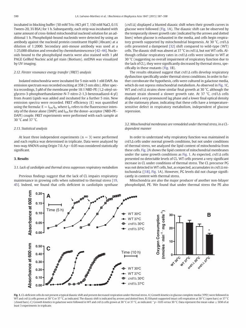
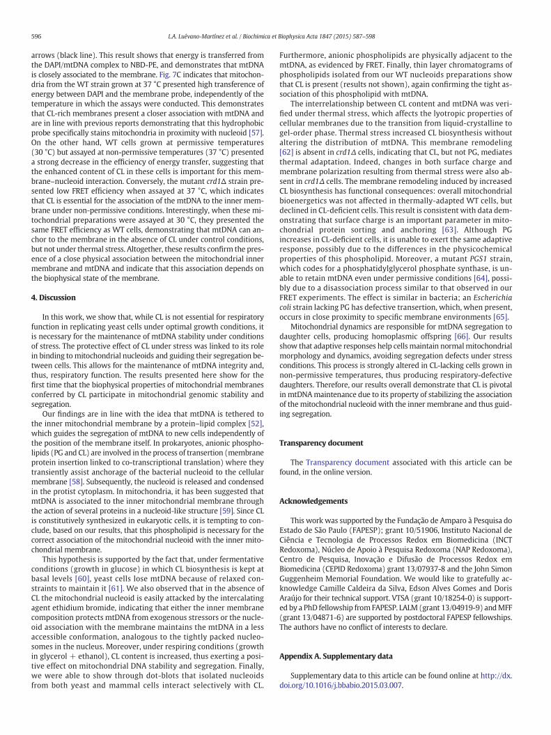
Fig. 1. CL-deficient cells do not present a typical diauxic shift andpresent decreased respiration uWT and crd1Δ cells grown at 30 °C or 37 °C, as indicated. The diauxic shift is indicated by arrows(closed bars). C) Growth kinetics in galactosewere followed inWT and crd1Δ cells grown at 30least 3 experiments in triplicate.
(crd1Δ) displayed a blunted diauxic shift when their growth curves inglucose were followed (Fig. 1A). The diauxic shift can be observed bythe temporarily slower growth rate (indicated by the arrows and dottedlines) when glucose is exhausted in the media, and cells begin respira-tory growth supported by mitochondrial biogenesis. At 30 °C, crd1Δcells presented a dampened [32] shift compared to wild-type (WT)cells. The diauxic shift was absent at 37 °C in crd1Δ, but notWT cells. Al-though cellular respiratory rates in crd1Δ cells were similar to WT at30 °C (suggesting no overall impairment of respiratory function due tothe lack of CL), theywere significantly decreased by thermal stress, spe-cifically in these mutants (Fig. 1B).
The results obtained suggest that crd1Δ cells develop respiratorydysfunction specifically under thermal stress conditions. In order to fur-ther corroborate the hypothesis, cells were cultured in galactose media,which do not repress mitochondrial metabolism. As observed in Fig. 1C,WT and crd1Δ strains show similar final growth at 30 °C, although themutant strain showed a slower growth rate. At 37 °C, crd1Δ cellsdisplayed a very pronounced lag phase and a lower final optical densityat the stationary phase, indicating that these cells have a temperature-sensitive defect in respiratory metabolism, independent of glucoserepression.
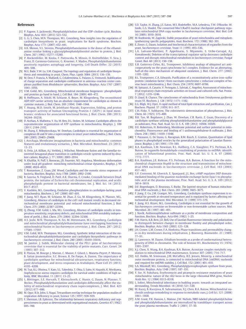
3.2.Mitochondrial membranes are remodeled under thermal stress, in a CL-dependent manner
In order to understand why respiratory function was maintained incrd1Δ cells under normal growth conditions, but not under conditionsof thermal stress, we analyzed the lipid content of mitochondria fromthese cells. Fig. 2A shows the lipid content of mitochondrialmembranesunder the same growth conditions as Fig. 1. As expected, crd1Δ cellspresented no detectable levels of CL. WT cells present a very significantincrease in CL under conditions of thermal stress. The CL precursor PGwasnot detected inWT cells, but, as expected, accumulates in crd1Δmi-tochondria ([18], Fig. 1A). However, PG levels did not change signifi-cantly in content with thermal stress.
Mitochondria are also the major producer of another non-bilayerphospholipid, PE. We found that under thermal stress the PE also
WT 300C
WT 370C
crd1Δ 370C
crd1Δ 300C
Co
WT 30
Co
WT
37Co
30Δcr
d1
Co
37Δcr
d1
0
10
20
30 B
*
Oxy
gen
Co
nsu
mp
tion
(nm
ole
s. m
in-1
. mg
cel
ls-1
)
WT 300C
WT 370C
crd1Δ 370C
crd1Δ 300C
me (10 h)
nder thermal stress. A)Growth kinetics in glucose completemedia (YPD)were followed inand dotted lines. B) Ethanol-supported intact cell respiration at 30 °C (open bars) or 37 °C°C or 37 °C, as indicated. * p b 0.05 versus 30 °C. Data represent themean value± SEM of at
WT Δ
crd1
0
30
60
90 B
*
*
AN
SA
Flu
ore
scen
ce(a
rbitr
ary
un
its)
Co
WT 30
Co
WT 37
Co
30Δcr
d1
Co
37Δcr
d1
0.0
0.1
0.2
0.3 C *
##
GP
em
issi
on
CLPG PE PC
0
20
40
60 WT 30°CWT 37°C
*
*
#
crd1Δ 300Ccrd1Δ 370C
A%
mo
l of P
ho
sph
olip
id 300C 370C
300C 370C
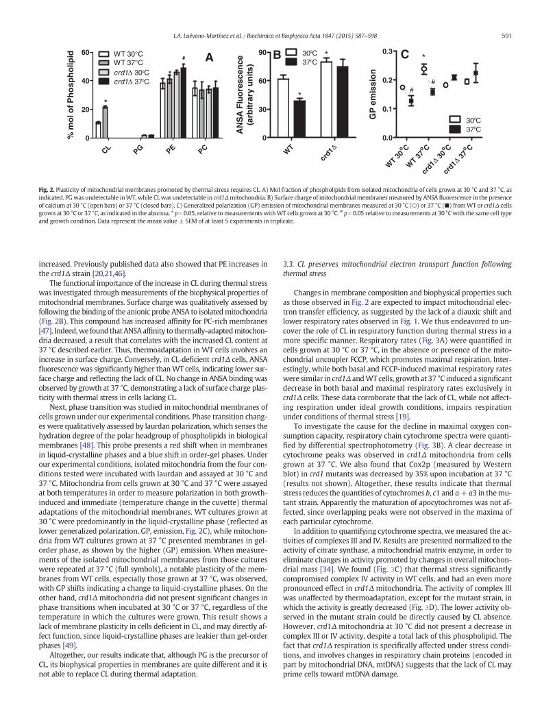
Fig. 2. Plasticity of mitochondrial membranes promoted by thermal stress requires CL. A) Mol fraction of phospholipids from isolated mitochondria of cells grown at 30 °C and 37 °C, asindicated. PGwas undetectable inWT, while CLwas undetectable in crd1Δmitochondria. B) Surface charge ofmitochondrial membranes measured by ANSA fluorescence in the presenceof calcium at 30 °C (open bars) or 37 °C (closed bars). C) Generalized polarization (GP) emission of mitochondrial membranes measured at 30 °C (○) or 37 °C (■) fromWT or crd1Δ cellsgrown at 30 °C or 37 °C, as indicated in the abscissa. * p b 0.05, relative tomeasurementswithWT cells grown at 30 °C. # p b 0.05 relative tomeasurements at 30 °Cwith the same cell typeand growth condition. Data represent the mean value ± SEM of at least 5 experiments in triplicate.
591L.A. Luévano-Martínez et al. / Biochimica et Biophysica Acta 1847 (2015) 587–598
increased. Previously published data also showed that PE increases inthe crd1Δ strain [20,21,46].
The functional importance of the increase in CL during thermal stresswas investigated through measurements of the biophysical properties ofmitochondrial membranes. Surface charge was qualitatively assessed byfollowing the binding of the anionic probe ANSA to isolatedmitochondria(Fig. 2B). This compound has increased affinity for PC-rich membranes[47]. Indeed,we found thatANSAaffinity to thermally-adaptedmitochon-dria decreased, a result that correlates with the increased CL content at37 °C described earlier. Thus, thermoadaptation in WT cells involves anincrease in surface charge. Conversely, in CL-deficient crd1Δ cells, ANSAfluorescence was significantly higher thanWT cells, indicating lower sur-face charge and reflecting the lack of CL. No change in ANSA binding wasobserved by growth at 37 °C, demonstrating a lack of surface charge plas-ticity with thermal stress in cells lacking CL.
Next, phase transition was studied in mitochondrial membranes ofcells grown under our experimental conditions. Phase transition chang-es were qualitatively assessed by laurdan polarization,which senses thehydration degree of the polar headgroup of phospholipids in biologicalmembranes [48]. This probe presents a red shift when in membranesin liquid-crystalline phases and a blue shift in order-gel phases. Underour experimental conditions, isolated mitochondria from the four con-ditions tested were incubated with laurdan and assayed at 30 °C and37 °C. Mitochondria from cells grown at 30 °C and 37 °C were assayedat both temperatures in order to measure polarization in both growth-induced and immediate (temperature change in the cuvette) thermaladaptations of the mitochondrial membranes. WT cultures grown at30 °C were predominantly in the liquid-crystalline phase (reflected aslower generalized polarization, GP, emission, Fig. 2C), while mitochon-dria from WT cultures grown at 37 °C presented membranes in gel-order phase, as shown by the higher (GP) emission. When measure-ments of the isolated mitochondrial membranes from those cultureswere repeated at 37 °C (full symbols), a notable plasticity of the mem-branes from WT cells, especially those grown at 37 °C, was observed,with GP shifts indicating a change to liquid-crystalline phases. On theother hand, crd1Δ mitochondria did not present significant changes inphase transitions when incubated at 30 °C or 37 °C, regardless of thetemperature in which the cultures were grown. This result shows alack of membrane plasticity in cells deficient in CL, and may directly af-fect function, since liquid-crystalline phases are leakier than gel-orderphases [49].
Altogether, our results indicate that, although PG is the precursor ofCL, its biophysical properties in membranes are quite different and it isnot able to replace CL during thermal adaptation.
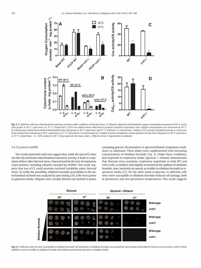
3.3. CL preserves mitochondrial electron transport function followingthermal stress
Changes in membrane composition and biophysical properties suchas those observed in Fig. 2 are expected to impact mitochondrial elec-tron transfer efficiency, as suggested by the lack of a diauxic shift andlower respiratory rates observed in Fig. 1. We thus endeavored to un-cover the role of CL in respiratory function during thermal stress in amore specific manner. Respiratory rates (Fig. 3A) were quantified incells grown at 30 °C or 37 °C, in the absence or presence of the mito-chondrial uncoupler FCCP, which promotes maximal respiration. Inter-estingly, while both basal and FCCP-induced maximal respiratory rateswere similar in crd1Δ andWT cells, growth at 37 °C induced a significantdecrease in both basal and maximal respiratory rates exclusively incrd1Δ cells. These data corroborate that the lack of CL, while not affect-ing respiration under ideal growth conditions, impairs respirationunder conditions of thermal stress [19].
To investigate the cause for the decline in maximal oxygen con-sumption capacity, respiratory chain cytochrome spectra were quanti-fied by differential spectrophotometry (Fig. 3B). A clear decrease incytochrome peaks was observed in crd1Δ mitochondria from cellsgrown at 37 °C. We also found that Cox2p (measured by Westernblot) in crd1 mutants was decreased by 35% upon incubation at 37 °C(results not shown). Altogether, these results indicate that thermalstress reduces the quantities of cytochromes b, c1 and a+ a3 in themu-tant strain. Apparently the maturation of apocytochromes was not af-fected, since overlapping peaks were not observed in the maxima ofeach particular cytochrome.
In addition to quantifying cytochrome spectra, we measured the ac-tivities of complexes III and IV. Results are presented normalized to theactivity of citrate synthase, a mitochondrial matrix enzyme, in order toeliminate changes in activity promoted by changes in overall mitochon-drial mass [34]. We found (Fig. 3C) that thermal stress significantlycompromised complex IV activity in WT cells, and had an even morepronounced effect in crd1Δ mitochondria. The activity of complex IIIwas unaffected by thermoadaptation, except for the mutant strain, inwhich the activity is greatly decreased (Fig. 3D). The lower activity ob-served in the mutant strain could be directly caused by CL absence.However, crd1Δ mitochondria at 30 °C did not present a decrease incomplex III or IV activity, despite a total lack of this phospholipid. Thefact that crd1Δ respiration is specifically affected under stress condi-tions, and involves changes in respiratory chain proteins (encoded inpart by mitochondrial DNA, mtDNA) suggests that the lack of CL mayprime cells toward mtDNA damage.
Basal
WT
FCCPW
T Δ
Basal
crd1 Δ
FCCP crd1
0
15
30
45
60
75 30oC
37oCA
*
*
*
Oxy
gen
Co
nsu
mp
tion
(nm
ole
s. m
in-1
. mg
pro
tein
-1)
WT Δ
crd1
0
20
40
60 C
*
*
Co
x ac
tivity
/CS
act
ivity
WT Δ
crd1
0
5
10
15
20
25 D
*C
ytb
c1
activ
ity/C
S a
ctiv
ityc b 3
a+a c b 3
a+a c b 3
a+a c b 3
a+a
0.0
0.1
0.2
0.3
0.4WT 30°C
WT 37°C
crd1Δ 30°C
crd1Δ 37°C
B
Cyt
och
rom
e co
nte
nt
(μm
ole
. mg
pro
tein
-1)
Fig. 3. CL-deficient cells lose mitochondrial respiratory proteins under conditions of thermal stress. A) Ethanol-supported mitochondrial oxygen consumption measured in WT or crd1Δcells grown at 30 °C (open bars) or 37 °C (closed bars). FCCP was added where indicated to promote maximal respiratory rates. Oxygen consumption was measured at 30 °C.B) Cytochrome content from isolated mitochondria from cells grown at 30 °C (open bars) and 37 °C (full bars). C) Cytochrome c oxidase (Cox) activity normalized activity to citrate syn-thase activity from cells grown at 30 °C (open bars) or 37 °C (closed bars). D) Cytochrome bc1 complex activity normalized to citrate synthase activity from cells grownat 30 °C (open bars)or 37 °C (closed bars). * p b 0.05, relative to 30 °C. Data represent the mean value ± SEM of at least 3 experiments in triplicate.
592 L.A. Luévano-Martínez et al. / Biochimica et Biophysica Acta 1847 (2015) 587–598
3.4. CL protects mtDNA
The results presented until now suggest that, while the lack of CL doesnot directly inactivatemitochondrial respiratory activity, it leads to respi-ratory defects after thermal stress, characterized by the lack of respiratorychain proteins, including subunits encoded by mtDNA. This result sug-gests that loss of CL could promote nucleoid instability under thermalstress. To verify this possibility, ethidium bromide accessibility to the mi-tochondrial nucleoid was analyzed by spot testing [50]. Cells were grownin galactose media. Aliquots were serially diluted and spotted in plates
Glucose
30° 37°
Eth
idiu
m B
rom
ide
Fig. 4. CL-deficient cells are more susceptible to ethidiumbromide. Cell sensitivity to ethidium bmtDNA is more accessible to ethidium bromide intercalation present poor growth in complete
containing glucose (fermentative) or glycerol/ethanol (respiratory condi-tions) as substrates. These plates were supplemented with increasingconcentrations of ethidium bromide (Fig. 4). Under these conditions,lack of growth in respiratory media (glycerol + ethanol) demonstratesthat thermal stress promotes respiratory ineptitude in both WT andcrd1Δ cells, a condition only slightlyworsenedby the addition of ethidiumbromide, since nucleoids are poorly accessible to ethidium bromide in re-spiratory media [51]. On the other hand, in glucose, CL-deficient cellswere more susceptible to ethidium bromide-induced cell damage, bothat permissive and non-permissive temperatures. This result suggests
Wild-type
crd1Δ
Wild-type
crd1Δ
Wild-type
crd1Δ
Glycerol + Ethanol
30° 37°
romide was assessed by spot testing as described inMaterials andmethods. Cells inwhichmedia.
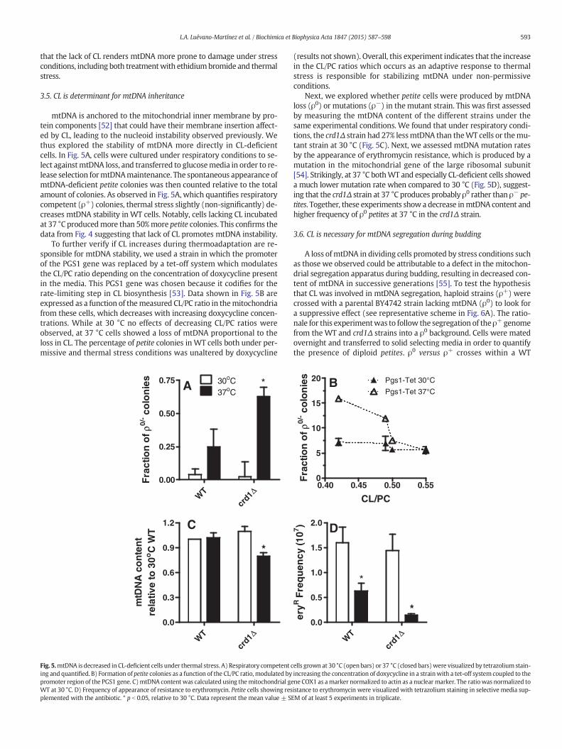
593L.A. Luévano-Martínez et al. / Biochimica et Biophysica Acta 1847 (2015) 587–598
that the lack of CL renders mtDNA more prone to damage under stressconditions, including both treatmentwith ethidiumbromide and thermalstress.
3.5. CL is determinant for mtDNA inheritance
mtDNA is anchored to the mitochondrial inner membrane by pro-tein components [52] that could have their membrane insertion affect-ed by CL, leading to the nucleoid instability observed previously. Wethus explored the stability of mtDNA more directly in CL-deficientcells. In Fig. 5A, cells were cultured under respiratory conditions to se-lect againstmtDNA loss, and transferred to glucosemedia in order to re-lease selection formtDNAmaintenance. The spontaneous appearance ofmtDNA-deficient petite colonies was then counted relative to the totalamount of colonies. As observed in Fig. 5A, which quantifies respiratorycompetent (ρ+) colonies, thermal stress slightly (non-significantly) de-creases mtDNA stability in WT cells. Notably, cells lacking CL incubatedat 37 °C producedmore than 50%more petite colonies. This confirms thedata from Fig. 4 suggesting that lack of CL promotes mtDNA instability.
To further verify if CL increases during thermoadaptation are re-sponsible for mtDNA stability, we used a strain in which the promoterof the PGS1 gene was replaced by a tet-off system which modulatesthe CL/PC ratio depending on the concentration of doxycycline presentin the media. This PGS1 gene was chosen because it codifies for therate-limiting step in CL biosynthesis [53]. Data shown in Fig. 5B areexpressed as a function of themeasured CL/PC ratio in themitochondriafrom these cells, which decreases with increasing doxycycline concen-trations. While at 30 °C no effects of decreasing CL/PC ratios wereobserved, at 37 °C cells showed a loss of mtDNA proportional to theloss in CL. The percentage of petite colonies in WT cells both under per-missive and thermal stress conditions was unaltered by doxycycline
WT Δ
crd1
0.00
0.25
0.50
0.75 30oC37oC
A *
*
Fra
ctio
n o
fρ0/
- co
lon
ies
WT Δ
crd1
0.0
0.3
0.6
0.9
1.2
*
*
mtD
NA
co
nte
nt
rela
tive
to 3
0oC
WT
*
C
Fig. 5.mtDNA is decreased in CL-deficient cells under thermal stress. A) Respiratory competent cing and quantified. B) Formation of petite colonies as a function of the CL/PC ratio, modulated bypromoter region of the PGS1 gene. C)mtDNA content was calculated using themitochondrial geWT at 30 °C. D) Frequency of appearance of resistance to erythromycin. Petite cells showing resplemented with the antibiotic. * p b 0.05, relative to 30 °C. Data represent the mean value ± S
(results not shown). Overall, this experiment indicates that the increasein the CL/PC ratios which occurs as an adaptive response to thermalstress is responsible for stabilizing mtDNA under non-permissiveconditions.
Next, we explored whether petite cells were produced by mtDNAloss (ρ0) or mutations (ρ−) in the mutant strain. This was first assessedby measuring the mtDNA content of the different strains under thesame experimental conditions. We found that under respiratory condi-tions, the crd1Δ strain had 27% lessmtDNA than theWT cells or themu-tant strain at 30 °C (Fig. 5C). Next, we assessed mtDNA mutation ratesby the appearance of erythromycin resistance, which is produced by amutation in the mitochondrial gene of the large ribosomal subunit[54]. Strikingly, at 37 °C bothWT and especially CL-deficient cells showeda much lower mutation rate when compared to 30 °C (Fig. 5D), suggest-ing that the crd1Δ strain at 37 °C produces probably ρ0 rather than ρ− pe-tites. Together, these experiments show a decrease inmtDNA content andhigher frequency of ρ0 petites at 37 °C in the crd1Δ strain.
3.6. CL is necessary for mtDNA segregation during budding
A loss ofmtDNA in dividing cells promoted by stress conditions suchas those we observed could be attributable to a defect in the mitochon-drial segregation apparatus during budding, resulting in decreased con-tent of mtDNA in successive generations [55]. To test the hypothesisthat CL was involved in mtDNA segregation, haploid strains (ρ+) werecrossed with a parental BY4742 strain lacking mtDNA (ρ0) to look fora suppressive effect (see representative scheme in Fig. 6A). The ratio-nale for this experimentwas to follow the segregation of the ρ+genomefrom the WT and crd1Δ strains into a ρ0 background. Cells were matedovernight and transferred to solid selecting media in order to quantifythe presence of diploid petites. ρ0 versus ρ+ crosses within a WT
WT Δ
crd1
0.0
0.5
1.0
1.5
2.0 D
*
*
eryR
Fre
qu
ency
(107 )
0.40 0.45 0.50 0.550
5
10
15
20 Pgs1-Tet 30°C
Pgs1-Tet 37°CB
CL/PC
Fra
ctio
n o
fρ0/
- co
lon
ies
ells grown at 30 °C (open bars) or 37 °C (closed bars)were visualized by tetrazolium stain-increasing the concentration of doxycycline in a strainwith a tet-off system coupled to thene COX1 as amarker normalized to actin as a nuclear marker. The ratiowas normalized toistance to erythromycin were visualized with tetrazolium staining in selective media sup-EM of at least 5 experiments in triplicate.
WT Δ
crd1
0
15
30
45 B *
Su
pp
resi
ven
ess
(%)
TMRM DAPI DIC
C
D
A
Fig. 6. CL-deficient cells have deficient mtDNA segregation under conditions of thermal stress. A) Representative scheme of themating experimental procedure. B) Percent of respiratoryincompetent ρ0 colonies aftermating at 30 °C (open bars) or 37 °C (closed bars)withWT and crd1Δ cellswith the genotype ρ+. * p b 0.05 relative to 30 °C. Data represent themean value±SEM of at least 5 experiments in triplicate. C) Confocal imaging of themitochondrial network stained with TMRM andmt-nucleoid (DAPI). D) Quantification of mtDNA segregation in zy-gote mitochondria expressed as area of TMRM fluorescence co-localized with DAPI fluorescence in all planes of a whole specimen z-stack, data from at least 10 zygotes per group in 3independent experiments. * p b 0.05 relative to 30 °C.
594 L.A. Luévano-Martínez et al. / Biochimica et Biophysica Acta 1847 (2015) 587–598
background (Mata ρ+ × Matα ρ0) produced only a small proportion ofrespiratory suppressed cells (~25%; Fig. 6B), both at 30 °C and 37 °C.This indicates that ρ+-WT cells were capable of distributing theirmtDNA to ρ0 cells, complementing the ρ0 phenotype and rescuing therespiratory phenotype. On the other hand, crossing Mata crd1Δ−ρ+
versusMatαWT-ρ0 at 37 °C resulted in a 40% increase in colonies lackingrespiratory capacity (Fig. 6B). This demonstrates that at non-permissivetemperatures the absence of CL affects mitochondrial segregation,impairing repopulation of the ρ0 cells with mtDNA.
In order to visually confirm the phenomenon indicated by this data,mitochondria and the distribution of mtDNA in mated cells were ana-lyzed at permissive and non-permissive temperatures in WT and CL-lacking strains. Fig. 6C shows typical confocal microscopies in whichthe mitochondrial mass from both strains was marked with TMRMand mtDNA was stained with DAPI (which marks predominantlymtDNA in yeast). The images indicate that, at 37 °C, the ρ+ genomefrom a crd1Δ strain is unable to segregate into a WT-ρ0 strain, sinceDAPI fluorescence is not evenly spread among the buds (see bottom
images). Interestingly, although DAPI fluorescence did not migratewithin the diploid, TMRM, which marks mitochondrial membranes,did. These results are depicted in the quantification shown in Fig. 6D,which gives an indirect measure of co-localization of mitochondriaand mtDNA. The results clearly indicate that CL is necessary formtDNA migration to mated, respiratory-incompetent, cells.
To further verify that mitochondrial exchange between cells is im-paired by the absence of cardiolipin, WT-ρ0 strains were transformedwith a plasmid containing a mitochondrially-tagged GFP and crossedwith DAPI-stained WT and crd1Δ ρ+ cells (Supplementary Fig. 1).Under these conditions, GFP-stainedmitochondria were widely distrib-uted into the zygote of WT cells. In the crd1Δ ×WT-ρ0 cross, GFP distri-bution is impaired and just a few small dot-like structures appear in theother lobules of the zygote, an effect that is strongly magnified at 37 °C.These results corroborate that mtDNA andmatrix content are mixed byindependent mechanisms between mitochondrial populations. SincemtDNA is anchored to the inner membrane, CL may be necessary forthis lipid–nucleoid interaction and consequent mtDNA exchange.
595L.A. Luévano-Martínez et al. / Biochimica et Biophysica Acta 1847 (2015) 587–598
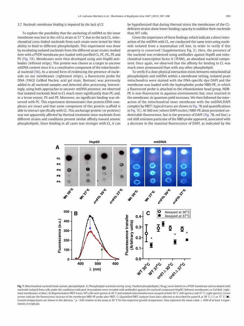
3.7. Nucleoid–membrane binding is impaired by the lack of CL
To explore the possibility that the anchoring of mtDNA to the innermembrane was lost in the crd1Δ strain at 37 °C due to the lack CL, mito-chondrial cross-linked nucleoids from each strain were tested for theirability to bind to different phospholipids. This experiment was doneby incubating isolated nucleoids from the different yeast strains studiedherewith a PVDFmembrane pre-loadedwith purified CL, PC, PG, PE andPS (Fig. 7A). Membranes were then developed using anti-Hsp60 anti-bodies (leftmost strips). This protein was chosen as a target to uncovermtDNA content since it is a constitutive component of themitochondri-al nucleoid [56]. As a second form of evidencing the presence of nucle-oids on our membranes (rightmost strips), a fluorescent probe forDNA (PAGE GelRed Nucleic acid gel stain; Biotium) was previouslyadded to all nucleoid samples and detected after processing. Interest-ingly, using both approaches to uncover mtDNA presence, we observedthat isolated nucleoids bind to CLmuchmore significantly than PG and,to a lesser extent, PS and PE. Moreover, no significant binding was ob-served with PC. This experiment demonstrates that protein/DNA com-plexes are intact and that some component of this protein scaffold isable to interact specificallywith CL. This anchorage protein (or proteins)was not apparently affected by thermal treatment since nucleoids fromdifferent strains and conditions present similar affinity toward anionicphospholipids. Since binding in all cases was stronger with CL, it can
A
B
400 500
500
400
300
200
100
0400
λ (nm)
Flu
ore
scen
ce (
a.u
.)
Fig. 7.Mitochondrial nucleoid binds anionic phospholipids. A) Phospholipid-nucleoid overlay anucleoids isolated from cells under the conditions indicated. Associationswere revealed with anmostmembranes, in blue). B) Representative FRET traces.WT cellswere grown at 30 °C and isolaarrows indicate the fluorescence increase of the membrane NBD-PE probe after FRET. C) QuantGrowth temperatures are shown in the abscissa. * p b 0.05 relative to the assay at 30 °C for theiments in triplicate.
be hypothesized that during thermal stress the membranes of the CL-deficient strains show lower binding capacity to stabilize their nucleoidsthan WT cells.
Given the importance of these findings,which indicate a direct inter-action of the mtDNAwith CL, we conducted the same tests using nucle-oids isolated from a mammalian cell line, in order to verify if thisproperty is conserved (Supplementary Fig. 2). Here, the presence ofthe nucleoid was evidenced using antibodies against Hsp60 and mito-chondrial transcription factor A (TFAM), an abundant nucleoid compo-nent. Once again, we observed that the affinity for binding to CL wasmuch more pronounced than with any other phospholipid.
To verify if a close physical interaction exists betweenmitochondrialphospholipids and mtDNA within a membrane setting, isolated yeastmitochondria were stained with the DNA-specific dye DAPI and themembrane was loaded with the hydrophobic probe NBD-PE, in whicha fluorescent probe is attached to the ethanolamine head group. NDB-PE is non-fluorescent in aqueous environments but, once inserted inthemembrane, its quantumyield increases.We then followed the inter-action of the mitochondrial inner membrane with the mtDNA/DAPIcomplex by FRET (typical traces are shown in Fig. 7B and quantificationsin Fig. 7C). At 560nm(where DAPI excites) NBD-PE alonepresented un-detectable fluorescence, but in the presence of DAPI (Fig. 7B, red line) ared shift emission particular of theNBDprobe appeared, associatedwitha decrease in the maximal fluorescence of DAPI, as indicated by the
Co
WT 30
Co
WT 37
Co
30Δcr
d1
Co
37Δcr
d1
0.0
0.1
0.2
0.3
0.4C
** *
Assayed at 30oCAssayed at 37oC
Flu
ores
cenc
e tr
ansf
er(A
rbit
rary
uni
ts)
ssay. Purified phospholipids (50 μg)were dotted on a PVDFmembrane and incubatedwithtibodies against the nucleoid component Hsp60 (leftmost membranes) or Gel Red (right-tedmitochondriawere assayed at both 30 °C (left spectra) and 37 °C (right spectra). Greenified FRET analyses from data collected as described for panel B, at 30 °C (○) or 37 °C (■).respective growth temperature. Data represent the mean value ± SEM of at least 3 exper-
596 L.A. Luévano-Martínez et al. / Biochimica et Biophysica Acta 1847 (2015) 587–598
arrows (black line). This result shows that energy is transferred fromthe DAPI/mtDNA complex to NBD-PE, and demonstrates that mtDNAis closely associated to the membrane. Fig. 7C indicates that mitochon-dria from the WT strain grown at 37 °C presented high transference ofenergy between DAPI and the membrane probe, independently of thetemperature in which the assays were conducted. This demonstratesthat CL-rich membranes present a closer association with mtDNA andare in line with previous reports demonstrating that this hydrophobicprobe specifically stains mitochondria in proximity with nucleoid [57].On the other hand, WT cells grown at permissive temperatures(30 °C) but assayed at non-permissive temperatures (37 °C) presenteda strong decrease in the efficiency of energy transfer, suggesting thatthe enhanced content of CL in these cells is important for this mem-brane–nucleoid interaction. Conversely, the mutant crd1Δ strain pre-sented low FRET efficiency when assayed at 37 °C, which indicatesthat CL is essential for the association of the mtDNA to the inner mem-brane under non-permissive conditions. Interestingly, when these mi-tochondrial preparations were assayed at 30 °C, they presented thesame FRET efficiency as WT cells, demonstrating that mtDNA can an-chor to the membrane in the absence of CL under control conditions,but not under thermal stress. Altogether, these results confirm the pres-ence of a close physical association between the mitochondrial innermembrane and mtDNA and indicate that this association depends onthe biophysical state of the membrane.
4. Discussion
In this work, we show that, while CL is not essential for respiratoryfunction in replicating yeast cells under optimal growth conditions, itis necessary for the maintenance of mtDNA stability under conditionsof stress. The protective effect of CL under stress was linked to its rolein binding to mitochondrial nucleoids and guiding their segregation be-tween cells. This allows for the maintenance of mtDNA integrity and,thus, respiratory function. The results presented here show for thefirst time that the biophysical properties of mitochondrial membranesconferred by CL participate in mitochondrial genomic stability andsegregation.
Our findings are in line with the idea that mtDNA is tethered tothe inner mitochondrial membrane by a protein–lipid complex [52],which guides the segregation of mtDNA to new cells independently ofthe position of the membrane itself. In prokaryotes, anionic phospho-lipids (PG and CL) are involved in the process of transertion (membraneprotein insertion linked to co-transcriptional translation) where theytransiently assist anchorage of the bacterial nucleoid to the cellularmembrane [58]. Subsequently, the nucleoid is released and condensedin the protist cytoplasm. In mitochondria, it has been suggested thatmtDNA is associated to the inner mitochondrial membrane throughthe action of several proteins in a nucleoid-like structure [59]. Since CLis constitutively synthesized in eukaryotic cells, it is tempting to con-clude, based on our results, that this phospholipid is necessary for thecorrect association of the mitochondrial nucleoid with the inner mito-chondrial membrane.
This hypothesis is supported by the fact that, under fermentativeconditions (growth in glucose) in which CL biosynthesis is kept atbasal levels [60], yeast cells lose mtDNA because of relaxed con-straints to maintain it [61]. We also observed that in the absence ofCL the mitochondrial nucleoid is easily attacked by the intercalatingagent ethidium bromide, indicating that either the inner membranecomposition protects mtDNA from exogenous stressors or the nucle-oid association with the membrane maintains the mtDNA in a lessaccessible conformation, analogous to the tightly packed nucleo-somes in the nucleus. Moreover, under respiring conditions (growthin glycerol + ethanol), CL content is increased, thus exerting a posi-tive effect on mitochondrial DNA stability and segregation. Finally,we were able to show through dot-blots that isolated nucleoidsfrom both yeast and mammal cells interact selectively with CL.
Furthermore, anionic phospholipids are physically adjacent to themtDNA, as evidenced by FRET. Finally, thin layer chromatograms ofphospholipids isolated from our WT nucleoids preparations showthat CL is present (results not shown), again confirming the tight as-sociation of this phospholipid with mtDNA.
The interrelationship between CL content and mtDNA was veri-fied under thermal stress, which affects the lyotropic properties ofcellular membranes due to the transition from liquid-crystalline togel-order phase. Thermal stress increased CL biosynthesis withoutaltering the distribution of mtDNA. This membrane remodeling[62] is absent in crd1Δ cells, indicating that CL, but not PG, mediatesthermal adaptation. Indeed, changes in both surface charge andmembrane polarization resulting from thermal stress were also ab-sent in crd1Δ cells. The membrane remodeling induced by increasedCL biosynthesis has functional consequences: overall mitochondrialbioenergetics was not affected in thermally-adapted WT cells, butdeclined in CL-deficient cells. This result is consistent with data dem-onstrating that surface charge is an important parameter in mito-chondrial protein sorting and anchoring [63]. Although PGincreases in CL-deficient cells, it is unable to exert the same adaptiveresponse, possibly due to the differences in the physicochemicalproperties of this phospholipid. Moreover, a mutant PGS1 strain,which codes for a phosphatidylglycerol phosphate synthase, is un-able to retain mtDNA even under permissive conditions [64], possi-bly due to a disassociation process similar to that observed in ourFRET experiments. The effect is similar in bacteria; an Escherichiacoli strain lacking PG has defective transertion, which, when present,occurs in close proximity to specific membrane environments [65].
Mitochondrial dynamics are responsible for mtDNA segregation todaughter cells, producing homoplasmic offspring [66]. Our resultsshow that adaptive responses help cells maintain normalmitochondrialmorphology and dynamics, avoiding segregation defects under stressconditions. This process is strongly altered in CL-lacking cells grown innon-permissive temperatures, thus producing respiratory-defectivedaughters. Therefore, our results overall demonstrate that CL is pivotalin mtDNAmaintenance due to its property of stabilizing the associationof themitochondrial nucleoid with the inner membrane and thus guid-ing segregation.
Transparency document
The Transparency document associated with this article can befound, in the online version.
Acknowledgements
This work was supported by the Fundação de Amparo à Pesquisa doEstado de São Paulo (FAPESP); grant 10/51906, Instituto Nacional deCiência e Tecnologia de Processos Redox em Biomedicina (INCTRedoxoma), Núcleo de Apoio à Pesquisa Redoxoma (NAP Redoxoma),Centro de Pesquisa, Inovação e Difusão de Processos Redox emBiomedicina (CEPID Redoxoma) grant 13/07937-8 and the John SimonGuggenheim Memorial Foundation. We would like to gratefully ac-knowledge Camille Caldeira da Silva, Edson Alves Gomes and DorisAraújo for their technical support. VTSA (grant 10/18254-0) is support-ed by a PhD fellowship fromFAPESP. LALM (grant 13/04919-9) andMFF(grant 13/04871-6) are supported by postdoctoral FAPESP fellowships.The authors have no conflict of interests to declare.
Appendix A. Supplementary data
Supplementary data to this article can be found online at http://dx.doi.org/10.1016/j.bbabio.2015.03.007.
597L.A. Luévano-Martínez et al. / Biochimica et Biophysica Acta 1847 (2015) 587–598
References
[1] P. Fagone, S. Jackowski, Phosphatidylcholine and the CDP–choline cycle, Biochim.Biophys. Acta 1831 (2013) 523–532.
[2] G. Li, S. Chen, M.N. Thompson, M.L. Greenberg, New insights into the regulation ofcardiolipin biosynthesis in yeast: implications for Barth syndrome, Biochim.Biophys. Acta 1771 (2007) 432–441.
[3] A.K. Menon, V.L. Stevens, Phosphatidylethanolamine is the donor of the ethanol-amine residue linking a glycosylphosphatidylinositol anchor to protein, J. Biol.Chem. 267 (1992) 15277–15280.
[4] P. Rockenfeller, M. Koska, F. Pietrocola, N. Minois, O. Knittelfelder, V. Sica, J.Franz, D. Carmona-Gutierrez, G. Kroemer, F. Madeo, Phosphatidylethanolaminepositively regulates autophagy and longevity, Cell Death Differ. 22 (2015)499–508.
[5] M.G. Baile, Y.W. Lu, S.M. Claypool, The topology and regulation of cardiolipin biosyn-thesis and remodeling in yeast, Chem. Phys. Lipids 3084 (2013) 136–139.
[6] M. Dezi, F. Francia, A.Mallardi, G. Colafemmina, G. Palazzo, G. Venturoli, Stabilizationof charge separation and cardiolipin confinement in antenna–reaction center com-plexes purified from Rhodobacter sphaeroides, Biochim. Biophys. Acta 1767 (2007)1041–1056.
[7] V.M. Gohil, M.L. Greenberg, Mitochondrial membrane biogenesis: phospholipidsand proteins go hand in hand, J. Cell Biol. 184 (2009) 469–472.
[8] B. Hoffmann, A. Stöckl, M. Schlame, K. Beyer, M. Klingenberg, The reconstitutedADP/ATP carrier activity has an absolute requirement for cardiolipin as shown incysteine mutants, J. Biol. Chem. 269 (1994) 1940–1944.
[9] T. Hoang, M.D. Smith, M. Jelokhani-Niaraki, Expression, folding, and protontransport activity of human Uncoupling Protein-1 (UCP1) in lipid mem-branes: evidence for associated functional forms, J. Biol. Chem. 288 (2013)36244–36258.
[10] D. Acehan, A. Malhotra, Y. Xu, M. Ren, D.L. Stokes, M. Schlame, Cardiolipin affects thesupramolecular organization of ATP synthase in mitochondria, Biophys. J. 100(2011) 2184–2192.
[11] M. Zhang, E. Mileykovskaya, W. Dowhan, Cardiolipin is essential for organization ofcomplexes III and IV into a supercomplex in intact yeast mitochondria, J. Biol. Chem.280 (2005) 29403–29408.
[12] I. Arechaga, Membrane invaginations in bacteria and mitochondria: commonfeatures and evolutionary scenarios, J. Mol. Microbiol. Biotechnol. 23 (2013)13–23.
[13] A. Ortiz, J.A. Killian, A.J. Verkleij, J. Wilschut, Membrane fusion and the lamellar-to-inverted-hexagonal phase transition in cardiolipin vesicle systems induced by diva-lent cations, Biophys. J. 77 (1999) 2003–2014.
[14] N. Khalifat, N. Puff, S. Bonneau, J.B. Fournier, M.I. Angelova, Membrane deformationunder local pH gradient: mimicking mitochondrial cristae dynamics, Biophys. J. 95(2008) 4924–4933.
[15] T. Romantsov, Z. Guan, J.M. Wood, Cardiolipin and the osmotic stress responses ofbacteria, Biochim. Biophys. Acta 1788 (2009) 2092–2100.
[16] R. Saxena, N. Fingland, D. Patil, A.K. Sharma, E. Crooke, Crosstalk between DnaAprotein, the initiator of Escherichia coli chromosomal replication, and acidicphospholipids present in bacterial membranes, Int. J. Mol. Sci. 14 (2013)8517–8537.
[17] V. Koshkin, M.L. Greenberg, Oxidative phosphorylation in cardiolipin-lacking yeastmitochondria, Biochem. J. 347 (2000) 687–691.
[18] F. Jiang, M.T. Ryan, M. Schlame, M. Zhao, Z. Gu, M. Klingenberg, N. Pfanner, M.L.Greenberg, Absence of cardiolipin in the crd1 null mutant results in decreased mi-tochondrial membrane potential and reduced mitochondrial function, J. Biol.Chem. 275 (2000) 22387–22394.
[19] Q. Zhong, V.M. Gohil, L. Ma, M.L. Greenberg, Absence of cardiolipin results in tem-perature sensitivity, respiratory defects, andmitochondrial DNA instability indepen-dent of pet56, J. Biol. Chem. 279 (2004) 32294–32300.
[20] A.S. Joshi, M.N. Thompson, N. Fei, M. Hüttemann, M.L. Greenberg, Cardiolipinand mitochondrial phosphatidylethanolamine have overlapping functions inmitochondrial fusion in Saccharomyces cerevisiae, J. Biol. Chem. 287 (2012)17589–17597.
[21] V.M. Gohil, M.N. Thompson, M.L. Greenberg, Synthetic lethal interaction of the mi-tochondrial phosphatidylethanolamine and cardiolipin biosynthetic pathways inSaccharomyces cerevisiae, J. Biol. Chem. 280 (2005) 35410–35416.
[22] M. Janitor, J. Subík, Molecular cloning of the PEL1 gene of Saccharomycescerevisiae that is essential for the viability of petite mutants, Curr. Genet. 24(1993) 307–312.
[23] B. Pineau, M. Bourge, J. Marion, C. Mauve, F. Gilard, L. Maneta-Peyret, P. Moreau,B. Satiat-Jeunemaître, S.C. Brown, R. De Paepe, A. Danon, The importance ofcardiolipin synthase for mitochondrial ultrastructure, respiratory function,plant development, and stress responses in Arabidopsis, Plant Cell 25 (2013)4195–4208.
[24] M. Tsai, R.L. Ohniwa, Y. Kato, S.L. Takeshita, T. Ohta, S. Saito, H. Hayashi, K. Morikawa,Staphylococcus aureus requires cardiolipin for survival under conditions of high sa-linity, BMC Microbiol. 11 (2011) 13–25.
[25] L. Böttinger, S.E. Horvath, T. Kleinschroth, C. Hunte, G. Daum, N. Pfanner, T.Becker, Phosphatidylethanolamine and cardiolipin differentially affect the sta-bility of mitochondrial respiratory chain supercomplexes, J. Mol. Biol. 423(2012) 677–686.
[26] M. Ogur, R.St. John, S. Nagai, Tetrazolium overlay technique for population studies ofrespiration deficiency in yeast, Science 125 (1957) 928–929.
[27] F. Sherman, I.B. Ephrussi, The relationship between respiratory deficiency and sup-pressiveness in yeast as determinedwith segregational mutants, Genetics 47 (1962)695–700.
[28] S.D. Taylor, H. Zhang, J.S. Eaton, M.S. Rodeheffer, M.A. Lebedeva, T.W. O'Rourke, W.Siede, G.S. Shadel, The conserved Mec1/Rad53 nuclear checkpoint pathway regu-lates mitochondrial DNA copy number in Saccharomyces cerevisiae, Mol. Biol. Cell16 (2005) 3010–3018.
[29] M. Kozłowski, W. Zagórski, Stable preparation of yeast mitochondria and mitoplastssynthesizing specific polypeptides, Anal. Biochem. 172 (1988) 382–391.
[30] E. Zinser, G. Daum, Isolation and biochemical characterization of organelles from theyeast, Saccharomyces cerevisiae, Yeast 11 (1995) 493–536.
[31] L.A. Luévano-Martínez, P. Appolinario, S. Miyamoto, S. Uribe-Carvajal, A.J.Kowaltowski, Deletion of the transcriptional regulator opi1p decreases cardiolipincontent and disrupts mitochondrial metabolism in Saccharomyces cerevisiae, FungalGenet. Biol. 60 (2013) 150–158.
[32] E.B. Gutierrez-Cirlos, B.L. Trumpower, Inhibitory analogs of ubiquinol act anti-cooperatively on the yeast cytochrome bc1 complex. Evidence for an alternating,half-of-the-sites mechanism of ubiquinol oxidation, J. Biol. Chem. 277 (2002)1195–1202.
[33] B.L. Trumpower, C.A. Edwards, Purification of a reconstitutively active iron–sulfurprotein (oxidation factor) from succinate-cytochrome c reductase complex of bo-vine heart mitochondria, J. Biol. Chem. 254 (1979) 8697–8706.
[34] M. Spinazzi, A. Casarin, V. Pertegato, L. Salviati, C. Angelini, Assessment of mitochon-drial respiratory chain enzymatic activities on tissues and cultured cells, Nat. Protoc.7 (2012) 1235–1246.
[35] D. Lloyd, B. Chance, The cytochromes of mitochondria from Tetrahymena pyriformisstrain ST, Biochem. J. 128 (1972) 1171–1182.
[36] E.G. Bligh, W.J. Dyer, A rapid method of total lipid extraction and purification, Can. J.Biochem. Physiol. 37 (1959) 911–917.
[37] C.H. Fiske, Y. Subbarow, The colorimetric determination of phosphorous, J. Biol.Chem. 66 (1925) 375–400.
[38] B.K. Tan, M. Bogdanov, J. Zhao, W. Dowhan, C.R. Raetz, Z. Guan, Discovery of acardiolipin synthase utilizing phosphatidylethanolamine and phosphatidylglycerolas substrates, Proc. Natl. Acad. Sci. U. S. A. 109 (2012) 16504–16509.
[39] D.E. Robertson, H. Rottenberg, Membrane potential and surface potential in mito-chondria. Fluorescence and binding of 1-anilinonaphthalene-8-sulfonate, J. Biol.Chem. 258 (1983) 11039–11048.
[40] T. Parasassi, G. De Stasio, G. Ravagnan, R.M. Rusch, E. Gratton, Quantitation of lipidphases in phospholipid vesicles by the generalized polarization of Laurdan fluores-cence, Biophys. J. 60 (1991) 179–189.
[41] B.A. Kaufman, S.M. Newman, R.L. Hallberg, C.A. Slaughter, P.S. Perlman, R.A.Butow, In organello formaldehyde crosslinking of proteins to mtDNA: identifi-cation of bifunctional proteins, Proc. Natl. Acad. Sci. U. S. A. 97 (2000)7772–7777.
[42] B.A. Kaufman, J.E. Kolesar, P.S. Perlman, R.A. Butow, A function for the mito-chondrial chaperonin Hsp60 in the structure and transmission of mitochon-drial DNA nucleoids in Saccharomyces cerevisiae, J. Cell Biol. 163 (2003)457–461.
[43] S.V. Consonni, M. Gloerich, E. Spanjaard, J.L. Bos, cAMP regulates DEP domain-mediated binding of the guanine nucleotide exchange factor Epac1 to phospha-tidic acid at the plasma membrane, Proc. Natl. Acad. Sci. U. S. A. 109 (2012)3814–3819.
[44] D.F. Bogenhagen, D. Rousseau, S. Burke, The layered structure of human mitochon-drial DNA nucleoids, J. Biol. Chem. 283 (2008) 3665–3675.
[45] F. Jiang, Z. Gu, J.M. Granger, M.L. Greenberg, Cardiolipin synthase expression is es-sential for growth at elevated temperature and is regulated by factors affecting mi-tochondrial development, Mol. Microbiol. 31 (1999) 373–379.
[46] F. Jiang, H.S. Rizavi, M.L. Greenberg, Cardiolipin is not essential for the growth ofSaccharomyces cerevisiae on fermentable or non-fermentable carbon sources, Mol.Microbiol. 26 (1997) 481–491.
[47] J. Slavík, Anilinonaphthalene sulfonate as a probe of membrane composition andfunction, Biochim. Biophys. Acta 694 (1982) 1–25.
[48] F.M. Harris, K.B. Best, J.D. Bell, Use of laurdan fluorescence intensity and polarizationto distinguish between changes in membrane fluidity and phospholipid order,Biochim. Biophys. Acta 1565 (2002) 123–128.
[49] J.H. Crowe, L.M. Crowe, F.A. Hoekstra, Phase transitions and permeability chang-es in dry membranes during rehydration, J. Bioenerg. Biomembr. 21 (1989)77–91.
[50] J.J. Lawrence, M. Daune, Ethidium bromide as a probe of conformational hetero-geneity of DNA in chromatin. The role of histone H1, Biochemistry 15 (1976)3301–3307.
[51] X.J. Chen, X. Wang, B.A. Kaufman, R.A. Butow, Aconitase couples metabolic reg-ulation to mitochondrial DNA maintenance, Science 307 (2005) 714–717.
[52] A.E. Hobbs, M. Srinivasan, J.M. McCaffery, R.E. Jensen, Mmm1p, a mitochondrialouter membrane protein, is connected to mitochondrial DNA (mtDNA) nucleoidsand required for mtDNA stability, J. Cell Biol. 152 (2001) 401–410.
[53] S.A. Minskoff, M.L. Greenberg, Phosphatidylglycerophosphate synthase from yeast,Biochim. Biophys. Acta 1348 (1997) 187–191.
[54] F. Sor, H. Fukuhara, Erythromycin and piramycin resistance mutations of yeastmitochondria: nature of the rib2 locus in the large ribosomal RNA gene, NucleicAcids Res. 12 (1984) 8313–8318.
[55] L. Solieri, Mitochondrial inheritance in budding yeasts: towards an integrated un-derstanding, Trends Microbiol. 18 (2010) 521–530.
[56] M. Kucej, B. Kucejova, R. Subramanian, X.J. Chen, R.A. Butow, Mitochondrial nu-cleoids undergo remodeling in response tometabolic cues, J. Cell Sci. 121 (2008)1861–1868.
[57] A.M. Grant, P.K. Hanson, L. Malone, J.W. Nichols, NBD-labeled phosphatidylcholineand phosphatidylethanolamine are internalized by transbilayer transport acrossthe yeast plasma membrane, Traffic 2 (2001) 37–50.

598 L.A. Luévano-Martínez et al. / Biochimica et Biophysica Acta 1847 (2015) 587–598
[58] K. Muchová, J. Jamroškovič, I. Barák, Lipid domains in Bacillus subtilis anucleate cells,Res. Microbiol. 161 (2010) 783–790.
[59] M. Albring, J. Griffith, G. Attardi, Association of a protein structure of probable mem-brane derivation with HeLa cell mitochondrial DNA near its origin of replication,Proc. Natl. Acad. Sci. U. S. A. 74 (1977) 1348–1352.
[60] G. Tuller, T. Nemec, C. Hrastnik, G. Daum, Lipid composition of subcellular mem-branes of an FY1679-derived haploid yeast wild-type strain grown on different car-bon sources, Yeast 15 (1999) 1555–1564.
[61] J.Z. Meyer, P.A. Whittaker, Respiratory repression and the stability of the mitochon-drial genome, Mol. Gen. Genet. 151 (1977) 333–342.
[62] J.R. Hazel, E.E. Williams, R. Livermore, N. Mozingo, Thermal adaptation in biologicalmembranes: functional significance of changes in phospholipid molecular speciescomposition, Lipids 26 (1991) 277–282.
[63] D. Steverding, C. Thiel, B. Kadenbach, N. Capitanio, S. Papa, Influence of surfacecharge on the incorporation and orientation of cytochrome c oxidase in liposomes,FEBS Lett. 257 (1989) 131–133.
[64] S.C. Chang, P.N. Heacock, E. Mileykovskaya, D.R. Voelker, W. Dowhan, Isolation andcharacterization of the gene (CLS1) encoding cardiolipin synthase in Saccharomycescerevisiae, J. Biol. Chem. 273 (1998) 14933–14941.
[65] I. Fishov, V. Norris, Membrane heterogeneity created by transertion is a global reg-ulator in bacteria, Curr. Opin. Microbiol. 15 (2012) 724–730.
[66] J. Nunnari, W.F. Marshall, A. Straight, A. Murray, J.W. Sedat, P. Walter, Mitochondrialtransmission duringmating in Saccharomyces cerevisiae is determined by mitochon-drial fusion and fission and the intramitochondrial segregation of mitochondrialDNA, Mol. Biol. Cell 8 (1997) 1233–1242.
Copyright © 2022 FDOKUMEN