Protein Oxidation Implicated as the Primary Determinant of Bacterial Radioresistance
11
Protein Oxidation Implicated as the Primary Determinant of Bacterial Radioresistance Michael J. Daly 1* , Elena K. Gaidamakova 1 , Vera Y. Matrosova 1 , Alexander Vasilenko 1 , Min Zhai 1 , Richard D. Leapman 2 , Barry Lai 3 , Bruce Ravel 3 , Shu-Mei W. Li 4 , Kenneth M. Kemner 3 , James K. Fredrickson 4 1 Department of Pathology, Uniformed Services University of the Health Sciences, Bethesda, Maryland, United States of America, 2 National Institute of Biomedical Imaging and Bioengineering, National Institutes of Health, Bethesda, Maryland, United States of America, 3 Biosciences Division and Advanced Photon Source, Argonne National Laboratory, Argonne, Illinois, United States of America, 4 Biological Sciences Division, Pacific Northwest National Laboratory, Richland, Washington, United States of America In the hierarchy of cellular targets damaged by ionizing radiation (IR), classical models of radiation toxicity place DNA at the top. Yet, many prokaryotes are killed by doses of IR that cause little DNA damage. Here we have probed the nature of Mn-facilitated IR resistance in Deinococcus radiodurans, which together with other extremely IR-resistant bacteria have high intracellular Mn/Fe concentration ratios compared to IR-sensitive bacteria. For in vitro and in vivo irradiation, we demonstrate a mechanistic link between Mn(II) ions and protection of proteins from oxidative modifications that introduce carbonyl groups. Conditions that inhibited Mn accumulation or Mn redox cycling rendered D. radiodurans radiation sensitive and highly susceptible to protein oxidation. X-ray fluorescence microprobe analysis showed that Mn is globally distributed in D. radiodurans, but Fe is sequestered in a region between dividing cells. For a group of phylogenetically diverse IR-resistant and IR-sensitive wild-type bacteria, our findings support the idea that the degree of resistance is determined by the level of oxidative protein damage caused during irradiation. We present the case that protein, rather than DNA, is the principal target of the biological action of IR in sensitive bacteria, and extreme resistance in Mn-accumulating bacteria is based on protein protection. Citation: Daly MJ, Gaidamakova EK, Matrosova VY, Vasilenko A, Zhai M, et al. (2007) Protein oxidation implicated as the primary determinant of bacterial radioresistance. PLoS Biol 5(4): e92. doi:10.1371/journal.pbio.0050092 Introduction The amount of DNA damage caused by a given dose of c- radiation for resistant and sensitive bacteria is very similar [1,2]. Yet, the range of ionizing radiation (IR) resistances is large [1–3], with a factor of 200 separating the most-resistant from the most-sensitive species [1]. For example, Deinococcus radiodurans can survive levels of IR (10 kGy) that induce approximately 100 DNA double-strand breaks (DSBs) per genome, whereas Shewanella oneidensis is killed by levels of IR (0.07 kGy) that result in less than 1 DSB per genome [1]. We have reported a relationship between intracellular Mn/Fe concentration ratios and bacterial survival following expo- sure to IR, in which the most-resistant cells contained about 300 times more Mn and about three times less Fe than the most-sensitive cells [1]. Furthermore, restricting Mn(II) during growth of D. radiodurans significantly lowered the Mn content of wild-type cells, and IR resistance to levels quantitatively similar to several highly sensitive D. radiodurans DNA repair mutants [1]. However, the nature of Mn- facilitated IR resistance was undefined, and the question of why many bacteria that encode a complement of repair functions are killed by doses of IR that cause little DNA damage has not been resolved [1,4,5]. Broad-based bioinformatic and experimental studies have converged on the conclusion that D. radiodurans uses a relatively conventional set of DNA repair and protection functions, but with far greater efficiency than IR-sensitive bacteria [1–12]. Despite these efforts, however, the molecular mechanisms underlying the extraordinary IR resistance of D. radiodurans and other Mn-accumulating bacteria remain poorly understood [3,4]. For example, recent work by Zahradka et al (2006) [7] showed that DNA polymerase I (PolA) of D. radiodurans supports very efficient DNA repli- cation at the earliest stages of recovery, and could account for the high fidelity of RecA-dependent DSB fragment assembly [7]. However, IR-sensitive D. radiodurans polA mutants are fully complemented by expression of the polA gene from the IR- sensitive Escherichia coli [12]. The reason why repair proteins, either native or cloned, in D. radiodurans cells function so much better after irradiation than in other organisms is unknown. We show that the amount of protein damage caused by a given dose of c- radiation for intrinsically resistant and sensitive bacteria is very different. High levels of protein protection during irradiation correlated with high intracellular Mn/Fe concen- tration ratios and high levels of resistance, whereas proteins in radiation-sensitive cells were highly susceptible to IR- induced oxidation. Academic Editor: Gregory A. Petsko, Brandeis University, United States of America Received October 30, 2006; Accepted February 2, 2007; Published March 20, 2007 This is an open-access article distributed under the terms of the Creative Commons Public Domain declaration which stipulates that, once placed in the public domain, this work may be freely reproduced, distributed, transmitted, modified, built upon, or otherwise used by anyone for any lawful purpose Abbreviations: D 10 , 10% survival value; dH 2 O, double-distilled and de-ionized water; DSB, double-strand break; HO , hydroxyl radical; HO 2 , hydroperoxyl radical; IR, ionizing radiation; LM, light microscopy; O 2 , superoxide ions; OD 600 , optical density at 600 nm; PolA, DNA polymerase I; ppm, parts per million; ROS, reactive oxygen species; TEM, transmission electron microscopy; XANES, X-ray-absorption near-edge structure; XRF, X-ray fluorescence * To whom correspondence should be addressed. E-mail: [email protected] PLoS Biology | www.plosbiology.org April 2007 | Volume 5 | Issue 4 | e92 0769 P L o S BIOLOGY
-
Upload
independent -
Category
Documents
-
view
3 -
download
0
Transcript of Protein Oxidation Implicated as the Primary Determinant of Bacterial Radioresistance

Protein Oxidation Implicated as the PrimaryDeterminant of Bacterial RadioresistanceMichael J. Daly
1*, Elena K. Gaidamakova
1, Vera Y. Matrosova
1, Alexander Vasilenko
1, Min Zhai
1, Richard D. Leapman
2,
Barry Lai3
, Bruce Ravel3
, Shu-Mei W. Li4
, Kenneth M. Kemner3
, James K. Fredrickson4
1 Department of Pathology, Uniformed Services University of the Health Sciences, Bethesda, Maryland, United States of America, 2 National Institute of Biomedical Imaging
and Bioengineering, National Institutes of Health, Bethesda, Maryland, United States of America, 3 Biosciences Division and Advanced Photon Source, Argonne National
Laboratory, Argonne, Illinois, United States of America, 4 Biological Sciences Division, Pacific Northwest National Laboratory, Richland, Washington, United States of America
In the hierarchy of cellular targets damaged by ionizing radiation (IR), classical models of radiation toxicity place DNAat the top. Yet, many prokaryotes are killed by doses of IR that cause little DNA damage. Here we have probed thenature of Mn-facilitated IR resistance in Deinococcus radiodurans, which together with other extremely IR-resistantbacteria have high intracellular Mn/Fe concentration ratios compared to IR-sensitive bacteria. For in vitro and in vivoirradiation, we demonstrate a mechanistic link between Mn(II) ions and protection of proteins from oxidativemodifications that introduce carbonyl groups. Conditions that inhibited Mn accumulation or Mn redox cyclingrendered D. radiodurans radiation sensitive and highly susceptible to protein oxidation. X-ray fluorescence microprobeanalysis showed that Mn is globally distributed in D. radiodurans, but Fe is sequestered in a region between dividingcells. For a group of phylogenetically diverse IR-resistant and IR-sensitive wild-type bacteria, our findings support theidea that the degree of resistance is determined by the level of oxidative protein damage caused during irradiation. Wepresent the case that protein, rather than DNA, is the principal target of the biological action of IR in sensitive bacteria,and extreme resistance in Mn-accumulating bacteria is based on protein protection.
Citation: Daly MJ, Gaidamakova EK, Matrosova VY, Vasilenko A, Zhai M, et al. (2007) Protein oxidation implicated as the primary determinant of bacterial radioresistance. PLoSBiol 5(4): e92. doi:10.1371/journal.pbio.0050092
Introduction
The amount of DNA damage caused by a given dose of c-radiation for resistant and sensitive bacteria is very similar[1,2]. Yet, the range of ionizing radiation (IR) resistances islarge [1–3], with a factor of 200 separating the most-resistantfrom the most-sensitive species [1]. For example, Deinococcusradiodurans can survive levels of IR (10 kGy) that induceapproximately 100 DNA double-strand breaks (DSBs) pergenome, whereas Shewanella oneidensis is killed by levels of IR(0.07 kGy) that result in less than 1 DSB per genome [1]. Wehave reported a relationship between intracellular Mn/Feconcentration ratios and bacterial survival following expo-sure to IR, in which the most-resistant cells contained about300 times more Mn and about three times less Fe than themost-sensitive cells [1]. Furthermore, restricting Mn(II)during growth of D. radiodurans significantly lowered the Mncontent of wild-type cells, and IR resistance to levelsquantitatively similar to several highly sensitive D. radioduransDNA repair mutants [1]. However, the nature of Mn-facilitated IR resistance was undefined, and the question ofwhy many bacteria that encode a complement of repairfunctions are killed by doses of IR that cause little DNAdamage has not been resolved [1,4,5].
Broad-based bioinformatic and experimental studies haveconverged on the conclusion that D. radiodurans uses arelatively conventional set of DNA repair and protectionfunctions, but with far greater efficiency than IR-sensitivebacteria [1–12]. Despite these efforts, however, the molecularmechanisms underlying the extraordinary IR resistance of D.radiodurans and other Mn-accumulating bacteria remainpoorly understood [3,4]. For example, recent work by
Zahradka et al (2006) [7] showed that DNA polymerase I(PolA) of D. radiodurans supports very efficient DNA repli-cation at the earliest stages of recovery, and could account forthe high fidelity of RecA-dependent DSB fragment assembly[7]. However, IR-sensitive D. radiodurans polA mutants are fullycomplemented by expression of the polA gene from the IR-sensitive Escherichia coli [12].The reason why repair proteins, either native or cloned, in
D. radiodurans cells function so much better after irradiationthan in other organisms is unknown. We show that theamount of protein damage caused by a given dose of c-radiation for intrinsically resistant and sensitive bacteria isvery different. High levels of protein protection duringirradiation correlated with high intracellular Mn/Fe concen-tration ratios and high levels of resistance, whereas proteinsin radiation-sensitive cells were highly susceptible to IR-induced oxidation.
Academic Editor: Gregory A. Petsko, Brandeis University, United States of America
Received October 30, 2006; Accepted February 2, 2007; Published March 20, 2007
This is an open-access article distributed under the terms of the Creative CommonsPublic Domain declaration which stipulates that, once placed in the public domain,this work may be freely reproduced, distributed, transmitted, modified, built upon,or otherwise used by anyone for any lawful purpose
Abbreviations: D10, 10% survival value; dH2O, double-distilled and de-ionizedwater; DSB, double-strand break; HO�, hydroxyl radical; HO2
�, hydroperoxyl radical;IR, ionizing radiation; LM, light microscopy; O2
��, superoxide ions; OD600, opticaldensity at 600 nm; PolA, DNA polymerase I; ppm, parts per million; ROS, reactiveoxygen species; TEM, transmission electron microscopy; XANES, X-ray-absorptionnear-edge structure; XRF, X-ray fluorescence
* To whom correspondence should be addressed. E-mail: [email protected]
PLoS Biology | www.plosbiology.org April 2007 | Volume 5 | Issue 4 | e920769
PLoS BIOLOGY

Results
Mn(II) Ions Protect Protein, but Not DNA, during In VitroIrradiation
In comparison to Fe(II), Mn(II) does not significantly reactwith dioxygen (O2) or hydrogen peroxide (H2O2) at physio-logical pH values in water. However, Mn(II) has been reportedto react strongly with superoxide radicals (O2
��). Forexample, as free ions or when complexed with lactate orsuccinate, Mn(II) can act as a potent scavenger of O2
��, withMn cycling between the divalent and trivalent states, releasingH2O2 as an intermediate [13]. In contrast, when complexedwith bicarbonate, Mn(II) catalyzes the disproportionation ofH2O2 [14]. Thus, the presence of Mn might affect the relativeabundance of reactive oxygen species (ROS) generated duringirradiation. During the c-radiolysis of water, solvatedelectrons (e�aq) react rapidly with O2 to form O2
�� [15],which could react with Mn(II) and protons (Hþ) to form H2O2
and Mn(III) [13]. Mn redox cycling would occur if Mn(III)generated during irradiation was reduced back to Mn(II) byan electron donor such as H2O2, a major and relatively stableproduct of the radiolysis of water (Figure 1).
The primary oxygen radicals generated in the radiolysis ofwater are hydroxyl radicals (HO�) and peroxyl radicals (R-O2�) [15]. On the basis of the different reactivities of HO� and
O2��with DNA and proteins, we tested the ability of Mn(II) to
scavenge these ROS during irradiation. DNA is readilydamaged by HO�, but insensitive to O2
�� [16]. In contrast,O2�� damages [4Fe-4S] cluster-containing enzymes such as
aconitase [16], and Mn(II) has been reported to protectenzymes from Fe-catalyzed inactivation by O2 in the presenceof electron donor systems [17,18]. For example, the restric-tion enzyme BamHI is readily deactivated by ROS generatedunder aerobic conditions in the presence of Fe(II) andascorbate, but not when O2 is absent or 4 mM Mn(II) ispresent [19].
Consistent with the propensity of HO�, but not O2��, to
damage DNA dissolved in double-distilled de-ionized water(dH2O) [15,16], 1% dimethylsulphoxide (DMSO, a HO�
scavenger) [15] conferred substantial protection on super-coiled plasmid DNA in vitro during aerobic irradiation, but 5mM Mn(II) did not (Figure 2A). We also tested BamHI for itssusceptibility to IR damage in vitro (Figure 2B and 2C). Thehighest IR dose that BamHI could survive and then functionafter irradiation under aerobic conditions in dH2O wasapproximately 50 Gy; in 1% DMSO, it was approximately 150Gy; and in 5 mM MnCl2, it was approximately 1,000 Gy. Sincethe deactivating IR dose for BamHI that has been irradiatedanaerobically in dH2O in the absence of DMSO or 5 mMMnCl2 was approximately 1,000 Gy (Figure 2B), we examinedwhether free Mn(II) ions might protect BamHI from O2-dependent modifications during irradiation. In vitro, 5 mMMnCl2 limited IR-induced oxidative protein damage underaerobic conditions in the presence or absence of Fe, asmeasured by assaying for carbonyl group (aldehydes andketones) generation into proteins at Lys, Arg, Pro, and Thrresidues by oxidative reactions [20] (Figure 2C). The level ofcarbonyl groups in proteins is widely used as a marker ofoxidative protein damage and has attracted a great deal ofattention due to its irreversible and unrepairable nature [21].Since not all oxidative modifications lead to carbonylderivatives, however, the levels of oxidation detected repre-sent minimal values. These results support our model thatMn(II) ions might protect proteins by scavenging peroxylradicals (O2
��, HO2�, and R-O2
�) and/or H2O2, but do notscavenge HO� generated during irradiation (Figure 1).
Bacterial Radioresistance Is Quantifiably Related to ProteinOxidationTo demonstrate a mechanistic link between solution-phase
radiochemistry of Mn ions (Figure 2) and their physiologicaltargets in vivo, we examined IR-induced protein damage in
Figure 1. Model of IR-Driven Mn and Fe Redox Cycling
(H2O)n þ c-IR, radiolysis of water exposed to ionizing radiation [15]:H2O! HO� þ Hþþ e� (IR-induced solvated electron); Primary radiolyticreaction [15]: 2 HO�! H2O2; IR-induced superoxide [15]: O2þ e�! O2
��;Fenton reaction [15]: Fe(II)þH2O2! Fe(III)þHO�þOH� (hydroxide ion);Haber-Weiss reaction [15]: Fe(III)þHO2
� ! Fe(II)þO2þHþ and 2 Fe(III)þH2O2 ! 2 Fe(II) þ O2 þ 2 Hþ; Mn oxidation [13]: Mn(II) þ O2
��þ 2 Hþ!Mn(III)þH2O2; Mn reduction [13]: 2 Mn(III)þH2O2! 2 Mn(II)þO2þ2 Hþ.Under IR, Fe(II,III) redox cycling is predicted to generate HO� and O2
��,whereas Mn(II,III) redox cycling is predicted to favor O2
�� scavengingwithout HO� production.doi:10.1371/journal.pbio.0050092.g001
PLoS Biology | www.plosbiology.org April 2007 | Volume 5 | Issue 4 | e920770
Radioprotection of Protein by Mn(II)
Author Summary
One original goal of radiobiology was to explain why cells are sosensitive to ionizing radiation (IR). Early studies in bacteriaincriminated DNA as the principal radiosensitive target, an assertionthat remains central to modern radiation toxicity models. Morerecently, the emphasis has shifted to understanding why bacteriasuch as Deinococcus radiodurans are extremely resistant to IR, byfocusing on DNA repair systems expressed during recovery fromhigh doses of IR. Unfortunately, as key features of DNA-centrichypotheses of extreme resistance have grown weaker, the study ofalternative cellular targets has lagged far behind, mostly because oftheir relative biological complexity. Recent studies have shown thatextreme levels of bacterial IR resistance correlate with highintracellular Mn(II) concentrations, and resistant and sensitivebacteria are equally susceptible to IR-induced DNA damage. Thecurrent work establishes a mechanistic link between Mn(II) andprotection of proteins from radiation damage. In contrast toresistant bacteria, naturally sensitive bacteria are highly susceptibleto IR-induced protein oxidation. We propose that sensitive bacteriasustain lethal levels of protein damage at radiation doses that elicitrelatively little DNA damage, and that extreme resistance in bacteriais dependent on protein protection.

IR-sensitive and IR-resistant bacteria (Figure 3). Cellularproteins in the most-sensitive bacteria were substantiallymore vulnerable to IR-induced oxidation than proteins in themost-resistant bacteria (Figure 3); and from the pattern ofoxidized bands, we infer that not all proteins in sensitivebacteria are equally susceptible to carbonylation. At 4 kGy,high levels of protein oxidation occurred in cells with thelowest intracellular Mn/Fe concentration ratios, whereas noprotein oxidation was detected in cells with the highest Mn/Feratios (see bottom of Figure 3 for bacterial IR survival valuesand Mn/Fe concentration ratios). In vitro, proteins fromresistant bacteria were readily carbonylated when exposed toIR in the presence of Fe (Figure 4A), which confirmed thatproteins in resistant bacteria are not inherently resistant tooxidation. Furthermore, we previously reported that D.radiodurans cells grown in defined rich medium without Mnsupplementation (No-Mn DRM) were depleted in Mn and, at10 kGy, displayed a 1,000-fold reduction in survival comparedto cells with normal Mn concentrations [1]. D. radiodurans cellsgrown in DRM without Mn were highly susceptible to proteinoxidation during irradiation (Figure 4B). In comparison, D.radiodurans cells with normal intracellular Mn concentrationswere sensitized to IR and protein oxidation when irradiatedat pH 10.5 (Figure 4C). Pseudomonas putida cells irradiatedanaerobically were equally sensitive to IR and as susceptibleto IR-induced protein carbonylation as cells irradiatedaerobically (Figure 4D). Thus, high levels of IR-inducedprotein oxidation in bacteria correlated with IR sensitivityin the presence or absence of atmospheric O2, and the IRresistance and level of protein oxidation in D. radiodurans cellswith normal intracellular Mn concentrations could becontrolled exogenously.
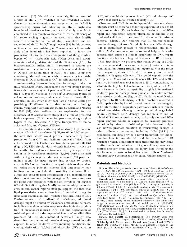
Generation of O2 and H2O2 in Anaerobic MnCl2 Solutionsduring IrradiationIn vitro, the stoichiometry of intermediates and end
products of Mn redox cycling is dependent on the concen-tration of reactants [13]. For example, Mn(III) accumulates ifH2O2 becomes limiting, whereas an excess of O2
�� and Hþ
favors the formation of H2O2 [13] (Figure 1). Using assaysbased on Rhodazine D, a sensitive colorimetric reagent formeasuring O2 and H2O2, we tested whether or not MnCl2solutions exposed anaerobically to 10 kGy generated thesespecies (Figures 5A and S1). Dissolved O2 and H2O2 react withthe pale yellow leuco form of Rhodazine D to produce a rosecolor, with the color proportional to the dissolved O2 orH2O2 concentration. Color development in an Ar-purgedsolution tested after irradiation (Figure 5A, column III), butnot upon re-purging with Ar (Figure 5A, column V), indicatedO2 formation. Color development in an Ar-purged solutiontested after irradiation and upon re-purging with Ar (Figure5A, column V), but not following catalase treatment (Figure5A, column VI), indicated H2O2 accumulation. Consistentwith the existence of a threshold concentration of Mn(II)needed for Mn redox cycling [13] under in vitro irradiationwere (1) exposure of anaerobic dH2O or MnCl2 solutions at
Figure 2. Mn(II) Protects Proteins, but Not DNA, during In Vitro
Irradiation
(A) DMSO-mediated DNA protection. pUC19 plasmid DNA was irradiatedaerobically to the indicated doses in dH2O, 1% DMSO (HO� scavenger), or5 mM MnCl2, followed by agarose gel electrophoresis (AGE). MnCl2 andDMSO were prepared in dH2O. IR, 60Co, aerobic at 0 8C. L, linear (2,686base pairs); Lp, pUC19 þ BamHI; M, size markers; OC, open circular; SC,supercoiled.(B) Mn(II)-mediated protein protection. BamHI enzyme was irradiatedaerobically to the indicated doses in dH2O, 1% DMSO, or 5 mM MnCl2,and then incubated with k-phage DNA for 1 h at 37 8C, followed by AGE.Inset (white border), top gel (dH2O): BamHI irradiated anaerobically. M,size markers; U, uncut k-DNA.(C) Western blot immunoassay of protein-bound carbonyl groups inBamHI irradiated aerobically to the indicated doses in the presence or
absence of 5 mM MnCl2 and/or 200 lM FeCl2. Approximately 220-ngBamHI were loaded per lane in the Western blot (W) and in theCoomassie-stained polyacrylamide denaturing gel (C); M, mixture ofartificial IgG-binding protein standards; S, wide-range protein standards.doi:10.1371/journal.pbio.0050092.g002
PLoS Biology | www.plosbiology.org April 2007 | Volume 5 | Issue 4 | e920771
Radioprotection of Protein by Mn(II)

concentrations below 0.1 mM to 10 kGy did not generatedetectable levels of O2 or H2O2 (Figure 5A and 5B); (2)exposure of anaerobic MnCl2 solutions at intermediateconcentrations (0.1–10 mM) to 10 kGy generated H2O2
(Figure 5A); and (3) from initially anaerobic conditions,exposure of MnCl2 solutions at high concentrations (1 M) to10 kGy yielded O2, but not H2O2 (Figure 5A), and copious Mndioxide precipitates (Figure S2); we infer that the accumu-lation of Mn dioxides was caused by a shortage of H2O2 [13],which is decomposed by c-radiolysis [15].
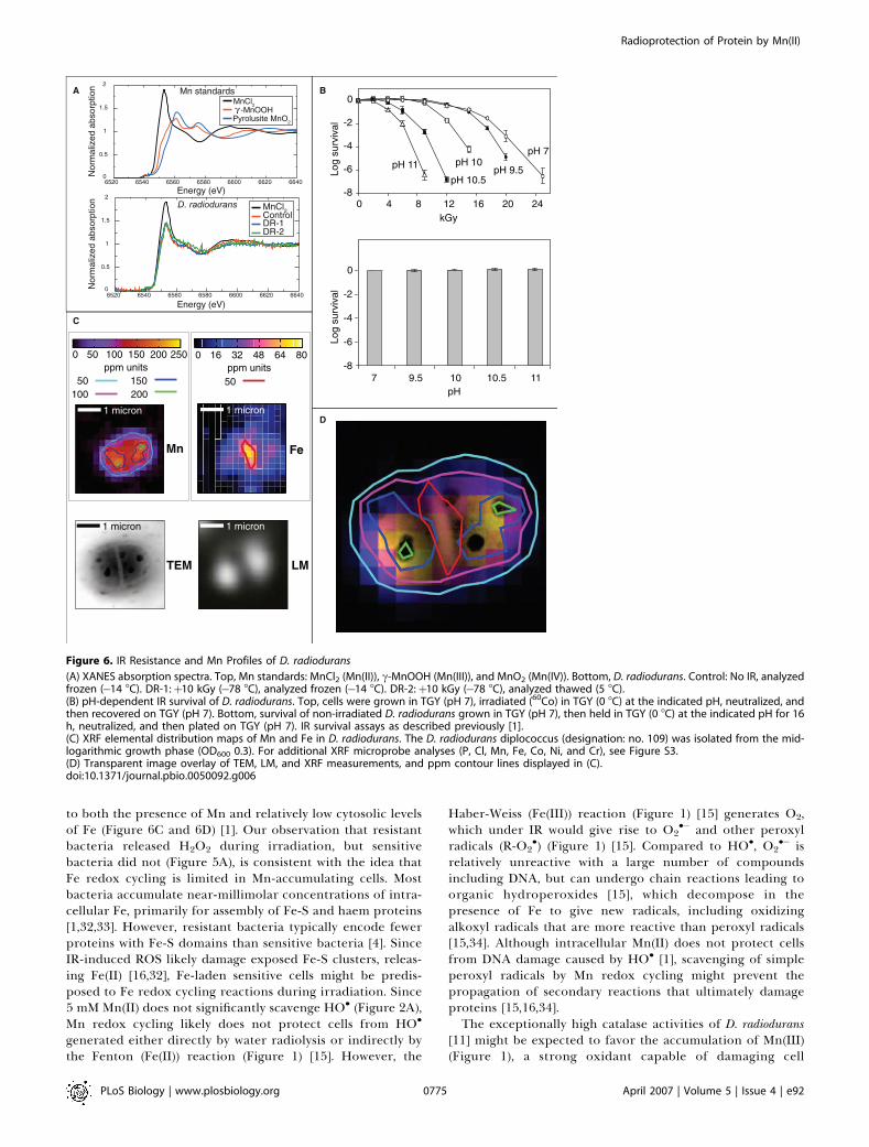
Mn-Accumulating Cells Release H2O2 during IrradiationThe obligate aerobic D. radiodurans accumulates 2 mM or
greater Mn (1 3 105 Mn atoms/cell, assuming an average cellvolume of 6.5 3 10�2 lm3) [1], which is present in cells asMn(II) (Figure 6A) [1], and the facultative anaerobic, radio-resistant bacterium Lactobacillus plantarum [1,22] accumulates20–25 mM Mn(II) [23]. For an irradiated cell containing 2–25mM Mn engaged in catalytic Mn redox cycling [13], intra-cellular O2
��, which does not easily cross biological mem-branes [16,24], might be reduced to H2O2, which is membranepermeable and could diffuse out of the cell and accumulate(Figure 1). When exposed to 10 kGy as cell suspensions indH2O at 0 8C and pre-conditioned to be anaerobic, H2O2 wasreleased by D. radiodurans (;2 3 10�5 M) and L. plantarum (;63 10�5 M), in which the presence of H2O2 after irradiation
was confirmed by catalase-treatment (Figure 5A); notably,H2O2 was consumed by the cells upon incubation at 32 8C(Figure 5A). We infer that H2O2 was produced by intracellularMn(II,III) redox cycling. In contrast, detectable levels of H2O2
or O2 were not released by non-irradiated D. radiodurans or L.plantarum control samples, nor by irradiated S. oneidensis(Figure 5A), which accumulates substantially more Fe thanMn and is extremely sensitive to IR [1,5].
Increasing pH Inhibits Mn Redox CyclingReducing the equilibrium concentration of Hþ with
hydroxide ions (OH�) is predicted to limit Mn redox cycling[13] and the accumulation of H2O2 (Figure 1). In testing thepossibility that survival of irradiated D. radiodurans isvulnerable to conditions that limit intracellular acidification,we first established that IR-driven Mn redox cycling could beinhibited in vitro by increasing the pH. In vitro, exposure toIR of anaerobic 10 mM MnCl2 at pH 9 or higher yielded O2
but not H2O2, whereas H2O2 was formed at pH values below 9(Figure 5B). Consistent with the prediction that Mn redoxcycling can be inhibited in vivo by OH�, D. radioduransirradiated at pH 10.5 did not release H2O2 or O2 (Figure 5A).In vivo, the 10% survival value (D10) [1] of D. radiodurans cellsgrown, irradiated, and recovered at pH 7 was 16 kGy, whereasthe D10 was 6 kGy for cells grown at pH 7, irradiated at pH10.5, and recovered at pH 7 (Figure 6B, top); survival of non-
Figure 3. In Vivo IR-Induced Oxidative Protein Damage
Samples were treated (þ) or not treated (�) with DNPH. For the Western blot (W) and for the Coomassie-stained polyacrylamide denaturing gel (C), 20-lg protein samples were loaded per lane. DR/Fe/0.4 kGy indicates that the D. radiodurans (non-irradiated) cell extract was adjusted to 500 lM FeCl2 andexposed to 0.4 kGy in vitro. Values for intracellular Mn/Fe concentration ratios and D10 at the bottom of the figure, as reported previously [1]. M, mixtureof artificial IgG-binding protein standards; O, oxidized protein standards; S, wide-range protein standards; SO, S. oneidensis.doi:10.1371/journal.pbio.0050092.g003
PLoS Biology | www.plosbiology.org April 2007 | Volume 5 | Issue 4 | e920772
Radioprotection of Protein by Mn(II)

irradiated D. radiodurans control samples held at a pH of 11 orlower for 16 h was approximately 100% (Figure 6B, bottom).In vivo, the pH-dependent loss in IR resistance of D.radiodurans (Figure 6B, top) correlated with a substantialincrease in oxidative protein damage during irradiation(Figure 4C). Thus, at pH values at which IR-driven Mn redoxcycling was inhibited in vitro (Figure 5B), the IR resistance ofD. radiodurans was significantly decreased, and the cells werehighly susceptible to IR-induced protein oxidation (Figure4C).
Distribution of Mn and Fe in D. radioduransBecause the formation of ROS during irradiation is
extremely rapid [15], an intracellular protection systemwhich is ubiquitous, but not highly dependent on theinduction of enzymes, stage of growth, or temperature overa range at which cells are metabolically active, could providea selective advantage to the host in some environments. Inthis context, we examined the intracellular distribution of Mnand Fe in D. radiodurans cells using X-ray fluorescence (XRF)microprobe analysis [25] (Figures 6C and S3). Within a
Figure 4. Additional Oxidative Protein Damage Assays
(A) In vitro IR-induced oxidative protein damage. Western blot (W) immunoassay of protein-bound carbonyl groups in D. radiodurans (non-irradiated)cell extract adjusted to 500 lM FeCl2 and irradiated to the indicated doses. A total of 20 lg of protein extract loaded per lane.(B) Mn-depleted, radiosensitive D. radiodurans cells [1] are highly susceptible to oxidative protein damage during irradiation. D. radiodurans was grownin defined rich medium without Mn supplementation (no-Mn DRM) [1] to OD600 0.8 and exposed aerobically to 10 kGy. A total of 20 lg of proteinextract loaded per lane.(C) Decreased survival of D. radiodurans irradiated at pH 10.5 correlates with oxidative protein damage. D. radiodurans was grown to OD600
approximately 0.9 in TGY (pH 7), adjusted to pH 10.5, and exposed aerobically to the indicated doses. A total of 20 lg of protein extract loaded per lane.(D) P. putida proteins are similarly susceptible to oxidative protein damage when cells are irradiated anaerobically (þAr) or aerobically (þO2). P. putidawas grown to OD600 approximately 0.9 in TGY, purged with ultra-high purity Ar, and irradiated in sealed tubes to 4 kGy.Values for intracellular Mn/Fe concentration ratios and D10 at the bottom of (B), (C), and (D), as reported previously [1]. A total of 20 lg of protein extractloaded per lane.C, Coomassie-stained polyacrylamide denaturing gel; M, mixture of artificial IgG-binding protein standards; O, oxidized protein standards; S, wide-rangeprotein standards; þ, DNPH treated;�, DNPH untreated.doi:10.1371/journal.pbio.0050092.g004
PLoS Biology | www.plosbiology.org April 2007 | Volume 5 | Issue 4 | e920773
Radioprotection of Protein by Mn(II)

representative diplococcus, whereas Mn(II) was globallydistributed, Fe was partitioned largely outside the cytoplasmin a region overlapping the septum between dividing cells(Figure 6D).
Discussion
We previously demonstrated a critical role for theaccumulation of Mn(II) in D. radiodurans in a mechanismtoward surviving IR that is not dependent on Mn-SOD (Mn-dependent superoxide dismutase) [1]. D. radiodurans containsfour to ten identical copies of its genome per cell, and whenirradiated to a dose of 10 kGy, generates more than 400genomic DSB fragments per cell [1,8,26]. Yet, this amount ofDNA damage in D. radiodurans does not typically lead to celldeath [1,8]. Bioinformatic and experimental reports generallysupport that genome configuration and copy number, andenzymatic protection and repair functions of D. radioduransdo not have unique properties that are essential orprerequisite for expression of the extreme-resistance pheno-type [1–12]. For example, D. radiodurans DNA repair andprotection genes do not differ greatly from their counter-parts in the IR-sensitive S. oneidensis, P. putida, or E. coli [1,6];several E. coli DNA repair genes have been shown to fullyrestore corresponding radiation-sensitive D. radiodurans mu-tants to wild-type levels of D. radiodurans resistance [12,27];Mn-SOD is not needed for survival of D. radiodurans followingacute irradiation or growth under 50 Gy/h [1,11]; non-homologous end joining of D. radiodurans chromosomal DSBfragments following IR is not observed [28]; and the productsof interchromosomal recombination in D. radiodurans follow-ing irradiation are consistent with the canonical version ofthe DSB repair model [8]. Over the last decade, severalhypotheses to reconcile these findings have built on the ideathat D. radiodurans might use mechanisms that restrict thediffusion of DNA DSB fragments produced followingirradiation, to facilitate repair [3,29]. However, transmissionelectron microscopy [TEM] of Mn-depleted, radiosensitive D.radiodurans displayed normal levels of chromosomal conden-sation [1], and cryoelectron microscopy of vitreous sectionsof D. radiodurans supports the conclusion that DNA fragmentsin D. radiodurans are mobile and that the arrangement of itsnucleoids does not play a key role in radioresistance [10,30].Consistently, a series of earlier molecular studies onirradiated D. radiodurans cells showed high levels of recombi-nation between homologous DSB fragments originating fromwidely separated genomic locations [8,28,31]. Evidencepresented here supports the idea that the extreme-resistancephenotype of D. radiodurans and other bacteria with highintracellular Mn/Fe concentration ratios is dependent on amechanism that protects proteins from oxidative damageduring irradiation, which could offset the need for highlyspecialized cellular repair systems.Previous work has shown that the linear density of DSBs
introduced into genomic DNA by a given dose of IR inextremely resistant and sensitive bacteria is essentially thesame [1,2]. In contrast, we find that protein damage isquantifiably related to bacterial radioresistance (Figures 3,4B, and 4C). The most-sensitive cells had very low Mn/Feconcentration ratios andwere highly susceptible to IR-inducedprotein oxidation, whereas the most-resistant cells had highintracellular Mn/Fe ratios and were relatively insusceptible toprotein oxidation. Although the mechanism by which Mnprotects proteins during irradiation remains unknown, ourresults provide insight into how Mn redox cycling couldattenuate the detrimental effects of Fe redox cycling.Scavenging of ROS in IR-resistant bacteria may be linked
Figure 5. Mn-Dependent IR-Driven O2 and H2O2 Generation
(A) Column I. No IR, aerobic; II. No IR, anaerobic (pre-conditioned bypurging with Ar [ultra-high purity]); III. 10 kGy, anaerobic; IV. 10 kGy,anaerobic, followed by incubation at 32 8C for 60 min; V. As for column III,but re-purged with Ar after irradiation; and VI. As for column III, buttreated with catalase (15,000 units) after irradiation and then re-purgedwith Ar. O2/H2O2 concentration determined by the Rhodazine D assay.See Figure S2 for additional assays. Irradiations (60Co) were at 0 8C. MnCl2solutions and bacteria (1.63109 cells/ml) were prepared in dH2O (pH ;6).(B) Standards.doi:10.1371/journal.pbio.0050092.g005
PLoS Biology | www.plosbiology.org April 2007 | Volume 5 | Issue 4 | e920774
Radioprotection of Protein by Mn(II)
to both the presence of Mn and relatively low cytosolic levelsof Fe (Figure 6C and 6D) [1]. Our observation that resistantbacteria released H2O2 during irradiation, but sensitivebacteria did not (Figure 5A), is consistent with the idea thatFe redox cycling is limited in Mn-accumulating cells. Mostbacteria accumulate near-millimolar concentrations of intra-cellular Fe, primarily for assembly of Fe-S and haem proteins[1,32,33]. However, resistant bacteria typically encode fewerproteins with Fe-S domains than sensitive bacteria [4]. SinceIR-induced ROS likely damage exposed Fe-S clusters, releas-ing Fe(II) [16,32], Fe-laden sensitive cells might be predis-posed to Fe redox cycling reactions during irradiation. Since5 mM Mn(II) does not significantly scavenge HO� (Figure 2A),Mn redox cycling likely does not protect cells from HO�
generated either directly by water radiolysis or indirectly bythe Fenton (Fe(II)) reaction (Figure 1) [15]. However, the
Haber-Weiss (Fe(III)) reaction (Figure 1) [15] generates O2,which under IR would give rise to O2
�� and other peroxylradicals (R-O2
�) (Figure 1) [15]. Compared to HO�, O2�� is
relatively unreactive with a large number of compoundsincluding DNA, but can undergo chain reactions leading toorganic hydroperoxides [15], which decompose in thepresence of Fe to give new radicals, including oxidizingalkoxyl radicals that are more reactive than peroxyl radicals[15,34]. Although intracellular Mn(II) does not protect cellsfrom DNA damage caused by HO� [1], scavenging of simpleperoxyl radicals by Mn redox cycling might prevent thepropagation of secondary reactions that ultimately damageproteins [15,16,34].The exceptionally high catalase activities of D. radiodurans
[11] might be expected to favor the accumulation of Mn(III)(Figure 1), a strong oxidant capable of damaging cell
Figure 6. IR Resistance and Mn Profiles of D. radiodurans
(A) XANES absorption spectra. Top, Mn standards: MnCl2 (Mn(II)), c-MnOOH (Mn(III)), and MnO2 (Mn(IV)). Bottom, D. radiodurans. Control: No IR, analyzedfrozen (�14 8C). DR-1:þ10 kGy (�78 8C), analyzed frozen (�14 8C). DR-2:þ10 kGy (�78 8C), analyzed thawed (5 8C).(B) pH-dependent IR survival of D. radiodurans. Top, cells were grown in TGY (pH 7), irradiated (60Co) in TGY (0 8C) at the indicated pH, neutralized, andthen recovered on TGY (pH 7). Bottom, survival of non-irradiated D. radiodurans grown in TGY (pH 7), then held in TGY (0 8C) at the indicated pH for 16h, neutralized, and then plated on TGY (pH 7). IR survival assays as described previously [1].(C) XRF elemental distribution maps of Mn and Fe in D. radiodurans. The D. radiodurans diplococcus (designation: no. 109) was isolated from the mid-logarithmic growth phase (OD600 0.3). For additional XRF microprobe analyses (P, Cl, Mn, Fe, Co, Ni, and Cr), see Figure S3.(D) Transparent image overlay of TEM, LM, and XRF measurements, and ppm contour lines displayed in (C).doi:10.1371/journal.pbio.0050092.g006
PLoS Biology | www.plosbiology.org April 2007 | Volume 5 | Issue 4 | e920775
Radioprotection of Protein by Mn(II)
components [13]. We did not detect significant levels ofMn(III) or Mn(IV) in irradiated or non-irradiated D. radio-durans by X-ray-absorption near-edge structure (XANES)spectroscopy (Figure 6A), indicating that Mn(III) might alsobe reduced by other mechanisms in vivo. Notably, when Mn iscomplexed with succinate or lactate in vitro, the efficiency ofMn redox cycling is greatly increased, such that Mn(III)reactivity is similar to that of Mn(II) complexes and re-reduction of Mn(III) by O2
��might occur [13]. In this context,metabolic pathway switching in D. radiodurans cells immedi-ately after irradiation has been reported to favor theproduction of succinate via up-regulation of the glyoxylatebypass of its tricarboxylic acid (TCA) cycle, and down-regulation of degradative steps of the TCA cycle [4,5,9]. Inbicarbonate/CO2 buffer, Mn(II) is reported to catalyze theoxidation of free amino acids such as leucine and alanine byH2O2 and the dismutation of H2O2 [35]. Thus, complexescontaining Mn and amino acids or organic acids mightscavenge H2O2 in addition to O2
�� in cells exposed to IR.An interesting feature of the systems for energy production
in D. radiodurans is that, unlike most other free-living bacteria,it uses the vacuolar type of proton ATP synthase instead ofthe F1F0 type [6]. Vacuolar (V)-type Hþ-ATPases are typical ofeukaryotes and archaea, and central players in intracellularacidification [36], which might facilitate Mn redox cycling byproviding Hþ (Figure 1). In this context, our findingsgenerally support bioinformatic studies by Karlin and Mrazekin 2001 [37], who proffered a new explanation for theresistance of D. radiodurans contingent on a role of predictedhighly expressed (PHX) genes for proteases, the glyoxylatebypass of the TCA cycle, ABC-type transporters of aminoacids and Mn, and (V)-type ATPases.
The speciation, distribution, and relatively high concen-tration of Mn in D. radiodurans [1] (Figure 6A and 6C) supportthe idea that Mn(II) could provide immediate cytosolicprotection from O2
��, and facilitate removal of H2O2 fromcells exposed to IR. Further, electron-dense granules (EDGs)(Figure 6C, TEM, circular dark ;0.2 lM inclusions), which arefrequently observed in electron microscopy images at thecenter of D. radiodurans nucleoids [1,4,10], were associatedwith the highest regional Mn concentrations (200 parts permillion [ppm]; 3.6 mM) (Figure 6D), perhaps to protectenzymic DNA repair functions, many of which are dependenton redox-active [4Fe-4S] clusters [38]. Importantly, ourfindings do not preclude the possibility that intracellularMn(II) also prevents lipid peroxidation in cell membranes. Inthis context, however, the lowest regional Mn concentrations(50 ppm) were associated with the cell envelope [6] (Figures6C and S3), indicating that Mn(II) predominantly protects thecytosol; and earlier reports strongly support the idea thatlipid peroxidation can be dissociated from lethal damage inirradiated mammalian and irradiated bacterial cells [34,39].During recovery of irradiated D. radiodurans, additionaldamage might be limited by secondary antioxidant defenses,including attendant cellular responses that limit the produc-tion of metabolism-induced ROS [4,9], and degradation ofoxidized proteins by the expanded family of subtilisin-likeproteases [6]. The Mn content of bacteria [1] might alsodetermine the amount of protein damage caused in cellsexposed to other oxidative stress-inducing conditions, in-cluding desiccation [1,6,24] and ultraviolet (UV) radiation
[5,12], and xenobiotic agents such as Cr(VI) and mitomycin-C(MMC) that elicit redox-related toxicity [40].Chromosomal DNA is an indispensable molecule whose
integrity must be conserved following exposure of a cell to IRto ensure survival [15], such that the functionality of DNArepair and replication systems ultimately determines if anirradiated cell lives or dies, even for the most IR-resistantbacteria [6,12,27]. Our findings that IR-induced cellularprotein damage (Figures 3 and 4), but not DNA damage[1,2], is quantifiably related to radioresistance, and intra-cellular Mn/Fe concentration ratios could help explain whybacteria that encode a similar repertoire of DNA repairfunctions display such large differences in IR resistance[1,3,5]. Specifically, we propose that redox cycling of Mn(II)that is accumulated in resistant bacteria [1] protects proteinsfrom oxidation during irradiation (Figure 1), with the resultthat enzyme systems involved in recovery survive andfunction with great efficiency. This could explain why thepolA gene of E. coli fully complements IR-, UV- and MMC-sensitive D. radiodurans polA mutants [12]. In comparison, weattribute the high level of radiation sensitivity of Fe-rich, Mn-poor bacteria to their susceptibility to global Fe-mediatedoxidative protein damage during irradiation under aerobicor anaerobic conditions (Figure 4D). Oxidative modificationof proteins by IR could disrupt cellular functions involved inDNA repair either by loss of catalytic and structural integrityor by interruption of regulatory pathways, which in extremelyradiation-sensitive cells might render protein damage lethalbefore significant DNA damage has accumulated [5]. Atsublethal IR doses in sensitive cells, oxidatively damaged DNArepair enzymes would be expected to passively promotemutations by misrepair. Oxidized proteins, however, mightalso actively promote mutation by transmitting damage toother cellular constituents, including DNA [34,41]. Inconclusion, our data provide a novel framework for under-standing how intracellular Mn and Fe contribute to IRresistance, which is important since these findings may cometo affect models of radiation toxicity, as well as approaches tocontrol recovery from radiation injury [42], including thedevelopment of systems for delivery into cells of Mn-basedradioprotective complexes or Fe-based radiosensitizers [43].
Materials and Methods
Strains. The wild-type strains used were as follows: D. radiodurans(ATCC BAA-816); D. geothermalis (DSM 11300); S. oneidensis (MR-1)(ATCC 700550); P. putida (ATCC 47054); Enterococcus faecium (ATCC19434); L. plantarum (ATCC 14917); and E. coli (K-12) (MG1655).
Growth and c-irradiations. Strains were cultured aerobically inundefined liquid rich medium [1] (TGY: 1% Bacto-tryptone, 0.1%glucose, 0.5% yeast extract) (pH 7) at 32 8C to an optical density at600 nm (OD600) of 0.9–1.0, unless indicated otherwise. For anaerobicirradiations, 6-ml 0–1,000 mM MnCl2 solutions in dH2O (pH ;6), orapproximately 1 3 1010 bacterial cells (;5 3 109 diplococci) re-suspended in 6-ml dH2O, were transferred to Quick-Seal ultra-centrifuge tubes (13 cm3) (Beckman Instruments, Palo Alto, Cal-ifornia, United States), unless indicated otherwise. The tubes werepurged at room temperature with ultra-high purity Ar (99.999%)(ValleyNational Gases, Frederick, Maryland, United States) for 5 min(200 cm3/min), sealed anaerobically, and irradiated on ice (0 8C) at 1.8Gy/s (60Co, Model 109; J. L. Shepard and Associates, San Fernando,California, United States).
pUC19 and BamHI IR sensitivities. The pUC19 assay wasperformed as follows. Supercoiled pUC19 (1 mg/ml) (New EnglandBiolabs, Ipswich, Massachusetts, United States) was diluted (1/25) indH2O, 1% DMSO, or 5 mM MnCl2; 50-ll aliquots of each of thethree pUC19 dilutions were irradiated (60Co) at 0 8C to the
PLoS Biology | www.plosbiology.org April 2007 | Volume 5 | Issue 4 | e920776
Radioprotection of Protein by Mn(II)

indicated doses. A total of 88 ng of each IR-treated pUC19 samplewas subjected to agarose (0.9%) gel electrophoresis in 1 3 TBE (Tris,borate, EDTA buffer) and 250-ng/ml ethidium bromide, at 47 V for14 h. Linearized plasmid (Lp) (pUC19 þ BamHI) was used as amarker in gels containing IR-treated supercoiled pUC19.
The BamHI assay was performed as follows. BamHI (700,000 U/ml)(New England Biolabs) was diluted (1/1,500) in dH2O, 1% DMSO, or 5mM MnCl2; 1,000-ll aliquots of each of the three BamHI dilutionswere irradiated (60Co) aerobically at 0 8C to the indicated doses. Foranaerobic BamHI irradiations, 5-ml aliquots of diluted BamHI/dH2Owere transferred to separate Quick-Seal ultracentrifuge tubes (13cm3) (Beckman Instruments), purged at room temperature with ultra-high purity Ar (99.999%) (Valley National Gases) for 5 min (200 cm3/min), and sealed anaerobically before irradiation. A total of 40 ll (23units) of each IR-treated BamHI sample was transferred to separatereaction mixes (final volume, 60 ll) containing 250-ng k-phage DNA(New England Biolabs), 50 mM NaCl, 10 mM Tris-HCl (pH 7.9), 10 mMMgCl2, and 1 mM dithiothreitol. BamHI/k DNA reactions wereincubated for 1 h at 37 8C, followed by agarose (0.7%/1 3 TBE) gelelectrophoresis at 23 V for 18 h.
Protein extractions. The 650-ml cultures of the indicated bacteriagrown in TGY to OD600 0.9 were harvested by centrifugation,resuspended in 30 ml TGY, and exposed to IR (0 8C). Irradiated andnon-irradiated (control) cells were washed and then resuspended inlysis buffer (50 mM potassium phosphate buffer [pH 7.0], 0 8C). A cellsuspension (;4 3 109 cells/ml, 2–4 ml) was adjusted to 1% (v/v) b-mercaptoethanol and passed through a French pressure cell (0 8C) at20,000 lb/in2, and the lysate centrifuged twice at 12,0003 g at 4 8C for30 min. The protein concentration of a supernatant was determinedby the Coomassie (Bradford) assay (BioRad, Hercules, California,United States), and the samples were then diluted with lysis buffer/1%b-mercaptoethanol to 20-lg/ll protein, divided into 50-ll aliquots,and stored at�80 8C. The addition of b-mercaptoethanol prior to celllysis and for storage was recommended in the protocols accompany-ing the OxyBlot Oxidation Detection Kit (Chemicon International,Temecula, California, United States) (see below) to prevent theoxidation of proteins during and after cell lysis. Assays for oxidativeprotein damage (Figure 3) were all conducted in parallel within 3 d ofprotein extraction.
Assay for oxidative protein damage. Protein oxidation in freshlyprepared extracts was measured using OxyBlot Protein OxidationDetection Kit (S7150) (Chemicon International), including theindicated molecular-weight markers. The carbonyl groups in theprotein side chains were derivatized to 2,4-dinitrophenylhydrazoneby reaction with 2,4-dinitrophenylhydrazine (DNPH) for 15 min in3% (w/v) SDS. Western blotting: the DNP-derivatized protein sampleswere separated by polyacrylamide denaturing gel electrophoresis(4%–20% gradient gels; BioRad) at 195 V for 50 min followed bytransferring proteins to a nitrocellulose membrane for 40 min(BioRad). The membranes were incubated with primary antibody,specific to the DNP moiety of the proteins. This step was followed byincubation with a horseradish peroxidase-antibody conjugate direc-ted against the primary antibody (secondary antibody: goat anti-rabbit IgG). The membranes were then treated with chemilumines-cent (SuperSignal) substrate (Pierce Biotechnology, Rockford, Illinois,United States) and imaged by exposure to light sensitive films(BIOMAX Light Film; Kodak, Rochester, New York, United States).
Rhodazine D O2/H2O2 assay. O2 and H2O2 concentrations weredetermined by the Rhodazine D assay (RDA) (CHEMetrics, Calverton,Virginia, United States). All Quick-Seal centrifuge tubes containingcells were centrifuged at 4 8C (2,000 3 g, 10 min) after irradiation.Supernatants were tested by the RDA. The RDA is suitable formeasuring 0.1–1.0 mg/l O2, and 1–10 3 10�5 M H2O2. Mn(III,IV) areoxidants that will also cause a positive RDA test result. However,Mn(III,IV) dioxides are insoluble in water (circa-neutral pH) andreadily removed from suspension by centrifugation (2,0003 g, 5 min)or standing. RDA results indicating 1 mg/l or more O2 or 103 10�5 Mor greater H2O2 were also tested by the Indigo Carmine assay(CHEMetrics), which yields a blue color suitable for measuring higherconcentrations of O2 and H2O2. Once a test solution had filled anassay vial, the open end was sealed anaerobically with vacuum grease,and the vial was stored in the dark.
XANES. Mn K-edge XANES spectra were measured [44] intransmission on standard compounds (MnCl2, c-MnOOH, andMnO2) and in fluorescence on the D. radiodurans samples. Themeasurements on c-MnOOH and MnO2 were made on fine powdersspread evenly onto adhesive tape and folded to make samples ofappropriate thickness for the transmission experiment. The 1 MMnCl2 solution and D. radiodurans cell suspensions (OD600 0.9) wereabsorbed on paper and cellulose acetate filters, respectively, frozen,
and then stored at �80 8C. For XANES measurements, samples weretransferred from dry ice (�78 8C) to a sample stage consisting of aninsulated Peltier stack kept under a helium atmosphere. Such sampleswere maintained at �14 8C during the analysis. The XANES spectrawere measured at the MRCAT [45] (Materials Research CollaborativeAccess Team) sector 10ID beamline at the Advanced Photon Source(APS). The 10ID is an insertion device beamline using APS undulator-A [46]. The undulator was tapered by approximately 2 keV to reducethe variation in incident radiation to less than 15% over the energyrange used in these measurements. The incident X-ray beam was theundulator fundamental and was monochromated using the (111)reflection of a liquid-nitrogen–cooled, double-crystal, silicon mono-chromator. Higher harmonic content of the incident beam wasrejected using a polished float glass mirror. Ionization chambers wereused to measure the incident and transmitted intensities, and werefilled with 10% nitrogen/90% helium and 100% nitrogen, respec-tively. The fluorescence spectra were measured with an ionizationdetector in the Stern-Heald geometry [47] filled with Ar gas, and useda chromium oxide filter of three absorption-lengths thickness toreduce the background signal. The incident X-ray beam was 1 mmsquare. Linearity tests [48] indicated less than 0.04% variation for a50% decrease in incident X-ray intensity. The XANES data wereprocessed using the Athena program [49]. All data scans were alignedin energy using a reference spectrum derived from metallicmanganese. Between five and 20 scans per sample were averaged toimprove measurement statistics. The averaged data were normalized[44] by regressing a line to the pre-edge region between approx-imately 6,400–6,500 eV. This line was subtracted from the data. Aquadratic polynomial was regressed to the region from approx-imately 6,600 eV to the end of the data range. This quadraticpolynomial was extrapolated to the edge energy (6,548.5 eV for thesamples containing D. radiodurans). The normalized data were dividedby the value of this polynomial at the edge energy. Notably, the edgepositions of XANES spectra of irradiated or non-irradiated D.radiodurans samples were essentially the same as for aqueous MnCl2solutions. This is consistent with oxygen atoms surrounding Mn(II) inD. radiodurans, and with no significant change in Mn valence duringirradiation, which would have been observed in cell samplescontaining 5% or more Mn(III,IV).
XRF. XRF microprobe analysis measurements [25] were made atbeamline 2ID-D at the APS [50]. The 2ID-D is an undulator beamlinewith Fresnel zone plates focusing optics that produced a focal spotwith a FWHM (full width at half maximum) spatial resolution ofapproximately 120 nm for these experiments. A silicon (111) double-crystal monochromator was used to shine 10-keV photons on theFresnel zone plates. The sample was rastered through the focal spot ofthe X-ray beam. For each pixel, the full XRF spectrum betweenapproximately 2 keV and 10 keV was measured using a silicon driftdetector. Thus, the distribution of elements between phosphorus andzinc on the periodic table of elements could be measured with 120-nm resolution throughout a cell and its periphery. XRF microprobemeasurements were made on D. radiodurans cells grown to OD600 0.3.The cells were deposited on grids as suspensions in TGY liquidmedium, which served to help maintain the structure and viability ofthe cells as they dried. The complete results for two distinctdiplococci (no. 109 and no. 110) are presented in Figure S3. Theposition coordinates of individual diplococci mounted on formvar-coated gold TEM grids were determined by light microscopy (LM).TEM grids were subsequently placed in the focal plane of the X-rayoptic, and the grid markers were used to locate the targeted cells.Data from 2ID-D were initially processed using the program MAPS[51]. The average XRF spectral intensities from 28 adjacent pixels inthe upper right-hand corners of XRF images, in areas beyond theboundaries of a cell (Figure S3), were used to determine thebackground signal for the elements under investigation. The averagebackground spectrum for an element being mapped was subtractedfrom all other pixels of the image. Next, a mathematical modelrepresenting the original morphology of cells was constructed inapproximate likeness to diplococci, which resemble adjoinedtruncated spheres. Within each pixel, the intensity of an element’sXRF signal was weighted by the average thickness of the diplococcussubtended by a respective pixel. The contour lines are straight-linesegments between points of equal intensity, in which linearinterpolation of the actual data was used to determine the locationsof the contour values. The contours were generated using thealgorithm Gnuplot (http://www.gnuplot.info). In combination, thisapproach limited the effect of cell thickness on the elementaldistribution maps, and facilitated correlation between electron-densegranules (EDGs) and Mn hotspots. Element distributions arepresented as mass parts per million (ppm).
PLoS Biology | www.plosbiology.org April 2007 | Volume 5 | Issue 4 | e920777
Radioprotection of Protein by Mn(II)

TEM. Whole cells deposited on formvar-coated gold TEM gridsand analyzed by XRF microprobe analysis were subsequently imagedin a FEI Tecnai TF30 transmission electron microscope equippedwith a field-emission gun and operated at an accelerating voltage of300 kV. An objective aperture with a diameter of 40 lm was selectedto optimize contrast due to differences in elastic scattering across thespecimen. Bright-field electron micrographs were recorded using aGatan Ultrascan Model 894 cooled CCD camera containing fourmega-pixels and operated with a four-port parallel read-out. Imageswere analyzed using Gatan Digital Micrograph software (version 3.4).Pixel intensities were transformed to a logarithmic scale to visualizethe EDGs (; 0.2 lm in diameter) in cells.
LM. LM was used to identify the position coordinates of discretediplococci on formvar-coated gold TEM grids before XRF microp-robe analysis (Figure S3); gold grids were supported by glass slidesduring LM. LM images were captured using a Leica DM RXA epi-fluorescent microscope (Leica, Wetzlar, Germany). Differentialinterference contrast images were recorded using a Scion Corpo-ration (Frederick, Maryland, United States) CCD camera andanalyzed with xyzGrabber.
Supporting Information
Figure S1. Additional Assays for O2/H2O2
(A) Indigo carmine assay (ICA) (CHEMetrics). Control (No IR). dH2Oand MnCl2 solutions were pre-conditioned by purging with O2-freeN2 (anaerobic).(B) As for (A), but exposed to IR (10 kGy).(C) O2/H2O2 was not detected in irradiated (10 kGy) TGY solidmedium. After irradiation of TGY agar under O2-free N2, anaerobicdH2O was equilibrated in the agar tubes for 24 h before testing withthe ICA and Rhodazine D assay (RDA).(D) RDA. IR-dependent O2/H2O2 formation in irradiated 2 mMMnCl2 solutions (anaerobic).(E) ICA. IR-dependent O2/H2O2 formation in irradiated MnCl2solutions (anaerobic).(F) As for (E), but O2/H2O2 determined by the RDA.(G) Reference ICA color standards. A sample tube is shown: 10 mMMnCl2 þ IR (10 kGy).(H) Reference RDA color standards.Theoretical rate constants for primary radiolytic reactions (TheRadiation Chemistry Data Center: http://www.rcdc.nd.edu/RCDC/RCDC.html) involving the formation and decomposition of ROS inanaerobic dH2O do not favor significant accumulation of H2O2 or O2,which we did not detect in irradiated anaerobic dH2O or diluteMnCl2 solutions (,0.1 mM) ([E] and [F]).
Found at doi:10.1371/journal.pbio.0050092.sg001 (289 KB PDF).
Figure S2. Oxidation of Mn(II) during c-Radiolysis of Water (60Co, 0 8C)
Column I. 1 M MnCl2, no IR (aerobic); II. 1 M MnCl2 þ 10 kGy(aerobic) (30 min after IR). Red-brown color indicates Mn(III); III. 1 MMnCl2þ10 kGy (aerobic) (24 h after IR). Black color indicates Mn(IV);IV. 10 mM MnCl2þ10 kGy (aerobic) (24 h after IR); and V. 1 M MnCl2þ 10 kGy (anaerobic, purged with Ar before IR) (24 h after IR).Notably, no Mn dioxides were precipitated during irradiation when 1M MnCl2 was purged continuously with Ar (unpublished data), whichfurther supports the idea that HO� do not readily react with Mn(II)(Figure 2A; main text).
Found at doi:10.1371/journal.pbio.0050092.sg002 (138 KB PDF).
Figure S3. LM, TEM, and XRF Element Distribution Maps of D.radiodurans(A) LM showing TEM grid-locations of diplococcus (group of twocells) no. 109 and no. 110.(B) No. 109. (C) No. 110. D. radiodurans (ATCC BAA-816) diplococciwere harvested from a mid-logarithmic culture (OD600 0.3) in TGY.The element distribution images ([B] and [C]) are plotted to differentscales designated by a single-color box, where yellow represents thehighest concentration and black the lowest; ppm values in paren-theses next to the element symbol correspond to yellow. For example,in (B), yellow corresponds to 200 ppm for the Mn map, but only 80ppm for the Fe map.
Found at doi:10.1371/journal.pbio.0050092.sg003 (699 KB PDF).
Acknowledgments
We are grateful to Carol Giometti (Biosciences Division) at ArgonneNational Laboratory for suggesting investigations on protein carbon-ylation, and Sofia del Castillo at Uniformed Services University of theHealth Sciences (USUHS) for technical support.
Author contributions. MJD, EKG, VYM, AV, RDL, BL, BR, KMK,and JKF conceived and designed the experiments. EKG, VYM, AV,MZ, RDL, BL, BR, S-MWL, and KMK performed the experiments.MJD, EKG, VYM, AV, RDL, BL, BR, S-MWL, KMK, and JKF analyzedthe data. MJD wrote the paper, with contributions from JKF.
Funding. This work was supported by grant DE-FG02-04ER63918 toMJD from the US Department of Energy (DOE), Office of Science (OS),Environmental Remediation Sciences Program (ERSP). The AdvancedPhoton Source and the Materials Research Collaborative Access Teamare supported by DOE, OS, Basic Energy Sciences. The work of KMKand BR at the APS was also supported by DOE, OS, ERSP. The work ofRDL was supported by the Intramural Program of the NationalInstitutes of Health.
Competing interests. The authors have declared that no competinginterests exist.
References1. Daly MJ, Gaidamakova EK, Matrosova VY, Vasilenko A, Zhai M, et al. (2004)
Accumulation of Mn(II) in Deinococcus radiodurans facilitates gamma-radiation resistance. Science 306: 1025–1028.
2. Gerard E, Jolivet E, Prieur D, Forterre P (2001) DNA protection mechanismsare not involved in the radioresistance of the hyperthermophilic archaeaPyrococcus abyssiandP. furiosus.MolGenetGenomics266: 72–78.
3. Cox MM, Battista JR (2005) Deinococcus radiodurans—The consummatesurvivor. Nat Rev Microbiol 3: 882–892.
4. Ghosal D, Omelchenko MV, Gaidamakova EK, Matrosova VY, Vasilenko A,et al. (2005) How radiation kills cells: Survival of Deinococcus radiodurans andShewanella oneidensis under oxidative stress. FEMS Microbiol Rev 29: 361–375.
5. Qiu X, Daly MJ, Vasilenko A, Omelchenko MV, Gaidamakova EK, et al.(2006) Transcriptome analysis applied to survival of Shewanella oneidensisMR-1 exposed to ionizing radiation. J Bacteriol 188: 1199–1204.
6. Makarova KS, Aravind L, Wolf YI, Tatusov RL, Minton KW, et al. (2001)Genome of the extremely radiation-resistant bacterium Deinococcus radio-durans viewed from the perspective of comparative genomics. MicrobiolMol Biol Rev 65: 44–79.
7. Zahradka K, Slade D, Bailone A, Sommer S, Averbeck D, et al. (2006)Reassembly of shattered chromosomes in Deinococcus radiodurans. Nature443: 569–573.
8. Daly MJ, Minton KW (1995) Interchromosomal recombination in theextremely radioresistant bacterium Deinococcus radiodurans. J Bacteriol 177:5495–5505.
9. Liu Y, Zhou J, Beliaev A, Stair J, Wu L, et al. (2003) Transcriptome dynamicsof Deinococcus radiodurans recovering from ionizing radiation. Proc NatlAcad Sci U S A 100: 4191–4196.
10. Eltsov M, Dubochet J (2005) Fine structure of the Deinococcus radiodurans
nucleoid revealed by cryoelectron microscopy of vitreous sections. JBacteriol 187: 8047–8054.
11. Markillie LM, Varnum SM, Hradecky P, Wong KK (1999) Targetedmutagenesis by duplication insertion in the radioresistant bacteriumDeinococcus radiodurans: radiation sensitivities of catalase (katA) and super-oxide dismutase (sodA) mutants. J Bacteriol 181: 666–669.
12. Gutman PD, Fuchs P, Minton KW (1994) Restoration of the DNA damageresistance of Deinococcus radiodurans DNA polymerase mutants by Escherichiacoli DNA polymerase I and Klenow fragment. Mutat Res 314: 87–97.
13. Archibald FS, Fridovich I (1982) The scavenging of superoxide radical bymanganous complexes: In vitro. Arch Biochem Biophys 214: 452–463.
14. Stadtman ER, Berlett BS, Chock PB (1990) Manganese-dependent dis-proportionation of hydrogen peroxide in bicarbonate buffer. Proc NatlAcad Sci U S A 87: 384–388.
15. von Sonntag C (1987) The chemical basis of radiation biology. London:Taylor & Francis. 515 p
16. Imlay JA (2003) Pathways of oxidative damage. Annu Rev Microbiol 57:395–418.
17. Levine RL (1983) Oxidative modification of glutamine synthetase. I.Inactivation is due to loss of one histidine residue. J Biol Chem 258:11823–11827.
18. Chou WY, Tsai WP, Lin CC, Chang GG (1995) Selective oxidativemodification and affinity cleavage of pigeon liver malic enzyme by theCu(2þ)-ascorbate system. J Biol Chem 270: 25935–25941.
19. Hlavaty JJ, Benner JS, Hornstra LJ, Schildkraut I (2000) Identification of themetal-binding sites of restriction endonucleases by Fe2þ-mediated oxida-tive cleavage. Biochemistry 39: 3097–3105.
20. Stadtman ER (1993) Oxidation of free amino acids and amino acid residuesin proteins by radiolysis and by metal-catalyzed reactions. Ann RevBiochem 62: 797–821.
PLoS Biology | www.plosbiology.org April 2007 | Volume 5 | Issue 4 | e920778
Radioprotection of Protein by Mn(II)
21. Nystrom T (2005) Role of oxidative carbonylation in protein qualitycontrol and senescence. EMBO J 24: 1311–1307.
22. Hastings JW, Holzapfel WH, Niemand JG (1986) Radiation resistance oflactobacilli isolated from radurized meat relative to growth and environ-ment. Appl Environ Microbiol 52: 898–901.
23. Archibald FS, Fridovich I (1981) Manganese, superoxide dismutase, andoxygen tolerance in some lactic acid bacteria. J Bacteriol 146: 928–936.
24. Kranner I, Birtic S (2005) A modulating role for antioxidants in desiccationtolerance. Integr Comp Biol 45: 734–740.
25. Kemner KM, Kelly SD, Lai B, Maser J, O’loughlin EJ, et al. (2004) Elementaland redox analysis of single bacterial cells by x-ray microbeam analysis.Science 306: 686–687.
26. Lin J, Qi R, Aston C, Jing J, Anantharaman TS, et al. (1999) Whole-genomeshotgun optical mapping of Deinococcus radiodurans. Science 285: 1558–1562.
27. Minton KW (1996) Repair of ionizing-radiation damage in the radiationresistant bacterium Deinococcus radiodurans. Mutat Res 363: 1–7.
28. Daly MJ, Minton KW (1996) An alternative pathway of recombination ofchromosomal fragments precedes recA-dependent recombination in theradioresistant bacterium Deinococcus radiodurans. J Bacteriol 178: 4461–4471.
29. Daly MJ, Minton KW (1995) Resistance to radiation. Science 270: 1318.30. Eltsov M, Dubochet J (2006) Study of the Deinococcus radiodurans nucleoid by
cryoelectron microscopy of vitreous sections: Supplementary comments. JBacteriol 188: 6053–6058.
31. Daly MJ, Minton KW (1997) Recombination between a resident plasmid andthe chromosome following irradiation of the radioresistant bacteriumDeinococcus radiodurans. Gene 187: 225–229.
32. Imlay JA (2006) Iron-sulphur clusters and the problem with oxygen. MolMicrobiol 59: 1073–1082.
33. Yamamoto Y, Fukui K, Koujin N, Ohya H, Kimura K, et al. (2004)Regulation of the intracellular free iron pool by Dpr provides oxygentolerance to Streptococcus mutans. J Bacteriol 186: 5997–6002.
34. Nauser T, Koppenol WH, Gebicki JM (2005) The kinetics of oxidation ofGSH by protein radicals. Biochem J 392: 693–701.
35. Berlett BS, Chock PB, Yim MB, Stadtman ER (1990) Manganese(II) catalyzesthe bicarbonate-dependent oxidation of amino acids by hydrogen peroxideand the amino acid-facilitated dismutation of hydrogen peroxide. ProcNatl Acad Sci U S A 87: 389–393.
36. Kane PM (2006) The where, when, and how of organelle acidification by theyeast vacuolar Hþ-ATPase. Microbiol Mol Biol Rev 70: 177–191.
37. Karlin S, Mrazek J (2001) Predicted highly expressed and putative aliengenes of Deinococcus radiodurans and implications for resistance to ionizingradiation damage. Proc Natl Acad Sci U S A 98: 5240–5245.
38. Boal AK, Yavin E, Lukianova OA, O’Shea VL, David SS, et al. (2005) DNA-
bound redox activity of DNA repair glycosylases containing [4Fe-4S]clusters. Biochemistry 44: 8397–8407.
39. Wolters H, Konings AWT (1982) Radiation effects on membranes. III. Theeffect of x-irradiation on survival of mammalian cells substituted bypolyunsaturated fatty acids. Radiat Res 92: 474–482.
40. Pagano G, Manini P, Bagchi D (2003) Oxidative stress-related mechanismsare associated with xenobiotics exerting excess toxicity to Fanconi anemiacells. Environ Health Perspect 111: 1699–1703.
41. Du J, Gebicki JM (2004) Proteins are major initial cell targets of hydroxylfree radicals. Int J Biochem Cell Biol 36: 2334–2343.
42. Daly MJ (2006) Modulating radiation resistance: Insights based on defensesagainst reactive oxygen species in the radioresistant bacterium Deinococcusradiodurans. Clin Lab Med 26: 491–504.
43. Stone WL, Smith M (2004) Therapeutic uses of antioxidant liposomes. MolBiotechnol 27: 217–230.
44. Stern EA, Heald SM (1983) Basic principles and applications of EXAFS. In:Koch EE, editor. Handbook of synchrotron radiation. New York: North-Holland. pp. 995–1014.
45. Segre CU, Leyarovska NE, Chapman LD, Lavender WM, Plag P, et al. (2000)The MRCAT insertion device beamline at the Advanced Photon Source. In:Pianetta P, Arthur J, Brennan S, editors. Synchrotron Radiation Instru-mentation: Eleventh U.S. National Conference. Stanford, California, 13–15October 1999. CP521. Melville (New York): American Insitute of Physics.pp. 419–422.
46. Cai Z, Dejus RJ, Den Hartog P, Feng Y, Gluskin E, et al. (1996) APSundulator radiation—First results. Rev Sci Instrum 67: CD-ROM.
47. Stern EA, Heald SM (1979) X-ray filter assembly for fluorescencemeasurements of x-ray absorption fine structure. Rev Sci Instrum 50:1579–1583.
48. Kemner KM, Kropf AJ, Bunker BA (1994) A low temperature total electronyield detector for x-ray absorption fine structure spectra. Rev Sci Instrum65: 3667–3669.
49. Ravel B, Newville M (2005) ATHENA, ARTEMIS, HEPHAESTUS: Dataanalysis for x-ray absorption spectroscopy using IFEFFIT. J SynchrotronRad 12: 537–541.
50. Cai Z, Lai B, Yun W, McNulty I, Khounsary A, et al. (2000) Performance of ahigh-resolution x-ray microprobe at the Advanced Photon Source. In:Pianetta P, Arthur J, Brennan S, editors. Synchrotron Radiation Instru-mentation: Eleventh U.S. National Conference. Stanford, California, 13–15October 1999. AIP CP521. Melville (New York): American Insitute ofPhysics. pp. 31–34.
51. Vogt S (2003) A set of software tools for analysis and visualization of 3D x-ray fluorescence data sets. J Phys IV France 104: 635–638.
PLoS Biology | www.plosbiology.org April 2007 | Volume 5 | Issue 4 | e920779
Radioprotection of Protein by Mn(II)