Characterization of a clinical polymer-drug conjugate using multiscale modeling
30
Characterization of a clinical polymer-drug conjugate using multiscale modeling Lili X. Peng 1 , Anthony Ivetac 2 , Sang Van 4 , Gang Zhao 4 , Akshay S. Chaudhari 1 , Lei Yu 4 , Stephen B. Howell 3 , J. Andrew McCammon 2 , and David A. Gough 1 1 Department of Bioengineering, University of California at San Diego, La Jolla, CA 2 Department of Chemistry and Biochemistry, University of California at San Diego, La Jolla, CA 3 Moores Cancer Center, University of California at San Diego, La Jolla, CA 4 Nitto Denko Technical Corporation, Oceanside, CA Abstract The molecular conformation of certain therapeutic agents has been shown to affect the ability to gain access to target cells, suggesting potential value in defining conformation of candidate molecules. This study explores how the shape and size of poly-γ-glutamyl-glutamate paclitaxel (PGG-PTX), an amphiphilic polymer-drug with potential chemotherapeutic applications, can be systematically controlled by varying hydrophobic and hydrophilic entities. Eighteen different formulations of PGG-PTX varying in three PTX loading fractions of 0.18, 0.24, and 0.37 and six spatial arrangements of PTX (‘clusters’, ‘ends,’ even’, ‘middle’, ‘random’, and ‘side) were explored. Molecular dynamics (MD) simulations of all-atom (AA) models of PGG-PTX were run until a statistical equilibrium was reached at 100 ns and then continued as coarse-grained (CG) models until a statistical equilibrium was reached at an effective time of 800 ns. Circular dichroism spectroscopy was used to suggest initial modeling configurations. Results show that a PGG-PTX molecule has a strong tendency to form coil shapes, regardless of the PTX loading fraction and spatial PTX arrangement, although globular shapes exist at f PTX = 0.24. Also, less uniform PTX arrangements such as ‘ends’, ‘middle’, and ‘side’ produce coil geometries with more curvature. The prominence of coil shapes over globules demonstrates that PGG-PTX may confer a long circulation half-life and high propensity for accumulation to tumor endothelia. This multiscale modeling approach may be advantageous for the design of cancer therapeutic delivery systems. Keywords polymer; amphiphilic; drug delivery; paclitaxel; multiscale modeling INTRODUCTION There is a continuing interest in the development of new chemotherapeutic agents for cancer therapy. 1 Although many existing cancer therapeutics are known to be highly effective in destroying cancer cells in vitro, physiological barriers may prevent optimal access of the therapeutic to its target. Various investigators have argued that the ability of a therapeutic to © 2010 Wiley Periodicals, Inc. Correspondence to: David A. Gough, [email protected]; UCSD Department of Bioengineering, 9500 Gilman Dr. m/c 0412, La Jolla, CA 92093-0412. NIH Public Access Author Manuscript Biopolymers. Author manuscript; available in PMC 2011 May 21. Published in final edited form as: Biopolymers. 2010 November ; 93(11): 936–951. doi:10.1002/bip.21474. NIH-PA Author Manuscript NIH-PA Author Manuscript NIH-PA Author Manuscript
Transcript of Characterization of a clinical polymer-drug conjugate using multiscale modeling

Characterization of a clinical polymer-drug conjugate usingmultiscale modeling
Lili X. Peng1, Anthony Ivetac2, Sang Van4, Gang Zhao4, Akshay S. Chaudhari1, Lei Yu4,Stephen B. Howell3, J. Andrew McCammon2, and David A. Gough1
1Department of Bioengineering, University of California at San Diego, La Jolla, CA2Department of Chemistry and Biochemistry, University of California at San Diego, La Jolla, CA3Moores Cancer Center, University of California at San Diego, La Jolla, CA4Nitto Denko Technical Corporation, Oceanside, CA
AbstractThe molecular conformation of certain therapeutic agents has been shown to affect the ability togain access to target cells, suggesting potential value in defining conformation of candidatemolecules. This study explores how the shape and size of poly-γ-glutamyl-glutamate paclitaxel(PGG-PTX), an amphiphilic polymer-drug with potential chemotherapeutic applications, can besystematically controlled by varying hydrophobic and hydrophilic entities. Eighteen differentformulations of PGG-PTX varying in three PTX loading fractions of 0.18, 0.24, and 0.37 and sixspatial arrangements of PTX (‘clusters’, ‘ends,’ even’, ‘middle’, ‘random’, and ‘side) wereexplored. Molecular dynamics (MD) simulations of all-atom (AA) models of PGG-PTX were rununtil a statistical equilibrium was reached at 100 ns and then continued as coarse-grained (CG)models until a statistical equilibrium was reached at an effective time of 800 ns. Circulardichroism spectroscopy was used to suggest initial modeling configurations. Results show that aPGG-PTX molecule has a strong tendency to form coil shapes, regardless of the PTX loadingfraction and spatial PTX arrangement, although globular shapes exist at fPTX = 0.24. Also, lessuniform PTX arrangements such as ‘ends’, ‘middle’, and ‘side’ produce coil geometries with morecurvature. The prominence of coil shapes over globules demonstrates that PGG-PTX may confer along circulation half-life and high propensity for accumulation to tumor endothelia. Thismultiscale modeling approach may be advantageous for the design of cancer therapeutic deliverysystems.
Keywordspolymer; amphiphilic; drug delivery; paclitaxel; multiscale modeling
INTRODUCTIONThere is a continuing interest in the development of new chemotherapeutic agents for cancertherapy.1 Although many existing cancer therapeutics are known to be highly effective indestroying cancer cells in vitro, physiological barriers may prevent optimal access of thetherapeutic to its target. Various investigators have argued that the ability of a therapeutic to
© 2010 Wiley Periodicals, Inc.Correspondence to: David A. Gough, [email protected]; UCSD Department of Bioengineering, 9500 Gilman Dr. m/c 0412, La Jolla,CA 92093-0412.
NIH Public AccessAuthor ManuscriptBiopolymers. Author manuscript; available in PMC 2011 May 21.
Published in final edited form as:Biopolymers. 2010 November ; 93(11): 936–951. doi:10.1002/bip.21474.
NIH
-PA Author Manuscript
NIH
-PA Author Manuscript
NIH
-PA Author Manuscript
successfully cross these barriers may depend on its physicochemical properties such asshape and size.1,2 For instance, wormlike, filamentous polymeric micelles remain in theblood ten times longer than their spherical counterparts.3,4 Ellipsoidal particles have shownto have a higher propensity to accumulate to the walls of tumor endothelia, than sphericalparticles.5 Also, it has been shown that nanoparticles within a size range of 2–100 nmaltered intracellular signaling pathways associated with basic cell function.6 These intriguingobservations suggest the shape and size of a therapeutic as a basis for design.
Since most therapeutic drugs are rigid, polar molecules, a flexible and hydrophilic entity isoften attached to the drug to alter its physicochemical properties. As drug delivery agents,polymers are known to confer benefits such as targeting drugs to the tumor whilesimultaneously minimizing toxicity by limiting drug exposure to normal tissue, increasingblood circulation half-life of drugs, and increasing adsorption of drugs to tumor tissue.7,8
The polymer-drug hybrid comprised of a rigid, hydrophobic therapeutic component and aflexible, biodegradable, and hydrophilic component may enhance the overall effectivenessof the drug.
This study focuses on developing a polymer-drug construct with potential applications forcancer chemotherapy: poly-γ-glutamyl-glutamate paclitaxel (PGG-PTX). A flexible,biocompatible, water-soluble polymer, poly-γ-glutamyl-glutamate (PGG) is covalentlyattached to paclitaxel (Taxol®, C47H51NO14), a widely used anticancer therapeutic knownfor its high efficacy in treating breast, ovarian, and lung cancers.9–11 Due to paclitaxel’shigh hydrophobicity and poor solubility, PGG, a hydrophilic, biodegradable carrier, is usedto effectively deliver paclitaxel to tumors while minimizing adverse toxicity effects.12 Forcomparison, a recent formulation of paclitaxel currently available on the market is paclitaxelbovine serum albumin, known as Abraxane®.13 The traditional method of designing cancertherapeutics usually invokes an empirical approach of trial-and-error testing of chemicalsubstances on animals and subsequently matching of apparent effects to treatments. Whileeffective, this procedure can be time-consuming and expensive.14 We used multiscalemodeling to elucidate the shape and size of various forms of a PGG-PTX molecule. Suchinformation would aid in the ultimate effort to identify potentially useful conformationalvariants, which would be otherwise expensive and time-consuming to achieve by systematicsynthesis. Our goal was to create a library of molecular conformations that can bebiologically tested for potential advantages in chemotherapy.
In the field of molecular modeling, docking methods have been used to predict the activityand affinity of rigid small molecule drug candidates to their protein targets.15–17 Althoughmolecular modeling methods have been applied to flexible systems such as polymers ineffort to predict their behavior,4,18–20 modeling both rigid and flexible components posesnew challenges and has yet to be attempted. Given the composite of the rigid paclitaxelmolecule and the flexible PGG polymer, full equilibrium of the system will never beattained from running molecular dynamic (MD) simulations. Therefore, a PGG-PTXmolecule is not expected to have a unique, singular equilibrated structure, but rather tend toa dynamic ensemble of molecular conformations, defined by a statistical equilibrium, a staterepresenting ongoing structural movement with statistical similarity. At this point, there islittle further change in the overall structure, although there may be minor movement on themolecular level. Our goal was to gain a general understanding of the range of candidatemorphologies that can be generated from PGG-PTX, as well as demonstrate a novel andunique application of multiscale modeling for characterizing cancer therapeutics.
Given the practicalities of running MD simulations, there is a need for specifying the initialmolecular structure. To construct the initial all-atom (AA) model of PGG-PTX, circulardichroism (CD) spectroscopy was used to gain information about the secondary structure of
Peng et al. Page 2
Biopolymers. Author manuscript; available in PMC 2011 May 21.
NIH
-PA Author Manuscript
NIH
-PA Author Manuscript
NIH
-PA Author Manuscript
PGG-PTX in solution.21 However, a concern arises: since MD simulations can not beinfinitely long, an acceptable approximation of the molecular structure is needed.22
Computational modeling tools can be advantageous for understanding the dynamical andstructural phenomena of molecular systems. Currently there exist a wide variety of methodssuitable for molecular systems ranging from proteins, polymers, and nucleic acids.23
However, AA MD simulations of relatively large systems (thousands to ten-thousands ofatoms) can require a lot of CPU power and be very expensive. Therefore, to minimizecomputational costs, coarse-grained (CG) models are implemented by reducing the amountof structural information yet maintaining the integrity of the structural information conferredby AA models. Examples of such CG MD approaches have been applied to a wide range ofmolecular events, such as investigating protein folding and elucidating protein-proteininteractions. Such CG simulation methods for polymer systems include Monte Carlomethods, dissipative particle dynamics, and Brownian dynamics.24,25 In this study,multiscale MD simulations of PGG-PTX were carried out by first developing AA modelsand then CG models in order to access the hundreds of nanoseconds regime whileminimizing CPU usage.
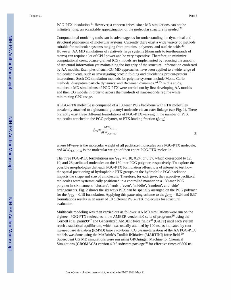
A PGG-PTX molecule is comprised of a 130-mer PGG backbone with PTX moleculescovalently attached to a glutamate-glutamyl molecule via an ester linkage (see Fig. 1). Therecurrently exist three different formulations of PGG-PTX varying in the number of PTXmolecules attached to the PGG polymer, or PTX loading fraction (fPTX):
(1)
where MWPTX is the molecular weight of all paclitaxel molecules on a PGG-PTX molecule,and MWPGG-PTX is the molecular weight of then entire PGG-PTX molecule.
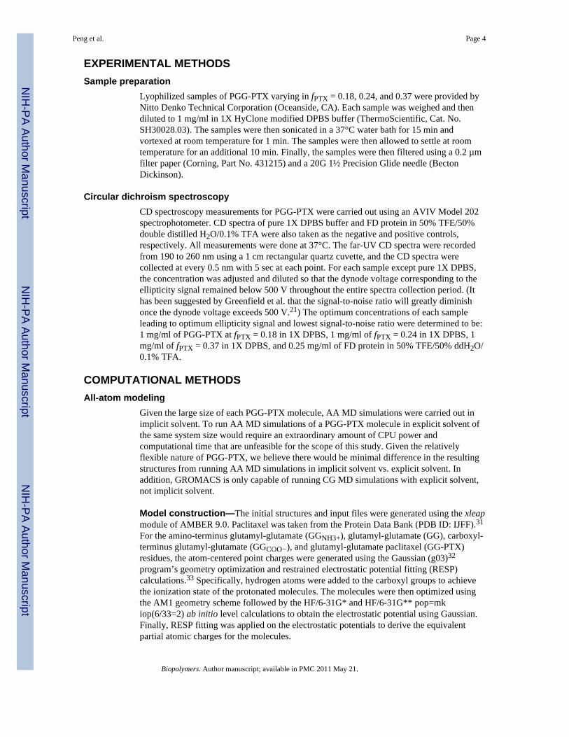
The three PGG-PTX formulations are fPTX = 0.18, 0.24, or 0.37, which correspond to 12,19, and 26 paclitaxel molecules on the 130-mer PGG polymer, respectively. To explore thepossible morphologies that each PGG-PTX formulation offers, it is of interest to test howthe spatial positioning of hydrophobic PTX groups on the hydrophilic PGG backboneimpacts the shape and size of a molecule. Therefore, for each fPTX, the respective paclitaxelmolecules were systematically positioned in a controlled manner on a 130-mer PGGpolymer in six manners: ‘clusters’, ‘ends’, ‘even’, ‘middle’, ‘random’, and ‘side’arrangements. Fig. 2 shows the six ways PTX can be spatially arranged on the PGG polymerfor the fPTX = 0.18 formulation. Applying this patterning scheme to the fPTX = 0.24 and 0.37formulations results in an array of 18 different PGG-PTX molecules for structuralevaluation.
Multiscale modeling was then carried out as follows: AA MD simulations were run on theeighteen PGG-PTX molecules in the AMBER version 9.0 suite of programs26 using theCornell et al. parm9927 and Generalized AMBER force fields28 (GAFF) until each systemreach a statistical equilibrium, which was usually attained by 100 ns, as indicated by root-mean-square deviation (RMSD) time evolutions. CG parameterization of the AA PGG-PTXmodels was done using the MARrink’s Toolkit INItiative (MARTINI) force field.29
Subsequent CG MD simulations were run using GROningen Machine for ChemicalSimulations (GROMACS) version 4.0.3 software package30 for effective times of 800 ns.
Peng et al. Page 3
Biopolymers. Author manuscript; available in PMC 2011 May 21.
NIH
-PA Author Manuscript
NIH
-PA Author Manuscript
NIH
-PA Author Manuscript
EXPERIMENTAL METHODSSample preparation
Lyophilized samples of PGG-PTX varying in fPTX = 0.18, 0.24, and 0.37 were provided byNitto Denko Technical Corporation (Oceanside, CA). Each sample was weighed and thendiluted to 1 mg/ml in 1X HyClone modified DPBS buffer (ThermoScientific, Cat. No.SH30028.03). The samples were then sonicated in a 37°C water bath for 15 min andvortexed at room temperature for 1 min. The samples were then allowed to settle at roomtemperature for an additional 10 min. Finally, the samples were then filtered using a 0.2 µmfilter paper (Corning, Part No. 431215) and a 20G 1½ Precision Glide needle (BectonDickinson).
Circular dichroism spectroscopyCD spectroscopy measurements for PGG-PTX were carried out using an AVIV Model 202spectrophotometer. CD spectra of pure 1X DPBS buffer and FD protein in 50% TFE/50%double distilled H2O/0.1% TFA were also taken as the negative and positive controls,respectively. All measurements were done at 37°C. The far-UV CD spectra were recordedfrom 190 to 260 nm using a 1 cm rectangular quartz cuvette, and the CD spectra werecollected at every 0.5 nm with 5 sec at each point. For each sample except pure 1X DPBS,the concentration was adjusted and diluted so that the dynode voltage corresponding to theellipticity signal remained below 500 V throughout the entire spectra collection period. (Ithas been suggested by Greenfield et al. that the signal-to-noise ratio will greatly diminishonce the dynode voltage exceeds 500 V.21) The optimum concentrations of each sampleleading to optimum ellipticity signal and lowest signal-to-noise ratio were determined to be:1 mg/ml of PGG-PTX at fPTX = 0.18 in 1X DPBS, 1 mg/ml of fPTX = 0.24 in 1X DPBS, 1mg/ml of fPTX = 0.37 in 1X DPBS, and 0.25 mg/ml of FD protein in 50% TFE/50% ddH2O/0.1% TFA.
COMPUTATIONAL METHODSAll-atom modeling
Given the large size of each PGG-PTX molecule, AA MD simulations were carried out inimplicit solvent. To run AA MD simulations of a PGG-PTX molecule in explicit solvent ofthe same system size would require an extraordinary amount of CPU power andcomputational time that are unfeasible for the scope of this study. Given the relativelyflexible nature of PGG-PTX, we believe there would be minimal difference in the resultingstructures from running AA MD simulations in implicit solvent vs. explicit solvent. Inaddition, GROMACS is only capable of running CG MD simulations with explicit solvent,not implicit solvent.
Model construction—The initial structures and input files were generated using the xleapmodule of AMBER 9.0. Paclitaxel was taken from the Protein Data Bank (PDB ID: IJFF).31
For the amino-terminus glutamyl-glutamate (GGNH3+), glutamyl-glutamate (GG), carboxyl-terminus glutamyl-glutamate (GGCOO−), and glutamyl-glutamate paclitaxel (GG-PTX)residues, the atom-centered point charges were generated using the Gaussian (g03)32
program’s geometry optimization and restrained electrostatic potential fitting (RESP)calculations.33 Specifically, hydrogen atoms were added to the carboxyl groups to achievethe ionization state of the protonated molecules. The molecules were then optimized usingthe AM1 geometry scheme followed by the HF/6-31G* and HF/6-31G** pop=mkiop(6/33=2) ab initio level calculations to obtain the electrostatic potential using Gaussian.Finally, RESP fitting was applied on the electrostatic potentials to derive the equivalentpartial atomic charges for the molecules.
Peng et al. Page 4
Biopolymers. Author manuscript; available in PMC 2011 May 21.
NIH
-PA Author Manuscript
NIH
-PA Author Manuscript
NIH
-PA Author Manuscript
Energy minimization and MD simulation—These steps were carried out using thesander module. The starting structures were minimized in implicit solvent using themodified Generalized-Born model of IGB=234 and the LPCO model35 using 250 steps ofsteepest descent followed by 1750 steps of conjugate gradient. To mimic the saltconcentration of blood plasma and PBS buffer, the ionic strength of the implicit solvent wasset to 140 mM. No periodic boundary conditions were applied. A non-bonded electrostaticcutoff of 16.0Å was used. Trajectory snapshots were saved every 100 steps for laterreprocessing.
MD simulations were carried out using AMBER 9.0 with the modified version (ff99SB)36 ofthe Cornell et al. parm9927 and GAFF force fields.28 MD simulations were carried out in theNVT ensemble (in which the number of atoms N, volume V, and temperature T were fixed)at 310K in an implicit solvent using the modified Generalized-Born model of IGB=2 and theLPCO model. No periodic boundary conditions were applied. Newton’s equations ofmotions were integrated with a time step of 2 fs. All bonds involving hydrogen atoms wereconstrained using the SHAKE algorithm37. Constant temperature scaling was also appliedwith a time constant of 0.5 ps. Langevin dynamics was used with a collision frequency of2.0 ps−1. The rotational and translational degree of freedom about the center of mass waseliminated. Trajectory snapshots were saved every 100 steps for later reprocessing. Eachcycle of the MD simulation was run for 0.1 ns, and this step was repeated until a statisticalequilibrium was reached at 100 ns.
Coarse-grained modelingFor the sake of minimizing computational costs as well as accessing longer length and timescales, the level of information in each AA PGG-PTX molecule was reduced while strivingto maintain the accuracy of the physicochemical information of each molecule. Given theamino acid-based nature of the glutamyl-glutamate backbone, the MARTINI force field wasselected for the CG parameterization for its successful application to proteins byexperimental validation of their structural properties as well as the reproduction ofthermodynamics.29,38–40 The MARTINI force field dictates that a group of roughly 4–5atoms are represented as an interaction center, or bead. These beads interact through a set ofshort-ranged Lennard-Jones potentials to reproduce characteristic properties resulting fromAA simulations. Charged groups interact via a Coulombic energy function, and bondedpotentials are used to describe the chemical connectivity of the beads. Detailedparameterization of the GGNH3+, GG, GGCOO−, and GG-PTX residues as well as water (W)and sodium (Na+) ions are described below.
Theory—The CG beads are considered as point masses, and their dynamics are describedby Newton’s equations of motion. The effective bonded and nonbonded interactions amongthe beads are described by a potential of the form, adapted from Marrink et al.40:
(2)
where the index m denotes one of the six components in the system: GGNH3+, GG,GGCOO−, GG-PTX, W, and Na+. The sum in Eqn. (2) describes the force potentials for thebonded CG beads: describes the forces between two bonded CG beads; accountsfor the forces used to sustain angles between sets of three bonded CG beads; and corresponds to the dihedral angle potential for quadruples of bonded CG beads. The
term accounts for the nonbonded interactions between all of the CG beads in thesystem.
Peng et al. Page 5
Biopolymers. Author manuscript; available in PMC 2011 May 21.
NIH
-PA Author Manuscript
NIH
-PA Author Manuscript
NIH
-PA Author Manuscript

Bonded interactions are characterized as one of the following:
(3)
(4)
(5)
where the indices i, j, k, and l represent four consecutive beads, is the distance betweentwo consecutive beads i and j, is the equilibrium CG bond length (or the average of allthe sampled conformations from the atomistic MD trajectory), is the angle betweenbeads i, j, and k, and is the equilibrium angle (the average of all the sampledconformations from the atomistic MD trajectory). The CG bead distance force constants
and the CG bead angle force constants depend onthe bead type and bead interaction.
The nonbonded interactions between two CG beads m and n at a distance rmn interact via avan der Waals (vdW) characterized by a Lennard-Jones 12-6 potential energy function aswell as a Coulombic potential for charged beads (bead type Q):
(6)
Where σmn refers to a typical closest distance of approach between two beads m and n, εmndenotes the strength of the interactions between beads m and n (one of five levels, I to V, foreach bead type, and the full interaction matrix is provided in Marrink et al.29 qm and qn arethe charges of the mth and nth beads, ε0 is the vacuum dielectric permittivity, and a relativedielectric constant of ε = 15 is assumed for all electrostatic interactions.
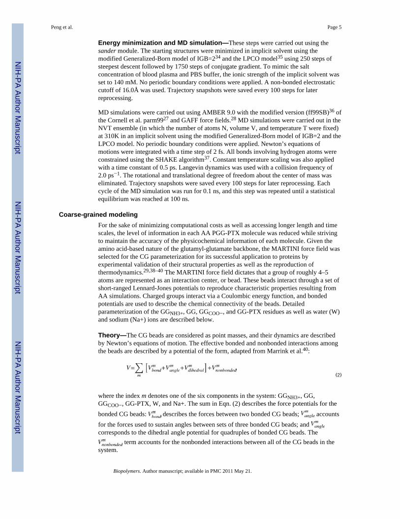
CG mapping—As aforementioned, the MARTINI force field dictates that approximately4–5 heavy atoms are mapped to one interaction center (bead). An exception to this ruleapplies to aromatic groups, such as the benzene molecules in paclitaxel: in order to preservethe geometric symmetry of these groups, a 2 heavy atoms-to-1 bead mapping scheme isapplied. The CG mapping of the GGNH3+, GG, GGCOO−, and GG-PTX residues aredepicted in Fig. 3. Given that the circular dichroism spectroscopy results show that PGG-PTX forms a random coil at 37°C (310 K), the bonded parameters of the GG backbone wereassigned based on the values for a random coil, as dictated by the MARTINI force field. Forthe negative and positive controls, description of the CG mapping for glutamate (G) andglutamate-benzene (G-B) residues is provided in Fig. S1 in Supplementary Materials. Inaddition, four water molecules are represented as a single P4 type bead, and each Na+ ion isrepresented by a single Q type bead with a +1 charge.
Peng et al. Page 6
Biopolymers. Author manuscript; available in PMC 2011 May 21.
NIH
-PA Author Manuscript
NIH
-PA Author Manuscript
NIH
-PA Author Manuscript
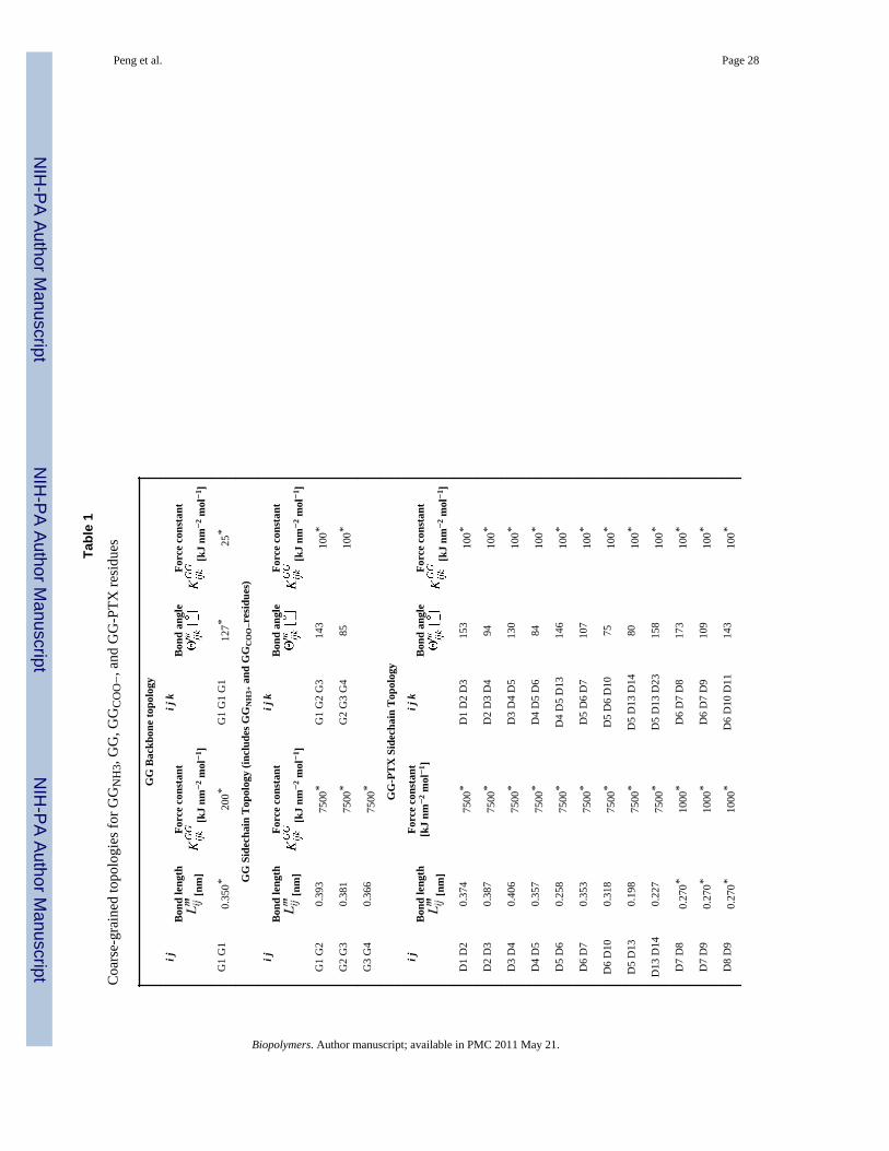
Parameterization of bonded interactions for a PGG-PTX molecule—Theequilibrium bond distances and angles were obtained from of the three ‘random’ PGG-PTXmolecules (fPTX = 0.18, 0.24, and 0.37) since they were the models that best mimic theexperimental formulations of PGG-PTX. The parameterization was carried out by firstextracting the time-dependent bond distances and angles from the 100 ns AA MDsimulations using ptraj module of AMBER 9.0. Then, the bonded parameters wereprocessed using the Boltzmann inversion procedure41 in MATLAB 7.042 to determine theequilibrium bond distances and angles.
Energy minimization and MD simulation—CG PGG-PTX models were constructed inGROMACS 4.0.3. Each PGG-PTX molecule was solvated in the center of the box withexplicit W beads of 2 nm thickness of surrounding the molecule. Due to the negativecharges imparted by the GG and GG-PTX residues in each system, Na+ ions (usually 234)were added in place of W beads to neutralize the system. The simulations were carried outunder NPT (the number of particles N, pressure P, and temperature T were fixed) conditions.For each simulation, the temperature was kept constant at 310 K with a coupling constant ofτT = 0.1 ps, and the pressure was weakly coupled to 1 bar with a relaxation time of τP = 0.5ps. The cutoff length for the nonbonded interactions is rcut = 1.2 nm. Lennard-Jones forceswere considered for rcut < 0.9 nm and Coulombic forces for rcut < 1.2 nm. The latter wascomputed every time step for 1.0 nm and once every 10 time steps for 0.9 nm < rcut < 1.2nm. The time step in the leap-frog integration scheme was 5 fs. The energies, coordinatesand velocities were written every 0.5 ps. For each of the eighteen systems, MD simulationswere run until a statistical equilibrium was obtained at 200 ns. It is worth noting that, due tothe smoothness of the CG potentials, CG dynamics are actually faster than AA interactions.To account for this difference, researchers have approximated CG results by scaling the timeaxis by a conversion factor of four, the effective speed up factor in the diffusional dynamicsof CG water compared to that of real water.29,40,43 Therefore, the effective time sampled foreach CG MD simulation was roughly 800 ns. Unless otherwise stated, the CG MDsimulation times reported in this paper are effective times.
RESULTS AND DISCUSSIONCircular dichroism spectroscopy
To construct a theoretical model of PGG-PTX practical for clinical applications, its initialmolecular configuration needs to be first defined. Circular dichroism spectroscopy was usedto determine the general structure of a PGG-PTX molecule, and the resulting structure wasregarded as the initial configuration for the AA PGG-PTX models.
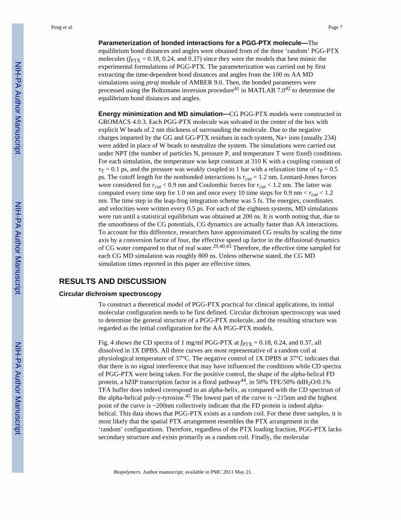
Fig. 4 shows the CD spectra of 1 mg/ml PGG-PTX at fPTX = 0.18, 0.24, and 0.37, alldissolved in 1X DPBS. All three curves are most representative of a random coil atphysiological temperature of 37°C. The negative control of 1X DPBS at 37°C indicates thatthat there is no signal interference that may have influenced the conditions while CD spectraof PGG-PTX were being taken. For the positive control, the shape of the alpha-helical FDprotein, a bZIP transcription factor in a floral pathway44, in 50% TFE/50% ddH2O/0.1%TFA buffer does indeed correspond to an alpha-helix, as compared with the CD spectrum ofthe alpha-helical poly-γ-tyrosine.45 The lowest part of the curve is ~215nm and the highestpoint of the curve is ~200nm collectively indicate that the FD protein is indeed alpha-helical. This data shows that PGG-PTX exists as a random coil. For these three samples, it ismost likely that the spatial PTX arrangement resembles the PTX arrangement in the‘random’ configurations. Therefore, regardless of the PTX loading fraction, PGG-PTX lackssecondary structure and exists primarily as a random coil. Finally, the molecular
Peng et al. Page 7
Biopolymers. Author manuscript; available in PMC 2011 May 21.
NIH
-PA Author Manuscript
NIH
-PA Author Manuscript
NIH
-PA Author Manuscript
configuration of PGG-PTX as a random coil does not impose any stipulations onconstruction of an AA model.
Comparison of all-atom and coarse-grained modelsFig. 5 shows the initial structures of the AA and CG models at of PGG-PTX molecule atfPTX = 0.37 with an ‘even’ PTX distribution. Neglecting the explicit solvent beads of the CGmodels, the size of the AA models (6189 heavy atoms) is roughly five times larger than itsCG counterpart (1222 beads), which is apparent in that the AA model appears to have ahigher image resolution than the CG model. Despite this difference, the structural integrityof the PGG-PTX molecule is still preserved in the CG model. From the perspectives of thexy and xz planes, the length and height of the AA and CG models are comparable, and thecross-sectional area is similar in AA and CG models from the yz plane. The paclitaxelmolecules are arranged in a spiral fashion around the PGG backbone, similar to the spiralpattern in the AA model.
AA and CG equilibrium bonded parameters—To assess the validity of the CG modelof PGG-PTX, a few equilibrium bond angles (G1-G2-G3, D3-D4-D5, D13-D14-D23, andD24-D25-D37) of the AA and CG PGG-PTX models were selected and compared. Asshown in Fig. 6, it is apparent that there is relatively good agreement between the AA andCG values. While there is not always a perfect overlap between the AA and CG curves, thisbehavior is to be expected for a system that has both flexible and rigid components. The AAand CG probability distributions for D13-D14-D23 and D24-D25-D27 are narrower thanthose for G1-G2-G3 and D3-D4-D5. An explanation for this difference is that the D13, D14,D23, D24, D25, and D27 beads are located in an area of the paclitaxel molecule with highsterical energy (D13, D14, D23, D25, and D26 are part of aromatic groups) thus limiting therange of orientations that the beads can sample. Contrariwise, G1, G2, G3, D3, D4, and D5are not bound to any aromatic structures, not to mention that they are within close vicinity ofthe glutamyl-glutamate backbone. The positions of these beads allow for a larger range ofaccessible orientations. In all, the probability distribution curves show that more flexiblecomponents of a PGG-PTX molecule have a wider range of accessible conformations, whilemore rigid components of a PGG-PTX molecule have a limited range of accessibleconformations.
RMSD and RMSF values—Figs. S4, S5, and S6 show the RMSD time evolutions from100-ns AA MD and 100-ns (or 25 ns non-effective time) CG MD simulations of PGG-PTXmolecules. It is apparent that there is relatively good agreement between the AA and CGRMSD values. Overall, the AA RMSD values rise to 3–5 nm by 100 ns, and CG RMSDvalues increase to 2–4 nm by 100 ns. Although these values are high, they are reasonablegiven the size of the PGG-PTX molecules. By 100 ns, most AA MD simulations haveattained a steady-state level. (While certain systems such as the fPTX = 0.18 ‘clusters’, fPTX= 0.18 ‘even’, and fPTX = 0.24 ‘even’ molecules have not yet attained a full steady-statelevel, they are adequately stabilized for this study.) On the other hand, the CG MDsimulations did not attain a steady-state by 100 ns and were subsequently extended to 800ns. To assess whether steady-state was reached by 800 ns, CG MD simulations wereextended further to an effective time of 2 µs. Fig 7 shows the CG RMSD values for alleighteen systems; most plots show that there is minimal variation in RMSD between 800 nsand 2 µs, indicating that 800 ns was long enough for a statistical equilibrium to be reached.
Figs. S7, S8, and S9 show the RMSF (by residue) values from 100 ns AA MD and 100 ns(or 25 ns non-effective time) CG MD simulations of PGG-PTX molecules. There isrelatively good agreement between the AA and CG RMSF values. The AA RMSF valuestypically fluctuated between 2–4 nm, with a few molecules having RMSF values near 8 nm.
Peng et al. Page 8
Biopolymers. Author manuscript; available in PMC 2011 May 21.
NIH
-PA Author Manuscript
NIH
-PA Author Manuscript
NIH
-PA Author Manuscript

The CG RMSF values are a bit lower by ~1–3 nm. This discrepancy is expected, given thatsome structural information was probably lost in the coarse-graining procedure and that thesystem is comprised of the flexible PGG polymer. Fig. 8 shows the CG RMSF values for alleighteen PGG-PTX molecules. Nearly all molecules fluctuate between 2–4 nm, regardless ofthe spatial PTX arrangement or PTX loading fraction. RMSF is also an indication ofmolecular flexibility; since higher RMSF values typically correspond to segments of PGG-PTX with unconjugated PGG (no paclitaxel molecules attached), those regions are the mostflexible. The distribution of hydrophobic PTX on the PGG backbone influences the degreeof residue fluctuation, which is further discussed below.
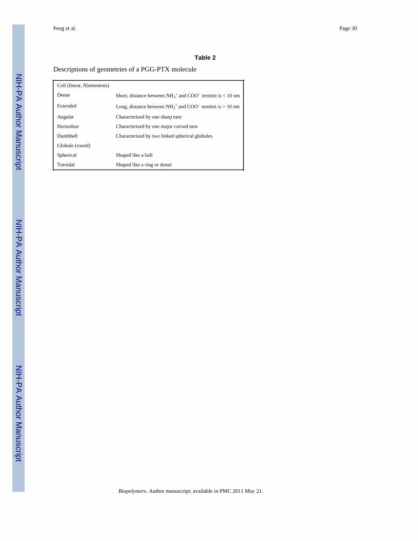
Molecular conformation of a PGG-PTX moleculeShape—It was of interest to investigate whether the shape of PGG-PTX can be predictedusing multiscale modeling. Fig. 9 shows the 800 ns CG MD simulations for the PGG-PTXmolecules. All molecules start out as linear at 0 ns and eventually adopt a coil (filamentous)or globular (round) shape by 800 ns. Since steady-state was attained by all systems by 800ns, the shape at this time was regarded as the official shape of each PGG-PTX molecule.There exists a wide variety of geometries among the molecules. The molecular shape ofeach molecule can be categorized into two main types: coil (filamentous, wormlike) andglobular (round). For each type, the conformation can be further characterized as one ormore subtypes (coil: angular, dense, extended, horseshoe, or dumbbell, globule: sphericaland toroidal). Descriptions of the geometries are summarized in Table 2.
The coil was the most popular type of geometry among all PGG-PTX molecules. Allmolecules with PTX loading fractions of 0.18 and 0.37 are coils, and at fPTX = 0.24, four ofthe six molecules are coils, whereas the other two are globules. The fPTX = 0.24 set also hasthe most diverse set of subtype geometries. All six subtypes are prevalent in the fPTX = 0.24molecules, including the spherical globule and toroidal globule that are not apparent in thefPTX = 0.18 and fPTX = 0.37 molecules. This evidence suggests that, out of the three PTXloading fractions, PGG-PTX at fPTX = 0.24 has the optimal balance of hydrophobic andhydrophilic forces to have the greatest concomitant effect on the geometry of the molecule.That is, at fPTX = 0.24, the geometry of PGG-PTX is influenced more by the spatialpositioning of PTX on the PGG backbone, rather than the PTX loading fraction. On theother hand, at fPTX = 0.18 and 0.37, the geometries of PGG-PTX is more influenced by themass fraction of hydrophobic PTX and hydrophilic PGG, rather than the spatial positioningof PTX on the PGG. In addition, there are certain spatial PTX arrangements for whosegeometries are the least affected by the PTX loading fraction. Such are the ‘middle’configuration, which maintains a dense coil for all three PTX loading fractions, and the‘random’ configuration, which maintains an extended coil regardless of the PTX loadingfraction. (This shape for the ‘random’ configuration is in good agreement with the previouscircular dichroism spectroscopy data on PGG-PTX that indicate that the shape of PGG-PTXis a random coil.) It is also interesting that the ‘side’ and ‘middle’ configurations are uniquein that they are characterized by sharp curvature: possessing an angled or curved turn. Thisbehavior is most likely attributed to the spatial PTX arrangement on the PGG backbone,which has a major influence on the flexibility of a PGG-PTX molecule. What sets the‘middle’ and ‘side’ configurations apart from the rest is that all of the PTX molecules areconcentrated into one group such that it leaves long, uninterrupted portions of the PGGbackbone unconjugated. These unconjugated portions are floppy and more flexible than thesegments of the PGG backbone with PTX molecules attached. A similar pattern is alsoemergent in the fPTX = 0.24 ‘ends’ molecule, a toroidal globule: two groups of paclitaxelmolecules located at each end, thus leaving the middle portion of the PGG backboneunconjugated. As for other configurations like ‘clusters’, ‘random,’ and ‘even’, thepaclitaxel molecules are arranged more uniformly along the PGG backbone such that the
Peng et al. Page 9
Biopolymers. Author manuscript; available in PMC 2011 May 21.
NIH
-PA Author Manuscript
NIH
-PA Author Manuscript
NIH
-PA Author Manuscript
portions of unconjugated PGG are much shorter (see Fig. 2). This relatively equal dispersionof hydrophobic forces minimizes the movement or fluctuation of the PGG-PTX molecule asa whole. For the ‘middle’ and ‘side’ configurations, this absence of hydrophobic drugmolecules removes the sterical hindrance imposed by the rigid PTX molecules and thusallows the unconjugated PGG portion to move about more freely. Generally speaking,configurations that have longer unconjugated PGG segments (‘middle’, ‘side’, and ‘ends’)tend to be more flexible and form more definitive, structured shapes as compared toconfigurations with a more even PTX distributions along the PGG backbone (‘clusters’,‘random’, and ‘even’).
Size—It was also of interest to examine the effects of PTX loading fraction and the spatialPTX arrangement on the size of a PGG-PTX molecule. Fig. 10 shows the 800 ns CG MDtime evolutions of the radius of gyration (Rg) for all 18 PGG-PTX molecules. There areseveral interesting trends apparent. It is evident that, at t = 0 ns, the Rg of most molecules is~6–8 nm, and over the 800 ns trajectory, each of the 18 PGG-PTX molecules decreasednearly 50% from its initial Rg. Fig. 9 confirms that, at t = 0 ns, all structures were stretchedout and extended, but by t = 200 ns, all of them had shrunken in size. Also, it is interestingto see that, as the PTX loading fraction increases, the greater the variability in Rg amongeach set of six PGG-PTX configurations. As shown in Fig. 10, at fPTX = 0.18, there is quitesome overlap between the plots, especially at 800 ns, indicating that all six fPTX = 0.18configurations have more or less the same size. At fPTX = 0.24, there is slightly less overlapin the plots, and at fPTX = 0.37, there is nearly no overlap at all. Suffice to say, higher PTXloading fractions most likely results in a larger range of candidate sizes of PGG-PTX. Inaddition, the degree of influence the spatial PTX arrangement on PGG exerts over the sizeof a PGG-PTX molecule is quite minimal. Regardless of the hierarchy of initial Rg at t = 0ns (from smallest to largest Rg: ‘middle’, ‘side’, ‘random’, ‘clusters’, ‘even’, and ‘ends’),once the molecules have stabilized, nearly all have a final Rg of ~4 nm.
Influence of PTX on molecular conformation—The effect of the presence ofpaclitaxel molecules on the conformation of a PGG molecule was also of interest. Toexamine the extent of this effect, two negative controls (CG models of hydrophilic polymersfree of any hydrophobic entities) and a positive control (CG model of a hydrophilic polymerconjugated with hydrophobic entities) were constructed. The negative controls are: a 130-mer poly-γ-glutamate molecule (PG), and a 130-mer poly-γ-glutamyl-glutamate (PGG)molecule. The positive control is a 130-mer poly-γ-glutamate molecule with covalentlyconjugated benzene molecules (PG-B) with a benzene loading fraction of 0.37 (fbenzene =0.37) and an ‘even’ benzene distribution. Fig. 11 shows the 800 ns CG MD simulations ofthe controls. During the 800 ns trajectory, both PG and PGG molecules wiggle around a lot,exhibiting flexible behavior. At 800 ns, they exhibit dense, horseshoe coil shapes. A similarbehavior is observed in the PG-B molecule, as it also flexible and contorts before it finallyadopts a dense coil at 800 ns. It is also worth noting that the benzene molecules self-assemble together to form three hydrophobic ‘cores’ located in the inner parts of themolecule.
Fig. 12 shows the 800 ns CG MD time evolutions of Rg for the controls. The PG and PG-Bmolecules both have an initial Rg ~ 13 nm and final Rg ~ 4 nm. Given that they both havecoil shapes, the benzene molecules have an insignificant effect on the coil geometry of a PGmolecule. Similarly, the PGG molecule and PGG-PTX molecule (fPTX = 0.37, ‘even’ PTXdistribution) each has an initial Rg ~ 7 nm and final Rg ~ 4 nm. Since both molecules aredense coils, this data suggests that, for the PGG-PTX (fPTX = 0.37 ‘even’) molecule, thepresence of PTX on PGG only also has a minor influence on the conformation of PGG. Itwas also interesting to see how the PTX molecules in the PGG-PTX (fPTX = 0.37 ‘even’)
Peng et al. Page 10
Biopolymers. Author manuscript; available in PMC 2011 May 21.
NIH
-PA Author Manuscript
NIH
-PA Author Manuscript
NIH
-PA Author Manuscript
molecule also self-assemble to form multiple inner hydrophobic ‘cores’, similar to the PG-Bmolecule.
Ability to cross physiological barriers in drug delivery—Wormlike, filamentousmicelles have been shown to result a longer circulation half-life than their sphericalcounterparts in drug delivery,3,4 and discoidal particles have been shown to have a highertendency to adhere and accumulate to the wall of tumor endothelia.5 By analogy, the shapesof the wormlike, filamentous micelles, and discoidal particles are structurally similar to thecoil-shaped PGG-PTX molecules. Since all 18 molecules adopt a general coil shape (with avariety of coil subtypes), with exception to the fPTX = 0.24 ‘ends’ and fPTX = 0.24 ‘even’molecules, it appears that the PTX loading fraction and spatial PTX arrangement have aminor concomitant influence on the molecular conformation of a PGG-PTX molecule. Byanalogy to the larger micelles and nanoparticles, we speculate that PGG-PTX formulationsmay confer a relatively long circulation half-life and propensity for accumulation, regardlessof the PTX loading fraction and spatial PTX arrangement. Finally, the radius of gyrationsizes of all PGG-PTX molecules range from 2–8 nm, implying that the actual molecular sizeis slightly greater. It has been shown that silver and gold nanoparticles of the 2–100 nm sizerange affects the intracellular signaling pathways implicated in basic cellular function.6Therefore, PGG-PTX molecules may also possess the ability to control cellular signalingpathways of tumor cells.
CONCLUSIONSThere have been suggestions that the molecular conformation of a cancer therapeuticinfluences its ability to reach its intended site in the body. In this study, the conformation ofPGG-PTX, a polymer-drug conjugate with potential applications in cancer therapy, wasexplored by systematically controlling the PTX loading fraction (fPTX = 0.18, 0.24, and0.37) and spatial patterning of PTX on PGG (‘clusters’, ‘ends’, ‘even’, ‘middle’, ‘random’,and ‘side’). Given that the CG models accurately reproduce the bonded parameters of theAA models and the good agreement between the AA and CG RMSD and RMSF trajectories,the CG models are suitable for accessing the nanosecond-microsecond regime. The 800 nsCG MD simulations have shown that, regardless of PTX loading fraction and PTXarrangement on PGG, a PGG-PTX molecule will most likely adopt a coil shape, suggestingthat it has a long circulation half-life and high inclination to accumulate towards walls oftumor endothelia. PGG-PTX molecules with less homogeneous PTX arrangements(characterized by long, unconjugated PGG sections) tend to result in geometries with highercurvature. Also, the sizes of all PGG-PTX molecules fall within the 2–100 nm range,suggesting that they may affect the basic function of tumor cells. In terms of producing thewidest variety of geometries, fPTX = 0.24 is the PTX loading fraction at which the PTXarrangements are the most sensitive to morphological change.
Supplementary MaterialRefer to Web version on PubMed Central for supplementary material.
AcknowledgmentsWork in the McCammon group is supported in part by NSF, NIH, Howard Hughes Medical Institute (HHMI),National Biomedical Computation Resource (NBCR), and Center of Theoretical and Biological Physics (CTBP).All simulations were performed the Granite supercomputer cluster at the UCSD Department of Bioengineering andthe Kryptonite supercomputer cluster at the National Biomedical Computation Resource (NBCR), whose work issupported by NBCR grant P41RR08605 from NCRR. The authors acknowledge the following people for theirassistance: David Black, Ingrid DeVries, Elizabeth Komives, and Stanley Opella for the circular dichroism
Peng et al. Page 11
Biopolymers. Author manuscript; available in PMC 2011 May 21.
NIH
-PA Author Manuscript
NIH
-PA Author Manuscript
NIH
-PA Author Manuscript
spectroscopy experiments; Sanjib Das for the synthesis of PGG-PTX, Robert Swift for the Boltzmann inversionprocedure; and Vincent Wong for the CG parameterization.
Financial support for this work was provided by UC Discovery Grant bio06-10568 and the Nitto Denko Technical(NDT) Corporation. Authors S. Van, G. Zhao, and L. Yu are full-time employees of NDT Corp., and authors S. B.Howell and D. A. Gough serve as consultants for NDT Corp. on an arrangement approved by the UCSD Conflict ofInterest Committee.
REFERENCES1. Ferrari M. Nat Rev. 2005; 5:161–171.2. Ferrari M. Nat Nano. 2008; 3:131–132.3. Geng Y, Dalhaimer P, Cai S, Tsai R, Tewari M, Minko T, Discher DE. Nature Nanotech. 2007;
2:249–255.4. Srinivas G, Shelley JC, Nielson SO, Discher DE, Klein ML. J Phys Chem B. 2004; 108:8150–8160.5. Decuzzi P, Pasqualini R, Arap W, Ferrari M. Pharma Res. 2008; 26:235–243.6. Jiang W, Kim BY, Rutka JT, Chan W. Nat Nano. 2008; 3:145–150.7. Duncan R. Nat Rev Drug Discov. 2003; 2:347–360. [PubMed: 12750738]8. Satchi-Fainaro, R.; Duncan, R. Polymer Therapeutics I. New York: Springer; 2006.9. Pujol, JL. Clinical Study Report for Study CA139-368. Montpellier, France: Bristol-Myers Squibb;
2005.10. Piccart MJ, Bertelsen K, James K, Cassidy J, Mangioni C, Simonsen E, Stuart G, Kaye S, Vergote
I, Blom R, Grimshaw R, Atkinson RJ, Swenerton KD, Trope C, Nardi M, Kaern J, Tumolo S,Timmers P, Roy JA, Lhoas F, Lindvall B, Bacon M, Birt A, Andersen JE, Zee B, Paul J, Baron B,Pecorelli S. J Natl Cancer Inst. 2000; 92:699–708. [PubMed: 10793106]
11. Yoshida, T. Clinical Study Report for Study CA139-387. Tokyo, Japan: Bristol-Myers Squibb;2009.
12. Wang, X.; Gang, Z.; Van, S.; Yu, L. Corporation. N. D, T., editor. USA: Nitto Denko TechnicalCorporation; 2009. p. 1-27.
13. A, editor. Abraxis Oncology. USA: Abraxis Bioscience; 2007. p. 1-26.14. Vauthier C, Fattal E, Labarre D. Chapter 26: From Polymer Chemistry and Physicochemistry to
Nanoparticulate Drug Carrier Design and Applications. 200315. Schames J, Henchman RH, Siegel JS, Sotriffer CA, Ni H, McCammon JA. J Med Chem. 2004;
47:1879–1881. [PubMed: 15055986]16. Amaro RE, Schnaufer A, Interthal H, Hol W, Stuart WD, McCammon JA. PNAS. 2008;
106:17278–17283. [PubMed: 18981420]17. Zewail, AH. Physical Biology: From Atoms to Medicine. Zewail, AH., editor. World Scientific
Publishing; 2008. p. 401-410.18. Baschnagel J, Binder K, Doruker P, Gusev AA, Hahn O, Kremer K, Mattice WL, Muller-Plathe F,
Murat M, Paul W, Santos S, Suter UW, Tries V. Advances in Polymer Science. 2000; 152:41–156.19. Beall GW, Muruesan S, Galloway HC, Koeck DC, Jarl J, Abrego F. Polymer. 2005; 46:11889–
11895.20. Faller R. Polymer. 2004; 45:3869–3876.21. Greenfield NJ. Nat Protoc. 2007; 1:2876–2890. [PubMed: 17406547]22. Cooke B, Schmidler SC. Biophys J. 2008; 95:4497–4511. [PubMed: 18676654]23. Adcock SA, McCammon JA. Chem Rev. 2006; 106:1589–1615. [PubMed: 16683746]24. Muller-Plathe F. Chem Phys Chem Rev. 2002; 3:754–769.25. Kenward M, Dorfman KD. Biophys J. 2009; 97:2785–2793. [PubMed: 19917233]26. Case DA, Cheatham TE, Darden T, Gohlke H, Luo R, Merz KM Jr, Onufriev A, Simmerling C,
Wang B, Woods R. J Comput Chem. 2005; 26:1668–1688. [PubMed: 16200636]27. Cornell WD, Cieplak P, Bayly CI, Gould IR, Merz KM Jr, Ferguson DM, Spellmeyer DC, Fox T,
Caldwell JW, Kollman PA. J Am Chem Soc. 1995; 117:5179–5197.
Peng et al. Page 12
Biopolymers. Author manuscript; available in PMC 2011 May 21.
NIH
-PA Author Manuscript
NIH
-PA Author Manuscript
NIH
-PA Author Manuscript

28. Wang J, Rolf RM, Caldwell JW, Kollman PA, Case DA. J Comput Chem. 2004; 25:1157–1174.[PubMed: 15116359]
29. Monticelli L, Kandasamy SK, Periole X, Larson RG, Tieleman DP, Marrink SJ. J Chem Theoryand Comput. 2008; 4:819–834.
30. Lindahl E, Hess B, van der Spoel D. J Mol Mod. 2001; 7:306–317.31. Bernstein FC, Koetzle TF, Williams GJ, Meyer EE Jr, Brice MD, Rodges JR, Kennard O,
Shimanouchi T, Tasumi M. J of Mol Biol. 1977; 112:535. [PubMed: 875032]32. Frisch, MJT.; G, W.; Schlegel, HB.; Gill, PMW.; Johnson, BG.; Robb, MA.; Cheeseman, JR.;
Keith, TA.; Petersson, GA.; Montgomery, JA.; Raghavachari, K.; Al-Laham, MA.; Zakrzewski,VG.; Ortiz, JV.; Foresman, JB.; Cioslowski, J.; Stefanov, BB.; Nanayakkara, A.; Challacombe,M.; Peng, CY.; Ayala, PY.; Chen, W.; Wong, MW.; Andres, JL.; Replogle, ES.; Gomperts, R.;Martin, RL.; Fox, DJ.; Binkley, JS.; Defrees, DJ.; Baker, J.; Stewart, JP.; Head-Gordon, M.;Gonzalez, C.; Pople, JA. Pittsburgh, PA: Gaussian, Inc.; 1995.
33. Cornell WD, Cieplak C, Bayly CI, Kollman PA. J Am Chem Soc. 1993; 115:9620–9631.34. Onufriev A, Bashford D, Case DA. Proteins: Struct, Funct, Bioinf. 2004; 55:383–394.35. Weiser J, Shenkin PS, Still WC. J Comput Chem. 20:217–230.36. Hornak V, Abel R, Okur A, Strockbine R, Roitberg A, Simmerling C. Proteins: Struct, Funct,
Bioinf. 2006; 65:712–725.37. Ryckaert J, Cicotti G, Berendsen H. J Comp Phys. 1977; 98:10089–10092.38. Tozzini V. Curr Opin in Struct Biol. 2005; 15:144–150. [PubMed: 15837171]39. Reith D, Putz M, Muller-Plathe F. J Comput Chem. 2003; 24:1624–1636. [PubMed: 12926006]40. Marrink SJ, Rissalada JH, Yefimov S, Tieleman DP, de Vries AH. J Phys Chem B. 2007;
111:7812–7824. [PubMed: 17569554]41. Trylska J, Tozzini V, McCammon JA. Biophys J. 2005; 89:1455–1463. [PubMed: 15951386]42. Mathworks. 1994–200843. Marrink SJ, de Vries AH, Mark AE. J Phys Chem B. 2004; 108:750–760.44. Abe M, Kobayashi Y, Yamamoto S, Daimon Y, Yamaguchi A, Ikeda Y, Ichinoki H, Notaguchi M,
Goto K, Araki T. Science. 2005; 5737:2876–2890.45. Yasui SC, Keiderling TA. Biopolymers. 1986; 25:5–15. [PubMed: 3947721]
Peng et al. Page 13
Biopolymers. Author manuscript; available in PMC 2011 May 21.
NIH
-PA Author Manuscript
NIH
-PA Author Manuscript
NIH
-PA Author Manuscript

FIGURE 1.Chemical structures of GG and GG-PTX. The GG residue differs slightly based on itsposition on the polymer, such as its position at the amino or carboxyl termini. For the GG-PTX residue, paclitaxel is covalently conjugated to a carboxylate group of glutamyl–glutamate via an ester linkage. GGNH3+ and GG-PTX each has a charge of −1, GG a chargeof −2, and GGCOO− a charge of −3. (Note: each twisted line represents the position towhich a preceding or subsequent residue is covalently attached.)
Peng et al. Page 14
Biopolymers. Author manuscript; available in PMC 2011 May 21.
NIH
-PA Author Manuscript
NIH
-PA Author Manuscript
NIH
-PA Author Manuscript

FIGURE 2.Abstract representation of the spatial PTX positioning patterns on the PGG backbone forfPTX = 0.18. Each PGG-PTX molecule is composed of 130 glutamyl–glutamate monomersand 12 paclitaxel molecules. Shows how 12 PTX are covalently attached to the PGGbackbone in six arrangements: ‘clusters’ (3 PTX groups with 4 PTX per group, spaced anequal number of residues apart), ‘ends’ (2 PTX groups with 6 PTX each, located at bothends), ‘even’ (each PTX is spaced an equal number of residues apart along PGG), ‘middle’(all PTX molecules positioned in the middle), ‘random’ (all PTX molecules located inrandom positions), and ‘side’ (all PTX molecules located at the amino terminus end).Numbers between residues denote number of repeating GG residues that are not amino- or
Peng et al. Page 15
Biopolymers. Author manuscript; available in PMC 2011 May 21.
NIH
-PA Author Manuscript
NIH
-PA Author Manuscript
NIH
-PA Author Manuscript

carboxyl-termini GG residues. The amino- and carboxyl-termini GG residues arerepresented by black dots at the ends of each line. The abstract representations for the fPTX =0.24 and 0.37 molecules are shown in Figs. S2 and S3, respectively, in SupplementaryMaterials.
Peng et al. Page 16
Biopolymers. Author manuscript; available in PMC 2011 May 21.
NIH
-PA Author Manuscript
NIH
-PA Author Manuscript
NIH
-PA Author Manuscript
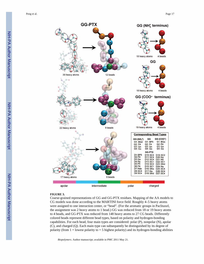
FIGURE 3.Coarse-grained representations of GG and GG-PTX residues. Mapping of the AA models toCG models was done according to the MARTINI force field. Roughly 4–5 heavy atomswere assigned to one interaction center, or “bead”. (For the aromatic groups in Paclitaxel,the assignment was 2 heavy atoms to 1 bead.) GG was reduced from 18 or 19 heavy atomsto 4 beads, and GG-PTX was reduced from 148 heavy atoms to 27 CG beads. Differentlycolored beads represent different bead types, based on polarity and hydrogen-bondingcapabilities. For each bead, four main types are considered: polar (P), nonpolar (N), apolar(C), and charged (Q). Each main type can subsequently be distinguished by its degree ofpolarity (from 1 = lowest polarity to = 5 highest polarity) and its hydrogen-bonding abilities
Peng et al. Page 17
Biopolymers. Author manuscript; available in PMC 2011 May 21.
NIH
-PA Author Manuscript
NIH
-PA Author Manuscript
NIH
-PA Author Manuscript

(denoted by a letter: donor (d), acceptor (a), both (da), or none (0)). Beads withnomenclature beginning ‘B’ and ‘S’ letters denote a backbone bead and an aromatic bead,respectively.
Peng et al. Page 18
Biopolymers. Author manuscript; available in PMC 2011 May 21.
NIH
-PA Author Manuscript
NIH
-PA Author Manuscript
NIH
-PA Author Manuscript
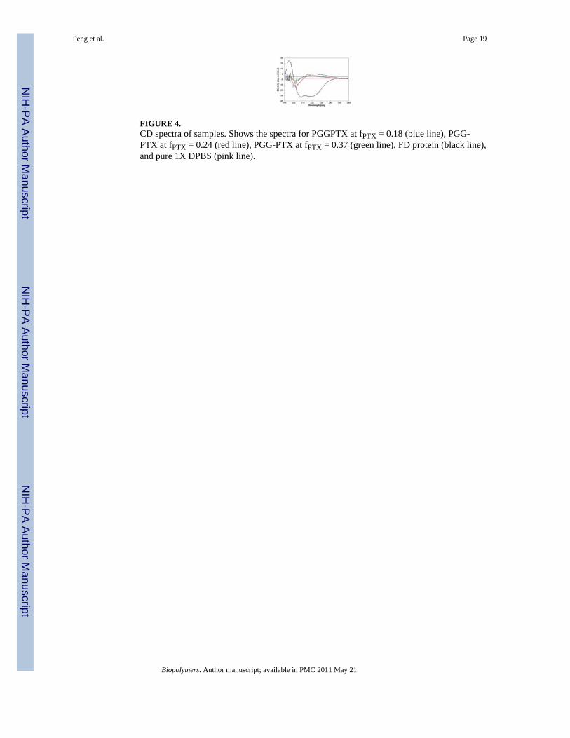
FIGURE 4.CD spectra of samples. Shows the spectra for PGGPTX at fPTX = 0.18 (blue line), PGG-PTX at fPTX = 0.24 (red line), PGG-PTX at fPTX = 0.37 (green line), FD protein (black line),and pure 1X DPBS (pink line).
Peng et al. Page 19
Biopolymers. Author manuscript; available in PMC 2011 May 21.
NIH
-PA Author Manuscript
NIH
-PA Author Manuscript
NIH
-PA Author Manuscript

FIGURE 5.AA and CG models of a PGG-PTX molecule. Shows the models for a fPTX = 0.37 moleculewith an ‘even’ PTX distribution. The GG (black) and PTX (grey) residues are portrayed.
Peng et al. Page 20
Biopolymers. Author manuscript; available in PMC 2011 May 21.
NIH
-PA Author Manuscript
NIH
-PA Author Manuscript
NIH
-PA Author Manuscript

FIGURE 6.Selected probability distributions of AA and CG bond angles between PGG-PTX atomgroups. Information was extracted from 100 ns AA MD and 100 ns CG MD simulations.Shows data for AA (black line) and CG models (grey line).
Peng et al. Page 21
Biopolymers. Author manuscript; available in PMC 2011 May 21.
NIH
-PA Author Manuscript
NIH
-PA Author Manuscript
NIH
-PA Author Manuscript

FIGURE 7.CG RMSD time evolutions of PGG-PTX molecules. PTX distributions include ‘clusters’(pink line), ‘ends’ (black line), ‘even’ (purple line), ‘middle’ (red line), ‘random’ (blue line),and ‘side’ (green line).
Peng et al. Page 22
Biopolymers. Author manuscript; available in PMC 2011 May 21.
NIH
-PA Author Manuscript
NIH
-PA Author Manuscript
NIH
-PA Author Manuscript
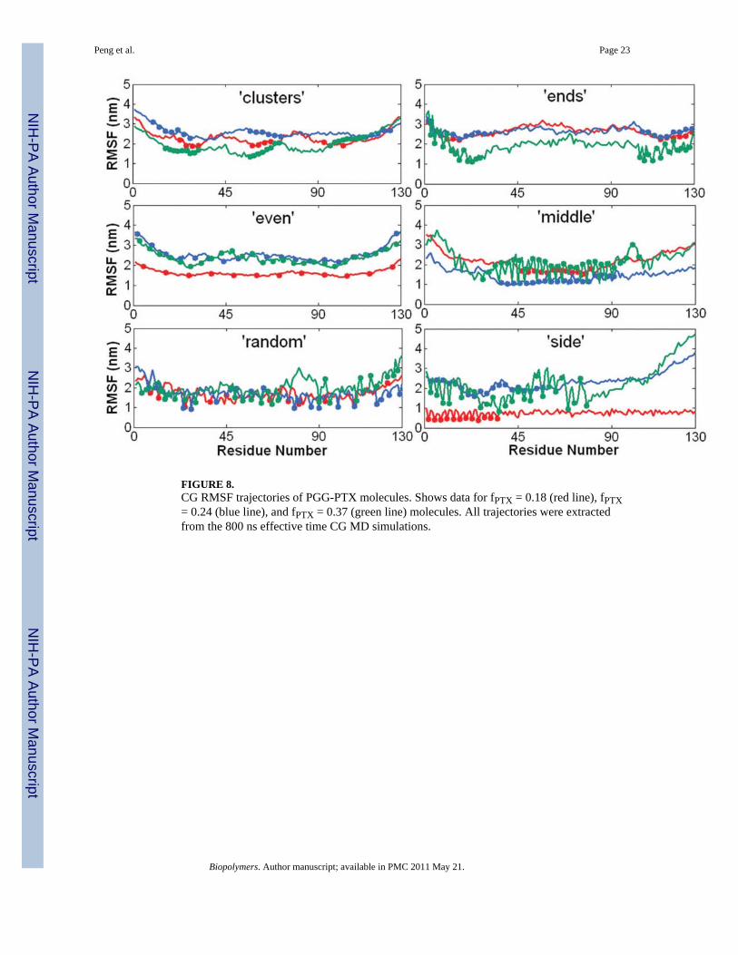
FIGURE 8.CG RMSF trajectories of PGG-PTX molecules. Shows data for fPTX = 0.18 (red line), fPTX= 0.24 (blue line), and fPTX = 0.37 (green line) molecules. All trajectories were extractedfrom the 800 ns effective time CG MD simulations.
Peng et al. Page 23
Biopolymers. Author manuscript; available in PMC 2011 May 21.
NIH
-PA Author Manuscript
NIH
-PA Author Manuscript
NIH
-PA Author Manuscript
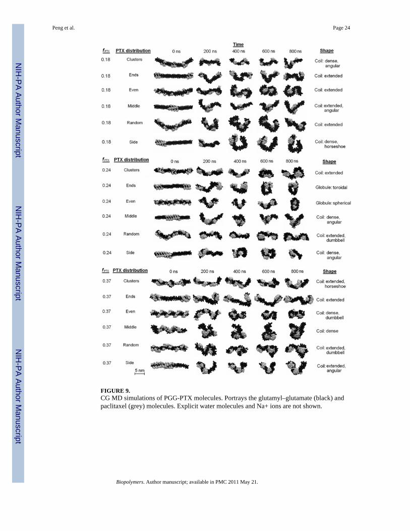
FIGURE 9.CG MD simulations of PGG-PTX molecules. Portrays the glutamyl–glutamate (black) andpaclitaxel (grey) molecules. Explicit water molecules and Na+ ions are not shown.
Peng et al. Page 24
Biopolymers. Author manuscript; available in PMC 2011 May 21.
NIH
-PA Author Manuscript
NIH
-PA Author Manuscript
NIH
-PA Author Manuscript
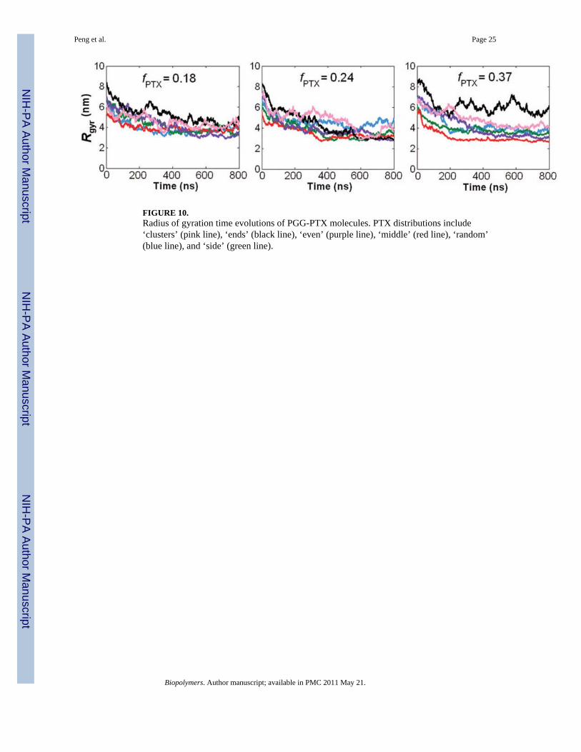
FIGURE 10.Radius of gyration time evolutions of PGG-PTX molecules. PTX distributions include‘clusters’ (pink line), ‘ends’ (black line), ‘even’ (purple line), ‘middle’ (red line), ‘random’(blue line), and ‘side’ (green line).
Peng et al. Page 25
Biopolymers. Author manuscript; available in PMC 2011 May 21.
NIH
-PA Author Manuscript
NIH
-PA Author Manuscript
NIH
-PA Author Manuscript

FIGURE 11.CG MD simulations of negative and positive controls. Shows the trajectories for PGG(black), PG (grey), and PG-B (blue). Explicit water molecules and Na+ ions are not shown.
Peng et al. Page 26
Biopolymers. Author manuscript; available in PMC 2011 May 21.
NIH
-PA Author Manuscript
NIH
-PA Author Manuscript
NIH
-PA Author Manuscript
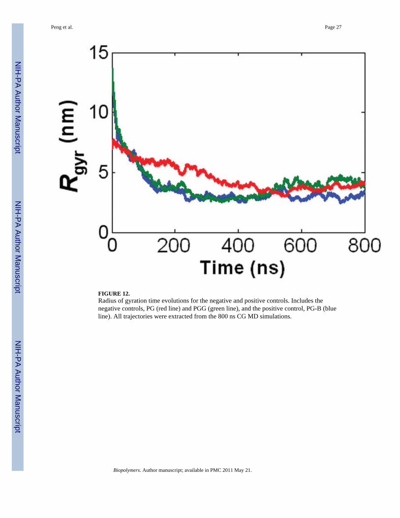
FIGURE 12.Radius of gyration time evolutions for the negative and positive controls. Includes thenegative controls, PG (red line) and PGG (green line), and the positive control, PG-B (blueline). All trajectories were extracted from the 800 ns CG MD simulations.
Peng et al. Page 27
Biopolymers. Author manuscript; available in PMC 2011 May 21.
NIH
-PA Author Manuscript
NIH
-PA Author Manuscript
NIH
-PA Author Manuscript
NIH
-PA Author Manuscript
NIH
-PA Author Manuscript
NIH
-PA Author Manuscript
Peng et al. Page 28
Tabl
e 1
Coa
rse-
grai
ned
topo
logi
es fo
r GG
NH
3, G
G, G
GC
OO−
, and
GG
-PTX
resi
dues
GG
Bac
kbon
e to
polo
gy
i jB
ond
leng
th
[nm
]
Forc
e co
nsta
nt
[kJ
nm−
2 mol−
1 ]
i j k
Bon
d an
gle
Forc
e co
nsta
nt
[kJ
nm−
2 mol−
1 ]
G1
G1
0.35
0*20
0*G
1 G
1 G
112
7*25
*
GG
Sid
echa
in T
opol
ogy
(incl
udes
GG
NH
3+ a
nd G
GC
OO−re
sidu
es)
i jB
ond
leng
th
[nm
]
Forc
e co
nsta
nt
[kJ
nm−
2 mol−
1 ]
i j k
Bon
d an
gle
Forc
e co
nsta
nt
[kJ
nm−
2 mol−
1 ]
G1
G2
0.39
375
00*
G1
G2
G3
143
100*
G2
G3
0.38
175
00*
G2
G3
G4
8510
0*
G3
G4
0.36
675
00*
GG
-PT
X S
idec
hain
Top
olog
y
i jB
ond
leng
th
[nm
]
Forc
e co
nsta
nt[k
J nm
−2 m
ol−
1 ]i j
kB
ond
angl
eFo
rce
cons
tant
[kJ
nm−
2 mol−
1 ]
D1
D2
0.37
475
00*
D1
D2
D3
153
100*
D2
D3
0.38
775
00*
D2
D3
D4
9410
0*
D3
D4
0.40
675
00*
D3
D4
D5
130
100*
D4
D5
0.35
775
00*
D4
D5
D6
8410
0*
D5
D6
0.25
875
00*
D4
D5
D13
146
100*
D6
D7
0.35
375
00*
D5
D6
D7
107
100*
D6
D10
0.31
875
00*
D5
D6
D10
7510
0*
D5
D13
0.19
875
00*
D5
D13
D14
8010
0*
D13
D14
0.22
775
00*
D5
D13
D23
158
100*
D7
D8
0.27
0*10
00*
D6
D7
D8
173
100*
D7
D9
0.27
0*10
00*
D6
D7
D9
109
100*
D8
D9
0.27
0*10
00*
D6
D10
D11
143
100*
Biopolymers. Author manuscript; available in PMC 2011 May 21.

NIH
-PA Author Manuscript
NIH
-PA Author Manuscript
NIH
-PA Author Manuscript
Peng et al. Page 29
D10
D11
0.27
0*10
00*
D6
D10
D12
105
100*
D10
D12
0.27
0*10
00*
D13
D14
D22
9510
0*
D11
D12
0.27
0*10
00*
D13
D14
D15
127
100*
D13
D23
0.22
375
00*
D13
D22
D24
8510
0*
D14
D23
0.34
275
00*
D14
D23
D22
6010
0*
D14
D15
0.38
275
00*
D14
D15
D16
5210
0*
D15
D16
0.51
975
00*
D14
D15
D17
3610
0*
D15
D17
0.60
975
00*
D15
D17
D22
5310
0*
D17
D18
0.57
875
00*
D15
D17
D18
8510
0*
D18
D19
0.28
975
00*
D15
D17
D19
4810
0*
D17
D19
0.46
975
00*
D16
D17
D24
3110
0*
D17
D22
0.13
875
00*
D17
D19
D21
120
100*
D19
D20
0.34
975
00*
D18
D19
D20
115
100*
D19
D21
0.24
975
00*
D18
D19
D21
121
100*
D22
D24
0.36
875
00*
D18
D17
D19
3010
0*
D22
D23
0.36
975
00*
D22
D18
D19
5410
0*
D24
D25
0.23
875
00*
D22
D19
D20
105
100*
D25
D26
0.27
0**
1000
*D
22 D
24 D
2512
510
0*
D25
D27
0.27
0**
1000
*D
23 D
22 D
1375
100*
D26
D27
0.27
0**
1000
*D
23 D
22 D
2496
100*
D24
D25
D26
128
100*
D24
D25
D27
165
100*
* Take
n fr
om th
e pr
edet
erm
ined
val
ues f
or b
ackb
one
or si
dech
ain
bond
ed p
aram
eter
s fro
m th
e M
AR
TIN
I for
ce fi
eld.
Biopolymers. Author manuscript; available in PMC 2011 May 21.
NIH
-PA Author Manuscript
NIH
-PA Author Manuscript
NIH
-PA Author Manuscript
Peng et al. Page 30
Table 2
Descriptions of geometries of a PGG-PTX molecule
Coil (linear, filamentous)
Dense Short, distance between NH3+ and COO− termini is < 10 nm
Extended Long, distance between NH3+ and COO− termini is > 10 nm
Angular Characterized by one sharp turn
Horseshoe Characterized by one major curved turn
Dumbbell Characterized by two linked spherical globules
Globule (round)
Spherical Shaped like a ball
Toroidal Shaped like a ring or donut
Biopolymers. Author manuscript; available in PMC 2011 May 21.