Multiscale Aspects of Cardiac Control
20
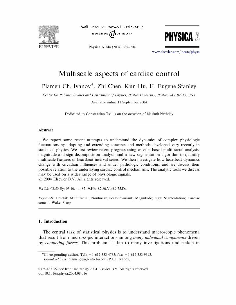
Physica A 344 (2004) 685–704 Multiscale aspects of cardiac control Plamen Ch. Ivanov , Zhi Chen, Kun Hu, H. Eugene Stanley Center for Polymer Studies and Department of Physics, Boston University, Boston, MA 02215, USA Available online 11 September 2004 Dedicated to Constantino Tsallis on the occasion of his 60th birthday Abstract We report some recent attempts to understand the dynamics of complex physiologic fluctuations by adapting and extending concepts and methods developed very recently in statistical physics. We first review recent progress using wavelet-based multifractal analysis, magnitude and sign decomposition analysis and a new segmentation algorithm to quantify multiscale features of heartbeat interval series. We then investigate how heartbeat dynamics change with circadian influences and under pathologic conditions, and we discuss their possible relation to the underlaying cardiac control mechanisms. The analytic tools we discuss may be used on a wider range of physiologic signals. r 2004 Elsevier B.V. All rights reserved. PACS: 02.50.Ey; 05.40.a; 87.19.Hh; 87.80.Vt; 89.75.Da Keywords: Fractal; Multifractal; Nonlinear; Scale-invariant; Magnitude; Sign; Segmentation; Cardiac control; Wake; Sleep 1. Introduction The central task of statistical physics is to understand macroscopic phenomena that result from microscopic interactions among many individual components driven by competing forces. This problem is akin to many investigations undertaken in ARTICLE IN PRESS www.elsevier.com/locate/physa 0378-4371/$ - see front matter r 2004 Elsevier B.V. All rights reserved. doi:10.1016/j.physa.2004.08.016 Corresponding author. Tel.: +1-617-353-4733; fax: +1-617-353-9393. E-mail address: [email protected] (P.Ch. Ivanov).
Transcript of Multiscale Aspects of Cardiac Control
Physica A 344 (2004) 685–704
Multiscale aspects of cardiac control
Plamen Ch. Ivanov�, Zhi Chen, Kun Hu, H. Eugene Stanley
Center for Polymer Studies and Department of Physics, Boston University, Boston, MA 02215, USA
Available online 11 September 2004
Dedicated to Constantino Tsallis on the occasion of his 60th birthday
Abstract
We report some recent attempts to understand the dynamics of complex physiologic
fluctuations by adapting and extending concepts and methods developed very recently in
statistical physics. We first review recent progress using wavelet-based multifractal analysis,
magnitude and sign decomposition analysis and a new segmentation algorithm to quantify
multiscale features of heartbeat interval series. We then investigate how heartbeat dynamics
change with circadian influences and under pathologic conditions, and we discuss their
possible relation to the underlaying cardiac control mechanisms. The analytic tools we discuss
may be used on a wider range of physiologic signals.
r 2004 Elsevier B.V. All rights reserved.
PACS: 02.50.Ey; 05.40.�a; 87.19.Hh; 87.80.Vt; 89.75.Da
Keywords: Fractal; Multifractal; Nonlinear; Scale-invariant; Magnitude; Sign; Segmentation; Cardiac
control; Wake; Sleep
1. Introduction
The central task of statistical physics is to understand macroscopic phenomena
that result from microscopic interactions among many individual components driven
by competing forces. This problem is akin to many investigations undertaken in
ARTICLE IN PRESS
www.elsevier.com/locate/physa
0378-4371/$ - see front matter r 2004 Elsevier B.V. All rights reserved.
doi:10.1016/j.physa.2004.08.016
�Corresponding author. Tel.: +1-617-353-4733; fax: +1-617-353-9393.
E-mail address: [email protected] (P.Ch. Ivanov).
biology. In particular, physiologic systems under autonomic regulation, such as
heart rate, are good candidates for a statistical physics approach, since (i)
physiologic systems often include many individual components, and (ii) physiologic
systems usually are driven by competing forces, e.g., parasympathetic versus
sympathetic stimuli. Physiologic systems often exhibit temporal structures which
are similar to those found in physical systems driven away from an equilibrium state.
Our work centers on human heart rate variability because: (i) the heart rate is
partially under autonomic control, (ii) interbeat interval variability is readily
measured by noninvasive means, (iii) analysis of heart rate dynamics may provide
important practical diagnostic and prognostic information not obtainable with
current approaches, and (iv) these tools can be generalized to the analysis of a wider
range of physiologic signals.
Beat-to-beat heart rate fluctuations are not simply due to known physiologic reflexes
in response to external factors, but rather they persist during rest, sleep, and controlled
constant routine conditions. In particular, two-point correlation analyses of heart rate
fluctuations reveal the presence of a robust interdependence between heartbeats, with
long-range power-law anticorrelations between the values of consecutive interbeat
intervals (i.e., an increase in one interbeat interval is more likely to be followed by a
decrease). These observations suggest a temporal organization, ‘‘hidden’’ in the
apparently noisy heartbeat fluctuations. Moreover, this ‘‘long-memory’’ structure is
characterized by a Fourier spectrum where the amplitudes of different frequency
components follow a power-law relationship of the form 1=f ; where f is the frequency[1–3]. A power-law relation implies invariance under change of frequency scale.
Fractal 1=f noise is also encountered in a certain class of physical systems [4–6],which for a given ‘‘critical’’ value of their parameters exhibit complex self-
organization among the individual components, leading to correlated interactions
over a broad range of scales. The behavior of some physical systems is characterized
by 1=f fluctuations. The presence of 1=f fractal structure in heart rate fluctuations isof particular interest because of its robust appearance in healthy subjects under
different free-running conditions. This fractal pattern breaks down with certain
pathologic perturbations such as congestive heart failure and sleep apnea, suggesting
that 1=f behavior in heart rate fluctuations represents important information aboutthe underlying cardiac control mechanisms.
Our findings [7,8] suggest that the complexity of the heart rate fluctuations cannot
be adequately described by a single scaling parameter, since cardiac dynamics exhibit
a higher level of multiscale complexity [9] which goes beyond 1=f fractal
organization. A large set of scaling parameters are needed. For example, nonlinear
features in heart rate dynamics are not accounted for by 1=f noise. Specifically, (i)the magnitude and the sign of heart rate fluctuations exhibit temporal structures
different from those found in 1=f noise, and (ii) there is a Fourier phase organizationin heart rate fluctuations not present in 1=f signals. We have tested and developedcomplementary methods to probe for a variety of independent characteristics of
heart rate fluctuations beyond 1=f behavior, and determined a set of indices whichcan better describe the complexity of cardiac dynamics under various physiologic
conditions and pathologic perturbations.
ARTICLE IN PRESS
P.Ch. Ivanov et al. / Physica A 344 (2004) 685–704686
1.1. Concepts and approaches from modern statistical physics
To probe how mechanisms of cardiac regulation generate complex heart rate
fluctuations, we have proposed an approach based on concepts and methods derived
from statistical physics. The traditional approach considers physiologic systems to
be governed by the classical principle of homeostasis which postulates that
physiologic systems return to equilibrium after perturbation [10–13]. Such systems
are often characterized by a single dominant time scale. Our preliminary results,
however, suggest that physiologic dynamics are characterized by long-range
correlations and the absence of a typical dominant time scale, and exhibit scale-
free power-law behavior over a broad range of scales [6]. Scale-free behavior is
associated with networks of multiple inputs and interactions [14,15].
1.1.1. Systems with characteristic scales
In order to understand the concept of ‘‘scale-free’’, we start with a discussion of
systems with a characteristic scale, the correlation length. Consider a physical
quantity which describes some properties of a system. This quantity may vary from
one location to another location (or may fluctuate in time). If the system cannot be
divided into many isolated sub-parts, the fluctuations of the physical quantity are
not completely uncorrelated. There is a well-defined scale for correlation length
related to the distance above which the physical properties are uncorrelated (or the
smallest scale of the sub-systems that can be treated as independent). This
characteristic scale has its practical role in simplifying the problem of describing
the system. Several models for studying physical systems take advantage of the
existence of this scale. For many physical systems, a similar scale in time also exists.
Furthermore, we note that the characteristic scales are frequently related to the
exponential decay of a specific quantity. For example, a half life is a characteristic
time scale for radioactive particle decay.
1.1.2. Scale-free systems without characteristic scales
Not every system contains a well-defined scale. In statistical physics, attention has
been directed at understanding systems that do not have any characteristic scale. A
classic example is a ferromagnetic Ising spin system at the critical point [4]. We will
use this example to illustrate important aspects of a scale-free system. Consider a
lattice with a spin on each lattice site. The spin can take two directions, denoted by 1
and �1; and interacts with its nearest neighbors through ferromagnetic interaction(lower energy when two spins align). If the temperature is high, then the
‘‘randomness’’ of the thermal noise dominates. Therefore, each spin is free to
choose its own direction with little correlation with its neighbors. If we gradually
lower the temperature, we will notice that spins are more correlated (with a short
correlation length). A physically meaningful way to define the correlation length for
the system is first to connect all nearest neighbor spins that are aligned in the same
direction as a ‘‘cluster’’, and then to relate the correlation length to the average
cluster size. Spins in different clusters know nothing about each other and, therefore,
the system can be divided into independent sub-systems (clusters). For high
ARTICLE IN PRESS
P.Ch. Ivanov et al. / Physica A 344 (2004) 685–704 687

temperature, the distribution of cluster size decays fast (it is extremely rare to find
large clusters), so a meaningful correlation scale exists. As we lower the temperature,
the size distribution varies (more chance to find large clusters) and the correlation
length increases. However, there is no dramatic change in the form of the
distribution function. If we continue to lower the temperature, suddenly, at
the critical temperature, the distribution function becomes a power law and the
correlation length diverges, i.e., the size of the largest cluster one can find becomes
comparable to the system size. At this point, the system can no longer be viewed as
many independent sub-systems and any characterization of the system must involve
the system as a whole: long-range power-law correlations appear.
1.1.3. Power-law scale-invariant correlations
Our preliminary work has focused primarily on the pursuit on the possibility that
the fluctuations in cardiac dynamics possess long-range power-law correlations.
Systems that have power-law correlations usually have certain scaling properties
related to fractal and nonlinear mechanisms, and are described by homogeneous
functions. The physical meaning of a homogeneous function is that the value of the
function at a new scale is simply related to the value of the function at the original
scale by a constant factor—scale invariant systems are identical under change of
scale. As such they obey functional equations of the form f ðlxÞ ¼ lpf ðxÞ; which statethat when the variable x is changed by a factor l; then the function is replaced by amultiple of itself. The solution of this functional equation is obtained by setting
l ¼ 1=x: f ðxÞ ¼ f ð1Þxp:We say that f ðxÞ is a power law with exponent p, and we seethat systems that are scale invariant of necessity obey power laws. The converse is
also true: any function gðxÞ obeying a power law of the form xp also obeys a
functional equation gðlxÞ ¼ lpgðxÞ: For this reason, a test for scale invariance is totest for power-law behavior.
The scale invariance concept generalizes to functions hðx; yÞ of more than onevariable: hðlax; lbyÞ ¼ lhðx; yÞ: If l ¼ ð1=yÞ1=b; the hðx; yÞ ¼ y1=bhðx=ya=b; 1Þ; so thatthe original function hðx; yÞ of two independent variables x and y is really a function
of a single ‘‘scaled variable’’ x=ya=b: This property is recognized in real data byplotting on the y-axis the ‘‘scaled’’ function hðx; yÞ=y1=b and on the x-axis the scaledvariable x=ya=b: If the original function hðx; yÞ is a scale invariant, then all the datawill ‘‘collapse’’ upon a single one-variable curve hðx=ya=b; 1Þ: Such ‘‘data collapse’’ isalso an indicator of scale invariant behavior [5–7].
Many systems in nature are scale invariant, and the ubiquity of such systems has
been popularized under the general term ‘‘fractal’’. Hence the words scale invariance,
scaling, power law, and fractal are used interchangeably. When applied to perfect
geometrical objects, such as the Sierpinski gasket construction, this scale invariance
is exact. However when applied to statistical objects, such as a heartbeat time series
where multiple extrinsic and intrinsic inputs of stochastic nature play an important
role, this scale invariance holds in a statistical sense, and such a scale invariant
system is not identical when examined on different scales, but rather has identical
statistical properties. Thus even though heart rate fluctuations may appear erratic,
they exhibit certain predictable statistical patterns.
ARTICLE IN PRESS
P.Ch. Ivanov et al. / Physica A 344 (2004) 685–704688
1.2. Nonstationarity in physiologic signals: limitations of traditional analysis
The healthy heartbeat is generally thought to be regulated according to the
classical principle of homeostasis whereby physiologic systems operate to reduce
variability and achieve an equilibrium-like state [10,12,4]. We find, however, that
under normal conditions, beat-to-beat fluctuations in heart rate display the kind of
long-range correlations typically exhibited by physical systems far from equilibrium,
such as those near a critical point. In healthy subjects such power-law correlations
are long-range: they extend over thousands of heartbeats [1,16–18]. In contrast, heart
rate time series from patients with congestive heart failure show a breakdown of this
long-range correlation behavior [19–22].
A major problem in quantifying long-range heart rate correlations is the presence
of nonstationarity. The signals obtained under constantly varying conditions raise
serious challenges to both technical and theoretical aspects of time series analyses.
Furthermore, nonstationarities are important features of the data under both
healthy and perturbed conditions [23–26].
Various problems related to nonstationarity are encountered when attempting to
extract both (a) static and (b) dynamic markers of physiologic data when
investigating sources of cardiac instability:
(a) Static properties such as averages, standard deviations and distribution
functions are widely used to characterize physiologic time series. However, while
physiologic signals under different conditions may have the same averages and
standard deviations—which is often the case with heartbeat time series—there may
be clear differences in the dynamics generating the signals. Hence additional
methods, sensitive to local patterns related to nonstationarities, are required if these
signals are to be distinguished. Further, it has been also hypothesized [27,20] that
even if the heartbeat fluctuations are different (e.g., smaller) during illness, the
pattern of heart rate variability might be otherwise very similar to that in health. In
such a case, the interbeat variations for normal and abnormal cardiac dynamics
would have the same distribution. Such assumptions are based on more conventional
studies of interbeat intervals.
(b) Dynamical properties such as the time ordering (correlations and frequency
content) of physiologic fluctuations are traditionally probed by means of correlation
and power-spectrum analyses. However, both of these methods assume that
the signal studied is stationary, and when applied to nonstationary time series can
lead to misleading results. For example, power-spectrum analysis cannot differ-
entiate between a signal where two different frequency components are present
and a signal where one of the frequency components is present in the first half of
the signal, and another frequency component is present in the second half. The
calculation of the power spectrum for these signals leads to almost identical results.
Similarly, the presence of linear or higher order polynomial trends can mask
true correlations in physiologic fluctuations, e.g., uncorrelated white noise super-
posed on a linear trend will appear strongly correlated. Thus, traditional methods
can be misleading if they are used as the only form of analysis for nonstationary
signals.
ARTICLE IN PRESS
P.Ch. Ivanov et al. / Physica A 344 (2004) 685–704 689

2. Review of recent work
2.1. Multifractality and cardiac regulation
One problem in physiologic research is the signal processing and analysis of
nonstationary time series [2,23,28–31]. Many physiologic time series are extremely
inhomogeneous and nonstationary, fluctuating in an irregular and complex manner.
As one approach to this problem, we have adapted and applied a wavelet-based
multifractal formalism to characterize and quantify specific aspects of the
nonstationarity in physiologic signals [8,19,32–35].
Previously, analyses of the fractal properties of physiologic fluctuations were
restricted to second-order linear characteristics such as the power spectrum and the
two-point autocorrelation function [2,3,36–38]. These analyses revealed that the
behavior of healthy, free-running physiologic systems is often characterized as 1=f -like [1,16–18,39–52]. Monofractal signals (such as classical 1=f noise) are
homogeneous, i.e., have the same scaling properties throughout the entire signal
[53–57]. Monofractal signals can therefore be indexed by a single exponent: the
Hurst exponent H [58].
On the other hand, multifractal signals are inhomogeneous and can be
decomposed into many subsets characterized by different local Hurst exponents h,
which quantify the local singular behavior and thus relate to the local scaling of the
time series. Since the local scaling properties change with time, multifractal signals
require many exponents to fully characterize their nonstationary properties [9,59,60].
The multifractal approach, a concept introduced in the context of multi-affine
functions [61,62,56], has the potential to describe a wide class of signals more
complex than those characterized by a single fractal dimension.
To avoid an ad hoc choice of the time scale over which the local Hurst exponent is
estimated, and to filter out polynomial trends masking the local singularities in the
heart rate signal, we have implemented a wavelet-based algorithm [63]. Wavelets are
designed to probe physiologic time series over a broad range of scales and have
recently been successfully used in analysis of ECG signals. Our wavelet decomposi-
tion reveals a robust self-similar hierarchical organization in the seemingly irregular
heartbeat fluctuations with bifurcations propagating from large to small scales [64,7].
To quantify these hierarchical cascades and to avoid inherent numerical instability in
the estimate of local singularities, we employed a ‘‘mean-field’’ approach—a concept
introduced in statistical physics [4]—which allows us to probe the collective behavior
of local singularities throughout the entire signal.
We performed a wavelet-based multifractal analysis to test the hypothesis that a
large number of exponents is required to characterize heterogeneous heartbeat
interval time series. We analyzed both the daytime (12:00–18:00) and nighttime
(0:00–6:00) continuous heartbeat time series records of healthy subjects [65] and the
daytime records of patients with congestive heart failure [66].
We found the heart rate in healthy subjects to be a multifractal signal; our finding
was the first demonstration of multifractality in physiologic dynamics. Multi-
fractality in heart rate cannot be explained by activity, as data from subjects during
ARTICLE IN PRESS
P.Ch. Ivanov et al. / Physica A 344 (2004) 685–704690

nocturnal hours also showed multifractality [19,8]. Furthermore, this multifractal
behavior could not be attributed to sleep-stage transitions, as we found multifractal
features during daytime hours as well, suggesting that intrinsic mechanisms may
account for these complex dynamics.
From a physiologic perspective, the detection of robust multifractal scaling in the
heart rate dynamics is of interest because it raises the possibility that the control
mechanisms regulating the heartbeat interact as part of a coupled cascade of
nonlinear feedback loops. In physical systems, such interactions are typically
associated with turbulent dynamics and certain systems operating far from
equilibrium [67,68,4]. Furthermore, our results indicate that the healthy heartbeat
is more complex than previously believed, posing a challenge to ongoing efforts to
develop realistic models of heart rate control.
We found that heart rate data from subjects with a pathological condition
associated with sudden death, e.g., congestive heart failure, show a clear loss of
multifractality [19,8]. For the heart failure subjects, the multifractal spectrum is non-
zero only over a very narrow range of exponents h indicating monofractal behavior.
Thus the degree of multifractality might prove useful as a diagnostic tool [64,69,70].
What gives rise to multifractality in healthy human heartbeat dynamics? This
intriguing question has important implications for basic signaling and feedback
mechanisms, and two possible answers can be considered. The first is that the
observed multifractality is primarily a consequence of the response of control
mechanisms to activity-related stimuli [71]. The second is that the heart rate control
mechanisms—even in the presence of weak external noise—endogenously generate
multifractal dynamics [72].
To confirm our initial findings suggesting that the observed multifractal dynamics
are related to intrinsic mechanisms of heart rate control, we next analyzed data taken
from healthy adult subjects under three control conditions: (i) a ‘‘constant routine’’
protocol where physical activity and postural changes were kept to a minimum, (ii)
sympathetic blockade, and (iii) parasympathetic blockade. Fig. 1(a) displays the
average multifractal spectra DðhÞ for six subjects under both regimens of daily and
constant routine. The identical width of the multifractal spectrum DðhÞ suggests that
the multifractality does not change with reduced physical activity under the constant
routine protocol (Fig. 1). This finding supports the hypothesis that the multi-
fractality in healthy heartbeat dynamics is endogenous to the regulation of the heart
rate [8,72].
To further test this hypothesis, we analyzed data from six additional subjects
(4 male, 2 female, ages: 21–34 yr), administered a beta-blocking drug [73,74], and
analyzed data from a placebo control group [75]. We analyzed eight datasets from
the six subjects from the second and/or third day of beta-blocker administration. The
multifractal spectrum curve DðhÞ for the group receiving the beta-blocker drug
differed from that of the control group. This result is consistent with decreased
multifractality—a shrinking of the multifractal spectrum—due to the suppression of
sympathetic control (Fig. 1(b)).
We also analyzed the multifractal properties of the heartbeat dynamics of healthy
individuals who were administered atropine, which suppresses the parasympathetic
ARTICLE IN PRESS
P.Ch. Ivanov et al. / Physica A 344 (2004) 685–704 691

control of the heartbeat, and of age-matched controls. Fig. 1c shows the multifractal
spectra for the two groups. The curve for the group of parasympathetic blockade is
nearly linear, indicating a marked loss of multifractality more apparent than with
sympathetic blockade. These results suggest the possibility that multifractality in
healthy heartbeat dynamics may arise, at least in part, from the interplay between
the two branches of the autonomic system and that the parasympathetic system
plays a particularly important role.
The interaction of autonomic and possibly other control mechanisms that
generate the complex multiscale dynamics of the heartbeat and other physiologic
signals will be a major challenge in our future efforts to model ‘‘real-world’’ signaling
mechanisms [15,76–82].
ARTICLE IN PRESS
0.0 0.1 0.2 0.3h
0.8
0.9
1.0D
(h)
Constant routineUsual daily activity
0.0 0.1 0.2 0.3h
0.8
0.9
1.0
D(h
)
Metoprolol
Placebo
0.0 0.1 0.2 0.3h
0.8
0.9
1.0
D(h
)
AtropinePlacebo
(a) (b)
(c)
Fig. 1. Multifractality and heart rate regulation (from Ref. [72]). (a) Constant routine study. DðhÞ measures
the fractal dimension of the subsets of the signal characterized by local Hurst exponents h. The two curves
have nearly identical widths indicating a similar degree of multifractality. This result is consistent with the
possibility that the activities of daily living do not account for the multifractal complexity of heart rate
dynamics. (b) Sympathetic blockade study. Heartbeat dynamics during sympathetic blockade with
metoprolol display a change in the multifractal spectrum, namely, decreased multifractality as evidenced
by the narrower distribution DðhÞ: (c) Parasympathetic blockade study. Suppression of parasympathetic
control with atropine leads to a collapse of the multifractal spectrum and to monofractal behavior similar
to the behavior observed for congestive heart failure, a condition associated with impaired vagal
regulatory mechanisms [19,8].
P.Ch. Ivanov et al. / Physica A 344 (2004) 685–704692

2.2. Magnitude and sign correlations in heartbeat dynamics
To further probe scaling and nonlinear features embedded in nonstationary
physiologic signals, we developed a new approach that involves (a) decomposing the
signal ‘‘increment’’ series (increments between successive heartbeat intervals) into
magnitude and sign series (Fig. 2), and (b) analyzing their respective scaling
properties using the detrended fluctuation analysis DFA method (Fig. 3) [83–85]. We
demonstrated that signals with identical long-range power-law correlations (1=fnoise) can exhibit different temporal organization for the magnitude and sign,
implying that this analysis goes beyond 1=f analysis. Further, we found that themagnitude series relates to the nonlinear properties of the original time series, and
that the value of the scaling exponent characterizing the correlations in the
magnitude series relates to the width of the multifractal spectrum [8,86,87]. On the
other hand, the sign series relates to the linear properties of the original signal.
We applied our approach to the heartbeat interval series obtained by Holter
monitoring. We previously found that heartbeat increment fluctuations exhibit
fractal-like scale-invariant properties and are anticorrelated over a broad range of
time scales (i.e., the power spectrum follows a power law where the amplitudes of the
higher frequencies are dominant) [20]. Using our new magnitude and sign approach,
we found that the magnitude of the heartbeat interval increments exhibits power-law
scaling behavior and is positively correlated, unlike the original heartbeat increment
time series which is anticorrelated (Fig. 3) [21].
Positive correlations in the magnitude series indicate that an increment with a
large magnitude is more likely to be followed by an increment with a large
magnitude. Anticorrelations in the sign series indicate that a positive increment is
ARTICLE IN PRESS
1180 1190 1200 1210 1220 1230
Beat number
-0.1
0.1
800 900 1000 1100 1200 13000.0
0.1
0.2
|∆R
R|
0 400 800 1200 1600 20000.4
0.6
0.8
1.0
RR
[se
c.]
-1
1
sig
n(∆
RR
)
∆RR
Healthy Heart
Fig. 2. Magnitude and sign decomposition (from Ref. [21]): (top) consecutive cardiac interbeat intervals;
(middle) magnitude series of the increments in the consecutive cardiac intervals shown at top; and
(bottom) sign series of the increments in the cardiac intervals shown at top.
P.Ch. Ivanov et al. / Physica A 344 (2004) 685–704 693

more likely to be followed by a negative increment. Our result for the temporal
organization of heartbeat fluctuations thus suggests that, under healthy conditions, a
large increment in the positive direction is more likely to be followed by a large
increment in the negative direction. We observe that this empirical rule holds over a
broad range of time scales from several up to hundreds of beats.
We also found a significant decrease in the short-range scaling exponent for the
sign series in heart failure, which may be related to perturbed vagal control affecting
relatively high frequency fluctuations. The simultaneous decrease of the long-range
scaling exponent of the magnitude series for heart failure patients indicates weaker
correlations and loss of nonlinearity that may be related to the observed loss of
multifractality in the same group of subjects suggesting impaired feedback
mechanisms of cardiac regulation.
2.3. Scale invariance in the nonstationarity of heartbeat fluctuations
Traditional methods of time series analysis do not address the problem of
nonstationarity in physiologic signals, and most of the more recent techniques
(e.g., wavelets and DFA) focus on removing effects of nonstationarity by filtering
out certain features of nonstationarity such as spikes or polynomial trends. However,
nonstationarity itself may carry information related to the dynamic regulation of
ARTICLE IN PRESS
10 100 1000n
10-1
10-3
10-2
10-3
10-2
100
F(n
)/n
F(n
)/n
original dataphase−randomized data
F(n
)/n
0.4
(b) |∆RR|
(c) sign(∆RR)
0.5
0.75
0
(a) ∆RR
Fig. 3. Root mean square fluctuation F ðnÞ vs. time scale n (in beat numbers) for � 6 h record (� 32; 000
data points) obtained using 2nd order DFA for (a) the heartbeat interval increment series DRRi (&) of a
healthy subject, (b) the integrated magnitude series jDRRij (&) and (c) the integrated sign series
signðDRRiÞ (&). The corresponding time series after Fourier phase randomization are presented with n:
The long-range correlation exponent a� 1 ¼ 0:74� 0:08 of the integrated magnitude series changes to 0.5
after the Fourier phase randomization indicating random behavior. This change in the scaling (after
removing the nonlinear features in the time series as a result of the Fourier phase randomization) suggests
that the magnitude series carries information about the nonlinear properties of heartbeat dynamics.
P.Ch. Ivanov et al. / Physica A 344 (2004) 685–704694

physiologic systems. Working under this hypothesis, we developed a new segmenta-
tion algorithm to quantify nonstationarity in physiologic data [25].
Segmentation method for nonstationary signals. The segmentation algorithm
[25,24] we have developed consists of the following procedure (Fig. 4):
� A sliding pointer is moved from left to right along the signal.
� At each position of the pointer, the mean of the subset of the signal to the left (mleft)
and to the right (mright) of the pointer is computed.
� To measure the difference between mleft and mright; the statistic t mleft�mright
sD
�
�
�
�
�
�is
calculated, where sD is the pooled variance.
� Next the position of the pointer for which t reaches its maximum value tmax is
determined, and the statistical significance of tmax is estimated.
� If this significance exceeds a selected threshold P0 (usually taken to be 95%), the
signal is cut at this point into two subsequences; otherwise the signal remains
undivided.
� If the sequence is cut, the procedure continues recursively for each of the two
resulting subsequences created by each cut.
� Before a new cut is accepted, the statistic t between the right-hand new segment
and its right neighbor (obtained by a previous cut) as well as between the left-hand
new segment and its left neighbor (also obtained by a previous cut) is calculated. If
both values of t have a statistical significance exceeding P0; we proceed with thenew cut; otherwise we do not cut. Thus all resulting segments have a statistically
significant difference in their means. The process stops when none of the possible
cutting points has a significance exceeding P0: As a result the signal is segmentedat the ‘‘significance level P0’’ (Fig. 4).
ARTICLE IN PRESS
0 500 1000 1500 2000x
0
10
t(x)
01020
t(x)
01020
t(x)
0-1
12
f(x)
tmax
x1tmax
x2 tmaxx1
(a)
(b)
(c)
(d)
Fig. 4. (a) An artificial time series f ðxÞ composed of three segments with different mean values. (b) Values
of the statistic tðxÞ obtained by moving the pointer along the time series; tmax is reached at x ¼ x1: IfPðtmaxÞXP0 ¼ 95%; we can cut the series at x1: (c) We iterate the procedure with the segment ½0; x1�: Since
PðtmaxÞX95% and t computed between ½x2;x1� and ½x2; 2000� is greater than 95%; the series is cut at x2: (d)
We iterate the procedure with the segment ½x1; 2000�; and find PðtmaxÞp95%; so this segment is not cut
(from Ref. [25]).
P.Ch. Ivanov et al. / Physica A 344 (2004) 685–704 695

The specific problem we addressed is how to partition a nonstationary time series
(composed of many segments with different mean values) in a way that maximizes
the difference in the mean values between adjacent segments. The segmentation
method leads to the partitioning of a time series into segments with well-defined
means, each significantly different from the mean of the adjacent segments (see
Fig. 5(a)–(d)). We then probed the nonstationarity in a signal through the statistical
analysis of the properties of the segments, thereby going beyond 1=f analysis.We considered 47 heart rate datasets from 18 healthy subjects, 17 cosmonauts
during orbital flight, and 12 patients with congestive heart failure. We analyzed the
cosmonaut data provided by NASA collaborator Dr. J. Fritsch-Yelle (Houston
Space Center). We separately analyzed six-hour subsets of each dataset correspond-
ing to the periods when the subject was awake or sleeping. To quantify the
nonstationarity in heart rate variability, we superimposed a plot of the segments
obtained by means of our segmentation algorithm (Fig. 5(a)–(d)) and analyzed the
ARTICLE IN PRESS
15000 15500 16000 16500 17000
0.0
5.0
10.0
0 5000 10000 15000 20000
0.0
5.0
10.0
15000 15500 16000 16500 170000.4
0.6
0.8
1.0
0 5000 10000 15000 200000.4
0.6
0.8
1.0
1.2
Int
erbe
at in
terv
al (
σ un
its)
Beat number
Int
erbe
at in
terv
al (
sec)
(a)
(b)
(c)
(d)
0 1 2
s ( jump size, σ units)
0
1
2
p(s
)
Healthy
Cosmonauts
Heart failure
(e)
Fig. 5. Probing nonstationarity using a segmentation algorithm (from Ref. 25]). (a) Analysis of 6 hours of
interbeat intervals for a healthy subject (upper curve) and a subject with heart failure (bottom curve). Note
the larger variability and patchiness for the healthy record. (b) Magnification of a small fraction (2000
beats) of the signals in (a). (c) Same signals as displayed in (a) after subtracting the global average and
dividing by the global standard deviation—the signal from the healthy subject is vertically offset; both
signals appear very similar after this normalization. (d) Magnification of a small fraction (2000 beats) of
the signals in (c). (e) Probability density functions of the absolute value of the difference between the mean
values (‘jumps’) of consecutive segments. Both healthy and cosmonaut subjects follow an identical
distribution while the heart-failure subjects follow a quite different distribution with a higher probability
for small jumps consistent with reports of smaller variability in heart failure subjects [88,89]. All
distributions are normalized to unit area.
P.Ch. Ivanov et al. / Physica A 344 (2004) 685–704696

absolute values of the differences between the mean values of consecutive segments,
called jumps.
In order to systematically compare the statistical properties of the jumps between
different individuals and different groups, we normalized each time series by
subtracting the global average (over six hours) and dividing by the global standard
deviation. In this way, all individual time series have a zero mean and unit standard
deviation (Fig. 5(c) and (d)). Such a normalization does not affect our segmentation
procedure. We found that heart rate fluctuation from both the healthy subjects on
earth and the cosmonauts under microgravity conditions follow identical distribu-
tions, but the distribution of the jumps obtained from the heart failure group are
markedly different—centered around lower values—indicating that even after
normalization there is a higher probability for smaller jumps compared to the
healthy subjects. This finding suggests a reduction in the response to autonomic inputs
for patients with congestive heart failure (Fig. 5(e)). Furthermore, these distributions
can be collapsed on top of each other by means of a homogeneous transformation
[25]. The ratio between the scaling parameters used in this transformation provides a
factor by which this feature of the heart rate variability is reduced for the subjects
with heart failure as compared to the healthy subjects. These observations extend to
previously reported results for the distribution of heartbeat fluctuations obtained by
means of wavelet and Hilbert transforms [7,36] and support the hypothesis that a
nonstationarity index itself carries relevant physiologic information.
2.4. Sleep-wake scaling differences in human heartbeat dynamics
To further probe the mechanisms of nonstationarity of heartbeat regulation, we
investigated circadian influences, a key modulator of many physiologic processes
[90,64]. The question we asked was whether there are characteristic differences in
the scaling behavior between sleep and wake cardiac dynamics. Differences in the
average value and standard deviation can be systematically observed in plots of the
interbeat intervals recorded from subjects during sleep and wake periods (Fig. 6(a)
and (b)) [90]. We hypothesized that changes in cardiac control during sleep and wake
periods may occur on all time scales and thus could lead to systematic changes in the
scaling properties of the heartbeat dynamics [91]. Elucidating the nature of these
sleep–wake rhythms could lead to a better understanding of the mechanisms of
cardiac regulation and its interaction with the mechanisms of sleep regulation.
We analyzed 30 datasets, each with 24 h of interbeat intervals, from 18 healthy
subjects and 12 patients with congestive heart failure. We analyzed the nocturnal and
diurnal fractions of the dataset of each subject, which corresponded to the six hours
from midnight to 6:00 am and noon to 6:00 pm. We found that at scales above
� 1min (n460) the data during wake hours display long-range power-law
correlations over two decades with average exponents awake � 1:05 for the healthygroup and awake � 1:2 for the heart failure patients. For the sleep data we found asystematic crossover at scale n � 60 beats followed by a scaling regime extending
over two decades characterized by a smaller exponent: asleep � 0:85 for the healthygroup and asleep � 0:95 for the heart failure group (Fig. 6(c)) [91]. Although the
ARTICLE IN PRESS
P.Ch. Ivanov et al. / Physica A 344 (2004) 685–704 697

values of the sleep and wake exponents vary from subject to subject, we found that
for all individuals studied, the heartbeat dynamics during sleep are characterized by
a smaller exponent, indicating stronger anticorrelated behavior in the heartbeat
fluctuations during sleep. The physiologic implications of this fundamental change in
the temporal properties of cardiac dynamics during sleep will be a focus of this
application.
To test the robustness of our results, we also analyzed 17 datasets from six
cosmonauts under conditions of microgravity during a long-term orbital flight on the
Mir space station [92]. Each dataset contained continuous six-hour periods of data
under both sleep and wake conditions. We found that for all cosmonauts the
heartbeat fluctuations exhibit an anticorrelated behavior with average scaling
exponents consistent with those found for the healthy terrestrial group: the exponent
is � 1:04 for the wake phase and � 0:82 for the sleep phase (Fig. 6(d)). Thus thelarger values for the wake phase scaling exponents cannot be a trivial artifact of
activity since cosmonauts are under completely different stress and physical
conditions compared to the terrestrial group. Furthermore, the larger value of the
average wake exponent for the heart failure group compared to the other two groups
cannot be attributed to external stimuli either, since patients with severe cardiac
disease are strongly restricted in their physical activity. Instead, our results suggest
that the observed scaling characteristics in the heartbeat fluctuations during sleep
and wake phases are related to intrinsic mechanisms of heart rate control, the
dynamics of which may change during the transition from wake to sleep [91,93,94].
Such a change in the dynamical state of cardiac regulation may be associated with
ARTICLE IN PRESS
0 5000 10000 15000
beat number
0.4
0.6
0.8
1.0
1.2
1.4
1.6
Int
erbe
at I
nter
val [
sec]
0 5000 10000 15000 20000 250000.4
0.6
0.8
1.0
1.2
1.4
1.6(a)
(b)
Healthy - Wake
Healthy - Sleep
10 100 1000 10000
n
F(n
)
103
102
101
100
10-1
103
102
101
100
10-1
Healthy(c)
(d)
slope=1
slope=0.8
Cosmonaut
slope=1.1
slope=0.8
Fig. 6. Statistical differences in heartbeat data during wake and sleep (from Ref. [91]). Consecutive
heartbeat intervals are plotted versus beat number for six hours recorded from the same healthy subject
during: (a) wake period: 12pm to 6pm and (b) sleep period: 12am to 6am. Plots of logF ðnÞ versus log n for
six-hour wake records (open circles) and sleep records (filled triangles) of (c) one typical healthy subject;
(d) one cosmonaut (during orbital flight). Here F ðnÞ is the DFA fluctuation function and n is the time scale
in beat numbers. Note the lower exponent at scales n460 beats for the sleep phase (filled triangles),
indicating systematic changes in the heartbeat dynamics during sleep.
P.Ch. Ivanov et al. / Physica A 344 (2004) 685–704698

previously reported peaks in cardiac vulnerability during sleep-wake transitions
[95–97] and may have important diagnostic and clinical applications.
2.5. Scale-invariant patterns in heartbeat dynamics during different sleep stages
Since our findings suggest different regimes of intrinsic regulation of the cardiac
dynamics during wake and sleep that may switch on and off with circadian rhythms
[91], we next investigated how the heart rhythms of healthy subjects change within
the different sleep stages [98]. We found that different sleep stages are associated with
specific types of correlations and different degrees of nonlinearity in heartbeat
dynamics [99], suggesting changes in the cardiac regulation with different sleep
stages.
We analyzed 24 records of interbeat intervals (� 7:5 h duration) obtained from 12
healthy individuals during sleep. Fig. 7(a) shows the heartbeat interval time series for
a typical healthy subject with periods of light sleep, deep sleep, REM sleep, and short
intermediate wake phases. The annotation and duration of the sleep stages were
determined based on standard procedures [100]. We next applied our magnitude and
sign approach.
The mean values of the scaling exponents obtained using our magnitude and sign
approach and their standard deviations for the different sleep stages are shown in
Fig. 7. We found a smaller scaling exponent amag for light sleep than for REM sleep,
indicating weaker long-range power-law correlations (Fig. 7). Surprisingly, we found
that in contrast to REM and light sleep, the magnitude series for deep sleep is
uncorrelated, with amag ¼ 1:5 suggesting random-walk-like Brownian behavior. The
ARTICLE IN PRESS
1 2 3 4 5 6 7 8time t [h]
0.8
1.0
1.2
1.4
RR
[sec
.]
wake stages
REM sleep
light sleep
deep sleep
wake REM light deep0.8
1.0
1.2
1.4
1.6
1.8
αsi
gnα
mag
(a) (b)
Fig. 7. Statistical differences and scaling in cardiac interbeat data during different sleep stages (from Ref.
[98]). (a) One-night record for a healthy subject. (b) The group average values and standard deviations of
the fluctuation exponents amag for the magnitude series and asign for the sign series for the different sleep/
wake phases. For each of the 24 records from 12 healthy subjects the corresponding second-order DFA
fluctuation functions F ðnÞ have been fit into the range of 8pnp13 and 11pnp150 heartbeats for asign and
amag; respectively, where the most significant differences between the sleep stages occur. Thus, the
correlation behavior of the heartbeat increments and their signs and magnitudes during daytime activity is
similar to the behavior we find in REM sleep, but significantly different from the behavior we observe in
deep sleep.
P.Ch. Ivanov et al. / Physica A 344 (2004) 685–704 699

long-range magnitude correlations found for REM sleep indicate nonlinear
contributions to the heartbeat regulation, which are reduced during light and deep
sleep [8]. Thus, the correlation behavior of the heartbeat increments and their signs
and magnitudes during daytime activity is similar to the behavior we find in REM
sleep, but quite different from the behavior we observe in deep sleep (Fig. 7).
3. Summary
A defining feature of physiologic systems is their complexity. Trying to decode the
remarkable range of behavior of living systems in health and disease has emerged as
a major focus of contemporary medicine. This enterprise suggests a need for
interdisciplinary collaborations among scientists with different backgrounds and the
need to foster of multidisciplinary approach to problems at the interface of physics
and physiology.
The general objectives of our work are to explore both the physiologic significance
and practical utility of heart rate fluctuations. This research should lead to deeper
understanding of the multiscale nature of cardiac control mechanisms, and to further
development of reliable statistical algorithms with which to identify and quantify
healthy and pathologic cardiac dynamics.
Acknowledgements
The results reviewed here represent a collaborative research effort with major
contributions from many individuals, including L.A.N. Amaral, Y. Ashkenazy,
R.M. Baevsky, P. Bernaola-Galvan, A. Bunde, P. Carpena, J. Fritsch-Yelle, A.L.
Goldberger, S. Havlin, J.W. Kantelhardt, T. Penzel, C.-K. Peng, J.-H. Peter, M.G.
Rosenblum, Y. Yamamoto. This work was supported by NIH/National Center for
Research Resources (P41 RR13622) and NIH Grants HL071972 and HL079046.
References
[1] M. Kobayashi, T. Musha, 1=f fluctuation of heartbeat period, IEEE Trans. Biomed. Eng. 29 (1982)456–457.
[2] J.B. Bassingthwaighte, L.S. Liebovitch, B.J. West, Fractal Physiology, Oxford University Press,
New York, 1994.
[3] M. Malik, A.J. Camm (Eds.), Heart Rate Variability, Futura, Armonk, NY, 1995.
[4] H.E. Stanley, Introduction to Phase Transitions and Critical Phenomena, Oxford University Press,
London, 1971.
[5] H.E. Stanley, Scaling, universality, and renormalization: three pillars of modern critical phenomena,
Rev. Mod. Phys. 71 (1999) S358–S366.
[6] H.E. Stanley, Power laws and universality, Nature 378 (1995) 554–554.
[7] P.Ch. Ivanov, M.G. Rosenblum, C.-K. Peng, J. Mietus, S. Havlin, H.E. Stanley, A.L. Goldberger,
Scaling behaviour of heartbeat intervals obtained by wavelet-based time-series analysis, Nature 383
(1996) 323–327.
ARTICLE IN PRESS
P.Ch. Ivanov et al. / Physica A 344 (2004) 685–704700

[8] P.Ch. Ivanov, M.G. Rosenblum, L.A.N. Amaral, Z.R. Struzik, S. Havlin, A.L. Goldberger, H.E.
Stanley, Multifractality in human heartbeat dynamics, Nature 399 (1999) 461–465.
[9] H.E. Stanley, P. Meakin, Multifractal phenomena in physics and chemistry, Nature 335 (1988)
405–409.
[10] C. Bernard, Les Phenomenes de la Vie, Paris, 1878.
[11] B. van der Pol, J. van der Mark, The heartbeat considered as a relaxation oscillation, and an
electrical model of the heart, Philos. Mag. 6 (1928) 763–775.
[12] W.B. Cannon, Organization for physiological homeostasis, Physiol. Rev. 9 (1929) 399–431.
[13] B.W. Hyndman, The role of rhythms in homeostasis, Kybernetik 15 (1974) 227–236.
[14] E.W. Montroll, M.F. Shlesinger, The wonderful world of random walks, in: L.J. Lebowitz, E.W.
Montroll (Eds.), Nonequilibrium Phenomena II: From Stochastics to Hydrodynamics, North-
Holland, Amsterdam, 1984, pp. 1–121.
[15] P.Ch. Ivanov, L.A.N. Amaral, A.L. Goldberger, H.E. Stanley, Stochastic feedback and the
regulation of biological rhythms, Europhys. Lett. 43 (1998) 363–368.
[16] Y. Yamamoto, R.L. Hughson, Coarse graining spectral analysis: new method for studying heart rate
variability, J. Appl. Physiol. 71 (1991) 1143–1150.
[17] Y. Yamamoto, R.L. Hughson, Extracting fractal components from time series, Physica D 68 (1993)
250–264.
[18] C.-K. Peng, J. Mietus, J.M. Hausdorff, S. Havlin, H.E. Stanley, A.L. Goldberger, Long-range anti-
correlations and non-Gaussian behavior of the heartbeat, Phys. Rev. Lett. 70 (1993) 1343–1346.
[19] P.Ch. Ivanov, L.A.N. Amaral, A.L. Goldberger, S. Havlin, M.G. Rosenblum, H.E. Stanley, Z.
Struzik, From 1=f noise to multifractal cascades in heartbeat dynamics, Chaos 11 (2001)
641–652.
[20] C.K. Peng, S. Havlin, H.E. Stanley, A.L. Goldberger, Quantification of scaling exponents and
crossover phenomena in nonstationary heartbeat time series, Chaos 5 (1995) 82–87.
[21] Y. Ashkenazy, P.Ch. Ivanov, S. Havlin, C.-K. Peng, A.L. Goldberger, H.E. Stanley, Magnitude and
sign correlations in heartbeat fluctuations, Phys. Rev. Lett. 86 (2001) 1900–1903.
[22] B. Kulessa, T. Srokowski, S. Drozdz, Long-time autocorrelation function of ECG signal for healthy
versus diseased human heart, Acta Phys. Pol. B 34 (2003) 3–15.
[23] H. Kantz, T. Schreiber, Nonlinear Time Series Analysis, Cambridge University Press, Cambridge,
1997.
[24] P. Bernaola-Galvan, I. Grosse, P. Carpena, J.L. Oliver, R. Roman-Roldan, H.E. Stanley, Finding
borders between coding and noncoding DNA regions by an entropic segmentation method, Phys.
Rev. Lett. 85 (2000) 1342.
[25] P. Bernaola-Galvan, P.Ch. Ivanov, L.A.N. Amaral, H.E. Stanley, Scale invariance in the
nonstationarity of physiological signals, Phys. Rev. Lett. 87 (2001) 168105(4).
[26] G.M. Viswanathan, C.-K. Peng, H.E. Stanley, A.L. Goldberger, Deviations from uniform power
law scaling in nonstationary time series, Phys. Rev. E 55 (1996) 845–849.
[27] A.A. Aghili, Rizwan-uddin, M.P. Griggin, J.R. Moorman, Scaling and ordering of neonatal rate
variability, Phys. Rev. Lett. 74 (1995) 1254–1257.
[28] M. Koebbe, G. Mayer-Kress, in: M. Casdagli, S. Eubank (Eds.), Nonlinear modeling and
forecasting, SFI studies in the science of complexity, Proc Vol. XII, Addison-Wesley, New York,
1992, pp. 361–378.
[29] T. Schreiber, Detecting and analyzing nonstationarity in a time series using nonlinear cross
predictions, Phys. Rev. Lett. 78 (1997) 843–846.
[30] A. Witt, J. Kurths, A. Pikovsky, Testing stationarity in time series, Phys. Rev. E 58 (1998)
1800–1810.
[31] P.E. Rapp, A guide to dynamical analysis, Integr. Physiol. Behav. Sci. 29 (1994) 311–327.
[32] H.E. Stanley, L.A.N. Amaral, A.L. Goldberger, S. Havlin, P.Ch. Ivanov, C.-K. Peng, Statistical
physics in physiology: monofractal and multifractal approaches, Physica A 270 (1999) 309–324.
[33] H.E. Stanley, L.A.N. Amaral, A.L. Goldberger, S. Havlin, P.Ch. Ivanov, C.-K. Peng, Monofractal
and multifractal approaches to complex biomedical signals, in: D.S. Broomhead, E.A. Luchinskaya,
P.V.E. McClintock, T. Mullin (Eds.), Stochastic and Chaotic Dynamics in the Lakes, Proceedings of
ARTICLE IN PRESS
P.Ch. Ivanov et al. / Physica A 344 (2004) 685–704 701
International Stochaos Workshop, Ambleside, Cumbria, UK, American Institute of Physics AIP
Conf. Proc. 502, Melville NY, 2000, pp. 133–145.
[34] A.L. Goldberger, L.A.N. Amaral, J.M. Hausdorff, P.Ch. Ivanov, C.-K. Peng, H.E. Stanley, Fractal
dynamics in physiology: alterations with disease and aging, Proc. Natl. Acad. Sci. USA 99 (Suppl. 1)
(2002) 2466–2472.
[35] A. Grossmann, J. Morlet, Mathematics and Physics: Lectures on Recent Results, World Scientific,
Singapore, 1985.
[36] P.F. Panter, Modulation, Noise, and Spectral Analysis Applied to Information Transmission, New
York, NY, 1965.
[37] R.I. Kitney, O. Rompelman, The Study of Heart-Rate Variability, Oxford University Press,
London, 1980.
[38] S. Akselrod, D. Gordon, F.A. Ubel, D.C. Shannon, A.C. Barger, R.J. Cohen, Power spectrum
analysis of heart rate fluctuation: a quantitative probe of beat-to-beat cardiovascular control,
Science 213 (1981) 220–222.
[39] M.F. Shlesinger, Fractal time and 1=f noise in complex systems, Ann. NY Acad. Sci. 504 (1987)
214–228.
[40] J.M. Hausdorff, Z. Ladin, J.Y. Wei, Footswitch system for measurement of the temporal parameters
of gait, J. Biomech. 28 (1995) 347–351.
[41] J.M. Hausdorff, P.L. Purdon, C.-K. Peng, Z. Ladin, J.Y. Wei, A.L. Goldberger, Fractal dynamics
of human gait: stability of long-range correlations in stride interval fluctuations, J. Appl. Physiol. 80
(1996) 1448–1457.
[42] S. Havlin, S.V. Buldyrev, A. Bunde, A.L. Goldberger, P.Ch. Ivanov, C.-K. Peng, H.E. Stanley,
Scaling in nature: from DNA through heartbeats to weather, Physica A 273 (1999) 46–69.
[43] H.E. Stanley, L.A.N. Amaral, P. Gopikrishnan, P.Ch. Ivanov, T.H. Keitt, V. Plerou, Scale
invariance and universality: organizing principles in complex systems, Physica A 281 (2000) 60–68.
[44] J. Kurths, A. Voss, P. Saparin, A. Witt, H.J. Kleiner, N. Wessel, Quantitative analysis of heart rate
variability, Chaos 5 (1995) 88–94.
[45] L.S. Liebovitch, Fractal analysis of channel mechanisms, Adv. Chem. Ser. 235 (1994) 357.
[46] S.B. Lowen, L.S. Liebovitch, J.A. White, Fractal ion-channel behavior generates fractal firing
patterns in neuronal models, Phys. Rev. E 59 (1999) 5970.
[47] Y.Q. Chen, M.Z. Ding, J.A.S. Kelso, Long memory processes (1=f ðaÞ type) in human coordination,
Phys. Rev. Lett. 79 (1997) 4501–4504.
[48] B.J. West, L. Griffin, Allometric control of human gait, Fractals 6 (1998) 101–108.
[49] L. Griffin, D.J. West, B.J. West, Random stride intervals with memory, J. Biol. Phys. 26 (2000)
185–202.
[50] L.A. Protsmman, H. Meeuwsen, P. Hamilton, B.J. West, J. Wilkerson, Nonlinear analysis of the
scaling properties of human gait: use of Hurst exponent as a clinical tool, J. Sport Exercise Psychol.
23 (2001) S67–S67.
[51] K. Hu, P.Ch. Ivanov, Z. Chen, M.F. Hilton, H. E Stanley, S.A. Shea, Intrinsic patters of human
activity: scaling and nonlinear dynamics in forearm motion, Physica A 337 (2004) 307.
[52] R. Karasik, N. Sapir, Y. Ashkenazy, P.Ch. Ivanov, I. Dvir, P. Lavie, S. Havlin, Correlation
differences in heartbeat fluctuations during rest and exercise, Phys. Rev. E 66 (2002) 062902(4).
[53] A. Bunde, S. Havlin (Eds.), Fractals and Disordered Systems, 2nd Edition, Springer, Berlin, 1996.
[54] H. Takayasu, Fractals in the Physical Sciences, Manchester University Press, Manchester UK, 1997.
[55] T.G. Dewey, Fractals in Molecular Biophysics, Oxford University Press, Oxford, 1997.
[56] A.-L. Barabasi, H.E. Stanley, Fractal Concepts in Surface Growth, Cambridge University Press,
Cambridge, 1995.
[57] S. Stoev, V. Pipiras, M.S. Taqqu, Estimation of the self-similarity parameter in linear fractional
stable motion, Signal Process 82j (2002) 1873–1901.
[58] H.E. Hurst, Long-term storage capacity of reservoirs, Trans. Am. Soc. Civ. Eng. 116 (1951)
770–808.
[59] P. Meakin, Fractals, Scaling and Growth Far from Equilibrium, Cambridge University Press,
Cambridge, 1997.
ARTICLE IN PRESS
P.Ch. Ivanov et al. / Physica A 344 (2004) 685–704702

[60] J. Feder, Fractals, Plenum, New York, 1988.
[61] T. Vicsek, A.-L. Barabasi, Multi-affine model for the velocity distribution in fully turbulent flows,
J. Phys. A: Math. Gen. 24 (1991) L845–L851.
[62] A.-L. Barabasi, P. Szepfalusy, T. Vicsek, Multifractality of multiaffine functions, Physica A 178
(1991) 17–28.
[63] J.F. Muzy, E. Bacry, A. Arneodo, The multifractal formalism revisited with wavelets, Int. J. Bifurc.
Chaos 4 (1994) 245–302.
[64] P.Ch. Ivanov, M.G. Rosenblum, C.-K. Peng, J. Mietus, S. Havlin, H.E. Stanley, A.L. Goldberger,
Scaling and universality in heart rate variability distributions, Physica A 249 (1998) 587–593.
[65] http://www.physionet.org/physiobank/database/nsrdb.
[66] http://www.physionet.org/physiobank/database/chfdb.
[67] C. Meneveau, K.R. Sreenivasan, Simple multifractal cascade model for fully developed turbulence,
Phys. Rev. Lett. 59 (1987) 1424–1427.
[68] U. Frisch, Turbulence, Cambridge University Press, Cambridge, 1995.
[69] S. Havlin, L.A.N. Amaral, Y. Ashkenazy, A.L. Goldberger, P.Ch. Ivanov, C.-K. Peng, H.E.
Stanley, Application of statistical physics to heartbeat diagnosis, Physica A 274 (1999) 99–110.
[70] L.A.N. Amaral, A.L. Goldberger, P.Ch. Ivanov, H.E. Stanley, Scale-independent measures and
pathologic cardiac dynamics, Phys. Rev. Lett. 81 (1998) 2388–2391.
[71] J.B. Carter, E.W. Banister, A.P. Blaber, Effect of endurance exercise on autonomic control of heart
rate, Sports Med. 33 (2003) 33–46.
[72] L.A.N. Amaral, P.Ch. Ivanov, N. Aoyagi, I. Hidaka, S. Tomono, A.L. Goldberger, H.E. Stanley, Y.
Yamamoto, Behavioral-independent features of complex heartbeat dynamics, Phys. Rev. Lett. 86
(2001) 6026–6029.
[73] J.J.V. McMurray, Major beta blocker mortality trials in chronic heart failure: a critical review,
Heart 82 (1999) 14–22.
[74] A.J. Moss, W. Zareba, W.J. Hall, P.J. Schwartz, R.S. Crampton, J. Benhorin, G.M. Vincent, E.H.
Locati, S.G. Priori, C. Napolitano, A. Medina, L. Zhang, J.L. Robinson, K. Timothy, J.A. Towbin,
M.L. Andrews, Effectiveness and limitations of beta-blocker therapy in congenital long-QT
syndrome, Circulation 101 (2000) 616–623.
[75] N. Aoyagi, K. Ohashi, S. Tomono, Y. Yamamoto, Temporal contribution of body movement to
very long-term heart rate variability in humans, Am. J. Physiol. 278 (2000) H1035–H1041.
[76] J.C. Marshall, Complexity, chaos, and incomprehensibility: parsing the biology of critical illness,
Crit. Care Med. 28 (2000) 2646–2648.
[77] K. Kotani, K. Takamasu, Y. Ashkenazy, H.E. Stanley, Y. Yamamoto, Model for cardiorespiratory
synchronization in humans, Phys. Rev. E 65 (2002) 051923(9).
[78] H. Seidel, H. Herzel, Bifurcations in a nonlinear model of the baroreceptor-cardiac reflex, Physica D
115 (1998) 145–162.
[79] V. Schulte-Frohlinde, Y. Ashkenazy, P.Ch. Ivanov, L. Glass, A.L. Goldberger, H.E. Stanley,
Noise effects on the complex patterns of abnormal heartbeats, Phys. Rev. Lett. 87 (2001)
068104(4).
[80] J.W. Kantelhardt, S. Havlin, P.Ch. Ivanov, Modeling transient correlations in heartbeat dynamics
during sleep, Europhys. Lett. 62 (2003) 147–153.
[81] J.F. Muzy, E. Bacry, Multifractal stationary random measures and multifractal random walks with
log infinitely divisible scaling laws, Phys. Rev. E 66 (2002) 056121.
[82] D.C. Lin, R.L. Hughson, Modeling heart rate variability in healthy humans: a turbulence analogy,
Phys. Rev. Lett. 86 (2001) 1650–1653.
[83] C.K. Peng, S.V. Buldyrev, J.H. Hausforff, S. Havlin, J.E. Mietus, M. Simons, H.E. Stanley, A.L.
Goldberger, Nonequilibrium dynamics as an indispensable characteristic of a healthy biological
system, Integr. Physiol. Behavioral. Sci. 29 (1994) 283–298.
[84] K. Hu, P.Ch. Ivanov, Z. Chen, P. Carpena, H.E. Stanley, Effect of trends on detrended fluctuation
analysis, Phys. Rev. E 64 (2001) 011114(19).
[85] Z. Chen, P.Ch. Ivanov, K. Hu, H.E. Stanley, Effect of nonstationarities on detrended fluctuation
analysis, Phys. Rev. E 65 (2002) 041107(15).
ARTICLE IN PRESS
P.Ch. Ivanov et al. / Physica A 344 (2004) 685–704 703

[86] Y. Ashkenazy, S. Havlin, P.Ch. Ivanov, C.-K. Peng, V. Schulte-Frohlinde, H.E. Stanley, Magnitude
and sign scaling in power-law correlated time series, Physica A 323 (2003) 19–41.
[87] Y. Ashkenazy, P.Ch. Ivanov, S. Havlin, C.-K. Peng, Y. Yamamoto, A.L. Goldberger, H.E. Stanley,
Decomposition of heartbeat time series: scaling analysis of the sign sequence, Comp. in Cardiol. 27
(2000) 139–142.
[88] M.M. Wolf, G.A. Varigos, D. Hunt, J.G. Sloman, Sinus arrhythmia in acute myocardial infarction,
Med. J. Aust. 2 (1978) 52–55.
[89] A.L. Goldberger, Non-linear dynamics for clinicians: chaos theory, and complexity at the bedside,
Lancet 347 (1996) 1312–1314.
[90] H. Molgaard, K.E. Sorensen, P. Bjerregaard, Circadian variation and influence of risk-factors on
heart-rate variability in healthy subjects, Am. J. Cardiol. 68 (1991) 777–784.
[91] P.Ch. Ivanov, A. Bunde, L.A.N. Amaral, S. Havlin, J. Fritsch-Yelle, R.M. Baevsky, H.E. Stanley,
A.L. Goldberger, Sleep-wake differences in scaling behavior of the human heartbeat: analysis of
terrestrial and long-term space flight data, Europhys. Lett. 48 (1999) 594–600.
[92] A.L. Goldberger, M.W. Bungo, R.M. Baevsky, B.S. Bennett, D.R. Rigney, J.E. Mietus, G.A.
Nikulina, J.B. Charles, Heart-rate dynamics during long-term space flight: report on MIR
cosmonauts, Am. Heart J. 128 (1994) 202–204.
[93] M.A. Carskadon, W.C. Dement, Normal human sleep: an overview, in: M.H. Kryger, T. Roth,
W.C. Dement (Eds.), Principles and Practice of Sleep Medicine, Saunders Co., Philadelphia, 2000,
pp. 15–25.
[94] D. Mcginty, R. Szymusiak, The sleep-wake switch: a neuronal alarm clock, Nature Med. 6 (2000)
510–511.
[95] J.E. Muller, P.H. Stone, Z.G. Turi, J.D. Rutherford, C.A. Czeisler, C. Parker, W.K. Poole, E.
Passamani, R. Roberts, T. Robertson, B.E. Sobel, J.T. Willerson, E. Braunwald, the MILIS Study
Group, Circadian variation in the frequency of onset of acute myocardial infarction, New Engl.
J. Med. 313 (1985) 1315–1322.
[96] J.E. Muller, P.L. Ludmer, S.N. Wilich, G.H. Tofler, G. Aylmer, I. Klangos, P.H. Stone, Circadian
variation in the frequency of sudden cardiac death, Circulation 75 (1987) 131–138.
[97] O. Stiedl, M. Meyer, Fractal dynamics in circadian cardiac time series of corticotropin-releasing
factor receptor subtype-2 deficient mice, J. Math. Biol. 47 (2003) 169–197.
[98] J.W. Kantelhardt, Y. Ashkenazy, P.Ch. Ivanov, A. Bunde, S. Havlin, T. Penzel, J.-H. Peter, H.E.
Stanley, Characterization of sleep stages by correlations of heartbeat increments, Phys. Rev. E 65
(2002) 051908(6).
[99] A. Bunde, S. Havlin, J.W. Kantelhardt, T. Penzel, J.H. Peter, K. Voigt, Correlated and uncorrelated
regions in heart-rate fluctuations during sleep, Phys. Rev. Lett. 85 (2000) 3736–3739.
[100] A. Rechtschaffen, A. Kales, A Manual of Standardized Terminology, Techniques, and Scoring
System for Sleep Stages of Human Subjects, US Government Printing Office, Washington DC, 1968.
ARTICLE IN PRESS
P.Ch. Ivanov et al. / Physica A 344 (2004) 685–704704