Cardiac NEC
130
Cardiac NEC This webinar is dedicated to Leyden. #preventNEC @NECsociety
-
Upload
khangminh22 -
Category
Documents
-
view
2 -
download
0
Transcript of Cardiac NEC

Cardiac NECThis webinar is dedicated to
Leyden.
#preventNEC@NECsociety


Jennifer CanvasserNEC Society
Melissa DlugoleckiNEC Society
Steven McElroyUniversity of Iowa
Amy HairTexas Children’s
Ravi Mangal PatelEmory University
Patrick McNamaraUniversity of Iowa
Kaitlin L'ItalienNationwide Children’s
Jonathan KaufmanNEC Society


Disclosure
• The NEC Society and featured faculty are
not providing medical advice.
• All faculty views are independent from
the NEC Society.

Leyden Saige


Leyden Saige

Preterm vs
Term (Cardiac) NEC
Steven McElroy, MDAssociate Professor Stead Family Dept of PediatricsMicrobiology and ImmunologyGraduate Program in [email protected] McELROY LAB

Disclosures• Evolve Biosystems collaboration
• Defensin Therapeutics collaboration
• Consultant for Abbott Nutrition
• Drawings created with BioRender.com

• An acute inflammatory disease process of the bowel
• Primarily a disease of premature infants
• 7% incidence worldwide in ELBW infants
• Mortality rate of 30% and can account for 10% of deaths in the NICU
Defining NEC for an audience that knows NEC
Image Source: SJ McElroy

Yee, Pediatrics 2012
NEC incidence is gestation-dependent…

How do we think NEC happens?
Image Source: J SpechtImage Source: J Specht

Mucus layer
microvasculature
Goblet cell
Stem cell
Paneth cell
Enterocyte

• An as of yet unknown factor allows for a weakening of the protective barrier of the intestine– Hypoxia– Inflammation– biome changes– genetic predisposition– Formula feeding factors– ???
Image Source: McElroy Lab

TNF
PAF
IL-6IL-8
Inf g
IL-17a
TNF
PAF
IL-6IL-8
Inf g
IL-17a
• This leads to:– Activation of Pattern
Recognition Receptors (TLRs)
– Disruption of Paneth cells
– Loss of barrier integrity
– Bacterial invasion
Image Source: McElroy Lab

• This leads to:
– Influx of inflammatory leukocytes
– Which create free radicals and induce further inflammation
O- O- O-
Image Source: McElroy Lab

Development of pneumatosis intestinalis
Histology from Cornell University Medical College
Pneumatosis
Radiograph and bowel picture with permission by SJ McElroy
Photo by SJ McElroy

But so far we have only talked about…
Photo by SJ McElroy

But NEC does happen at term…
Photo by SJ McElroy• 10-20% of neonates with NEC are term
• 0.4% of all term infants admitted to a NICU develop NEC
• Important to note: Term infants have essentially a fully mature intestine

Where does our information come from?• Three large cohort studies
• Christensen and Lambert (2013)• 11,596 infants from Intermountain Health
from 2001-2011
• Li (2017)• 70,326 from Children’s Hospital of
Chongquing from 1996-2015
• Overman (2019)• 170 infants from CS Mott Children’s Hospital
from 2003-2012
Photo by SJ McElroy

Facts about term NEC
• These infants have associated co-morbidities
• Intrauterine growth restriction• Sepsis• Asphyxia• Formula feeding• Neonatal abstinence syndrome• Congenital Heart Disease
Photo by SJ McElroy

Facts about term NEC
• Typically present at 6-22 days of life• Later onset seems to be more consistent
with cardiac disease
• Bloody stools and radiologic changes are most often the first signs
• Most commonly have colon as site of NEC• Small intestine is the site of preemie NEC
Photo by SJ McElroy

NEC and Congenital Heart Disease
Lipman “One Portrait of One Man” Weisman Museum of Art
• Risk factors:– Prematurity– Impaired Systemic Perfusion– Overfeeding (esp with formula) – Hypoplastic Left Heart Syndrome (may be as high
as 8% develop NEC)– Truncus arteriosus
• Associated mortality– Preemies: ~30% – Non-cyanotic CHD: 39%– Cyanotic CHD: 57%– Preterm with CHD: 43%

Asphyxia can induce NEC-like injury
• Severe decreases in piglet intestinal blood flow in both SMA and abdominal aorta cause NEC-like injury
Gellen B Pediatr Surg Int 2003
Image Source: McElroy Lab

Term infants with CHD and NEC have abnormal blood flow patterns
• Persistent diastolic reversal in the abdominal aorta is associated with NEC in infants with CHD
Carlo Pediatrics 2007
• Study looked at 18 infants with CHD and NEC compared to 20 infants with CHD alone (matched diagnosis and age)
Image Source: Gray’s Anatomy

• Fifty-one infants with HLHS who received an MRI.• Those with feeding intolerance or NEC had lower aortic flow
Papneja Int J Cardiovasc Imaging 2020
• Retrospective cohort study looking at clinical characteristics in 36 surgical NEC infants
• Infants with cardiac diagnoses (PDA or CHD) had higher levels of macroscopic necrosis and of intraoperative bacteria compared to preterm NEC infants
• suggesting increased intestinal necrosis and ability of bacteria to invade tissue
Diez Frontiers Pediatr 2020
Term infants with CHD and NEC have abnormal blood flow patterns

This reduced blood flow may predispose to secondary injury
Photo by SJ McElroy
Blood cast of abdominal organs: Body Worlds
Berger JN, et al, J Ped Surg, 2019

Thanks for listening! I will take questions at the end.

A Review of the Types of Critical Congenital Heart Disease Associated with Greater Risk of NEC
Kaitlin L’Italien, MD MSPediatric Cardiologist, Nationwide Children’s Hospital
Assistant Professor of Pediatrics, The Ohio State University College of Medicine
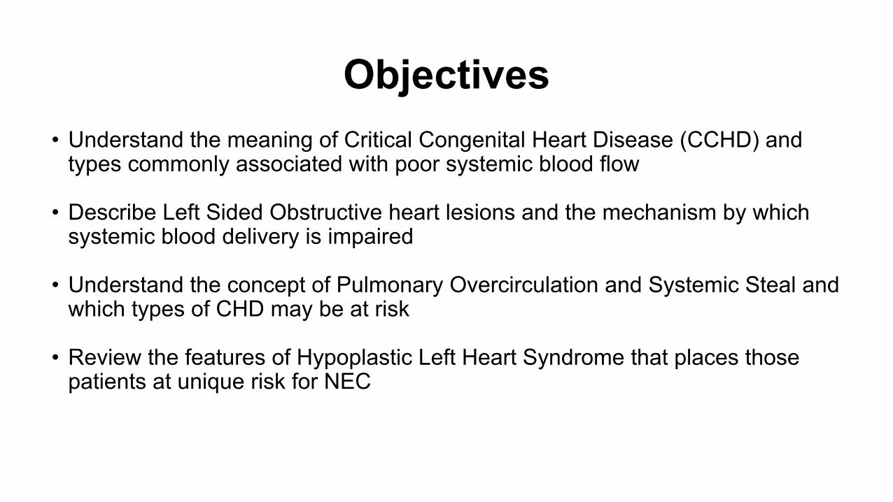
Objectives• Understand the meaning of Critical Congenital Heart Disease (CCHD) and
types commonly associated with poor systemic blood flow
• Describe Left Sided Obstructive heart lesions and the mechanism by which systemic blood delivery is impaired
• Understand the concept of Pulmonary Overcirculation and Systemic Steal and which types of CHD may be at risk
• Review the features of Hypoplastic Left Heart Syndrome that places those patients at unique risk for NEC

Disclosures• None

The Normal Heart

The Normal Heart
Deoxygenated blood returns from the body via the superior and inferior vena cavae

The Normal Heart
And returns to the Right Atrium

The Normal Heart
Exits the right atrium through the Tricuspid valve

The Normal Heart
And enters the Right Ventricle

The Normal Heart
Right ventricle pumps the blood through pulmonary valve

The Normal Heart
And into the main and branch pulmonary arteries

The Normal Heart
The branch pulmonary arteries bring the blood to the lungs

The Normal Heart
The blood flow through the right sided heart chambers will provide the “Pulmonary Blood Flow” AKA “Qp”

The Normal Heart
Oxygenated blood returns from the lungs via the pulmonary veins

The Normal Heart
The blood enters into the left atrium

The Normal Heart
Exits the left atrium through the Mitral valve

The Normal Heart
Enters the Left ventricle which pumps the blood to the body

The Normal Heart
The blood passes through the Aortic valve

The Normal Heart
The blood moves into the Aorta

The Normal Heart
And out to the body

The Normal Heart
The blood flow through the left sided heart chambers will provide the “Systemic Blood Flow” AKA “Qs”

The Normal Heart
Qp = Qs in a Normal Heart

Definitions:Congenital Heart Disease
(CHD)Critical Congenital Heart Disease
(CCHD)
Any heart malformation that is present from birth
CHD that requires surgery or intervention in the first year of life

One category of severe CCHD is a “Ductal Dependent” Heart Lesion
• CCHD that requires a Patent Ductus Arteriosus to help supply blood to the Pulmonary or Systemic Circulation
• Babies require a medication, Prostaglandin, to keep the PDA open after birth
• Patient will require CHD surgery as a neonate
Definitions:

CHD with Higher Risk of NECMcElhinney et al. in Pediatrics, 2000:
• Hypoplastic Left Heart Syndrome (HLHS)• Left Ventricular Outflow Tract
Obstruction/Coarctation• Any Single Ventricle Heart Disease (with or
without arch obstruction)• Palliation with a Shunt (BTT or Central)• Truncus Arteriosus/AP Window• Low Systemic Cardiac Output State/Shock

Lau et al. in J Ped Surg, 2018:
CHD with Higher Risk of NEC

What Problems May Reduce Systemic Circulation in Patients with CHD?

What Problems May Reduce Systemic Circulation in Patients with CHD?
Mechanical Obstruction

What Problems May Reduce Systemic Circulation in Patients with CHD?
Mechanical Obstruction Too much pulmonary blood flow and not enough systemic (Qp>>Qs)

Mechanical Obstruction Left Sided Obstructive Congenital Heart Disease
What Problems May Reduce Systemic Circulation in Patients with CHD?

Left Sided Obstructive Lesions
Interrupted Aortic Arch

Left Sided Obstructive Lesions
Interrupted Aortic Arch

Left Sided Obstructive Lesions
Interrupted Aortic Arch

Left Sided Obstructive Lesions
Interrupted Aortic Arch

Left Sided Obstructive Lesions
Interrupted Aortic Arch

Left Sided Obstructive Lesions
• Obstructed flow of oxygenated blood out to body= SHOCKà tachycardia, tachypnea, poor perfusion
• Depending upon severity of obstruction may be ductal dependent and/or require neonatal surgery for correction
• Severe obstruction decreases gut perfusion and raises risk for NEC

Too much pulmonary blood flow and not enough systemic (Qp>>Qs)
What Problems May Reduce Systemic Circulation in Patients with CHD?

Too much pulmonary blood flow and not enough systemic (Qp>>Qs)
Left to Right Shunts
What Problems May Reduce Systemic Circulation in Patients with CHD?

Too much pulmonary blood flow and not enough systemic (Qp>>Qs)
Left to Right Shunts
Mixing Lesions (with Qp>Qs)
What Problems May Reduce Systemic Circulation in Patients with CHD?

Left to Right Shunts

Left to Right Shunts

Left to Right Shunts

Left to Right Shunts

Left to Right Shunts

Left to Right Shunts

Left to Right Shunts• Key Concept: Blood will always move from higher pressure chamber to lower
pressure chamber
• Unless there is some other problem that increases the pressure, blood will ALWAYS move from Left to Right
LV to RV LA to RA Aorta to MPA

Left to Right Shunts• Key Concept: Blood will always move from higher pressure chamber to lower
pressure chamber
• Unless there is some other problem that increases the pressure, blood will ALWAYS move from Left to Right
LV to RV LA to RA Aorta to MPA

Left to Right Shunts• Key Concept: Blood will always move from higher pressure chamber to lower
pressure chamber
• Unless there is some other problem that increases the pressure, blood will ALWAYS move from Left to Right
LV to RV LA to RA Aorta to MPA

Left to Right Shunts• Key Concept: Blood will always move from higher pressure chamber to lower
pressure chamber
• Unless there is some other problem that increases the pressure, blood will ALWAYS move from Left to Right
LV to RV LA to RA Aorta to MPA

Left to Right Shunts
CHD with L to R shunts à extra blood flow to the lungs (increased Qp) and decreased blood flow to the body (decreased Qs)
LV to RV LA to RA Aorta to MPA

Left to Right Shunts
• Pulmonary “overcirculation” (high Qp) and relative decreased systemic output (low Qs) --> Sx of tachypnea, poor feeding, FTT
• In severe cases where Qp>>>Qs can lead to gut hypoperfusion and risk of NEC
• Can lead to irreversible pulmonary vascular changes à pulmonary hypertension
• May require CHD surgery based on symptoms/overall size of shunt and likelihood of spontaneous closure

Too much pulmonary blood flow and not enough systemic (Qp>>Qs)
Left to Right Shunts
What Problems May Reduce Systemic Circulation in Patients with CHD?

Too much pulmonary blood flow and not enough systemic (Qp>>Qs)
Left to Right Shunts
Mixing Lesions (with Qp>Qs)
What Problems May Reduce Systemic Circulation in Patients with CHD?

Mixing Lesions

Mixing LesionsAll the pulmonary venous return (oxygenated blood)

Mixing LesionsAll the pulmonary venous return (oxygenated blood)
All the systemic venous return (deoxygenated blood)

Mixing LesionsAll the pulmonary venous return (oxygenated blood)
All the systemic venous return (deoxygenated blood)
Completely Mixed !

Mixing Lesions
Tricuspid AtresiaTotal Anomalous Pulmonary Venous
Return (TAPVR)

Mixing Lesions
Tricuspid AtresiaTotal Anomalous Pulmonary Venous
Return (TAPVR)
Versions of “Single Ventricle CHD”

Mixing Lesions
Tricuspid AtresiaTotal Anomalous Pulmonary Venous
Return (TAPVR)
Versions of “Single Ventricle CHD”

Mixing Lesions
Tricuspid AtresiaTotal Anomalous Pulmonary Venous
Return (TAPVR)
Versions of “Single Ventricle CHD”

Mixing Lesions
Tricuspid AtresiaTotal Anomalous Pulmonary Venous
Return (TAPVR)
Versions of “Single Ventricle CHD”

Mixing Lesions
Tricuspid AtresiaTotal Anomalous Pulmonary Venous
Return (TAPVR)
Versions of “Single Ventricle CHD”

Mixing Lesions
• Presentation is variable and depends whether “too much” or “too little” pulmonary blood flow, rarely well balanced
• If “too much” of the mixed blood is going to the lungsà pulmonary overcirculationà tachypnea, high sats, difficulty feeding, poor systemic perfusion
• If Qp>>>Qs risk for gut hypoperfusion and NEC
• If “too little” of the mixed blood is going to the lungà signs of significant cyanosis
• May be ductal dependent for pulmonary or systemic blood flow

Why do patients with HLHS seem to have increased risk of NEC?

Why do patients with HLHS seem to have increased risk of NEC?
They have potential for both problems!!

Why do patients with HLHS seem to have increased risk of NEC?
Left Sided Obstruction
They have potential for both problems!!

Why do patients with HLHS seem to have increased risk of NEC?
Mixing Lesion with potential for Qp>>Qs
They have potential for both problems!!

Why do patients with HLHS seem to have increased risk of NEC?
Mixing Lesion with potential for Qp>>Qs
They have potential for both problems!!
Left Sided Obstruction

Thank You!

Amy B. Hair, M.D.Assistant Professor of Pediatrics
Program Director of Neonatal NutritionProgram Director of Intestinal Rehabilitation Team
Section of NeonatologyDepartment of Pediatrics
Baylor College of MedicineTexas Children’s Hospital
Feeding Practices in Infants with Congenital Heart Disease

Neonatal Nutrition Program

• I receive research grants from:
• Prolacta Bioscience® for the Human Milk Cream Length of Stay Multicenter Study (Study PI) and Cardiac Study; Fresenius Kabi for SMOF Randomized Trial
Disclosures

• Discuss current feeding practices
• Summarize the literature regarding feeding practices and identify knowledge gaps
• Review the benefits of human milk for infants
• Review recent studies of human milk use for Congenital Heart Disease infants
Objectives:

Feeding Practices in Infants with Congenital Heart Disease
https://www.babygaga.com/15-things-that-can-cause-congenital-heart-defects-in-babies/

• Data from premature neonates extrapolated to other populations
• Paucity of data for optimal feeding practices specifically for infants with ductal-dependent lesions • Lack of consensus and wide variation in feeding practice• No prospective studies• Very few human milk studies
Feeding Practices in Infants with Congenital Heart Disease
Slicker et al. 2016

• Standardization of care can lead to improvement in outcomes
• Use is often at discretion of provider (comfort level, experience)
• Impedes well-designed, multi-center studies from being conducted
Feeding Protocols
Petrillo-Albarano et al. 2006, Barr et al, 2004, del Castillo et al. 2010, Gephart et al. 2013

“Wide variations in practices exist in the nutritional care between European PICUs, which reflects the absence of local protocols and scientific society-endorsed guidelines.”
Feeding Protocols
Tume et al. 2018

Literature Review
Pubmed search: enteral feeding congenital heart disease

Literature Review

• Systematic review and Meta-analysis to summarize the evidence for pre-operative feeds in infants with ductal-dependent congenital heart disease
• Majority of studies focus on post-operative feeding and hospital outcomes
• Insufficient evidence that pre-operative enteral feeds adversely affect post-operative outcomes
• Illustrates need for well-designed, prospective study Dr. Kataria-Hale

• Necrotizing enterocolitis
• Mesenteric hypoperfusion• Particularly ductal-dependent due to
risk of ductal steal
• Vulnerable vascular system undergoing cardiac surgery à ischemia
Risk of Feeding
Scahill et al. 2017

Risk of NOT Feeding
Enteral Feeding
Atrophy of intestinal mucosa
Loss of important intestinal cell wall
barriers
Abnormal increase in gut permeability
Delayed postnatal intestinal
development and
maturation
Motility problems

• Breast milk: best source of nutrition when safe & available- ê Infection- ê Necrotizing enterocolitis (NEC)- ê Chronic lung disease- é Neurodevelopmental outcomes
• Promote postnatal intestinal development & maturation à healthy intestinal microbiota
Early Feedings with Human Milk for Premature Infants
Ballard et al. 2013, Cortez et al. 2013, Hair et al. 2016.
xxx00.#####.ppt 12/30/18 1:07:45 PM

Non-Nutritional Components of Human MilkAntimicrobialsecretory IgA, IgM, IgG lactoferrinlysozymecomplement C3leukocytesbifidus factorlipids and fatty acidsantiviral mucins, GAGsoligosaccharides
Hormonesfeedback inhibitor of lactation (FIL) insulinprolactinthyroid hormones (T2, T3, Reverse T3)corticosteroids, ACTHoxytocincalcitoninparathyroid hormoneerythropoietinprogesteroneestrogen
Growth factors epidermal (EGF)nerve (NGF)insulininsulin-like (IGF)transforming (TGF)taurinepolyaminesgastringastric inhibitory peptide (GIP)Gastric regulatory peptide (GRP)neurotensinpeptide histidine methionine (PHM)Peptide YY (PYY)Anti-inflammatory
tumor necrosis factorinterleukinsinterferon-gprostaglandinsa1-antichymotrypsina1-antitrypsinplatelet-activating factor: acetyl hydrolase
Digestive enzymesamylasebile acid-stimulating esterasebile acid-stimulating lipaseslipoprotein lipaseProteases
Transporterslactoferrin (Fe)xanthine oxidaseglutathione peroxidasealkaline phosphatasefolate bindercobalamin binderIgF binderthyroxine bindercorticosteroid binder

• Is it safe to feed neonates with ductal-dependent CHD preoperatively?
• Do the benefits outweigh the risks?
• Does the type of cardiac lesion matter?
• What about the use of human milk?• Does feeding CHD infants human milk confer the same benefits as it does for
premature infants?• Does human milk lead to an improvement in outcomes for this population as it
does in premature infants?
Knowledge Gap

TCH Experience
• Retrospective study of 546 infants with complex CHD at Texas Children’s Hospital from 2010-2016
• Evaluate risk of pre-operative NEC with human milk feeding• Overall incidence of pre-operative NEC 3.3% • An exclusive unfortified human milk diet was associated with a
significantly lower risk of pre-operative NEC (OR 0.17, 95% CI 0.04 -0.84, P = .03)

• Examples of barriers we have encountered:• No dedicated refrigerators for mother’s own milk• Limited lactation support
• Limited lactation education for all providers and caregivers
• Extreme fear of NEC
• Lack of evidence-based protocols• Lack of literature to guide practice
• Nutrition “doesn’t matter”
Challenges over the past 10 years at TCH:


Oral Care with Colostrum
©Kataria-Hale

Proportion of Infants Receiving Formula Before Surgery
0
10
20
30
40
50
60
70
80
90
100
Pre-Protocol Post-Protocol
HumanMilk
Formula41.6%
*P<0.0001Fisher’s Exact Test
Kataria-Hale and Hair, et al. Under review

• If there is one intervention you can do in your unit, it would be to increase the use of mother’s own milk!
• Guidelines are coming
• Neonatal Heart Society in conjunction with other societies is working on the Neonatal Cardiac Care Collaborative
• Research Studies are pending
• Donor milk use• Ongoing nutrition study in single ventricle babies- using a donor human milk-
derived term fortifier
What’s next?

Pediatrics
• Neonatal Cardiac Nutrition• Jeramy Roddy, MD• Scott Osborne, MD• Jasmeet Kataria-Hale, MD
• Acacia Cognata, MD• Paul Checchia, MD• Joseph Hagan, ScD• Mohan Pammi, MD• Murali Premkumar, MD• Sharda Gowda, MD
• Neonatal Dietitians
Acknowledgements• Neonatal Nutrition Research Group
- Heeju Yang- Jana Unger, RD, MS- Laura Gollins, RD, MBA- Pam Gordon, RNC-NIC
TCH Milk Banks and Lactation• Nancy Hurst, PhD• Kristina Tucker, RN, MS

Acknowledgements

#preventNEC@NECsociety
Thank you!
Discussion & Questions