Monocrotophos Induced Apoptosis in PC12 Cells: Role of Xenobiotic Metabolizing Cytochrome P450s
Upload
independentCategory
view
0download
0
HuR’s role in gemcitabine efficacy in pancreatic cancer: HuRupregulates the expression of the gemcitabine metabolizingenzyme, deoxycytidine kinase
Christina L. Costantino1,*, Agnieszka K. Witkiewicz2,*, Yuki Kuwano Y4,4, Joseph A.Cozzitorto1, Eugene P. Kennedy1, Abhijit Dasgupta3, Judith C. Keen5, Charles J. Yeo1,Myriam Gorospe4,4, and Jonathan R. Brody11 Department of Surgery, Jefferson Center for Pancreatic, Biliary and Related Cancers, ThomasJefferson University, Philadelphia, PA 191072 Department of Pathology, Kimmel Cancer Center, Thomas Jefferson University, Philadelphia, PA191073 Department of Pharmacology and Experimental Therapeutics, Kimmel Cancer Center, ThomasJefferson University, Philadelphia, PA 191074 LCMB, National Institute on Aging-IRP, NIH, Baltimore, Maryland 212245 Robert Wood Johnson Medical School, University of Medicine and Dentistry of New Jersey,Camden, New Jersey 08106
AbstractRNA-binding protein HuR binds U- or AU-rich sequences in the 3′-untranslated regions (UTRs) oftarget mRNAs, stabilizing them and/or modulating their translation. Given HuR’s links with cancer,we studied the consequences of modulating HuR levels in pancreatic cancer cells. HuR-overexpressing cancer cells, in some instances, are up to 30-fold more sensitive to treatment withgemcitabine (GEM), the main chemotherapeutic component of treatment regimens for pancreaticductal adenocarcinoma (PDA), compared to control cells. In pancreatic cancer cells, HuR associateswith deoxycytidine kinase (dCK) mRNA, which encodes the enzyme that metabolizes and therebyactivates GEM. GEM exposure to pancreatic cancer cells, enriches the association between HuR anddCK mRNA and increases cytoplasmic HuR levels. Accordingly, HuR overexpression elevates,while HuR silencing reduces, dCK protein expression in pancreatic cancer cells. In a clinical correlatestudy of GEM treatment, we found a 7-fold increase in risk of mortality in PDA patients with lowcytoplasmic HuR levels compared to patients with high HuR levels, after adjusting for othertreatments and demographic variables. These data support the notion that HuR is a key mediator ofGEM efficacy in cancer cells, at least in part through its ability to regulate dCK levels post-transcriptionally. We propose that HuR levels in PDA modulate the therapeutic efficacy of GEM,thus serving as a marker of the clinical utility of this common chemotherapeutic agent and a potentialtarget for intervention in pancreatic cancer.
Corresponding Author: Jonathan R. Brody, Department of Surgery, Thomas Jefferson University, 1015 Walnut Street, Curtis 611A,Philadelphia, PA 19107, Tel: 215-955-2693, [email protected].*CLC and AKW contributed equally4YK and MG were supported by the NIA-IRP, NIH
NIH Public AccessAuthor ManuscriptCancer Res. Author manuscript; available in PMC 2010 June 1.
Published in final edited form as:Cancer Res. 2009 June 1; 69(11): 4567–4572. doi:10.1158/0008-5472.CAN-09-0371.
NIH
-PA Author Manuscript
NIH
-PA Author Manuscript
NIH
-PA Author Manuscript

Keywordspancreatic ductal adenocarcinoma; pancreatic cancer; HuR; gemcitabine; deoxycytidine kinase;mRNA binding protein; gemcitabine response; RNA binding proteins
IntroductionPancreatic ductal adenocarcinoma (PDA) is the fourth leading cause of cancer-related deathsin the United States (1). Currently, two therapeutic options that provide the best clinical benefitare surgical resection and chemotherapy regimens that include gemcitabine (GEM) (2′,2′-difluorodeoxycytidine). For over ten years, GEM has been the reference drug for the treatmentof this often fatal disease (2). GEM is also utilized to treat other malignancies including non-small-cell lung, breast, gastric, and ovarian cancers. GEM utilizes the same key metabolicenzyme for activation within the cell, deoxycytidine kinase (dCK), as does a previouslydeveloped and related nucleoside analog cytarabine (Ara-C) (3). dCK phosphorylates theprodrug, GEM, generating the active metabolites gemcitabine di- and triphosphates that inhibitDNA chain elongation and cause cellular death (4). The levels of dCK correlate with overallpatient survival following GEM-based therapy in PDA specimens (p=0.0425)(4).
The stress-response protein Hu antigen R (HuR) is an RNA-binding protein that regulates geneexpression post-transcriptionally. Like other related Hu/elav proteins, HuR harbors threeconserved RNA recognition motifs through which it binds to target mRNAs (5) that frequentlyhave AU- or U-rich stretches in their 3′-untranslated regions (UTRs). HuR is predominantlynuclear, but in response to various stimuli, it is mobilized to the cytoplasm, prolongs targetmRNA half-life, and can modulate target mRNA translation (5). Many HuR target mRNAsencode stress-response, immune-response, cell cycle regulatory proteins, oncogenes, andtumor suppressor genes (5). HuR modulates these transcripts in response to stimuli such astherapeutic agents (i.e. tamoxifen and prostaglandin), nutrient depletion (polyamines, aminoacid starvation), heat shock, immune stimuli, short-wavelength UV irradiation, oxidants, andtranscriptional inhibitors (actinomycin D) (5–7).
Although HuR has never been reported to be mutated in cancer, it has been proposed tocontribute to the tumorigenesis process (8–10). Elevated cytoplasmic accumulation of HuRboth correlates with high-grade malignancy and serves as a prognostic factor of poor clinicaloutcome in some cancers (11–14). Given the extensive research examining HuR’s role incancer and stress response over the past decade (5), we explored the role of HuR in PDA andthe relationship of HuR to chemotherapeutic efficacy.
MethodsTransfection of pancreatic cancer cell lines
HuR cDNA sequence was cloned into the pcDNA 3.1.Zeo vector (Invitrogen) for stabletransfection of pancreatic cancer cell lines MiaPaca2, PL-5, and Hs766t (15). Pooled cellsremained under selection media containing Zeocin (Invitrogen) for several months aftertransfection. Mia.HuR, PL5.HuR, and Hs766t.HuR denote HuR overexpressing lines; Mia.EV,PL5.EV, and Hs766t denote empty vector or control lines. HuR and control siRNA sequencesand transfection conditions were as described (7).
ImmunofluorescenceCells were plated on LabTek II™ Chamber slides (Fisher Scientific) and fixed in 3%paraformaldehyde (20 min, RT). Cells were washed with PBS and permeabilized using 0.5%
Costantino et al. Page 2
Cancer Res. Author manuscript; available in PMC 2010 June 1.
NIH
-PA Author Manuscript
NIH
-PA Author Manuscript
NIH
-PA Author Manuscript

Triton X-100/1% normal goat serum (Vector Laboratories) in PBS (15 min). After washes in1% goat serum/PBS, cells were incubated (1:50 dilution, 1 hr, RT) with mouse anti-HuR (SantaCruz) or anti-deoxycytidine kinase (dCK, Abnova) primary antibodies. After washes in PBS,cells were incubated for 1 hr with goat anti-mouse secondary antibody (1:400, Alexa Fluor647, Molecular Probes). Nuclei were stained with DAPI and cells were evaluated under a ZeissLSM-510 Confocal Laser Microscope.
Drug sensitivity assayMia.HuR, Mia.EV, Hs766t.HuR, Hs766t, and PL5.HuR, PL5.EV cells were seeded (1000cells/well) in 96-well plates and treated with Etoposide, 5-Fluorouracil, Cis-platin,Staurosporine, Nocodazole, Colcemid, Ara-C (Sigma), and GEM (Gemzar, Eli-Lilly,Indianapolis, IN) for 6–7 days. After treatment, cells were washed with PBS and lysed with100 μL of water/well; cell viability was quantified by staining of double-stranded DNA withQuant-iT™ PicoGreen (Invitrogen) and analyzed with a TECAN SpectraFluor.
ImmunoblotWhole-cell, cytoplasmic, and nuclear lysates were prepared as described (7), and protein wassize-fractionated by SDS-PAGE (10% acrylamide). Membranes were blocked for 1 h in 5%Milk/TBS-T and incubated overnight with monoclonal antibodies (Santa Cruz) recognizingHuR, dCK, the cytoplasmic marker α-Tubulin, or the nuclear marker hnRNP. Membranes werewashed with TBS-T and incubated with secondary antibodies; and the resulting signals werevisualized by chemiluminescence (Millipore). Total protein was visualized with Fast Green(USB).
Cell cycle analysis and apoptosis assayMia.EV and Mia.HuR cell lines were either left untreated or treated with 0.03 μM GEM for48 h. For cell cycle analysis, cells were then fixed in 100% ethanol, and stained with apropidium iodide solution containing RNAse A (Sigma Aldrich). For apoptosis assays, cellswere resuspended at 106 cells/mL and incubated in Annexin V and Propidium Iodide, followingthe manufacturers’ protocol (FITC Annexin V, BD Pharmigen). Both assays were analyzed byflow cytometry.
RNA-binding: biotin pulldown and RNP IP assaysMiaPaCa2 cells were treated with 4 μM GEM and collected 48 hours later. For biotin pulldownanalysis (7), cytoplasmic extracts were isolated using the NE-PERR® Nuclear andCytoplasmic Extraction Reagents Kit (Pierce Biotechnology). Probes for biotin pull-downanalysis were synthesized as described (7) using the following PCR primers (sense andantisense, respectively) containing the T7 RNA polymerase promoter sequenceCCAAGCTTCTAATACGACTCACTATAGGGAGA (T7):(T7)GATCTTGCTGAAGACTACAGGC and TTATTAGCGT CTTTTCAATTCTACAAA fordCK 3′UTR; (T7)CTCAACGACCACTTTGTCAAGC andCACAGGGTACTTTATTGATGGTACAT for GAPDH 3′UTR (16) (see supplemental figurefor depiction of UTR positions). Biotinylated probes were synthesized using the MAXIscriptT7 kit (Ambion) and Biotinylated dCTP (Enzo Life Sciences).
For immunoprecipitation of endogenous RNA-protein complexes (RNP IP) from cytoplasmic(450 μg) extracts, reactions were carried out as described (7), using protein A-Sepharose beads(Sigma) that were precoated with 30 μg of either mouse immunoglobulin G1 (IgG1; BDBiosciences), or anti-HuR antibodies. After IP, the RNA in the IP materials was isolated andreverse-transcribed. GAPDH and dCK transcripts were quantified by real-time PCR analysisusing each specific primers: AGCAAGGCATTCCTCTTGAA and
Costantino et al. Page 3
Cancer Res. Author manuscript; available in PMC 2010 June 1.
NIH
-PA Author Manuscript
NIH
-PA Author Manuscript
NIH
-PA Author Manuscript

CTACAGGCAGCCAAATGGTT for dCK, TGCACCACCAACTGCTTAGC andCTCATGACCACAGTCCATGCC for GAPDH. The relative levels of dCK product was firstnormalized to GAPDH product in all IP samples, then fold enrichments in HuR IP werecompared with IgG IP, as described (7).
Case selection and immunohistochemistryHuR immunostaining was performed on 32 resected PDAspecimens from the ThomasJefferson University pathology archives after IRB approval. All patients received GEM, aloneor in combination with Xeloda (2 patients), radiation therapy (8 patients) or both (2 patients).The experienced pancreatic pathologist (A.K.W.) reviewed all cases in a blinded fashion andclassified the tumors as well differentiated(n=6), moderately differentiated (n=22), or poorlydifferentiated(n=12). For each case, representative sections were selected forimmunohistochemical analysis of HuR cytoplasmic and nuclear staining patterns, which werescored using the following scale: 0 for no staining; 1 for weak and/or focal (<10% of the cells)staining; 2 for moderate or strong staining (10–50% of the cells); and 3 for moderate or strongstaining (>50% of the cells). Combined scores 0 and 1 represented low expression, whilecombined scores 2 and 3 represented high expression.
Results and DiscussionHuR overexpression preferentially sensitized pancreatic cancer cell lines to the nucleosideanalogs GEM and Ara-C
Stable HuR overexpression in the indicated pancreatic cancer cell lines was confirmed byimmunoblot and immunofluorescence analyses (Figure 1A and B). Contrary to previous studiesin colon cancer cell lines (9), isogenic, transfected cell lines grew roughly at the same rates(Figure 1C). Treatment with the indicated chemotherapeutic agents (but not GEM) showed nodifference in sensitivity in HuR-overexpressing cells (Figure 1D).
By contrast, cell lines overexpressing HuR were found to be strikingly more sensitive to GEMthan were control lines, as assessed both by PicoGreen measurement (Figure 2A) and bystaining with crystal violet even when cells were treated with low concentrations of GEM(Figure 2B). HuR-overexpressing cell lines were similarly selectively more sensitive to Ara-C, another anti-cancer agent that utilizes dCK (Figure 2C). After GEM treatment, HuR-overexpressing cells showed selective enrichment in the S-phase of the cell division cycle andincreased apoptosis (Figure 2D) as compared to the control cells.
HuR localization and association with dCK mRNA upon GEM treatmentGEM treatment did not alter whole-cell HuR levels in parental MiaPaCa2 cells, butsignificantly increased the cytoplasmic HuR levels, as determined by immunoblot andimmunofluorescence analyses (Figure 3A). Given that the dCK 3′UTR region contained 8putative hits of an HuR recognition motif (17), we tested if HuR associates with dCK mRNAusing two different RNA-binding assays. First, MiaPaCa2 cytoplasmic extracts were incubatedwith equimolar amounts of biotinylated transcripts spanning the dCK 3′UTR and the GAPDH3′UTR (a control RNA, not a target of HuR); the resulting complexes were analyzed by HuRimmunoblot. As shown in Figure 3B (left), HuR bound the dCK 3′UTR much more stronglythan the GAPDH 3′UTR. Second, we tested the association of HuR with the endogenous dCKmRNA by using a ribonucleoprotein immunoprecipitation (RNP IP) assay. As shown, whileGEM did not alter overall dCK protein or mRNA levels (Figure 3A, C), it significantlyincreased HuR’s association with dCK mRNA (Figure 3B, right).
Inhibition of HuR expression using small interfering (si)RNA(7) did not alter dCK mRNAlevels (Figure 3C, right) but decreased dCK protein levels (Figure 3D, left) regardless of GEM
Costantino et al. Page 4
Cancer Res. Author manuscript; available in PMC 2010 June 1.
NIH
-PA Author Manuscript
NIH
-PA Author Manuscript
NIH
-PA Author Manuscript

treatment. Conversely, HuR-overexpressing cells displayed higher dCK signals (Figure 3D,right). Together, these data show for the first time that 1) GEM exposure to cancer cellsincreases cytoplasmic HuR levels (Figure 3A), 2) HuR associates with dCK mRNA (Figure3B), and 3) HuR regulates dCK protein levels (Figure 3D).
HuR localization and expression in PDA specimensWe detected primarily weak to moderate nuclear HuR expression in normal pancreatic ductaland acinarcells (Figure 4A). Strong nuclear expression of HuR was found inwell-, moderately,and poorly differentiated PDAs. Cytoplasmic HuR accumulation was associated with poorlydifferentiated PDAs (Figure 4B and data not shown). Figure 4C shows the Kaplan-Meieroverall survival curves of patients receiving GEM, stratified by their HuR status. The mediansurvival time for patients on GEM was 619 days, with 21 deaths out of the 32 patients whoreceived GEM. A univariate Cox regression model gives a hazard ratio of low to high HuR of4.48, with a 95% confidence interval of (1.49 to 13.5). Adjusting for age, sex, Xeloda use andradiation therapy in this patient group gives an adjusted hazard ratio of 7.34 (p=0.0022) witha 95% confidence interval of (2.05 to 26.22). These data indicate a greater than 7-fold increaserisk of mortality in patients with low cytoplasmic HuR levels (compared to high cytoplasmicHuR levels) among patients receiving GEM, after adjusting for variables as mentioned above.
PerspectiveAs elevated cytoplasmic HuR has been widely correlated with advanced malignancy, thefinding that high cytoplasmic HuR levels were associated with an increased therapeuticefficacy of GEM in pancreatic cancer was unexpected. Our results that HuR regulates dCKprotein concentration and that cytoplasmic HuR levels predict GEM response in our patientcohort lead us to hypothesize that HuR could be a key molecule involved in GEM efficacy incancer. It is intriguing to postulate that part of HuR’s survival repertoire may be to increasedCK levels to process deoxyribonucleosides for survival, however in the presence ofnucleoside analogs (such as GEM) HuR’s augmentation of dCK may be deleterious. Furtherstudies are warranted to investigate if HuR dictates GEM effectiveness by regulating additionaltarget mRNAs, and to assess if HuR is a suitable marker for GEM response in larger cancerpatient cohorts for which GEM therapy is utilized.
Supplementary MaterialRefer to Web version on PubMed Central for supplementary material.
References1. Jemal A, Siegel R, Ward E, et al. Cancer statistics, 2008. CA Cancer J Clin 2008;58:71–96. [PubMed:
18287387]2. Burris HA 3rd, Moore MJ, Andersen J, et al. Improvements in survival and clinical benefit with
gemcitabine as first-line therapy for patients with advanced pancreas cancer: a randomized trial. J ClinOncol 1997;15:2403–13. [PubMed: 9196156]
3. Li ZR, Campbell J, Rustum YM. Effect of 3-deazauridine on the metabolism, toxicity, and antitumoractivity of azacitidine in mice bearing L1210 leukemia sensitive and resistant to cytarabine. CancerTreat Rep 1983;67:547–54. [PubMed: 6190558]
4. Sebastiani V, Ricci F, Rubio-Viqueira B, et al. Immunohistochemical and genetic evaluation ofdeoxycytidine kinase in pancreatic cancer: relationship to molecular mechanisms of gemcitabineresistance and survival. Clin Cancer Res 2006;12:2492–7. [PubMed: 16638857]
5. Hinman MN, Lou H. Diverse molecular functions of Hu proteins. Cell Mol Life Sci 2008;65:3168–81. [PubMed: 18581050]
Costantino et al. Page 5
Cancer Res. Author manuscript; available in PMC 2010 June 1.
NIH
-PA Author Manuscript
NIH
-PA Author Manuscript
NIH
-PA Author Manuscript

6. Hostetter C, Licata LA, Witkiewicz A, et al. Cytoplasmic accumulation of the RNA binding proteinHuR is central to tamoxifen resistance in estrogen receptor positive breast cancer cells. Cancer BiolTher 2008;7:1496–506. [PubMed: 18769129]
7. Kuwano Y, Kim HH, Abdelmohsen K, et al. MKP-1 mRNA stabilization and translational control byRNA-binding proteins HuR and NF90. Mol Cell Biol 2008;28:4562–75. [PubMed: 18490444]
8. Lopez de Silanes I, Lal A, Gorospe M. HuR: post-transcriptional paths to malignancy. RNA Biol2005;2:11–3. [PubMed: 17132932]
9. Lopez de Silanes I, Fan J, Yang X, et al. Role of the RNA-binding protein HuR in colon carcinogenesis.Oncogene 2003;22:7146–54. [PubMed: 14562043]
10. Lopez de Silanes I, Fan J, Galban CJ, Spencer RG, Becker KG, Gorospe M. Global analysis of HuR-regulated gene expression in colon cancer systems of reducing complexity. Gene Expr 2004;12:49–59. [PubMed: 15473260]
11. Yoo PS, Sullivan CA, Kiang S, et al. Tissue Microarray Analysis of 560 Patients with ColorectalAdenocarcinoma: High Expression of HuR Predicts Poor Survival. Ann Surg Oncol. 2008
12. Heinonen M, Fagerholm R, Aaltonen K, et al. Prognostic role of HuR in hereditary breast cancer.Clin Cancer Res 2007;13:6959–63. [PubMed: 18056170]
13. Denkert C, Weichert W, Winzer KJ, et al. Expression of the ELAV-like protein HuR is associatedwith higher tumor grade and increased cyclooxygenase-2 expression in human breast carcinoma.Clin Cancer Res 2004;10:5580–6. [PubMed: 15328200]
14. Denkert C, Weichert W, Pest S, et al. Overexpression of the embryonic-lethal abnormal vision-likeprotein HuR in ovarian carcinoma is a prognostic factor and is associated with increasedcyclooxygenase 2 expression. Cancer Res 2004;64:189–95. [PubMed: 14729623]
15. Brody JR, Witkiewicz A, Williams TK, et al. Reduction of pp32 expression in poorly differentiatedpancreatic ductal adenocarcinomas and intraductal papillary mucinous neoplasms with moderatedysplasia. Mod Pathol 2007;20:1238–44. [PubMed: 17906614]
16. Casolaro V, Fang X, Tancowny B, et al. Posttranscriptional regulation of IL-13 in T cells: role of theRNA-binding protein HuR. J Allergy Clin Immunol 2008;121:853–9. e4. [PubMed: 18279945]
17. Lopez de Silanes I, Zhan M, Lal A, Yang X, Gorospe M. Identification of a target RNA motif forRNA-binding protein HuR. Proc Natl Acad Sci U S A 2004;101:2987–92. [PubMed: 14981256]
Costantino et al. Page 6
Cancer Res. Author manuscript; available in PMC 2010 June 1.
NIH
-PA Author Manuscript
NIH
-PA Author Manuscript
NIH
-PA Author Manuscript

Figure 1.Characterization of HuR-overexpressing pancreatic cancer cell lines. (A) Immunoblot analysisof HuR expression in lysates from MiaPaCa2 (Mia.HuR and Mia.EV) and Hs766T (Hs766t.HuR and Hs766t) cells. Fast Green staining confirmed the equality of protein loading. (B)Immunofluorescence to detect HuR and nuclei (DAPI). (C) Mia.HuR and Mia.EV cellproliferation rates, as determined by direct cell counts. (D) Cell survival was measured byPicoGreen after incubation of cells for 5–7 days with the indicated compounds. Data show themeans (and S.E.M.) from 3 measurements in a single experiment; each experiment isrepresentative of at least three individual experiments. ▲, Mia.HuR cells; ■, Mia.EV cells.
Costantino et al. Page 7
Cancer Res. Author manuscript; available in PMC 2010 June 1.
NIH
-PA Author Manuscript
NIH
-PA Author Manuscript
NIH
-PA Author Manuscript

Figure 2.Stable expression of HuR renders cells hypersensitive to the nucleoside analogs GEM and Ara-C. (A) Survival of MiaPaCa2, Hs766t, and PL5 cell lines was measured by the PicoGreen assayafter 5–7 days of incubation with the indicated GEM doses. Graphs represent singleexperiments (S.E.M.); each experiment is representative of >three individual experiments. ▲,HuR expressing cells; ■, control cells. (B) Crystal violet-stained flasks of Mia.HuR andMia.EV cultures after GEM treatment (0.1 μM, 7 days). (C) Sensitivity of MiaPaCa2 cells toAra-C treatment was measured as explained in panel (A). (D) FACS analysis of cells treatedwith GEM (0.03 μM) for 48 h, depicting the percentages of cells in G1, S, and G2/M
Costantino et al. Page 8
Cancer Res. Author manuscript; available in PMC 2010 June 1.
NIH
-PA Author Manuscript
NIH
-PA Author Manuscript
NIH
-PA Author Manuscript

compartments (left). Measurement of apoptotic fractions (Materials and Methods) in culturestreated as explained in panel (Right).
Costantino et al. Page 9
Cancer Res. Author manuscript; available in PMC 2010 June 1.
NIH
-PA Author Manuscript
NIH
-PA Author Manuscript
NIH
-PA Author Manuscript

Figure 3.HuR associates with dCK mRNA and promotes dCK protein expression in MiaPaCa2 cells.(A) Western blot analysis of HuR levels in whole-cell and cytoplasmic lysates after treatmentof MiaPaCa2 cells with GEM (1 μM) for the indicated times (left). Immunofluorescenceanalysis of HuR levels and localization (red) in cells treated with 4 μM GEM for 24 h; nucleiwere distinguished by staining with DAPI (blue) (right). (B) Biotin pulldown analysis of HuRRNP complexes. Cytoplasmic extracts were incubated with biotinylated transcripts spanningthe DCK or GAPDH 3′UTRs. The association of HuR with biotinylated RNAs was tested byWestern blot analysis. Positive control: HuR cytoplasmic lysate. Negative controls: ‘Probeonly’ lanes contain only biotinylated RNAs that were not incubated with protein lysates. Shown
Costantino et al. Page 10
Cancer Res. Author manuscript; available in PMC 2010 June 1.
NIH
-PA Author Manuscript
NIH
-PA Author Manuscript
NIH
-PA Author Manuscript

is a representative blot (right). HuR binding to dCK mRNA was tested by RNP IP analysis(Materials and Methods) in MiaPaCa2 cells treated with GEM for the times indicated; GEMmRNA levels in HuR and IgG IP samples were first normalized to GAPDH mRNA levels inthe same IP reactions, and plotted as fold enrichment in dCK mRNA in HuR IP compared withIgG IP. Data show the means and standard deviation from 3 independent experiments (left).(C) dCK mRNA levels were measured in cells that were left untransfected (left) or weretransfected with either a control siRNA or HuR siRNA(7) and tested 48 h later (right). (D)Western blot analysis of HuR, dCK, and α-Tubulin in cells expressing normal or silenced HuRlevels (left). Immunofluorescence analysis of dCK levels (indicated by the arrow) andlocalization (red) in cells expressing normal or elevated HuR levels; nuclei were visualized bystaining with DAPI (blue) (right).
Costantino et al. Page 11
Cancer Res. Author manuscript; available in PMC 2010 June 1.
NIH
-PA Author Manuscript
NIH
-PA Author Manuscript
NIH
-PA Author Manuscript
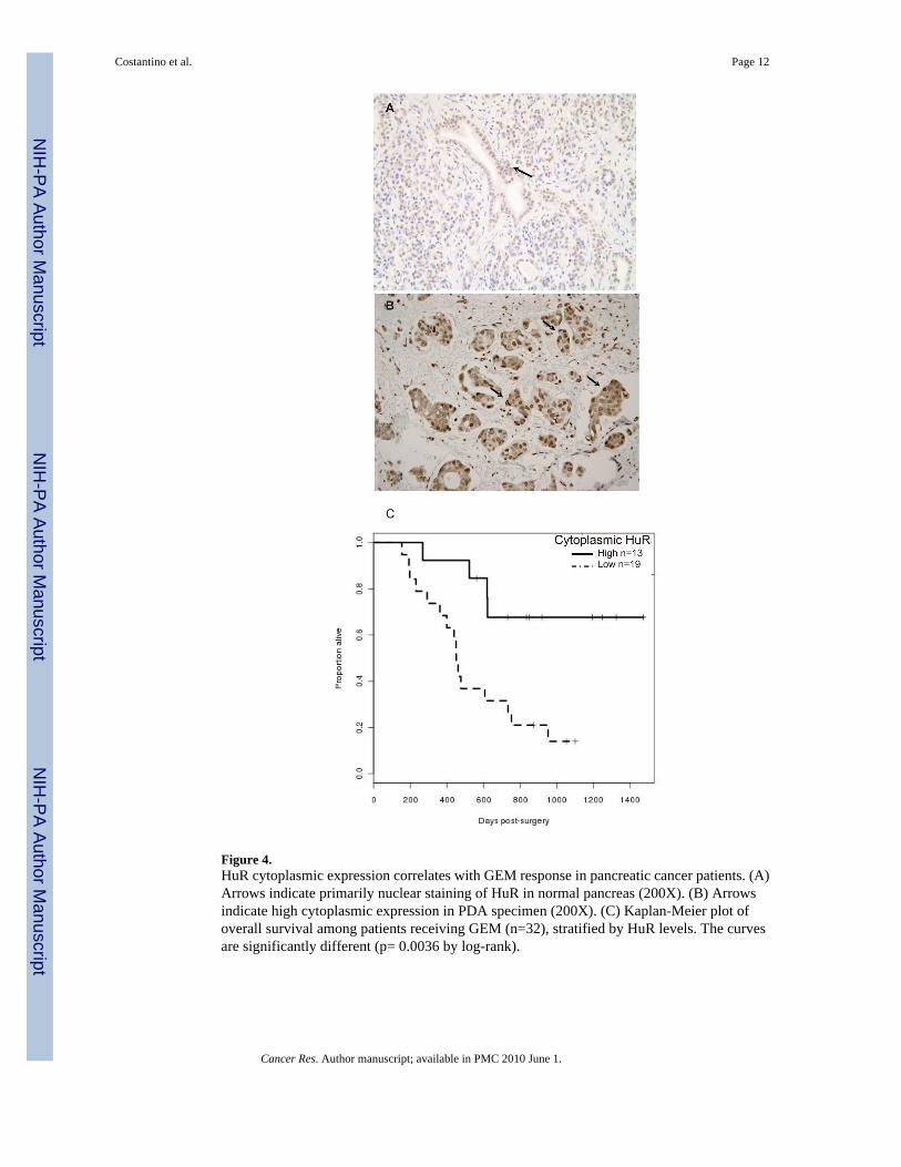
Figure 4.HuR cytoplasmic expression correlates with GEM response in pancreatic cancer patients. (A)Arrows indicate primarily nuclear staining of HuR in normal pancreas (200X). (B) Arrowsindicate high cytoplasmic expression in PDA specimen (200X). (C) Kaplan-Meier plot ofoverall survival among patients receiving GEM (n=32), stratified by HuR levels. The curvesare significantly different (p= 0.0036 by log-rank).
Costantino et al. Page 12
Cancer Res. Author manuscript; available in PMC 2010 June 1.
NIH
-PA Author Manuscript
NIH
-PA Author Manuscript
NIH
-PA Author Manuscript
Copyright © 2022 FDOKUMEN