
Mitochondria, calcium and cell death: A deadly triad in neurodegeneration
10
Review Mitochondria, calcium and cell death: A deadly triad in neurodegeneration Fulvio Celsi a,b , Paola Pizzo c , Marisa Brini d,e , Sara Leo a,b , Carmen Fotino a,b , Paolo Pinton a,b , Rosario Rizzuto c, ⁎ a Department of Experimental and Diagnostic Medicine, Section of General Pathology, Interdisciplinary Center for the Study of Inflammation (ICSI), Italy b Emilia Romagna Laboratory BioPharmaNet, University of Ferrara, Via Borsari 46, 44100 Ferrara, Italy c Department of Biomedical Sciences, University of Padova, Via Colombo 3, 35121 Padova, Italy d Department of Biochemistry, University of Padova, Viale G. Colombo, 3 35131 Padova, Italy e Department of Experimental Veterinary Sciences, University of Padova, Viale dell' Università 16, 35020 Legnaro, Italy abstract article info Article history: Received 17 December 2008 Received in revised form 23 February 2009 Accepted 24 February 2009 Available online 4 March 2009 Keywords: Mitochondria Calcium Neurodegenerative disease Alzheimer's disease Parkinson's disease Huntington's disease Mitochondrial Ca 2+ accumulation is a tightly controlled process, in turn regulating functions as diverse as aerobic metabolism and induction of cell death. The link between Ca 2+ (dys)regulation, mitochondria and cellular derangement is particularly evident in neurodegenerative disorders, in which genetic models and environmental factors allowed to identify common traits in the pathogenic routes. We will here summarize: i) the current view of mechanisms and functions of mitochondrial Ca 2+ homeostasis, ii) the basic principles of organelle Ca 2+ transport, iii) the role of Ca 2+ in neuronal cell death, and iv) the new information on the pathogenesis of Alzheimer's, Huntington's and Parkinson's diseases, highlighting the role of Ca 2+ and mitochondria. © 2009 Elsevier B.V. All rights reserved. 1. Mitochondria in calcium signalling The notion that mitochondria are active players in cellular calcium homeostasis dates back to the demonstration of the chemiosmotic theory, based on the concept of a major proton electrochemical gradient that could drive the rapid accumulation of cations across the ion-impermeant mitochondrial inner membrane. Such a notion was corroborated by the direct measurement of Ca 2+ uptake by isolated mitochondria, and the functional, albeit not molecular, elucidation of the transporters [1]. However, the experiments that in the 80s drove massive interest into calcium as a ubiquitous second messenger also led to the gradual decline in the attention on mitochondrial Ca 2+ homeostasis. On the one hand, it appeared clear that the endoplasmic reticulum (ER), through resident Ca 2+ channels (those sensitive to inositol 1,4,5 trisphosphate, IP 3 R, and to the plant alkaloid ryanodine, RyR) acts as the intracellular pool of Ca 2+ mobilized upon cell stimulation. On the other, the development of accurate fluorescent indicators for the measurement of Ca 2+ concentration in living cells showed that cytosolic Ca 2+ concentration fluctuates between approx. 0.1 μM at rest to 2–3 μM at the peak of the rise elicited by the opening of plasma membrane or ER Ca 2+ channels. Under those conditions, the low affinity of the mitochondrial Ca 2+ transporters should allow little Ca 2+ uptake into the organelle. Thus, the prevalent notion was that mitochondrial Ca 2+ accumulation is negligible in physiological condi- tions, and could become relevant only upon massive Ca 2+ overload, that could take place in severe cellular dysfunction. This situation was completely reversed when tools were developed that allowed the selective measurement of mitochondrial Ca 2+ concentration ([Ca 2+ ] m ) in living cells. Targeting to mitochondria the Ca 2+ -sensitive photoprotein aequorin [2] demonstrated that a rapid [Ca 2+ ] m peak, reaching values well above those of the bulk cytosol, parallels the [Ca 2+ ] rise evoked in the cytoplasm by cell stimulation [3]. Similar conclusions could be reached also with fluorescent indicators, such as the positively charged Ca 2+ indicator rhod 2 (that accumulates within the organelle) [4] and the more recently developed GFP-based fluorescent indicators [5]. With the latter probes, endowed with a much stronger signal than the photoprotein, single-cell imaging of organelle Ca 2+ could be carried out [6]. With these tools in hands, not only the notion was confirmed that mitochondria promptly respond to cytosolic [Ca 2+ ] rises, but also that the [Ca 2+ ] c oscillations, the typical response to agonists of many cell types, are paralleled by rapid spiking of [Ca 2+ ] m , thus providing a frequency-mediated signal specifically decoded within the mitochon- dria, as clearly shown in hepatocytes [7], cardiomyocytes [8], and HeLa cells [9]. The apparent discrepancy between the promptness and amplitude of the mitochondrial Ca 2+ response and the low affinity of Biochimica et Biophysica Acta 1787 (2009) 335–344 ⁎ Corresponding author. Tel.: +39 0498276481; fax: +39 0498276049. E-mail address: [email protected] (R. Rizzuto). 0005-2728/$ – see front matter © 2009 Elsevier B.V. All rights reserved. doi:10.1016/j.bbabio.2009.02.021 Contents lists available at ScienceDirect Biochimica et Biophysica Acta journal homepage: www.elsevier.com/locate/bbabio
Transcript of Mitochondria, calcium and cell death: A deadly triad in neurodegeneration
Biochimica et Biophysica Acta 1787 (2009) 335–344
Contents lists available at ScienceDirect
Biochimica et Biophysica Acta
j ourna l homepage: www.e lsev ie r.com/ locate /bbab io
Review
Mitochondria, calcium and cell death: A deadly triad in neurodegeneration
Fulvio Celsi a,b, Paola Pizzo c, Marisa Brini d,e, Sara Leo a,b, Carmen Fotino a,b,Paolo Pinton a,b, Rosario Rizzuto c,⁎a Department of Experimental and Diagnostic Medicine, Section of General Pathology, Interdisciplinary Center for the Study of Inflammation (ICSI), Italyb Emilia Romagna Laboratory BioPharmaNet, University of Ferrara, Via Borsari 46, 44100 Ferrara, Italyc Department of Biomedical Sciences, University of Padova, Via Colombo 3, 35121 Padova, Italyd Department of Biochemistry, University of Padova, Viale G. Colombo, 3 35131 Padova, Italye Department of Experimental Veterinary Sciences, University of Padova, Viale dell' Università 16, 35020 Legnaro, Italy
⁎ Corresponding author. Tel.: +39 0498276481; fax:E-mail address: [email protected] (R. Rizzuto)
0005-2728/$ – see front matter © 2009 Elsevier B.V. Adoi:10.1016/j.bbabio.2009.02.021
a b s t r a c t
a r t i c l e i n f oArticle history:Received 17 December 2008Received in revised form 23 February 2009Accepted 24 February 2009Available online 4 March 2009
Keywords:MitochondriaCalciumNeurodegenerative diseaseAlzheimer's diseaseParkinson's diseaseHuntington's disease
Mitochondrial Ca2+ accumulation is a tightly controlled process, in turn regulating functions as diverse asaerobic metabolism and induction of cell death. The link between Ca2+ (dys)regulation, mitochondria andcellular derangement is particularly evident in neurodegenerative disorders, in which genetic models andenvironmental factors allowed to identify common traits in the pathogenic routes. Wewill here summarize: i)the current view of mechanisms and functions of mitochondrial Ca2+ homeostasis, ii) the basic principles oforganelle Ca2+ transport, iii) the role of Ca2+ in neuronal cell death, and iv) the new information on thepathogenesis of Alzheimer's, Huntington's and Parkinson's diseases, highlighting the role of Ca2+ andmitochondria.
© 2009 Elsevier B.V. All rights reserved.
1. Mitochondria in calcium signalling
The notion that mitochondria are active players in cellular calciumhomeostasis dates back to the demonstration of the chemiosmotictheory, based on the concept of a major proton electrochemicalgradient that could drive the rapid accumulation of cations across theion-impermeant mitochondrial inner membrane. Such a notion wascorroborated by the direct measurement of Ca2+ uptake by isolatedmitochondria, and the functional, albeit not molecular, elucidation ofthe transporters [1].
However, the experiments that in the 80s drove massive interestinto calcium as a ubiquitous second messenger also led to the gradualdecline in the attention on mitochondrial Ca2+ homeostasis. On theone hand, it appeared clear that the endoplasmic reticulum (ER),through resident Ca2+ channels (those sensitive to inositol 1,4,5trisphosphate, IP3R, and to the plant alkaloid ryanodine, RyR) acts asthe intracellular pool of Ca2+ mobilized upon cell stimulation. On theother, the development of accurate fluorescent indicators for themeasurement of Ca2+ concentration in living cells showed thatcytosolic Ca2+ concentration fluctuates between approx. 0.1 μMat restto 2–3 μM at the peak of the rise elicited by the opening of plasma
+39 0498276049..
ll rights reserved.
membrane or ER Ca2+ channels. Under those conditions, the lowaffinity of the mitochondrial Ca2+ transporters should allow little Ca2+
uptake into the organelle. Thus, the prevalent notion was thatmitochondrial Ca2+ accumulation is negligible in physiological condi-tions, and could become relevant only uponmassive Ca2+ overload, thatcould take place in severe cellular dysfunction.
This situationwas completely reversedwhen tools were developedthat allowed the selective measurement of mitochondrial Ca2+
concentration ([Ca2+]m) in living cells. Targeting to mitochondriathe Ca2+-sensitive photoprotein aequorin [2] demonstrated that arapid [Ca2+]m peak, reaching values well above those of the bulkcytosol, parallels the [Ca2+] rise evoked in the cytoplasm by cellstimulation [3]. Similar conclusions could be reached also withfluorescent indicators, such as the positively charged Ca2+ indicatorrhod 2 (that accumulates within the organelle) [4] and the morerecently developed GFP-based fluorescent indicators [5]. With thelatter probes, endowed with a much stronger signal than thephotoprotein, single-cell imaging of organelle Ca2+ could be carriedout [6]. With these tools in hands, not only the notion was confirmedthat mitochondria promptly respond to cytosolic [Ca2+] rises, but alsothat the [Ca2+]c oscillations, the typical response to agonists of manycell types, are paralleled by rapid spiking of [Ca2+]m, thus providing afrequency-mediated signal specifically decoded within the mitochon-dria, as clearly shown in hepatocytes [7], cardiomyocytes [8], and HeLacells [9]. The apparent discrepancy between the promptness andamplitude of the mitochondrial Ca2+ response and the low affinity of

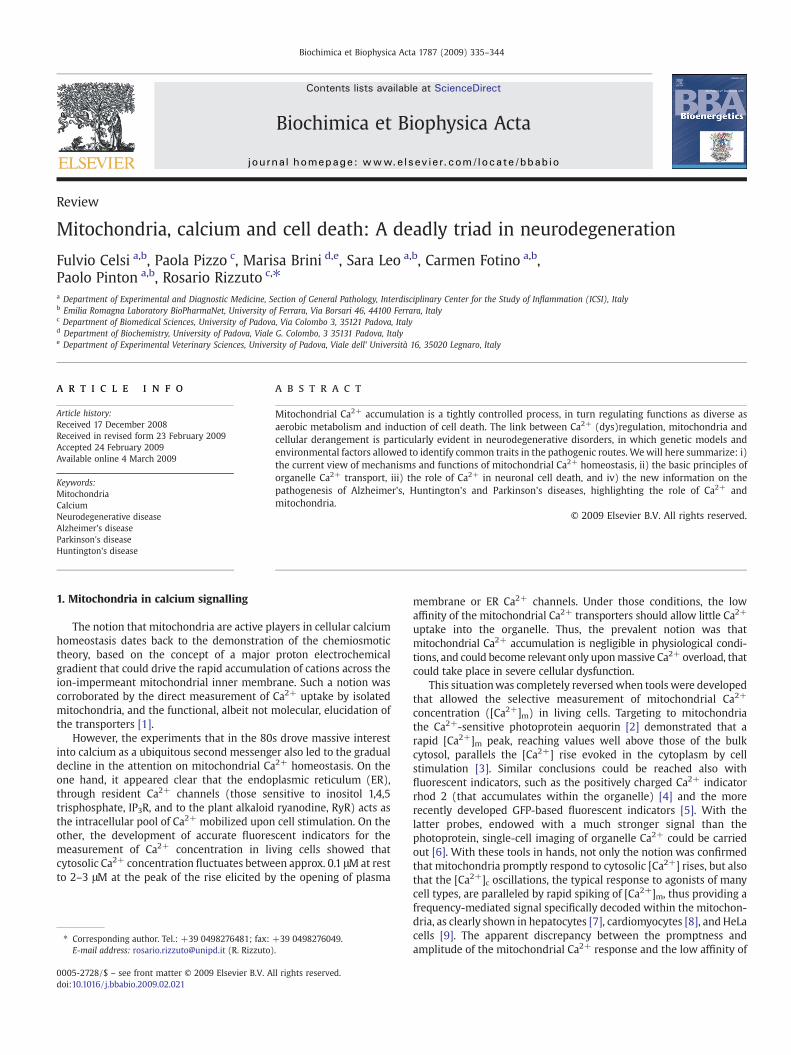
Fig. 1. Schematic map of mitochondrial Ca2+ transporters. Mitochondria accumulateCa2+ in the matrix via an electrogenic Ca2+ uniporter (MCU) that acts to equilibrate Ca2+
according the electrochemical gradient generated by the respiratory chain (ETC). VoltageDependent Anion Channel (VDAC) controls the Ca2+ diffusion through the outermitochondrial membrane (OMM), thus facilitating mitochondrial Ca2+ accumulation. Asto the efflux pathways, a Na+/Ca2+ and a H+/Ca2+ exchangers have been shown tooperate. The permeability transition pore (PTP) opening plays different roles: its briefopening could allow rapid Ca2+ release, but its long-lasting openings (potentiated byapoptotic stimuli and Ca2+ itself) could trigger cell death process. IMS, intermembranespace; IMM, inner mitochondrial membrane.
336 F. Celsi et al. / Biochimica et Biophysica Acta 1787 (2009) 335–344
the organelle transporters was reconciled by the demonstration thatmitochondria are in close contact with the source of the cytosolic Ca2+
rise (i.e. the ER via IP3Rs and RyRs and the plasma membrane via awide variety of voltage- and agonist-sensitive Ca2+ channels). Thus,upon cell stimulation they are exposed to microdomains of high [Ca2+]that greatly exceed the values measured in the cytosol and well matchthe affinity of mitochondrial Ca2+ transporters [10].
As soon as the concept was established that a Ca2+ rise in thecytosol is paralleled by a cycle of mitochondrial Ca2+ uptake, andsubsequent release (through the pathways that will be brieflydescribed later), the identification of the functional significance ofthis process became a primary goal. Also in this task, the finebiochemical work carried out in the 70s provided a hypothesis to test:three key metabolic enzymes (the pyruvate, ketoglutarate andisocitrate dehydrogenases) were shown to be activated by Ca2+ bydifferent mechanisms: in the case of pyruvate dehydrogenase througha Ca2+-dependent dephosphorylation step, in the latter two casesthrough the direct binding of Ca2+ to the enzyme complex [11].Recently, also some metabolite transporters were shown to beregulated by Ca2+ and participate in the enhancement of aerobicmetabolism upon cell stimulation [12]. Thus, an obvious role formitochondrial Ca2+ accumulation could be inferred, i.e. that of rapidlyupregulating mitochondrial ATP production in stimulated cells. Thepossibility of directly monitoring, in parallel, Ca2+ and ATP levelswithin mitochondria and in the cytosol proved that this is indeed thecase [13]. Interestingly, this route for controlling mitochondrialmetabolic output proved to be affected in mitochondrial geneticdisorders. In cybrids harboring the tRNAlys mutation of MERRF(myoclonic epilepsy with ragged-red fibers), mitochondrial Ca2+
responses were reduced, and, accordingly, ATP production upon cellstimulation, and the pharmacological correction of the Ca2+ alterationalso restored the metabolic dysfunction [14,15].
In addition to the functionofmetabolic coupling,mitochondrial Ca2+
accumulation was shown to underlie a role for these organelles inshaping the spatio/temporal patterning of cytosolic Ca2+ rises.Mitochondria, distinctly from cytosolic proteins, are highly sophisti-cated, “tunable” buffers that vary their activity in different phases andfunctional states of the cell; indeed, their number, shape, distribution[16] and most likely their responsiveness to Ca2+ [17] are controlled byconverging signalling pathways. This Ca2+ buffering activity influencescytosolic Ca2+ signals in two conceptually different ways, i.e., 1) byacting as high-capacity sinks placed on the way of a propagating Ca2+
wave and 2) by clearing Ca2+ in restricted microdomains (such as themicroenvironment of a Ca2+ channel). In thefirst case, spatial clusters ofmitochondria have been demonstrated to isolate functionally distinctdomains of polarized cells, namely, a mitochondrial “firewall” wasshown to prevent the spread of Ca2+ signals from the apical (secretory)region of pancreatic acinar cell from the basolateral region, containingthe nucleus [18]. Similarly, neuronal mitochondria have been shown tobuffer [Ca2+] increases in defined cellular regions, e.g., the presynapticmotoneuron ending [19]. As to the second case, a thoroughlyinvestigated example is the regulation of Ca2+ release through IP3Rs.In Xenopus oocytes, the mitochondria energization state (and thusthe capacity to accumulate Ca2+) was shown tomodify the propagatingCa2+ waves induced by IP3 [20]. In permeabilized blowfly salivaryglands, it was observed that perfusion of IP3 induced ER [Ca2+]oscillations, the frequency of which increased with the dose of IP3.Such an effect was observed only upon energization of mitochondria,implying a primary role of these organelles in regulating Ca2+
microdomains in the proximity of IP3Rs and thus the oscillatory paceof stimulated cells [21]. In mammals, this effect has been seen in manycell systems, includinghepatocytes,HeLa cells, astrocytes, and BHKcells.As to the cellular consequence, very different effects were observedgiven the bell-shaped sensitivity of IP3Rs to Ca2+ concentration on thecytosolic side. In astrocytes and hepatocytes, cytosolic excitabilityappeared enhanced when mitochondrial Ca2+ uptake was inhibited,
indicating that mitochondrial clearance of the Ca2+ microdomainreduced the positive Ca2+ feedback on the IP3R and/or bufferedsubstantial Ca2+ loads [22]. Conversely, in BHK cells, inhibition ofmitochondrial Ca2+uptake resulted in reduction of ERCa2+ release [23],thus indicating that mitochondria prevent the Ca2+-dependent inacti-vation of the channel.
The interest in the process of mitochondrial Ca2+ homeostasisdramatically increased when it became apparent that also cell death iscausally linked to organelle Ca2+ loading. On the one hand, it wasclear that cellular Ca2+ overload, such as that caused by hyperstimu-lation of ionotropic glutamate receptors, leads to Ca2+ cycling acrossthe mitochondrial membranes, collapse of the proton gradient andbioenergetic catastrophe, thus leading to cell death by necrosis, asdiscussed in more detail later in this review. On the other hand, Ca2+
proved to sensitize cells to apoptotic challenges, acting on themitochondrial checkpoint. This notion, subsequently confirmed bythe study of other anti- and pro-apoptotic proteins, emerged from theanalysis of the effect of Bcl-2 on Ca2+ signalling. We refer to otherspecifically focused reviews for a detailed coverage of this topic [24].Briefly, we here summarize that Bcl-2, by partially emptying the ERCa2+ store, reduces the release from this organelle and the loading ofmitochondria. As a consequence, the efficacy of apoptotic challengesin opening the permeability transition pore (PTP), causing mitochon-drial morphological alterations and releasing caspase cofactors, suchas cytochrome c, is greatly reduced [25].
2. The basics of mitochondrial Ca2+ transport
For entering the mitochondrial matrix, Ca2+ needs to cross twolipid bilayers, the outer and inner mitochondrial membranes (Fig. 1).
337F. Celsi et al. / Biochimica et Biophysica Acta 1787 (2009) 335–344
The outer mitochondrial membrane (OMM) is permeable to ions andsmall proteins (MWb10 kDa) due to the abundance of a largeconductance channel, known as mitochondrial porin or voltage-dependent anion channel (VDAC). It should be noted, however, thatthe channel appears to be gated in vivo, and permeability is controlledby ATP and other regulatory factors [26]. Ca2+ diffusion through theOMM was thus traditionally considered not to be a limiting factor inmitochondrial Ca2+ uptake. Recent data showed that the availabilityand selective placement of VDAC channels at ER/mitochondriacontact sites facilitate mitochondrial Ca2+ accumulation, in keepingwith the idea that the latter process requires the fast and efficienttransfer of Ca2+ microdomains from the mouth of the Ca2+ channelslocated in neighbouring ER or plasma membranes to the transportersof the ion-impermeant inner membrane (IMM) [27]. The IMM is anion-impermeable membrane, with a much larger extension of theOMM (and consequent formation of foldings into the internal space,known as cristae). The activity of respiratory chain complexes allowsthe translocation of H+ in the space between the two membranes,which consequently generates an electrochemical gradient (ΔμH)composed of a chemical (ΔpH) and electrical (ΔψH) component. Inmitochondria, most of the ΔμH established by the respiratory chain issupposed to be in the form of ΔψH (around −180 mV), whichprovides a huge driving force for Ca2+ entry into the organelle. Indeed,collapse of the ΔψH by protonophores, such as p-[trifluoromethoxyl]-phenyl-hydrazone (FCCP), abolishes mitochondrial Ca2+ uptake.
Mitochondrial Ca2+ traffic takes place essentially through twopathways: i) an electrogenic pathway, the mitochondrial Ca2+
uniporter (MCU), corresponding to the channel activity demonstratedby Clapham et al. [28] that acts to equilibrate Ca2+ with itselectrochemical gradient, and thus accumulates the cation into thematrix; ii) two exchangers (with H+ and Na+, mostly expressed innonexcitable and excitable cells, respectively), that utilize theelectrochemical gradient of the monovalent cations to prevent theattainment of electrical equilibrium (that would imply, for amitochondrial membrane potential, Δψm, of −180 mV and a cytosolicCa2+ concentration of 0.1 μM, accumulation of Ca2+ into thematrix upto 0.1 M).
Numerous works has given the biochemical properties of thesetwo pathways, and it is possible to summarize as it follows:
i) Given the huge driving force for accumulation, studies in isolatedorganelles with clamped ΔΨ (by establishing K+ diffusionpotentials with valinomycin in non-respiring mitochondria)allowed to estimate a VmaxN1400 nmol Ca2+ (mg protein)−1
min−1 and an apparent Kmb10 μM in sucrose-based media [29].Also in isolated mitochondria, a number of inhibitors wereidentified. Ruthenium compounds (typically Ruthenium Red,RR), represent a class of non-competitive inhibitors, but unfortu-nately their poor permeability across cellular membranes hasmade it of little use in studies in intact cells. A second class ofinhibitors is divalent cations that are themselves transported bythe uniporter (e.g. Sr2+, Mn2+, Ba2+ and lanthanides).ii) As to the efflux pathways, studies in isolated organelles allowedto estimate their properties. The mitochondrial Na+/Ca2+
exchanger (mNCX) has a Vmax ranging between 2.6 (liver) and18 nmol Ca2+ (mg protein)−1 min−1 (heart). The dependence onNa+ is sigmoidal, with typical Km values of about 8–10 mM Na+.Ca2+ efflux is inhibited by Sr2+, Ba2+, Mg2+ or Mn2+, and by avariety of compounds of pharmacological interest such asdiltiazem, verapamil and other blockers of the voltage-dependentcalcium channels, and more specifically by CGP37157 [30]. As tothe H+/Ca2+ exchanger (mHCX), (1) it saturates at Ca2+ loads of25 nmol (mg protein)−1; (2) its Vmax is not influenced by theconcentration of inorganic phosphate and does not exceed a rate of
1.2 nmol Ca2+ (mg protein)−1 min−1; and (3) it extrudes Ca2+
against a gradient that is much higher than thermodynamicallypermissible for an electroneutral H+/Ca2+ exchanger [31]. Thus,either Ca2+ efflux occurs via a nH+/Ca2+ exchanger with nN2, orit has an active component. Accordingly, Ca2+ efflux is inhibitedrather than stimulated by small depolarizations [32].
2.1. The permeability transition pore
This high-conductance channel (PTP) mediating mitochondrialswelling, postulated on the basis of experimental evidence datingback more than 40 years [33], has attracted enormous interest in thelast decade, when its role in mitochondrial dysfunction andmitochondria-dependent cell routes has become clear. It is a high-conductance, non-selective channel that exhibits a prominentdependence on matrix Ca2+ and is inhibited by Cyclosporin A (CsA).Reversible PTP openings have been resolved both in individualisolated mitochondria [34] and in intact cells [35]. PTP could play arole in various conditions. Long-lasting openings, triggered byapoptotic challenges and potentiated by Ca2+-mediated cellularsignals [36] cause morphological transitions of mitochondria under-lying the release of caspase cofactors into the cytosol, and theinitiation of the cell death process. Conversely, given the large size andlack of selectivity of the PTP (providing charge compensation withinthe channel and allowing maximal Ca2+ flux at zero potential) briefPTP openings could allow at least in principle rapid Ca2+ release fromthe organelle [37].
3. Calcium routes to neurodegeneration
It was hypothesized already in the 70s that prolonged stimulationof N-methyl-D-aspartate (NMDA) ionotropic glutamate receptors caninduce massive cell death in the brain (excitotoxicity), by causingCa2+ and Na+ overload in post-synaptic neurons [38]. Excitotoxicityplays a central role in promoting cell death during neuronal ischemiaand substantial work has been placed in clarifying the differentphases of the process, and their reversibility, and the potentialpharmacological targets. It is now accepted that a primary Ca2+
increase occurs, as a consequence of direct entry through NMDAreceptors, but also following depolarization, and hence opening ofvoltage-gated Ca2+ channels, release from internal stores andreversal of the plasma membrane Na+/Ca2+ exchanger (NCX). Celldeath, however, does not depend on this initial Ca2+ rise, but ratherinvariably follows a delayed massive accumulation of Ca2+, occurringa few hours after the toxic challenge and representing a no-returntransition into the death process. Recent work has highlightedevents occurring in the interphase between the two Ca2+ rises, andsuggested a progression route into the secondary Ca2+ overload anddelayed cell death. Specifically, Bano et al. [39] showed that calpain-mediated cleavage on NCX is a critical step for allowing the delayedCa2+ deregulation: inhibition of NCX cleavage protects from exci-totoxic challenges, and conversely downregulation of the exchangersensitizes to sub-lethal stimuli. As to mitochondria, direct measure-ment of matrix Ca2+ showed that the impairment of their Ca2+
uptake capacity is downstream of the delayed Ca2+ dysregulation,and not the cause of it. Thus, the delayed Ca2+ increase is not due tothe sudden discharge from the mitochondrial buffer that still retainsthe capacity to accumulate Ca2+. Rather, the delayed, massive influxof Ca2+ into the cell leads to organelle Ca2+ overload, collapse of theelectrochemical proton gradient and bioenergetic catastrophe, lead-ing to necrotic cell death [40].
Interestingly, such a commitment mechanism appears to closelymatch an emerging paradigm clarified in a number of apoptoticroutes. Indeed, we and other showed that mitochondrial involvementin apoptosis (morphological transitions and release of caspase

338 F. Celsi et al. / Biochimica et Biophysica Acta 1787 (2009) 335–344
cofactors) often utilizes Ca2+ as a key sensitizing factor. In agreementwith this notion, Bcl-2 reduces the state of filling of intracellular Ca2+
stores, thereby reducing mitochondrial loading upon physiologicaland pathological challenges and protecting cells from apoptotic death[25]. The pro-apoptotic protein Bax antagonizes this signalling effect,by augmenting the state of filling of intracellular Ca2+ stores [41]. Inaddition, closely mimicking the data described above in excitotoxicity,early cleavage of plasma membrane Ca2+ pumps (PMCA) by caspaseswas shown to greatly enhance the efficacy of apoptotic challenges inneurons [39] and hepatocytes [42]. In this context, one can envision acommon route to neurodegeneration in neurons, in which Ca2+ playsan important regulatory role and eventually, by affecting themitochondrial checkpoint, renders cell death obligatory. Thus, whilethe fate into a rapid necrotic outcome or a more controlled apoptoticelimination will depend on the intensity of the insult and on thebioenergetic balance of the cell, the observation of common signallingthemes and molecular targets highlights the extensive crosstalkbetween the different death pathways and the potential strategies forpharmacological intervention.
4. Looking into complex models: the pathogenesis ofneurodegenerative disorders
4.1. Alzheimer's disease (AD)
Alzheimer's disease (AD) is a devastating neurological disorderclinically characterized by impairment of cognitive function andchanges in behavior and personality. Morphological hallmarks of thepathology are cortical and hippocampal atrophy, accumulation ofabnormal fibers in neuronal cell bodies (neurofibrillary tanglescomposed of a hyperphosphorylated form of the microtubular proteintau), and the presence, in the extracellular space, of senile plaques, the
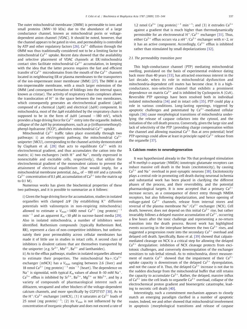
Fig. 2. Presenilins (PSs), Amyloid Precursor Protein (APP) and peptide β−amyloid (Aβ) can afthe expression/sensitivity of ER Ca2+ release channels (RyR and IP3R) leading to an exaggerabut not the FAD mutants, were reported to form Ca2+ permeable leak channels in the ER proAPP can interact with the mitochondrial TIM/TOM protein import complex. The presenmitochondrial translocation that, in turn, inhibited the entry of nuclear-encoded mitochenzymatic activities of cytochrome c oxidase (Cyt c-OX) and inhibition of mitochondrial AIntra-mitochondrial Aβwas demonstrated to directly interact with cyclophilin D (CypD), theADmitochondriamore sensitive to PTP opening. The origin of intra-mitochondrial Aβ peptideIMS, intermembrane space; IMM, inner mitochondrial membrane.
major component of which is the peptide β-amyloid (Aβ) in two mostfrequent forms, Aβ40 and Aβ42. These derive from the transmem-brane protein APP (Amyloid Precursor Protein), that can be alter-natively processed by three different enzymes, named α, β, and γsecretases. The combined action of β and γ secretases leads to theformation of a soluble fragment (sAPPβ) and of Aβ, together with itscytosolic counterpart AICD (APP Intra-Cellular Domain) [43].
Although the majority of AD cases are sporadic, a significantfraction of AD is inherited in a dominant pattern. Mutations in thegenes encoding for APP and for Presenilin-1 and -2 (PS1 and PS2), twoproteins belonging to the γ-secretase enzymatic complex, have beenlinked to the familial form of AD (FAD; see [44] for a recent review).Since the majority of FAD mutations have been found to increase theAβ42/Aβ40 ratio, the initial hypothesis was that the disease wasdependent on the enhanced fibrillization of the more amyloidogenicAβ42 [45].
Although this concept has never been questioned, about a decadeago various experimental observations suggested that an alteration inintracellular Ca2+ homeostasis could also contribute to the develop-ment of FAD, and more in general to the pathogenesis of AD. Indeed,PSsmutationswere shown to alter the expression, or the sensitivity, ofER Ca2+ release channels (RyR and IP3R) in different cell models[46–49] and in neurons from Tg AD mice [50,51], leading to the “Ca2+
overload” hypothesis [45,52], i.e. the idea that exaggerated ER Ca2+
release affects cellular targets such as mitochondria, favouring cellulardemise (Fig. 2). The Ca2+ overload mechanism has however remainedlargely mysterious until it was found that wt PSs, but not the FADmutants, can form Ca2+ permeable leak channels in the ER [53,54],thus providing a clear case for enhanced Ca2+ release in thepathological model. Although the data are straightforward, andsupport a very appealing model, the following work in otherlaboratories was not entirely consistent with the first formulation of
fect mitochondrial functionality by differentmeans. FAD-linked PSsmutationsmay alterted ER Ca2+ release that in turn may cause abnormal mitochondrial Ca2+ uptake. wt PSs,viding a clear explanation for the enhanced Ca2+ release found in different AD models.ce of an acidic domain within APP sequence may be responsible for an incompleteondrial proteins. Mitochondrial Aβ accumulation has been correlated with impairedβ-binding alcohol dehydrogenase (ABAD), leading to mitochondrial oxidative damage.PTP component that binds CsA and renders the channel more sensitive to Ca2+, makings is however unclear. ER, endoplasmic reticulum; OMM outermitochondrial membrane.
339F. Celsi et al. / Biochimica et Biophysica Acta 1787 (2009) 335–344
the hypothesis (see for example [55–59]). Specifically, Zatti et al.[60–62] showed that some FAD-linked PS2 mutations caused areduction, not an increase, in ER/Golgi Ca2+ levels. This experimentaldiscrepancy, while not disproving the Ca2+ hypothesis, may thus allowtwo possible conclusions. The first is that the system is likely to be verycomplex, with additional unidentified regulatory elements, and the useof different cell systems and experimental approaches (e.g. silencing oroverexpression, knock-out or knock-in models) may trigger equallydifferent compensatory mechanisms and calcium effects in the cells.Secondly, given that the discrepanciesmostly refer to PS2, a speculative,but appealing, possibility is that PS2 and PS1 play distinct roles in ER/Golgi Ca2+ handling. In particular, FAD-PS1 mutations, that cause anincrease in the ER Ca2+, unavoidably exacerbate cell death. Conversely,PS2 mutants, by favouring low ER Ca2+ levels, might confer relativeprotection to other routes of cell intoxication, such as Aβ peptides andoxidative damage. This hypothesis would be consistent with the abovedescribed role of the ER/mitochondrial Ca2+ relationship, and with theclinical observation that FAD-linkedPS2mutationshavebeenassociatedto milder phenotypes [60–62].
No matter how the synergistic “Ca2+ hit” occurs, mitochondrialdysfunction appears an obligatory downstream step in the pathogen-esis of AD. Decreased cytochrome c oxidase activity, increased free-radical generation leading to oxidative stress and reduced energymetabolism was described in the brain of AD patients before Aβplaques formation [63–67]. Moreover, electron microscopy analysis ofmitochondria in various regions of AD brain revealed significantmorphological organelle alterations, such as reduced size of mito-chondrial cristae [68].
As to the mechanism, converging evidence points to a role for Aβpeptides and the PTP and, possibly, to a Ca2+-sensitization step.Endogenous as well as ectopically expressed wt or FAD-linkedSwedish APP have been found to localize to mitochondria in differentcell types [69–71]. In isolated mitochondria, Aβ peptides wereshown to inhibit mitochondrial respiration [72,73] and, in thepresence of Ca2+, cause the opening of PTP. The involvement of PTPwas further reinforced by the analysis of mouse models in whichcyclophilin D, (CypD), the PTP component that binds CsA and rendersthe channel more sensitive to Ca2+, was knocked out [74,75].Interestingly, this genetic alteration substantially improves thecognitive abilities of a mouse model of AD and alleviates Aβ-mediatedreduction of long-term potentiation [76]. Moreover, intra-mitochon-drial Aβ was demonstrated to directly interact with CypD, thusproviding amolecular basis for this pathogenic mechanism [76]. Otherputative damaging effects of Aβ were reported. In PC12 cells, Aβblocked the entry of nuclear-encoded proteins into mitochondriacausing decreased mitochondrial membrane potential, increased ROSproduction, oxygen glucose deprivation and altered mitochondrialmorphology [77]. In another study, Aβ increased neuronal ROSproduction, activated mitochondrial fission proteins Drp1 and Fis1and caused mitochondrial fragmentation [78]. Finally, in the mito-chondrial matrix Aβ peptides were shown to interact with and inhibitthe activity of mitochondrial Aβ-binding alcohol dehydrogenase(ABAD), leading to mitochondrial oxidative damage, increasedcarbonylation of mitochondrial proteins and impaired activity ofrespiratory complexes. Some of these alterations were found to occurin AD mouse models before the development of senile plaques,suggesting that mitochondrial dysfunction is an early event in thepathogenesis of the disease [79].
Thus calcium andmitochondriamay represent the common themelinking the various reported aspects of the pathogenesis of AD. Severalaspects remain however to be solved, and we wish to point out twocrucial ones. The first is a clear understanding of the dysregulation ofcalcium signalling, as discussed above. The second one refers to theorigin of intra-mitochondrial Aβ, if a primary role is attributed to thismolecule. Experiments of limited trypsin proteolysis and chemicalcross-linking showed that APP interacts with the mitochondrial TIM/
TOM protein import complex and its orientation is such that the N-terminal resides inside the mitochondria while the large C-terminalpart of the protein faces the cytosol [69,71]. Thus, although thepresence within mitochondria of γ-secretase constituents and func-tional γ-secretase activity has been reported [80], it is unlikely that Aβis being produced on the matrix side. Moreover, the mitochondrialpresence of functional β-secretase, essential for the generation of theC99 peptide, substrate for γ-secretase, has not been reported yet. As todirect permeation from the ER, the Golgi complex or secretory vesiclesto the cytosol, and then across the outer and the inner mitochondrialmembranes (the latter notoriously highly impermeable to mostsolutes, including ions), this audacious possibility awaits experimen-tal confirmation.
4.2. Huntington's disease (HD)
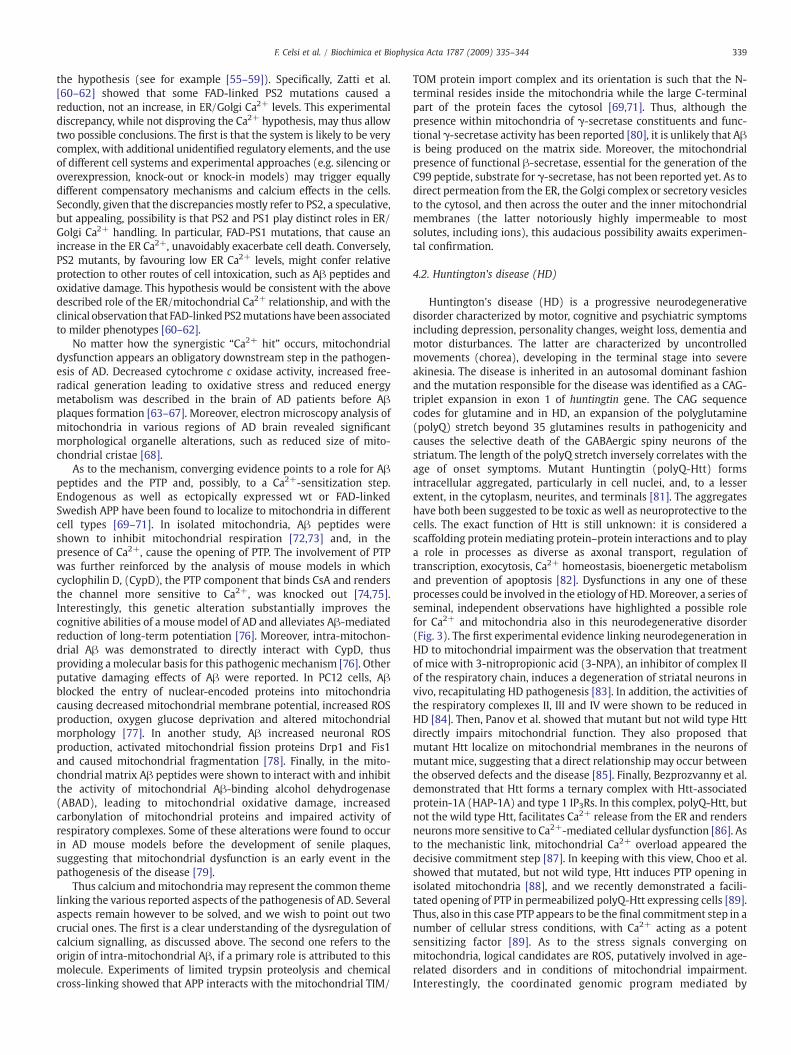
Huntington's disease (HD) is a progressive neurodegenerativedisorder characterized by motor, cognitive and psychiatric symptomsincluding depression, personality changes, weight loss, dementia andmotor disturbances. The latter are characterized by uncontrolledmovements (chorea), developing in the terminal stage into severeakinesia. The disease is inherited in an autosomal dominant fashionand the mutation responsible for the disease was identified as a CAG-triplet expansion in exon 1 of huntingtin gene. The CAG sequencecodes for glutamine and in HD, an expansion of the polyglutamine(polyQ) stretch beyond 35 glutamines results in pathogenicity andcauses the selective death of the GABAergic spiny neurons of thestriatum. The length of the polyQ stretch inversely correlates with theage of onset symptoms. Mutant Huntingtin (polyQ-Htt) formsintracellular aggregated, particularly in cell nuclei, and, to a lesserextent, in the cytoplasm, neurites, and terminals [81]. The aggregateshave both been suggested to be toxic as well as neuroprotective to thecells. The exact function of Htt is still unknown: it is considered ascaffolding proteinmediating protein–protein interactions and to playa role in processes as diverse as axonal transport, regulation oftranscription, exocytosis, Ca2+ homeostasis, bioenergetic metabolismand prevention of apoptosis [82]. Dysfunctions in any one of theseprocesses could be involved in the etiology of HD.Moreover, a series ofseminal, independent observations have highlighted a possible rolefor Ca2+ and mitochondria also in this neurodegenerative disorder(Fig. 3). The first experimental evidence linking neurodegeneration inHD to mitochondrial impairment was the observation that treatmentof mice with 3-nitropropionic acid (3-NPA), an inhibitor of complex IIof the respiratory chain, induces a degeneration of striatal neurons invivo, recapitulating HD pathogenesis [83]. In addition, the activities ofthe respiratory complexes II, III and IV were shown to be reduced inHD [84]. Then, Panov et al. showed that mutant but not wild type Httdirectly impairs mitochondrial function. They also proposed thatmutant Htt localize on mitochondrial membranes in the neurons ofmutant mice, suggesting that a direct relationship may occur betweenthe observed defects and the disease [85]. Finally, Bezprozvanny et al.demonstrated that Htt forms a ternary complex with Htt-associatedprotein-1A (HAP-1A) and type 1 IP3Rs. In this complex, polyQ-Htt, butnot the wild type Htt, facilitates Ca2+ release from the ER and rendersneuronsmore sensitive to Ca2+-mediated cellular dysfunction [86]. Asto the mechanistic link, mitochondrial Ca2+ overload appeared thedecisive commitment step [87]. In keeping with this view, Choo et al.showed that mutated, but not wild type, Htt induces PTP opening inisolated mitochondria [88], and we recently demonstrated a facili-tated opening of PTP in permeabilized polyQ-Htt expressing cells [89].Thus, also in this case PTP appears to be the final commitment step in anumber of cellular stress conditions, with Ca2+ acting as a potentsensitizing factor [89]. As to the stress signals converging onmitochondria, logical candidates are ROS, putatively involved in age-related disorders and in conditions of mitochondrial impairment.Interestingly, the coordinated genomic program mediated by
Fig. 3. Mutant Huntingtin (mutHtt) impairs mitochondrial function by transcriprional and non-trascriptional mechanisms. The transcriptional effects are mediated by nucleartranslocation of mutHtt. One important consequence of the regulation of gene transcription is the downregulation of PGC1α, and thus the reduced expression of nuclear-encodedmitochondrial proteins involved in the respiratory chains and in the oxidative-stress defense. MutHtt also associates with the outer mitochondrial membrane (OMM) directlyaffecting the PTP opening susceptibility andmaking striatal neurons more vulnerable to excitotoxic stimuli. MutHtt reduces complex II activity and the treatment with the complex IIinhibitor 3-nitropropionic acid (3-NPA) induces striatal neurodegeneration in vitro and in vivo. The association of mutHtt with HAP-1A and with the IP3R type I facilitates ER Ca2+
release, thus making mitochondria more susceptible to Ca2+ overload. ER, endoplasmic reticulum; IMS, intermembrane space; IMM, inner mitochondrial membrane.
340 F. Celsi et al. / Biochimica et Biophysica Acta 1787 (2009) 335–344
Peroxisome proliferator-activated receptor-coactivator (PGC-1α), atranscription factor that upregulates ROS-scavenging systems uponoxidative stress [90], appears dysfunctional in HD striata [91], and alsoin cellular models of the disease [92]. Indeed, in both cases the level ofthe PGC-1α transcript is downregulated, and, at least in part, thescavenging enzymes are accordingly reduced. This observationappears very appealing, as recent work has highlighted PGC-1αtranscriptional regulation as a promising drug target [93].
Another important way in which mitochondrial function could beimpaired inHD is through abnormal axonal trafficking ofmitochondriato and from the synapse. PolyQ-Htt has beenproposed to inhibit axonaltransport through severalmechanisms and, recently, itwas shown thatfragments of Htt associate with mitochondria thus interfering withtheir microtubule-associated transport [94]. Overall, the data so farobtained appear very coherent, and highlight a straightforwardpathogenic route, based on the triggering of a calcium-dependentmitochondrial dysfunction. What remains to be assessed is how theputative damaging effects of polyQ-Htt are coordinately regulated. Aremitochondrial respiratory deficiencies and calcium-mediated orga-nelle dysfunction two independent effects of mutated Htt, or are theycausally linked? Or does the latter gradually induce the former, in avicious cycle of ROS production and genetic damage that can be furtherexacerbated by toxic challenges affecting the brain?And in this picture,why isn't PGC-1α and its anti-oxidant program upregulated? Relativeto the respiratory deficiencies, recent work on cultured striatalneurons transfected with a polyQ-Htt showed downregulation ofcomplex II components at the protein, but not at the mRNA, level [95].Complex II deficiency appears thus due to a post-transcriptional(proteolytic) effect. Whether a calcium-sensitive protease (e.g.calpains) is involved, as was reported in other non genetic models ofneurodegeneration [39], has yet to be demonstrated.
4.3. Parkinson's disease (PD)
Parkinson's disease (PD) is a chronic progressive neurodegenera-tive disorder clinically characterized by motor impairments involving
resting tremor, progressive rigidity, bradykinesia and posturalinstability. PD pathology is characterized most prominently by lossof dopaminergic neurons in the substantia nigra and formation ofintraneuronal protein aggregates called Lewy body.
Mitochondrial involvement in PD has been an established notionfor many years, since the recognition of the mechanism of action ofMPTP (1-methyl 4-phenyl-1,2,3,6-tetrahydropyridine). This com-pound is formed during production of synthetic heroine, and itsmetabolite MPP+ is a mitochondrial toxin, that blocks complex I ofthe respiratory chain. In drug abusers, it causes a form of PD that isclinically indistinguishable from the sporadic variety. Further studiescorroborated this observation. Biochemical analysis of PD samplesrevealed the presence of a mild, systemic defect of complex I, andchronic exposure to the most classical complex I inhibitor (rotenone)accurately recapitulated the pathological, biochemical, and behavioralfeatures of PD. The mechanism of complex I inhibition toxicityprobably involves oxidative stress, caused by the block of therespiratory chain, but the selective vulnerability of dopaminergicneurons still remain elusive to explain [96].
The recent identification of a cohort of genes involved in thefamilial forms of PD further corroborates the notion of mitochondrialinvolvement, as apparently unrelated proteins seem to share thisorganelle as a common theme, and possibly point to a signalling rolefor Ca2+ (Fig. 4). Specifically, mutations were reported in genesencoding for α-synuclein, DJ-1, Leucin Rich-Repeated Kinase (LRRK2),ubiquitin C-terminal hydrolase L1 (UCHL1), phosphatase-tensinhomologue (PTEN)-induced kinase 1 (PINK1), and parkin, as well aswithin the mtDNA (for a recent review, see [97]). Mutations in thegene encoding the mitochondrial serine protease HtrA2 have alsobeen linked to PD in several families.
α-synuclein mutations are linked to autosomal dominant familiarPD. Inclusions immunopositive for α-synuclein are found in Levy'sbody, raising to the possibility that the toxicity of the protein is due toan abnormal form of aggregation or fibril formation, in analogy onwhat happens in AD. In a recent study [98] the different levels of α-synuclein oligomerization have been linked to cell death. In particular,

341F. Celsi et al. / Biochimica et Biophysica Acta 1787 (2009) 335–344
an heterogeneous mixture of small oligomers of α-synuclein can leadto Ca2+ dysregulation, probably via a pore-forming mechanism.Calpain, a major Ca2+-activated protease, can cleave α-synuclein,producing a truncated form more prone to aggregate, thus leading toformation of protofibrils [99]. Overall, these observations suggest thata vicious cycle of Ca2+ dysregulation (coordinately activated by theoligomers and, possibly, excitotoxic stimulation) and fibril depositioneventually leads to severe Ca2+ overload, to the point of mitochondrialpermeability transition and commitment to neuronal cell death.
Parkin has been identified as a ubiquitin-protein ligase (E3) thatacts along with the ubiquitin-conjugating enzymes (E2s) in selectivityof ubiquitination and recognition of substrates [100]. Inactivation ofparkin leads to reduction in UPS-mediated degradation of itssubstrates, among which a glycosylated form of α-synuclein. Inter-estingly, Drosophila parkin null mutants exhibited defects in mito-chondrial function with signs of increased oxidative stress, muscledegeneration and male sterility. Reduced levels of mitochondrialproteins involved in mitochondrial oxidative phosphorylation werealso reported in parkin-knock-out mice, which exhibited normal brainmorphology, but increased striatal extracellular dopamine levels[101]. Overexpression of parkin in cultured cells prevents mitochon-drial swelling and stress-induced apoptosis. The protein appearslocalized in the mitochondrial matrix, where it enhances mitochon-drial gene transcription and biogenesis in proliferating cells, but theexact mechanism for the protective function of parkin inmitochondriais unknown [102]. Recently, parkin has been shown to promote theselective clearance of damaged mitochondria through the mitophagicprocess [103] further reinforcing the concept that it has a neuropro-tective role.
PINK1 is highly conserved protein, containing a catalytic serine-threonine kinase domain, ubiquitously expressed in the human brain.It is unambiguously localized to mitochondrial membranes, and itsoverexpression protects cells from mitochondrial depolarization andapoptosis induced by the proteosomal inhibitor MG132, and frommitochondrially-induced apoptosis triggered by staurosporine [104].
Fig. 4. Mutations in familial PD-linked genes encoding α-synuclein (α-syn), parkin, pinpathways. Mutant α-syn promotes Ca2+ influx and mitochondrial Ca2+ overload and mutaprotein targets: genetic data suggest that pink1 is upstream of parkin, and that all of them emitochondrial-induced apoptosis. LRRK2 mutations induce apoptotic death through cytochinhibition by MPTP or rotenone causes dopaminergic degeneration. Mutations in mtDNmitochondrial membrane; IMS, intermembrane space; IMM, inner mitochondrial membran
Loss of function mutations are responsible for male sterility, andmuscle and dopaminergic neuronal degeneration in Drosophila [105].Overexpression of parkin was shown to rescue the mitochondriadysfunction caused by PINK1 deficiency (but did not rescue theincreased sensitivity of PINK1 mutant flies to apoptosis), suggestingthat the two proteins could cooperate (probably regulating thebalance between mitochondrial fission and fusion, [106]) in preser-ving mitochondrial integrity in various stress conditions [107].Interestingly, mutant PINK1 has been shown to exacerbate mitochon-drial alterations (disturbing the mitochondrial Ca2+ fluxes) promotedby mutant α-synuclein, thus suggesting a cooperative role of thesetwo proteins [108].
LRRK2 is a recently found gene, mutated in familiar late-onset PD.Its product is a kinase [109] and a significant fraction of the protein isassociated with mitochondria [110]. Pathogenic PD mutations arelinked to activation of the intrinsic apoptotic pathway, withcytochrome c released into the cytosol and activation of caspase-3[111].
Finally, little is known of the function of the DJ-1 gene product. DJ-1 expression is particularly abundant in brain regionwhere it is largelycytoplasmic except for a pool localized in mitochondria. In particular,DJ-1 was reported to be located in the mitochondrial intermembranespace and in the matrix, while very little, if any, is associated withouter or inner mitochondrial membranes. This finding suggests thatDJ-1 may act within mitochondria but its precise neuroprotectivemechanism remains obscure (for a review, see [112]). Recent findingssuggest that DJ-1 is an atypical peroxiredoxin-like peroxidase [113]indicating that it may play a role in reducing mitochondrial oxidativestress.
These data highlight mitochondria involvement in PD. Obviously,an intriguing possibility is that mitochondrial Ca2+ homeostasis, andthus the sensitivity to Ca2+-mediated challenges, are directly orindirectly controlled by these proteins. Work on this topic is underway in several laboratories, including ours, and it is easy to predictthat also in this model of neurodegeneration the concerted action of
k1, DJ-1 and LRRK2 cause mitochondrial dysfunctions through common intersectingnt parkin exacerbates this effect. DJ-1, PINK1 and Parkin may act in series on the samexert a protective role preserving mitochondrial functions, morphology and preventingrome c release and activation of caspase-3. Complex I activity is reduced in PD and itsA-encoded complex I subunits also cause PD. PM, plasma membrane; OMM outere.
342 F. Celsi et al. / Biochimica et Biophysica Acta 1787 (2009) 335–344
Ca2+ and toxic challenges on mitochondria will prove to representthe mechanism leading through time to neuronal dysfunction.Indeed, preliminary evidence from our laboratory, demonstratingan alteration in mitochondrial Ca2+ homeostasis in a PD model,supports this possibility. Specifically, by using recombinant aequorinto evaluate Ca2+ fluxes [3] we observed an alteration of mitochon-drial Ca2+ signals in SH-SY5Y cells overexpressing wild type ormutated (G2019S and R1441C) LRRK2, associated with PD (Celsi inpreparation).
Overall, the picture emerging from the study of the pathogenesis ofthe neurodegenerative disorders appears terribly complex, with manyuncertainties and gaps to fill. Nevertheless, the common role ofmitochondria dysfunction (metabolic, morphologic or dynamic)appears very clear, and provides not only a leading theme for furtherstudies, but also a promising pharmacological target in thesedevastating diseases.
Acknowledgements
Experimental work in the authors' laboratories is supported bygrants from the Italian Ministry of Education (Prin 2005), ItalianMinistry of University (FIRB grant no. RBIN042Z2Y), The EuropeanUnion (FP7 “MyoAGE”), the Emilia-Romagna region, the ItalianAssociation for Cancer Research (AIRC), local funds from FerraraUniversity, Telethon, The Italian Multiple Sclerosis Foundation (FISM),the Italian Space Agency (ASI), NIH (Grant #1P01AG025532-01A1)and the United Mitochondrial Disease Foundation (UMDF).
References
[1] E. Carafoli, Historical review: mitochondria and calcium: ups and downs of anunusual relationship, Trends Biochem. Sci. 28 (2003) 175.
[2] F. De Giorgi, M. Brini, C. Bastianutto, R. Marsault, M. Montero, P. Pizzo, R. Rossi, R.Rizzuto, Targeting aequorin and green fluorescent protein to intracellularorganelles, Gene 173 (1996) 113.
[3] P. Pinton, A. Rimessi, A. Romagnoli, A. Prandini, R. Rizzuto, Biosensors for thedetection of calcium and pH, in: A.P.a.E. Liza (Ed.), Methods in Cell BiologyMi-tochondria, 2nd Edition, Academic Press, 2007, pp. 297–325.
[4] D.F. Babcock, J. Herrington, P.C. Goodwin, Y.B. Park, B. Hille, Mitochondrialparticipation in the intracellular Ca2+ network, J. Cell Biol. 136 (1997) 833.
[5] T. Knöpfel, J. Dìez-Garcìa, W. Akemann, Optical probing of neuronal circuitdynamics: genetically encoded versus classical fluorescent sensors, TrendsNeurosci. 29 (2006) 160.
[6] R. Rudolf, M. Mongillo, R. Rizzuto, T. Pozzan, Looking forward to seeing calcium,Nat. Rev. Mol. Cell. Biol. 4 (2003) 579.
[7] L.D. Robb-Gaspers, P. Burnett, G.A. Rutter, R.M. Denton, R. Rizzuto, A.P. Thomas,Integrating cytosolic calcium signals into mitochondrial metabolic responses,EMBO J. 17 (1998) 4987.
[8] E.N. Dedkova, L.A. Blatter, Mitochondrial Ca2+ and the heart, Cell Calcium 44(2008) 77.
[9] P. Pinton, M. Brini, C. Bastianutto, R.A. Tuft, L. Pradier, R. Rizzuto, New light onmitochondrial calcium, BioFactors 8 (1998) 243.
[10] R. Rizzuto, T. Pozzan, Microdomains of intracellular Ca2+: molecular determi-nants and functional consequences, Physiol. Rev. 86 (2006) 369.
[11] R.M. Denton, J.G. McCormack, Ca2+ as a second messenger within mitochondriaof the heart and other tissues, Annu. Rev. Physiol. 52 (1990) 451.
[12] F.M. Lasorsa, P. Pinton, L. Palmieri, G. Fiermonte, R. Rizzuto, F. Palmieri,Recombinant expression of the Ca2+-sensitive aspartate/glutamate carrierincreases mitochondrial ATP production in agonist-stimulated chinese hamsterovary cells, J. Biol. Chem. 278 (2003) 38686.
[13] L.S. Jouaville, P. Pinton, C. Bastianutto, G.A. Rutter, R. Rizzuto, Regulation ofmitochondrial ATP synthesis by calcium: evidence for a long-term metabolicpriming, Proc. Natl. Acad. Sci. U. S. A. 96 (1999) 13807.
[14] M. Brini, P. Pinton, M.P. King, M. Davidson, E.A. Schon, R. Rizzuto, A calciumsignaling defect in the pathogenesis of a mitochondrial DNA inherited oxidativephosphorylation deficiency, Nat. Med. 5 (1999) 951.
[15] H.J. Visch, G.A. Rutter,W.J.H. Koopman, J.B. Koenderink, S. Verkaart, T. de Groot, A.Varadi, K.J. Mitchell, L.P. van den Heuvel, J.A.M. Smeitink, P.H.G.M. Willems,Inhibition of mitochondrial Na+–Ca2+ exchange restores agonist-induced ATPproduction and Ca2+ handling in human complex I deficiency, J. Biol. Chem. 279(2004) 40328.
[16] S. Herzig, J.C. Martinou, Mitochondrial dynamics: to be in good shape to survive,Curr. Mol. Med. 8 (2008) 131.
[17] J.B. Hoek, J.L. Farber, A.P. Thomas, X.Wang, Calcium ion-dependent signalling andmitochondrial dysfunction: mitochondrial calcium uptake during hormonalstimulation in intact liver cells and its implication for the mitochondrialpermeability transition, Biochim. Biophys. Acta 1271 (1995) 93.
[18] H. Tinel, J.M. Cancela, H. Mogami, J.V. Gerasimenko, O.V. Gerasimenko, A.V.Tepikin, O.H. Petersen, Active mitochondria surrounding the pancreatic acinargranule region prevent spreading of inositol trisphosphate-evoked local cytosolicCa(2+) signals, EMBO J. 18 (1999) 4999.
[19] G. David, J.N. Barrett, E.F. Barrett, Evidence thatmitochondria buffer physiologicalCa2+ loads in lizard motor nerve terminals, J. Physiol. 509 (1998) 59.
[20] M. Falcke, J.L. Hudson, P. Camacho, J.D. Lechleiter, Impact of mitochondrial Ca2+cycling on pattern formation and stability, Biophys. J. 77 (1999) 37.
[21] B. Zimmermann, Control of InsP3-induced Ca2+ oscillations in permeabilizedblowfly salivary gland cells: contribution of mitochondria, J. Physiol. 525 Pt 3:707–19. (2000) 707.
[22] E. Boitier, R. Rea, M.R. Duchen, Mitochondria exert a negative feedback on thepropagation of intracellular Ca2+waves in rat cortical astrocytes, J. Cell Biol. 145(1999) 795.
[23] B. Landolfi, S. Curci, L. Debellis, T. Pozzan, A.M. Hofer, Ca2+ homeostasis in theagonist-sensitive internal store: functional interactions between mitochondriaand the ER measured in situ in intact cells, J. Cell Biol. 142 (1998) 1235.
[24] C. Hetz, L. Glimcher, The daily job of night killers: alternative roles of the BCL-2family in organelle physiology, Trends Cell Biol. 18 (2008) 38.
[25] P. Pinton, D. Ferrari, E. Rapizzi, V.F. Di, T. Pozzan, R. Rizzuto, The Ca2+concentration of the endoplasmic reticulum is a key determinant of ceramide-induced apoptosis: significance for the molecular mechanism of Bcl-2 action,EMBO J. 20 (2001) 2690.
[26] T. Rostovtseva, S. Bezrukov, VDAC regulation: role of cytosolic proteins andmitochondrial lipids, J. Bioenerg. Biomembranes 40 (2008) 163.
[27] G. Szabadkai, K. Bianchi, P. Varnai, D. De Stefani, M.R.Wieckowski, D. Cavagna, A.I.Nagy, T. Balla, R. Rizzuto, Chaperone-mediated coupling of endoplasmicreticulum and mitochondrial Ca2+ channels, J. Cell Biol. 175 (2006) 901.
[28] Y. Kirichok, G. Krapivinsky, D.E. Clapham, The mitochondrial calcium uniporter isa highly selective ion channel, Nature 427 (2004) 360.
[29] M. Bragadin, T. Pozzan, G.F. Azzone, Kinetics of Ca2+ carrier in rat livermitochondria, Biochemistry 18 (1979) 5972.
[30] D.A. Cox, M.A. Matlib, Modulation of intramitochondrial free Ca2+ concentrationby antagonists of Na(+)–Ca2+ exchange, Trends Pharmacol. Sci. 14 (1993) 408.
[31] D.E. Wingrove, T.E. Gunter, Kinetics of mitochondrial calcium transport. II. Akinetic description of the sodium-dependent calcium efflux mechanism of livermitochondria and inhibition by ruthenium red and by tetraphenylphosphonium,J. Biol. Chem. 261 (1986) 15166.
[32] P. Bernardi, G.F. Azzone, Regulation of Ca2+ efflux in rat liver mitochondria. Roleof membrane potential, Eur. J. Biochem. 134 (1983) 377.
[33] G.F. Azzone, A. Azzi, Volume changes induced by inorganic phosphate in livermitochondria, Biochem. J. 94 (1965) 10C.
[34] J. Huser, C.E. Rechenmacher, L.A. Blatter, Imaging the permeability pore transitionin single mitochondria, Biophys. J. 74 (1998) 2129.
[35] V. Petronilli, G. Miotto, M. Canton, M. Brini, R. Colonna, P. Bernardi, L.F. Di,Transient and long-lasting openings of themitochondrial permeability transitionpore can bemonitored directly in intact cells by changes inmitochondrial calceinfluorescence, Biophys. J. 76 (1999) 725.
[36] G. Szalai, R. Krishnamurthy, G. Hajnoczky, Apoptosis driven by IP(3)-linkedmitochondrial calcium signals, EMBO J. 18 (1999) 6349.
[37] P. Bernardi, V. Petronilli, The permeability transition pore as a mitochondrialcalcium release channel: a critical appraisal, J. Bioenerg. Biomembr. 28 (1996) 131.
[38] J.W. Olney, Brain lesions, obesity, and other disturbances in mice treated withmonosodium glutamate, Science 164 (1969) 719.
[39] D. Bano, K.W. Young, C.J. Guerin, R. Lefeuvre, N.J. Rothwell, L. Naldini, R. Rizzuto,E. Carafoli, P. Nicotera, Cleavage of the plasma membrane Na+/Ca2+ exchangerin excitotoxicity, Cell 120 (2005) 275.
[40] M.P. Mattson, Calcium and neurodegeneration, Aging Cell 6 (2007) 337.[41] L. Scorrano, S.A. Oakes, J.T. Opferman, E.H. Cheng, M.D. Sorcinelli, T. Pozzan, S.J.
Korsmeyer, BAX and BAK regulation of endoplasmic reticulum Ca2+: a controlpoint for apoptosis, Science 300 (2003) 135.
[42] M. Chami, A. Prandini, M. Campanella, P. Pinton, G. Szabadkai, J.C. Reed, R.Rizzuto, Bcl-2 and Bax exert opposing effects on Ca2+ signaling, which do notdepend on their putative pore-forming region, J. Biol. Chem. 279 (2004) 54581.
[43] G. Thinakaran, E.H. Koo, Amyloid precursor protein trafficking, processing, andfunction, J. Biol. Chem. 283 (2008) 29615.
[44] L. Bertram, R.E. Tanzi, Thirty years of Alzheimer's disease genetics: theimplications of systematic meta-analyses, Nat. Rev. Neurosci. 9 (2008) 768.
[45] G. Thinakaran, S.S. Sisodia, Presenilins and Alzheimer disease: the calciumconspiracy, Nat. Neurosci. 9 (2006) 1354.
[46] Q. Guo, K. Furukawa, B.L. Sopher, D.G. Pham, J. Xie, N. Robinson, G.M. Martin, M.P.Mattson, Alzheimer's PS-1 mutation perturbs calcium homeostasis and sensitizesPC12 cells to death induced by amyloid beta-peptide, NeuroReport 8 (1996) 379.
[47] M.A. Leissring, I. Parker, F.M. LaFerla, Presenilin-2 mutations modulate amplitudeand kinetics of inositol 1, 4,5-trisphosphate-mediated calcium signals, J. Biol.Chem. 274 (1999) 32535.
[48] S.L. Chan, M. Mayne, C.P. Holden, J.D. Geiger, M.P. Mattson, Presenilin-1mutations increase levels of ryanodine receptors and calcium release in PC12cells and cortical neurons, J. Biol. Chem. 275 (2000) 18195.
[49] S.Y. Lee, D.Y. Hwang, Y.K. Kim, J.W. Lee, I.C. Shin, K.W. Oh, M.K. Lee, J.S. Lim, D.Y.Yoon, S.J. Hwang, J.T. Hong, PS2 mutation increases neuronal cell vulnerability toneurotoxicants through activation of caspase-3 by enhancing of ryanodinereceptor-mediated calcium release, FASEB J. 20 (2006) 151.
[50] M.A. Leissring, I. Parker, F.M. LaFerla, Presenilin-2 mutations modulate amplitudeand kinetics of inositol 1, 4,5-trisphosphate-mediated calcium signals, J. Biol.Chem. 274 (1999) 32535.
343F. Celsi et al. / Biochimica et Biophysica Acta 1787 (2009) 335–344
[51] I.F. Smith, K.N. Green, F.M. LaFerla, Calcium dysregulation in Alzheimer's disease:recent advances gained from genetically modified animals, Cell Calcium 38(2005) 427.
[52] F.M. LaFerla, Calcium dyshomeostasis and intracellular signalling in Alzheimer'sdisease, Nat. Rev. Neurosci. 3 (2002) 862.
[53] H. Tu, O. Nelson, A. Bezprozvanny, Z. Wang, S.F. Lee, Y.H. Hao, L. Serneels, S.B. De,G. Yu, I. Bezprozvanny, Presenilins form ER Ca2+ leak channels, a functiondisrupted by familial Alzheimer's disease-linked mutations, Cell 126 (2006) 981.
[54] O. Nelson, H. Tu, T. Lei, M. Bentahir, S.B. De, I. Bezprozvanny, Familial Alzheimerdisease-linked mutations specifically disrupt Ca2+ leak function of presenilin 1,J. Clin. Invest. 117 (2007) 1230.
[55] N.N. Kasri, S.L. Kocks, L. Verbert, S.S. Hebert, G. Callewaert, J.B. Parys, L. Missiaen,S.H. De, Up-regulation of inositol 1,4,5-trisphosphate receptor type 1 isresponsible for a decreased endoplasmic-reticulum Ca2+ content in presenilindouble knock-out cells, Cell Calcium 40 (2006) 41.
[56] K.N. Green, A. Demuro, Y. Akbari, B.D. Hitt, I.F. Smith, I. Parker, F.M. LaFerla, SERCApump activity is physiologically regulated by presenilin and regulates amyloidbeta production, J. Cell. Biol. 181 (2008) 1107.
[57] M.A. Busche, G. Eichhoff, H. Adelsberger, D. Abramowski, K.H. Wiederhold, C.Haass, M. Staufenbiel, A. Konnerth, O. Garaschuk, Clusters of hyperactive neuronsnear amyloid plaques in a mouse model of Alzheimer's disease, Science 321(2008) 1686.
[58] K.H. Cheung, D. Shineman, M. Muller, C. Cardenas, L. Mei, J. Yang, T. Tomita, T.Iwatsubo, V.M. Lee, J.K. Foskett, Mechanism of Ca2+ disruption in Alzheimer'sdisease by presenilin regulation of InsP3 receptor channel gating, Neuron 58(2008) 871.
[59] L. Fedrizzi, D. Lim, E. Carafoli, M. Brini, Interplay of the Ca2+-binding proteinDREAM with presenilin in neuronal Ca2+ signaling, J. Biol. Chem. 283 (2008)27494.
[60] M. Giacomello, L. Barbiero, G. Zatti, R. Squitti, G. Binetti, T. Pozzan, C. Fasolato, R.Ghidoni, P. Pizzo, Reduction of Ca2+ stores and capacitative Ca2+ entry isassociated with the familial Alzheimer's disease presenilin-2 T122R mutationand anticipates the onset of dementia, Neurobiol. Dis. 18 (2005) 638.
[61] G. Zatti, A. Burgo, M. Giacomello, L. Barbiero, R. Ghidoni, G. Sinigaglia, C. Florean,S. Bagnoli, G. Binetti, S. Sorbi, P. Pizzo, C. Fasolato, Presenilin mutations linked tofamilial Alzheimer's disease reduce endoplasmic reticulum and Golgi apparatuscalcium levels, Cell Calcium 39 (2006) 539.
[62] G. Zatti, R. Ghidoni, L. Barbiero, G. Binetti, T. Pozzan, C. Fasolato, P. Pizzo, Thepresenilin 2 M239I mutation associated with familial Alzheimer's diseasereduces Ca2+ release from intracellular stores, Neurobiol. Dis. 15 (2004) 269.
[63] A. Nunomura, G. Perry, G. Aliev, K. Hirai, A. Takeda, E.K. Balraj, P.K. Jones, H.Ghanbari, T. Wataya, S. Shimohama, S. Chiba, C.S. Atwood, R.B. Petersen, M.A.Smith, Oxidative damage is the earliest event in Alzheimer disease, J.Neuropathol. Exp. Neurol. 60 (2001) 759.
[64] W.D. Parker Jr., Cytochrome oxidase deficiency in Alzheimer's disease, Ann. N. Y.Acad. Sci. 640 (1991) 59.
[65] I. Maurer, S. Zierz, H.J. Moller, A selective defect of cytochrome c oxidase ispresent in brain of Alzheimer disease patients, Neurobiol. Aging 21 (2000) 455.
[66] M.T. Lin, M.F. Beal, Mitochondrial dysfunction and oxidative stress in neurode-generative diseases, Nature 443 (2006) 787.
[67] P.H. Reddy, M.F. Beal, Are mitochondria critical in the pathogenesis ofAlzheimer's disease? Brain Res. Brain Res. Rev. 49 (2005) 618.
[68] S.J. Baloyannis, Mitochondrial alterations in Alzheimer's disease, J. Alzheimers.Dis. 9 (2006) 119.
[69] H.K. Anandatheerthavarada, G. Biswas, M.A. Robin, N.G. Avadhani, Mitochon-drial targeting and a novel transmembrane arrest of Alzheimer's amyloidprecursor protein impairs mitochondrial function in neuronal cells, J. Cell Biol.161 (2003) 41.
[70] U. Keil, A. Bonert, C.A. Marques, I. Scherping, J. Weyermann, J.B. Strosznajder, F.Muller-Spahn, C. Haass, C. Czech, L. Pradier, W.E. Muller, A. Eckert, Amyloid beta-induced changes in nitric oxide production and mitochondrial activity lead toapoptosis, J. Biol. Chem. 279 (2004) 50310.
[71] H.J. Park, S.S. Kim, Y.M. Seong, K.H. Kim, H.G. Goo, E.J. Yoon, d.S. Min, S. Kang, H.Rhim, Beta-amyloid precursor protein is a direct cleavage target of HtrA2 serineprotease. Implications for the physiological function of HtrA2 in the mitochon-dria, J. Biol. Chem. 281 (2006) 34277.
[72] C.S. Casley, J.M. Land, M.A. Sharpe, J.B. Clark, M.R. Duchen, L. Canevari, Beta-amyloid fragment 25–35 causes mitochondrial dysfunction in primary corticalneurons, Neurobiol. Dis. 10 (2002) 258.
[73] P.J. Crouch, R. Blake, J.A. Duce, G.D. Ciccotosto, Q.X. Li, K.J. Barnham, C.C. Curtain,R.A. Cherny, R. Cappai, T. Dyrks, C.L. Masters, I.A. Trounce, Copper-dependentinhibition of human cytochrome c oxidase by a dimeric conformer of amyloid-beta1–42, J. Neurosci. 25 (2005) 672.
[74] T. Nakagawa, S. Shimizu, T. Watanabe, O. Yamaguchi, K. Otsu, H. Yamagata, H.Inohara, T. Kubo, Y. Tsujimoto, Cyclophilin D-dependent mitochondrial perme-ability transition regulates some necrotic but not apoptotic cell death, Nature 434(2005) 652.
[75] A.C. Schinzel, O. Takeuchi, Z. Huang, J.K. Fisher, Z. Zhou, J. Rubens, C. Hetz, N.N.Danial, M.A. Moskowitz, S.J. Korsmeyer, Cyclophilin D is a component ofmitochondrial permeability transition and mediates neuronal cell death afterfocal cerebral ischemia, Proc. Natl. Acad. Sci. U. S. A 102 (2005) 12005.
[76] H. Du, L. Guo, F. Fang, D. Chen, A. Sosunov, M.McKhann, Y. Yan, C.Wang, H. Zhang,J.D. Molkentin, F.J. Gunn-Moore, J.P. Vonsattel, O. Arancio, J.X. Chen, S.D. Yan,Cyclophilin D deficiency attenuates mitochondrial and neuronal perturbationand ameliorates learning and memory in Alzheimer's disease, Nat. Med. 14(2008) 1097.
[77] D. Sirk, Z. Zhu, J.S. Wadia, N. Shulyakova, N. Phan, J. Fong, L.R. Mills, Chronicexposure to sub-lethal beta-amyloid (Abeta) inhibits the import of nuclear-encoded proteins to mitochondria in differentiated PC12 cells, J. Neurochem. 103(2007) 1989.
[78] M.J. Barsoum, H. Yuan, A.A. Gerencser, G. Liot, Y. Kushnareva, S. Graber, I. Kovacs,W.D. Lee, J. Waggoner, J. Cui, A.D. White, B. Bossy, J.C. Martinou, R.J. Youle, S.A.Lipton, M.H. Ellisman, G.A. Perkins, E. Bossy-Wetzel, Nitric oxide-inducedmitochondrial fission is regulated by dynamin-related GTPases in neurons,EMBO J. 25 (2006) 3900.
[79] M. Manczak, T.S. Anekonda, E. Henson, B.S. Park, J. Quinn, P.H. Reddy,Mitochondria are a direct site of A beta accumulation in Alzheimer's diseaseneurons: implications for free radical generation and oxidative damage indisease progression, Hum. Mol. Genet. 15 (2006) 1437.
[80] C.A. Hansson, S. Frykman, M.R. Farmery, L.O. Tjernberg, C. Nilsberth, S.E.Pursglove, A. Ito, B. Winblad, R.F. Cowburn, J. Thyberg, M. Ankarcrona, Nicastrin,presenilin, APH-1, and PEN-2 form active gamma-secretase complexes inmitochondria, J. Biol. Chem. 279 (2004) 51654.
[81] E. Sapp, C. Schwarz, K. Chase, P.G. Bhide, A.B. Young, J. Penney, J.P. Vonsattel, N.Aronin, M. DiFiglia, Huntingtin localization in brains of normal and Huntington'sdisease patients, Ann. Neurol. 42 (1997) 604.
[82] E. Cattaneo, C. Zuccato, M. Tartari, Normal huntingtin function: an alternativeapproach to Huntington's disease, Nat. Rev. Neurosci. 6 (2005) 919.
[83] E. Brouillet, C. Jacquard, N. Bizat, D. Blum, 3-Nitropropionic acid: a mitochondrialtoxin to uncover physiopathological mechanisms underlying striatal degenera-tion in Huntington's disease, J. Neurochem. 95 (2005) 1521.
[84] T. Milakovic, G.V. Johnson, Mitochondrial respiration and ATP production aresignificantly impaired in striatal cells expressing mutant huntingtin, J. Biol.Chem. 280 (2005) 30773.
[85] A.V. Panov, C.A. Gutekunst, B.R. Leavitt, M.R. Hayden, J.R. Burke, W.J. Strittmatter,J.T. Greenamyre, Early mitochondrial calcium defects in Huntington's disease area direct effect of polyglutamines, Nat. Neurosci. 5 (2002) 731.
[86] T.S. Tang, H. Tu, E.Y. Chan, A. Maximov, Z. Wang, C.L. Wellington, M.R. Hayden, I.Bezprozvanny, Huntingtin and huntingtin-associated protein 1 influenceneuronal calcium signaling mediated by inositol-(1,4,5) triphosphate receptortype 1, Neuron 39 (2003) 227.
[87] I. Bezprozvanny, M.R. Hayden, Deranged neuronal calcium signaling andHuntington disease, Biochem. Biophys. Res. Commun. 322 (2004) 1310.
[88] Y.S. Choo, G.V. Johnson, M. MacDonald, P.J. Detloff, M. Lesort, Mutant huntingtindirectly increases susceptibility of mitochondria to the calcium-induced perme-ability transition and cytochrome c release, Hum. Mol. Genet. 13 (2004) 1407.
[89] D. Lim, L. Fedrizzi, M. Tartari, C. Zuccato, E. Cattaneo, M. Brini, E. Carafoli, Calciumhomeostasis and mitochondrial dysfunction in striatal neurons of Huntingtondisease, J. Biol. Chem. 283 (2008) 5780.
[90] J. St-Pierre, S. Drori, M. Uldry, J.M. Silvaggi, J. Rhee, S. Jager, C. Handschin, K.Zheng, J. Lin, W. Yang, D.K. Simon, R. Bachoo, B.M. Spiegelman, Suppression ofreactive oxygen species and neurodegeneration by the PGC-1 transcriptionalcoactivators, Cell 127 (2006) 397.
[91] P. Weydt, V.V. Pineda, A.E. Torrence, R.T. Libby, T.F. Satterfield, E.R. Lazarowski, M.L. Gilbert, G.J. Morton, T.K. Bammler, A.D. Strand, L. Cui, R.P. Beyer, C.N. Easley, A.C.Smith, D. Krainc, S. Luquet, I.R. Sweet, M.W. Schwartz, A.R. La Spada,Thermoregulatory and metabolic defects in Huntington's disease transgenicmice implicate PGC-1alpha in Huntington's disease neurodegeneration, Cell.Metab. 4 (2006) 349.
[92] L. Cui, H. Jeong, F. Borovecki, C.N. Parkhurst, N. Tanese, D. Krainc, Transcriptionalrepression of PGC-1alpha by mutant huntingtin leads to mitochondrialdysfunction and neurodegeneration, Cell 127 (2006) 59.
[93] Z. Arany, B.K. Wagner, Y. Ma, J. Chinsomboon, D. Laznik, B.M. Spiegelman, Geneexpression-based screening identifies microtubule inhibitors as inducers of PGC-1alpha and oxidative phosphorylation, Proc. Natl. Acad. Sci. U. S. A. 105 (2008)4721.
[94] A.L. Orr, S. Li, C.E. Wang, H. Li, J. Wang, J. Rong, X. Xu, P.G. Mastroberardino, J.T.Greenamyre, X.J. Li, N-terminal mutant huntingtin associates with mitochondriaand impairs mitochondrial trafficking, J. Neurosci. 28 (2008) 2783.
[95] P. Majumder, S. Raychaudhuri, B. Chattopadhyay, N.P. Bhattacharyya, Increasedcaspase-2, calpain activations and decreasedmitochondrial complex II activity incells expressing exogenous huntingtin exon 1 containing CAG repeat in thepathogenic range, Cell. Mol. Neurobiol. 27 (2007) 1127.
[96] J.T. Greenamyre, T.B. Sherer, R. Betarbet, A.V. Panov, Complex I and Parkinson'sdisease, IUBMB Life 52 (2001) 135.
[97] A.H. Schapira, Mitochondria in the aetiology and pathogenesis of Parkinson'sdisease, Lancet Neurol. 7 (2008) 97.
[98] K.M. Danzer, D. Haasen, A.R. Karow, S. Moussaud, M. Habeck, A. Giese, H.Kretzschmar, B. Hengerer, M. Kostka, Different species of alpha-synucleinoligomers induce calcium influx and seeding, J. Neurosci. 27 (2007) 9220.
[99] B.M. Dufty, L.R. Warner, S.T. Hou, S.X. Jiang, T. Gomez-Isla, K.M. Leenhouts, J.T.Oxford, M.B. Feany, E. Masliah, T.T. Rohn, Calpain-cleavage of alpha-synuclein:connecting proteolytic processing to disease-linked aggregation, Am. J. Pathol.170 (2007) 1725.
[100] T.M. Dawson, Parkin and defective ubiquitination in Parkinson's disease, J. NeuralTransm. Suppl. (2006) 209.
[101] J.J. Palacino, D. Sagi, M.S. Goldberg, S. Krauss, C. Motz, M. Wacker, J. Klose, J. Shen,Mitochondrial dysfunction and oxidative damage in parkin-deficient mice, J. Biol.Chem. 279 (2004) 18614.
[102] H. Jiang, Y. Ren, J. Zhao, J. Feng, Parkin protects human dopaminergicneuroblastoma cells against dopamine-induced apoptosis, Hum. Mol. Genet. 13(2004) 1745.

344 F. Celsi et al. / Biochimica et Biophysica Acta 1787 (2009) 335–344
[103] D. Narendra, A. Tanaka, D.F. Suen, R.J. Youle, Parkin is recruited selectively toimpaired mitochondria and promotes their autophagy, J. Cell Biol. 183 (2008) 795.
[104] A.Wood-Kaczmar, S. Gandhi, Z. Yao, A.S. Abramov, E.A. Miljan, G. Keen, L. Stanyer,I. Hargreaves, K. Klupsch, E. Deas, J. Downward, L. Mansfield, P. Jat, J. Taylor, S.Heales, M.R. Duchen, D. Latchman, S.J. Tabrizi, N.W.Wood, PINK1 is necessary forlong term survival and mitochondrial function in human dopaminergic neurons,PLoS ONE 3 (2008) e2455.
[105] J. Park, S.B. Lee, S. Lee, Y. Kim, S. Song, S. Kim, E. Bae, J. Kim, M. Shong, J.M. Kim, J.Chung, Mitochondrial dysfunction in Drosophila PINK1 mutants is complemen-ted by parkin, Nature 441 (2006) 1157.
[106] H. Deng, M.W. Dodson, H. Huang, M. Guo, The Parkinson's disease genes pink1and parkin promote mitochondrial fission and/or inhibit fusion in Drosophila,Proc. Natl. Acad. Sci. U. S. A. 105 (2008) 14503.
[107] Y. Yang, S. Gehrke, Y. Imai, Z. Huang, Y. Ouyang, J.W. Wang, L. Yang, M.F. Beal, H.Vogel, B. Lu, Mitochondrial pathology and muscle and dopaminergic neurondegeneration caused by inactivation of Drosophila Pink1 is rescued by Parkin,Proc. Natl. Acad. Sci. U. S. A. 103 (2006) 10793.
[108] R. Marongiu, B. Spencer, L. Crews, A. Adame, C. Patrick, M. Trejo, B. Dallapiccola,E.M. Valente, E. Masliah, Mutant Pink1 induces mitochondrial dysfunction in
a neuronal cell model of Parkinson's disease by disturbing calcium flux,J. Neurochem. (2009).
[109] A.B. West, D.J. Moore, S. Biskup, A. Bugayenko, W.W. Smith, C.A. Ross, V.L.Dawson, T.M. Dawson, Parkinson's disease-associated mutations in leucine-richrepeat kinase 2 augment kinase activity, Proc. Natl. Acad. Sci. U. S. A. 102 (2005)16842.
[110] S. Biskup, D.J. Moore, F. Celsi, S. Higashi, A.B. West, S.A. Andrabi, K. Kurkinen, S.W.Yu, J.M. Savitt, H.J. Waldvogel, R.L. Faull, P.C. Emson, R. Torp, O.P. Ottersen, T.M.Dawson, V.L. Dawson, Localization of LRRK2 to membranous and vesicularstructures in mammalian brain, Ann. Neurol. 60 (2006) 557.
[111] C. Iaccarino, C. Crosio, C. Vitale, G. Sanna, M.T. Carri, P. Barone, Apoptoticmechanisms in mutant LRRK2-mediated cell death, Hum. Mol. Genet. 16 (2007)1319.
[112] M.W. Dodson, M. Guo, Pink1, Parkin, DJ-1 and mitochondrial dysfunction inParkinson's disease, Curr. Opin. Neurobiol. 17 (2007) 331.
[113] E. Andres-Mateos, C. Perier, L. Zhang, B. Blanchard-Fillion, T.M. Greco, B. Thomas,H.S. Ko, M. Sasaki, H. Ischiropoulos, S. Przedborski, T.M. Dawson, V.L. Dawson, DJ-1 gene deletion reveals that DJ-1 is an atypical peroxiredoxin-like peroxidase,Proc. Natl. Acad. Sci. U. S. A. 104 (2007) 14807.