Emerging Therapeutic Approaches to Combat the Pandemicity of the Deadly Ebola Virus
12
American Journal of Phytomedicine and Clinical Therapeutics www.ajpct.org Review Article Emerging Therapeutic Approaches to Combat the Pandemicity of the Deadly Ebola Virus Minyahil Alebachew Woldu* 1 , Jimma Likisa Lenjisa 2 , Gobezie Temesgen Tegegne 2 and Gizaw Dabessa Satessa 2 1 Department of pharmacology and clinical pharmacy, School of pharmacy, college of health sciences, Addis Ababa University, Addis Ababa-Ethiopia 2 Clinical pharmacy unit, department of pharmacy, college of medicine and health sciences, Ambo University, Ambo-Ethiopia ABSTRACT Background: Since the identification of Ebola Virus (EBOV) in 1976, significant filovirus research has focused on developing antiviral therapies. However, despite promising vaccine candidates, no licensed prophylactics currently exist for preventing or treating filovirus infections. Pathogenesis: The Ebola genome encodes only seven genes, which mediate the entry, replication, and egress of the virus from the host cell. Bats have been identified as a reservoir for Ebola viruses but it remains unclear if transmission to an end host involves intermediate hosts. Diagnosis: Diagnosis within a few days after symptoms begin involves antigen-capture enzyme-linked immunosorbent assay (ELISA) testing, IgM ELISA, polymerase chain reaction (PCR) and Virus isolation. Clinical pictures: Initial signs and symptoms are nonspecific and may include fever, chills, myalgias, and malaise. Sufferers experience nausea, vomiting, internal bleeding and organ failure before they die. Treatment: There are no approved treatments or vaccines available for Ebola virus disease (EVD) until today; however, there are a bunch of therapeutic approaches on the track which could have the real impact on control and prevention of this global threat. Among these, the one announced by the WHO opens some ones eyebrow and gives the real glimmer of hope to tackle EVD. The two “front running” vaccines on the track are cAd3-ZEBOV, a chimpanzee derived adenovirus vaccine developed by GlaxoSmithKline in conjunction with the US National Institute of Allergies and Infectious Diseases, and rVSV-ZEBOV, developed by the Public Health Agency of Canada and now licensed to a US company called New Address for Correspondence Department of pharmacology and clinical pharmacy, School of pharmacy, college of health sciences, Addis Ababa University, Addis Ababa-Ethiopia. E-mail: [email protected]
-
Upload
addisababa -
Category
Documents
-
view
0 -
download
0
Transcript of Emerging Therapeutic Approaches to Combat the Pandemicity of the Deadly Ebola Virus
American Journal of Phytomedicine and Clinical Therapeutics www.ajpct.org
Review Article
Emerging Therapeutic Approaches to Combat the Pandemicity of the Deadly Ebola Virus
Minyahil Alebachew Woldu*1, Jimma Likisa Lenjisa2, Gobezie Temesgen Tegegne2 and Gizaw Dabessa Satessa2
1Department of pharmacology and clinical pharmacy, School of pharmacy, college of health sciences, Addis Ababa University, Addis Ababa-Ethiopia 2Clinical pharmacy unit, department of pharmacy, college of medicine and health sciences, Ambo University, Ambo-Ethiopia
ABSTRACT
Background: Since the identification of Ebola Virus (EBOV) in 1976, significant filovirus research has focused on developing antiviral therapies. However, despite promising vaccine candidates, no licensed prophylactics currently exist for preventing or treating filovirus infections. Pathogenesis: The Ebola genome encodes only seven genes, which mediate the entry, replication, and egress of the virus from the host cell. Bats have been identified as a reservoir for Ebola viruses but it remains unclear if transmission to an end host involves intermediate hosts. Diagnosis: Diagnosis within a few days after symptoms begin involves antigen-capture enzyme-linked immunosorbent assay (ELISA) testing, IgM ELISA, polymerase chain reaction (PCR) and Virus isolation. Clinical pictures: Initial signs and symptoms are nonspecific and may include fever, chills, myalgias, and malaise. Sufferers experience nausea, vomiting, internal bleeding and organ failure before they die. Treatment: There are no approved treatments or vaccines available for Ebola virus disease (EVD) until today; however, there are a bunch of therapeutic approaches on the track which could have the real impact on control and prevention of this global threat. Among these, the one announced by the WHO opens some ones eyebrow and gives the real glimmer of hope to tackle EVD. The two “front running” vaccines on the track are cAd3-ZEBOV, a chimpanzee derived adenovirus vaccine developed by GlaxoSmithKline in conjunction with the US National Institute of Allergies and Infectious Diseases, and rVSV-ZEBOV, developed by the Public Health Agency of Canada and now licensed to a US company called New
Address for
Correspondence
Department of pharmacology and clinical pharmacy, School of pharmacy, college of health sciences, Addis Ababa University, Addis Ababa-Ethiopia.
E-mail: [email protected]

Woldu et al_________________________________________________ ISSN 2321 – 2748
AJPCT[2][11][2014]1287-1298
Link. Conclusions: Many promising vaccines are moving through pre-clinical or clinical trials, but mass immunization is unlikely due to the localized and sporadic nature of EBOV infections. Post-exposure interventions are therefore necessary for the treatment of cases as they occur.
Keywords: EBOV, Ebola virus, EHF, Ebola hemorrhagic fever, Filoviridae, Filovirus.
BACKGROUND
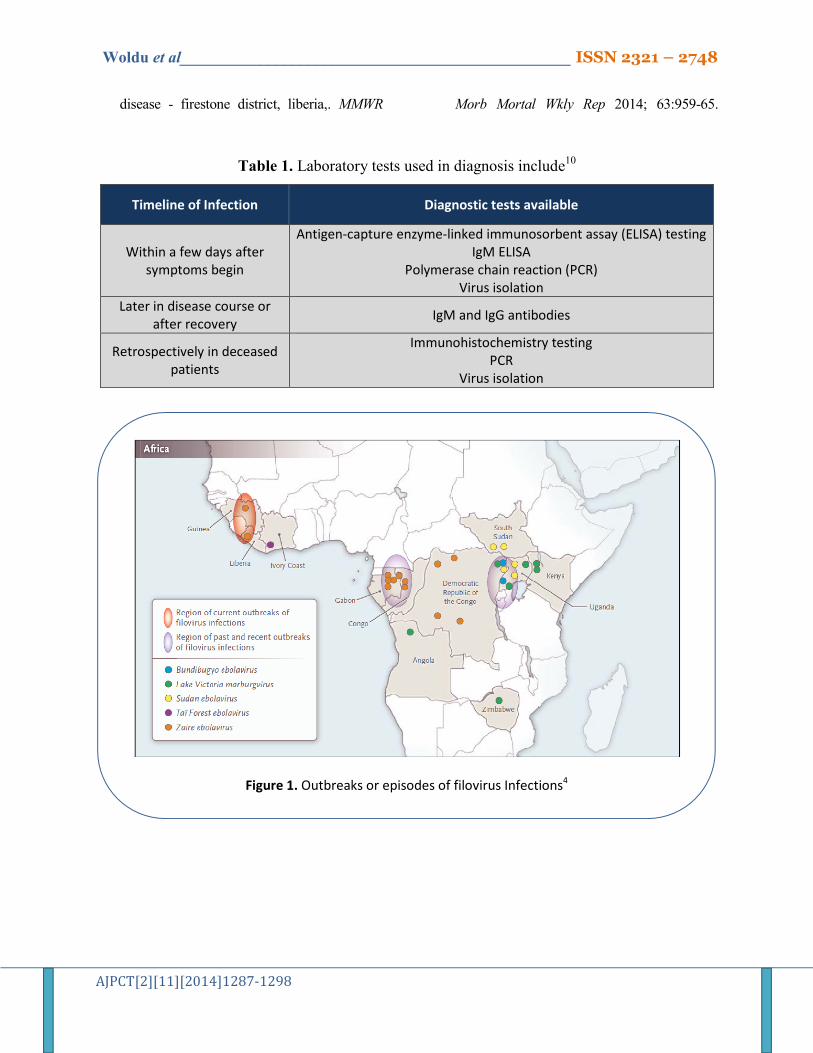
An Ebola Hemorrhagic Fever (EHF) is zoonotic pathogen caused by the Filoviridae family, Ebolavirus (EBOV)1,2. Two genera compose the family Filoviridae: Ebolavirus and Marburgvirus3. There are five antigenically distinct species of Ebola viruses: Zaire ebolavirus (EBOV), Sudan ebolavirus (SUDV), Bundibugyo ebolavirus (BDBV), Tai Forest ebolavirus (TAFV), and Reston ebolavirus (RESTV) (4). All of these viruses cause disease in humans except RESTV, which is pathogenic in nonhuman primates3 (Figure 1).
EBOV has been endemic to sub-Saharan Africa and has high mortality rates in humans1,2. Currently it represents a significant challenge to global public health, causing a severe hemorrhagic fever with case fatality rates of up to 90%5,6.
Filovirus outbreak is one of the most fatal viral diseases worldwide affecting humans and nonhuman primates (NHPs)7 and therefore, EBOV is identified as a biosafety level 4 pathogens and CDC category A agents of bioterrorism6. Furthermore, the lack of preventive vaccines and FDA-approved therapeutics has struck fear that the EBOV could become a pandemic threat8.
Since the identification of EBOV in 1976, significant filovirus research has focused on developing antiviral therapies. However, despite promising vaccine candidates, no licensed prophylactics
currently exist for preventing or treating filovirus infections9.
Case definition for Ebola virus disease (EVD)
Person under investigation (PUI) A person who has both consistent
symptoms and risk factors as follows10: Clinical criteria, which includes fever of
greater than 38.6 degrees Celsius or 101.5 degrees Fahrenheit, and additional symptoms such as severe headache, muscle pain, vomiting, diarrhea, abdominal pain, or unexplained hemorrhage; AND
1. Epidemiologic risk factors within the past 21 days before the onset of symptoms, such as contact with blood or other body fluids or human remains of a patient known to have or suspected to have EVD; residence in or travel to an area where EVD transmission is active; or direct handling of bats or non-human primates from disease-endemic areas.
Probable case
A PUI whose epidemiologic risk factors include high or low risk exposure (s)10. Confirmed case
A case with laboratory-confirmed diagnostic evidence of Ebola virus infection10.
Woldu et al_________________________________________________ ISSN 2321 – 2748
AJPCT[2][11][2014]1287-1298
Pathogenesis
The Ebola genome The Ebola genome encodes only
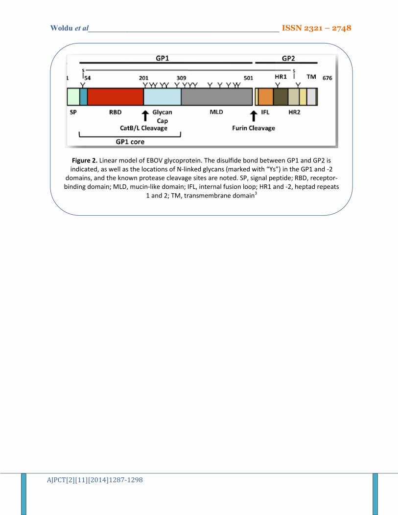
seven genes, which mediate the entry, replication, and egress of the virus from the host cell8. EBOV entry requires the virion surface-associated glycoprotein (GP) that is composed of a trimer of heterodimers (GP1/GP2). The GP1 subunit contains two heavily glycosylated domains, the glycan cap and the mucin-like domain (MLD). The glycan cap contains only N-linked glycans, whereas the MLD contains both N- and O-linked glycans3 (Figure 2).
The genomes of filoviruses (Marburg virus (MARV) and EBOV) are ∼19 kb, single stranded, negative sense RNA molecules encoding a total of 7 genes (NP, VP35, VP40, GP, VP30, VP24, L) separated by intergenic regions of varying lengths and flanked by untranslated regions (UTRs) at the 3′ and 5′ ends9.
Viral protein 35 (VP35), encoded by filoviruses, is a multifunctional dsRNA binding protein that plays important roles in viral replication, innate immune evasion, and pathogenesis. The multifunctional nature of these proteins also presents opportunities to develop countermeasures that target distinct functional regions11. The EBOV matrix protein is VP40, which is also found localized under the lipid envelope of the virus where it bridges the viral lipid envelope and nucleocapsid. The VP40 is effectively a peripheral protein that mediates the plasma membrane binding and budding of the virus prior to egress8 (Figure 2&3).
Mode of transmission
Bats have been identified as a reservoir for Ebola viruses but it remains unclear if transmission to an end host involves intermediate hosts. In 2013, one of the Ebola species was found in Philippine pigs raising concerns regarding animal health and food safety2.
Once infected, humans transmit the disease through body fluids, including breast milk. Funerals, which can bring dozens of people in contact with the remains of Ebola patients, are particularly dangerous and gatherings for suspected victims of the virus have also been outlawed12. It is also being afraid that EBOV can become airborne13.
EBOV entry is rapid, with viral protein expression detectable within 2 h after infection5. After the initial transmission, the viruses can spread from person to person through contact with body fluids or contaminated needles14.
The virulence nature of Ebola
Ebola virus enters the patient through mucous membranes, breaks in the skin, or parenterally and infects many cell types, including monocytes, macrophages, dendritic cells, endothelial cells, fibroblasts, hepatocytes, adrenal cortical cells and epithelial cells. The incubation period may be related to the infection route (e.g., 6 days for injection versus 10 days for contact)15.
The virulence of EBOV can be attributed to several immunoevasion mechanisms: an early inhibition of innate immunity started by the down regulation of type I interferon, epitope masking and subversion of the adaptive humoural immunity by secreting a truncated form of the viral glycoprotein16.
Diagnosis
The currently available screening technique include wild-type or recombinant viruses13. Diagnosing Ebola in an person who has been infected for only a few days is difficult, because the early symptoms, such as fever, are nonspecific to Ebola infection and are seen often in patients with more commonly occurring diseases, such as malaria and typhoid fever10. (See table 1.)
Platelet counts are often decreased in the 50,000 to 100,000 range. Amylase may be
Woldu et al_________________________________________________ ISSN 2321 – 2748
AJPCT[2][11][2014]1287-1298
elevated, reflecting pancreatic involvement (inflammation/infection). Hepatic transami-nases are elevated with aspartate aminotransferase (AST) exceeding alanine aminotransferase (ALT); these values may peak at more than 1,000 IU/L. Proteinuria may be present. Prothrombin (PT) and partial thromboplastin times (PTT) are prolonged and fibrin degradation products are elevated, consistent with disseminated intravascular coagulation (DIC)15.
Clinical pictures
Patients with EVD generally have abrupt onset of fever and symptoms typically 8 to12 days after exposure (incubation period for current outbreak has a mean of approximately 9 to 11 days). Initial signs and symptoms are nonspecific and may include fever, chills, myalgias, and malaise. Due to these nonspecific symptoms, particularly early in the course, EVD can often be confused with other more common infectious diseases such as malaria, typhoid fever, meningococcemia, and other bacterial infections (e.g., pneumonia). Sufferers experience nausea, vomiting, internal bleeding and organ failure before they die13,15,17.
The most common signs and symptoms reported from West Africa during the current outbreak from symptom-onset to the time the case was detected include: fever (87%), fatigue (76%), vomiting (68%), diarrhea (66%), and loss of appetite (65%). Unexplained bleeding has been reported from only 18% of patients, most often blood in the stool (about 6%). Patients may develop a diffuse erythematous maculopapular rash by day 5 to 7 (usually involving the neck, trunk, and arms) that can desquamate. Pregnant women may experience spontaneous miscarriages15.
Treatment
Current treatment approaches There are no approved treatments
available for EVD. Clinical management should focus on supportive care of complications, such as hypovolemia, electrolyte abnormalities, hematologic abnormalities, refractory shock, hypoxia, hemorrhage, septic shock, multi-organ failure, and DIC15. There is also no FDA-approved vaccine available for Ebola10.
Patients with Ebola have been treated using the following measures17: Intensive fluid resuscitation (IV fluids) Oral rehydration (drinking water) Nutritional support Electrolyte (body salt) management Dialysis and mechanical ventilation in
cases of end-organ damage (failure of the kidneys and lungs)
Pressors to maintain adequate blood pressure and blood circulation
Blood and blood products Analgesics for pain relief Antipyretics for fever Antiemetics for nausea and vomiting Antidiarrheal agents Antibiotics for accompanying infections
Emerging targets and novel therapeutic approaches
Antibody-mediated therapeutics Currently, monoclonal antibody
(mAbs)-based therapies are the most efficient at reversing the progression of a lethal Ebola virus infection in nonhuman primates, which recapitulate the human disease with the highest similarity. Novel combinations of mAbs can even fully cure lethally infected animals after clinical symptoms and circulating virus have been detected, days into the infection. These new developments have reopened the door for using antibody-based therapies for filovirus infections18.
Woldu et al_________________________________________________ ISSN 2321 – 2748
AJPCT[2][11][2014]1287-1298
Monkeys which were deliberately infected with the Ebola virus have been successfully cured in a trial of a new antibody drug cocktail, raising the possibility of a human cure for the deadly virus19. These cocktail of 3 different siRNAs (against L, VP24 and VP35) encapsulated in stable nucleic acid-lipid particles (SNALPs) provided up to 100% protection against Ebola virus when given 30 min post-exposure20,21.
These Monoclonal antibody (mAbs)-based therapies are becoming the most efficient at reversing the progression of the infection in nonhuman primates, which recapitulate the human disease with the highest similarity6. Such a recombinant human monoclonal antibody directed against the envelope GP of Ebola has been demonstrated to possess neutralizing activity. Therefore, Ebola neutralizing antibody may be useful in vaccine development or as a passive prophylactic agent22.
One study also showed that interleukin (IL)-10 signaling pathway modulates Ebola pathogenesis. IL-10-/- mice and wild-type mice receiving antisense targeting IL-10 signaling via disrupting expression through aberrant splice altering were resistant to Ebola virus infection23. In other similar study, mAbs and Ad-Vectored IFN-α therapy rescue Ebola-infected non-human primates when administered after the detection of viremia and symptoms24.
Other drugs and drug-like small molecules
A novel synthetic adenosine analogue, BCX4430, a synthetic drug-like small molecule that provides protection from Ebola and Marburg virus infection also reported to be effective in animal models. The study resulted in lower viremia, reduced clotting times and reduced liver enzyme levels were observed in treated animals20,25. Post-exposure intramuscular administration of BCX4430 protects against Ebola virus and Marburg virus disease in rodent models. Most
importantly, BCX4430 completely protects cynomolgus macaques from Marburg virus infection when administered as late as 48 hours after infection25.
Experimental drugs used for sepsis (rAPC and rNAPc2) were the first effective post-exposure therapies for Ebola virus infection with 17% - 33% survival but administration required continuous intravenous infusion20.
Investigational New Drug (IND) applications have been also approved by the US Food and Drug Administration (FDA) and phase I clinical trials have been initiated for two small-molecule therapeutics: anti-sense phosphorodiamidate morpholino oligomers (PMOs: AVI-6002, AVI-6003) and lipid nanoparticle/small interfering RNA (LNP/siRNA: TKM-Ebola)26.
One study also come up with a successful treatment of advanced Ebola virus infection with the pyrazinecarboxamide derivative T-705 (favipiravir), in a small animal model. The study further suggest that Initiation of T-705 administration at day 6 post infection induced rapid virus clearance, reduced biochemical parameters of disease severity, and prevented a lethal outcome in 100% of the animals27.
One study reported that st. amiodarone, a multi-ion channel inhibitor and adrenoceptor antagonist, is a potent inhibitor of filovirus cell entry at concentrations that are routinely reached in human serum during anti-arrhythmic therapy. A similar effect was observed with the amiodarone-related agent dronedarone and the L-type calcium channel blocker verapamil. Inhibition of filovirus entry was observed with most but not all cell types tested and was accentuated by the pre-treatment of cells, indicating a host cell-directed mechanism of action28.
One study reported that, the cytoprotective enzyme heme oxygenase-1 (HO-1) such as cobalt protoporphyrin (CoPP), which acts as a selective HO-1
Woldu et al_________________________________________________ ISSN 2321 – 2748
AJPCT[2][11][2014]1287-1298
inducer, suppresses Ebola virus replication. Since, HO-1 is an enzyme that catalyzes the rate-limiting step in heme degradation, has antioxidative properties and protects EBOV infected cells from various stresses as it has also recently shown to have antiviral activity, potently by inhibiting the replication of viruses such as hepatitis C virus and human immunodeficiency virus29.
The first clinical trial of drugs to treat Ebola virus disease is set to launch at the beginning of November 2014, with first results available by the end of the year, as per the report by the BMJ. The partnership behind the trial, which includes Oxford University, the International Severe Acute Respiratory and Emerging Infections Consortium, the Global Health Network, the Institut Pasteur, and the Institut Pasteur de Dakar, is preparing the groundwork at record speed, having been awarded a £3.2m (€4m; $5.1m) grant by the Wellcome Trust at the end of September30.
Drugs targeting viral proteins
Ebola virus-like particles (eVLP) comprising of virus protein (VP40), glycoprotein, and nucleoprotein protect rodents and nonhuman primates from lethal EBOV infection, representing as a candidate vaccine for EBOV infection. eVLP stimulate the expression of proinflammatory cytokines and interferons (IFNs) and IFN-stimulated genes (ISGs) in murine bone marrow-derived dendritic cells (BMDCs) and macrophages. eVLP stimulate early innate immune responses through toll-like receptor (TLR) and type I IFN signaling pathways31.
A number of studies have demonstrated specific deletions or mutations of VP40 to abrogate viral egress but to date pharmacological inhibition of VP40 has not been demonstrated8. Functional validation and the establishment of therapeutic approaches toward multifunctional proteins such as VP35, particularly for nonenzymatic targets, are often challenging11.
FDA also approved selective estrogen receptor modulators (SERMs), including clomiphene and toremifene, which act as potent inhibitors of EBOV infection. Anti-EBOV activity was confirmed for both of these SERMs in an in vivo mouse infection model. This anti-EBOV activity occurred even in the absence of detectable estrogen receptor expression, and both SERMs inhibited virus entry after internalization, suggesting that clomiphene and toremifene are not working through classical pathways associated with the estrogen receptor32.
Vaccines
Passive immunization for Ebola virus was successful in 2012, after over 15 years of failed attempts leading to skepticism that the approach would ever be of potential benefit18. In fact, vaccine for EBOV was Successful in primates in 200833.
In 2010, vaccines have been tried in monkeys by containing pieces of the protein-sugar coat (glycoprotein) from the Zaire and Sudan EDOV, inoculated into a type of common cold virus. Once infected, the monkeys' immune cells chop the Ebola glycoprotein into small pieces and display them on their surface, where they stimulate a response from other immune cells. The monkeys got four "priming" shots, followed a year later with a booster shot. Four other macaques got no vaccine. All eight animals were inoculated with ordinarily lethal doses of the new Ebola virus. The vaccinated animals all survived, and the unvaccinated monkeys all died34,35. The researchers will use the new results to "optimize" a vaccine that might eventually be approved for human use. To do that, they'll have to show it works in larger animal studies34.
The World Health Organization has also said that the first vaccines against the Ebola virus may be in use among frontline workers in West Africa by the beginning of 2015. The two “front running” vaccines are

Woldu et al_________________________________________________ ISSN 2321 – 2748
AJPCT[2][11][2014]1287-1298
cAd3-ZEBOV, a chimpanzee derived adenovirus vaccine developed by GlaxoSmithKline in conjunction with the US National Institute of Allergies and Infectious Diseases, and rVSV-ZEBOV, developed by the Public Health Agency of Canada and now licensed to a US company called New Link30.
Limitations of the experimental treatments
The findings of animal model studies cannot be directly extrapolated to human use, because even though it is encouraging, such findings must be taken with a grain of salt. The result has to be further repeated in volunteer human patients or people at risk of getting the disease with a multicenter, multination, and randomized clinical trial study design. Ethical clearance and approval of the study from the responsible organ, which is still a challenging one.
General precautions and approaches for prevention
General medical support is critical and should include replacement of coagulation factors and heparin if disseminated intravascular coagulation develops. Such care must be administered with strict attention to barrier isolation. All body fluids (blood, saliva, urine, and stool) contain infectious virions and should be handled with great care22.
Livestock vaccination against Ebola seems currently not justified but proper preparedness may include experimental vaccine approaches2. Resistance to Ebola infection is regulated by IL-10 and can be targeted in a prophylactic manner to protect against lethal hemorrhagic virus challenge23.
Many promising vaccines are moving through pre-clinical or clinical trials, but mass immunization is unlikely due to the localized and sporadic nature of EBOV infections. Post-exposure interventions are therefore necessary for the treatment of cases as they occur, but have been harder to develop as
death from an EBOV infection typically occurs within 6–9 days in non-human primates (NHPs). This leaves a very short window for the treatment to reduce virus replication until the immune response expands sufficiently to control the infection36.
Everyone working in the isolation area must follow the protocols and procedures. And should follow a buddy system where should have to be responsible for his/herself and the colleagues: Because one mistake could be deadly37. Should wear protective clothing, including masks, gloves, gowns, and eye protection and should have to practice proper infection control and sterilization measures, isolate patients with Ebola from other patients and should also avoid direct contact with the bodies of people who have died from Ebola10.
Multidisciplinary teams should oversee the outbreak response, address case detection, manage cases in a dedicated unit, and reintegrate convalescent patients into the community. The team should also create a robust risk communication, prevention, and social mobilization campaign to boost community awareness of Ebola and how to prevent transmission38. The isolation area is divided into separate tents for patients with suspected, probable, and confirmed Ebola virus infection37.
Practicing careful hygiene is very important. For example, wash hands with soap and water or an alcohol-based hand sanitizer and avoid contact with blood and body fluids. Avoid items that may have come in contact with an infected person’s blood or body fluids (such as clothes, bedding, needles, and medical equipment). Avoid funeral or burial rituals that require handling the body of someone who has died from Ebola. Avoid contact with bats and nonhuman primates or blood, fluids, and raw meat prepared from these animals15.
Woldu et al_________________________________________________ ISSN 2321 – 2748
AJPCT[2][11][2014]1287-1298
CONCLUSION
Early recognition is critical for infection control. Health care providers should be alert for and evaluate any patients suspected of having EBOV. Many promising vaccines are moving through pre-clinical or clinical trials, but mass immunization is unlikely due to the localized and sporadic nature of EBOV infections. Post-exposure interventions are therefore necessary for the treatment of cases as they occur.
Competing interests
The Authors’ declare that there are no competing interests.
Authors’ contributions
MAW has made substantial contributions to conception and gathering of information and has been involved in drafting the manuscript or revising it critically for important intellectual content; and has given final approval of the version to be published. JLL has made substantial contributions to conception and design, or acquisition of data, or analysis and interpretation of data. GTT has been involved in drafting the manuscript or revising it critically for important intellectual content. GDS has been involved in drafting the manuscript or revising it critically for important intellectual content. All Authors read and approved the final manuscript.
Author information 1. Minyahil Alebachew Woldu: BPharm, MSc in Clinical Pharmacy,
fulltime Lecturer: department of Pharmacology and Clinical pharmacy, College of Medicine and Health Sciences, Addis Ababa University; Addis Ababa- Ethiopia. Email: [email protected]. Tel: +251-91-2648527.
2. Jimma Likisa Lenjisa: BPharm, MSc in Clinical Pharmacy,
fulltime Lecturer: department of
Pharmacy, College of Medicine and Health Sciences, Ambo University; Ambo- Ethiopia. Email: jimmapharm@ gmail.com.
3. Gobezie Temesgen Tegegne: BPharm, MSc in Clinical Pharmacy,
fulltime Lecturer: department of Pharmacy, College of Medicine and Health Sciences, Ambo University; Ambo- Ethiopia. Email: gobepharm@ gmail.com.
4. Gizaw Dabessa Satessa: BSc, MD., fulltime Lecturer: department
of Pharmacy, College of Medicine and Health Sciences, Ambo University; Ambo-Ethiopia. Email: gizaw_dhab@ yahoo.com.
ACKNOWLEDGMENTS
The authors’ are grateful to all the health care professionals, researchers and governments working to prevent this global terror risking their life, family and community. We are also grateful to all the information resources included in this review article with specially thanks to NIH, NEJM and BMJ.
Operational definitions
EVD exposure risk levels Levels of exposure risk are defined as
follows10:
High risk exposures A high risk exposure includes any of
the following: Percutaneous (e.g., needle stick) or
mucous membrane exposure to blood or body fluids of EVD patient
Direct skin contact with, or exposure to blood or body fluids of, an EVD patient without appropriate personal protective equipment (PPE)
Processing blood or body fluids of a confirmed EVD patient without
Woldu et al_________________________________________________ ISSN 2321 – 2748
AJPCT[2][11][2014]1287-1298
appropriate PPE or standard biosafety precautions
Direct contact with a dead body without appropriate PPE in a country where an EVD outbreak is occurring
Low risk exposures
A low risk exposure includes any of the following. Household contact with an EVD patient Other close contact with EVD patients in
health care facilities or community settings. Close contact is defined as,
1. Being within approximately 3 feet (1 meter) of an EVD patient or within the patient’s room or care area for a prolonged period of time (e.g., health care personnel, household members) while not wearing recommended personal protective equipment (i.e., standard, droplet, and contact precautions; see Infection Prevention and Control Recommendations).
2. Having direct brief contact (e.g., shaking hands) with an EVD patient while not wearing recommended personal protective equipment.
Brief interactions, such as walking by a person or moving through a hospital, do not constitute close contact.
No known exposure
Having been in a country in which an EVD outbreak occurred within the past 21 days and having had no high or low risk exposures.
Acronyms and abbreviations ALT - Alanine Aminotransferase AST - Aspartate Aminotransferase BDBV - Bundibugyo Ebolavirus DIC - Disseminated Intravascular Coagulation EBOV - Ebolavirus EHF - Ebola Hemorrhagic Fever
ELISA - Enzyme-Linked Immuno-sorbent Assay EVD - Ebola Virus Disease GP - Glycoprotein mAbs - Monoclonal Antibody MARV - Marburg Virus MLD - Mucin-like Domain NHPs - Nonhuman Primates PCR - Polymerase Chain Reaction PT - Prothrombin PTT - Partial Thromboplastin Times PUI - Person under investigation RESTV - Reston Ebolavirus SUDV - Sudan Ebolavirus TAFV - Tai Forest Ebolavirus REFERENCES 1. Eri Nakayama, Saijo M. Animal models for
Ebola and Marburg virus infections. Frontiers in Microbiology | Virology 2013; 4:1-20.
2. Feldmann F, H. F. Ebola: facing a new transboundary animal disease? Dev Biol (Basel) 2013; 135:201-9.
3. Lennemann NJ, Rhein BA, Ndungo E, Chandran K, Qiu X, W. M. Comprehensive functional analysis of N-linked glycans on Ebola virus GP1. MBio 2014 5:e00862-13.
4. Heinz Feldmann. Ebola - A Growing Threat? N engl j med 2014.
5. Hoenen T1, Groseth A, Callison J, Takada A, H. F. A novel Ebola virus expressing luciferase allows for rapid and quantitative testing of antivirals. Antiviral Res 2013; 99:207-13.
6. Qiu X1 KG. Antibody therapy for Ebola: Is the tide turning around? Hum Vaccin Immunother 2014 6:4.
7. Marzi A, H. F. Ebola virus vaccines: an overview of current approaches. Expert Rev Vaccines 2014; 13:521-31.
8. Stahelin RV. Could the Ebola virus matrix protein VP40 be a drug target? Expert Opin Ther Targets 2014 18:115-20.
9. Uebelhoer LS, Albariño CG, McMullan LK, et al. High-throughput, luciferase-based reverse genetics systems for identifying inhibitors of Marburg and Ebola viruses. Antiviral Res 2014; 106:86-94.
Woldu et al_________________________________________________ ISSN 2321 – 2748
AJPCT[2][11][2014]1287-1298
10. NIH. Turning Discovery Into Health®. 9000 Rockville Pike, Bethesda, Maryland 20892. National Institutes of Health; 2014.
11. Binning JM, Wang T, Luthra P, et al. Development of RNA aptamers targeting Ebola virus VP35. Biochemistry 2013 52:8406-19.
12. Andrew Green. West Africa struggles to contain Ebola outbreak. The Lancet 2014 383:1196.
13. Jennifer Carpenter. Vaccine developed against Ebola. Science reporter, BBC News 2011.
14. Ebola virus and Marburg virus. Mayo Foundation 2014.
15. NIH. Ebola virus disease Information for Clinicians in U.S. Healthcare Settings 2014.
16. Wong G, Kobinger GP, X. Q. Characterization of host immune responses in Ebola virus infections. Expert Rev Clin Immunol 2014.
17. Tackling the Troubling Ebola Virus. About.com, 2014.
18. Xiangguo Qiu, Gary P Kobinger. Antibody therapy for Ebola: Is the tide turning around? Landes Bioscience 2014; 10.
19. Ian Steadman. Ebola virus cured in monkeys with triple drug cocktail. Science 2012.
20. Darryl Falzarano, Heinz Feldmann. Possible leap ahead in filovirus therapeutics. Cell Research advance online publication 2014.
21. Pettitt J, Zeitlin L, Kim do H, et al. Therapeutic intervention of Ebola virus infection in rhesus macaques with the MB-003 monoclonal antibody cocktail. Sci Transl Med 2013 5:199ra13.
22. Ebola Virus Infection Treatment & Management. 2013.
23. Panchal RG, Mourich DV, Bradfute S, et al. Induced IL-10 Splice Altering Approach to Antiviral Drug Discovery. Nucleic Acid Ther 2014.
24. Qiu X, Wong G, Fernando L, et al. mAbs and Ad-Vectored IFN-α therapy rescue ebola-infected nonhuman primates when administered after the detection of viremia and symptoms. Sci Transl Med 2013 5:207ra143.
25. Warren TK, Wells J, Panchal RG, et al. Protection against filovirus diseases by a
novel broad-spectrum nucleoside analogue BCX4430. Nature 2014 508:402-5.
26. Choi JH, MA C. Emerging targets and novel approaches to Ebola virus prophylaxis and treatment. Bio Drugs 2013 27:565-83.
27. Oestereich L, Lüdtke A, Wurr S, Rieger T, Muñoz-Fontela C, S. G. Successful treatment of advanced Ebola virus infection with T-705 (favipiravir) in a small animal model. Antiviral Res 2014 105:17-21.
28. Gehring G, Rohrmann K, Atenchong N, et al. The clinically approved drugs amiodarone, dronedarone and verapamil inhibit filovirus cell entry. J Antimicrob Chemother 2014
29. Hill-Batorski L, Halfmann P, Neumann G, Y. K. The cytoprotective enzyme heme oxygenase-1 suppresses Ebola virus replication. J Virol 2013 87:13795-802.
30. Anne Gulland. Ebola drug trial is to start next month. BMJ 2014; 349:g6436.
31. Ayithan N, Bradfute SB, Anthony SM, et al. Ebola virus-like particles stimulate type I interferons and proinflammatory cytokine expression through the toll-like receptor and interferon signaling pathways. J Interferon Cytokine Res 2014 34:79-89.
32. Lisa M. Johansen, Jennifer M. Brannan, Sue E. Delos, et al. FDA-Approved Selective Estrogen Receptor Modulators Inhibit Ebola Virus Infection. Sci Transl Med June 19; 5(190): 190ra79 2013 5:90ra79.
33. Microbiology SfG. Vaccine for Ebola Virus Successful In Primates. ScienceDaily 2008.
34. Experimental Ebola Vaccine Also Works On New Virus. NPR, 2010.
35. O'Brien LM, Stokes MG, Lonsdale SG, et al. Vaccination with recombinant adenoviruses expressing Ebola virus glycoprotein elicits protection in the interferon alpha/beta receptor knock-out mouse. Virology 2014 452:324-33.
36. Xiangguo Qiu, Jonathan Audet, Gary Wong, et al. Sustained protection against Ebola virus infection following treatment of infected nonhuman primates with ZMAb. Scientific Reports; 3:1-9.
37. Anja Wolz, R.N. Face to Face with Ebola - An Emergency Care Center in Sierra Leone. N engl j med 2014.
38. Reaves EJ, Mabande LG, Thoroughman DA, Arwady MA, JM. M. Control of ebola virus
Woldu et al_________________________________________________ ISSN 2321 – 2748
AJPCT[2][11][2014]1287-1298
disease - firestone district, liberia,. MMWR Morb Mortal Wkly Rep 2014; 63:959-65.
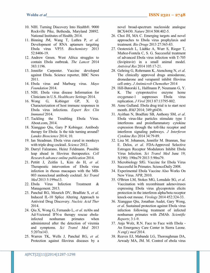
Table 1. Laboratory tests used in diagnosis include10
Timeline of Infection Diagnostic tests available
Within a few days after symptoms begin
Antigen-capture enzyme-linked immunosorbent assay (ELISA) testing IgM ELISA
Polymerase chain reaction (PCR) Virus isolation
Later in disease course or after recovery
IgM and IgG antibodies
Retrospectively in deceased patients
Immunohistochemistry testing PCR
Virus isolation
Figure 1. Outbreaks or episodes of filovirus Infections4
Woldu et al_________________________________________________ ISSN 2321 – 2748
AJPCT[2][11][2014]1287-1298
Figure 2. Linear model of EBOV glycoprotein. The disulfide bond between GP1 and GP2 is indicated, as well as the locations of N-linked glycans (marked with “Ys”) in the GP1 and -2
domains, and the known protease cleavage sites are noted. SP, signal peptide; RBD, receptor-binding domain; MLD, mucin-like domain; IFL, internal fusion loop; HR1 and -2, heptad repeats
1 and 2; TM, transmembrane domain3