Neuroprotective effects of citicoline in in vitro models of retinal neurodegeneration
Upload
independentCategory
view
1download
0
Folate deprivation induces neurodegeneration: roles of oxidative stressand increased homocysteine
Pei I. Ho,a,b David Ashline,a,b,c Sirikarnt Dhitavat,a,b Daniela Ortiz,a Scott C. Collins,d
Thomas B. Shea,a,b,d,* and Eugene Rogersa,b,c
a Center for Cellular Neurobiology and Neurodegeneration Research, University of Massachusetts–Lowell, Lowell, MA 01854, USAb Department of Biochemistry, University of Massachusetts–Lowell, Lowell, MA 01854, USA
c Department of Health and Clinical Sciences, University of Massachusetts–Lowell, Lowell, MA 01854, USAd Department of Biological Sciences, University of Massachusetts–Lowell, Lowell, MA 01854, USA
Received 29 March 2002; revised 5 November 2002; accepted 14 February 2003
Abstract
Clinical studies suggest a relationship between folate deficiency and neurological and disorders including Alzheimer’s disease (AD). Toinvestigate mechanisms underlying this association, we examined the consequences of folate deprivation on neuronal cultures. Culturingembryonic cortical neurons and differentiated SH-SY-5Y human neuroblastoma cells in folate-free medium induced neurodegenerativechanges characteristic of those observed in AD, including increased cytosolic calcium, reactive oxygen species (ROS), phospho-� andapoptosis. In accord with clinical studies, generation of the neurotoxic amino acid homocysteine (HC) was likely to contribute to thesephenomena, since (1) a significant increase in HC was detected following folate deprivation, (2) addition of the inhibitor of HC formation,3-deazaadenosine, both prevented HC formation and eliminated the increase in ROS that normally accompanied folate deprivation, (3) directaddition of HC in the presence of folate induced the neurotoxic effects that accompanied folate deprivation, and (4) an antagonist of NMDAchannels that blocks HC-induced calcium influx also blocked calcium influx following folate deprivation. Folate deprivation decreased thereduced form of glutathione, indicating a depletion of oxidative buffering capacity. This line of reasoning was supported by an increase inglutathione and reduction in ROS following supplementation of folate-deprived cultures with the cell-permeant glutathione precursor,N-acetyl-L-cysteine, or vitamin E. Folate deprivation potentiated ROS and apoptosis induced by amyloid-�, while folate supplementationat higher concentrations prevented generation of ROS by amyloid-�, suggesting that folate levels modulate the extent of amyloid-�neurotoxicity. These findings underscore the importance of folate metabolism in neuronal homeostasis and suggest that folate deficiencymay augment AD neuropathology by increasing ROS and excitotoxicity via HC generation.© 2003 Elsevier Science (USA). All rights reserved.
Introduction
Folate deficiency has long been implicated in cardiovas-cular disorders, and only recently is it becoming apparentthat it also contributes to many neurological and psycho-logical disorders including dementia, impaired cognition,depression, psychosis, and Alzheimer’s disease (AD; e.g.,Allen et al., 1998; Clarke et al., 1998; Hassing et al., 1999;Nilsson et al., 1994; Renvall et al., 1989; Wang et al., 2001;Gray, 1989; Nilsson-Ehle, 1998). Low serum folate levelsare strongly associated with atrophy of the cerebral cortex
(Snowdon et al., 2000). In addition, while levels of folate incerebral spinal fluid are normally three-to fourfold higher inthan blood, spinal fluid levels of folate are significantlylower in AD patients (Serot et al., 2001; Wevers et al.,1994). Diminished folate levels are associated with a two-fold increased risk in developing AD (Wang et al., 2001).
The full range of mechanisms by which deficiencies infolate may contribute to neurodegeneration is unclear. How-ever, one likely major impact of folate deprivation is in-creases in homocysteine (HC), since folate is a necessarycofactor for the enzyme (5,10-methyenetetralhydrofolate re-ductase) that mediates conversion of HC to methionine(Fiskerstrand et al., 1997; Pietrzik and Bronstrup, 1998).Elevated HC levels are also associated with neuropsychiat-
* Corresponding author. Fax: �1-508-934-3044.E-mail address: [email protected] (T.B. Shea).
R
Available online at www.sciencedirect.com
Neurobiology of Disease 14 (2003) 32–42 www.elsevier.com/locate/ynbdi
0969-9961/03/$ – see front matter © 2003 Elsevier Science (USA). All rights reserved.doi:10.1016/S0969-9961(03)00070-6

ric disorders including AD (Allen et al., 1998; Bell et al.,1992; Miller, 2000). HC is toxic to cultured neurons andneuronal cells (Kruman et al., 2000; Ho et al., 2001a) andpotentiates neurotoxicity induced by amyloid-� (Ho et al.,2001a; White et al., 2001). Folate deficiency has beenshown to elevate HC, compromise neuronal homesostasis,and render neurons more sensitive to amyloid toxicity inmouse models of AD and Parkinson’s disease (Duan et al.,2002; Kruman et al., 2002). To address more fully theneurotoxic effects of folate deprivation, we examined theconsequences of folate deficiency on cortical neurons anddifferentiated human neuroblastoma cells in culture.
Materials and methods
Cell culture and treatment
Murine cortical neurons from embryonic day 12 werecultured for 3 days with serum-free Neurobasal mediumwith N2 supplements. SH-SY-5Y human neuroblastomacells were cultured in DMEM containing 10% fetal calfserum and were differentiated for 7 days with 10 �Mretinoic acid (Ekinci et al., 1999). To monitor the conse-quences of folate deprivation, both culture types receivedDMEM lacking folate (Sigma) for 2 h–7 days. Corticalneurons continued to receive B27 supplements in the pres-ence and absence of folate. Additional SH-SY-5Y culturesreceived 10–250 �M HC (Ho et al., 2001a), 1 �M MK-801(antagonist of NMDA calcium channels; e.g., Gray andPatel, 1995), 1 �M nimopridine (antagonist of the L volt-age-sensitive calcium channel; Ueda et al., 1997), amy-loid-� 25–35; A�; 20 �M for SH-SY-5Y cells, 10 �M forcortical neurons; Ekinci et al., 1999), 25 mM 3-deazaade-nosine (DZA; (Jeong et al., 1999), 10 mM N-acetyl-L-cysteine (NAC; Huang et al., 2002; Schulz et al., 2000), or0.15 mg/L vitamin E (as �-tocopherol; Ekinci et al., 1999;Huang et al., 2002; Shea and Rogers, 2002b) in the presenceand absence of folate.
DMEM, utilized during experimental treatments for bothcell types, contains 4 mg/L folate. The concentration offolate in serum is approximately 10 �g/L; folate-free me-dium supplemented with 10% serum therefore contains ap-proximately 1 �g/L folate. Folate-free medium with orwithout serum therefore contains vastly reduced folate ver-sus medium containing folate, and the presence or absenceof serum was likely to have minimal, if any, impact uponcells in folate-free medium. Reference to such conditions as“folate-deprived” or “receiving medium lacking folate” isintended to reflect this major difference in folate concentra-tion rather than necessarily referring to a complete absenceof folate. Moreover, both cortical neurons and SH-SY-5Ycells are likely to retain some intracellular folate that mayprovide for certain metabolic processes during the relativelyshort incubation period (2 h) utilized in most of theseexperiments.
Analyses of calcium influx, reactive oxygen species (ROS),phospho-�, and apoptosis
To monitor cytosolic calcium, cells were rinsed withserum-free medium and received 10 �l/ml Fluo-3 (ace-toxymethyl ester; Molecular Probes) for 30 min and livingcultures were then visualized under fluorescein UV optics.For monitoring of intracellular peroxide concentrations asan index of ROS, cultures received 10 �l/ml DFCD (2�7�-dichlorofluorescein diacetate; Kodak) for 20 min. Cultureswere then visualized under fluorescein UV optics. Apopto-sis was also monitored by two methods, all of which havepreviously been utilized to quantify neuronal apoptosis inthese cultures (Ekinci and Shea, 2000; Ekinci et al., 1999:Huang et al., 2000) as follows: externalized phosphatidylserine (PS) was assayed by adding 5 �l merocyanine/ml ofculture medium to cultures 10 min before the experimentalendpoint, with incubation continued at 37°C (Ekinci et al.,1999; Ekinci and Shea, 2000). Cultures were then visualizedunder rhodamine UV optics. Additional cultures were incu-bated for 10 min at 37°C with DAPI (0.1 �g/ml), whichselectively binds to DNA (Huang et al., 2000). The percent-age of cells exhibiting shrunken, pycnotic nuclei was quan-tified for DAPI (Ekinci and Shea, 2000; Huang et al., 2000),and the percentage of cells displaying intense externalizedPS was quantified for merocyanine staining (Ekinci et al.,1999). To monitor mitochondrial membrane potential, cellscultured for 2 h, 3 days, or 6 days in the presence or absenceof folate were rinsed with fresh DMEM and incubated in 1.5�M/ml of 5,5�,6,6�-tetrachloro-1,1�,3,3�-tetraethylbenzimi-dazolylcarbocyanine iodide (“JC-1,” Molecular Probes) for10 min at 37°C (Wadia et al., 1998) and washed again withmedium and images were acquired under fluorescence andrhodamine UV optics. JC-1 is a cationic dye that exhibitspotential-dependent accumulation in mitochondria, indi-cated by a fluorescence emission shift from green to red.Mitochondrial depolarization is determined by a decrease inthe red/green fluorescence intensity ratio (Reers et al.,1991). For changes in � phosphorylation, cultures werefixed in 4% paraformaldehyde, rinsed in Tris-buffered sa-line (TBS), blocked with 5% bovine serum albumin, andthen incubated overnight at 4°C with a 1:50 dilution ofPHF-1 (for phospho-�) or 5E2 (for total �) followed byrhodamine- or fluorescein-conjugated secondary antibodies.The above methodologies have been routinely utilized formonitoring neurodegenerative changes in these cultures(e.g., Ekinci et al., 1999, 2000; Ekinci and Shea, 2000; Hoet al., 2001a).
Images were captured using a Dage CCL-72 cameraoperated by NIH Image via a Scion LG-3 frame grabber andstored as PICT files on a PowerPC Macintosh. Identicalillumination and capture settings were used for all images.Multiple fields (5–10) at 20� were captured from duplicateor triplicate cultures. Relative fluorescent intensity wasquantified from stored images using NIH Image. Statistical
33P.I. Ho et al. / Neurobiology of Disease 14 (2003) 32–42

analyses were carried out by Student’s t test or ANOVA.Values were considered statistically different if P � 0.05.
Immunoblot analysis
SH-SY-5Y cells cultured in the presence or absence offolate were rinsed with Tris-buffered saline and harvested in1% Triton X-100 in 50 mM Tris–HCl (pH 6.8) containing 5mM EDTA, 50 �g/ml leupeptin, 1 mM PMSF, 1 mM Nafluoride, and 1 mM Na–vanadate. Homogenates were sedi-mented at 13,000g for 15 min and the resulting supernatantwas subjected to SDS–gel electrophoresis followed bytransfer to nitrocellulose and immunoblot analysis withPHF-1 as described (e.g., Ekinci et al., 1999). Immunoblotswere scanned and the relative intensity of PHF-1 immuno-reactivity was determined with NIH Image by encircling allPHF-1 immunoreactive bands with the program’s freehandtool and the density of all bands was summed.
Determination of HC and glutathione levels
HC and total glutathione were monitored in differenti-ated SH-SY-5Y cells by HPLC using a modified version ofthe methodology of Araki and Sako (1987). Cultures wererinsed 2� with TBS, scraped from the plate in a smallvolume of TBS, and homogenized on ice. Medium was alsoutilized for analysis of HC along with cellular lysates, sinceHC is normally exported from cells (Christensen et al.,1991). Aliquots of medium or lysates (100 �l) were com-bined with 30 �l of 30 �M cystamine (as an internalstandard) and 10 �l of tricarboxyethylphosphine (100mg/ml in 0.05 M HCl). Samples were vortexed, incubated atroom temperature for 10 min, and then centrifuged at10,000g for 10 min. A total of 80 �l of the resultingsupernatant was combined with 160 �l of 2 M boric acidcontaining 4 mM EDTA (pH 10.5), followed by 80 �l of 1.0mg/ml 7-fluorobenzo-2-oxa-1,3-diazide-4-sulfonate in thesame buffer. Samples were incubated for 1 h at 60°C andthen injected (50 �l) into a Hewlett Packard Model 1090HPLC equipped with a Model 1046A fluorescence detectorand a Hewlett Packard 4.6 � 60 mm high-speed analyticalcolumn packed with 3 �M ODs (C18) Hypersil silica. Theisocratic mobile phase consisted of 2 vol of methanol/ 98vol of 0.1 M phosphate buffer (pH 2.0). HC and totalglutathione concentrations were then determined by com-parison of peak height ratios to the cystamine internal stan-dard.
For analysis of reduced glutathione, culture medium wasreplaced with fresh medium containing the reduced gluta-thione-reactive dye, 5-chloromethylfluorescein diacetate (5�M; Molecular Probes), for 30 min at 37°C, after whichcells received fresh medium and were incubated an addi-tional 30 min according to the manufacturer’s instructions.Cells were then fixed with paraformaldehyde and perme-abilized with 0.1% saponin in 50 mM Tris (pH 6.8; Ekinci
et al., 1999) and images were recorded under phase and UVoptics as above using NIH Image software (version 1.62).
Results
Folate deprivation increases ROS and cytosolic calcium
Cultured embryonic murine cortical neurons and differ-entiated SH-SY-5Y human neuroblastoma cells were de-prived of folate for 2 h. This treatment induced a markedincrease in cytosolic calcium (Fig. 1) and ROS (Fig. 2).
Folate deficiency induces rapid accumulation of phospho-�
Cultures deprived of folate for 2 h were fixed and pro-cessed for phospho-� immunoreactivity using a monoclonalantibody (PHF-1). Folate deficiency induced 3.5- and 1.5-fold increases in perikaryal PHF-1 immunofluorescencewithin differentiated SH-SY-5Y cells and cortical neurons,respectively (Figs. 3 and 4). In addition, a focal increase inPHF-1 immunoreactivity was noted within the area at thebase of the major neurite that markedly exceeded the overallperikaryal increase (arrows; Figs. 3 and 4). Increased PHF-1immunoreactivity following folate deprivation was also ob-served following immunoblot analyses of SH-SY-5Y cells(Fig. 3). While overall perikaryal phosphorylation for bothcell types was increased by 1.5- to 2-fold within 2 h of folatedeprivation (Figs. 3 and 4), densitometric comparison fur-ther indicated that the focal increase in phospho-� at theneurite base was 10.5-fold in cortical neurons and 12.5-fold
Fig. 1. Folate deprivation increased cytosolic calcium. Cortical neurons anddifferentiated SH-SY-5Y neuroblastoma were cultured for 2 h in thepresence or absence of folate and were processed for cytosolic calciumlevels using the calcium indicator Fluo-3 as described under Materials andMethods. Micrographs present representative SH-SY-5Y cells cultured inthe presence and absence of folate as indicated. The accompanying graphspresent the mean � SEM Fluo-3 fluorescent intensity from �50 cells fromtwo separate cultures of cortical neurons and SH-SY-5Y cells cultured inthe presence and absence of folate as indicated from two independentexperiments each. Note the marked increase in cytosolic calcium in bothcell types following folate deprivation (P � 0.05; Student’s t test).
34 P.I. Ho et al. / Neurobiology of Disease 14 (2003) 32–42

in SH-SY-5Y cells. Additional cultures were immuno-stained with monoclonal antibody 5E2, which reacts with �independent of its phosphorylation state. These analysesdemonstrated that steady-state levels of � were not alteredfollowing 2 h folate deprivation and therefore indicated thatfolate deprivation induced a specific increase in phospho-�(Fig. 5). The increase in phospho-� following folate depri-vation was reversible; when cultures that had been deprivedof folate for 4 h were cultured for an additional 20 h withmedium containing folate, perikaryal phospho-� levels de-clined to levels observed in cultures that had not beendeprived of folate (Table 1).
Folate deprivation induces apoptosis
To ascertain whether folate deprivation induced apopto-sis in differentiated SH-SY-5Y cells, we monitored changesin externalized PS and fragmentation of DNA, which rep-resent early and late markers of apoptosis (Collins et al.,1997; Rimon et al., 1997). Differentiated SH-SY-5Y cells
and cortical neurons were cultured in the presence or ab-sence of folate-containing or folate-free medium for 3 and 6days. Cultures deprived of folate demonstrated a markedincrease in externalization of PS and formation of DAPI-
Fig. 2. Folate deprivation generates increased ROS. Cortical neurons andSH-SY-5Y neuroblastoma were cultured for 2 h in the presence or absenceof folate as indicated and were processed for ROS using DFCD as de-scribed under Materials and Methods. Micrographs present representativecortical neurons cultured in the presence and absence of folate as indicated.The accompanying graphs present DFCD fluorescent intensity (mean �SEM) from �50 cells from two separate cultures of cortical neurons andSH-SY-5Y cells � folate from two independent experiments each. Notethe marked increase in ROS in both cell types following folate deprivation(P � 0.05; Student’s t test).
Fig. 3. Folate deprivation induces accumulation of phospho-� in humanneuroblastoma. Differentiated SH-SY-5Y cells were cultured for 2 h in thepresence (�folate) or absence (�folate) of folate and then fixed andprocessed for PHF-1 immunofluorescence (A) or immunoblot analysis (B).Note the marked increase in perikaryal PHF-1 immunofluorescence infolate-deprived cells (A). Note further that increased PHF-1 immunofluo-rescence is concentrated in many cells at the base of the major neurite(arrows indicate representative cells; not all such clusters of immunofluo-rescence are indicated). The accompanying graph (A) presents densitomet-ric analyses of PHF-1 immunoreactivity within representative perikarya ofcells cultured in the presence and absence of folate. Note the markedincrease in PHF-1 immunoreactivity following folate deprivation (P �
0.05; Student’s t test). Immunoblot analysis of Triton-soluble homogenatesfrom SH-SY-5Y cells (B) also demonstrated a marked increase in PHF-1immunoreactivity in cells cultured without (�) versus with (�) folate.Densitometric analysis of immunoblots, presented in the accompanyinggraph, demonstrated an approximate twofold increase in PHF-1 immuno-reactivity following folate deprivation.
Fig. 4. Folate deprivation induces accumulation of phospho-� in murinecortical neurons. Cortical neurons were cultured in the presence (�folate)or absence (�folate) of folate for 2 h, after which they were processed forPHF-1 immunofluorescence. Note the marked increase in overall PHF-1immunoreactivity in folate-deprived cells. Note further the focal increase atthe base of the major neurite (arrows). The accompanying graph presentsdensitometric analyses of PHF-1 immunoreactivity within 50 representa-tive cells cultured in the presence and absence of folate (P � 0.05;Student’s t test).
35P.I. Ho et al. / Neurobiology of Disease 14 (2003) 32–42

labeled apoptotic bodies by the 3rd day, which had doubledby the 6th day (Fig. 6A). In accordance with the priorstudies of Duan et al. (2002), folate-deprived cells alsoexhibited a substantial increase in mitochondrial trauma, asindicated by a loss of mitochondrial membrane potential(Fig. 6B).
HC formation contributes to ROS generation followingfolate deprivation
One likely major impact of folate deprivation is in-creased HC production (Fiskerstrand et al., 1997). We firstconfirmed that folate deprivation indeed increased HC pro-duction by HPLC analyses. In accordance with prior studiesthat demonstrate that HC is either rapidly metabolized orexported (e.g., Christensen et al., 1991) and that increasedHC formation increases its export rather than intracellularaccumulation (Blom, 2000), HC levels were undetectable(�1 nM) within cell lysates both before and after folatedeprivation. HC levels within medium of cells cultured for2 h in the absence of folate increased 10-fold (to approx.300 nM) from levels in medium of cells cultured in thepresence of folate (approx. 30 nM; Fig. 7). Additional dif-ferentiated SY-SY-5Y cells were treated with DZA, whichinhibits HC formation (Jeong et al., 1999), simultaneouslywith folate deprivation. As above (Fig. 2), folate deprivationfor 2 h increased ROS vs cultures receiving folate. How-ever, inclusion of 25 �M DZA simultaneously with folatedeprivation attenuated this increase in ROS by approxi-mately 50% (Fig. 8; P � 0.005 vs folate deprivation withoutDZA; Student’s t test). Cytosolic calcium was increased ina dose-dependent manner following treatment of additionalcultures with 10–250 �M HC for 2 h in the presence offolate (Table 2). In addition to increasing cytosolic calcium,our prior studies (Ho et al., 2001a) have demonstrated that
ROS and apoptosis are also increased following treatmentof these cells with HC. These data collectively demonstratethat HC, in the presence of folate, can directly induce theneurotoxic effects observed herein following folate depri-vation. HC has been reported to induce calcium influx viaNMDA channels (Ho et al., 2001a). If folate deprivationindeed induced calcium influx via HC formation and export,MK-801 should prevent calcium influx following folatedeprivation. As anticipated (Ho et al., 2001a), treatmentwith MK-801 prevented the increase in cytosolic calciumthat normally accompanies HC treatment in the presence offolate (Fig. 9), while nimoprodine, an antagonist of distinctcalcium channel, did not inhibit HC-induced calcium influx(Fig. 9; see also Ho et al., 2001a). Similarly, MK-801, butnot nimopridine, prevented the increase in cytosolic calciumthat normally accompanies folate deprivation for 2 h (Fig.9). While these data do not exclude some additional as yetundescribed cytotoxic effects of folate deprivation, they areconsistent with folate deprivation inducing an increase incytosolic calcium via HC formation and export. Immuno-staining of cultures treated under these conditions withPHF-1 demonstrated that MK-801 also attenuated by 36.9� 9.5% the increase in � phosphorylation following folatedeprivation. Since increased � phosphorylation is known tobe a downstream consequence of calcium influx in differ-entiated SH-SY-5Y cells (e.g. Ekinci et al., 1999), includingthat resulting from HC treatment (Ho et al., 2001a, 2002),these data are also consistent with HC mediating the neu-rotoxic effects of folate deprivation by increasing calciuminflux via the NMDA channel.
Folate deprivation compromises neuronal oxidativebuffering capacity
Since folate deprivation increased ROS (Fig. 1), weexamined whether folate deficiency perturbed endogenousantioxidants such as glutathione (Schulz et al., 2000). Folatedeficiency for as long as 24 h did not alter total glutathionelevels; however, reduced glutathione levels were signifi-cantly depleted within 2 h of folate deficiency (Table 3).
Table 1Folate deprivation induces a reversible increase in phospho-�
Cultureconditions
Mean � SEMa P value vscontrol
% Increase vscontrol
� Folate 4 h 40.3 � 3.9 — —� Folate 24 h 38.1 � 3.1 0.56 �5.5� Folate 4 h 52.0 � 3.4 0.01 29� Folate 4 h,
� folate 20 h39.7 � 2.6 0.71 �1.5
Note. Differentiated SH-SY-5Y cells were cultured for 4–24 h in thepresence of folate or for 4 h in the absence of folate, followed by anadditional 20 h in the presence of folate, and probed for PHF-1 immuno-reactivity. Note that the increase in phospho-�, levels resulting from 4-hfolate deprivation declined to control levels following restoration of folate.
a Values present arbitrary densitometric units.
Fig. 5. Folate deprivation does not alter total � levels. DifferentiatedSH-SY-5Y cells were cultured for 2 h in the presence (�folate) or absence(�folate) of folate and then fixed and processed for total � immunofluo-rescence using monoclonal antibody 5E2, which reacts with � regardless ofits phosphorylation state, in addition to PHF-1 to detect phospho-�. Valuesrepresent the mean � SEM of PHF-1 or 5E2 immunofluorescence in �50cells. Note the lack in change (P � 0.05) of total � (as reflected by 5E2immunofluorescence), in contrast to the marked increase in phospho-� (asreflected by PHF-1 immunofluorescence; P � 0.05; Student’s t test).
36 P.I. Ho et al. / Neurobiology of Disease 14 (2003) 32–42

N-Acetyl-L-cysteine and vitamin E prevent the increase inROS resulting from folate deprivation
NAC, a cell-permeant glutathione precursor (Schulz etal., 2000), protects both SH-SY-5Y cells and cultured cor-tical neurons from oxidative stress (Hatanaka et al., 1996;Oliveri et al., 2001) and increases intracellular glutathione(Ou et al., 1999). Since folate deprivation induces ROS, wetherefore treated cultures with 10 mM NAC simultaneouslywith folate deprivation. NAC not only prevented the in-
crease in ROS that normally resulted from folate depriva-tion, but, moreover, reduced ROS in cultures with or with-out folate to 25–35% lower than levels in untreated controlcultures (Fig. 8). Similar results were obtained when folate-deprived cultures were treated with vitamin E (Fig. 8).
NAC increases cysteine and glutathione in SH-SY-5Y cells
NAC directly scavenges ROS and also provide a sourceof intracellular cysteine for synthesis of glutathione during
Fig. 6. Folate deprivation induces apoptosis and disrupts mitochondrial homeostasis. A: Differentiated SH-SY-5Y cells received medium containing orlacking folate for 3 or 6 days and were then fixed and stained with merocyanine to reveal externalized PS as an early indicator of apoptosis as described underMaterials and Methods. Panels labeled “merocyanine” present representative phase-contrast and corresponding epifluorescent images of cells cultured in thepresence or absence of folate for 3 days and then stained with merocyanine; arrows denote intense merocyanine labeling, which corresponds to shrunken cellsin the accompanying phase-contrast image. Cortical neurons cultured for 3 or 6 days in the presence or absence of folate were stained with DAPI as describedunder Materials and Methods; epifluorescent images are presented in panels labeled “DAPI”; arrows denote shrunken, punctate nuclei, which denote neuronsundergoing apoptosis. The accompanying graph presents the percentage (mean � standard deviation) of apoptotic cells obtained from 3–10 randomly selectedmicroscopic fields from duplicate SH-SY-5Y or cortical cultures stained with merocyanine or DAPI, respectively. Note the marked increase in apoptotic cellsfollowing 3–6 days in the absence of folate (P � 0.05 0 vs 3 days and 3 vs 6 days for all three assays; Student’s t test). Cortical neurons deprived of folateexhibited increases in merocyanine labeling similar to SH-SY-5Y cells, and SH-SY-5Y cells deprived of folate exhibited increases in DAPI staining to similarcortical neurons (not shown). B: Representative images of cortical neurons cultured for 2 h–3 days in the presence or absence of folate as indicated and thenstained with the cationic dye JC-1 as described under Materials and Methods. Images are merged from the combined exposures obtained under fluoresceinand rhodamine optics, which reveal depolarized or healthy mitochondria, respectively. The accompanying graph presents the percentage change in fluorescentsignal obtained for healthy (rhodamine) and degenerate (fluorescein) mitochondria at 3 and 6 days in the presence and absence of folate versus levels observedat 2 h. Note the progressive increase in depolarized mitochondria within 3–6 days in the absence of folate (P � 0.05 0 vs 3 days; P � 0.05 3 vs 6 days forall three assays; Student’s t test).
37P.I. Ho et al. / Neurobiology of Disease 14 (2003) 32–42
conditions of oxidative stress (Mitchell et al., 1973, Dringenand Hamprecht, 1999; Ou et al., 1999). We therefore deter-mined whether NAC mediated its protective effects follow-ing folate deprivation in part via increasing total glutathi-one. NAC increased both total intracellular glutathione andcysteine levels independent of the presence or absence offolate (Fig. 10, Table 4). These increases are likely tocontribute to the ability of NAC to buffer the extent of ROSgenerated following folate deprivation (Fig. 7).
Folate modulates amyloid-� (A-�) neurotoxicity
Since folate modulates intracellular glutathione in SH-SY-5Y cells (above) and therefore regulates their capacityto buffer oxidative stress, we examined the impact of alter-ing folic acid on A� neurotoxicity. Folate deprivation in-duced a 64 � 5% increase in ROS vs cells cultured in thepresence of folate, while A� treatment in the presence of
folate induced a 34 � 4% increase. However, A� treatmentof folate-deprived cells induced a 144 � 10% increase (Fig.11). If folate deprivation and A� induced an additive effecton ROS, an increase of 98 � 9% would have been expected.The observed increase following A� treatment markedlyexceeds this value, indicating that the combined influence ofA� treatment and folate deprivation exerts a synergisticeffect on ROS. This is consistent with our prior reportindicating that HC potentiates the neurotoxic effects of A�(Ho et al., 2001a). Folate deprivation also augmented theextent of apoptosis induced by A�, even under conditions(24 h of folate deprivation) in which significant apoptosiswas not induced by folate deprivation alone (Fig. 12).
We next examined whether folate supplementation couldinfluence A�-induced ROS generation. Additional cultures
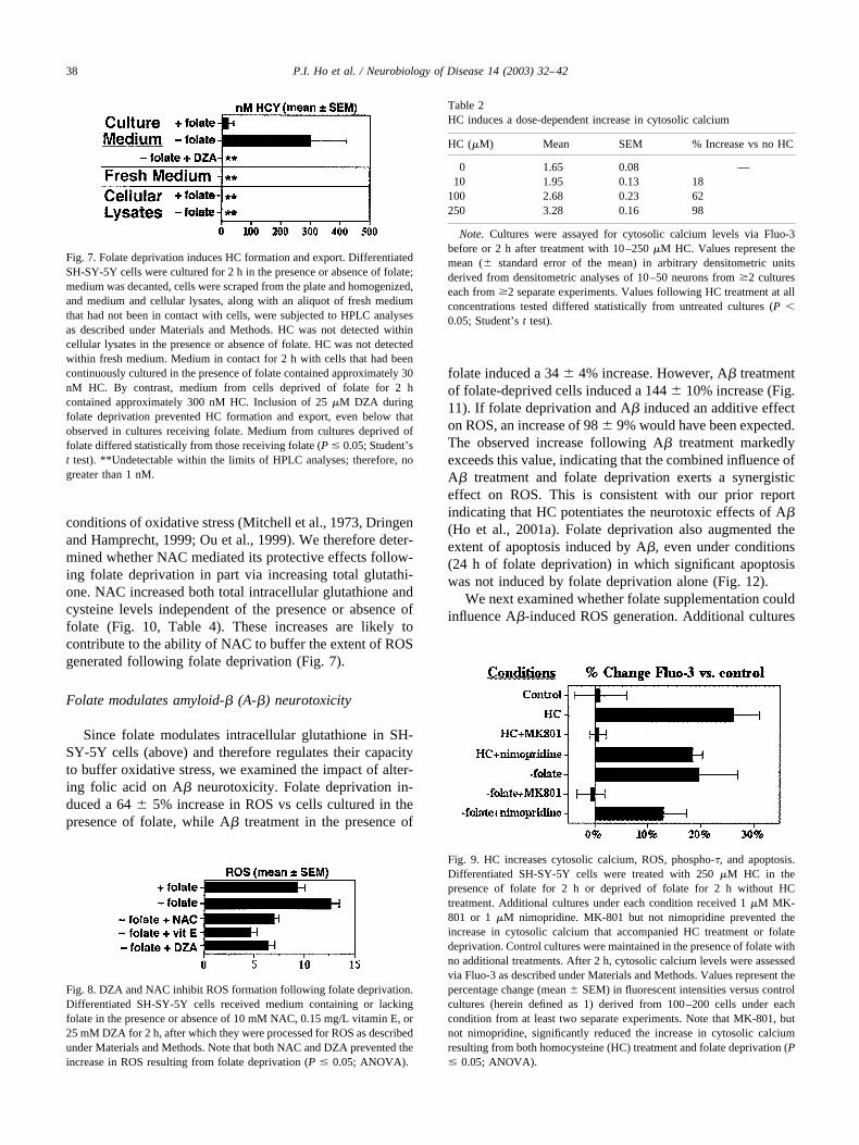
Fig. 8. DZA and NAC inhibit ROS formation following folate deprivation.Differentiated SH-SY-5Y cells received medium containing or lackingfolate in the presence or absence of 10 mM NAC, 0.15 mg/L vitamin E, or25 mM DZA for 2 h, after which they were processed for ROS as describedunder Materials and Methods. Note that both NAC and DZA prevented theincrease in ROS resulting from folate deprivation (P � 0.05; ANOVA).
Table 2HC induces a dose-dependent increase in cytosolic calcium
HC (�M) Mean SEM % Increase vs no HC
0 1.65 0.08 —10 1.95 0.13 18
100 2.68 0.23 62250 3.28 0.16 98
Note. Cultures were assayed for cytosolic calcium levels via Fluo-3before or 2 h after treatment with 10–250 �M HC. Values represent themean (� standard error of the mean) in arbitrary densitometric unitsderived from densitometric analyses of 10–50 neurons from �2 cultureseach from �2 separate experiments. Values following HC treatment at allconcentrations tested differed statistically from untreated cultures (P �0.05; Student’s t test).
Fig. 9. HC increases cytosolic calcium, ROS, phospho-�, and apoptosis.Differentiated SH-SY-5Y cells were treated with 250 �M HC in thepresence of folate for 2 h or deprived of folate for 2 h without HCtreatment. Additional cultures under each condition received 1 �M MK-801 or 1 �M nimopridine. MK-801 but not nimopridine prevented theincrease in cytosolic calcium that accompanied HC treatment or folatedeprivation. Control cultures were maintained in the presence of folate withno additional treatments. After 2 h, cytosolic calcium levels were assessedvia Fluo-3 as described under Materials and Methods. Values represent thepercentage change (mean � SEM) in fluorescent intensities versus controlcultures (herein defined as 1) derived from 100–200 cells under eachcondition from at least two separate experiments. Note that MK-801, butnot nimopridine, significantly reduced the increase in cytosolic calciumresulting from both homocysteine (HC) treatment and folate deprivation (P� 0.05; ANOVA).
Fig. 7. Folate deprivation induces HC formation and export. DifferentiatedSH-SY-5Y cells were cultured for 2 h in the presence or absence of folate;medium was decanted, cells were scraped from the plate and homogenized,and medium and cellular lysates, along with an aliquot of fresh mediumthat had not been in contact with cells, were subjected to HPLC analysesas described under Materials and Methods. HC was not detected withincellular lysates in the presence or absence of folate. HC was not detectedwithin fresh medium. Medium in contact for 2 h with cells that had beencontinuously cultured in the presence of folate contained approximately 30nM HC. By contrast, medium from cells deprived of folate for 2 hcontained approximately 300 nM HC. Inclusion of 25 �M DZA duringfolate deprivation prevented HC formation and export, even below thatobserved in cultures receiving folate. Medium from cultures deprived offolate differed statistically from those receiving folate (P � 0.05; Student’st test). **Undetectable within the limits of HPLC analyses; therefore, nogreater than 1 nM.
38 P.I. Ho et al. / Neurobiology of Disease 14 (2003) 32–42

received medium supplemented with folic acid at 2–10�the normal medium concentration in DMEM for 2 h in thepresence and absence of 20 �M A�. The A�-induced in-crease in ROS was prevented by 5� folate. In addition, 10�folate also marginally decreased ROS in both A�-treatedcells and otherwise untreated cells (Fig. 11).
Discussion
Deficiencies in folate have been clinically correlatedwith AD (Allen et al., 1998; Clarke et al., 1998; Gray, 1989;Hassing et al., 1999; Nilsson et al., 1996; Nilsson-Ehle,1998; Renvall et al., 1989; Serot et al., 2001; Snowdon etal., 2000; Wang et al., 2001; for review, see Mattson andShea, 2002). However, the direct impact of folate depriva-tion on cultured neurons and neuronal cells, and the mech-anism(s) by which it could induce neurodegeneration, hasnot been demonstrated. Our findings demonstrate that folatedeprivation induced increases in ROS, cytosolic calcium,phospho-�, and ultimate apoptosis. These findings provideevidence that folate plays a direct role in neuronal healthapart from considerations of its effect on vascular integrity.
One major mechanism by which folate deprivation is likelyto promote neurotoxicity is by extracellular accumulation ofHC (Pietrzik and Bronstrup, 1998; for review, see Shea et al.,2002). Notably, similar consequences (increased cytosolic cal-cium, ROS, and apoptosis) were observed following directaddition of HC to cultured neuronal cells both herein and inprior studies (Ho et al., 2001a; Kruman et al., 2000; White etal., 2001). As shown herein, folate deprivation induced HCaccumulation within medium, while addition of DZA pre-vented both this increase and the increased generation of ROSthat normally accompanies folate deprivation. Moreover, theincrease in cytosolic calcium that normally accompanied folatedeprivation was blocked by cotreatment with an antagonist ofthe NMDA channel, which is known to be the mechanism bywhich HC induces calcium influx (Ho et al., 2001a, and ref-erences therein). By reducing some aspects of the toxicity offolate deprivation following cotreatment with DZA or nimo-pridine, these findings confirm and extend the indication that
increased HC mediates a portion of the neurotoxicity resultingfrom folate deprivation in mice (Duan et al., 2002; Kruman etal., 2002). These findings support the likelihood that increasedHC formation and its obligate export (Blom, 2000; Christensenet al., 1991) mediate at least in part the consequences of folatedeprivation on cultured neurons and neuronal cells. Notably,since HC was undetectable within cells even following folatedeprivation, our data also indicate that HC is exported againsta concentration gradient.
Folate deprivation under the conditions utilized herein de-
Fig. 10. NAC increases cysteine and glutathione. Differentiated SH-SY-5Ycells with and without NAC treatment were subjected to HPLC along with50 �M each of thiol standards as indicated. Note the marked increase incysteine and glutathione following NAC treatment; NAC is also detected incultures where added.
Table 3Folate deprivation depletes reduced glutathione in SH-SY-5Y cells
Total glutathionea Reduced glutathione
2 h 24 h 2 h 24 h
� Folate 20.8 � 2.2b 28.5 � 2.6 19.5 � 0.9 18.4 � 0.7� Folate 20.7 � 1.7 31.6 � 2.4 13.8 � 0.6* 14.9 � 0.6*
Note. Total and reduced glutathione were determined in SH-SY-5Ylysates by HPLC and 5-chloromethylfluorescein diacetate, respectively, asdescribed under Materials and Methods.
a Nanograms per milligram of proteinb SD, n � 4 for all treatments.* Statistically different from values obtained �folate; P � 0.05, Stu-
dent’s t test.
39P.I. Ho et al. / Neurobiology of Disease 14 (2003) 32–42

pleted reduced, but not total, glutathione. It remains to bedetermined whether prolonged folate deprivation may alsodeplete total glutathione. Nevertheless, depletion in reducedglutathione indicates that folate deprivation compromised theneuron’s capacity to buffer the consequences of oxidativestress. This notion is consistent with both increased ROS gen-eration following folate deprivation and alleviation of ROS
generation by the addition of NAC, which both has antioxidantcapabilities and is a precursor for glutathione (Dringen et al.,1999; Mitchell et al., 1973). These latter findings with NACare consistent with increased HC levels as mediating the neu-rotoxicity of folate deprivation, since direct addition of HCalso increases ROS generation in cultured neurons (Ho et al.,2001a; Upchurch et al., 1997), and NAC alleviates HC-in-duced ROS generation and resultant apoptosis (Ho et al.,2001a; Huang et al., 2002). The pivotal nature of ROS gener-ation in AD neuropathology has been demonstrated by preven-tion of all downstream consequences of A� treatment of cul-tured neurons (e.g., � phosphorylation, apoptosis) by timelyapplication of antioxidants (Ekinci et al., 2000; Dhitavant et al.,2001).
Deficiencies in vitamin B12 modulate the impact of folatedeficiency on AD (Allen et al., 1998; Wang et al., 2001). B12deficiency correlates with AD and senile dementia of the Alz-heimer type (Clarke et al., 1998; McCaddon and Kelly, 1992;Levitt and Karlinsky, 1992; Regland et al., 1988, 1992; Wanget al., 2001; Wynn and Wynn, 1998a, 1998b). Like that offolate deficiency, the impact of B12 deficiency on AD is likelyto be in part due to HC accumulation. HC is normally rem-ethylated to form methionine (Hultberg et al., 1995; Kreis andGoddenow, 1978). However, the activity of the enzymes thatconvert HC to methionine (methionine synthase and 5,10-methylenetetrahydrofolate reductase) is decreased in dementia(Trolin et al., 1995) and requires B12 and folate for activity(Allen et al., 1997; Fiskerstrand et al., 1997; Jakuboswski,1997). Our findings are consistent with the suggestion thatimpaired nutrition, including concomitant deficiencies in folateand B12, in demented patients may hasten overall patientdecline (Gray, 1989). Importantly, dietary supplementation offolate is sufficient to modulate HC levels under normal con-ditions (Lucock et al., 1996) and may also be useful to lessenpotential synergistic effects on the progression of AD. Notably,our data demonstrate that supplementation with folate at five-fold more than the concentration normally present in culture
Table 4N-Acetyl cysteine increases total glutathione and cysteine levels inSHSY5Y neuroblastoma cells
Conditions NAC (�)a NAC (�)
Total GSHb Cysb Total GSH Cys
Folate (�) 28.2 � 8.7c 4.5 � 1.7 38.7 � 1.6 27.0 � 1.8Folate (�) 32.0 � 2.7 5.6 � 0.8 39.8 � 1.7 26.7 � 0.2
Note. Total cysteine and glutathione were determined in SH-SY-5Ylysates by HPLC (see Materials and Methods). Cultures were treated for24 h in the presence and absence of 10 mM NAC. Note that NAC increasedboth total glutathione and cysteine to similar respective degrees irrespec-tive of the presence or absence of folate (P � 0.05; Student’s t test).
a N-Acetylcysteine.b Nanograms per milligram of proteinc SD, n � 4 for each treatment.
Fig. 11. Folate regulates A�-induced ROS generation. Differentiated SH-SY-5Y cells received DMEM that contained normal levels of folate (con-trol; 1� folate), medium lacking folate (� folate), and medium supple-mented with folic acid at 2–10x the concentration present in DMEM withand without 20 �M A� as indicated for 2 h, after which ROS werequantified via DFCD as described under Materials and Methods. Valuesrepresent means � SEM from duplicate cultures from �50 cells fromduplicate cultures for each condition from two separate experiments; as-terisks denote statistical differences between cultures treated with and nottreated with A�. Note that folate deprivation both increased ROS andenhanced the A�-induced increase in ROS, while folate supplementationprogressively prevented A�-induced ROS. Asterisks indicate significantdifferences in ROS levels in the presence and absence of various concen-trations of folate and/or A� (P � 0.05; Student’s ANOVA with Fisher’sPLSD); lack of an asterisk indicates that values � A� were statisticallyidentical. Cultures deprived of folate and treated with A� displayed sig-nificantly (P � 0.001) increased ROS versus those deprived of folate in theabsence of A� or treated with A� in the presence of folate. Culturesreceiving 5 or 10� folate displayed statistically identical levels of ROS (P� 0.23 and 0.88, respectively) in the presence and absence of A� all ofthese cultures displayed significantly (P � 0.001) reduced ROS versuscultures receiving 1� folate without A� (ANOVA with Fisher’s PLSD).
Fig. 12. Folate regulates A�-induced apoptosis. Cortical neurons werecultured for 24 h in the presence or absence of folate and 10 �M A� asindicated and then stained with DAPI as described under Materials andMethods. The graph presents the percentage (mean � standard deviation)of apoptotic cells obtained from randomly selected microscopic fields fromduplicate cortical cultures. While folate deprivation alone did not induce astatistically significant increase in apoptotic cells within 24 h (but does soby 3–6 days; e.g., Fig. 6), treating folate-deprived neurons with A�resulted in significantly (P � 0.05) more apoptotic cells than did A�-treatment of neurons cultured in the presence of folate (ANOVA withFisher’s PLSD).
40 P.I. Ho et al. / Neurobiology of Disease 14 (2003) 32–42

medium was able to prevent oxidative stress induced by A� incultured neurons. This finding leaves open the possibility thatdietary levels of folate that may be adequate to maintain neu-ronal homeostasis under normal conditions may be inadequatefor neuronal survival under pathological conditions such as AD(Shea and Rogers, 2002a).
The findings of the present study support the clinicalassociations of folate deficiency with AD by demonstratingdirect neurotoxic consequences of folate deficiency in cul-ture and demonstrate further that folate deprivation impactsparameters of neurotoxicity associated with affected neu-rons in AD, including increasing cytosolic calcium, ROS,phospho-�, and apoptosis as well as enhancing A� neuro-toxicity. As with A� treatment, the initial influx of calciumobserved following folate deprivation is benign providedthat ROS are quenched (Ho et al., 2001a). The ability tomanipulate, and in some cases reverse, these consequencesby timely supplementation with folate or antioxidants sug-gests that such approaches may have clinical relevance inAD. This is further supported by the alleviation of oxidativedamage in transgenic mice lacking apoplipoprotein E byfolate (Ho et al., 2001b; Shea and Rogers, 2002b; Shea etal., 2002), since certain apoplipoprotein E alleles result inincreased prevalence and earlier age of onset of AD (Grow-don, 2001). In addition to dietary deficiencies in folate,congenital errors in folate metabolism, including the poten-tial impact of certain polymorphisms in 5,10-methylenetet-rahydrofolate reductase that may impair folate usage undercertain conditions, may contribute to the nature and pro-gression of AD and/or PD (Ueland et al., 2001; Regland etal., 1999; Wevers et al., 1994; Zittlun, 1995). Further stud-ies to determine the impact of folate deficiency on neuronalpathology in situ are warranted.
Acknowledgments
This research was supported by the Office of Collabora-tive Research at UMass–Lowell. We thank the anonymousreviewer for suggesting that we examine mitochondrialhealth during folate deprivation.
References
Allen, R.H., Stabler, S.P., Lindenbaum, J., 1998. Eur. J. Pediatr. 157,S122–S126.
Araki, A., Sako, Y., 1987. Determination of free and total homocysteine inhuman plasma by high-performance liquid chromatography with fluo-rescence detection. J. Chromatogr. 422, 43–52.
Bell, I.R., Edman, J.S., Selhub, J., Morrow, F.D., Marby, D.W., Kayne,H.L., Cole, J.O., 1992. Plasma homocysteine in vascular disease andnonvascular dementia of depressed elderly people. Acta Psychiat.Scand. 86, 386–390.
Blom, H.J., 2000. Consequences of homocysteine export and oxidation inthe vascular system. Semin. Thromb. Hemost. 26, 227–232.
Christensen, B., Refsum, H., Vintermyr, O., Ueland, P.M., 1991. Homo-cysteine export from cells cultured in the presence of physiological or
superfluous levels of methionine: methionine loading or non-trans-formed, transformed, proliferating, and quiescent cells in culture.J. Cell Physiol. 146, 52–62.
Clarke, R., Smith, A.D., Jobst, K.A., Refsum, H., Sutton, L., Ueland, P.M.,1998. Folate, vitamin B12, and serum total homocysteine levels inconfirmed Alzheimer disease. Arch. Neurol. 55, 1449–1455.
Collins, J.A., Schandi, C.A., Young, K.K., Vesely, J., Willingham, M.C.,1997. Major DNA fragmentation is a late event in apoptosis. J. Histo-chem. Cytochem. 45, 923–934.
Dhitavant, S., Rogers, E., Shea, T.B., 2001. Differential efficacy of li-pophilic and cytosolic antioxidants on generation of reactive oxygenspecies by amyloid-beta. J. Alz. Dis. 3, 525–529.
Dringen, R., Hamprecht, 1999. N-Acetylcysteine, but not methionine or2-oxothiazolidine-4-carboxylate, serves as cysteine donor for the syn-thesis of glutathione in cultured neurons derived from embryonal ratbrain. Neurosci. Lett. 259, 79–82.
Duan, W., Ladenheim, B., Cutler, R.G., Kruman, I.I., Cadet, J.L., Mattson,M.P., 2002. Dietary folate defeciency and elevated homocysteine levelsendanger dopaminergic neurons in models of Parkinson’s disease.J. Neurochem. 80, 101–110.
Ekinci, F.J., Shea, T.B., 2000. Hyperphosphorylation of tau does notcorrelate with neurodegeneration in cortical neurons in culture. J. Alz.Dis. 2, 7–12.
Ekinci, F.J., Linsley, M.-D., Shea, T.B., 2000. Beta-Amyloid-inducedcalcium influx induces apoptosis in culture by oxidative stress ratherthan tau phosphorylation. Mol. Brain Res. 76, 389–395.
Ekinci, F.J., Malik, K.M., Shea, T.B., 1999. Beta-Amyloid induces calciuminflux and neurodegeneration by MAP kinase-mediated activation ofthe L voltage-sensitive calcium channel. J. Biol. Chem. 274, 30322–30327.
Fiskerstrand, T., Ueland, P.M., Refsum, H., 1997. Folate depletion inducedby methotrexate affects methionine synthase activity and its suscepti-bility to inactivation by nitrous oxide. J. Pharmacol. Exp. Ther. 282,1305–1311.
Gray, G.E., 1989. Nutrition and dementia. J. Am. Diet Assoc. 89, 1795–1802.
Gray, C.W., Patel, A.J., 1995. Neurodegeneration mediated by glutamateand beta-amyloid peptide: a comparison and possible interaction. BrainRes. 691, 169–179.
Growdon, J., 2001. Incorporating biomarkers into clinical drug trials inAlzheimer’s disease. J. Alz. Dis. 3, 287–292.
Hassing, L., Wahlin, A., Winblad, B., Backman, L., 1999. Further evidenceon the effects of vitamin B12 and folate levels on episodic memoryfunctioning: a population-based study of healthy very old adults. Biol.Psychiatry 45, 1472–1480.
Hatanaka, Y., Suzuki, K., Kawasaki, Y., Endo, Y., Taniguchi, N., Takei,N., 1996. A role of peroxides in Ca ionophore-induced apoptosis incultured rat cortical neurons. Biochem. Biophys. Res. Commun. 227,513–518.
Ho, P., Collins, S.C., Dhitavat, S., Ortiz, D., Ashline, D., Rogers, E., Shea,T.B., 2001a. Homocysteine potentiates beta-amyloid neurotoxicity:role of oxidative stress. J. Neurochem. 78, 249–253.
Ho, P., Ashline, D., Dhitavat, S., Ortiz, D., Collins, S.C., Rogers, E., Shea,T.B., 2001b. Folate deprivation induces neurodegeneration: roles ofoxidative stress and increased homocysteine. Mol. Biol. Cell 12, 120a.
Ho, P.I., Ortiz, D., Rogers E., Shea, T.B., 2002. Multiple aspects ofhomocysteine neurotoxicity: glutamate excitotoxicity, kinase hyperac-tivation and DNA damage. J. Neurosci. Res., in press.
Huang, H.M., Ou, H.C., Hsieh, S.J., 2000. Antioxidants prevent amyloidpeptide-induced apoptosis and alteration of calcium homeostasis incultured cortical neurons. Life Sci. 66, 1879–1892.
Huang, R.F., Huang, H.M., Lin, B.S., Hung, C.Y., Lu, H.T., 2002. N-Acetylcysteine, vitamin C and vitamin E diminish homocysteine thio-lactone-induced apoptosis in human promyeloid HL-60 cells. J. Nutr.132, 2151–2156.
Hultberg, B., Andersson, A., Isaksson, A., 1995. Metabolism of homocys-teine, its relation to the other cellular thiols and its mechanism of cell
41P.I. Ho et al. / Neurobiology of Disease 14 (2003) 32–42

damage in a cell culture line (human histiocytic cell line U-937).Biochim. Biophys. Acta 1269, 6–12.
Jakubowski, H., 1997. Metabolism of homocysteine thiolactone in humancell cultures. Possible mechanism for pathological consequences ofelevated homocysteine levels. J. Biol. Chem. 272, 1935–1942.
Jeong, S.-Y., Ahn, S.-G., Lee, J.-H., Kim, H.-S., Kim, J.-W., Rhim, H.,Jeong, S.-W., Kim, I.-K., 1999. 3-Deazaadenosine, S-adenosylhomo-cysteine hydrolase inhibitor, has dual effects on NF-kB regulation.J. Biol. Chem. 274, 18981–18988.
Kreis, W., Goodenow, M., 1987. Methionine requirement and replacementby homocysteine in tissue cultures of selected rodent and humanmalignant and normal cells. Cancer Res. 2259–2262.
Kruman, I.I., Culmsee, C., Chan, S., Kruman, Y., Guo, Z., Penix, L.,Mattson, M.P., 2000. Homocysteine elicits a DNA damage response inneurons that promotes apoptosis and hypersensitivity to excitotoxicity.J. Neurosci. 20, 6920–6926.
Kruman, I.I., Kumaravel, T.S., Lohani, A., Pedersen, W.A., Cutler, R.G.,Kruman, Y., Haughey, N., Lee, J., Evans, M., Mattson, M.P., 2002.Folic acid deficiency and homocysteine impair DNA repair in hip-pocampal neurons and sensitize them to amyloid toxicity in experi-mental models of Alzheimer’s disease. J. Neurosci. 22, 1752–1762.
Levitt, A.J., Karlinsky, H., 1992. Folate, vitamin B12 and cognitive im-pairment in patients with Alzheimer’s disease. Acta Psychiatr. Scand.86, 301–305.
Lucock, M.D., Daskalakis, I.G., Wild, J., Anderson, A., Schorah, C.J.,Lean, M.E.J., Levene, M.I., 1996. The influence of dietary folate andmethionine on the metabolic disposition of endotoxic homocysteine.Biochem. Mol. Med. 59, 104–111.
Mattson, M.M., Shea, T.B., 2002. Folate and homocysteine in neuralplasticity and neurodegenerative disorders. Trends Neurosci., in press.
McCaddon, A., Kelly, C.L., 1992. Familial Alzheimer’s disease and vita-min B12 deficiency. Age Ageing 23, 334–337.
Miller, J.W., 2000. Homocysteine, Alzheimer’s disease and cognitive func-tion. Nutrition 16, 675–677.
Mitchell, J.R., Jollow, D.J., Potter, W.Z., Gillette, J.R., Brodie, B.B., 1973.Acetaminophen-induced hepatic necrosis. IV. Protective role of gluta-thione. J. Pharmacol. Exp. Ther. 187, 211–217.
Mosharov, E., Cranfored, M.R., Banerjee, R., 2000. The quantitativelyimportant relationship between homocysteine metabolism and glutathi-one synthesis by the transsulfuration pathway and its regulation byredox changes. Biochemistry 39, 13005–13011.
Nilsson, K., Gustafson, L., Faldt, R., Anderson, A., Hultberg, B., 1994.Plasma homocysteine in relation to serum cobalamin and blood folatein a psychogeriatric population. Eur. J. Clin. Invest. 24, 600–606.
Nilsson-Ehle, H., 1998. Age-related changes in cobalamin (vitamin B12)handling. Implications for therapy. Drugs Aging 12, 277–292.
Oliveri, G., Baysang, G., Meier, F., Muller-Spahn, F., Stahelin, H.B.,Brockhaus Brack, C.H., 2001. N-Acetyl-L-cysteine protects SHSY5Yneuroblastoma cells from oxidative stress and cell cytotoxicity: effectson �-amyloid secretion and tau phosphorylation. J. Neurochem. 76,224–233.
Ou, Y.C., White, C.C., Krejsa, C.M., Ponce, R.A., Kavanaugh, T.J., Faust-man, E.M., 1999. The role of intracellular glutathione in methylmer-cury-induced toxicity in embryonic neuronal cells. Neurotoxicology20, 793–804.
Pietrzik, K., Bronstrup, A., 1998. Vitamins B12, B6 and folate as deter-minants of homocysteine concentration in the healthy population. Eur.J. Pediatr. 157, S135–S138.
Reers, M., Smith, T.W., Chen, L.B., 1991. Aggregate formation of acarbocyanide as a quantitative fluorescent indicator of membrane po-tential. Biochemistry 30, 4480–4486.
Regland, B., Blennow, K., Germgard, T., Kock-Schmidt, A.-C., Gottfries,C.-G., 1999. The role of the polymorphic genes apolipoprotein E andmethylene-tetrahydrofolate reductase in the development of dementiaof the Alzheimer type. Dement. Geriatr. Cogn. Disord. 10, 245–251.
Regland, B., Gottfries, C.G., Oreland, L., Svennerholm, L., 1988. Low B12levels related to high activity of platelet MAO in patients with dementiadisorders. A retrospective study. Acta Psychiatr. Scand. 78, 451–457.
Regland, B., Abrahamsson, L., Blennow, K., Gottfries, C.G., Wallin, A.,1992. Vitamin B12 in CSF: reduced CSF/serum B12 ratio in dementedmen. Acta Neurol. Scand. 85, 276–281.
Renvall, M.J., Spindler, A.A., Ramsdell, J.W., Paskvan, M., 1989. Nutri-tional status of free-living Alzheimer’s patients. Am. J. Med. Sci. 298,20–27.
Rimon, G., Bazenet, C.E., Philpott, K.L., Rubin, L.L., 1997. Increasedsurface phosphatidylserine is an early marker of neuronal apoptosis.J. Neurosci. Res. 48, 563–570.
Schulz, J.B., Kindenau, J., Dichgans, J., 2000. Glutathione, oxidative stressand neurodegeneration. Eur. J. Biochem. 267, 4904–4911.
Serot, J.M., Christman, D., Dubost, T., Bene, M.C., Faure, G.C., 2001.CSF-folate levels are decreased in late-onset AD patients. J. NeuralTransm. 108, 93–99.
Shea, T.B., Rogers, E., 2002a. Homocysteine as a risk factor for Alzhei-mer’s disease. N. Engl. J. Med. 346, 2007.
Shea, T.B., Rogers, E., 2002b. Folate quenches oxidative damage in brainsof apolipoprotein E-deficient mice: augmentation by vitamin E. Mol.Brain Res., in press.
Shea, T.B., Lyons-Wieler, J., Rogers, E., 2002. Homocysteine, folatedeprivation and Alzheimer’s disease. J. Alz. Dis., in press.
Snowdon, D.A., Tully, C.L., Smith, C.D., Riley, D., Markesbery, W., 2000.Serum folate and the severity of atrophy of the neocortex in Alzheimerdisease: findings from the Nun Study. Am. J. Clin. Nutr. 71, 993–998.
Trolin, C.G., Regland, B., Oreland, L., 1995. Decreased methionine ad-enosyltransferase activity in erythrocytes of patients with dementiadisorders. Eur. Neuropsychopharmacol. 5, 107–114.
Ueda, K., Shinohara, S., Yagami, T., Asakura, K., Kawasaki, K., 1997.Amyloid beta protein potentiates Ca2� influx through L-type voltage-sensitive Ca2� channels: a possible involvement of free radicals.J. Neurochem. 68, 265–271.
Ueland, P.M., Hustad, S., Schneede, J.M., Refsum, H., Vollset, S.E., 2001.Biological and clinical implications of the MTHFR C677T polymor-phism. Trends Pharm. Sci. 22, 195–201.
Upchurch, G.R., Welch, G.N., Fabian, A.J., Freedman, J.E., Johnson, J.L.,Keaney, J.F., Loscalzo, J., 1997. Homocysteine decreases bioavailablenitric oxide by a mechanism involving glutathione peroxidase. J. Biol.Chem. 272, 17012–17017.
Wadia, J.S., Chalmers-Redman, R.M., Ju, W.J., Carlile, G.W., Phillips,J.L., Fraser, A.D., Tatton, W.G., 1998. Mitochondrial membrane po-tential and nuclear changes in apoptosis caused by serum and nervegrowth factor withdrawal: time course and modification by (�)-depre-nyl. J. Neurosci. 18, 932–947.
Wang, H.-X., Wahlin, A., Basun, H., Fastom, J., Winblad, B., Fratiglioni,L., 2001. Vitamin B12 and folate in relation to the development ofAlzheimer’s disease. Neurology 56, 1188–1194.
Wevers, R.A., Hansen, S.I., Hubar, J.L.MvH., Holm, J., Hoier-Madsen, M.,Jongen, P.J.H., 1994. Folate deficiency in cerebrospinal fluid associatedwith a defect in folate binding protein in the centra nervous system.J. Neurol. Neurosurg. Psych. 57, 223–226.
White, A.R., Huang, X., Jobling, M.F., Barrow, C.H., Beyreuther, K.,Masters, C.L., Bush, A.I., Cappai, R., 2001. Homocysteine potentiatescopper- and amyloid beta peptide-mediated toxicity in primary cul-tures: possible risk factors in the Alzheimer’s-type neurodegenerativepathways. J. Neurochem. 76, 1509–1520.
Wynn, M., Wynn, A., 1998a. The danger of B12 deficiency in the elderly.Nutr. Health 12, 215–226.
Wynn, M., Wynn, A., 1998b. Fortification of grain products with folate:should Britain follow the American example? Nutr. Health 12, 147–161.
Zuttiun, J., 1995. Congenital effors of folate metabolism. Baillierels Clin.Haematol. 8, 603–616.
42 P.I. Ho et al. / Neurobiology of Disease 14 (2003) 32–42
Copyright © 2022 FDOKUMEN