Translational upregulation of folate receptors is mediated by homocysteine via RNA-heterogeneous...
17
The Journal of Clinical Investigation | January 2004 | Volume 113 | Number 2 285 Introduction Folates are critical for the perpetuation of one-carbon metabolism and DNA synthesis (1), and folate recep- tors (FRs) have been shown to mediate the cellular uptake of the physiological folate 5-methyltetrahydro- folate in several malignant and normal cells (reviewed in ref. 2). The native membrane-associated FR glyco- protein of cervical cancer (HeLa-IU 1 ) cells is hydropho- bic and binds a significant amount of Triton X-100 detergent upon solubilization (3). This hydrophobici- ty is conferred primarily by its glycosylphosphatidyli- nositol (GPI)-anchored tail, which can be cleaved off by GPI-specific phospholipase C and/or D to give rise to a hydrophilic species. Alternatively, endoproteolytic cleavage of the GPI anchor and the C-terminal end of the FR by an endogenous membrane-associated metal- loprotease can also yield a soluble hydrophilic species (2, 4). Although FRs are upregulated in folate deficien- cy and downregulated with repletion of folate (5–12), the molecular and biochemical link between folate defi- ciency and upregulation of FRs is not clear. When a cervical carcinoma cell line (HeLa-IU 1 ) that was stably propagated in 2.3 μM folate (called HeLa- IU 1 -HF cells here, for “high folate”) was adapted to growth in low physiological concentrations of folate (9 nM) (called HeLa-IU 1 -LF cells here, for “low folate”), the cells exhibited biochemical evidence of folate deficien- cy and FRs were markedly upregulated ninefold. How- ever, the FR mRNA was disproportionately low (being increased by only twofold), suggesting that upregula- tion of FRs was a function of translational or post- translational alterations in FR metabolism. Previously we demonstrated that the interaction of an 18-base cis- element in the 5′-untranslated region (5′-UTR) of FR-α isoform mRNA with a cytosolic trans-factor is critical for translational upregulation of FRs (13). More recent- ly, we determined that the cytosolic FR mRNA–binding trans-factor is identical to the 43-kDa heterogeneous nuclear ribonucleoprotein E1 (hnRNP E1) (14). HnRNP E1 is abundant in reticulocyte lysates (15), so the addition of recombinant hnRNP E1 to the in vitro translation mixture does not increase FR translation. However, anti–hnRNP E1 specifically inhibited trans- Received for publication October 13, 2000, and accepted in revised form October 21, 2003. Address correspondence to: A´ sok C. Antony, Indiana Cancer Research Institute, R4-266, 1044 West Walnut Street, Indianapolis, Indiana 46202-5121, USA. Phone: (317) 274-3589; Fax: (317) 274-0396; E-mail: [email protected]. Conflict of interest: The authors have declared that no conflict of interest exists. Nonstandard abbreviations used: folate receptor (FR); glycosylphosphatidylinositol (GPI); high folate (-HF); low folate (-LF); untranslated region (UTR); heterogeneous nuclear ribonucleoprotein E1 (hnRNP E1); pteroylglutamate (PteGlu); Dulbecco’s PBS (D-PBS); chloramphenicol acetyltransferase (CAT); with a cis-element (+cis); lacking a cis-element (–cis). Translational upregulation of folate receptors is mediated by homocysteine via RNA-heterogeneous nuclear ribonucleoprotein E1 interactions A´ sok C. Antony, 1 Ying-Sheng Tang, 1 Rehana A. Khan, 1 Mangatt P. Biju, 1 Xiangli Xiao, 1 Qing-Jun Li, 1 Xin-Lai Sun, 1 Hiremagalur N. Jayaram, 2 and Sally P. Stabler 3 1 Division of Hematology-Oncology, Department of Medicine, and 2 Department of Biochemistry and Molecular Biology, Indiana University School of Medicine, and the Richard L. Roudebush Veterans Affairs Medical Center, Indianapolis, Indiana, USA 3 Division of Hematology, Department of Medicine, University of Colorado Health Sciences Center, Denver, Colorado, USA Cellular acquisition of folate is mediated by folate receptors (FRs) in many malignant and normal human cells. Although FRs are upregulated in folate deficiency and downregulated following folate repletion, the mechanistic basis for this relationship is unclear. Previously we demonstrated that interaction of an 18-base cis-element in the 5′-untranslated region of FR mRNA and a cystolic trans- factor (heterogeneous nuclear ribonucleoprotein E1 [hnRNP E1]) is critical for FR synthesis. How- ever, the molecular mechanisms controlling this interaction, especially within the context of FR reg- ulation and folate status, have remained obscure. Human cervical carcinoma cells exhibited progressively increasing upregulation of FRs after shifting of folate-replete cells to low-folate media, without a proportionate rise in FR mRNA or rise in hnRNP E1. Translational FR upregulation was accompanied by a progressive accumulation of the metabolite homocysteine within cultured cells, which stimulated interaction of the FR mRNA cis-element and hnRNP E1 as well as FR biosynthesis in a dose-dependent manner. Abrupt reversal of folate deficiency also led to a rapid parallel reduc- tion in homocysteine and FR biosynthesis to levels observed in folate-replete cells. Collectively, these results suggest that homocysteine is the key modulator of translational upregulation of FRs and establishes the linkage between perturbed folate metabolism and coordinated upregulation of FRs. J. Clin. Invest. 113:285–301 (2004). doi:10.1172/JCI200411548.
Transcript of Translational upregulation of folate receptors is mediated by homocysteine via RNA-heterogeneous...
The Journal of Clinical Investigation | January 2004 | Volume 113 | Number 2 285
IntroductionFolates are critical for the perpetuation of one-carbonmetabolism and DNA synthesis (1), and folate recep-tors (FRs) have been shown to mediate the cellularuptake of the physiological folate 5-methyltetrahydro-folate in several malignant and normal cells (reviewedin ref. 2). The native membrane-associated FR glyco-protein of cervical cancer (HeLa-IU1) cells is hydropho-bic and binds a significant amount of Triton X-100detergent upon solubilization (3). This hydrophobici-ty is conferred primarily by its glycosylphosphatidyli-nositol (GPI)-anchored tail, which can be cleaved off byGPI-specific phospholipase C and/or D to give rise to ahydrophilic species. Alternatively, endoproteolyticcleavage of the GPI anchor and the C-terminal end of
the FR by an endogenous membrane-associated metal-loprotease can also yield a soluble hydrophilic species(2, 4). Although FRs are upregulated in folate deficien-cy and downregulated with repletion of folate (5–12),the molecular and biochemical link between folate defi-ciency and upregulation of FRs is not clear.
When a cervical carcinoma cell line (HeLa-IU1) thatwas stably propagated in 2.3 µM folate (called HeLa-IU1-HF cells here, for “high folate”) was adapted togrowth in low physiological concentrations of folate (9nM) (called HeLa-IU1-LF cells here, for “low folate”), thecells exhibited biochemical evidence of folate deficien-cy and FRs were markedly upregulated ninefold. How-ever, the FR mRNA was disproportionately low (beingincreased by only twofold), suggesting that upregula-tion of FRs was a function of translational or post-translational alterations in FR metabolism. Previouslywe demonstrated that the interaction of an 18-base cis-element in the 5′-untranslated region (5′-UTR) of FR-αisoform mRNA with a cytosolic trans-factor is criticalfor translational upregulation of FRs (13). More recent-ly, we determined that the cytosolic FR mRNA–bindingtrans-factor is identical to the 43-kDa heterogeneousnuclear ribonucleoprotein E1 (hnRNP E1) (14).HnRNP E1 is abundant in reticulocyte lysates (15), sothe addition of recombinant hnRNP E1 to the in vitrotranslation mixture does not increase FR translation.However, anti–hnRNP E1 specifically inhibited trans-
Received for publication October 13, 2000, and accepted in revised formOctober 21, 2003.
Address correspondence to: Asok C. Antony, Indiana CancerResearch Institute, R4-266, 1044 West Walnut Street,Indianapolis, Indiana 46202-5121, USA. Phone: (317) 274-3589;Fax: (317) 274-0396; E-mail: [email protected] of interest: The authors have declared that no conflict ofinterest exists.Nonstandard abbreviations used: folate receptor (FR);glycosylphosphatidylinositol (GPI); high folate (-HF); low folate (-LF); untranslated region (UTR); heterogeneous nuclearribonucleoprotein E1 (hnRNP E1); pteroylglutamate (PteGlu);Dulbecco’s PBS (D-PBS); chloramphenicol acetyltransferase(CAT); with a cis-element (+cis); lacking a cis-element (–cis).
Translational upregulation of folate receptors is mediatedby homocysteine via RNA-heterogeneous nuclearribonucleoprotein E1 interactions
Asok C. Antony,1 Ying-Sheng Tang,1 Rehana A. Khan,1 Mangatt P. Biju,1 Xiangli Xiao,1
Qing-Jun Li,1 Xin-Lai Sun,1 Hiremagalur N. Jayaram,2 and Sally P. Stabler3
1Division of Hematology-Oncology, Department of Medicine, and2Department of Biochemistry and Molecular Biology, Indiana University School of Medicine, and the Richard L. RoudebushVeterans Affairs Medical Center, Indianapolis, Indiana, USA
3Division of Hematology, Department of Medicine, University of Colorado Health Sciences Center, Denver, Colorado, USA
Cellular acquisition of folate is mediated by folate receptors (FRs) in many malignant and normalhuman cells. Although FRs are upregulated in folate deficiency and downregulated following folaterepletion, the mechanistic basis for this relationship is unclear. Previously we demonstrated thatinteraction of an 18-base cis-element in the 5′-untranslated region of FR mRNA and a cystolic trans-factor (heterogeneous nuclear ribonucleoprotein E1 [hnRNP E1]) is critical for FR synthesis. How-ever, the molecular mechanisms controlling this interaction, especially within the context of FR reg-ulation and folate status, have remained obscure. Human cervical carcinoma cells exhibitedprogressively increasing upregulation of FRs after shifting of folate-replete cells to low-folate media,without a proportionate rise in FR mRNA or rise in hnRNP E1. Translational FR upregulation wasaccompanied by a progressive accumulation of the metabolite homocysteine within cultured cells,which stimulated interaction of the FR mRNA cis-element and hnRNP E1 as well as FR biosynthesisin a dose-dependent manner. Abrupt reversal of folate deficiency also led to a rapid parallel reduc-tion in homocysteine and FR biosynthesis to levels observed in folate-replete cells. Collectively, theseresults suggest that homocysteine is the key modulator of translational upregulation of FRs andestablishes the linkage between perturbed folate metabolism and coordinated upregulation of FRs.
J. Clin. Invest. 113:285–301 (2004). doi:10.1172/JCI200411548.
286 The Journal of Clinical Investigation | January 2004 | Volume 113 | Number 2
lation of FRs in vitro in a dose-dependent manner, andthese effects could be reversed in a dose-dependentmanner by either purified human placental trans-factoror hnRNP E1. These results confirmed the critical roleof the interaction of cis-element and hnRNP E1 in thetranslation of FRs (14). The recent identification of asignificant correlation between the expression of FRsand hnRNP E1 in the cervixes of normal women andthose with cervical dysplasia and cervical cancer furthersupports the idea of a physiologic relationship (16).However, the obvious “missing link” to a more com-prehensive understanding of the translational regula-tion of FRs (via involvement of these components) infolate deficiency was the precise trigger(s) for the inter-action of FR mRNA cis-element and the hnRNP E1resulting in upregulation of FRs.
This paper describes investigations into the func-tional role of this RNA-protein interaction in the regu-lation of FRs in HeLa-IU1 cells within the context offolate homeostasis. We report that the marked upreg-ulation of FRs observed in folate deficiency is mediat-ed at the translational level by the metabolite homo-cysteine, which accumulates in folate deficiency andincreases the interaction of the cis-element and hnRNPE1, which, in turn, results in increased biosynthesis ofFRs. To our knowledge, this is the first time that amechanistic linkage between substrate build-up fromperturbed folate metabolism secondary to nutritionalfolate deficiency and the coordinated upregulation ofa protein that is integrally involved in folate metabo-lism has been characterized at the translational level.
MethodsMaterials. Nondialyzed and dialyzed fetal bovine serum,rabbit nonimmune serum, and other chemicals of thehighest analytical grade were from Sigma Chemical Co.(St. Louis, Missouri, USA). The random hexanucleotideprimer kit for all radiolabeled DNA probes (specificactivity, ∼0.3 mCi/µg DNA) and QuickChange Site-Directed Mutagenesis Kit were from Stratagene (La Jolla,California, USA). The β-actin DNA was from Oncor(Gaithersburg, Maryland, USA). The plasmid pc32, apUC 8 plasmid containing full-length human FR-αcDNA, was a gift from P.C. Elwood (National CancerInstitute, Medicine Branch, NIH, Bethesda, Maryland,USA) (17). All oligodeoxynucleotides were synthesizedby Invitrogen (San Diego, California, USA). The primersfor RT-PCR of FR cDNA were synthesized based on dataprovided (18). The [3,5,7′,9′-3H]pteroylglutamate([3,5,7′,9′-3H]PteGlu) potassium salt (specific activity,∼50 Ci/mmol); [α-32P]UTP (specific activity, >800Ci/mmol); and [35S]cysteine (specific activity, >1,000Ci/mmol) were from Amersham (Arlington Heights, Illi-nois, USA). IgGsorb, a 10% suspension of formalin-fixedStaphylococcus aureus cells bearing protein A, was fromThe Enzyme Center (Boston, Massachusetts, USA).
Cell culture. The addition of nondialyzed fetal bovineserum to a final concentration of 10% in folate-freeminimum essential media (MEM) (Gibco-BRL,
Gaithersburg, Maryland, USA) led to a final extracellu-lar concentration of 5-methyl-tetrahydrofolate of 9 nM;this media is referred to as MEM-LF. HeLa-IU1 cells (3)cultured at 37°C in 5% CO2 and propagated in MEMcontaining 10% nondialyzed fetal bovine serum and 2.3µM PteGlu (this media is referred to as MEM-HF) weredesignated HeLa-IU1-HF cells. HeLa-IU1-LF cells werederived from HeLa-IU1-HF cells that were abruptlytransferred to MEM-LF and stably adapted to growthin this media for over 6 months.
Analysis of metabolites related to folate metabolism in thegrowth media and in cells. The supernatant of HeLa-IU1-HF cells (1.5 × 10⁷) propagated in low-folate media(MEM-LF) over several weeks was assayed for variousmetabolites as follows: Briefly, cells that were plated in100- × 20-mm dishes were fed twice a week with freshMEM-LF. By the end of each week, growth media incontact with cells for 3 days was aspirated and frozen(–80°C). The concentration of total homocysteine,methionine, total cysteine, and cystathionine wasmeasured in these samples by gas chromatography–mass spectrometry (19). S.P. Stabler was “blinded” tosample identity for all experiments by the use of codedsamples (University of Colorado Health Sciences Cen-ter, Denver, Colorado, USA). In preliminary studies, weconsistently failed to measure intracellular homocys-teine concentrations in HeLa-IU1-HF and HeLa-IU1-LFcells after washing the cells with large volumes of bufferin order to separate homocysteine in the supernatantfrom cells. These results were consistent with theknown propensity of homocysteine to rapidly moveout of cells (20). Accordingly, efflux of homocysteinewas minimized first by release of adherent cells with arubber policeman (into the growth media used by thecells) and then by centrifugation of the suspension oversilicone fluid to separate the media from cells. Thisapproach has been validated as a very efficient methodfor the separation of cells from an aqueous phase (21,22); thus, when [14C]inulin is added to a cell suspensionthat is centrifuged through a silicone matrix, less than0.01% of [14C]inulin contaminates the cell pellet. TheHeLa-IU1-HF and HeLa-IU1-LF cell pellets were thenlysed and analyzed for homocysteine, methionine, cys-teine, and cystathionine concentrations (19). Briefly,7 × 105 HeLa-IU1-HF cells were seeded separately in100- × 20-mm Falcon tissue culture dishes (BectonDickinson, Franklin Lakes, New Jersey, USA) in MEM-LFand were propagated for various times (up to 12 weeks)in this medium. By the end of each week, cell samplesthat had been uniformly fed with fresh MEM-LF 3 daysprior to the day of harvest were analyzed for cell num-ber, protein, and intracellular homocysteine. Accord-ingly, four dishes from each group were randomlyselected and the number of cells was determined aftertrypsin digestion. The adherent cells from another fourdishes from each group were released by being scrapedwith a rubber policeman and cells were resuspended inthe same (“spent”) growth medium. After being trans-ferred to preweighed sterile 15-ml polypropylene coni-
The Journal of Clinical Investigation | January 2004 | Volume 113 | Number 2 287
cal Falcon tubes, cells were centrifuged at 1,000 g for 10minutes. After aspiration of the supernatant, the pelletwas digested in 25 µl of 1 N NaOH overnight at 37°C andthe protein content was determined. Cells from anotherfour dishes were also similarly scraped, resuspended inspent medium, and immediately applied to the tops of15-ml Falcon tubes containing 5 ml of Versilube F50 sil-icone fluid (General Electric, Waterford, New York, USA).After centrifugation (2,400 g for 10 minutes), the mediawas aspirated and tubes were immediately frozen on dryice for 30 minutes to solidify the oil. The cell pellet wascut out from the bottom of the tube and resuspended in500 µl of fresh growth medium in an Eppendorf tube andsubjected to four cycles of freezing and thawing (eachcycle consisted of 5 minutes of freezing on dry ice fol-lowed by 20 minutes of thawing at 22°C). The releasedintracellular contents were separated from other celldebris by centrifugation at 12,000 g for 10 minutes at4°C, and the supernatant was filtered through a 0.22-µmfilter and analyzed for homocysteine, methionine, cys-teine, and cystathionine. The concentration of homocys-teine, methionine, cysteine, and cystathionine in 500 µlof fresh medium was also measured and this value wassubtracted from the concentration of these thiols deter-mined in spent medium and the cell pellet as a functionof time (see Results). The data on homocysteine wereexpressed as micromolar and nanomoles per milligramof protein. These experiments were repeated four differ-ent times and the data were expressed as the mean ± SD.In other experiments, the extent of intracellular accumu-lation of homocysteine following exposure of HeLa-IU1-HF cells to 500 µM and 1000 µM of homocysteine wasalso determined after rapid separation over silicone. Forthe experiments that used pharmacological concentra-tions of homocysteine, an additional control was includ-ed in which each concentration of homocysteine in vehi-cle was also applied to the silicone column (in the absenceof cells) and the amount of homocysteine recovered inthe “cell pellet” fraction was measured. Because this valueat each concentration represented nonspecific passage ofhomocysteine through silicone, it was subtracted fromthe values from studies using cells.
Radiolabeled folic acid (PteGlu) binding studies. Binding ofradiolabeled [3H]PteGlu to intact cells on each weekduring long-term culture (Figure 1) was carried out asdescribed (23), with the following modifications. Cells(2 × 10⁶) were cultured in T-75 flasks to 80% confluence,and endogenous (FR-bound) folate was removed bywashing cells using the following buffers in sequence (at4°C): 10 ml Dulbecco’s PBS (D-PBS), four washes; 10 ml0.05 M sodium acetate-acetic acid (pH 4.5) containing0.15 M NaCl (30 seconds per wash), two washes; 0.01 Msodium phosphate containing 0.15 M NaCl (pH 7.4),one wash. After cells were released by being incubatedwith isotonic trypsin–EDTA (Gibco BRL) for 5 minutesat 37°C, they were washed twice in 10 ml D-PBS.
Binding of radioligand to FRs on intact cells wasdetermined at 4°C in 15-ml sterile tubes by incuba-tion of 1 × 10⁶ cells with varying concentrations of
[3H]PteGlu (0.0625–128 nM) in the absence or presenceof a 1,000-fold excess of unlabeled folic acid for 1 hour(equilibrium binding time at 4°C) in a final volume of 1ml D-PBS. The cells were washed four times with 10 mlD-PBS. After the cell pellet containing bound [3H]PteGluwas digested in 50 µl of 1 N NaOH overnight, the sam-ple was analyzed by liquid scintillation spectrometry.Specific [3H]PteGlu binding was determined by sub-tracting the value of nonspecific [3H]PteGlu binding(obtained in duplicate samples containing 1,000-foldexcess of unlabeled folic acid) from total [3H]PteGlubinding (determined from samples containing radiola-beled [3H]PteGlu alone). Nonspecific [3H]PteGlu bind-ing was less than 10% of total binding in all experiments.The total number of FRs per cell by the end of each weekin long-term culture in low-folate medium (Figure 1) wascalculated based on Scatchard analysis of the data. Eachdata point represents four independent experiments.
Western blot analysis. Cells were lysed overnight in abuffer containing 2% SDS, 20 mM EDTA, 1 µg/ml apro-tinin, and 1 mM PMSF (all from Sigma). Fifteen micro-grams of total protein was analyzed by 4–20% SDS-PAGE followed by Western blotting. The proteinstransferred to PVDF membranes were first incubatedwith rabbit polyclonal anti–human placental FR anti-serum (dilution, 1:1500) overnight at 4°C, followed byincubation for 1 hour with a 1:15,000 dilution of per-oxidase-coupled goat anti-rabbit antibody. Antibody-protein complexes were then detected using the ECLdetection kit (Amersham) followed by autoradiography.A densitometric comparison of the intensity of the FRsignal from cells at the end of each week in long-termculture (Figure 1) with that value of the FR signalobtained at time zero allowed for an estimate of the foldincrease in FR expression in cells as a function of time.
Morphometric and morphological studies. HeLa-IU1-HFand HeLa-IU1-LF cells at 80% confluency were washedwith D-PBS and harvested after exposure to isotonictrypsin-EDTA. Cells (1 × 10⁴) were then transferred toglass slides, and total cell and nuclear diameters werequantitatively determined (24).
RT-PCR. Total cellular RNA from HeLa-IU1-HF andHeLa-IU1-LF cells was isolated, poly(A)+ RNA was puri-fied (Invitrogen), and reverse transcription was carriedout (Boehringer Mannheim, Indianapolis, Indiana,USA). The cDNA fragments of different FR isoformswere amplified (Perkin-Elmer, Branchburg, New Jersey,USA) using similar amplification conditions andprimers specific for the FR-α, FR-β, and FR-γ isoforms(18), except that one specific primer for FR-α was 5′-GGAGGCTCAGACAAGGATTG-3′. The PCR productswere separated by electrophoresis through 2% agarosegels, stained with ethidium bromide, and visualized byUV illumination. After Southern transfer, hybridiza-tion was accomplished with 32P-labeled FR cDNA fol-lowed by autoradiography (25).
FR mRNA expression and stability. FR mRNA expressionwas determined in HeLa-IU1-HF and HeLa-IU1-LF cells,and HeLa-IU1-HF cells cultured in low-folate medium

288 The Journal of Clinical Investigation | January 2004 | Volume 113 | Number 2
for up to 12-weeks as described (25). Briefly, RNA wasextracted from cells cultured in 10-cm dishes, and 20µg total RNA per lane was separated by electrophoresisthrough 1.5% agarose/2.2 M formaldehyde gels andtransferred to nylon membranes (Roche DiagnosticsCorp., Indianapolis, Indiana, USA). The probes werelabeled with [digoxigenin]dUTP using the PCR DIGprobe synthesis kit (Roche Diagnostics Corp.) andmembranes were developed using the DIG luminescentdetection kit (Roche Diagnostics Corp.) and autoradi-ography. The relative expression of FR mRNA at theend of each week (see Figure 1) was determined as fol-lows: The densitometric-scanning data of the FRmRNA signal for each sample was first normalized tothe corresponding β-actin mRNA signal of that sampleusing an IS-1000 digital imaging system (AlphaInnotech, Torrance, California, USA). The fold increase
in FR mRNA expression in cells as a function of timewas then determined by dividing the individual valueof FR mRNA expression for each week by the valueobtained at week zero (Figure 1).
For the determination of FR mRNA stability, 1 × 10⁶
HeLa-IU1-HF cells or 2 × 10⁶ HeLa-IU1-LF cells were splitinto 100- × 20-mm dishes. Three days later, cells wereexposed to 5 µg/ml of actinomycin D for 2, 4, 6, and 8hours. Total cellular RNA (10 µg) from unexposed andexposed cells was then analyzed on Northern blots (3).
Nuclear run-on assay. Five micrograms of denaturedFR-α cDNA and β-actin DNA were hybridized to filtersblotted with equal amounts of [α-32P]UTP nuclear RNAfrom 2 × 10⁷ HeLa-IU1-HF or HeLa-IU1-LF cells (26).
Biosynthetic radiolabeling of hydrophobic FR. For biosyn-thetic studies, dialyzed fetal bovine serum (1,000-Mr
cut-off filter) was used whenever cysteine-free MEM or
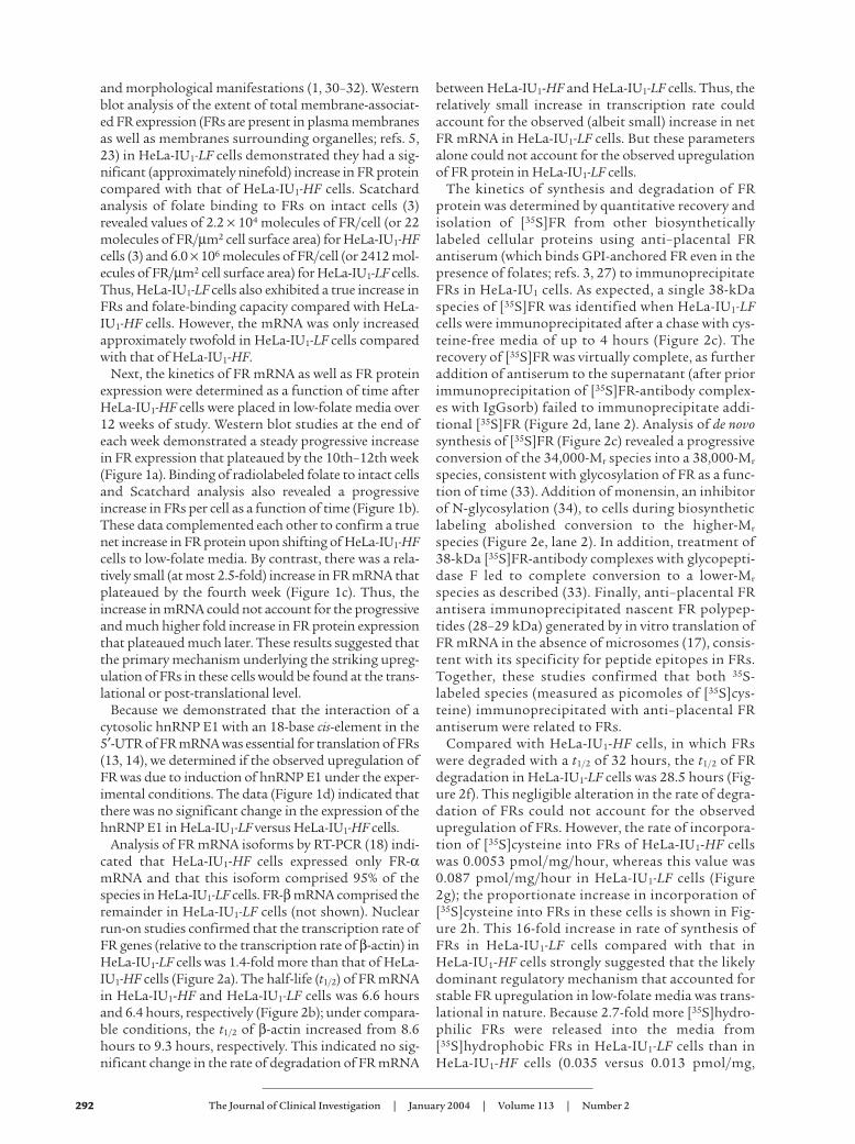
Figure 1Analysis of FR protein, FR mRNA, and hnRNP E1 inHeLa-IU1-HF cells propagated in low-folate media over12 weeks. (a) Western blot analysis of the expressionof FR protein as a function of time after shift of HeLa-IU1-HF cells to low-folate media. Each data point (foldincrease of FR protein as a function of time) representsthe mean ± SD of four independent evaluations. TheWestern blot data shown represent one of the fourstudies. (b) Determination of the cell surface receptornumber by [3H]folate-binding to intact cells andScatchard analysis as a function of time after shiftingof HeLa-IU1-HF cells to low-folate media. Each datapoint represents the mean ± SD of four independentevaluations. (c) Northern blot analysis of FR mRNArelative to β-actin mRNA expression as a function oftime after shifting of HeLa-IU1-HF cells to low-folatemedia. Signals from Northern blot analysis of 10 µg oftotal cellular RNA from cells for the indicated timeswere quantitated by densitometry. The quantity of theFR mRNA was normalized to that of β-actin mRNA.Each data point (fold increase of FR mRNA as a func-tion of time) represents the mean ± SD of four inde-pendent evaluations. The Northern blot data shownrepresent one of the four studies. (d) Northwestern gelanalysis of the FR mRNA–binding hnRNP E1 fromHeLa-IU1-HF cells and HeLa-IU1-LF cells probed withFR-α mRNA cis-element (3). The FR-α RNA sequence5′-CUCCAUUCCCACUCCCUG-3′ was labeled by invitro transcription. Densitometric analysis of the sig-nals is shown in the panel.

The Journal of Clinical Investigation | January 2004 | Volume 113 | Number 2 289
MEM containing [35S]cysteine was used. However, dur-ing chase periods, regular (cysteine-replete) media con-taining nondialyzed fetal bovine serum was utilized.Briefly, 2 × 10⁶ HeLa-IU1-HF cells or HeLa-IU1-LF cellswere cultured in 100- × 20-mm dishes to about 80%confluence. The monolayers were rinsed three timeswith D-PBS and “starved” for 18 hours in 8 ml cysteine-free MEM-HF or MEM-LF, respectively. For determina-tion of the synthetic rate of FRs, 6 × 10⁶ to 8 × 10⁶ cellswere pulsed with [35S]cysteine (250 µCi) in 4 ml of cys-teine-free MEM-HF or MEM-LF, respectively, for vari-ous times indicated (up to 4 hours) at 37°C (see Figure2). After being rinsed twice with D-PBS, the cells wereharvested with a plastic cell scraper into 10 ml D-PBScontaining 20 mM EDTA, 100 µg/ml phenylmethyl-sulfonyl fluoride, and 1 µg/ml aprotinin (buffer A) andwere centrifuged at 500 g for 5 minutes. Cell pelletswere then solubilized in 1 ml of buffer A containing1.5% Triton X-114 at 4°C for 24 hours. After centrifu-gation at 13,600 g for 15 minutes at 4°C, the super-natant was transferred to a fresh tube and an aliquotwas removed for the determination of protein concen-tration. After four cycles of temperature-inducedphase-separation (27) in buffer A for the removal of free[35S]cysteine, the Triton X-114 micellar phase was
reconstituted to 1 ml with buffer A. The samples con-taining hydrophobic [35S]FR were subsequently sub-jected to immunoprecipitation with anti–placental FRantiserum (see below). To determine the degradationrate of FR, cysteine-depleted cells were pulsed with[35S]cysteine (400 µCi) in 4 ml of cysteine-free MEM-HFor MEM-LF for 8 hours at 37°C followed by chase withMEM-HF or MEM-LF, respectively. At various latertimes, hydrophobic [35S]FR was quantitated by specif-ic immunoprecipitation (described below).
For biosynthetic labeling of FR of HeLa-IU1-HF cellswith [3H]leucine, cells were starved overnight withleucine-free MEM-HF before being pulsed with[3H]leucine for 0.5, 1, and 2 hours in the absence andpresence of 0.5 mM or 1 mM dl-homocysteine.
Validation that the procedure for immunoprecipitation of denovo–synthesized FR by anti-FR antiserum includes incom-pletely glycosylated forms. We conducted experiments tovalidate the fact that our protocol using anti-placentalFR antiserum and IgGsorb immunoprecipitated allfull-length de novo synthesized [35S]FR polypeptides inHeLa-IU1 cells. First, HeLa-IU1-LF cells were pulsed forup to four hours with [35S]cysteine followed byimmunoprecipitation of 20 µg of total cellular proteinsat various times (from 30 minutes to four hours) with
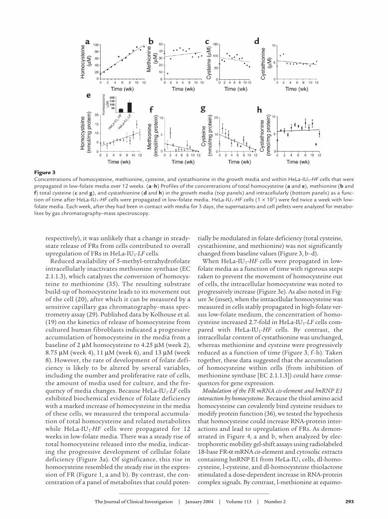
Figure 2Determination of the locus of upregulation of FR in HeLa-IU1-LF cells. (a) FR gene transcription. Nuclei (5 × 10⁷) from HeLa-IU1-HF and HeLa-IU1-LF cells were used in nuclear run-on transcription assays. Equal amounts of [α-32P]UTP nuclear RNA were hybridized to filters blotted with5 µg of denatured plasmid DNA containing human β-actin cDNA or human FR cDNA, followed by autoradiography. (b) Analysis of FR mRNAstability in HeLa-IU1-HF (open circles) and HeLa-IU1-LF (filled circles) cells exposed to actinomycin D for the indicated times followed by North-ern blot analysis. The curve-fitting analysis of FR mRNA was determined by linear regression. (c–e) Validation that the procedure for immuno-precipitation of the de novo–synthesized FR with anti-FR antiserum includes incompletely glycosylated forms (see Methods for details). (f)Analysis of the rates of FR protein degradation of either six million HeLa-IU1-HF cells (open circles) or HeLa-IU1-LF cells (filled circles) thatwere pulsed with [35S]cysteine, chased with high-folate media (open circles) or low-folate media (filled circles) for the various times indicat-ed, and immunoprecipitated [35S]FR was determined. The curve-fitting analyses in protein half-life studies were determined by linear regres-sion. (g) Analysis of the rates of FR protein synthesis of HeLa-IU1-HF cells (filled diamonds) and HeLa-IU1-LF cells (filled squares). 6 × 10⁶ cellswere pulsed with [35S]cysteine for the indicated time, and immunoprecipitated [35S]FR was determined by liquid scintillation counting. Therewas less than 15% variation from the mean for each data point of triplicate samples. The data are representative of a typical experiment thatwas repeated on three different occasions. (h) Comparison of the intensity of immunoprecipitated [35S]FR signals from HeLa-IU1-HF andHeLa-IU1-LF cells after 10% SDS-PAGE, Western blotting, and autoradiography.
290 The Journal of Clinical Investigation | January 2004 | Volume 113 | Number 2
anti-placental FR antiserum followed by 12.5% SDS-PAGE and fluorography of the gel (Figure 2c). Next,total cellular [35S]cysteine-labeled proteins from HeLa-IU1-LF cells (20 µg) were incubated with 20 µl anti-pla-cental FR antiserum and immunoprecipitated withIgGsorb (Figure 2d, lane 1); the remaining supernatantwas also subjected to the same (anti-placental FR anti-serum plus IgGsorb) treatment and analyzed (Figure2d, lane 2). Finally, HeLa-IU1-LF cells were starved forone hour with cysteine-free MEM-LF without (Figure2e, lane 1) or with (Figure 2e, lane 2) 1 µM monensinand pulsed for two hours with [35S]cysteine in theabsence or presence of monensin, respectively, beforeimmunoprecipitation of [35S]FR.
Specific immunoprecipitation of [35S]FR and UV cross-linked RNA-protein complexes. Ten microliters of nonim-mune serum was incubated with solubilized [35S]FR orUV-cross-linked RNA-protein complexes at 4°C for 2hours on a shaking platform. IgGsorb (200 µl) wasthen added and samples were incubated for 2 hours,followed by centrifugation at 13,600 g for 5 minutes at4°C to remove nonspecifically adsorbed proteins. Thesupernatant was aspirated and divided into six aliquotsof 20 µg protein each. After the addition of 20 µl ofrabbit anti–human placental FR antiserum (28) oranti–hnRNP E1 antiserum (14) or nonimmune serum(to three aliquots each), the final volume of each sam-ple was brought up to 500 µl with D-PBS containing1% Triton X-100 and 20 mM EDTA. Following incuba-tion at 4°C for 18 hours, 200 µl of IgGsorb was added.After incubation for 2 hours and centrifugation at13,600 g for 5 minutes at 4°C, the pellets (20 µl each)were washed with 1 ml of buffer A four times and wereresuspended in 0.48 ml D-PBS. An aliquot (50 µl) wasmixed with 10 ml of counting cocktail and analyzed forradioactivity in a β-scintillation counter. The dataobtained with nonimmune serum was subtracted fromthat obtained with anti–placental FR antiserum oranti–hnRNP E1 antiserum to derive values for specific[35S]cysteine incorporated into FRs or specific [32P]cis-element RNA-hnRNP E1 complexes immunoprecipi-tated. The remaining samples (from triplicates) werethen combined, concentrated to 100 µl, and analyzedby SDS-PAGE and autoradiography.
Analysis of hydrophilic FR released into the growth media.After HeLa-IU1-HF and HeLa-IU1-LF cells were pulsedfor 8 hours with [35S]cysteine, the [35S]hydrophilic FRs(35S-labeled hydrophilic FRs) released into the mediawere concentrated by ultrafiltration (Mr cutoff =10,000) using Diaflo filters (Amicon, Danvers, Massa-chusetts, USA) (28), and quantitated by immunopre-cipitation with anti–placental FR antiserum.
Northwestern blot analysis and gel-shift assays. Possible dif-ferences in the interaction of the 18-base cis-element inthe 5′-UTR of FR mRNA and cytosolic 43-kDa hnRNPE1 from HeLa-IU1 cells propagated under various extra-cellular folate concentrations were assessed by North-western blot analysis (13). Gel-shift assays were used todetermine the effect of homocysteine and other agents in
increasing the interaction of the radiolabeled 18-base FRmRNA cis-element or its mutants with the specifichnRNP E1 from cytosolic extracts of HeLa-IU1-HF cells(S-100 fraction) that were dialyzed in buffer withoutdithiothreitol (DTT) (13). Briefly, [32P]cis-element (10,000cpm) was allowed to react for 30 minutes at 22°C with 20µg of dialyzed S-100 fraction from HeLa-IU1-HF cellswith various concentrations of L-methionine, L-cysteine,DL-homocysteine thiolactone, DL-homocysteine, D-homo-cystine, L-homocystine, β-mercaptoethanol, glutathione,or DTT. After the addition of RNase T1 and heparin andincubation for 20 minutes, RNA-protein complexes wereseparated by electrophoresis (6% native PAGE; acry-lamide:bis-acrylamide, 60:1) followed by autoradiography(13). In other experiments, the effect of folic acid or 5-methyl-tetrahydrofolate in modulating the RNA-proteininteraction in the absence or presence of homocysteinewas also assessed using dialyzed S-100 fractions. Briefly,S-100 fractions from HeLa-IU1-HF cells were extensivelydialyzed in DTT-free buffer. Next, 20 µg of the dialyzedsample, which was supplemented with 1 mM dl-homo-cysteine, was allowed to react for 30 minutes at 22°C with[32P]cis-element (10,000 cpm), and increasing concentra-tions (10 nM to 100 µM) of either folic acid or 5-methyl-tetrahydrofolate was added to the RNA-protein mixture;freshly prepared 5-methyltetrahydrofolate was addedwithin 15 minutes of the opening of a new vial. After theaddition of RNaseT1 and heparin and incubation foranother 20 minutes, the intensities of the RNA-proteincomplex signals were determined (13).
Plasmid construction. The plasmid containing chloram-phenicol acetyltransferase (CAT) and the cis-element[pCAT (+cis)] was constructed by replacing a 223-bpintron from the pCAT3 control vector (Promega, Madi-son, Wisconsin, USA) with a 200-bp fragment of the 5′-UTR of FR cDNA containing the 18-base cis-element atthe HindIII site. The pCAT (–cis) plasmid, a control plas-mid with deletion of the 18-base cis-element, was pre-pared using site-directed mutagenesis in which theprimers 5′-GCTCCACTCCTGGGCCTGTCTCCTAG-3′ and5′-CTAGGAGACAGGCCCAGGAGTGGAGC-3′ were used.The plasmids pN, pC1, pC2, and pC12 were constructedby subcloning the four pairs of oligodeoxynucleotides(shown below) into the pSPT18 vector that was linearizedwith EcoRI and HindIII to generate the 18-base cis-ele-ment RNA and its mutants. The orientation and pointmutations were confirmed in all plasmids sequencing:pN: 5′-p-AATTCGCTCCATT CCCACTCCCTGA-3′ 3′-GCG-AGGTAAGGGTGAGGGACTTCGA-p-5′ pC1: 5′-p-AATTCGC-TACATT CCCACTCCCTGA-3′ 3′-GCGATGTAAGGGT-GAGGGACTTCGA-p-5′; pC2: 5′-p-AATTCGCTCCATTCCCACTACCTGA-3′; 3′-GCGAGGTAAGGGTGATGGACTTC-GA-p-5′; pC12: 5′-p-AATTCGCTACATTCCC ACTACCTGA-3′; 3′-GCGATGTAAGGGTGATGGACTTCGA-p-5′.
Transfection of HeLa-IU1-HF and HeLa-IU1-LF cells withCAT reporter constructs driven by a 5′-UTR of FR mRNA inwhich the cis-element is retained [pCAT (+cis)] or deleted[pCAT (–cis)]. The pSV-β-galactosidase (Promega) wascotransfected with either pCAT (+cis) or pCAT (–cis),
The Journal of Clinical Investigation | January 2004 | Volume 113 | Number 2 291
which permitted assay for both β-galactosidase activi-ty (by a β-Gal reporter gene assay using a chemilumi-nescence assay) and CAT ELISA (Roche DiagnosticsCorp.). The extent of β-galactosidase activity served asan internal control for transfection efficiency of pCAT(+cis) or pCAT (–cis) into HeLa-IU1-HF and HeLa-IU1-LFcells. The results of negative control cell extracts, pre-pared from nontransfected cells and assayed for bothβ-galactosidase and CAT, were routinely subtractedfrom the results obtained from transfected cell extracts.Briefly, cells were plated 1 day before being transfectedin culture media (MEM-HF or MEM-LF for HeLa-IU1-HFor HeLa-IU1-LF cells, respectively) at a density of 2.5 × 10⁵
cells/well using six-well plates to achieve 50–80% con-fluency on the day of transfection. The FuGENE-6(Roche Diagnostics Corp.) transfection reagent wasused with each plasmid DNA [either pCAT (+cis) orpCAT (–cis) and pSV-β-galactosidase] at a ratio of 3:2 (3µl FuGene-6, 2 µg DNA of each plasmid, and 97 µlserum-free media) per well, and transfection was car-ried out according to the manufacturer’s instructions.
Determination of the identity of the protein within RNA-pro-tein complexes induced by intracellular homocysteine in HeLa-IU1-LF cells by gel supershift assays. Cytosolic proteinextracts (S-100 fraction) from HeLa-IU1 cells were pre-pared as described below. Briefly, HeLa-IU1-HF andHeLa-IU1-LF cells were cultured in MEM-HF and MEM-LF media, respectively, for 3 days to approximately 80%confluence. After cells were harvested with a rubberpoliceman, the cell suspensions were centrifuged overVersilube F50 silicone fluid to separate media and cellsas described above. Cells were incubated in two packedcell volumes of buffer A (10 mM HEPES, pH 7.9, con-taining 1.5 mM MgCl2 and 10 mM KCl) for 10 minutesat 4°C, and then were lysed by 30 strokes of a Kontes all-glass Dounce homogenizer (B-type pestle). After confir-mation of cell lysis and recentrifugation (at 800 g for 10minutes at 4°C), the supernatant was mixed with 0.11volumes of buffer B (300 mM HEPES, pH 7.9, contain-ing 30 mM MgCl2 and 1.4 M KCl), and centrifuged for60 minutes at 100,000 g (in a Beckman Type SW 55.Tirotor). The protein concentration of the supernatant wasmeasured by bicinchonic acid assay (Pierce, Rockford,Illinois, USA) and 500-µl aliquots were frozen at –80°C(S-100 fraction). RNA-protein binding reactions werecarried out with 800 µg cytosolic extracts from HeLa-IU1-HF or HeLa-IU1-LF cells and 1 × 10⁶ cpm of [32P]cis-element in 10 mM HEPES, pH 7.6, containing 3 mMMgCl2, 40 mM KCl, 5% glycerol, and 600 ng/µl yeasttransfer RNA in a final volume of 300 µl. After sampleswere incubated at room temperature for 30 min, 500units of RNase T1 and 20 µl of heparin solution (100mg/ml) were added and incubation was continued foranother 30 minutes. Aliquots of 15 µl were then incu-bated with anti–hnRNP E1 antiserum or nonimmuneserum overnight followed by a gel-shift assay (14) todemonstrate a supershift with antiserum and the iden-tity of the protein within the cis-element and trans-factorcomplex. The remainder of the RNA-protein complexes
were transferred to 24-well plates and irradiated from adistance of 1 cm by a UV lamp (300 nm, 70,000 µW/cm2;Fotodyne Inc., New Berlin, Wisconsin, USA) for 1 hourat 4°C followed by specific immunoprecipitation of UVcross-linked RNA-protein complexes.
Statistical analysis. The curve-fitting analyses weredetermined by linear regression (Microsoft Excel).Unless otherwise stated, all experiments were carriedout on three different occasions with less than 15%variation from the mean for each data point.
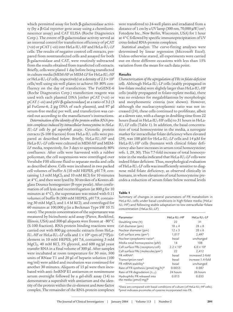
ResultsCharacterization of the upregulation of FRs in folate-deficientcells. Although HeLa-IU1-LF cells (stably propagated inlow-folate media) were slightly larger than HeLa-IU1-HFcells (stably propagated in folate-replete media), therewas no evidence for megaloblastosis by morphologicand morphometric criteria (not shown). However,although the nuclear:cytoplasmic ratio was not in-creased (24), these cells continued to proliferate, albeitat a slower rate, with a change in doubling time from 22hours (basal in HeLa-IU1-HF cells) to 31 hours in HeLa-IU1-LF cells (Table 1). In addition, the basal concentra-tion of total homocysteine in the media, a surrogatemarker for intracellular folate deficiency when elevated(29), was 108 µM for HeLa-IU1-LF cells and 14 µM forHeLa-IU1-HF cells (humans with clinical folate defi-ciency also have increases in serum total homocysteine;refs. 1, 29, 30). This 7.7-fold increase of total homocys-teine in the media indicated that HeLa-IU1-LF cells wereindeed folate deficient. Thus, morphological evaluationof HeLa-IU1-LF cells was insufficiently sensitive to diag-nose mild folate deficiency, as observed clinically inhumans, in whom elevations of total homocysteine pre-cedes a reduction of serum folate below normal levels
Table 1Summary of changes in several parameters of FR metabolism inHeLa-IU1 cells under basal conditions in high-folate media (HeLa-IU1-HF) and following stable adaptation to low extracellular folateconcentration (HeLa-IU1-LF)
Parameter HeLa-IU1-HF HeLa-IU1-LFDoubling time (h) 22 31Cell diameter (µm) 18 ± 4 28 ± 8Nuclear diameter (µm) 12 ± 3 20 ± 6Cell surface area (µm2) 1,017 2,497Nuclear:cytoplasmic ratioA basal unchangedMedia total homocysteine (µM) 14 108Cell surface FRs (receptors/cell) 2.2 × 10⁴ 6.0 × 10⁶
Cell surface FRs (molecules/µm2) 22 2,412FR mRNAA basal increased 2-foldTranscription rateA basal increase 1.4-foldFR mRNA stabilityA basal unchangedRate of FR synthesis (pmol/mg/h)B 0.0053 0.087Rate of FR degradation (t1/2) 24 hours 26 hoursHydrophilic FR released into 0.013 0.035the media (pmol/mg)B
AData are compared with basal conditions of culture (of HeLa-IU1-HF cells).Bpmol indicates picomoles of cysteine incorporated into FR.
292 The Journal of Clinical Investigation | January 2004 | Volume 113 | Number 2
and morphological manifestations (1, 30–32). Westernblot analysis of the extent of total membrane-associat-ed FR expression (FRs are present in plasma membranesas well as membranes surrounding organelles; refs. 5,23) in HeLa-IU1-LF cells demonstrated they had a sig-nificant (approximately ninefold) increase in FR proteincompared with that of HeLa-IU1-HF cells. Scatchardanalysis of folate binding to FRs on intact cells (3)revealed values of 2.2 × 10⁴ molecules of FR/cell (or 22molecules of FR/µm2 cell surface area) for HeLa-IU1-HFcells (3) and 6.0 × 10⁶ molecules of FR/cell (or 2412 mol-ecules of FR/µm2 cell surface area) for HeLa-IU1-LF cells.Thus, HeLa-IU1-LF cells also exhibited a true increase inFRs and folate-binding capacity compared with HeLa-IU1-HF cells. However, the mRNA was only increasedapproximately twofold in HeLa-IU1-LF cells comparedwith that of HeLa-IU1-HF.
Next, the kinetics of FR mRNA as well as FR proteinexpression were determined as a function of time afterHeLa-IU1-HF cells were placed in low-folate media over12 weeks of study. Western blot studies at the end ofeach week demonstrated a steady progressive increasein FR expression that plateaued by the 10th–12th week(Figure 1a). Binding of radiolabeled folate to intact cellsand Scatchard analysis also revealed a progressiveincrease in FRs per cell as a function of time (Figure 1b).These data complemented each other to confirm a truenet increase in FR protein upon shifting of HeLa-IU1-HFcells to low-folate media. By contrast, there was a rela-tively small (at most 2.5-fold) increase in FR mRNA thatplateaued by the fourth week (Figure 1c). Thus, theincrease in mRNA could not account for the progressiveand much higher fold increase in FR protein expressionthat plateaued much later. These results suggested thatthe primary mechanism underlying the striking upreg-ulation of FRs in these cells would be found at the trans-lational or post-translational level.
Because we demonstrated that the interaction of acytosolic hnRNP E1 with an 18-base cis-element in the5′-UTR of FR mRNA was essential for translation of FRs(13, 14), we determined if the observed upregulation ofFR was due to induction of hnRNP E1 under the exper-imental conditions. The data (Figure 1d) indicated thatthere was no significant change in the expression of thehnRNP E1 in HeLa-IU1-LF versus HeLa-IU1-HF cells.
Analysis of FR mRNA isoforms by RT-PCR (18) indi-cated that HeLa-IU1-HF cells expressed only FR-αmRNA and that this isoform comprised 95% of thespecies in HeLa-IU1-LF cells. FR-β mRNA comprised theremainder in HeLa-IU1-LF cells (not shown). Nuclearrun-on studies confirmed that the transcription rate ofFR genes (relative to the transcription rate of β-actin) inHeLa-IU1-LF cells was 1.4-fold more than that of HeLa-IU1-HF cells (Figure 2a). The half-life (t1/2) of FR mRNAin HeLa-IU1-HF and HeLa-IU1-LF cells was 6.6 hoursand 6.4 hours, respectively (Figure 2b); under compara-ble conditions, the t1/2 of β-actin increased from 8.6hours to 9.3 hours, respectively. This indicated no sig-nificant change in the rate of degradation of FR mRNA
between HeLa-IU1-HF and HeLa-IU1-LF cells. Thus, therelatively small increase in transcription rate couldaccount for the observed (albeit small) increase in netFR mRNA in HeLa-IU1-LF cells. But these parametersalone could not account for the observed upregulationof FR protein in HeLa-IU1-LF cells.
The kinetics of synthesis and degradation of FRprotein was determined by quantitative recovery andisolation of [35S]FR from other biosyntheticallylabeled cellular proteins using anti–placental FRantiserum (which binds GPI-anchored FR even in thepresence of folates; refs. 3, 27) to immunoprecipitateFRs in HeLa-IU1 cells. As expected, a single 38-kDaspecies of [35S]FR was identified when HeLa-IU1-LFcells were immunoprecipitated after a chase with cys-teine-free media of up to 4 hours (Figure 2c). Therecovery of [35S]FR was virtually complete, as furtheraddition of antiserum to the supernatant (after priorimmunoprecipitation of [35S]FR-antibody complex-es with IgGsorb) failed to immunoprecipitate addi-tional [35S]FR (Figure 2d, lane 2). Analysis of de novosynthesis of [35S]FR (Figure 2c) revealed a progressiveconversion of the 34,000-Mr species into a 38,000-Mr
species, consistent with glycosylation of FR as a func-tion of time (33). Addition of monensin, an inhibitorof N-glycosylation (34), to cells during biosyntheticlabeling abolished conversion to the higher-Mr
species (Figure 2e, lane 2). In addition, treatment of38-kDa [35S]FR-antibody complexes with glycopepti-dase F led to complete conversion to a lower-Mr
species as described (33). Finally, anti–placental FRantisera immunoprecipitated nascent FR polypep-tides (28–29 kDa) generated by in vitro translation ofFR mRNA in the absence of microsomes (17), consis-tent with its specificity for peptide epitopes in FRs.Together, these studies confirmed that both 35S-labeled species (measured as picomoles of [35S]cys-teine) immunoprecipitated with anti–placental FRantiserum were related to FRs.
Compared with HeLa-IU1-HF cells, in which FRswere degraded with a t1/2 of 32 hours, the t1/2 of FRdegradation in HeLa-IU1-LF cells was 28.5 hours (Fig-ure 2f). This negligible alteration in the rate of degra-dation of FRs could not account for the observedupregulation of FRs. However, the rate of incorpora-tion of [35S]cysteine into FRs of HeLa-IU1-HF cellswas 0.0053 pmol/mg/hour, whereas this value was0.087 pmol/mg/hour in HeLa-IU1-LF cells (Figure2g); the proportionate increase in incorporation of[35S]cysteine into FRs in these cells is shown in Fig-ure 2h. This 16-fold increase in rate of synthesis ofFRs in HeLa-IU1-LF cells compared with that inHeLa-IU1-HF cells strongly suggested that the likelydominant regulatory mechanism that accounted forstable FR upregulation in low-folate media was trans-lational in nature. Because 2.7-fold more [35S]hydro-philic FRs were released into the media from[35S]hydrophobic FRs in HeLa-IU1-LF cells than inHeLa-IU1-HF cells (0.035 versus 0.013 pmol/mg,
The Journal of Clinical Investigation | January 2004 | Volume 113 | Number 2 293
respectively), it was unlikely that a change in steady-state release of FRs from cells contributed to overallupregulation of FRs in HeLa-IU1-LF cells.
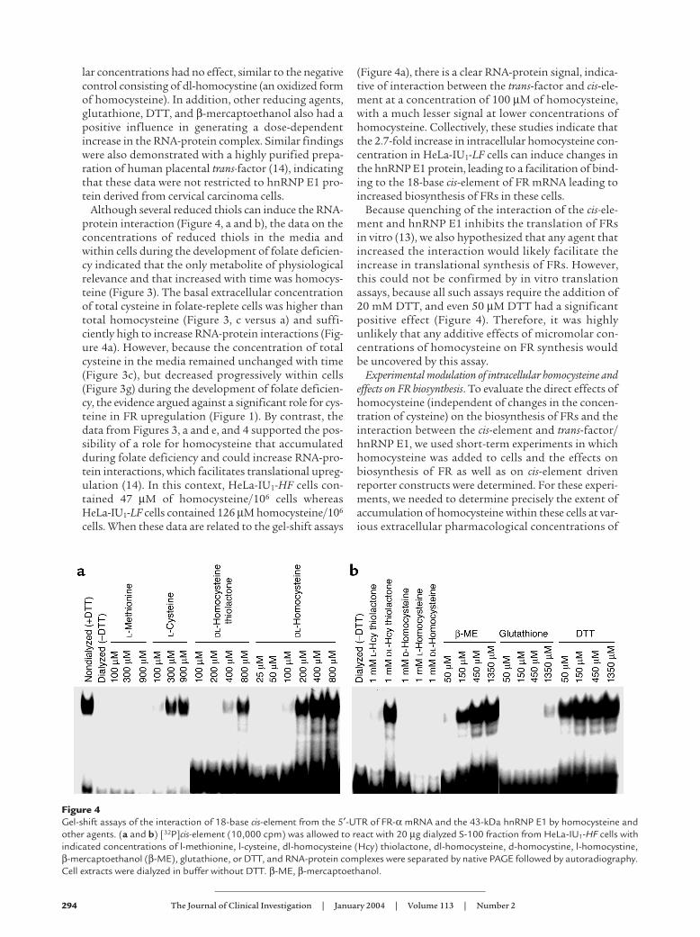
Reduced availability of 5-methyl-tetrahydrofolateintracellularly inactivates methionine synthase (EC2.1.1.3), which catalyzes the conversion of homocys-teine to methionine (35). The resulting substratebuild-up of homocysteine leads to its movement outof the cell (20), after which it can be measured by asensitive capillary gas chromatography–mass spec-trometry assay (29). Published data by Kolhouse et al.(19) on the kinetics of release of homocysteine fromcultured human fibroblasts indicated a progressiveaccumulation of homocysteine in the media from abaseline of 2 µM homocysteine to 4.25 µM (week 2),8.75 µM (week 4), 11 µM (week 6), and 13 µM (week8). However, the rate of development of folate defi-ciency is likely to be altered by several variables,including the number and proliferative rate of cells,the amount of media used for culture, and the fre-quency of media changes. Because HeLa-IU1-LF cellsexhibited biochemical evidence of folate deficiencywith a marked increase of homocysteine in the mediaof these cells, we measured the temporal accumula-tion of total homocysteine and related metaboliteswhile HeLa-IU1-HF cells were propagated for 12weeks in low-folate media. There was a steady rise oftotal homocysteine released into the media, indicat-ing the progressive development of cellular folatedeficiency (Figure 3a). Of significance, this rise inhomocysteine resembled the steady rise in the expres-sion of FR (Figure 1, a and b). By contrast, the con-centration of a panel of metabolites that could poten-
tially be modulated in folate deficiency (total cysteine,cystathionine, and methionine) was not significantlychanged from baseline values (Figure 3, b–d).
When HeLa-IU1-HF cells were propagated in low-folate media as a function of time with rigorous stepstaken to prevent the movement of homocysteine outof cells, the intracellular homocysteine was noted toprogressively increase (Figure 3e). As also noted in Fig-ure 3e (inset), when the intracellular homocysteine wasmeasured in cells stably propagated in high-folate ver-sus low-folate medium, the concentration of homo-cysteine increased 2.7-fold in HeLa-IU1-LF cells com-pared with HeLa-IU1-HF cells. By contrast, theintracellular content of cystathionine was unchanged,whereas methionine and cysteine were progressivelyreduced as a function of time (Figure 3, f–h). Takentogether, these data suggested that the accumulationof homocysteine within cells (from inhibition ofmethionine synthase [EC 2.1.1.3]) could have conse-quences for gene expression.
Modulation of the FR mRNA cis-element and hnRNP E1interaction by homocysteine. Because the thiol amino acidhomocysteine can covalently bind cysteine residues tomodify protein function (36), we tested the hypothesisthat homocysteine could increase RNA-protein inter-actions and lead to upregulation of FRs. As demon-strated in Figure 4, a and b, when analyzed by elec-trophoretic mobility gel-shift assays using radiolabeled18-base FR-α mRNA cis-element and cytosolic extractscontaining hnRNP E1 from HeLa-IU1 cells, dl-homo-cysteine, l-cysteine, and dl-homocysteine thiolactonestimulated a dose-dependent increase in RNA-proteincomplex signals. By contrast, l-methionine at equimo-
Figure 3Concentrations of homocysteine, methionine, cysteine, and cystathionine in the growth media and within HeLa-IU1-HF cells that werepropagated in low-folate media over 12 weeks. (a–h) Profiles of the concentrations of total homocysteine (a and e), methionine (b andf) total cysteine (c and g), and cystathionine (d and h) in the growth media (top panels) and intracellularly (bottom panels) as a func-tion of time after HeLa-IU1-HF cells were propagated in low-folate media. HeLa-IU1-HF cells (1 × 10⁷) were fed twice a week with low-folate media. Each week, after they had been in contact with media for 3 days, the supernatants and cell pellets were analyzed for metabo-lites by gas chromatography–mass spectroscopy.
294 The Journal of Clinical Investigation | January 2004 | Volume 113 | Number 2
lar concentrations had no effect, similar to the negativecontrol consisting of dl-homocystine (an oxidized formof homocysteine). In addition, other reducing agents,glutathione, DTT, and β-mercaptoethanol also had apositive influence in generating a dose-dependentincrease in the RNA-protein complex. Similar findingswere also demonstrated with a highly purified prepa-ration of human placental trans-factor (14), indicatingthat these data were not restricted to hnRNP E1 pro-tein derived from cervical carcinoma cells.
Although several reduced thiols can induce the RNA-protein interaction (Figure 4, a and b), the data on theconcentrations of reduced thiols in the media andwithin cells during the development of folate deficien-cy indicated that the only metabolite of physiologicalrelevance and that increased with time was homocys-teine (Figure 3). The basal extracellular concentrationof total cysteine in folate-replete cells was higher thantotal homocysteine (Figure 3, c versus a) and suffi-ciently high to increase RNA-protein interactions (Fig-ure 4a). However, because the concentration of totalcysteine in the media remained unchanged with time(Figure 3c), but decreased progressively within cells(Figure 3g) during the development of folate deficien-cy, the evidence argued against a significant role for cys-teine in FR upregulation (Figure 1). By contrast, thedata from Figures 3, a and e, and 4 supported the pos-sibility of a role for homocysteine that accumulatedduring folate deficiency and could increase RNA-pro-tein interactions, which facilitates translational upreg-ulation (14). In this context, HeLa-IU1-HF cells con-tained 47 µM of homocysteine/106 cells whereasHeLa-IU1-LF cells contained 126 µM homocysteine/106
cells. When these data are related to the gel-shift assays
(Figure 4a), there is a clear RNA-protein signal, indica-tive of interaction between the trans-factor and cis-ele-ment at a concentration of 100 µM of homocysteine,with a much lesser signal at lower concentrations ofhomocysteine. Collectively, these studies indicate thatthe 2.7-fold increase in intracellular homocysteine con-centration in HeLa-IU1-LF cells can induce changes inthe hnRNP E1 protein, leading to a facilitation of bind-ing to the 18-base cis-element of FR mRNA leading toincreased biosynthesis of FRs in these cells.
Because quenching of the interaction of the cis-ele-ment and hnRNP E1 inhibits the translation of FRsin vitro (13), we also hypothesized that any agent thatincreased the interaction would likely facilitate theincrease in translational synthesis of FRs. However,this could not be confirmed by in vitro translationassays, because all such assays require the addition of20 mM DTT, and even 50 µM DTT had a significantpositive effect (Figure 4). Therefore, it was highlyunlikely that any additive effects of micromolar con-centrations of homocysteine on FR synthesis wouldbe uncovered by this assay.
Experimental modulation of intracellular homocysteine andeffects on FR biosynthesis. To evaluate the direct effects ofhomocysteine (independent of changes in the concen-tration of cysteine) on the biosynthesis of FRs and theinteraction between the cis-element and trans-factor/hnRNP E1, we used short-term experiments in whichhomocysteine was added to cells and the effects onbiosynthesis of FR as well as on cis-element drivenreporter constructs were determined. For these experi-ments, we needed to determine precisely the extent ofaccumulation of homocysteine within these cells at var-ious extracellular pharmacological concentrations of
Figure 4Gel-shift assays of the interaction of 18-base cis-element from the 5′-UTR of FR-α mRNA and the 43-kDa hnRNP E1 by homocysteine andother agents. (a and b) [32P]cis-element (10,000 cpm) was allowed to react with 20 µg dialyzed S-100 fraction from HeLa-IU1-HF cells withindicated concentrations of l-methionine, l-cysteine, dl-homocysteine (Hcy) thiolactone, dl-homocysteine, d-homocystine, l-homocystine,β-mercaptoethanol (β-ME), glutathione, or DTT, and RNA-protein complexes were separated by native PAGE followed by autoradiography.Cell extracts were dialyzed in buffer without DTT. β-ME, β-mercaptoethanol.
The Journal of Clinical Investigation | January 2004 | Volume 113 | Number 2 295
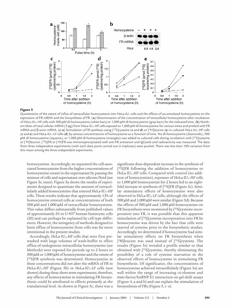
homocysteine. Accordingly, we separated the cell-asso-ciated homocysteine from the higher concentrations ofhomocysteine extant in the supernatant by passing themixture of cells and supernatant over silicone fluid (seeFigure 3e, inset). Figure 5a shows the results of experi-ments designed to quantitate the amount of extracel-lularly added homocysteine that entered HeLa-IU1-HFcells. These results indicate that approximately 13% ofhomocysteine entered cells at concentrations of both500 µM and 1,000 µM of extracellular homocysteine.This value differs substantially from published valuesof approximately 2% in U-937 human histiocytic cells(20) and can perhaps be explained by cell type differ-ences. However, the stringency of methods designed tolimit efflux of homocysteine from cells was far moreintentional in the present studies.
Accordingly, HeLa-IU1-HF cells that were first pre-washed with large volumes of wash-buffer to effectefflux of endogenous intracellular homocysteine (seeMethods) were exposed for periods up to 2 hours to500 µM or 1,000 µM of homocysteine and the extent of[35S]FR synthesis was determined. Homocysteine atthese concentrations did not alter the mRNA of FR inHeLa-IU1-HF (Figure 5b) or HeLa-IU1-LF cells (notshown) during these short-term experiments; therefore,any effects of homocysteine in stimulating FR biosyn-thesis could be attributed to effects primarily at thetranslational level. As shown in Figure 5c, there was a
significant dose-dependent increase in the synthesis of[35S]FR following the addition of homocysteine toHeLa-IU1-HF cells. Compared with control (no addi-tion of homocysteine), exposure of HeLa-IU1-HF cellsto 1,000 µM homocysteine for 2 hours led to an eight-fold increase in synthesis of [35S]FR (Figure 5c). Simi-lar stimulatory effects of homocysteine were alsoobserved in HeLa-IU1-LF cells, although the effects of500 µM and 1,000 µM were similar (Figure 5d). Becausethe effects of 500 µM and 1,000 µM homocysteine onFR biosynthesis were monitored by [35S]cysteine incor-poration into FR, it was possible that this apparentstimulation of [35S]cysteine incorporation into FR byhomocysteine was driven by the fact that cells werestarved of cysteine prior to the biosynthetic studies.Accordingly, we determined if homocysteine had simi-lar stimulatory effects on FR biosynthesis when[3H]leucine was used instead of [35S]cysteine. Theresults (Figure 5e) revealed a profile similar to thatobtained with [35S]cysteine, thereby eliminating thepossibility of a role of cysteine starvation in theobserved effects of homocysteine in stimulating FRbiosynthesis. Of significance, the concentrations ofhomocysteine achieved intracellularly (Figure 5a) arewell within the range of increasing cis-element andtrans-factor/hnRNP E1 interaction on gel-shift assays(Figure 4, a and b) and can explain the stimulation ofbiosynthesis of FRs (Figure 5, c–e).
Figure 5Quantitation of the extent of influx of extracellular homocysteine into HeLa-IU1 cells and the effects of accumulated homocysteine on theexpression of FR mRNA and the biosynthesis of FR. (a) Determination of the concentration of intracellular homocysteine after incubationof HeLa-IU1–HF cells with 500 µM dl-homocysteine (white bars) or 1,000 µM dl-homocysteine (gray bars) for the indicated time. (b) North-ern blots of total cellular mRNA (3 µg) from HeLa-IU1–HF cells exposed to 1,000 µM dl-homocysteine for various times and probed with FRmRNA and β-actin mRNA. (c–e) Stimulation of FR synthesis using [35S]cysteine (c and d) or [3H]leucine (e) in cultured HeLa-IU1–HF cells(c and e) and HeLa-IU1–LF cells (d) by various concentrations of homocysteine as a function of time. No dl-homocysteine (diamonds), 500µM dl-homocysteine (squares), or 1,000 µM dl-homocysteine (triangles) was added to cultured cells during incubation with [35S]cysteineor [3H]leucine. [35S]FR or [3H]FR was immunoprecipitated with anti-FR antiserum and IgGsorb and radioactivity was measured. The datafrom three independent experiments (with each data point carried out in triplicate) were pooled. There was less than 10% variation fromthis mean among the three independent experiments.
296 The Journal of Clinical Investigation | January 2004 | Volume 113 | Number 2
Homocysteine exerted a stimulatory effect on cellu-lar FR biosynthesis within 30 minutes. This duration ismuch shorter than the observed effects of homocys-teine on transcriptional regulation of many othergenes, which required 6 hours of exposure (37). Fur-thermore, our documentation that homocysteine didnot induce changes in FR mRNA levels supported theconclusion that the effect of homocysteine was prima-rily in modulating RNA-protein interactions. Thus, inthe model involving a gradual development of nutri-tional folate deficiency, the data strongly argued for acausal role for homocysteine in translational upregu-lation of FRs. Collectively these data supported theconclusion that the build-up of homocysteine in nutri-tional folate deficiency stimulated the interaction of aspecific cis-element and hnRNP E1 that led to the trans-lational upregulation of FRs.
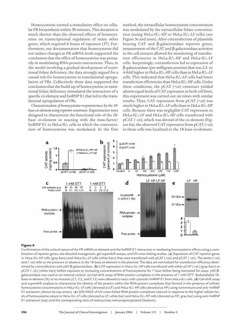
Characterization of homocysteine-responsiveness by the 18-base cis-element using reporter constructs. Experiments weredesigned to characterize the functional role of the 18-base cis-element in reacting with the trans-factor/hnRNP E1 in HeLa-IU1 cells in which the concentra-tion of homocysteine was modulated. In the first
method, the intracellular homocysteine concentrationwas modulated by the extracellular folate concentra-tion (using HeLa-IU1-HF or HeLa-IU1-LF cells) (seeFigure 3e and inset). After cotransfection of plasmidsbearing CAT and β-galactosidase reporter genes,measurement of the CAT and β-galactosidase activitiesin the cell extracts allowed for monitoring of transfec-tion efficiencies in HeLa-IU1-HF and HeLa-IU1-LFcells. Surprisingly, cotransfection led to expression ofβ-galactosidase (per milligram protein) that was 2.5- to4-fold higher in HeLa-IU1-HF cells than in HeLa-IU1-LFcells. This indicated that HeLa-IU1-LF cells had lowertransfection efficiencies than HeLa-IU1-HF cells. Underthese conditions, the pCAT (+cis) construct yieldedalmost equal levels of CAT expression in both cell lines;this experiment was carried out six times with similarresults. Thus, CAT expression from pCAT (+cis) wasmuch higher in HeLa-IU1-LF cells than in HeLa-IU1-HFcells. Because there was negligible CAT expression inHeLa-IU1-LF and HeLa-IU1-HF cells transfected withpCAT (–cis), which was devoid of the cis-element (Fig-ure 6a), the observed CAT expression from pCAT (+cis)in these cells was localized to the 18-base cis-element.
Figure 6Confirmation of the critical nature of the FR mRNA cis-element and the hnRNP E1 interaction in mediating homocysteine effects using a com-bination of reporter genes, site-directed mutagenesis, gel-supershift assays, and UV cross-linking studies. (a) Expression of CAT reporter genesin HeLa-IU1-HF cells (gray bars) and HeLa-IU1-LF cells (white bars) that were transfected with pCAT (+cis) and pCAT (–cis). The terms (+cis)and (–cis) refer to the presence or absence or the 18-base cis-element in the plasmid. The data are normalized for transfection efficiency deter-mined by cotransfection with pSV-β-galactosidase. (b) CAT expression in HeLa-IU1-HF cells transfected with either pCAT (+cis) (gray bars) orpCAT (–cis) (white bars) before exposure to increasing concentrations of homocysteine for 1 hour before being harvested for assay. pSV-β-galactosidase was used as an internal control. (c) Gel-shift assay of RNA-protein complexes in the presence of 1 mM DTT. Radiolabeled 18-base cis-element (N) or its mutants (C1, C2, and C12) were allowed to react with cytosolic hnRNP E1 from HeLa-IU1 cells. (d) Gel-shift assayand supershift analysis to characterize the identity of the protein within the RNA-protein complexes that formed in the presence of cellularhomocysteine concentrations in HeLa-IU1-LF cells (denoted as LF) and HeLa-IU1-HF cells (denoted as HF) using nonimmune and anti–hnRNPE1 antiserum (shown by top arrow). (e) SDS-PAGE of UV cross-linked RNA-protein complexes induced in response to the higher cellular lev-els of homocysteine extant in HeLa-IU1-LF cells (denoted as LF; white bar) and HeLa-IU1-HF cells (denoted as HF; gray bar) using anti–hnRNPE1 antiserum (top) and the corresponding ratio of radioactivity immunoprecipitated (bottom).

The Journal of Clinical Investigation | January 2004 | Volume 113 | Number 2 297
Because HeLa-IU1-LF cells had a 2.7-fold greater concen-tration of intracellular homocysteine than HeLa-IU1-HFcells (Figure 3e, inset) at levels at which RNA-proteininteraction could be demonstrated (Figure 4a), theseresults continued to highlight the likely importance ofthe 18-base cis-element in homocysteine responsiveness.
To further confirm that the cis-element was integralto homocysteine responsiveness, we sought to deter-mine if short-term exposure of HeLa-IU1-HF cells trans-fected with pCAT (+cis) to progressively increasingintracellular concentrations of homocysteine (for 1hour) led to a predictable dose response compared withcells transfected with pCAT (–cis). The results (Figure6b) indicated that whereas HeLa-IU1-HF cells trans-fected with pCAT (–cis) exhibited no increase in signalwith increasing concentrations of homocysteine, thosecells transfected with pCAT (+cis) exhibited doseresponsiveness to homocysteine. When these resultswere correlated with the actual measurements of intra-cellular homocysteine under similar experimental con-ditions (Figure 5a), the concentrations of intracellularhomocysteine achieved following incubation with 50,100, 250, 500, 750, and 1,000-µM of dl-homocysteinewere 6.5, 13, 32.5, 65, 97.5, and 130 µM, respectively.Furthermore, when these results are compared withthose values of homocysteine used in gel-shift experi-ments to demonstrate RNA-protein interactions (Fig-ure 4), it is readily apparent that the concentrationsachieved intracellularly were well within the range forendogenous trans-factor/hnRNP E1 (Figure 1d) tointeract with the 18-base cis-element proximal to theCAT gene to give the observed signals. Thus, all of thesedata (Figures 1–6) can now be reconciled to explain theprogressive increase in FR upregulation when the extra-cellular folate concentration is limited. On the basis ofthese experiments, we conclude that the data demon-strate that the accumulated homocysteine within cellsduring folate deficiency is sufficient to trigger the inter-action of the trans-factor/hnRNP-E1 with the 18-basecis-element within the 5′-UTR of FR mRNA to result intranslational upregulation of FRs.
Next we determined if point mutation of the 18-base cis-element sequence would abrogate RNA-pro-tein complex formation in the induced state. Based oncharacterization of the RNA-protein interaction, wehad suggested that the CUCC motif in the 18-base cis-element sequence [CUCC(AU)-4-6-bases-CUCC]could be critical in binding hnRNP E1 (13, 14).Accordingly, we mutated cytidine 5′-triphosphate atspecific positions in the 18-mer cis-element (detailedin Methods) and compared the binding to hnRNP E1.The results (Figure 6c) revealed that a point mutationin either the first or second (or both) CUCC motif inthe 18-base cis-element led to a reduction in RNA-pro-tein complex formation in the induced state. This ver-ified that the interaction of endogenous hnRNP E1was critically dependent on the integrity of the 18-base cis-element in the 5′-UTR of FR mRNA and con-firmed our earlier predictions (13).
Next we determined if more radiolabeled cis-elementinteracted with endogenous hnRNP E1 from HeLa-IU1-LF cells than from HeLa-IU1-HF cells because of thehigher cellular concentration of homocysteine offolate-depleted cells (Figure 3e). We used UV cross-link-ing of the RNA-protein complexes (13), which allowedus to eliminate the use of an exogenous reducing agentto trigger RNA-protein complex formation (whichwould confound analysis of the independent effect ofintracellular homocysteine). Accordingly, cytosolichnRNP E1 from HeLa-IU1-HF and HeLa-IU1-LF cellsthat was isolated under conditions that avoided effluxof cellular homocysteine (see Methods) was incubatedwith [32P]cis-element, and after cross-linking of RNA-protein complexes (13), the mixture was allowed toreact with anti–hnRNP E1 antiserum or nonimmuneserum and was analyzed further. The results of gel-shiftassay, immunoprecipitation, and SDS-PAGE of theanti–hnRNP E1 antiserum–bound RNA-protein com-plexes in HeLa-IU1-LF cells and HeLa-IU1-HF cells areshown in Figure 6, d and e. These concordant resultsdemonstrated that the greater amount of homocys-teine present in HeLa-IU1-LF cells (Figure 3e) led to agreater interaction of the cis-element and hnRNP E1,compared with the data from HeLa-IU1-HF cells. Thisis shown by the stronger signal in the gel-shifted band,a greater amount of immunoprecipitated material byradioactive counts, and a stronger signal on SDS-PAGEin the sample from HeLa-IU1-LF cells. Moreover, thesupershift of the signal with anti–hnRNP E1 antiserum(Figure 6d) indicated that the [32P]cis-element RNA-protein complex contained hnRNP E1.
When we used RNA cis-element probes with very highspecific activity in experiments similar to that shown inFigure 4, the lowest concentrations of homocysteinethat could stimulate interaction of the cis-element andtrans-factor/(hnRNP E1) were 20–25 µM, values thatwere consistent with mild folate deficiency (1, 30). Thissuggests that smaller concentrations of intracellularhomocysteine have the potential to modulate the inter-action, indicating that this regulatory mechanism isoperative before a much higher “critical threshold”concentration is achieved. The data (Figure 6, d and e)support this conclusion. Such a system probably allowsfor regulation of FRs even during short-term depriva-tion of dietary folate. Future studies will focus on char-acterizing the concordance of fluctuating intracellularhomocysteine concentrations with changes in FRexpression in real-time.
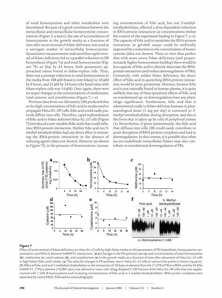
Mechanism(s) of downregulation of FRs during acute rever-sal of folate deficiency. As demonstrated in Figure 4,methionine did not stimulate the RNA-protein inter-action. This predicts that reversal of folate deficiency,which is associated with conversion of homocysteine tomethionine, would result in less RNA-protein interac-tion and downregulation of FRs. So in the next experi-mental system, HeLa-IU1-LF cells were abruptlyexposed to high-folate media and over the ensuing 24hours the rate of FR biosynthesis and concentrations
298 The Journal of Clinical Investigation | January 2004 | Volume 113 | Number 2
of total homocysteine and other metabolites weredetermined. Because of a good correlation between theintracellular and extracellular homocysteine concen-trations (Figure 3, a and e), the rate of accumulation ofhomocysteine in the growth media as a function oftime after acute reversal of folate deficiency was used asa surrogate marker of intracellular homocysteine.Quantitative measurements indicated that rapid rever-sal of folate deficiency led to a parallel reduction in FRbiosynthesis (Figure 7a) and total homocysteine (Fig-ure 7b) so that by 24 hours, both parameters ap-proached values found in folate-replete cells. Thus,there was a prompt reduction in total homocysteine inthe media from 108 µM (basal in low-folate) to 43 µMby 8 hours, and 21 µM by 24 hours (the basal value withfolate-replete cells was 14 µM). Once again, there wereno major changes in the concentrations of methionine,total cysteine, and cystathionine (Figure 7, c–e).
Previous data from our laboratory (38) predicted thatat the high concentrations of folic acid in media used topropagate HeLa-IU1-HF cells, folic acid could easily pas-sively diffuse into cells. Therefore, rapid replenishmentof folic acid to folate-deficient HeLa-IU1-LF cells (Figure7) introduced a new variable (folic acid) that could influ-ence RNA-protein interaction. Neither folic acid nor 5-methyl-tetrahydrofolate had any direct effect in increas-ing the RNA-protein interaction in the absence ofreducing agents (data not shown). However (as shownin Figure 7f), in the presence of homocysteine, increas-
ing concentrations of folic acid, but not 5-methyl-tetrahydrofolate, effected a dose-dependent reductionin RNA-protein interaction (at concentrations withinthe context of the experiment leading to Figure 7, a–e).The capacity of folic acid to modulate the RNA-proteininteraction in gel-shift assays could be artificiallyimproved by a reduction in the concentration of homo-cysteine (data not shown). These in vitro data predictthat with more severe folate deficiency (and propor-tionately higher homocysteine buildup) there would beless capacity of folic acid to directly dissociate the RNA-protein interaction (and induce downregulation of FRs).Conversely, with milder folate deficiency, the directeffect of folic acid in quenching RNA-protein interac-tion would be more prominent. However, because folicacid is not naturally found in human plasma, it is quiteunlikely that any of these (putative) effects of folic acidon translational up- or downregulation have any physi-ologic significance. Furthermore, folic acid that isadministered orally to folate-deficient humans in phar-macological doses (1 mg per day) is converted to 5-methyl-tetrahydrofolate during absorption, and this isthe form that is taken up by cells of peripheral tissues(1). Nevertheless, if given intravenously, the folic acidthat diffuses into cells (38) could easily contribute toacute disruption of RNA-protein complexes and lead todownregulation. In this context, it is possible that other(as-yet-undefined) intracellular folates may also con-tribute to translational downregulation of FRs.
Figure 7Effect of acute reversal of folate deficiency on HeLa-IU1-LF cells by high-folate media on the parameters of FR biosynthesis, homocysteine con-centration, and RNA cis-element–hnRNP E1 interactions. (a–e) Changes in the FR synthesis rate (a) and concentrations of total homocysteine(b), methionine (c), total cysteine (d), and cystathionine (e) in the growth media as a function of time after placement of HeLa-IU1–LF cellsin high-folate (folic acid) media. (a) The value for changes in FR synthetic rate in HeLa-IU1–LF cells at various time points is shown (squares).(f) Effect of folic acid and 5-methyltetrahydrofolate on the interaction of 18-base cis-element from the 5′-UTR of FR-α mRNA and the 43-kDahnRNP E1. [32P]cis-element (10,000 cpm) was allowed to react with 20 µg dialyzed S-100 fraction from HeLa-IU1-HF cells that was supple-mented with 1 mM dl-homocysteine and increasing concentrations of folic acid or 5-methyl-tetrahydrofolate. RNA-protein complexes wereseparated by native PAGE, followed by autoradiography.
The Journal of Clinical Investigation | January 2004 | Volume 113 | Number 2 299
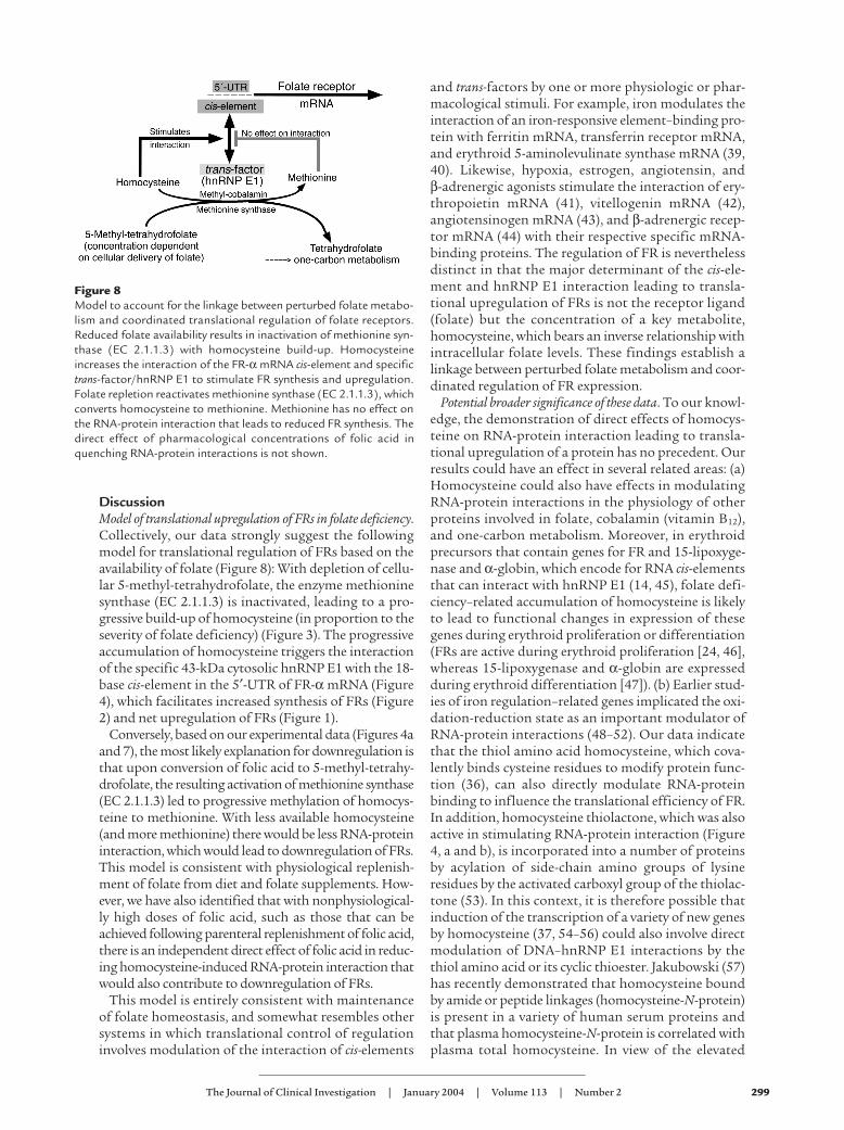
DiscussionModel of translational upregulation of FRs in folate deficiency.Collectively, our data strongly suggest the followingmodel for translational regulation of FRs based on theavailability of folate (Figure 8): With depletion of cellu-lar 5-methyl-tetrahydrofolate, the enzyme methioninesynthase (EC 2.1.1.3) is inactivated, leading to a pro-gressive build-up of homocysteine (in proportion to theseverity of folate deficiency) (Figure 3). The progressiveaccumulation of homocysteine triggers the interactionof the specific 43-kDa cytosolic hnRNP E1 with the 18-base cis-element in the 5′-UTR of FR-α mRNA (Figure4), which facilitates increased synthesis of FRs (Figure2) and net upregulation of FRs (Figure 1).
Conversely, based on our experimental data (Figures 4aand 7), the most likely explanation for downregulation isthat upon conversion of folic acid to 5-methyl-tetrahy-drofolate, the resulting activation of methionine synthase(EC 2.1.1.3) led to progressive methylation of homocys-teine to methionine. With less available homocysteine(and more methionine) there would be less RNA-proteininteraction, which would lead to downregulation of FRs.This model is consistent with physiological replenish-ment of folate from diet and folate supplements. How-ever, we have also identified that with nonphysiological-ly high doses of folic acid, such as those that can beachieved following parenteral replenishment of folic acid,there is an independent direct effect of folic acid in reduc-ing homocysteine-induced RNA-protein interaction thatwould also contribute to downregulation of FRs.
This model is entirely consistent with maintenanceof folate homeostasis, and somewhat resembles othersystems in which translational control of regulationinvolves modulation of the interaction of cis-elements
and trans-factors by one or more physiologic or phar-macological stimuli. For example, iron modulates theinteraction of an iron-responsive element–binding pro-tein with ferritin mRNA, transferrin receptor mRNA,and erythroid 5-aminolevulinate synthase mRNA (39,40). Likewise, hypoxia, estrogen, angiotensin, and β-adrenergic agonists stimulate the interaction of ery-thropoietin mRNA (41), vitellogenin mRNA (42),angiotensinogen mRNA (43), and β-adrenergic recep-tor mRNA (44) with their respective specific mRNA-binding proteins. The regulation of FR is neverthelessdistinct in that the major determinant of the cis-ele-ment and hnRNP E1 interaction leading to transla-tional upregulation of FRs is not the receptor ligand(folate) but the concentration of a key metabolite,homocysteine, which bears an inverse relationship withintracellular folate levels. These findings establish alinkage between perturbed folate metabolism and coor-dinated regulation of FR expression.
Potential broader significance of these data. To our knowl-edge, the demonstration of direct effects of homocys-teine on RNA-protein interaction leading to transla-tional upregulation of a protein has no precedent. Ourresults could have an effect in several related areas: (a)Homocysteine could also have effects in modulatingRNA-protein interactions in the physiology of otherproteins involved in folate, cobalamin (vitamin B12),and one-carbon metabolism. Moreover, in erythroidprecursors that contain genes for FR and 15-lipoxyge-nase and α-globin, which encode for RNA cis-elementsthat can interact with hnRNP E1 (14, 45), folate defi-ciency–related accumulation of homocysteine is likelyto lead to functional changes in expression of thesegenes during erythroid proliferation or differentiation(FRs are active during erythroid proliferation [24, 46],whereas 15-lipoxygenase and α-globin are expressedduring erythroid differentiation [47]). (b) Earlier stud-ies of iron regulation–related genes implicated the oxi-dation-reduction state as an important modulator ofRNA-protein interactions (48–52). Our data indicatethat the thiol amino acid homocysteine, which cova-lently binds cysteine residues to modify protein func-tion (36), can also directly modulate RNA-proteinbinding to influence the translational efficiency of FR.In addition, homocysteine thiolactone, which was alsoactive in stimulating RNA-protein interaction (Figure4, a and b), is incorporated into a number of proteinsby acylation of side-chain amino groups of lysineresidues by the activated carboxyl group of the thiolac-tone (53). In this context, it is therefore possible thatinduction of the transcription of a variety of new genesby homocysteine (37, 54–56) could also involve directmodulation of DNA–hnRNP E1 interactions by thethiol amino acid or its cyclic thioester. Jakubowski (57)has recently demonstrated that homocysteine boundby amide or peptide linkages (homocysteine-N-protein)is present in a variety of human serum proteins andthat plasma homocysteine-N-protein is correlated withplasma total homocysteine. In view of the elevated
Figure 8Model to account for the linkage between perturbed folate metabo-lism and coordinated translational regulation of folate receptors.Reduced folate availability results in inactivation of methionine syn-thase (EC 2.1.1.3) with homocysteine build-up. Homocysteineincreases the interaction of the FR-α mRNA cis-element and specifictrans-factor/hnRNP E1 to stimulate FR synthesis and upregulation.Folate repletion reactivates methionine synthase (EC 2.1.1.3), whichconverts homocysteine to methionine. Methionine has no effect onthe RNA-protein interaction that leads to reduced FR synthesis. Thedirect effect of pharmacological concentrations of folic acid inquenching RNA-protein interactions is not shown.

300 The Journal of Clinical Investigation | January 2004 | Volume 113 | Number 2
homocysteine within cells during folate deficiency, ourstudies predict that homocysteine could (either direct-ly or indirectly) alter the function of hnRNP E1 andincrease its affinity for the cis-element. To resolve thisissue, we have initiated site-directed mutagenesis ofspecific amino acid residues that may be involved with-in a putative RNA-binding pocket of hnRNP E1 (Y.S.Tang, R.A. Khan, S. Xiao, H.N. Jayaram, and A.C.Antony, unpublished observations); our preliminarydata appear to support a direct effect of homocysteineon hnRNP E1. (c) Because chronic hyper-homocys-teinemia is a major risk factor in a variety of occlusivevascular diseases (58–65), some neural tube defects(66–68), congenital heart defects (69), and, as demon-strated more recently, dementia and Alzheimer disease(70), the direct effects of homocysteine on RNA-proteinand possibly DNA-protein interactions could have arole in the etiology and pathogenesis of these diseases(71). Moreover, since approximately 13% of homocys-teine can enter cells from the extracellular media, it ispossible that with significant levels of chronic hyper-homocysteinemia, as noted with congenital defects infolate and cobalamin metabolism (72), entry of thethiol intracellularly has the potential to trigger addi-tional effects on gene transcription and/or translation.(d) Finally, because of the recent documentation of arole of FRs in mediating the cellular entry of Ebolavirus (73), our demonstration that acute downregula-tion can occur within 24 hours of exposure to highconcentrations of folic acid may be of some practicalimportance. Aerosolized Ebola virus can be transmit-ted between humans through the respiratory route. Asbronchial glands and alveolar lining cells (includingtype I and type II pneumocytes) highly overexpress FRs(74), it is theoretically possible that rapid downregula-tion of FRs by folic acid may help to reduce the risk ofuptake of virus among noninfected individuals (includ-ing health care personnel entering an epidemic zone).
AcknowledgmentsThis work was supported in part by NIH grantsR01CA58919 and R01HD39295 (to A.C. Antony) andR01AG09834 (to S.P. Stabler), and a Veterans AffairsMerit Review Award (to A.C. Antony).
1. Antony, A.C. 2000. Megaloblastic anemias. In Hematology: basic principlesand practice. R. Hoffman et al., editors. Churchill-Livingstone. New York,New York, USA. 446–485.
2. Antony, A.C. 1996. Folate receptors. Annu. Rev. Nutr. 16:501–521.3. Sun, X.L., et al. 1995. Transduction of folate receptor cDNA into cervi-
cal carcinoma cells using recombinant adeno-associated virions delayscell proliferation in vitro and in vivo. J. Clin. Invest. 96:1535–1547.
4. Antony, A.C. 1992. The biological chemistry of folate receptors. Blood.79:2807–2820.
5. McHugh, M., and Cheng, Y.C. 1979. Demonstration of a high affinityfolate binder in human cell membranes and its characterization in cul-tured human KB cells. J. Biol. Chem. 254:11312–11318.
6. Kane, M.A., et al. 1988. Influence on immunoreactive folate-binding pro-teins of extracellular folate concentration in cultured human cells. J. Clin.Invest. 81:1398–1406.
7. Henderson, G.B., Tsuji, J.M., and Kumar, H.P. 1988. Mediated uptake offolate by a high-affinity binding protein in sublines of L1210 cells adapt-ed to nanomolar concentrations of folate. J. Membr. Biol. 101:247–258.
8. Jansen, G., et al. 1989. Expression of a folate binding protein in L1210cells grown in low folate medium. Cancer Res. 49:1959–1963.
9. Sadasivan, E., and Rothenberg, S.P. 1989. The complete amino acidsequence of a human folate binding protein from KB cells deter-mined from the cDNA [erratum 1990, 265:1821]. J. Biol. Chem.264:5806–5811.
10. Hsueh, C.T., and Dolnick, B.J. 1993. Altered folate-binding proteinmRNA stability in KB cells grown in folate-deficient medium. Biochem.Pharmacol. 45:2537–2545.
11. Zhu, W.Y., Alliegro, M.A., and Melera, P.W. 2001. The rate of folate recep-tor α (FRα) synthesis in folate depleted CHL cells is regulated by a trans-lational mechanism sensitive to media folate levels, while stable overex-pression of its mRNA is mediated by gene amplification and an increasein transcript half-life. J. Cell. Biochem. 81:205–219.
12. Sadasivan, E., Regec, A., and Rothenberg, S.P. 2002. The half-life of thetranscript encoding the folate receptor α in KB cells is reduced by cytoso-lic proteins expressed in folate-replete and not in folate-depleted cells.Gene. 291:149–158.
13. Sun, X.L., and Antony, A.C. 1996. Evidence that a specific interactionbetween an 18-base cis-element in the 5′-untranslated region of humanfolate receptor-α mRNA and a 46-kDa cytosolic trans-factor is criticalfor translation. J. Biol. Chem. 271:25539–25547.
14. Xiao, X., et al. 2001. Isolation and characterization of a folate receptormRNA-binding trans-factor from human placenta: Evidence favoringidentity with heterogeneous nuclear ribonucleoprotein E1. J. Biol. Chem.276:41510–41517.
15. Ostareck, D.H., et al. 1997. mRNA silencing in erythroid differentiation:hnRNP K and hnRNP E1 regulate 15-lipoxygenase translation from the3′ end. Cell. 89:597–606.
16. Pillai, M.R., et al. 2003. Expression of folate receptors and heterogeneousnuclear ribonucleoprotein-E1 in women with human papillomavirus-mediated transformation of cervical tissue to cancer. J. Clin. Path.56:569–574.
17. Elwood, P.C. 1989. Molecular cloning and characterization of thehuman folate-binding protein cDNA from placenta and malignant tis-sue culture (KB) cells. J. Biol. Chem. 264:14893–14901.
18. Shen, F., Ross, J.F., Wang, X., and Ratnam, M. 1994. Identification of anovel folate receptor, a truncated receptor, and receptor type β inhematopoietic cells: cDNA cloning, expression, immunoreactivity, andtissue specificity. Biochemistry. 33:1209–1215.
19. Kolhouse, J.F., Stabler, S.P., and Allen, R.H. 1993. Identification and per-turbation of mutant human fibroblasts based on measurements ofmethylmalonic acid and total homocysteine in the culture media. Arch.Biochem. Biophys. 303:355–360.
20. Hultberg, B., Andersson, A., and Isaksson, A. 1995. Metabolism of homo-cysteine, its relation to the other cellular thiols and its mechanism of celldamage in a cell culture line (human histiocytic cell line U-937). Biochim.Biophys. Acta. 1269:6–12.
21. Jayaram, H.N., Zhen, W., and Gharehbaghi, K. 1993. Biochemical con-sequences of resistance to tiazofurin in human myelogenous leukemicK562 cells. Cancer Res. 53:2344–2348.
22. Sun, X.L., et al. 1999. Modulation of the cytotoxicity of 3′-azido-3′-deoxythymidine and methotrexate after transduction of folate recep-tor cDNA into human cervical carcinoma: identification of a correla-tion between folate receptor expression and thymidine kinase activity.Cancer Res. 59:940–946.
23. Antony, A.C., Kane, M.A., Portillo, R.M., Elwood, P.C., and Kolhouse, J.F.1985. Studies of the role of a particulate folate-binding protein in theuptake of 5-methyltetrahydrofolate by cultured human KB cells. J. Biol.Chem. 260:14911–14917.
24. Antony, A.C., et al. 1991. Megaloblastic hematopoiesis in vitro. Interac-tion of anti-folate receptor antibodies with hematopoietic progenitorcells leads to a proliferative response independent of megaloblasticchanges. J. Clin. Invest. 87:313–325.
25. Sambrook, J., Fritsch, E.F., and Maniatis, T. 1989. Molecular cloning: a lab-oratory manual. 2nd edition. Cold Spring Harbor Laboratory Press. Plain-view, New York, USA. 5.33–5.56.
26. Weber, B., Horiguchi, J., Luebbers, R., Sherman, M., and Kufe, D. 1989.Posttranscriptional stabilization of c-fms mRNA by a labile protein dur-ing human monocytic differentiation. Mol. Cell Biol. 9:769–775.
27. Verma, R.S., Gullapalli, S., and Antony, A.C. 1992. Evidence that thehydrophobicity of isolated, in situ, and de novo-synthesized nativehuman placental folate receptors is a function of glycosyl-phos-phatidylinositol anchoring to membranes. J. Biol. Chem. 267:4119–4127.
28. Antony, A.C., Utley, C., Van Horne, K.C., and Kolhouse, J.F. 1981. Isola-tion and characterization of a folate receptor from human placenta. J. Biol. Chem. 256:9684–9692.
29. Stabler, S.P., et al. 1988. Elevation of total homocysteine in the serum ofpatients with cobalamin or folate deficiency detected by capillary gaschromatography-mass spectrometry. J. Clin. Invest. 81:466–474.
30. Allen, R.H., Stabler, S.P., Savage, D.G., and Lindenbaum, J. 1993. Meta-bolic abnormalities in cobalamin (vitamin B12) and folate deficiency.FASEB J. 7:1344–1353.
31. Savage, D.G., Lindenbaum, J., Stabler, S.P., and Allen, R.H. 1994. Sensi-
The Journal of Clinical Investigation | January 2004 | Volume 113 | Number 2 301
tivity of serum methylmalonic acid and total homocysteine determina-tions for diagnosing cobalamin and folate deficiencies. Am. J. Med.96:239–246.
32. Stabler, S., Allen, R., Savage, D., and Lindenbaum, J. 1991. Diagnosis ofcobalamin deficiency. Blood. 77:1854.
33. Luhrs, C.A. 1991. The role of glycosylation in the biosynthesis and acqui-sition of ligand-binding activity of the folate-binding protein in culturedKB cells. Blood. 77:1171–1180.
34. Peters, B.P., et al. 1983. The use of drugs to dissect the pathway for secre-tion of the glycoprotein hormone chorionic gonadotropin by culturedhuman trophoblastic cells. J. Biol. Chem. 258:14505–14515.
35. Banerjee, R.V., and Matthews, R.G. 1990. Cobalamin-dependent methio-nine synthase. FASEB J. 4:1450–1459.
36. Hajjar, K.A., et al. 1998. Tissue plasminogen activator binding to theannexin II tail domain. Direct modulation by homocysteine. J. Biol. Chem.273:9987–9993.
37. Chacko, G., Ling, Q., and Hajjar, K.A. 1998. Induction of acute transla-tional response genes by homocysteine. Elongation factors-1α, -β, and -δ.J. Biol. Chem. 273:19840–19846.
38. Antony, A.C., Kane, M.A., Krishnan, S.R., Kincade, R.S., and Verma, R.S.1989. Folate (pteroylglutamate) uptake in human red blood cells, ery-throid precursors and KB cells at high extracellular folate concentra-tions. Evidence against a role for specific folate-binding and transportproteins. Biochem. J. 260:401–411.
39. Constable, A., Quick, S., Gray, N.K., and Hentze, M.W. 1992. Modula-tion of the RNA-binding activity of a regulatory protein by iron in vitro:switching between enzymatic and genetic function? Proc. Natl. Acad. Sci.U. S. A. 89:4554–4558.
40. Melefors, O., et al. 1993. Translational control of 5-aminolevulinate syn-thase mRNA by iron-responsive elements in erythroid cells. J. Biol. Chem.268:5974–5978.
41. Rondon, I.J., et al. 1991. Hypoxia up-regulates the activity of a novel ery-thropoietin mRNA binding protein. J. Biol. Chem. 266:16594–16598.
42. Dodson, R.E., and Shapiro, D.J. 1994. An estrogen-inducible proteinbinds specifically to a sequence in the 3′ untranslated region of estrogen-stabilized vitellogenin mRNA. Mol. Cell Biol. 14:3130–3138.
43. Klett, C., Bader, M., Ganten, D., and Hackenthal, E. 1994. Mechanism bywhich angiotensin II stabilizes messenger RNA for angiotensinogen.Hypertension. 23:I120–I125.
44. Huang, L.Y., Tholanikunnel, B.G., Vakalopoulou, E., and Malbon, C.C.1993. The Mr 35,000 β-adrenergic receptor mRNA-binding proteininduced by agonists requires both an AUUUA pentamer and U-richdomains for RNA recognition. J. Biol. Chem. 268:25769–25775.
45. Antony, A.C., Tang, Y.-S., Xiao, X., Mackins, J.Y., and Stabler, S.P. 2001.Mechanism of translational up-regulation of folate receptors (FR). 20thAnnual Convention of the Indian Association for Cancer Research (AbstractBook), Jan. 19-21, 2001. Ahmedabad, Gujerat State, India.
46. Antony, A.C., et al. 1987. Effect of perturbation of specific folate recep-tors during in vitro erythropoiesis. J. Clin. Invest. 80:1618–1623.
47. Ostareck-Lederer, A., Ostareck, D.H., and Hentze, M.W. 1998. Cytoplas-mic regulatory functions of the KH-domain proteins hnRNPs K andE1/E2. Trends Biochem. Sci. 23:409–411.
48. Rouault, T.A., Hentze, M.W., Caughman, S.W., Harford, J.B., and Klaus-ner, R.D. 1988. Binding of a cytosolic protein to the iron-responsive ele-ment of human ferritin messenger RNA. Science. 241:1207–1210.
49. Hentze, M.W., Rouault, T.A., Harford, J.B., and Klausner, R.D. 1989. Oxi-dation-reduction and the molecular mechanism of a regulatory RNA-protein interaction. Science. 244:357–359.
50. Lin, J.J., et al. 1990. Derepression of ferritin messenger RNA translationby hemin in vitro. Science. 247:74–77.
51. Goessling, L.S., Daniels-McQueen, S., Bhattacharyya-Pakrasi, M., Lin,J.J., and Thach, R.E. 1992. Enhanced degradation of the ferritin repres-
sor protein during induction of ferritin messenger RNA translation.Science. 256:670–673.
52. Rouault, T.A., Stout, C.D., Kaptain, S., Harford, J.B., and Klausner, R.D.1991. Structural relationship between an iron-regulated RNA-bindingprotein (IRE-BP) and aconitase: functional implications. Cell64:881–883.
53. Jakubowski, H. 1997. Metabolism of homocysteine thiolactone inhuman cell cultures. Possible mechanism for pathological consequencesof elevated homocysteine levels. J. Biol. Chem. 272:1935–1942.
54. Lubec, B., et al. 1996. Homocysteine increases cyclin-dependent kinasein aortic rat tissue. Circulation. 94:2620–2625.
55. Tsai, J.C., et al. 1996. Induction of cyclin A gene expression by homocys-teine in vascular smooth muscle cells. J. Clin. Invest. 97:146–153.
56. Kokame, K., Kato, H., and Miyata, T. 1996. Homocysteine-respondentgenes in vascular endothelial cells identified by differential display analy-sis. GRP78/BiP and novel genes. J. Biol. Chem. 271:29659–29665.
57. Jakubowski, H. 2002. Homocysteine is a protein amino acid inhumans. Implications for homocysteine-linked disease. J. Biol. Chem.277:30425–30428.
58. Graham, I.M., et al. 1997. Plasma homocysteine as a risk factor for vas-cular disease. The European Concerted Action Project. JAMA.277:1775–1781.
59. Mayer, E.L., Jacobsen, D.W., and Robinson, K. 1996. Homocysteine andcoronary atherosclerosis. J. Amer. Coll. Cardiol. 27:517–527.
60. Aronow, W.S., Ahn, C., and Schoenfeld, M.R. 1997. Association betweenplasma homocysteine and extracranial carotid arterial disease in olderpersons. Am. J. Cardiol. 79:1432–1433.
61. Dennis, V.W., and Robinson, K. 1996. Homocysteinemia and vasculardisease in end-stage renal disease. Kidney Int. Suppl. 57:S11–S17.
62. Robinson, K., et al. 1996. Hyperhomocysteinemia confers an inde-pendent increased risk of atherosclerosis in end-stage renal diseaseand is closely linked to plasma folate and pyridoxine concentrations.Circulation. 94:2743–2748.
63. Konecky, N., et al. 1997. Correlation between plasma homocyst(e)ine andaortic atherosclerosis. Am. Heart J. 133:534–540.
64. Refsum, H., Ueland, P.M., Nygard, O., and Vollset, S.E. 1998. Homocys-teine and cardiovascular disease. Annu. Rev. Med. 49:31–62.
65. Goddijn-Wessel, T.A., et al. 1996. Hyperhomocysteinemia: a risk factorfor placental abruption or infarction. Eur. J. Obstet. Gynecol. Reprod. Biol.66:23–29.
66. Whitehead, A.S., et al. 1995. A genetic defect in 5,10 methylenetetrahy-drofolate reductase in neural tube defects. Quart. J. Med. 88:763–766.
67. van der Put, N.M., et al. 1995. Mutated methylenetetrahydrofolate reduc-tase as a risk factor for spina bifida. Lancet. 346:1070–1071.
68. Posey, D.L., Khoury, M.J., Mulinare, J., Adams, M.J., Jr., and Ou, C.Y.1996. Is mutated MTHFR a risk factor for neural tube defects? Lancet.347:686–687.
69. Rosenquist, T.H., Ratashak, S.A., and Selhub, J. 1996. Homocysteineinduces congenital defects of the heart and neural tube: effect of folicacid. Proc. Natl. Acad. Sci. U. S. A. 93:15227–15232.
70. Seshadri, S., et al. 2002. Plasma homocysteine as a risk factor for demen-tia and Alzheimer’s disease. N. Engl. J. Med. 346:476–483.
71. Antony, A.C., and Hansen, D.K. 2000. Hypothesis: folate-responsive neu-ral tube defects and neurocristopathies. Teratology. 62:42–50.
72. Rosenblatt, D.S., and Whitehead, V.M. 1999. Cobalamin and folate defi-ciency: acquired and hereditary disorders in children. Semin. Hematol.36:19–34.
73. Chan, S.Y., et al. 2001. Folate receptor-alpha is a cofactor for cellularentry by Marburg and Ebola viruses. Cell. 106:117–126.
74. Weitman, S.D., et al. 1992. Cellular localization of the folate receptor:potential role in drug toxicity and folate homeostasis. Cancer Res.52:6708–6711.