Hydrogen Sulfide Regulates Homocysteine-Mediated Glomerulosclerosis
14
Fax +41 61 306 12 34 E-Mail [email protected] www.karger.com Original Report: Laboratory Investigation Am J Nephrol 2010;31:442–455 DOI: 10.1159/000296717 Hydrogen Sulfide Regulates Homocysteine-Mediated Glomerulosclerosis Utpal Sen Charu Munjal Natia Qipshidze Oluwasegun Abe Riyad Gargoum Suresh C. Tyagi Department of Physiology and Biophysics, University of Louisville School of Medicine, Louisville, Ky., USA molecule-1 and vascular cell adhesion molecule-1, as well as increased macrophage infiltration, were detected in WT 1K, CBS+/– 2K and CBS+/– 1K mice. These changes were amelio- rated with H 2 S supplementation. Conclusion: Together, these results suggest that increased oxidative stress and de- creased H 2 S in HHcy causes matrix remodeling and inflam- mation resulting in glomerulosclerosis and reduced renal function. Copyright © 2010 S. Karger AG, Basel Introduction It is now well established that hyperhomocysteinemia (HHcy), an increased plasma homocysteine (Hcy) level, is a potent inducer of endothelial dysfunction, particu- larly in small vessels [1]. HHcy promotes atherosclerosis and thrombosis in susceptible animals, such as cystathio- nine -synthase heterozygous (CBS+/–) mice fed with a high methionine diet [2]. The pathophysiological mecha- nisms of these effects, however, are less understood in the kidney, particularly in the process of glomerulosclerosis. An increase in the Hcy level has been shown to induce oxidative stress through reactive oxygen species in the kidney [3, 4] and has been reported to induce local oxida- tive stress, mesangial expansion and podocyte dysfunc- Key Words Collagen Matrix metalloproteinase Inflammation Fibrosis Hypertension Renal dysfunction Abstract Background/Aims: In this study we tested the hypothesis that H 2 S regulates collagen deposition, matrix metallopro- teinases (MMP) and inflammatory molecules during hyper- homocysteinemia (HHcy) resulting in attenuation of glomer- ulosclerosis and improved renal function. Materials and Methods: A genetic model of HHcy, cystathionine -syn- thase heterozygous (CBS+/–) and wild-type (WT) 2-kidney (2K) mice were used in this study and supplemented with or without NaHS (30 mol/l, H 2 S donor) in drinking water for 8 weeks. To expedite the renal damage associated with HHcy, uninephrectomized (1K) mice of similar groups were also used. Results: Results demonstrated that NAD(P)H oxidase (p47 phox subunit) and blood pressure were upregulated in WT 1K, CBS+/– 2K and CBS+/– 1K mice with downregulation of H 2 S production and reduced glomerular filtration rate. These changes were normalized with H 2 S supplementation. Both pro- and active MMP-2 and -9 and collagen protein ex- pressions and glomerular depositions were also upregulated in WT 1K, CBS+/– 2K and CBS+/– 1K mice. Increased expres- sions of inflammatory molecules, intercellular cell adhesion Received: January 11, 2010 Accepted: March 8, 2010 Published online: April 16, 2010 Nephrolo gy American Journal of Utpal Sen Department of Physiology and Biophysics University of Louisville School of Medicine 500 South Preston Street, A-1214, Louisville, KY 40202 (USA) Tel. +1 502 852 3627, Fax +1 502 852 6239, E-Mail u0sen001 @ louisville.edu © 2010 S. Karger AG, Basel Accessible online at: www.karger.com/ajn
-
Upload
independent -
Category
Documents
-
view
1 -
download
0
Transcript of Hydrogen Sulfide Regulates Homocysteine-Mediated Glomerulosclerosis

Fax +41 61 306 12 34E-Mail [email protected]
Original Report: Laboratory Investigation
Am J Nephrol 2010;31:442–455 DOI: 10.1159/000296717
Hydrogen Sulfide RegulatesHomocysteine-Mediated Glomerulosclerosis
Utpal Sen Charu Munjal Natia Qipshidze Oluwasegun Abe Riyad Gargoum Suresh C. Tyagi
Department of Physiology and Biophysics, University of Louisville School of Medicine, Louisville, Ky. , USA
molecule-1 and vascular cell adhesion molecule-1, as well as increased macrophage infiltration, were detected in WT 1K, CBS+/– 2K and CBS+/– 1K mice. These changes were amelio-rated with H 2 S supplementation. Conclusion: Together, these results suggest that increased oxidative stress and de-creased H 2 S in HHcy causes matrix remodeling and inflam-mation resulting in glomerulosclerosis and reduced renal function. Copyright © 2010 S. Karger AG, Basel
Introduction
It is now well established that hyperhomocysteinemia (HHcy), an increased plasma homocysteine (Hcy) level, is a potent inducer of endothelial dysfunction, particu-larly in small vessels [1] . HHcy promotes atherosclerosis and thrombosis in susceptible animals, such as cystathio-nine � -synthase heterozygous (CBS+/–) mice fed with a high methionine diet [2] . The pathophysiological mecha-nisms of these effects, however, are less understood in the kidney, particularly in the process of glomerulosclerosis.
An increase in the Hcy level has been shown to induce oxidative stress through reactive oxygen species in the kidney [3, 4] and has been reported to induce local oxida-tive stress, mesangial expansion and podocyte dysfunc-
Key Words Collagen � Matrix metalloproteinase � Inflammation � Fibrosis � Hypertension � Renal dysfunction
Abstract Background/Aims: In this study we tested the hypothesis that H 2 S regulates collagen deposition, matrix metallopro-teinases (MMP) and inflammatory molecules during hyper-homocysteinemia (HHcy) resulting in attenuation of glomer-ulosclerosis and improved renal function. Materials and Methods: A genetic model of HHcy, cystathionine � -syn-thase heterozygous (CBS+/–) and wild-type (WT) 2-kidney (2K) mice were used in this study and supplemented with or without NaHS (30 � mol/l, H 2 S donor) in drinking water for 8 weeks. To expedite the renal damage associated with HHcy, uninephrectomized (1K) mice of similar groups were also used. Results: Results demonstrated that NAD(P)H oxidase (p47 phox subunit) and blood pressure were upregulated in WT 1K, CBS+/– 2K and CBS+/– 1K mice with downregulation of H 2 S production and reduced glomerular filtration rate. These changes were normalized with H 2 S supplementation. Both pro- and active MMP-2 and -9 and collagen protein ex-pressions and glomerular depositions were also upregulated in WT 1K, CBS+/– 2K and CBS+/– 1K mice. Increased expres-sions of inflammatory molecules, intercellular cell adhesion
Received: January 11, 2010 Accepted: March 8, 2010 Published online: April 16, 2010
NephrologyAmerican Journal of
Utpal Sen Department of Physiology and BiophysicsUniversity of Louisville School of Medicine 500 South Preston Street, A-1214, Louisville, KY 40202 (USA) Tel. +1 502 852 3627, Fax +1 502 852 6239, E-Mail u0sen001 @ louisville.edu
© 2010 S. Karger AG, Basel
Accessible online at:www.karger.com/ajn

Glomerular Remodeling and Hydrogen Sulfide
Am J Nephrol 2010;31:442–455 443
tion resulting in renal fibrosis [5] . In rat mesangial cell culture, Hcy promotes collagen accumulation and is as-sociated with increased NAD(P)H activity [5] . This sug-gests that Hcy induces local oxidative stress, cellular dys-function and extracellular matrix metabolism in the glo-merulus, all of which are associated with increased NAD(P)H activity.
The three enzymes CBS, cystathionine � -lyase (CSE) and 3-mercaptopyruvate sulfurtransferase (3MST) me-tabolize Hcy to produce H 2 S in the body [6–9] . The phys-iological function of endogenous H 2 S is not clear and could be multifaceted; however, it is involved in the regu-lation of vascular tone [10] and protects neuronal cells from oxidative stress by increasing the intracellular con-centration of antioxidant (glutathione) [11] . Increasing evidence suggests the potential antioxidant properties of H 2 S in normal and pathophysiological conditions [12–14] . In addition, recent reports have demonstrated that H 2 S is a potential anti-inflammatory substance [10, 15, 16] . However, the physiological role of H 2 S in HHcy-as-sociated renal remodeling is incompletely defined.
Matrix metalloproteinases (MMPs) degrade both the collagenous and noncollagenous components of the extracellular matrix, and are thereby actively involvedin matrix turnover. Gelatinases, members of the family of MMPs, digest these products into smaller peptides. Among gelatinases, MMP-2 and MMP-9 have gained po-tential interest because of their capability to disrupt the kidney architecture [17–19] . Similarly, experimental evi-dence implicated sustained elevation of cell adhesion molecules, such as intercellular cell adhesion molecule-1 (ICAM-1) and vascular cell adhesion molecule-1 (VCAM-1), in chronic inflammatory disorder, which leads to scle-rosis [20] . HHcy has been reported to increase the expres-sion of ICAM-1 [19] and VCAM-1 [19, 21] in experimen-tal models by independent laboratories, including our own. In addition, macrophage infiltration in the kidney is one of the most important events for progression of ne-phropathy [22] . Despite these facts, however, the major contributing factors of HHcy-associated inflammation in the process of glomerulosclerosis still need to be de-fined comprehensively.
In this study we tested the hypothesis that HHcy-in-duced oxidative stress upregulates collagen deposition in the glomerulus leading to glomerulosclerosis through modulation of MMPs and inflammatory molecules. In addition, the regulatory role of H 2 S to modulate this re-nal remodeling process was determined in an HHcy kidney.
Materials and Methods
Animals Wild-type (WT, C57BL/6J) and CBS+/– mice aged 8 weeks
were obtained from Jackson Laboratories (Bar Harbor, Me., USA) and bred at the animal care facility at the University of Louisville. Genotypes of these mice were determined and 10-week-old male mice were used for this study. Mice were divided into 2 sets. The first set of mice had 2 normal kidneys (2K) and were divided into the following groups: WT, CBS+/–, WT + H 2 S [30 � mol/l for 8 weeks, NaHS (sodium hydrosulfide) was used as a donor of H 2 S] and CBS+/– + H 2 S (30 � mol/l for 8 weeks). The second set of mice were uninephrectomized (1K) and separated as the 4 groups above. At the end of the experiments, mice were deeply anesthe-tized and sacrificed to harvest the tissues. All animal procedures were in accordance with the National Institute of Health Guide-lines for Animal Research and were approved by the Institutional Animal Care and Use Committee of the University of Louisville School of Medicine.
H 2 S Treatment H 2 S was supplemented to mice through drinking water in the
form of NaHS, which in an aqueous solution produces an equal concentration of H 2 S. Typically, physiological ranges of H 2 S vary from 10–100 � mol/l [12, 23] and can rise as high as 160 � mol/l in the human plasma during septic shock [24] . To keep H 2 S supple-mentation within the physiological range, we supplemented ani-mals with 30 � mol/l of H 2 S. The density of H 2 S is about 18% higher than the air; therefore, the evaporation rate of H 2 S from the solutions is normally low. Moreover, the water bottle had a drinking tube with a small aperture, which minimized evapora-tion. In order to make sure animals took the appropriate amount of H 2 S through drinking water, approximately every 12 h we changed the water with fresh H 2 S in the appropriate groups. Within this period we observed a negligible amount of H 2 S loss (approx. 2–3%). Also, water consumption in treated versus non-treated groups was recorded, but no differences were found. No apparent toxicities of H 2 S were noticed other than its effect on blood pressure (reported in ‘Results’).
Cell Culture Mouse kidney mesangial cells were purchased from ATTC,
and were cultured and maintained in T-25 flasks. The culture me-dium was DMEM/F-12 (50/50) containing 10% fetal calf serum and antibiotics (Cellgro; Mediatech Inc., Herndon, Va., USA). Cells were trypsinized as described earlier [19] and plated onto 8-well chamber slides. After experiments, the cells were processed for immunostaining and visualized under a laser scanning confo-cal microscope (FluoView 1000, Olympus) using an appropriate filter.
Antibodies and Reagents Rat monoclonal antibody F4/80 was purchased from Abcam
(Cambridge, Mass., USA). Anti-p47 phox , MMP-2 and -9, collagen IV, ICAM-1, VCAM-1 and HRP-linked secondary antibodies were from Santa Cruz Biotechnology (Santa Cruz, Calif., USA). Anti- � -actin antibody, NaHS and other analytical reagents were from Sigma-Aldrich (St. Louis, Mo., USA). Polyvinylidene difluo-ride membrane was from Bio-Rad (Hercules, Calif., USA).
Sen/Munjal/Qipshidze/Abe/Gargoum/Tyagi
Am J Nephrol 2010;31:442–455444
Cryosectioning At the end of the experiments, kidneys were excised and cryo-
preserved in Peel-A-Way disposable plastic tissue embedding molds (Polysciences Inc., Warrington, Pa., USA) containing tis-sue freezing media (Triangle Biomedical Sciences, Durham, N.C., USA). These molds were kept frozen (–70 ° C) until serial 5- � m (for histological staining) or 2- � m (for immunostaining) tissue sections were made in cryocut (Leica CM 1850). Cryosections were placed on Superfrost Plus microscope slides, air-dried and processed for staining.
Western Blots The kidney cortical tissues were minced into fine particles
with scissors and incubated with protein extraction buffer (0.01 mol/l cacodylic acid pH 5.0, 0.15 mol/l NaCl, 1 � mol/l ZnCl 2 , 0.02 mol/l CaCl 2 , 0.0015 mol/l NaN 3 and 0.01% v/v Triton X-100) over-night at 4 ° C with gentle agitation. The extracted protein was col-lected and pH rose to 7.4 by adding 1.5 mol/l Tris (pH 8.8.). An equal amount of protein was analyzed by 10% SDS-PAGE, trans-ferred to a polyvinylidene difluoride membrane and probed with appropriate antibodies followed by reprobing with � -actin as de-scribed earlier [19] .
Immunostaining Cryosectioned (2- � m) renal tissue samples and cells in the
8-well chamber slides were washed with PBS (pH 7.4) and blocked with 1% BSA for 15 min followed by 2 washes with PBS (5 min each). Slides were then fixed with 4% paraformaldehyde contain-ing 0.25% L - � -lysophosphatidylcholine for 30 min. After washing with PBS (3 ! , 5 min each), samples were blocked with 1% BSA for 1 h. Slides were then washed twice, primary antibody (p47 phox ,1: 100 dilutions in 1% BSA) was added, and incubated overnight at 4 ° C with gentle agitation. Excess antibody was washed with PBS (3 ! , 5 min each) and secondary fluorescence-conjugated anti-body (1: 500 dilutions in 1% BSA) was added and incubated for 2 h. Unbound secondary antibody was removed by PBS wash (3 ! , 5 min each), mounted on slides and visualized with fluorescence in a laser scanning confocal microscope (Olympus FluoView 1000) with an appropriate filter.
Masson’s Trichrome Staining Cryosectioned kidney tissue samples (5- � m) were processed
for Masson’s trichrome (Richard-Allan Scientific, Kalamazoo, Mich., USA) staining according to the manufacturer’s instruc-tions for detecting collagen. Collagen appeared as a blue color.
RT-PCR Total RNA was extracted from renal cortical tissue using
Trizol Reagent (Gibco BRL) according to the manufacturer’s in-structions. The cDNA template was synthesized using a Promega kit. Primer sequences for mouse collagen IV were used as de-scribed elsewhere [25] .
Peroxidase-Based DAB Immunohistochemical Staining Kidney tissues were cryosectioned (2- � m thickness; Leica,
CM 1850)and placed on slides, with the slides brought to room temperature before being washed with PBS (pH 7.4, 2 ! , 5 min each). The sections were then blocked with 1% BSA (in PBS)for 15 min, washed with PBS (2 ! , 5 min each) and fixed with4% paraformaldehyde containing L - � -lysophosphatidylcholine
(0.25% w/v) for 1 h. Following fixation, sections were washed with PBS (3 ! , 5 min each) and then blocked again with 1% BSA for1 h. The blocking solution was discarded and the slides were washed with PBS (2 ! , 5 min each) followed by incubation with primary antibody (1: 100 in 1% BSA) overnight at 4 ° C. Slides were washed with PBS (3 ! , 5 min each) to remove excess antibody, fol-lowed by incubation with HRP-conjugated secondary antibody (1: 250 in 1% BSA) for 2 h at room temperature. Slides were then washed with PBS (3 ! , 5 min each) and DAB substrate-chromogen was applied, incubated at 37 ° C for 30 min, rinsed with distilled water and mounted in mounting medium. DAB staining was vi-sualized under a microscope with dark brown end-product at the site of the target antigen.
Measurement of Glomerular Filtration Rate Alzet Mini-Osmotic Pumps (Durect Corp., Cupertino, Calif.,
USA) with a constant flow rate (8 � l/h) filled with 200 � l of 5% FITC-inulin (dialyzed) were inserted into the peritoneal cavity through a small midline incision under light anesthesia. Animals were then placed in metabolic cages. Both collected plasma and urine samples were buffered at pH 7.4 with 500 mmol/l HEPES before taking the FITC fluorescence value to minimize the effect of pH [26] . The titrated 50- � l samples were then loaded onto each well of a 96-well plate. Fluorescence was determined using Spectra Max (Molecular Device, Sunnyvale, Calif., USA) with 485-nm excitation and read at 538-nm emission. Glomerular filtration rate (GFR) was calculated based on inulin clearance using the 24-hour urinary FITC-inulin excretion rate (urinary fluorescence counts/24 h) divided by the concentration of plasma FITC-inulin (fluorescence counts/ � l).
Statistical Analysis Values are given as means 8 SEM from ‘n’ numbers of animals
in each group as mentioned in each of the figure legends. The dif-ference between mean values of multiple groups were analyzed by one-way analysis of variance (ANOVA) followed by Scheffe’s post hoc analysis. Comparisons between groups were made with the use of Student’s independent t test. Significance was accepted atp ! 0.05.
Results
Endogenous Production of Tissue H 2 S and CSE Expression Was Attenuated in HHcy Mice CBS+/– mice are hyperhomocysteinemic, and previ-
ously we reported that 1K CBS+/– mice further devel-oped HHcy [18] . Also, 1K WT exhibited a moderate in-crease in plasma Hcy compared to their 2K littermates [18] . Here, we sought to determine whether the increased plasma level of Hcy had any causative effect on endoge-nous production of H 2 S in the renal cortical tissue. Our results showed that H 2 S production was marginally low in 1K WT and was further downregulated in 2K CBS+/– mice compared to 2K WT mice ( fig. 1 a). However, in 1K CBS+/– mice, H 2 S production was significantly attenu-

Glomerular Remodeling and Hydrogen Sulfide
Am J Nephrol 2010;31:442–455 445
ated compared to both 2K littermates and 2K WT mice. Interestingly, supplementation of H 2 S (30 � mol/l) im-proved endogenous generation of H 2 S in these animal groups ( fig. 1 a). These results clearly suggest that HHcy attenuates endogenous H 2 S production and that H 2 S sup-plementation normalizes H 2 S production.
CSE is one of the enzymes involved in the metabolism of Hcy and production of H 2 S. In order to assess whether HHcy has any modulatory role in the expression of CSE, we measured renal cortical CSE expression by Western blot. Our results showed that CSE expression was down-regulated in 1K WT and 1K CBS+/– mice ( fig. 1 b). How-ever, supplementation of H 2 S ameliorated CSE expres-sion, suggesting that H 2 S plays a role in CSE expression during HHcy.
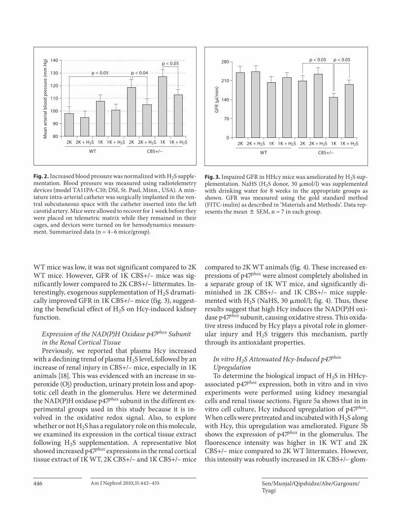
H 2 S Reduced Increased Blood Pressure inCBS+/– Mice We used a 1K model to expedite the glomerular dam-
age; however, the 1K animal often develops hypertension and, consequently, renal damage. In order to assess the degree of hypertension and the role of H 2 S treatment in regulating blood pressure, we measured blood pressure; the summarized data is shown in figure 2 . 1K WT ani-
mals developed a small increase of blood pressure com-pared to 2K WT, which was not significant. There was, however, a significant increase of blood pressure in 2K CBS+/– mice compared to 2K WT animals. 1K CBS+/– mice showed a further increase of blood pressure (6 to 7 mm Hg), but this was not significant compared to 2K CBS+/– animals. Interestingly, H 2 S supplementation normalized this increase of blood pressure in 1K WT and 2K and 1K CBS+/– mice. The heart rate, however, did not change between groups with or without H 2 S supplemen-tation (data not shown). These results specifically suggest that H 2 S acts at least through two different mechanisms: (1) through reducing blood pressure, which has immense impact on glomerular damage; and (2) through its anti-oxidant properties, which reduces oxidative damages in-duced by HHcy.
H 2 S Supplementation Improved GFR To assess the impact of HHcy on renal function and
any regulatory role of H 2 S on it, GFR was measured us-ing the gold standard method (FITC-inulin clearance) as described in ‘Materials and Methods’. 2K CBS+/– mice showed a marginally low GFR compared to their age-matched 2K WT littermates ( fig. 3 ). Although GFR in 1K
02K
5
10
15
H2S
pro
duct
ion
(nm
ol/g
/min
)
2K + H2S 1K
WTa
1K + H2S 2K 2K + H2S 1K 1K + H2S
CBS+/–
*
†
‡
02K
0.5
1.0
2K + H2S 1K
WTb
1K + H2S 2K 2K + H2S 1K 1K + H2S
CBS+/–
* *
††
�-Actin
CSE
1.5
CSE/
�-a
ctin
(fol
d ch
ange
s, ×
2K
WT)
Fig. 1. Attenuated renal cortical tissue H 2 S production and CSE expressions were normalized by H 2 S treatment. a In the presence of 10 mmol/l L -cysteine and 2 mmol/l pyridoxol 5 � -phosphate, H 2 S production in the kidney cortex tissue homogenates was measured following the procedure as described in the ‘Materials and Methods’. * p ! 0.01 vs. 2K WT; † p ! 0.05 vs. 2K CBS+/–;
‡ p ! 0.05 vs. 1K CBS+/–. b Representative Western blot showed CSE expression in the kidney cortex tissue in different experi-mental animal groups. The bar diagram showed densitometric analyses of CSE expression against � -actin loading control. Data represents the mean 8 SEM, n = 4–7. * p ! 0.01 vs. respective 2K mice and † p ! 0.01 vs. respective 1K mice.
Sen/Munjal/Qipshidze/Abe/Gargoum/Tyagi
Am J Nephrol 2010;31:442–455446
WT mice was low, it was not significant compared to 2K WT mice. However, GFR of 1K CBS+/– mice was sig-nificantly lower compared to 2K CBS+/– littermates. In-terestingly, exogenous supplementation of H 2 S dramati-cally improved GFR in 1K CBS+/– mice ( fig. 3 ), suggest-ing the beneficial effect of H 2 S on Hcy-induced kidney function.
Expression of the NAD(P)H Oxidase p47 phox Subunit in the Renal Cortical Tissue Previously, we reported that plasma Hcy increased
with a declining trend of plasma H 2 S level, followed by an increase of renal injury in CBS+/– mice, especially in 1K animals [18] . This was evidenced with an increase in su-peroxide (O2
–�) production, urinary protein loss and apop-totic cell death in the glomerulus. Here we determined the NAD(P)H oxidase p47 phox subunit in the different ex-perimental groups used in this study because it is in-volved in the oxidative redox signal. Also, to explore whether or not H 2 S has a regulatory role on this molecule, we examined its expression in the cortical tissue extract following H 2 S supplementation. A representative blot showed increased p47 phox expressions in the renal cortical tissue extract of 1K WT, 2K CBS+/– and 1K CBS+/– mice
compared to 2K WT animals ( fig. 4 ). These increased ex-pressions of p47 phox were almost completely abolished in a separate group of 1K WT mice, and significantly di-minished in 2K CBS+/– and 1K CBS+/– mice supple-mented with H 2 S (NaHS, 30 � mol/l; fig. 4 ). Thus, these results suggest that high Hcy induces the NAD(P)H oxi-dase p47 phox subunit, causing oxidative stress. This oxida-tive stress induced by Hcy plays a pivotal role in glomer-ular injury and H 2 S triggers this mechanism, partly through its antioxidant properties.
In vitro H 2 S Attenuated Hcy-Induced p47 phox Upregulation To determine the biological impact of H 2 S in HHcy-
associated p47 phox expression, both in vitro and in vivo experiments were performed using kidney mesangial cells and renal tissue sections. Figure 5 a shows that in in vitro cell culture, Hcy induced upregulation of p47 phox . When cells were pretreated and incubated with H 2 S along with Hcy, this upregulation was ameliorated. Figure 5 b shows the expression of p47 phox in the glomerulus. The fluorescence intensity was higher in 1K WT and 2K CBS+/– mice compared to 2K WT littermates. However, this intensity was robustly increased in 1K CBS+/– glom-
02K
70
140
280
210
GFR
(μl/
min
)
2K + H2S 1K
WT
1K + H2S 2K 2K + H2S 1K 1K + H2S
CBS+/–
p < 0.05 p < 0.05
Mea
n ar
teria
l blo
od p
ress
ure
(mm
Hg)
802K
100
120
90
110
130
140
2K + H2S 1K
WT
1K + H2S 2K 2K + H2S 1K
p < 0.05
p < 0.05 p < 0.04
1K + H2S
CBS+/–
Fig. 2. Increased blood pressure was normalized with H 2 S supple-mentation. Blood pressure was measured using radiotelemetry devices (model TA11PA-C10; DSI, St. Paul, Minn., USA). A min-iature intra-arterial catheter was surgically implanted in the ven-tral subcutaneous space with the catheter inserted into the left carotid artery. Mice were allowed to recover for 1 week before they were placed on telemetric matrix while they remained in their cages, and devices were turned on for hemodynamics measure-ment. Summarized data (n = 4–6 mice/group).
Fig. 3. Impaired GFR in HHcy mice was ameliorated by H 2 S sup-plementation. NaHS (H 2 S donor, 30 � mol/l) was supplemented with drinking water for 8 weeks in the appropriate groups as shown. GFR was measured using the gold standard method (FITC-inulin) as described in ‘Materials and Methods’. Data rep-resents the mean 8 SEM, n = 7 in each group.

Glomerular Remodeling and Hydrogen Sulfide
Am J Nephrol 2010;31:442–455 447
erulus, indicating higher expression of p47 phox compared to other nontreated groups. When a similar group of mice was treated with H 2 S, this induction of p47 phox was at-tenuated ( fig. 5 b). Together, these results suggest that Hcy induces p47 phox and H 2 S downregulates p47 phox during HHcy.
Induction of MMP-2 and -9 in the Renal Cortex Oxidative stress regulates MMPs, which play a role in
collagen remodeling in the matrix during HHcy [18, 27, 28] . Therefore, we measured the expressions of MMP-2 and -9 in the renal cortex tissue-extracted protein by Western blot. Summarized data in figure 6 showed that both pro- and active forms of MMP-9 were increased sig-nificantly in 1K WT, 2K CBS+/– and 1K CBS+/– mice. Interestingly, H 2 S inhibited expression of both these forms in the cortex. The expression of the active form of MMP-2 was higher in 1K WT animals compared to 2K WT, whereas both pro- and active forms of this protein-ase were found upregulated in 2K and 1K CBS+/– ani-mals ( fig. 6 ). Like MMP-9, upregulation of MMP-2 was also attenuated, at least in 1K WT and 1K CBS+/– mice, with the supplementation of H 2 S ( fig. 6 ). These results suggest that H 2 S plays a role in mitigating MMP-2 and -9 inductions during HHcy.
Exogenous H 2 S Modulated Collagen Remodeling in the Glomerulus Collagen is one of the major components of extracel-
lular matrix, and excessive collagen accumulation is re-lated to the pathophysiology of tissue remodeling. Here, we sought to determine collagen deposition in the glom-erulus of WT and CBS+/– mice in different experimental groups. The results, as shown in figure 7 , suggest that ex-ogenous H 2 S ameliorates Hcy-induced collagen deposi-tion in the glomerulus.
Collagen IV is a structural component of glomerular basal lamina, and excessive accumulation of collagen IV in the lamina adversely affects glomerular function. Therefore, we measured collagen IV expression in the re-nal cortical tissue-extracted protein. In accordance with histological collagen distribution in the kidney tissues, Western blot analyses of cortical tissues demonstrated that, although 2K CBS+/– mice had higher amounts of collagen IV expression compared to 2K WT, the presence of collagen protein in 1K CBS+/– was much higher than in 1K WT and 2K CBS+/– mice ( fig. 8 ). H 2 S, however, normalized this protein expression in the renal cortical tissue ( fig. 8 ). Previously, we reported increased renal in-jury in 1K CBS+/– mice as a result of elevated plasma Hcy level and Hcy-induced oxidative stress [18] . The present results confirm that this injury leads to excessive accu-mulation of collagen leading to glomeruloslerosis. H 2 S, however, ameliorates collagen expression, probably through its anti-oxidant properties.
Additionally, the collagen IV gene expression level was measured and normalized with internal control GAPDH ( fig. 9 ). This suggests that expression of protein level was due to transcriptional regulation of the colla-gen IV gene.
H 2 S Acted as an Anti-Inflammatory Substance Inflammation causes upregulation of inflammatory
molecules, such as ICAM-1 and VCAM-1, leading to in-flammatory disorder and sclerosis. Therefore, we mea-sured these molecules in our next experiment. As shown in the representative figure ( fig. 10 ), increased expres-sions of the inflammatory molecules ICAM-1 and VCAM-1 were observed in 1K WT as well as in 2K and 1K CBS+/– mice, and were significantly higher com-pared to 2K WT control animals. We reported earlier that these mice were hyperhomocysteinemic with low plasma H 2 S levels [18] . Interestingly, exogenous supple-mentation of H 2 S in the similar groups of animals at-tenuated these adhesion molecule expressions ( fig. 10 ). These results indicate that Hcy induces the inflamma-
p < 0.01 p < 0.01
p < 0.05 p < 0.05
02K
2
6
4
2K + H2S 1K
WT
1K + H2S 2K 2K + H2S 1K 1K + H2S
CBS+/–
*
**
�-Actin
p47phox
8
p47ph
ox/�
-act
in(fo
ld c
hang
es, ×
2K
WT)
Fig. 4. H 2 S mitigated NAD(P)H oxidase (p47 phox subunit) in the HHcy kidney. Western blot was performed to measure the p47 phox subunit of NAD(P)H oxidase in the kidney cortex tissue extract using anti-p47 phox antibody. Data represents the mean 8 SEM,n = 5–7. * Significant difference (p ! 0.05) compared to 2K WT, and * * significant difference (p ! 0.05) compared to 2K CBS+/–.

Sen/Munjal/Qipshidze/Abe/Gargoum/Tyagi
Am J Nephrol 2010;31:442–455448
DAPI Mergedp47phox
Cont
rol
Hcy
a
Hcy
+ H
2S
H2S
2K 1K2K + H2S 1K + H2S
2K 1K2K + H2S 1K + H2S
WT
CBS+
/–
b
Fig. 5. H 2 S diminished Hcy-induced p47 phox up-regulation in mesangial cells as well as in the glomerulus. a Kidney mesangial cells were cul-tured in 8-well chamber slides. Cells were pre-treated with H 2 S (NaHS, 30 � mol/l) 15 min be-fore cells were exposed to Hcy (50 � mol/l) for48 h (NaHS and Hcy were freshly added after ev-ery 12 h). Cells were fixed, permeabilized, blocked with 1% BSA in PBS, and immunostained with anti-p47 phox antibody secondarily conjugated with FITC. Also, cells were counterstained with DAPI. Fluorescence images were taken under the laser scanning confocal microscope (FluoView 1000, Olympus). Green fluorescence, as indicated by arrows, indicated p47 phox expression. Repre-sentative images from independent experiments (n = 5). b Representative images of p47 phox immu-nostained glomerulus (n = 5–7 animals/group) from different experimental animal groups.
Colo
r ver
sion
ava
ilabl
e on
line

Glomerular Remodeling and Hydrogen Sulfide
Am J Nephrol 2010;31:442–455 449
tory molecules ICAM-1 and VCAM-1 in the renal cortex, and H 2 S acts as an anti-inflammatory substance which downregulates these molecules in Hcy-induced renal cortex.
H 2 S Supplementation Inhibited Macrophage Infiltration Macrophage infiltration and accumulation in the kid-
ney is one of the most important events for the progres-sion of glomerulosclerosis [22] . In our next experiment, we determined macrophage infiltration in the glomeru-lus. To do this, we immunostained kidney sections (2- � m thickness) with murine F4/80 antibody, one of the mark-ers of macrophages. As shown in the representative im-ages ( fig. 11 ), increased expression of F4/80 antigen was observed in the glomerulus of 1K WT and both 2K and 1K CBS+/– mice compared to 2K WT control animals. The abundance of F4/80 antigen expression in the glom-erulus was attenuated with H 2 S supplementation in sim-ilar groups of animals ( fig. 11 ). These results indicate that increased plasma Hcy and a subsequent decrease in plas-ma H 2 S level induces macrophage infiltration in the glomerulus, whereas supplementation of H 2 S limits this infiltration associated with HHcy, thus preventing glo-merulosclerosis.
Discussion
We previously reported that the decreased plasma lev-el of H 2 S in WT 1K, CBS+/– 2K and CBS+/– 1K animals was strongly associated with increased plasma Hcy and that supplementation of H 2 S ameliorated HHcy-associat-ed chronic renal failure [18] . Results from the present study demonstrated that abilities of renal cortical tissue to generate endogenous H 2 S were impaired in 1K WT mice with further disabilities in both 2K and 1K CBS+/– mice ( fig. 1 a). This may be due to downregulation of one of the Hcy-metabolizing enzymes, CSE ( fig. 1 b), in addi-tion to one copy of mutant CBS gene in CBS+/– mice. Hcy is one of the precursors of endogenous H 2 S production. Therefore, an increased Hcy level and downregulation of its metabolizing enzyme will essentially decrease plasma H 2 S level, a potent vasorelaxant gas. This will conse-quently result in hypertension, as we have observed in our study ( fig. 2 ).
Our results also suggested that supplementation of H 2 S resulted in normalization of blood pressure associ-ated with HHcy ( fig. 2 ). In addition to that, NAD(P)H oxidase (p47 phox subunit) was upregulated in WT 1K and
CBS+/– 2K and 1K mice. The increased expression of this protein was normalized with H 2 S supplementation, sug-gesting the anti-oxidant properties of H 2 S. Increased MMP-2 and -9, collagen protein expression, and histo-logical collagen depositions were at least in part due to an increased oxidative-redox state. During HHcy, supple-mentation of H 2 S normalized MMPs expressions and collagen accumulation in the glomerulus, suggesting the antifibrotic properties of H 2 S. Increased expressions of inflammatory molecules, ICAM-1 and VCAM-1, along with increased macrophage marker protein F4/80, were augmented with the decline of H 2 S generation in the mouse-exhibited HHcy, whereas H 2 S supplementation prevented inflammation. Together, these results suggest that HHcy causes a decrease in endogenous H 2 S genera-tion, which eventually leads to hypertension, oxidative stress, matrix remodeling and chronic inflammation re-sulting in glomerulosclerosis and reduced glomerular function (GFR; fig. 3 ).
Renal dysfunction is recognized as a risk factor for in-creased cardiovascular morbidity and mortality [29] , and chronic kidney disease is a graded cardiovascular risk factor that leads from renal impairment to end-stage re-
02K
2
6
4
2K + H2S 1K
WT
1K + H2S 2K 2K + H2S 1K 1K + H2S
CBS+/–
** ******
** *
*
*
GAPDH
MMP-2
MMP-9 – Pro– Active– Pro– Active
GAPDH
8
MMP-9MMP-2
Expr
essi
on/G
APD
H(fo
ld c
hang
es, ×
2K
WT)
†
†
Fig. 6. H 2 S attenuated MMP-2 and -9 expressions in the kidney. Western blot analyses were performed to measure MMP-2 and -9 in the kidney cortical tissue extract using anti-MMP-2 and anti-MMP-9 antibodies, respectively. The bar diagram indicated den-sitometric analyses of expressed protein. Data represents the mean 8 SEM; n = 5–7 per group. * p ! 0.05 vs. respective 2K; * * significant differences (p ! 0.05) vs. respective 1K; † p ! 0.05 vs. 2K WT.

Sen/Munjal/Qipshidze/Abe/Gargoum/Tyagi
Am J Nephrol 2010;31:442–455450
nal failure [30] . The kidney also plays a major role in Hcy metabolism [31] and plasma total Hcy increases with the declines of renal function [29] . Hcy, however, in the mam-malian tissue can be metabolized further using three en-dogenous enzymes (CBS, CSE and 3MST) to produce H 2 S. Although H 2 S has been known for decades as a tox-ic gas with intoxication effects on the central nervous sys-tem and respiratory system, recently it has been docu-mented as an important physiological gaseous molecule with antioxidant [13, 18] , antihypertensive [32] , anti-in-flammatory [10, 16] and antifibrotic [33, 34] properties.
It is important to point out that both Hcy and cysteine are substrates for H 2 S generation, and that the CSE en-zyme mainly uses cysteine to produce H 2 S endogenously. Biochemically during HHcy, Hcy competes for binding to CSE with cysteine; therefore, increases in Hcy will de-crease H 2 S production from cysteine through substrate inhibition [35, 36] . The reaction occurs as follows:
u u CSE f (availability to bind with cysteine) Hcy d i i Cysteine i H 2 S f CBS CBS, 3MST
Previously we reported a decreased occurrence of plasma H 2 S levels during HHcy [18] . In the present study we ob-served decreased renal production of H 2 S in HHcy that resulted in hypertension ( fig. 2 ). This result indicates that above substrate inhibition, a mechanism may be playing a role in producing H 2 S during HHcy. Furthermore, pro-tein homocysteinylation is a major reaction in the pres-ence of thiolactone and homocysteinylation led to protein damage. This manifested as multimerization and precip-itation of extremely modified proteins [37] . Therefore, it may also be possible that Hcy at elevated levels homocys-teinylates its metabolizing enzymes, including CSE, re-sulting in damage and precipitation of modified CSE. Therefore, we detected low expression of CSE protein in the renal tissue ( fig. 1 b). Although this was not within the
2K 1K2K + H2S 1K + H2S
2K 1K2K + H2S 1K + H2S
WT
CBS+
/–
Colo
r ver
sion
ava
ilabl
e on
line
Fig. 7. Excessive collagen deposition in HHcy mice was amelio-rated by H 2 S supplementation. Histological kidney tissue sections were stained with Masson trichrome (collagen appears as blue, and indicated by arrows). 2K CBS+/– mice showed increased col-lagen deposition in the glomerular basement membrane, com-pared to 2K WT. This collagen deposition in the glomerulus of 1K
CBS+/– mice was even higher than in 2K CBS+/– mice. When a separate group of 1K CBS+/– mice was supplemented with H 2 S (NaHS, 30 � mol/l in drinking water) for 8 weeks postsurgery, the collagen appeared much less in the glomerulus. Notably, their 1K WT littermates had very little collagen appearance and this disap-peared with NaHS supplementation (n = 7/group; ! 200).

Glomerular Remodeling and Hydrogen Sulfide
Am J Nephrol 2010;31:442–455 451
scope of the present study, this potential mechanism does require further experimentation.
Hcy contains a reactive sulfhydryl group and, like most thiols (RSH), can undergo oxidation to the disulfide (RSSR) at physiological pH in the presence of O 2 [38] . A variety of reactive oxygen species can be produced upon Hcy oxidation, including superoxide (O2
–�) and hydrogen peroxide (H 2 O 2 ) [39, 40] . Increasing evidence suggests that reactive oxygen species play an important role in the pathophysiology of glomerular dysfunction, interstitial fibrosis and glomerulosclerosis [5, 41] . Multiple enzymes contribute to exacerbating oxidative stress in different tissues and cells; however, studies have demonstrated that NAD(P)H oxidase is a major enzyme to produce super-oxide (O2
–�) in the kidney under physiological conditions [42] . Initially, NAD(P)H was characterized in neutrophils including membrane subunits p22 phox and gp91 phox and cytoplasmic subunits p47 phox , p40 phox , p67 phox , and Rac GTPase [43, 44] . It is generally accepted that in the event of NAD(P)H oxidase activation, the p47 phox subunit plays a vital role [45, 46] . Therefore, we tested the expression of p47 phox both in in vivo and in vitro experimental condi-tions. Our results suggested that p47 phox was upregulated in the animal groups exhibiting HHcy ( fig. 4 , and 5 b) and
p < 0.01p < 0.05
p < 0.05 p < 0.01
02K
1
3
2
2K + H2S 1K
WT
1K + H2S 2K 2K + H2S 1K 1K + H2S
CBS+/–
*
�-Actin
Collagen IV
4
Colla
gen
IV/�
-act
in(fo
ld c
hang
es, ×
2K
WT)
p < 0.01 p < 0.05
p < 0.01
02K
1
4
3
2
2K + H2S 1K
WT
1K + H2S 2K 2K + H2S 1K 1K + H2S
CBS+/–
*
**
GAPDH
Collagen IV
5
Colla
gen
IV/G
APD
H(fo
ld c
hang
es, ×
2K
WT)
02K
1
3
2
2K + H2S 1K
WT
1K + H2S 2K 2K + H2S 1K 1K + H2S
CBS+/–
* *
�-Actin
VCAM-1
ICAM-1
4 ICAM-1VCAM-1
Expr
essi
on/�
-act
in(fo
ld c
hang
es, ×
2K
WT)
††
‡
##
‡
Fig. 8. H 2 S-attenuated collagen IV protein expression. Kidney cortex tissues were analyzed by Western blot using anticollagen IV antibody. While collagen expression was high in 1K WT and both 2K and 1K CBS+/– mice, this increased expression was ame-liorated with H 2 S supplementation (NaHS, 30 � mol/l) for 8 weeks postsurgery. The bar diagram indicated densitometric analyses of the blots. Data represents the mean 8 SEM; n = 5–7/group. * Sig-nificant difference (p ! 0.05) compared to 2K WT.
Fig. 9. Expression of collagen IV gene in the kidney cortical tis-sues. Renal cortical collagen protein was analyzed by Western blot. The bar diagram showed densitometric analyses of blots. Data represents the mean 8 SEM, n = 5–7/group. * p ! 0.01 com-pared to WT 2K; * * p ! 0.05 compared to CBS+/– 2K.
Fig. 10. Inflammatory molecule ICAM-1 and VCAM-1 were mit-igated by H 2 S. Representative Western blot showed ICAM-1 and VCAM-1 expressions in the kidney cortex tissue in different ex-perimental animal groups. The bar diagram indicated densito-metric analyses of expressed protein in the respective blots. Data represents the mean 8 SEM, n = 5–7/group. * Significant differ-ence (p ! 0.01) compared to 2K WT littermates; † p ! 0.01 vs. 1K WT; # p ! 0.01 vs. 2K CBS+/–; ‡ p ! 0.01 vs. 1K CBS+/–.

Sen/Munjal/Qipshidze/Abe/Gargoum/Tyagi
Am J Nephrol 2010;31:442–455452
crepancy could be due to the difference in H 2 S level be-tween these two HHcy animal models. However, the dis-crepancy in p47 phox levels between the present study and a previous report using the same type of cultured cells in vitro is puzzling [48] . The other report did not show a change in p47 phox levels by Hcy in cultured renal mesan-gial cells [48] . One possible reason may be due to Hcy stimulation for a prolonged period of time (48 h) in the present study versus the earlier report (16 h). However, the in vivo result of the present study suggests an upregu-lation of p47 phox in the glomerulus which is diminished by H 2 S treatment.
associated with a decrease in renal production of H 2 S ( fig. 1 a). In an in vitro experiment using mesangial cells, we observed a similar increase of p47 phox expression dur-ing HHcy, and this increased expression was normalized in the presence of H 2 S ( fig. 5 a). Thus, from our study it is very clear that in the event of HHcy, p47 phox -mediated oxidative stress induces glomerular injury and H 2 S plays a key role in this mechanism.
It is very interesting that the current report showed p47 phox was upregulated in HHcy ( fig. 4 ), which is incon-sistent with a previous report showing that p47 phox was not changed in a different HHcy model [47] . This dis-
02K
1
2K + H2S 1K 1K + H2S 2K 2K + H2S 1K 1K + H2S
2K 2K + H2S 1KW
T1K + H2S
2K 2K + H2S 1K 1K + H2S
CBS+
/–
2
Fold
cha
nges
(× 2
K W
T)
* ***
†
‡
‡‡
Fig. 11. Increased macrophage infiltration was mitigated by H 2 S. a Kidney tissue cryosections (2- � m) were immunostained with macrophage F4/80 marker and visualized with DAB (3,3 � -diaminobenzidine) staining. Increased brown staining, as indi-cated by arrows, in 1K WT, 2K CBS+/– and 1K CBS+/– mice in-dicated more macrophage infiltration in the glomerulus. In the
similar groups, H 2 S supplementation prevented this infiltration. Representative images (n = 5/group); data represent the mean 8 SEM. b Bar diagram showed intensity changes of DAB staining against 2K WT glomerulus. * p ! 0.05 vs. 2K WT; * * p ! 0.05 vs. 1K WT; †,‡ p ! 0.05 vs. 2K CBS+/– and ‡‡ p ! 0.05 vs. 1K CBS+/–.
Colo
r ver
sion
ava
ilabl
e on
line

Glomerular Remodeling and Hydrogen Sulfide
Am J Nephrol 2010;31:442–455 453
References 1 Lentz SR: Mechanisms of homocysteine-in-duced atherothrombosis. J Thromb Hae-most 2005; 3: 1646–1654.
2 Dayal S, Bottiglieri T, Arning E, Maeda N, Malinow MR, Sigmund CD, Heistad DD, Faraci FM, Lentz SR: Endothelial dysfunc-tion and elevation of S-adenosylhomocyste-ine in cystathionine beta-synthase-deficient mice. Circ Res 2001; 88: 1203–1209.
MMP-2 and -9 are collagenases with more affinity for collagen IV and have been shown to have a renoprotective role by promoting turnover and, therefore, preventing matrix accumulation [49, 50] . On the contrary, elevated expressions and activities of MMP-2 and -9 have been observed in the fibrotic renal cortex; therefore, they are documented as playing an important role in matrix ac-cumulation associated with progressive renal scarring (22, 95). MMP-2 and -9 also degrade elastin more effi-ciently than other MMPs [51] . Because the turnover of collagen is faster than elastin, oxidatively modified col-lagen (glycated collagen) is deposited faster than elastin or any other ultra-structural matrix proteins [52] .
We have previously reported that an increase of MMP-2 and -9 activity was observed in the renal tissue from the animals exhibiting HHcy and lower levels of plasma H 2 S. In the present study we measured increased levels of MMP-2 and -9, collagen IV mRNA, and protein with the overall increase of deposition of collagen in the glomeru-lus of WT 1K, CBS+/– 2K and CBS+/– 1K mice. These results support our previous data [18] and suggest that MMP-2 and -9 as well as collagen remodeling are associ-ated with HHcy-related chronic renal failure. Supple-mentation of H 2 S can prevent these remodeling process-es by regulating MMPs and collagen during HHcy-asso-ciated pathogenesis in the kidney ( fig. 6–9 ).
Typically, glomerulosclerosis refers to a hardening of the glomerulus which frequently occurs due to collagen deposition. Glomerulosclerosis may be total where the entire glomerulus becomes hard and nonfunctional. Whereas partial glomerulosclerosis is often known as fo-cal segmental glomerulosclerosis where part of the glom-erulus becomes scar tissue. Early stages of glomeruloscle-rosis may produce little symptoms of scar tissue, which is collagen accumulation in our study ( fig. 7 ); however, the most important warning sign is proteinuria. We have previously reported that HHcy animal groups developed proteinuria, whereas H 2 S treatment partially prevented urinary protein loss [18] . Our present study further con-firms our previous findings that the protein loss was pri-marily due to focal segmental glomerulosclerosis.
The excessive production of reactive oxygen species mediates renal fibrotic injury through activation of pro-inflammatory molecules. Recent studies of cytokines, chemokines and adhesion molecules have enhanced our understanding of molecular mechanisms of leukocyte trafficking and their activation in the inflammatory phase of various renovascular diseases [53] . Increased ex-pressions of ICAM-1 and VCAM-1 have been shown as a potential mechanism facilitating lymphocytic infiltra-
tion and organ dysfunction [54] . More importantly, ex-perimental evidence has implicated sustained elevation of cell adhesion molecules, such as ICAM-1 and VCAM-1, in chronic inflammatory disorder, which leads to scle-rosis [20] . HHcy has now been recognized as a patho-physiological stimulus of vascular endothelial dysfunc-tion and has been shown to increase expression of ICAM-1 [19] and VCAM-1 [19, 21] in experimental mod-els. However, an Hcy-induced inflammatory reaction in the glomerulus during renal diseases was clearly not de-fined. The present study further supports our previous findings [18] and demonstrates that macrophage infiltra-tion and increased expression of ICAM-1 and VCAM-1 ( fig. 10 , 11 ) are associated with HHcy. These events are partially, if not completely, preventable by H 2 S treatment, suggesting the possible protective role of H 2 S through its antioxidant properties during HHcy-associated renal in-flammation and remodeling. However, the relationship between oxidant stress and inflammation, i.e. which is the upstream effector in HHcy, was not adequately ad-dressed in the present study because inflammation has also been shown to cause oxidant stress. Blocking either pathway may clarify this question. Nevertheless, the pres-ent results reveal that oxidative stress is one of the, if not only, upstream effectors of HHcy.
In summary, we demonstrated that increased oxida-tive stress in HHcy-associated renal cortical tissue is due to, in part, reduced renal H 2 S production and upregula-tion of the p47 phox subunit of NAD(P)H oxidase. These lead to MMPs and collagen remodeling in the glomerulus along with the increase in inflammatory response result-ing in glomerulosclerosis, reduced glomerular function and hypertension. These structural and functional changes are associated with HHcy and are preventable by H 2 S supplementation.
Acknowledgement
This study was supported, in part, by NIH grants HL-71010, HL-74185, HL-88012 and NS-51568.

Sen/Munjal/Qipshidze/Abe/Gargoum/Tyagi
Am J Nephrol 2010;31:442–455454
3 Diez N, Perez R, Hurtado V, Santidrian S: Hyperhomocysteinaemia induced by dietary folate restriction causes kidney oxidative stress in rats. Br J Nutr 2005; 94: 204–210.
4 Zhang F, Siow YL, O K: Hyperhomocystein-emia activates NF-kappaB and inducible ni-tric oxide synthase in the kidney. Kidney Int 2004; 65: 1327–1338.
5 Yi F, Li PL: Mechanisms of homocysteine-induced glomerular injury and sclerosis. Am J Nephrol 2008; 28: 254–264.
6 Shibuya N, Mikami Y, Kimura Y, Nagahara N, Kimura H: Vascular endothelium ex-presses 3-mercaptopyruvate sulfurtransfer-ase and produces hydrogen sulfide. J Bio-chem 2009; 146: 623–626.
7 Shibuya N, Tanaka M, Yoshida M, Ogas-awara Y, Togawa T, Ishii K, Kimura H: 3-mercaptopyruvate sulfurtransferase pro-duces hydrogen sulfide and bound sulfane sulfur in the brain. Antioxid Redox Signal 2009; 11: 703–714.
8 Geng B, Yang J, Qi Y, Zhao J, Pang Y, Du J, Tang C: H 2 S generated by heart in rat and its effects on cardiac function. Biochem Bio-phys Res Commun 2004; 313: 362–368.
9 Swaroop M, Bradley K, Ohura T, Tahara T, Roper MD, Rosenberg LE, Kraus JP: Rat cys-tathionine beta-synthase. Gene organization and alternative splicing. J Biol Chem 1992; 267: 11455–11461.
10 Lowicka E, Beltowski J: Hydrogen sulfide (H 2 S) – the third gas of interest for pharma-cologists. Pharmacol Rep 2007; 59: 4–24.
11 Kimura Y, Dargusch R, Schubert D, Kimura H: Hydrogen sulfide protects HT22 neuro-nal cells from oxidative stress. Antioxid Re-dox Signal 2006; 8: 661–670.
12 Yonezawa D, Sekiguchi F, Miyamoto M, Taniguchi E, Honjo M, Masuko T, Nishika-wa H, Kawabata A: A protective role ofhydrogen sulfide against oxidative stressin rat gastric mucosal epithelium. Toxicolo-gy 2007; 241: 11–18.
13 Wei HL, Zhang CY, Jin HF, Tang CS, Du JB: Hydrogen sulfide regulates lung tissue-oxi-dized glutathione and total antioxidant ca-pacity in hypoxic pulmonary hypertensive rats. Acta Pharmacol Sin 2008; 29: 670–679.
14 Fu Z, Liu X, Geng B, Fang L, Tang C: Hydro-gen sulfide protects rat lung from ischemia-reperfusion injury. Life Sci 2008; 82: 1196–1202.
15 Zanardo RC, Brancaleone V, Distrutti E,Fiorucci S, Cirino G, Wallace JL: Hydrogen sulfide is an endogenous modulator of leuko-cyte-mediated inflammation. FASEB J 2006; 20: 2118–2120.
16 Hu LF, Wong PT, Moore PK, Bian JS: Hydro-gen sulfide attenuates lipopolysaccharide-induced inflammation by inhibition of p38 mitogen-activated protein kinase in microg-lia. J Neurochem 2007; 100: 1121–1128.
17 Rao VH, Lees GE, Kashtan CE, Nemori R, Singh RK, Meehan DT, Rodgers K, Berridge BR, Bhattacharya G, Cosgrove D: Increased expression of MMP-2, MMP-9 (type IV col-lagenases/gelatinases), and MT1-MMP in canine X-linked Alport syndrome (XLAS). Kidney Int 2003; 63: 1736–1748.
18 Sen U, Basu P, Abe OA, Givvimani S, Tyagi N, Metreveli N, Shah KS, Passmore JC, Tyagi SC: Hydrogen sulfide ameliorates hyper-homocysteinemia-associated chronic renal failure. Am J Physiol Renal Physiol 2009; 297:F410–F419.
19 Sen U, Tyagi N, Kumar M, Moshal KS, Ro-driguez WE, Tyagi SC: Cystathionine-beta-synthase gene transfer and 3-deazaadenosine ameliorate inflammatory response in endo-thelial cells. Am J Physiol Cell Physiol 2007; 293:C1779–C1787.
20 Silverman MD, Tumuluri RJ, Davis M, Lopez G, Rosenbaum JT, Lelkes PI: Homocysteine upregulates vascular cell adhesion mole-cule-1 expression in cultured human aortic endothelial cells and enhances monocyte ad-hesion. Arterioscler Thromb Vasc Biol 2002; 22: 587–592.
21 Hofmann MA, Lalla E, Lu Y, Gleason MR, Wolf BM, Tanji N, Ferran LJ Jr, Kohl B, Rao V, Kisiel W, Stern DM, Schmidt AM: Hyper-homocysteinemia enhances vascular in-flammation and accelerates atherosclerosis in a murine model. J Clin Invest 2001; 107: 675–683.
22 Kikuchi Y, Imakiire T, Yamada M, Saigusa T, Hyodo T, Hyodo N, Suzuki S, Miura S: Mizoribine reduces renal injury and macro-phage infiltration in non-insulin-dependent diabetic rats. Nephrol Dial Transplant 2005; 20: 1573–1581.
23 Richardson CJ, Magee EA, Cummings JH: A new method for the determination of sul-phide in gastrointestinal contents and whole blood by microdistillation and ion chroma-tography. Clin Chim Acta 2000; 293: 115–125.
24 Li L, Bhatia M, Zhu YZ, Zhu YC, Ramnath RD, Wang ZJ, Anuar FB, Whiteman M, Salto-Tellez M, Moore PK: Hydrogen sulfide is a novel mediator of lipopolysaccharide-in-duced inflammation in the mouse. FASEB J 2005; 19: 1196–1198.
25 Maezawa Y, Yokote K, Sonezaki K, Fujimoto M, Kobayashi K, Kawamura H, Tokuyama T, Takemoto M, Ueda S, Kuwaki T, Mori S, Wahren J, Saito Y: Influence of C-peptide on early glomerular changes in diabetic mice. Diabetes Metab Res Rev 2006; 22: 313–322.
26 Lorenz JN, Gruenstein E: A simple, nonra-dioactive method for evaluating single-nephron filtration rate using FITC-inulin. Am J Physiol 1999; 276:F172–F177.
27 Rodriguez WE, Tyagi N, Joshua IG, Pass-more JC, Fleming JT, Falcone JC, Tyagi SC: Pioglitazone mitigates renal glomerular vas-cular changes in high-fat, high-calorie-in-duced type 2 diabetes mellitus. Am J Physiol Renal Physiol 2006; 291:F694–F701.
28 Li N, Chen YF, Zou AP: Implications of hy-perhomocysteinemia in glomerular sclero-sis in hypertension. Hypertension 2002; 39: 443–448.
29 Rodionov RN, Lentz SR: The homocysteine paradox. Arterioscler Thromb Vasc Biol 2008; 28: 1031–1033.
30 Schiffrin EL, Lipman ML, Mann JF: Chronic kidney disease: effects on the cardiovascular system. Circulation 2007; 116: 85–97.
31 Friedman AN, Bostom AG, Selhub J, Levey AS, Rosenberg IH: The kidney and homocys-teine metabolism. J Am Soc Nephrol 2001; 12: 2181–2189.
32 Yang G, Wu L, Jiang B, Yang W, Qi J, Cao K, Meng Q, Mustafa AK, Mu W, Zhang S, Sny-der SH, Wang R: H 2 S as a physiologic vaso-relaxant: hypertension in mice with deletion of cystathionine gamma-lyase. Science 2008; 322: 587–590.
33 Fang L, Li H, Tang C, Geng B, Qi Y, Liu X: Hydrogen sulfide attenuates the pathogene-sis of pulmonary fibrosis induced by bleomy-cin in rats. Can J Physiol Pharmacol 2009; 87: 531–538.
34 Mishra PK, Tyagi N, Sen U, Givvimani S,Tyagi SC: H 2 S ameliorates oxidative andproteolytic stresses and protects the heart against adverse remodeling in chronic heart failure. Am J Physiol Heart Circ Physiol 2010; 298:H451–H456.
35 Stabler SP, Steegborn C, Wahl MC, Olive-riusova J, Kraus JP, Allen RH, Wagner C, Mudd SH: Elevated plasma total homocyste-ine in severe methionine adenosyltransfer-ase I/III deficiency. Metabolism 2002; 51: 981–988.
36 Chang L, Geng B, Yu F, Zhao J, Jiang H, Du J, Tang C: Hydrogen sulfide inhibits myocar-dial injury induced by homocysteine in rats. Amino Acids 2008; 34: 573–585.
37 Jakubowski H: Protein homocysteinylation: possible mechanism underlying pathologi-cal consequences of elevated homocysteine levels. FASEB J 1999; 13: 2277–2283.
38 Jacobsen DW: Hyperhomocysteinemia and oxidative stress: Time for a reality check? Ar-terioscler Thromb Vasc Biol 2000; 20: 1182–1184.
39 Jacobsen DW, Troxell LS, Brown KL: Cataly-sis of thiol oxidation by cobalamins and co-binamides – reaction-products and kinetics. Biochemistry 1984; 23: 2017–2025.
40 Misra HP: Generation of superoxide free radical during the autoxidation of thiols. J Biol Chem 1974; 249: 2151–2155.
41 Brezniceanu ML, Liu F, Wei CC, Chenier I, Godin N, Zhang SL, Filep JG, Ingelfinger JR, Chan JS: Attenuation of interstitial fibrosis and tubular apoptosis in db/db transgenic mice overexpressing catalase in renal proxi-mal tubular cells. Diabetes 2008; 57: 451–459.

Glomerular Remodeling and Hydrogen Sulfide
Am J Nephrol 2010;31:442–455 455
42 Griendling KK, Sorescu D, Lassegue B, Ushio-Fukai M: Modulation of protein ki-nase activity and gene expression by reactive oxygen species and their role in vascular physiology and pathophysiology. Arterio-scler Thromb Vasc Biol 2000; 20: 2175–2183.
43 Brandes RP, Kreuzer J: Vascular NADPH ox-idases: molecular mechanisms of activation. Cardiovasc Res 2005; 65: 16–27.
44 Takeya R, Ueno N, Kami K, Taura M, Koh-jima M, Izaki T, Nunoi H, Sumimoto H:Novel human homologues of p47 phox and p67 phox participate in activation of super-oxide-producing NADPH oxidases. J Biol Chem 2003; 278: 25234–25246.
45 Madamanchi NR, Vendrov A, Runge MS: Oxidative stress and vascular disease. Arte-rioscler Thromb Vasc Biol 2005; 25: 29–38.
46 Sellmayer A, Obermeier H, Danesch U, Aep-felbacher M, Weber PC: Arachidonic acidincreases activation of NADPH oxidase in monocytic U937 cells by accelerated translo-cation of p47-phox and co-stimulation of protein kinase C. Cell Signal 1996; 8: 397–402.
47 Ungvari Z, Csiszar A, Edwards JG, Kamin-ski PM, Wolin MS, Kaley G, Koller A: In-creased superoxide production in coronary arteries in hyperhomocysteinemia: role of tumor necrosis factor-alpha, NAD(P)H oxi-dase, and inducible nitric oxide synthase. Arterioscler Thromb Vasc Biol 2003; 23: 418–424.
48 Yi F, Zhang AY, Janscha JL, Li PL, Zou AP: Homocysteine activates NADH/NADPH oxidase through ceramide-stimulated Rac GTPase activity in rat mesangial cells. Kid-ney Int 2004; 66: 1977–1987.
49 Endo T, Nakabayashi K, Sekiuchi M, Kuroda T, Soejima A, Yamada A: Matrix metallopro-teinase-2, matrix metalloproteinase-9, and tissue inhibitor of metalloproteinase-1 in the peripheral blood of patients with various glomerular diseases and their implication in pathogenetic lesions: study based on an en-zyme-linked assay and immunohistochemi-cal staining. Clin Exp Nephrol 2006; 10: 253–261.
50 Rysz J, Banach M, Stolarek RA, Pasnik J, Cialkowska-Rysz A, Koktysz R, Piechota M, Baj Z: Serum matrix metalloproteinases MMP-2 and MMP-9 and metalloproteinase tissue inhibitors TIMP-1 and TIMP-2 in dia-betic nephropathy. J Nephrol 2007; 20: 444–452.
51 Senior RM, Griffin GL, Fliszar CJ, Shapiro SD, Goldberg GI, Welgus HG: Human 92- and 72-kilodalton type IV collagenases are elastases. J Biol Chem 1991; 266: 7870–7875.
52 Camp TM, Tyagi SC, Senior RM, Hayden MR: Gelatinase B(MMP-9) an apoptotic fac-tor in diabetic transgenic mice. Diabetologia 2003; 46: 1438–1445.
53 Bonventre JV: Pathophysiology of acute kid-ney injury: roles of potential inhibitors of in-flammation. Contrib Nephrol 2007; 156: 39–46.
54 Anderson JA, Lentsch AB, Hadjiminas DJ, Miller FN, Martin AW, Nakagawa K, Ed-wards MJ: The role of cytokines, adhesion molecules, and chemokines in interleukin-2-induced lymphocytic infiltration in C57BL/6 mice. J Clin Invest 1996; 97: 1952–1959.