Hydrogen Sulfide and Urogenital Tract
26
Hydrogen Sulfide and Urogenital Tract Roberta d’Emmanuele di Villa Bianca, Giuseppe Cirino, and Raffaella Sorrentino Contents 1 Introduction ................................................................................. 112 2 Hydrogen Sulfide in Male Sexual Function ................................................ 113 2.1 The Male Reproductive System ...................................................... 113 2.2 Erectile Function and Dysfunction ................................................... 114 2.3 Hydrogen Sulfide and Erectile Function and Dysfunction .......................... 114 2.4 Mechanism(s) of Action that Sustain the Effect of H 2 S in Erection ............... 118 2.4.1 Ion Channels and EDHF as H 2 S Target ..................................... 118 2.4.2 Testosterone, Gender Difference, and Biosynthesis of H 2 S ................ 120 2.4.3 Interaction Between H 2 S and NO ............................................ 121 2.5 Hydrogen Sulfide and Vas Deferens ................................................. 122 2.6 Hydrogen Sulfide and Human Prostate .............................................. 123 3 Hydrogen Sulfide in Female Sexual Function ............................................. 123 4 Hydrogen Sulfide and Bladder ............................................................. 124 4.1 Bladder Function ...................................................................... 124 4.2 Animal Studies ........................................................................ 125 4.3 Human Studies ........................................................................ 126 5 Hydrogen Sulfide and Urethra .............................................................. 129 6 Conclusions and Future Directions ......................................................... 129 References ....................................................................................... 131 Abstract In this chapter the role played by H 2 S in the physiopathology of urogenital tract revising animal and human data available in the current relevant literature is discussed. H 2 S pathway has been demonstrated to be involved in the mechanism R. d’Emmanuele di Villa Bianca • G. Cirino (*) • R. Sorrentino Department of Pharmacy, University of Naples, Federico II, Via D. Montesano, 49, Naples 80131, Italy e-mail: [email protected] # Springer International Publishing Switzerland 2015 P.K. Moore, M. Whiteman (eds.), Chemistry, Biochemistry and Pharmacology of Hydrogen Sulfide, Handbook of Experimental Pharmacology 230, DOI 10.1007/978-3-319-18144-8_5 111 [email protected]
Transcript of Hydrogen Sulfide and Urogenital Tract

Hydrogen Sulfide and Urogenital Tract
Roberta d’Emmanuele di Villa Bianca, Giuseppe Cirino,and Raffaella Sorrentino
Contents
1 Introduction . . . . . . . . . . . . . . . . . . . . . . . . . . . . . . . . . . . . . . . . . . . . . . . . . . . . . . . . . . . . . . . . . . . . . . . . . . . . . . . . . 112
2 Hydrogen Sulfide in Male Sexual Function . . . . . . . . . . . . . . . . . . . . . . . . . . . . . . . . . . . . . . . . . . . . . . . . 113
2.1 The Male Reproductive System . . . . . . . . . . . . . . . . . . . . . . . . . . . . . . . . . . . . . . . . . . . . . . . . . . . . . . 113
2.2 Erectile Function and Dysfunction . . . . . . . . . . . . . . . . . . . . . . . . . . . . . . . . . . . . . . . . . . . . . . . . . . . 114
2.3 Hydrogen Sulfide and Erectile Function and Dysfunction . . . . . . . . . . . . . . . . . . . . . . . . . . 114
2.4 Mechanism(s) of Action that Sustain the Effect of H2S in Erection . . . . . . . . . . . . . . . 118
2.4.1 Ion Channels and EDHF as H2S Target . . . . . . . . . . . . . . . . . . . . . . . . . . . . . . . . . . . . . 118
2.4.2 Testosterone, Gender Difference, and Biosynthesis of H2S . . . . . . . . . . . . . . . . 120
2.4.3 Interaction Between H2S and NO . . . . . . . . . . . . . . . . . . . . . . . . . . . . . . . . . . . . . . . . . . . . 121
2.5 Hydrogen Sulfide and Vas Deferens . . . . . . . . . . . . . . . . . . . . . . . . . . . . . . . . . . . . . . . . . . . . . . . . . 122
2.6 Hydrogen Sulfide and Human Prostate . . . . . . . . . . . . . . . . . . . . . . . . . . . . . . . . . . . . . . . . . . . . . . 123
3 Hydrogen Sulfide in Female Sexual Function . . . . . . . . . . . . . . . . . . . . . . . . . . . . . . . . . . . . . . . . . . . . . 123
4 Hydrogen Sulfide and Bladder . . . . . . . . . . . . . . . . . . . . . . . . . . . . . . . . . . . . . . . . . . . . . . . . . . . . . . . . . . . . . 124
4.1 Bladder Function . . . . . . . . . . . . . . . . . . . . . . . . . . . . . . . . . . . . . . . . . . . . . . . . . . . . . . . . . . . . . . . . . . . . . . 124
4.2 Animal Studies . . . . . . . . . . . . . . . . . . . . . . . . . . . . . . . . . . . . . . . . . . . . . . . . . . . . . . . . . . . . . . . . . . . . . . . . 125
4.3 Human Studies . . . . . . . . . . . . . . . . . . . . . . . . . . . . . . . . . . . . . . . . . . . . . . . . . . . . . . . . . . . . . . . . . . . . . . . . 126
5 Hydrogen Sulfide and Urethra . . . . . . . . . . . . . . . . . . . . . . . . . . . . . . . . . . . . . . . . . . . . . . . . . . . . . . . . . . . . . . 129
6 Conclusions and Future Directions . . . . . . . . . . . . . . . . . . . . . . . . . . . . . . . . . . . . . . . . . . . . . . . . . . . . . . . . . 129
References . . . . . . . . . . . . . . . . . . . . . . . . . . . . . . . . . . . . . . . . . . . . . . . . . . . . . . . . . . . . . . . . . . . . . . . . . . . . . . . . . . . . . . . 131
Abstract
In this chapter the role played by H2S in the physiopathology of urogenital tract
revising animal and human data available in the current relevant literature is
discussed. H2S pathway has been demonstrated to be involved in the mechanism
R. d’Emmanuele di Villa Bianca • G. Cirino (*) • R. Sorrentino
Department of Pharmacy, University of Naples, Federico II, Via D. Montesano,
49, Naples 80131, Italy
e-mail: [email protected]
# Springer International Publishing Switzerland 2015
P.K. Moore, M. Whiteman (eds.), Chemistry, Biochemistry and Pharmacology of
Hydrogen Sulfide, Handbook of Experimental Pharmacology 230,
DOI 10.1007/978-3-319-18144-8_5
111
underlying penile erection in human and experimental animal. Both
cystathionine-β synthase (CBS) and cystathionine-γ lyase (CSE) are expressed
in the human corpus cavernosum and exogenous H2S relaxes isolated human
corpus cavernosum strips in an endothelium-independent manner. Hydrogen
sulfide pathway also accounts for the direct vasodilatory effect operated by
testosterone on isolated vessels. Convincing evidence suggests that H2S can
influence the cGMP pathway by inhibiting the phosphodiesterase 5 (PDE-5)
activity. All these findings taken together suggest an important role for the H2S
pathway in human corpus cavernosum homeostasis. However, H2S effect is not
confined to human corpus cavernosum but also plays an important role in human
bladder. Human bladder expresses mainly CBS and generates in vitro detectable
amount of H2S. In addition the bladder relaxant effect of the PDE-5 inhibitor
sildenafil involves H2S as mediator.
In conclusion the H2S pathway is not only involved in penile erection but also
plays a role in bladder homeostasis. In addition the finding that it involved in the
mechanism of action of PDE-5 inhibitors strongly suggests that modulation of
this pathway can represent a therapeutic target for the treatment of erectile
dysfunction and bladder diseases.
Keywords
Bladder • Corpus cavernosum • Erectile dysfunction (ED) • Benign prostatic
hyperplasia (BPH) • Lower urinary tract symptoms (LUTS) • PDE-5 inhibitor •
CBS • CSE • 3MST
1 Introduction
Hydrogen sulfide (H2S) presence in mammalian tissues has been known since the
1980s, but H2S has been considered mainly as a metabolic waste product with no
potential physiological activity. The first evidence, indicating H2S as an endo-
genous mediator was published in 1996 by Abe and Kimura (1996) that described
a role for this mediator in the brain. Some years, after this first evidence was
published, many research groups have focused their interest on this new pathway
as described in the other chapters. H2S together with nitric oxide (NO) and carbon
monoxide (CO) is now classified as gasotransmitter. Like the other two gasses, it
has the ability to diffuse easily through the cellular membranes. Hydrogen sulfide
can also interact with proteins such as ion channels or enzymes regulating their state
either directly via chemical modification such as by sulfuration and sulfhydration or
indirectly via second messengers.
The role of H2S in the homeostatic control of our body is now consistently
supported by the literature, and in the other chapters of the present book,
its synthesis, measurement, chemistry, and interaction have been reported. This
chapter will deal with the role played by H2S in the physiopathology of urogenital
tract revising animal and human data available in the current relevant literature.
112 R. d’Emmanuele di Villa Bianca et al.

2 Hydrogen Sulfide in Male Sexual Function
2.1 The Male Reproductive System
Male reproductive system consists of a number of sex organs that concur to the
human reproductive process localized around the pelvic region. Briefly, the main
male sex organs are the penis and the testicles important for producing semen and
sperm. In particular, the penis and the scrotum represent the external genital organs.
The penile erectile apparatus consists of two vascularized paired corpora cavernosa.
The corpus spongiosum together with the urethra is related to the ventral area of the
penis. The corpora cavernosa, the corpus spongiosum, and the glans penis are
composed of septa of smooth muscle and erectile tissue that enclose vascular
cavities. The tunica albuginea forms a thick fibrous coat to the spongy tissue of
the corpora cavernosa and corpus spongiosum, and it consists of two layers.
The muscles involved in penile function are the ischiocavernosum and bulbo-
spongiosus. The ischiocavernous muscle inserts into the medial and inferior surface
of the corpora and increases penile turgidity during erection. In the flaccid state, the
smooth muscle fibers are tonically contracted by the sympathetic system. During
erection, trabeculae smooth muscle is relaxed, and this allows a cascade of events.
The arterioles of the penis are dilated while the veins are passively compressed
between the tunica albuginea and the peripheral sinusoids reducing the venous
outflow. This process is known as the venous-occlusive mechanism, and it leads to
erection that is sustained by an increase of intra-cavernous blood pressure of about
100 mmHg. The contraction of the ischiocavernous muscle further increases the
intra-cavernous pressure leading to the rigid erection phase.
The main role of the scrotum is to hold and protect the testes. It also contains
numerous nerves and blood vessels. Between the male internal genital organs, there
is the epididymis, a whitish mass of tightly coiled tubes cupped against the testicles.
It acts as a maturation and storage for sperm before they pass into the vas deferens,
which carry sperm to the ampullary gland and prostatic ducts. Testosterone is the
most important sexual hormone in males and is released by the testes. This hormone
has a crucial role in the development of sperm and is also responsible for the
development of physical characteristics in men such as facial hair and deep voice.
Moreover the accessory glands, such as the seminal vesicles and the prostate gland,
provide fluids that lubricate the duct system and nourish the sperm cells.
In particular, the prostate gland surrounds the ejaculatory ducts at the base of the
male urethra, just below the bladder. The prostate gland is responsible for the proof
semen, a liquid mixture of sperm cells, prostate fluid, and seminal fluid. This gland
is also responsible for making the semen milky in appearance by mixing calcium to
the semen coming from seminal vesicle. The semen remains cloudy and clumpy
until the prostatic pro fibrinolysis is formed into fibrinolysis and lysis of the
fibrinogen from the seminal vesicle fluids occurs.
Hydrogen Sulfide and Urogenital Tract 113

2.2 Erectile Function and Dysfunction
Erection is a vascular event which relies upon interaction of neural and
humoral mechanisms at various levels. Indeed, since the penis receives innervations
from sacral parasympathetic (pelvic), thoracolumbar sympathetic, and somatic
(pudenda) nerves, the erection phenomenon requires participation of these three
systems. Thus, erection is a consequence of a complex integration of several
signals. In simple words, erection is essentially a spinal reflex that can be initiated
by recruitment of penile afferents, but also by visual, olfactory, and imaginary
stimuli. All these stimuli may contribute to the increase in intra-cavernous pressure
(Cirino et al. 2006) and thus to penile erection. Erectile dysfunction (ED) is defined
as the consistent or recurrent inability of a man to attain and/or maintain a penile
erection sufficient for sexual activity (2nd International Consultation on Sexual
Dysfunction-Paris, June 28th–July 1st, 2003). Of note, ED shares many of the risk
factors that contribute to the development and the progression of cardiovascular
diseases such as age, hypercholesterolemia, obesity, diabetes, and smoking
(Brunner et al. 2005). Several clinical studies have shown that ED is often
associated with cardiovascular disturbance and therefore it could be taken in
account as an early sign of cardiovascular diseases (Dong et al. 2011; Nehra
et al. 2012). This hypothesis relies on the fact that penile artery size is smaller as
compared to coronary arteries. Therefore, it is feasible that the same level of
endothelial dysfunction can cause a more significant reduction of blood flow in
erectile tissues compared to that elicited in coronary circulation. Therefore, ED
could be considered a prognostic factor for possible cardiovascular problems
(Gandaglia et al. 2014).
2.3 Hydrogen Sulfide and Erectile Function and Dysfunction
The corpora cavernosa, as briefly reported above, are composed of sinusoids
bearing a single layer of endothelial cells surrounded by multiple layers of smooth
muscle cells. Thus, the corpora cavernosa are vascular organs. Among the vaso-
dilator agents, it is undisputed that nitric oxide (NO) is considered one of the most
important endogenous factors since it is released not only by endothelial cells but
also from plexus nerve. Recently hydrogen sulfide (H2S), as discussed in the other
chapters, has been shown to possess an important role in the modulation of smooth
muscle cell tone. In particular, it has been demonstrated that H2S relaxes smooth
muscle cells, and this finding together with others have lead to study the role of this
mediator in the penile physiology. To date, the role of this gas in corpus
cavernosum function has been addressed in animal models as well as in the
human tissues (d’Emmanuele di Villa Bianca et al. 2011; Qiu et al. 2012). The
first evidence was published in 2006 by Srilatha and coworkers. These authors
showed that the intra-cavernous injection of sodium hydrogen sulfide (NaHS)
resulted in a significant increase in penile length and cavernous pressure in primates
measured by using laser Doppler flow meter and cutaneous probe. To acquire
114 R. d’Emmanuele di Villa Bianca et al.

further evidence on the role of H2S in penile erection, a study on rats was also
performed. Administration of dl-propargylglycine [PAG, a cysthatione-γ lyase
(CSE) inhibitor] to rats resulted in a significant reduction in cavernous nerve
stimulation-evoked perfusion pressure. This study suggested a possible role for
endogenous H2S in facilitating nerve-mediated penile tumescence (Srilatha
et al. 2006). In 2009, d’Emmanuele di Villa Bianca and coworkers demonstrated
the role and function of H2S in human. The authors showed that both cysthatione-β
synthase (CBS) and CSE, the main enzymes involved in H2S synthesis, are
expressed in the human corpus cavernosum (CC) (Fig. 1a, b). Moreover, by using
human CC homogenates, as enzyme source, they confirmed that the enzymes
present in human CC can efficiently convert L-Cys (the substrate) to H2S
(Fig. 1c). CBS and CSE are localized within muscular trabeculae and smooth
muscle component of the penile artery (Fig. 2). Interestingly, the enzymes appear
to be differently distributed within the penile structure. In particular, CSE but not
Fig. 1 CBS and CSE:
activity, Western blot
analysis, and qRT-PCR of
human penile tissue.
(a) HCC-expressed mRNA
for both CBS and CSE as
determined by qRT-PCR.
(b) Representative Western
blot analysis for CBS and
CSE. (c) HCC homogenate
produced H2S under basal
conditions (open bar).
Incubation of HCC
homogenate with
10 mM L-Cys caused a
significant increase in the H2S
production compared with
basal values (**P< 0.001).
PAG (10 mM), 1 mM AOAA,
or 10 mM PAG plus 1 mM
AOAA significantly inhibited
the L-Cys-induced increase in
H2S production ({P< 0.01).
Data represent the mean SEM
from 3 or 4 different human
specimens (Reprinted with
permission from
d’Emmanuele di Villa Bianca
et al. 2009)
Hydrogen Sulfide and Urogenital Tract 115

CBS is expressed in peripheral nerves (Fig. 2). Functional studies conducted by
using human CC isolated strips demonstrated that both exogenous H2S and L-Cys
cause a concentration-dependent relaxation of human CC strips. This relaxant
Fig. 2 Immunochemistry for CBS and CSE in HCC. Immunohistochemical detection of CSE and
CBS in HCC tissue. Immunoreactivity and nuclear staining appear brown (DAB) and blue
(hematoxylin counterstain), respectively. CSE was detected in trabecular muscular tissue (a and
b, black arrows) and vascular smooth muscle cells (c, white arrows). Immunoreactivity for CBS
was mostly observed in trabecular muscular tissue (d, black arrows). Results illustrated are from a
single experiment and are representative of three different specimens. (Original magnification,
200.) Immunohistochemical detection of CSE and CBS in HCC nerve fibers. Immunoreactivity
and nuclear staining appear brown (DAB) and blue (hematoxylin counterstain), respectively. CSE
was detected in nerve fibers in cryostat (e, arrows) and not in paraffin (g) sections. Both cryostat
(f) and paraffin (h) sections lacked immunoreactivity for CBS. Results illustrated are from a single
experiment and are representative of three different specimens. (Original magnification, 200.)
(Reprinted with permission from d’Emmanuele di Villa Bianca et al. 2009)
116 R. d’Emmanuele di Villa Bianca et al.

effect was inhibited by the CBS inhibitor, aminoxyacetic acid (AOAA) and only
slightly reduced by the endothelial NO-synthase inhibitor, L-NAME. Electrophysio-
logical experiments performed using peripheral nerve electrical field stimulation of
human penile tissue demonstrated that H2S pathway is involved in penile homeo-
stasis. Indeed, EFS of human CC strips, under resting conditions, caused an increase
in tension that was significantly potentiated by inhibiting CSE and/or CBS with the
selective inhibitors PAG and/or AOAA (Fig. 3). The role of H2S pathway in erectile
function was confirmed also in vivo by using an experimental animal model of
penile erection. Using this model, it was demonstrated that either NaHS or L-Cys
administration cause an increase in the intra-cavernous pressure in anesthetized rats
(d’Emmanuele di Villa Bianca et al. 2009).
These studies have revealed that there are important differences between the
human and rat tissue for what concerns the H2S pathway. In fact, in rats, only CSE
is expressed. This finding is also confirmed by the fact that CSE but not CBS
inhibition significantly decreased H2S production when rat cavernosal tissue
homogenates were used as enzyme source to generate H2S from L-cysteine
in vitro. Concerning the role of CSE and H2S in rat corpus cavernosum, it has
been also shown that CSE inhibition by PAG causes a significant increase in
non-adrenergic non-cholinergic-induced relaxation leading to an enhanced
Fig. 3 EFS of HCC strips.
EFS caused a frequency-
related increase in basal tone.
Incubation of HCC strips with
10 mM PAG for 30 or 60 min
(a) or with 1 mM AOAA for
30 or 60 min (b) significantly
increased the EFS-induced
contraction [*P< 0.05;
**P< 0.01 vs. control
(CTR)]. Tissue responses to
EFS are expressed as force in
dynes per milligram of tissue.
Experiments were performed
on three different specimens
(Reprinted with permission
from d’Emmanuele di Villa
Bianca et al. 2009)
Hydrogen Sulfide and Urogenital Tract 117

neurogenic relaxation of rat corpus cavernosum induced by EFS. The authors
hypothesize that, since H2S does not have a direct constrictor effect in corporal
tissue, this effect might be due to either inhibition of NO synthesis by endogenous
H2S or to a direct chemical reaction between H2S and NOS products (Ghasemi
et al. 2012). Thus, while CSE in rats accounts for the production of H2S, in human,
the H2S pathway is sustained by both CBS and CSE, which are also differently
distributed in the human corpus cavernosum (Fig. 2) indicating that human and rat
penile tissues are different for what concerns the H2S pathway involvement in
penile erectile mechanism(s).
2.4 Mechanism(s) of Action that Sustain the Effect of H2Sin Erection
2.4.1 Ion Channels and EDHF as H2S TargetIon channels are intimately involved in the biochemical events associated with
smooth muscle function, and their activation/inactivation is tightly associated with
tumescence/detumescence function during erection. As already reported in the
previous chapters, H2S activates within the vasculature the adenosine triphosphate
(ATP)-activated potassium (KATP) channel. The mechanism of activation proposed
involves S-sulfhydration by H2S of the cysteine residues located on a specific
subunit of the extracellular loop of the KATP channel complex leading to opening
of this channel (Jiang et al. 2010; Mustafa et al. 2011). In particular, potassium
channels are important in mediating physiologically relevant relaxation responses
in human, rabbit, and rat corpus cavernosum strips (Christ et al. 1993; Mirone
et al. 2000; Karicheti and Christ 2001; Spektor et al. 2002; Ruiz Rubio et al. 2004).
In this context, it has been proposed that KATP channels could have an important
role as modulator of corporal smooth muscle tone (Christ et al. 1993). Indeed, in
diabetes, the KATP channels-mediated relaxation is significantly reduced in human
CC strips (Venkateswarlu et al. 2002). Interestingly, in human CC strips
glybenclamide, a selective KATP channel inhibitor, significantly impairs H2S-
induced relaxation (d’Emmanuele di Villa Bianca et al. 2009) indicating that
these channels are involved in H2S effect. KATP channels may also act by increasing
cyclic adenosine monophosphate (cAMP) and thus cause relaxation. In fact, KATP
channels are a physiologically important target of the adenylate cyclase/cAMP/
PKA signaling pathway (Nelson et al. 2011), confirming indirectly the possible
involvement of this channel as proposed by Srilatha et al. in 2007. In fact, the effect
of NaHS in rabbit CC can be blocked by using MDL 12,330A, an adenylate cyclase
inhibitor (Srilatha et al. 2007). On this basis, it is feasible to hypothesize that in
diabetes, where there is a reduced activity of these channels, the H2S pathways may
be involved in ED associated with the diabetes. However, these issues need to be
addressed more carefully in order to define the contribution of this mechanism to
the physiology of erection as well as its involvement in ED.
Due to the ability of H2S to directly interact with proteins, another channel that
seems to be its target is the calcium-dependent potassium (KCa) channel.
118 R. d’Emmanuele di Villa Bianca et al.

The physiological role of this channel in human CCSM cells have been
demonstrated (Christ et al. 1993). As mentioned before, NO is a key mediator in
erectile function, and it derives from both peripheral nervous terminations and of
course from endothelial cells. However, the existence of an unidentified endothelial
factor that promotes smooth muscle hyperpolarization and relaxation, resistant to
NO synthase (NOS) and cyclooxygenase (COX) inhibition, has been clearly
established (Busse et al. 2002). This factor, named endothelial-derived hyper-
polarizing factor (EDHF), has particular functional relevance in small arteries.
This pathway involves the activation of two populations of endothelial potassium
channels, the small/intermediate conductance and the large KCa channels (Grgic
et al. 2009). It has been clearly demonstrated that in human penile resistance
arteries, as opposite to human CC, there is a significant component in the relaxation
response to acetylcholine that is resistant to NOS and COX inhibition attributed to
EDHF-like activity (Angulo et al. 2003). This EDHF-like activity is impaired in
human penile resistance arteries from diabetic patients. In this framework fits H2S
since there are significant data supporting H2S as an EDHF (Wang 2003, 2009;
d’Emmanuele di Villa Bianca et al. 2011; Tang et al. 2013).
Inhibitors of the phosphodiesterase-5 (PDE-5) are drugs largely used to treat
men with ED to increase the half-life of cyclic guanosine monophosphate (cGMP)
in the corpus cavernosum facilitating its relaxation and therefore penile erection.
However, although sildenafil has been shown to be efficacious for treating ED, the
percentage of efficacy in diabetic patients is clearly reduced when compared to
nondiabetic men (Rendell et al. 1999; Price et al. 1998; Vickers and Satyanarayana
2002). Indeed, the presence of ED and diabetes in a patient represents a prognostic
factor for a poor response to sildenafil but also with the other new PDE-5 inhibitors
tadalafil (Saenz de Tejada et al. 2002) and vardenafil (Goldstein et al. 2003). The
mechanism(s) responsible for this poor clinical outcome is not, as yet, understood.
One hypothesis formulated is related to the impairment of EDHF in human penile
resistance vessels of diabetes patients that is not restored by PDE-5 treatment. This
hypothesis is supported by a study where it has been shown that dobesilate, a drug
that specifically enhances endothelium-dependent relaxation attributed to EDHF,
restores the endothelial function in penile arteries of diabetic patients (Angulo
et al. 2003). Therefore, it is also feasible, and it needs to be explored the possibility,
that the combination of an H2S donor and a PDE-5 inhibitor may overcome the
poor outcome of PDE-5 inhibitors in diabetic patients and could be beneficial in the
oral treatment of diabetic ED.
Other H2S target may be the calcium permeable channels. In fact, other authors
have reported the ability of H2S to interfere not only with potassium channels but
also with calcium homeostasis particularly in neurons, cardiomyocytes, and endo-
thelial cells. Although some effects of H2S on calcium signals are secondary to
KATP modulation, there is growing consensus about the existence of a direct effect
of H2S on Ca2+-permeable channels (Munaron et al. 2013). The modulation/activa-
tion of these channels seems to be tightly related to the H2S concentration. At the
present stage, there are no data available in the current literature on CCSM cells in
both animal models and human tissues.
Hydrogen Sulfide and Urogenital Tract 119

2.4.2 Testosterone, Gender Difference, and Biosynthesis of H2STestosterone (T), as already reported above, plays a critical role not only in human
male sexual behavior, but several studies have indicated a relation between andro-
gen levels and sexual interest, libido, and the frequencies of orgasm and nocturnal
erections. Moreover, there is a growing body of evidence that low T levels are
associated with an increased cardiovascular and cancer mortality (Hackett
et al. 2014). Thus, T seems to have a protective effect in man as occurs for estrogen
in woman. For example, T plasma levels negatively correlate with hypertension,
diabetes, and severe coronary artery disease. While it is clear that T restoration to a
physiological level is beneficial in hypogonadal subjects, the positive role of T
treatment is questionable in men who are not clearly hypogonadal or eugonadal.
Another important well-accepted concept is that T level correlates to ED (Isidori
et al. 2014). In particular, patients with organic ED have lower free T levels than
patients with psychogenic ED. Moreover, a strong positive correlation exists
between free T levels and the degree of trabecular smooth muscle relaxation, as
measured by resistive index at dynamic duplex ultrasound (Aversa et al. 2000). The
role of T in regulating CCSM and penile arterial tone has been extensively explored
in animal models. In this regards, a dual separate effect with or without the
endothelium has been shown. The effect in presence of endothelium is related to
NO; in particular, there are several evidences supporting a role for androgens in
regulating the expression and activity of NOS isoforms in the corpus cavernosum in
animal models (Traish et al. 2007). The increase in T plasma levels in response to
sexual stimulation raised the question of its biological significance. To this aim,
several indirect evidences support the presence of a local vascular effect of T in
men. In fact, it has been demonstrated that penile erection in the healthy male is
associated with a significant increase in T level in both circulating and in the
cavernous plasma. Moreover, in the flaccid state, the T level in cavernous plasma
is significantly lower when compared to the systemic concentration, and on this
basis, the authors conclude that these differential evaluation could represent a
diagnostic tool (Becker et al. 2000). Interestingly, during the penile tumescence
and rigidity, T level in corpus cavernosum increases significantly in comparison to
the flaccid state, and this increase is not observed in ED patients (Becker
et al. 2001). These results further support the hypothesis that T, through the
androgen receptor, has a direct effect on the cavernous smooth muscle. Androgen
receptors are present in the human CC (Schultheiss et al. 2003), and T induces
relaxation by activating smooth muscle KATP channels in human CC strips
(Yildiz et al. 2009). This study was the first to report a direct non-genomic relaxant
effect of T on human CC in vitro. The authors have demonstrated that T causes
a rapid vasorelaxation partially mediated by an increase of potassium efflux through
KATP channels, but not involving BKCa, voltage-dependent inward rectifier
K channel, or voltage K channels. Thus, T-induced relaxation on smooth muscle
CC seems to involve an endothelium-dependent (particularly trough NO pathway)
and -independent (probably by K channels) mechanism (Aversa et al. 2000). It has
been confirmed that these channels are also involved in T-induced relaxation within
the vasculature and that T effect involves H2S pathway. In fact, it has been shown
120 R. d’Emmanuele di Villa Bianca et al.

that T causes an increase in H2S level involving the L-Cys/H2S pathway in rat aorta
(Bucci et al. 2009). The increase of H2S induced by T and its vasodilatory effect
was prevented by the androgen receptor antagonist nilutamide in rat aorta rings
indicating that the interaction with the androgen receptor is a key issue (Bucci
et al. 2009). In this context, it has been recently shown the heat shock protein
90, which plays a role in the activation of androgen receptor, is also involved in
CSE activation. In the same work, the presence of a similar mechanism for proges-
terone and 17-β-estradiol was ruled out. This latter observation well fit with the
finding that H2S levels in human blood, collected from male healthy volunteers,
were higher than those in female samples (Brancaleone et al. 2014).
Thus, if T level is related to ED, it could well be that an impairment of
T-mediated H2S production plays a role in ED. On this specific subject, there is
only one study available, performed using an in vivo rat model. In this work, the
role of endogenous H2S in ED induced by androgen deficiency has been
investigated. Serum T levels were significantly reduced after 2 and 4 weeks from
castration, and in this condition, the H2S pathway was significantly impaired as well
as there was a reduction in intra-cavernous pressure increase elicited by electrical
stimulation. On the other hand, T replacement resulted efficacious to restore H2S
release and to improve intra-cavernous pressure in rats. These in vivo data support
the hypothesis that H2S pathway may be one of the mechanisms underlying
androgen role in erection (Zuo et al. 2014). Moreover, it has been demonstrated
that aging significantly impairs NO and H2S level both in plasma and corpus
cavernosum tissue. A reduction of the intra-cavernous pressure is countered by
NaHS or sildenafil after 10 weeks of treatment. The link between T and H2S was
further confirmed by Srilatha and coauthors, who have shown a marked increase in
T or estradiol levels after NaHS supplementation. These data support the idea that
ED in aging may be also linked to a derangement in the H2S pathway accompanied
by low T levels (Srilatha et al. 2012).
All these findings support the importance of androgens in regulating smooth
muscle function in the penis. A possible clinical application could be the use of a
combination of PDE-5, T, and/or H2S donors/substrate (such as L-Cysteine) to be
used in patients nonresponder to PDE-5 therapy (Isidori et al. 2014).
2.4.3 Interaction Between H2S and NOThere is a deep discussion in literature concerning the role of NO/cGMP pathway in
H2S effects, and this issue is under investigation in several anatomical district (see
other chapters). For example, it has been demonstrated that CSE activity is
upregulated by NO (Zhao et al. 2001), and it is partially inhibited by NOS blockade
(Zhao and Wang 2002). The data available in the current literature demonstrated
that H2S (1) inhibits eNOS activity partly through inhibition of eNOS phosphory-
lation by reducing Akt phosphorylation (Geng et al. 2007), (2) stimulates the
activity of several upstream kinase such as Akt and in turn activates eNOS by
phosphorylation (Cai et al. 2007), (3) acts as PDE inhibitor (Bucci et al. 2010),
(4) directly causes eNOS phosphorylation (Altaany et al. 2014), (5) is mutually
required with NO in order to elicit angiogenesis and vasodilatation (Coletta
Hydrogen Sulfide and Urogenital Tract 121

et al. 2012). Therefore, there are several evidence implying a cross talk at vascular
level between NO and H2S. As opposite, few studies are available on human CC
tissues. The relaxing effect of H2S on human CC strips has been demonstrated to be
endothelium independent (d’Emmanuele di Villa Bianca et al. 2009), but this result
does not exclude a possible indirect effect of H2S since, in presence of the
endothelium blockade of NOS, L-NAME causes a significant inhibition of H2S-
induced relaxation at higher concentration. This latter result is in line with a recent
study by Meng and coworkers (Meng et al. 2013) demonstrating that incubation of
rat CC tissues with NaHS leads to an increase in eNOS but not nNOS mRNA. In
this study, the authors show that the increased mRNA expression of eNOS
correlates with protein expression as well as with NO production. However,
caveolin-1 expression, a dominant inhibitory interaction partner of eNOS, was
not modified by H2S. On this basis, the authors conclude that H2S could be
particularly useful in improving the clinical outcome of ED patients, whose erectile
impairment involves a weakened function of endothelial-derived NO. However,
whether H2S increases the soluble guanylil cyclase activity directly was not exam-
ined. The possible role of H2S on modulating the NO/cGMP pathway has been,
instead, suggested in vascular studies, where it has been shown that H2S increases
cGMP levels acting as a PDE inhibitor delaying cGMP degradation (Bucci
et al. 2010; Coletta et al. 2012). Preliminary data indicate that an increase in
cGMP level drive by PDE-5 can cause an increase in H2S production in mice
corpus cavernosum (Dikmen et al. 2013). Many aspects concerning the interaction
between these two gasses are still matter of debate in the current relevant literature.
A recent review addresses in depth the possible role of this cross talk in erectile
function/dysfunction (Yetik-Anacak et al. 2014).
2.5 Hydrogen Sulfide and Vas Deferens
The vas deferens transports sperm from the epididymis to the ejaculatory ducts in
anticipation of ejaculation. Due to the role of H2S on erectile function, its presence
and role in vas deferens have also been evaluated. Both CBS and CSE are function-
ally expressed in the vas deferens of rat, mice, and human. The endogenous H2S
causes a smooth muscle relaxation of vas deferens (Li et al. 2011). The same group
has also demonstrated that NaHS-induced effect is mediated by BKCa channel.
Indeed, they observed a consistent reversion of the relaxant effect of rat vas
deferens-induced relaxation by performing a pharmacological modulation with
iberiotoxin or tetraethylammonium. The authors also demonstrated that H2S modu-
lation of KCa channels requires a redox signaling. Indeed, N-ethylmaleimide, a
sulfhydryl alkylation compound protecting thiols from oxidation, inhibited NaHS
relaxation as opposite to DTT, a strong reducing agent, that did not affect the H2S
response of vas deferens. Besides, the presence of the BKCa channels in rat vas
deferens smooth muscle cells was also confirmed. Finally, the involvement of the
NO pathway, the transient receptor potential (TRP) channels and of the KATP
channels were excluded (Li et al. 2012).
122 R. d’Emmanuele di Villa Bianca et al.

2.6 Hydrogen Sulfide and Human Prostate
Regarding the role of H2S in the prostate, it has been reported by Guo and
coworkers (Guo et al. 2012) that in human prostatic tissues (obtained from patients
undergoing surgery for prostatic cancer) and cells, this pathway is physiologically
present. The presence of H2S pathway has been also confirmed by using biopsy
from cancer-free human prostate (Gai et al. 2013). Moreover, Guo and coworkers
have demonstrated that both cell activity and CBS/CSE protein levels are higher in
the androgen-dependent prostate cancer cell LNCaP than in all the other cell lines
evaluated and that dihydrotestosterone downregulates this activity. Varying H2S
levels, as well as CBS/CSE expression, in human prostate stromal and epithelial
compartments have been also described. In particular, prostate epithelium
expresses both CBS and CSE as opposite to stromal where only CSE is expressed.
Overall, these data suggest that H2S pathway can be involved in prostate cancer and
benign prostatic hyperplasia. These results also further confirm that a link between
H2S and T exists. In physiological condition, T can modulate the H2S synthesis
contributing to erectile function. In pathologic condition, such as androgen-
dependent prostate cancer, where H2S pathway is over expressed, it may represent
a potential therapeutic target (Guo et al. 2012). Indeed, H2S does not alter signifi-
cantly androgen receptor expression or its phosphorylation but inhibits androgen
receptor transactivation probably at the DNA-binding level. This effect may
involve a posttranslational regulation of androgen receptor by S-sulfhydration
leading to conformational change and alteration of protein function. Thus, H2S
may decrease the genomic effect associated with androgen receptor activation
exhibiting an anti-proliferating effect, data that are in agreement with the higher
cell proliferating rate associated with aging in prostate tissues of CSE knock-out
mice (Zhao et al. 2014).
3 Hydrogen Sulfide in Female Sexual Function
There is very little published on the possible involvement of the H2S pathway in
female sexual physiology. In 2009, a pilot study has been published suggesting that
H2S pathway plays a physiological role in female sexual apparatus. In particular,
the authors have studied the effect of exogenous H2S in vaginal and clitoral
cavernosal smooth muscle strips from New Zealand white female rabbits. By
using H2S donors and several inhibitors towards different enzymes and channels,
the authors have shown that H2S vasodilatory effect involves cAMP, (cyclic
adenosine 3050-monophosphate), NO-cGMP (cyclic guanosine monophosphate),
and KATP channels. Of particular interest is the finding that inhibition of H2S-
induced relaxation was observed only when a combination of both adenylate- and
guanylate- cyclase inhibitors was used indicating that both nucleotides may concur
to H2S effect. Thus, H2S pathway appears to be involved also in female sexual
responses. Moreover, these data further support the evidence that there exists an
interplay between NO and H2S pathways (Srilatha et al. 2009).
Hydrogen Sulfide and Urogenital Tract 123

4 Hydrogen Sulfide and Bladder
4.1 Bladder Function
The urinary bladder has two important functions: storage of urine and emptying.
Storage of urine occurs at low pressure, which implies that the bladder relaxes
during the filling phase. The wall of the bladder is composed of 3 layers:
1. Outer layer of loose connective tissue, containing blood and lymphatic vessels
and nerves.
2. Middle layer, consisting of a mass of interlacing smooth muscle fibers and
elastic tissue—this is called the detrusor muscle.
3. Inner layer composed of transitional epithelium.
The storage and periodic elimination of urine are dependent upon the reciprocal
activity of two functional units in the urinary tract: a reservoir (urinary bladder) and
an outlet (bladder neck, smooth and striated muscle of the urethra). During urine
storage, the outlet is closed, and the bladder is quiescent, allowing intravesical
pressure to remain low over a wide range of bladder volumes. During voiding, the
muscles of outlet relax and the bladder smooth muscles contract, raising
intravesical pressure and inducing urine flow. These changes are coordinated by
three sets of nerves (parasympathetic, sympathetic, and somatic) emerging from the
sacral and thoracolumbar levels of spinal cord. Thus, when the bladder is filled, the
relaxation of the wall stimulates the afferent fibers, and input is transmitted to the
cortex eliciting the micturition reflex. During the micturition, the detrusor muscle
and the longitudinal muscle of the neck and the urethral sphincter contract by
activation of parasympathetic nerves. Contextually, an inhibitory input of the
somatic nerves causes a relaxation of the external sphincter allowing the urine
ejection. Different signaling molecules contribute to the fine tuning of the urinary
bladder, acting both in autocrine and paracrine manner. The receptors involved are
specifically distributed among different types of cells within the urinary bladder
structure. Various neurotransmitters, including acetylcholine, norepinephrine,
dopamine, serotonin, excitatory and inhibitory amino acids, adenosine thri-
phosphate, nitric oxide, and neuropeptides, are implicated in the neuronal regu-
lation of micturition (de Groat and Yoshimura 2001). Several disorders can affect
the bladder such as cystitis, urinary stones, bladder cancer, urinary incontinence,
hematuria, urinary retention, cystocele, bed-wetting, dysuria, overactive bladder,
and lower urinary tract symptoms (LUTS). Disturbances of storage function may
result in LUTS, such as urgency, frequency, and urge incontinence, the components
of the overactive bladder syndrome (Abrams et al. 2002). The overactive bladder
syndrome, which may be due to involuntary contractions of the smooth muscle of
the bladder (detrusor) during the storage phase, is a common problem (Milsom
et al. 2001). H2S has been shown to be involved in the bladder function in different
species such as trout, rat, pig, and human (Fusco et al. 2012; Gai et al. 2013;
Fernandes et al. 2013a, b; Patacchini et al. 2004; Dombkowski et al. 2006).
124 R. d’Emmanuele di Villa Bianca et al.

4.2 Animal Studies
H2S is endogenously produced in urinary bladder of trout, mice, and rat implying
that the contribution of this pathway to bladder function is conserved in different
species. Similarly, to human tissues, homogenates of bladder can generate detect-
able amount of H2S in basal or stimulated conditions (Dombkowski et al. 2006;
Matsunami et al. 2012; Gai et al. 2013). The expression of the enzymes responsible
of H2S production has been reported in bladder of rat, pig, and mice by using
different methods. CSE and CBS were detected by immunohistochemistry in nerve
fibers and are widely distributed within the smooth muscle layer of the pig bladder
neck (Fernandes et al. 2013a). CBS, CSE, and 3MST have been also shown to be
present in rat bladder (Gai et al. 2013). In mouse bladder, only the presence of CSE
has been reported so far, (Matsunami et al. 2012). However, the presence of CBS
cannot be excluded since it has not been evaluated (Matsunami et al. 2012).
In the urinary tract, H2S donors caused smooth muscle relaxation and contrac-
tion depending on the species and experimental conditions used. NaHS (exogenous
source of H2S) or L-cysteine (the substrate) relaxes rat bladder strips in a
concentration-dependent manner (Gai et al. 2013). This effect can be inhibited by
incubating tissues with glybenclamide, a KATP channel inhibitor, CSE, and CBS
blockers (Gai et al. 2013). In trout bladder, NaHS, as well as Na2S (exogenous
donor of H2S less used in the current literature), inhibits spontaneous contractions
and relaxes precontracted strips (Dombkowski et al. 2006). In trout bladder, the H2S
response does not appear to be mediated by KATP channels as occurs in human and
rat since glybenclamide was ineffective. Similarly, the other types of K+ channels
were not involved in H2S-induced relaxation, as well. The rat urinary bladder can
be indirectly contracted through H2S stimulation of capsaicine-sensitive nerves as
reported by Patacchini and coworkers (Patacchini et al. 2004, 2005). This discre-
pancy between rat and human could be due to the different derivation of the tissue
(Kardong 2005). In rat, H2S has a contractile effect that is mediated by the
activation of sensory neurons in a ruthenium red-sensitive but not capsazepine-
dependent manner. Thus, it is feasible that in this case, the molecular target of H2S
could be either a receptorial domain located on the transient receptor potential
vanilloid receptor 1 (TRPV1) cation channel, independent from those bound by
vanilloids and other TRPV1 activators, or another ruthenium red-sensitive TRP
cation channels co-expressed on primary afferent neuron terminals (Patacchini
et al. 2005). The super family of TRP includes the transient receptor potential
ankyrin (TRPA) (Nilius et al. 2007), which has been found on capsaicine-sensitive
primary sensory neurons (Story et al. 2003; Bautista et al. 2005). The TRPA1
activators allyl isothiocyanate and cinnamaldehyde contract rat urinary bladder
through stimulation of the capsaicin-sensitive nerves (Andrade et al. 2006;
Patacchini et al. 1990). Therefore, an action of H2S also on TRPA1 in urinary
bladder has been suggested. This hypothesis is based upon the following findings i)
co-localization of TRPA1 and TRPV1 in rat bladder afferents ii) H2S mimics the
TRPA1 agonist cinnamaldehyde effect stimulating the micturition reflex after
protamine sulfate pretreatment (Streng et al. 2008). Thus, TRPA1 represents a
molecular target for H2S in rat bladder.
Hydrogen Sulfide and Urogenital Tract 125

H2S-mediated O2 sensing has been demonstrated in a variety of O2-sensing
tissues in vertebrate cardiovascular and respiratory systems, including smooth
muscle in systemic and respiratory blood vessels and airways, carotid artery,
adrenal medulla, and other peripheral as well as central chemoreceptors (Olson
2014). Concerning the possible involvement of this mechanism in urinary tract, it
has been shown that H2S is involved in O2 sensing/signal transduction in the trout
urinary bladder (Dombkowski et al. 2006) showing some similitude with mamma-
lian tissue (Olson 2014).
An upregulation of CSE has been found in cyclophosphamide-induced cystitis in
mouse, suggesting a role for the endogenous H2S in the pathogenesis of this
experimental model (Matsunami et al. 2012). In addition, a link between H2S and
Cav3.2 T-type channel has also been suggested in the cystitis-related nociceptive
changes (Matsunami et al. 2012).
The control of the bladder neck tone is important during the voiding phase either
in physiological or in pathological conditions. In this regard, CBS and CSE, as
reported above, have been found expressed in nerve fibers in the pig bladder neck
smooth muscle layer suggesting the involvement of H2S as mediator released
following muscle distension (Fernandes et al. 2013a). Both electrical field or
GYY4137, an H2S donor, relaxed the pig bladder neck strips through KATP channel
activation as well as COX1-derived prostanoids (Fernandes et al. 2013a, b)
indicating also a functional role at this level. These studies provided also evidence
for a role of endogenous H2S released from nerves involvement in the inhibitory
transmission of the outflow region (Fernandes et al. 2013a). Additionally, H2S
promotes the release of inhibitory neuropeptide by the activation of TRPA1 and
TRPV1 (Fernandes et al. 2013b). Therefore, at the present stage, KATP channels,
TRPA1, and TRPV1 are considered feasible targets for H2S in the bladder function.
4.3 Human Studies
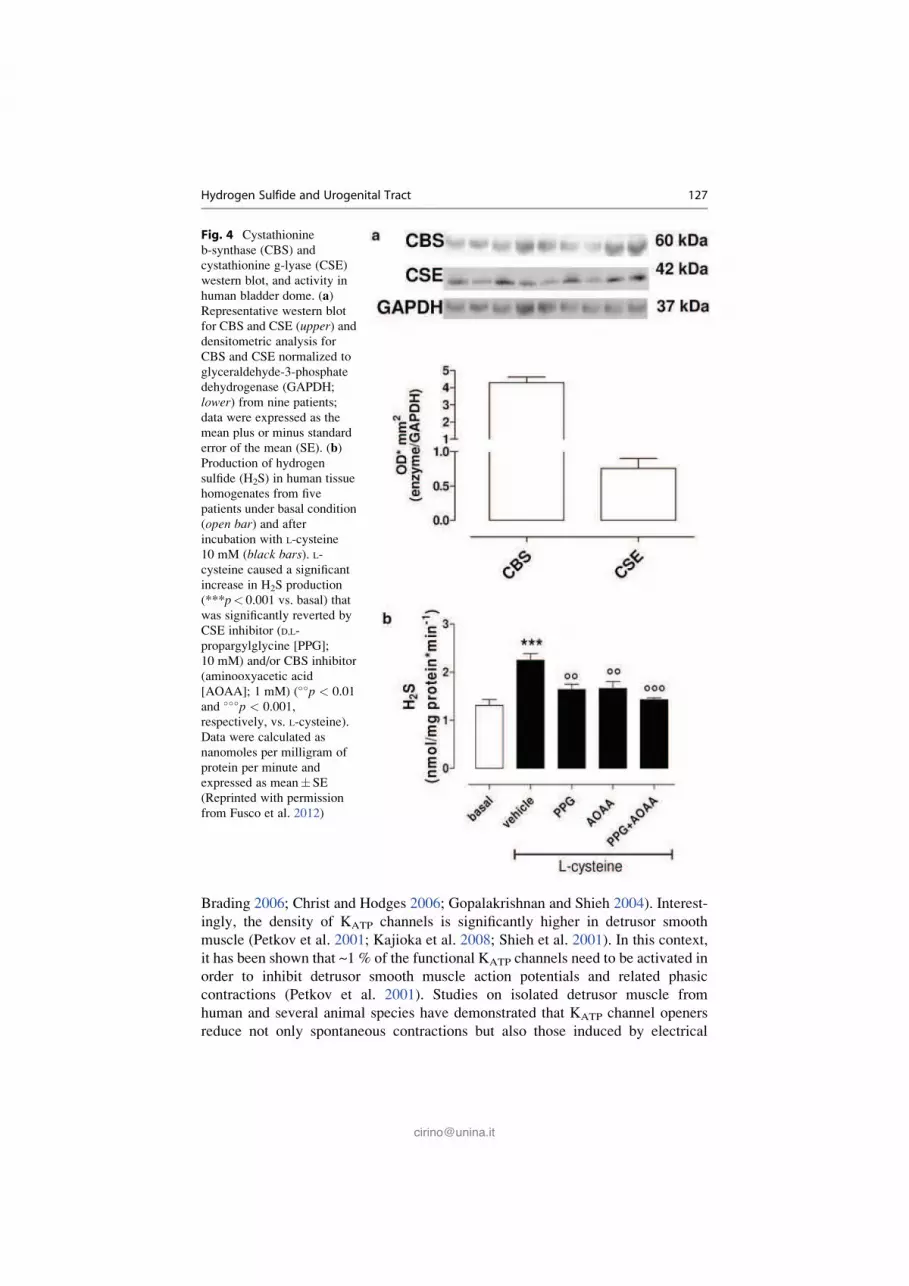
Human bladder expresses all three enzymes responsible for the H2S production
namely CBS, CSE, and 3-MST (Fusco et al. 2012; Gai et al. 2013). Indeed, human
bladder homogenate generates a basal level of production of H2S that can be
enhanced by incubation with L-cysteine (the substrate). This effect is reversed by
either PAG (CSE inhibitor) or AOAA (CBS inhibitor) (Fig. 4). Functional studies
performed using isolated human bladder strips have demonstrated that both sodium
hydrogen sulfide (NaHS) or L-cysteine relax human bladder strips precontracted by
carbachol. Thus, H2S pathway is involved in the regulation of bladder homeostasis.
As discussed, KATP channels are activated by H2S. It is known that urinary
bladder smooth muscles express KATP channels, and they have been shown to be
involved in the regulation of bladder contractility (Andersson 1992; Bonev and
Nelson 1993). The presence of mRNA for sulfonylurea receptors has been
demonstrated in both pig and human detrusor (Buckner et al. 2000). The detrusor
smooth muscle expresses a variety of K+ channels that are responsible for the
detrusor smooth muscle excitability and contractility (Andersson and Arner 2004;
126 R. d’Emmanuele di Villa Bianca et al.
Brading 2006; Christ and Hodges 2006; Gopalakrishnan and Shieh 2004). Interest-
ingly, the density of KATP channels is significantly higher in detrusor smooth
muscle (Petkov et al. 2001; Kajioka et al. 2008; Shieh et al. 2001). In this context,
it has been shown that ~1 % of the functional KATP channels need to be activated in
order to inhibit detrusor smooth muscle action potentials and related phasic
contractions (Petkov et al. 2001). Studies on isolated detrusor muscle from
human and several animal species have demonstrated that KATP channel openers
reduce not only spontaneous contractions but also those induced by electrical
Fig. 4 Cystathionine
b-synthase (CBS) and
cystathionine g-lyase (CSE)
western blot, and activity in
human bladder dome. (a)
Representative western blot
for CBS and CSE (upper) and
densitometric analysis for
CBS and CSE normalized to
glyceraldehyde-3-phosphate
dehydrogenase (GAPDH;
lower) from nine patients;
data were expressed as the
mean plus or minus standard
error of the mean (SE). (b)
Production of hydrogen
sulfide (H2S) in human tissue
homogenates from five
patients under basal condition
(open bar) and after
incubation with L-cysteine
10 mM (black bars). L-
cysteine caused a significant
increase in H2S production
(***p< 0.001 vs. basal) that
was significantly reverted by
CSE inhibitor (D,L-
propargylglycine [PPG];
10 mM) and/or CBS inhibitor
(aminooxyacetic acid
[AOAA]; 1 mM) (��p < 0.01
and ���p < 0.001,
respectively, vs. L-cysteine).
Data were calculated as
nanomoles per milligram of
protein per minute and
expressed as mean� SE
(Reprinted with permission
from Fusco et al. 2012)
Hydrogen Sulfide and Urogenital Tract 127

stimulation, carbachol, and low external K+ concentrations (Andersson 1993).
Interestingly, the relaxant effect elicited by H2S is inhibited by glybenclamide, a
KATP channel inhibitor (Gai et al. 2013). Therefore, it is feasible to hypothesize that
once released, H2S causes relaxation of the detrusor muscle that involves KATP
channel activation thereby contributing to the tonic regulation of the bladder tone.
Bladder tone regulation is a key issue in LUTS. LUTS markedly increase with age
in both males and females, and it represents a major problem in the elderly
population. Recently, the PDE-5 inhibitor tadalafil has been approved by FDA for
LUTS treatment. In addition, it has been reported that sildenafil, a well-known
PDE-5 inhibitor, can directly relax human bladder strips and that this effect
involves the H2S pathway (Fusco et al. 2012). Indeed, sildenafil incubation of
human bladder samples causes a time- and concentration-dependent increase in
H2S production (Fig. 5). In conclusion, the human data, available in the current
Fig. 5 Sildenafil-induced hydrogen sulfide (H2S) production in human bladder dome. (a) Silden-
afil 10 mM caused a time-dependent increase in H2S production (***p < 0.001 vs vehicle; **p <
0.01 vs. 30 min and 45 min). (b) Sildenafil incubation for 30 min caused a concentration-
dependent increase in H2S production compared to vehicle (*p < 0.05 and **p < 0.001
vs. vehicle). (c) Combination of CBS and/or CSE inhibitors (DL-propargylglycine [PPG] 10 mM
and/or aminooxyacetic acid [AOAA] 1 mM) significantly inhibited sildenafil-induced H2S pro-
duction (�p < 0.05 and ��p < 0.01 vs sildenafil 10 mM; **p < 0.01 vs. vehicle). Data were
calculated as nanomoles per milligram of protein per minute and expressed as mean� SEM for
five patients (Reprinted with permission from Fusco et al. 2012)
128 R. d’Emmanuele di Villa Bianca et al.

relevant literature, suggest that the H2S pathway is involved not only in the
physiology of human bladder function but also in LUTS as well as in overactive
bladder. However, further studies are necessary in order to better define the role of
this pathway.
5 Hydrogen Sulfide and Urethra
The urethra together with smooth muscles in the bladder controls the storage and
the voiding of urine. The presence as well as the ability of CBS, CSE, and 3-MST to
convert L-cysteine into H2S has been reported both in human and rat urethra (Gai
et al. 2013). On the functional side, H2S has been shown to have no effect on the
basal tone but to relax the human urethra strips (Gratzke et al. 2009). The relaxing
effect on human urethra strips has been proposed to involve the TRPA1 receptors
that are expressed on nerves fibers, urothelium, and interstitial cells (Gratzke
et al. 2009).
6 Conclusions and Future Directions
On the basis of the findings present in the current literature, from non-mammalian
and mammalian, including human, it is clear that H2S is endogenously produced,
and the enzymes responsible of its biosynthesis are constitutively present in the
genitourinary tract. In addition, H2S appears to be a phylogenetically ancient and
versatile regulatory molecule. Nevertheless, the H2S response in the genitourinary
tract, as reported above, appears to be specie dependent. Therefore, more studies are
needed in order to clearly define the role of this pathway in human since the
translation of preclinical data to human is not always possible. For what concerns,
drugs used in therapy that have been proposed to involve H2S as mediator, there are
some considerations that can be made. It is now well established that patients’
response to a therapy is not equally effective, and who once were called
nonresponders are now considered as a specific sub group. Indeed, it is now clear
that genetics plays a great role in determining the nature of the drug responses.
However, it is also true that, most likely, there are other therapeutic targets that have
not as yet been defined within a specific pathology. In this regard, in both ED and in
pathologies associated with bladder dysfunction, there is a need to find new
therapies. Indeed, PDE-5 inhibitors often do not resolve the ED in diabetes patients
or fail to act in a certain number of eligible patients with no comorbidities. In fact,
the efficacy of PDE-5 inhibitor, which is a mainstay in the treatment of ED, is
negatively associated with the nerve and endothelium damage. These latter features
are associated with several pathologies such as cardiovascular disease, diabetes,
obesity, and post-prostatectomy state and in turn lead to an impairment in the
signaling of the NO/cGMP pathway. In this context, the H2S pathway represents
an attractive target since a link between PDE-5 inhibitors and H2S pathway has
been already shown. Indeed, tadalafil limits myocardial infarction through H2S
Hydrogen Sulfide and Urogenital Tract 129

signaling (Salloum et al. 2009), and sildenafil causes an increase in H2S production
by CBS and CSE activities in human bladder (Fusco et al. 2012). A very prelimi-
nary attempt to develop a drug working on H2S and cGMP pathways has been taken
by Shukla and coworkers who have synthesized and characterized an H2S-donating
derivative of sildenafil (ACS6) (Shukla et al. 2009). For what concerns the bladder,
it has to be stressed that the widely used antimuscarinic drugs often fails to alleviate
the LUTS symptoms. Also in this case, by looking at the recent findings,
highlighted in this chapter, the H2S pathway represents an attractive therapeutic
target that may allow to develop new drugs. In Fig. 6 are shown the drugs for which
the involvement of H2S in their mechanism of action has been proposed.
Another possible therapeutic approach that rises from what has been discussed is
the possibility to modulate H2S levels by exogenous supplementation. Intuitively,
the easiest way should be to use H2S donors that release the gas slowly, in order to
reproduce as much as possible, the physiological conditions. Alternatively, the H2S
production could be enhanced by inducing H2S synthesis by using L-cysteine or
other substrates. However, this approach could fail in condition where a down-
regulation of the enzyme CBS and CSE occurs. Several studies have addressed
these issues in preclinical setting (Kashfi and Olson 2013). The activity of garlic-
Fig. 6 Drugs proposed to involve hydrogen sulfide (H2S) pathway in their mechanism of action.
The cartoon reproduces a smooth muscle cell of the urogenital tract, where the involvement of H2S
in testosterone and sildenafil mechanism of action is depicted. Adenosine triphosphate-activated
potassium channel (KATP); Androgen receptor AR; calcium-dependent potassium channel (KCa);
cyclic guanosine monophosphate (cGMP); Cysthatione-β synthase (CBS); Cysthatione-γ lyase
(CSE), phosphodiesterase-5 (PDE-5)
130 R. d’Emmanuele di Villa Bianca et al.

derived molecules, generally considered as H2S releasers following metabolization,
such as diallyl disulfide, diallyl sulfide, diallyl trisulfide, and diallyl tetrasulfide, has
been profusely investigated (Jacob et al. 2008). Garlic and its bioactive component,
the S-allyl cysteine, have been shown to restore erectile function in diabetic rats by
preventing ROS formation through modulation of NADPH oxidase subunit expres-
sion (Yang et al. 2013). In particular, anti-inflammatory and anticancer effects have
been demonstrated. However, no data, concerning their efficacy, on urogenital tract
are available. The GYY4137 is actually the most attracting synthetic H2S donor. It
inhibits lipid accumulation exhibiting anti-atherosclerotic activity both in vitro and
in vivo (Xu et al. 2014) and exerts anti-inflammatory (Li et al. 2013) and anticancer
(Kashfi 2014) activity. Also in this case, there are no data available on GYY4137
effect in urogenital tract diseases.
References
Abe K, Kimura H (1996) The possible role of hydrogen sulfide as an endogenous neuromodulator.
J Neurosci 16:1066–1071
Abrams P, Cardozo L, Fall M, Griffiths D, Rosier P, Ulmsten U, van Kerrebroeck P, Victor A,
Wein A (2002) The standardization of terminology of lower urinary tract function: report from
the Standardisation Sub-committee of the International Continence Society. Neurourol Urodyn
21:167–178
Altaany Z, Ju Y, Yang G, Wang R (2014) The coordination of S-sulfhydration, S-nitrosylation,
and phosphorylation of endothelial nitric oxide synthase by hydrogen sulfide. Sci Signal 7:ra87
Andersson KE (1992) Clinical pharmacology of potassium channel openers. Pharmacol Toxicol
70:244–254
Andersson KE (1993) Pharmacology of lower urinary tract smooth muscles and penile erectile tissues.
Pharmacol Rev 45:253–308
Andersson KE, Arner A (2004) Urinary bladder contraction and relaxation: physiology and
pathophysiology. Physiol Rev 84:935–986
Andrade EL, Ferreira J, Andre E, Calixto JB (2006) Contractile mechanisms coupled to TRPA1
receptor activation in rat urinary bladder. Biochem Pharmacol 72:104–114
Angulo J, Cuevas P, Fernandez A, Gabancho S, Allona A, Martın-Morales A, Moncada I,
Videla S, Saenz de Tejada I (2003) Diabetes impairs endothelium-dependent relaxation of
human penile vascular tissues mediated by NO and EDHF. Biochem Biophys Res Commun
312:1202–1208
Aversa A, Isidori AM, De Martino MU, Caprio M, Fabbrini E, Rocchietti-March M, Frajese G,
Fabbri A (2000) Androgens and penile erection: evidence for a direct relationship between free
testosterone and cavernous vasodilation in men with erectile dysfunction. Clin Endocrinol
(Oxf) 53:517–522
Bautista DM, Movahed P, Hinman A, Axelsson HE, Sterner O, H€ogestatt ED, Julius D, Jordt SE,
Zygmunt PM (2005) Pungent products from garlic activate the sensory ion channel TRPA1.
Proc Natl Acad Sci USA 102:12248–12252
Becker AJ, Uckert S, Stief CG, Truss MC, Machtens S, Scheller F, Knapp WH, Hartmann U,
Jonas U (2000) Cavernous and systemic testosterone levels in different phases of human penile
erection. Urology 56:125–129
Becker AJ, Uckert S, Stief CG, Scheller F, Knapp WH, Hartmann U, Jonas U (2001) Cavernous
and systemic testosterone plasma levels during different penile conditions in healthy males and
patients with erectile dysfunction. Urology 58:435–440
Hydrogen Sulfide and Urogenital Tract 131

Bonev AD, Nelson MT (1993) ATP-sensitive potassium channels in smooth muscle cells from
guinea pig urinary bladder. Am J Physiol Cell Physiol 264:C1190–C1200
Brading AF (2006) Spontaneous activity of lower urinary tract smooth muscles: correlation
between ion channels and tissue function. J Physiol 570:13–22
Brancaleone V, Vellecco V, Matassa DS, d’Emmanuele di Villa Bianca R, Sorrentino R, Ianaro A,
Bucci M, Esposito F, Cirino G (2014) Crucial role of androgen receptor in vascular H2S
biosynthesis induced by testosterone. Br J Pharmacol 172(6):1505–1515
Brunner H, Cockcroft JR, Deanfield J, Donald A, Ferrannini E, Halcox J, Kiowski W, Luscher TF,
Mancia G, Natali A, Oliver JJ, Pessina AC, Rizzoni D, Rossi GP, Salvetti A, Spieker LE,
Taddei S, Webb DJ, Working Group on Endothelins and Endothelial Factors of the European
Society of Hypertension (2005) Endothelial function and dysfunction. Part II: association with
cardiovascular risk factors and diseases. A statement by the working group on endothelins and
endothelial factors of the european society of hypertension. J Hypertens 23:233–246
Bucci M, Mirone V, Di Lorenzo A, Vellecco V, Roviezzo F, Brancaleone V, Imbimbo C, Cirino G
(2009) Hydrogen sulphide is involved in testosterone vascular effect. Eur Urol 56:378–383
Bucci M, Papapetropoulos A, Vellecco V, Zhou Z, Pyriochou A, Roussos C, Roviezzo F,
Brancaleone V, Cirino G (2010) Hydrogen sulfide is an endogenous inhibitor of phospho-
diesterase activity. Arterioscler Thromb Vasc Biol 30:1998–2004
Buckner SA, Milicic I, Daza A, Davis-Taber R, Scott VE, Sullivan JP, Brioni JD (2000)
Pharmacological and molecular analysis of ATP-sensitive K channels in the pig and
human detrusor. Eur J Pharmacol 400:287–295
Busse R, Edwards G, Feletou M, Fleming I, Vanhoutte PM, Weston AH (2002) EDHF:
bringing the concepts together. Trends Pharmacol Sci 23:374–380
Cai WJ, Wang MJ, Moore PK, Jin HM, Yao T, Zhu YC (2007) The novel proangiogenic effect of
hydrogen sulfide is dependent on Akt phosphorylation. Cardiovasc Res 76:29–40
Christ GJ, Hodges S (2006) Molecular mechanisms of detrusor and corporal myocyte contraction:
identifying targets for pharmacotherapy of bladder and erectile dysfunction. Br J Pharmacol
147(Suppl 2):S41–S55
Christ GJ, Brink PR, Melman A, Spray DC (1993) The role of gap junctions and ion channels in
the modulation of electrical and chemical signals in human corpus cavernosum smooth muscle.
Int J Impot Res 5:77–96
Cirino G, Fusco F, Imbimbo C, Mirone V (2006) Pharmacology of erectile dysfunction in man.
Pharmacol Ther 111:400–423
Coletta C, Papapetropoulos A, Erdelyi K, Olah G, M�odis K, Panopoulos P, Asimakopoulou A,
Ger€o D, Sharina I, Martin E, Szabo C (2012) Hydrogen sulfide and nitric oxide are
mutually dependent in the regulation of angiogenesis and endothelium-dependent vaso-
relaxation. Proc Natl Acad Sci USA 109:9161–9166
d’Emmanuele di Villa Bianca R, Sorrentino R, Maffia P, Mirone V, Imbimbo C, Fusco F,
De Palma R, Ignarro LJ, Cirino G (2009) Hydrogen sulfide as a mediator of human corpus
cavernosum smooth-muscle relaxation. Proc Natl Acad Sci USA 106:4513–4518
d’Emmanuele di Villa Bianca R, Sorrentino R, Coletta C, Mitidieri E, Rossi A, Vellecco V
et al (2011) Hydrogen sulfide-induced dual vascular effect involves arachidonic acid cascade
in rat mesenteric arterial bed. J Pharmacol Exp Ther 337:59–64
de Groat WC, Yoshimura N (2001) Pharmacology of the lower urinary tract. Annu Rev Pharmacol
Toxicol 41:691–721
Dikmen A, d’Emmanuele di Villa Bianca R, Mitidieri E, Donnarumma E, Sevin G, Cirino G,
Sorrentino R, Yetik-Anacak G (2013) New mechanism for the beneficial effect of sildenafil on
erectile function: H2S. Nitric Oxide 31:S38
Dombkowski RA, Doellman MM, Head SK, Olson KR (2006) Hydrogen sulfide mediates
hypoxia-induced relaxation of trout urinary bladder smooth muscle. J Exp Biol 209:3234–3340
Dong JY, Zhang YH, Qin LQ (2011) Erectile dysfunction and risk of cardiovascular disease: meta-
analysis of prospective cohort studies. J Am Coll Cardiol 58:1378–1385
132 R. d’Emmanuele di Villa Bianca et al.

Fernandes VS, Ribeiro AS, Barahona MV, Orensanz LM, Martınez-Saenz A, Recio P,
Martınez AC, Bustamante S, Carballido J, Garcıa-Sacristan A, Prieto D, Hernandez M
(2013a) Hydrogen sulfide mediated inhibitory neurotransmission to the pig bladder neck:
role of KATP channels, sensory nerves and calcium signaling. J Urol 190:746–756
Fernandes VS, Ribeiro AS, Martınez MP, Orensanz LM, Barahona MV, Martınez-Saenz A,
Recio P, Benedito S, Bustamante S, Carballido J, Garcıa-Sacristan A, Prieto D, Hernandez
M (2013b) Endogenous hydrogen sulfide has a powerful role in inhibitory neurotransmission to
the pig bladder neck. J Urol 189:1567–1573
Fusco F, d’Emmanuele di Villa Bianca RD, Mitidieri E, Cirino G, Sorrentino R, Mirone V (2012)
Sildenafil effect on the human bladder involves the L-cysteine/hydrogen sulfide pathway:
a novel mechanism of action of phosphodiesterase type 5 inhibitors. Eur Urol 62:1174–1180
Gai JW, Wahafu W, Guo H, Liu M, Wang XC, Xiao YX, Zhang L, Xin ZC, Jin J (2013)
Further evidence of endogenous hydrogen sulphide as a mediator of relaxation in human and
rat bladder. Asian J Androl 15:692–696
Gandaglia G, Briganti A, Jackson G, Kloner RA, Montorsi F, Montorsi P, Vlachopoulos C (2014)
A systematic review of the association between erectile dysfunction and cardiovascular disease.
Eur Urol 65:968–978
Geng B, Cui Y, Zhao J, Yu F, Zhu Y, Xu G, Zhang Z, Tang C, Du J (2007) Hydrogen sulfide
downregulates the aortic L-arginine/nitric oxide pathway in rats. Am J Physiol Regul Integr
Comp Physiol 293:R1608–R1618
Ghasemi M, Dehpour AR, Moore KP, Mani AR (2012) Role of endogenous hydrogen sulfide in
neurogenic relaxation of rat corpus cavernosum. Biochem Pharmacol 83:1261–1268
Goldstein I, Young JM, Fischer J, Bangerter K, Segerson T, Taylor T (2003) Vardenafil a
new phosphodiesterase type 5 inhibitor, in the treatment of erectile dysfunction in men with
diabetes: a multicenter double-blind placebo-controlled fixed-dose study. Diabetes Care 26:
777–783
Gopalakrishnan M, Shieh CC (2004) Potassium channel subtypes as molecular targets for over-
active bladder and other urological disorders. Expert Opin Ther Targets 8:437–458
Gratzke C, Streng T, Waldkirch E, Sigl K, Stief C, Andersson KE, Hedlund P (2009) Transient
receptor potential A1 (TRPA1) activity in the human urethra—evidence for a functional role
for TRPA1 in the outflow region. Eur Urol 55:696–704
Grgic I, Kaistha BP, Hoyer J, K€ohler R (2009) Endothelial Ca +�activated K+ channels in
normal and impaired EDHF-dilator responses–relevance to cardiovascular pathologies and
drug discovery. Br J Pharmacol 157:509–526
Guo H, Gai JW, Wang Y, Jin HF, Du JB, Jin J (2012) Characterization of hydrogen sulfide and its
synthases, cystathionine β-synthase and cystathionine γ-lyase, in human prostatic tissue and
cells. Urology 79:483, e1-5
Hackett G, Kirby M, Sinclair AJ (2014) Testosterone deficiency, cardiac health, and older men.
Int J Endocrinol 2014:143763
Isidori AM, Buvat J, Corona G, Goldstein I, Jannini EA, Lenzi A, Porst H, Salonia A, Traish AM,
Maggi M (2014) A critical analysis of the role of testosterone in erectile function: from
pathophysiology to treatment-a systematic review. Eur Urol 65:99–112
Jacob C, Anwar A, Burkholz T (2008) Perspective on recent developments on sulfur-containing
agents and hydrogen sulfide signaling. Planta Med 74:1580–1592
Jiang B, Tang G, Cao K, Wu L, Wang R (2010) Molecular mechanism for H(2)S-induced
activation of K(ATP) channels. Antioxid Redox Signal 12:1167–1178
Kajioka S, Nakayama S, Asano H, Seki N, Naito S, Brading AF (2008) Levcromakalim and
MgGDP activate small conductance ATP-sensitive K+ channels of K+ channel pore 6.1/
sulfonylurea receptor 2A in pig detrusor smooth muscle cells: uncoupling of cAMP signal
pathways. J Pharmacol Exp Ther 327:114–123
Kardong KV (2005) The urogenital system. In: Vertebrates: compartive anatomy, function,
evolution. McGraw Hill Higher Education, New York, pp 577–578 (Chapter 14)
Hydrogen Sulfide and Urogenital Tract 133

Karicheti V, Christ GJ (2001) Physiological roles for K+ channels and gap junctions in
urogenital smooth muscle: implications for improved understanding of urogenital function,
disease and therapy. Curr Drug Targets 2:1–20
Kashfi K (2014) Anti-cancer activity of new designer hydrogen sulfide-donating hybrids.
Antioxid Redox Signal 20:831–846
Kashfi K, Olson KR (2013) Biology and therapeutic potential of hydrogen sulfide and
hydrogen sulfide-releasing chimeras. Biochem Pharmacol 85:689–703
Li J, Li Y, Du Y, Mou K, Sun H, Zang Y, Liu C (2011) Endogenous hydrogen sulfide as a mediator
of vas deferens smooth muscle relaxation. Fertil Steril 95:1833–1835
Li Y, Zang Y, Fu S, Zhang H, Gao L, Li J (2012) H2S relaxes vas deferens smooth muscle by
modulating the large conductance Ca2 +�activated K+ (BKCa) channels via a redox mecha-
nism. J Sex Med 9:2806–2813
Li L, Fox B, Keeble J, Salto-Tellez M, Winyard PG, Wood ME, Moore PK, Whiteman M (2013)
The complex effects of the slow-releasing hydrogen sulfide donor GYY4137 in a model of
acute joint inflammation and in human cartilage cells. J Cell Mol Med 17:365–376
Matsunami M, Miki T, Nishiura K, Hayashi Y, Okawa Y, Nishikawa H, Sekiguchi F, Kubo L,
Ozaki T, Tsujiuchi T, Kawabata A (2012) Involvement of the endogenous hydrogen sulfide/Ca
(v) 3.2 T-type Ca2+ channel pathway in cystitis-related bladder pain in mice. Br J Pharmacol
167:917–928
Meng J, Ganesan Adaikan P, Srilatha B (2013) Hydrogen sulfide promotes nitric oxide production
in corpus cavernosum by enhancing expression of endothelial nitric oxide synthase. Int J Impot
Res 25:86–90
Milsom I, Abrams P, Cardozo L, Roberts RG, Thuroff J, Wein AJ (2001) How widespread are the
symptoms of an overactive bladder and how are they managed? A population-based prevalence
study. BJU Int 87:760–766
Mirone V, Sorrentino R, di Villa BR, Imbimbo C, Palmieri A, Fusco F, Tajana G, Cirino G (2000)
A standardized procedure for using human corpus cavernosum strips to evaluate drug activity.
J Pharmacol Toxicol Methods 44:477–482
Munaron L, Avanzato D, Moccia F, Mancardi D (2013) Hydrogen sulfide as a regulator of
calcium channels. Cell Calcium 53:77–84
Mustafa AK, Sikka G, Gazi SK, Steppan J, Jung SM, Bhunia AK, Barodka VM, Gazi FK, Barrow
RK, Wang R, Amzel LM, Berkowitz DE, Snyder SH (2011) Hydrogen sulfide as endothelium-
derived hyperpolarizing factor sulfhydrates potassium channels. Circ Res 109:1259–1268
Nehra A, Jackson G, Miner M, Billups KL, Burnett AL, Buvat J, Carson CC, Cunningham GR,
Ganz P, Goldstein I, Guay AT, Hackett G, Kloner RA, Kostis J, Montorsi P, Ramsey M,
Rosen R, Sadovsky R, Seftel AD, Shabsigh R, Vlachopoulos C, Wu FC (2012) The Princeton
III Consensus recommendations for the management of erectile dysfunction and cardio-
vascular disease. Mayo Clin Proc 87:766–778
Nelson CP, Rainbow RD, Brignell JL, Perry MD, Willets JM, Davies NW, Standen NB,
Challiss RA (2011) Principal role of adenylyl cyclase 6 in K+ channel regulation and vaso-
dilator signalling in vascular smooth muscle cells. Cardiovasc Res 91:694–702
Nilius B, Owsianik G, Voets T, Peters JA (2007) Transient receptor potential cation channels in
disease. Physiol Rev 87:165–217
Olson KR (2014) Hydrogen sulfide as an oxygen sensor. Antioxid Redox Signal 22(5):377–397
Patacchini R, Maggi CA, Meli A (1990) Capsaicin-like activity of some natural pungent
substances on peripheral endings of visceral primary afferents. Naunyn Schmiedebergs Arch
Pharmacol 342:72–77
Patacchini R, Santicioli P, Giuliani S, Maggi CA (2004) Hydrogen sulfide (H2S) stimulates
capsaicin-sensitive primary afferent neurons in the rat urinary bladder. Br J Pharmacol
142:31–34
Patacchini R, Santicioli P, Giuliani S, Maggi CA (2005) Pharmacological investigation of hydro-
gen sulfide (H2S) contractile activity in rat detrusor muscle. Eur J Pharmacol 509:171–177
134 R. d’Emmanuele di Villa Bianca et al.

Petkov GV, Heppner TJ, Bonev AD, Herrera GM, Nelson MT (2001) Low levels of KATP channel
activation decrease excitability and contractility of urinary bladder. Am J Physiol Regul Integr
Comp Physiol 280:R1427–R1433
Price DE, Gingell JC, Gepi-Attee S, Warcham K, Yates P, Boolell M (1998) Sildenafil: study of a
novel oral treatment for erectile dysfunction in diabetic men. Diabet Med 15:821–825
Qiu X, Villalta J, Lin G, Lue TF (2012) Role of hydrogen sulfide in the physiology of penile erection.
J Androl 33:529–535
Rendell MS, Rajfer J, Wicker PA, Smith MD (1999) Sildenafil for treatment of erectile dys-
function in men with diabetes. A randomized controlled trial. JAMA 281:421–426
Ruiz Rubio JL, Hernandez M, Arcos L, Benedito S, Recio P, Garcıa P, Garcıa-Sacristan A,
Prieto D (2004) Role of ATP-sensitive K+ channels in relaxation of penile resistance arteries.
Urology 63:800–805
Saenz de Tejada I, Anglin G, Knight JR, Emmick JT (2002) Effects of tadalafil on erectile
dysfunction in men with diabetes. Diabetes Care 25:2159–2164
Salloum FN, Chau VQ, Hoke NN, Abbate A, Varma A, Ockaili RA, Toldo S, Kukreja RC (2009)
Phosphodiesterase-5 inhibitor, tadalafil, protects against myocardial ischemia/reperfusion
through protein-kinase g-dependent generation of hydrogen sulfide. Circulation 120:S31–S36
Schultheiss D, Badalyan R, Pilatz A, Gabouev AI, Schlote N, Wefer J, von Wasielewski R,
Mertsching H, Sohn M, Stief CG, Jonas U (2003) Androgen and estrogen receptors in the
human corpus cavernosum penis: immunohistochemical and cell culture results. World J Urol
21:320–324
Shieh CC, Feng J, Buckner SA, Brioni JD, Coghlan MJ, Sullivan JP, Gopalakrishnan M (2001)
Functional implication of spare ATP-sensitive K+ channels in bladder smooth muscle cells.
J Pharmacol Exp Ther 296:669–675
Shukla N, Rossoni G, Hotston M, Sparatore A, Del Soldato P, Tazzari V, Persad R, Angelini GD,
Jeremy JY (2009) Effect of hydrogen sulphide-donating sildenafil (ACS6) on erectile function
and oxidative stress in rabbit isolated corpus cavernosum and in hypertensive rats. BJU Int
103:1522–1529
Spektor M, Rodriguez R, Rosenbaum RS, Wang HZ, Melman A, Christ GJ (2002) Potassium
channels and human corporeal smooth muscle cell tone: further evidence of the physiological
relevance of the Maxi-K channel subtype to the regulation of human corporeal smooth muscle
tone in vitro. J Urol 167:2628–2635
Srilatha B, Adaikan PG, Moore PK (2006) Possible role for the novel gasotransmitter hydrogen
sulphide in erectile dysfunction–a pilot study. Eur J Pharmacol 535:280–282
Srilatha B, Adaikan PG, Li L, Moore PK (2007) Hydrogen sulphide: a novel endogenous gaso-
transmitter facilitates erectile function. J Sex Med 4:1304–1311
Srilatha B, Hu L, Adaikan GP, Moore PK (2009) Initial characterization of hydrogen sulfide
effects in female sexual function. J Sex Med 6:1875–1884
Srilatha B, Muthulakshmi P, Adaikan PG, Moore PK (2012) Endogenous hydrogen sulfide
insufficiency as a predictor of sexual dysfunction in aging rats. Aging Male 15:153–158
Story GM, Peier AM, Reeve AJ, Eid SR, Mosbacher J, Hricik TR, Earley TJ, Hergarden AC,
Andersson DA, Hwang SW, McIntyre P, Jegla T, Bevan S, Patapoutian A (2003) ANKTM1,
a TRP-like channel expressed in nociceptive neurons, is activated by cold temperatures.
Cell 112:819–829
Streng T, Axelsson HE, Hedlund P, Andersson DA, Jordt SE, Bevan S, Andersson KE,
H€ogestatt ED, Zygmunt PM (2008) Distribution and function of the hydrogen sulfide-sensitive
TRPA1 ion channel in rat urinary bladder. Eur Urol 53:391–399
Tang G, Yang G, Jiang B, Ju Y, Wu L, Wang R (2013) H2S is an endothelium-derived
hyperpolarizing factor. Antioxid Redox Signal 19:1634–1646
Traish AM, Goldstein I, Kim NN (2007) Testosterone and erectile function: from basic research to
a new clinical paradigm for managing men with androgen insufficiency and erectile dys-
function. Eur Urol 52:54–70
Hydrogen Sulfide and Urogenital Tract 135

Venkateswarlu K, Giraldi A, Zhao W, Wang HZ, Melman A, Spektor M, Christ GJ (2002)
Potassium channels and human corporeal smooth muscle cell tone: diabetes and relaxation
of human corpus cavernosum smooth muscle by adenosine triphosphate sensitive potassium
channel openers. J Urol 168:355–361
Vickers MA, Satyanarayana R (2002) Phosphodiesterase type 5 inhibitors for the treatment of
erectile dysfunction in patients with diabetes mellitus. Int J Impot Res 14:466–471
Wang R (2003) The gasotransmitter role of hydrogen sulfide. Antioxid Redox Signal 5:493–501
Wang R (2009) Hydrogen sulfide: a new EDRF. Kidney Int 76:700–704
Xu S, Liu Z, Liu P (2014) Targeting hydrogen sulfide as a promising therapeutic strategy for
atherosclerosis. Int J Cardiol 172:313–317
Yang J, Wang T, Yang J, Rao K, Zhan Y, Chen RB, Liu Z, Li MC, Zhuan L, Zang GH, Guo SM,
Xu H, Wang SG, Liu JH, Ye ZQ (2013) S-allyl cysteine restores erectile function through
inhibition of reactive oxygen species generation in diabetic rats. Andrology 1:487–494
Yetik-Anacak G, Sorrentino R, Linder AE, Murat N (2014) Gas what: NO is not the only answer to
sexual function. Br J Pharmacol 172(6):1434–1454
Yildiz O, Seyrek M, Irkilata HC, Yildirim I, Tahmaz L, Dayanc M (2009) Testosterone
might cause relaxation of human corpus cavernosum by potassium channel opening action.
Urology 74:229–232
Zhao W, Wang R (2002) H2S-induced vasorelaxation and underlying cellular and
molecular mechanisms. Am J Physiol Heart Circ Physiol 283:H474–H480
Zhao W, Zhang J, Lu Y, Wang R (2001) The vasorelaxant effect of H2S as a novel endogenous
gaseous KATP channel opener. EMBO J 20:6008–6016
Zhao K, Li S, Wu L, Lai C, Yang G (2014) Hydrogen sulfide represses androgen receptor
transactivation by targeting at the second zinc finger module. J Biol Chem 289:20824–20835
Zuo C, Huang YM, Jiang R, Yang HF, Cheng B, Chen F (2014) Endogenous hydrogen sulfide and
androgen deficiency-induced erectile dysfunction in rats. Zhonghua Nan Ke Xue 20:605–612
136 R. d’Emmanuele di Villa Bianca et al.