Biliary tract cancer - AIGO
17
Seminar 428 www.thelancet.com Vol 397 January 30, 2021 Biliary tract cancer Juan W Valle, R Katie Kelley, Bruno Nervi, Do-Youn Oh, Andrew X Zhu Biliary tract cancers, including intrahepatic, perihilar, and distal cholangiocarcinoma as well as gallbladder cancer, are low-incidence malignancies in most high-income countries, but represent a major health problem in endemic areas; moreover, the incidence of intrahepatic cholangiocarcinoma is rising globally. Surgery is the cornerstone of cure; the optimal approach depends on the anatomical site of the primary tumour and the best outcomes are achieved through management by specialist multidisciplinary teams. Unfortunately, most patients present with locally advanced or metastatic disease. Most studies in advanced disease have pooled the various subtypes of biliary tract cancer by necessity to achieve adequate sample sizes; however, differences in epidemiology, clinical presentation, natural history, surgical therapy, response to treatment, and prognosis have long been recognised. Additionally, the identification of distinct patient subgroups harbouring unique molecular alterations with corresponding targeted therapies (such as isocitrate dehydrogenase-1 mutations and fibroblast growth factor receptor-2 fusions in intrahepatic cholangiocarcinoma, among others) is changing the treatment paradigm. In this Seminar we present an update of the causes, diagnosis, molecular classification, and treatment of biliary tract cancer. Introduction Biliary tract cancer refers to a spectrum of invasive adenocarcinomas, including cholangiocarcinoma (cancers arising in the intrahepatic, perihilar, or distal biliary tree), and gallbladder carcinoma. In this Seminar we discuss epidemiology and risk factors, classification of the various subtypes of biliary tract cancer, diagnosis, and treatment (including surgery and adjuvant therapy in early-stage disease through to the latest developments in molecular profiling, targeted therapies, and immunotherapy for advanced disease) and provide some perspectives for the future. Epidemiology and risk factors Incidence and causes vary between biliary tract cancer subgroups and geographical regions (figure 1). 1,2 The incidence of cholangiocarcinoma is low in high-income countries (from 0·35 cases per 100 000 to 2 per 100 000 annually); however, in endemic regions of Thailand and China, the incidence is up to 40-times higher. 3,4 The incidence of intrahepatic cholangiocar- cinoma in high-income countries is rising; data from the UK, the USA, and other countries have shown a consistent and steady rise in incidence from 0·1 cases per 100 000 to 0·6 per 100 000 over the past 30 years. 3,5–7 Surveillance, Epidemiology, and End Results Program data (1973–2012) have shown only a slight incidence increase in extrahepatic cholangiocarcinoma from 0·95 cases per 100 000 to 1·02 per 100 000; however, intra- hepatic cholangiocarcinoma incidence has increased from 0·44 per 100 000 to 1·18 cases per 100 000; an average annual percentage change of 2·3% (4·4% over the past 10 years) 8 even correcting for the following coding errors. International Classification of Diseases and Related Health Problems (ICD) codes for cholangiocarcinoma have changed three times (ICD-0–1 to ICD-0–2 in 1993, and ICD-0–3 in 2001) with perihilar cholangiocarcinoma misclassified as intrahepatic cholangiocarcinoma during these changes 9 and with versions adopted inconsistently globally. The new ICD-11 classification 10 includes specific codes for intrahepatic cholangiocarcinoma (2C12.10), hilar cholangiocarcinoma (2C18.0), adenocarcinoma of biliary tract, distal bile duct (2C15.0), and adenocarcinoma of the gallbladder (2C13.0); aiming to harmonise future epidemiological data. Moreover, cases of intrahepatic cholangiocarcinoma might be misclassified as metastatic cancer of unknown primary (CUP); 11 a number of criteria and new tests, including the newly developed albumin in- situ hybridisation assay, can differentiate between intra- hepatic cholangiocarcinoma and CUP. 12 Regarding gallbladder carcinoma, an estimated 219 420 new cases and 165 087 deaths were reported worldwide in 2018, 13 with substantial variation by gender and geographical region globally. The highest rates are observed in women from southern Chile (27 cases per 100 000) followed by regions of northern India (21·5 cases per 100 000), Poland (14 cases per 100 000), south Pakistan (11·3 cases per 100 000), and Japan (7 cases per 100 000). The incidence is relatively uniform or decreasing in high-income countries, 14 probably because of the increase in routine cholecystectomy. The varying regional incidence of cholangiocarcinoma reflects different underlying risk factors. In general, risk factors for the disease include primary sclerosing cholangitis, Caroli’s disease, hepatolithiasis, and liver fluke infections. Others include cirrhosis, hepatitis B and hepatitis C infection, obesity-associated liver disease, and Lancet 2021; 397: 428–44 Division of Cancer Sciences, University of Manchester, Manchester, UK (Prof J W Valle MD); Department of Medical Oncology, The Christie NHS Foundation Trust, Manchester, UK (Prof J W Valle); Helen Diller Family Comprehensive Cancer Center, University of California, San Francisco, CA, USA (R K Kelley MD); Department of Hematology Oncology, School of Medicine, Pontificia Universidad Católica de Chile, Santiago, Chile (B Nervi MD); Division of Medical Oncology, Department of Internal Medicine, Seoul National University Hospital, Cancer Research Institute, Seoul National University College of Medicine, Seoul, Korea (Prof D-Y Oh PhD); Massachusetts General Hospital Cancer Center, Harvard Medical School, Boston, MA, USA (Prof A X Zhu MD); Jiahui International Cancer Center, Jiahui Health, Shanghai, China (Prof A X Zhu) Correspondence to: Prof Juan W Valle, Division of Cancer Sciences, University of Manchester, Manchester, M20 4BX, UK [email protected] Search strategy and selection criteria We searched MEDLINE and PubMed databases, using the terms “biliary tract cancer”, “cholangiocarcinoma” or “gallbladder cancer”, focusing on randomised trials and other high-quality studies published in English from Jan 1, 1995, to March 31, 2020. Publications within the past 5 years were prioritised, although older, relevant, high-quality studies were also selected. Meeting abstracts (from peer-reviewed congresses) were also included if deemed to be of high quality and could potentially change practice. Downloaded for AdminAigo AdminAigo ([email protected]) at Italian Hospital Gastroenterologists and Endoscopists Association from ClinicalKey.com by Elsevier on February 13, 2021. For personal use only. No other uses without permission. Copyright ©2021. Elsevier Inc. All rights reserved.
-
Upload
khangminh22 -
Category
Documents
-
view
0 -
download
0
Transcript of Biliary tract cancer - AIGO
Seminar
428 www.thelancet.com Vol 397 January 30, 2021
Biliary tract cancerJuan W Valle, R Katie Kelley, Bruno Nervi, Do-Youn Oh, Andrew X Zhu
Biliary tract cancers, including intrahepatic, perihilar, and distal cholangiocarcinoma as well as gallbladder cancer, are low-incidence malignancies in most high-income countries, but represent a major health problem in endemic areas; moreover, the incidence of intrahepatic cholangiocarcinoma is rising globally. Surgery is the cornerstone of cure; the optimal approach depends on the anatomical site of the primary tumour and the best outcomes are achieved through management by specialist multidisciplinary teams. Unfortunately, most patients present with locally advanced or metastatic disease. Most studies in advanced disease have pooled the various subtypes of biliary tract cancer by necessity to achieve adequate sample sizes; however, differences in epidemiology, clinical presentation, natural history, surgical therapy, response to treatment, and prognosis have long been recognised. Additionally, the identification of distinct patient subgroups harbouring unique molecular alterations with corresponding targeted therapies (such as isocitrate dehydrogenase-1 mutations and fibroblast growth factor receptor-2 fusions in intrahepatic cholangiocarcinoma, among others) is changing the treatment paradigm. In this Seminar we present an update of the causes, diagnosis, molecular classification, and treatment of biliary tract cancer.
IntroductionBiliary tract cancer refers to a spectrum of invasive adenocarcinomas, including cholangiocarcinoma (cancers arising in the intrahepatic, perihilar, or distal biliary tree), and gallbladder carcinoma. In this Seminar we discuss epidemiology and risk factors, classification of the various subtypes of biliary tract cancer, diagnosis, and treatment (including surgery and adjuvant therapy in early-stage disease through to the latest developments in molecular profiling, targeted therapies, and immunotherapy for advanced disease) and provide some perspectives for the future.
Epidemiology and risk factorsIncidence and causes vary between biliary tract cancer subgroups and geographical regions (figure 1).1,2 The incidence of cholangiocarcinoma is low in high-income countries (from 0·35 cases per 100 000 to 2 per 100 000 annually); however, in endemic regions of Thailand and China, the incidence is up to 40-times higher.3,4 The incidence of intrahepatic cholangiocar-cinoma in high-income countries is rising; data from the UK, the USA, and other countries have shown a consistent and steady rise in incidence from 0·1 cases per 100 000 to 0·6 per 100 000 over the past 30 years.3,5–7 Surveillance, Epidemiology, and End Results Program data (1973–2012) have shown only a slight incidence
increase in extra hepatic cholangiocarcinoma from 0·95 cases per 100 000 to 1·02 per 100 000; however, intra-hepatic cholangiocarcinoma incidence has increased from 0·44 per 100 000 to 1·18 cases per 100 000; an average annual percentage change of 2·3% (4·4% over the past 10 years)8 even correcting for the following coding errors. International Classification of Diseases and Related Health Problems (ICD) codes for cholangiocarcinoma have changed three times (ICD-0–1 to ICD-0–2 in 1993, and ICD-0–3 in 2001) with perihilar cholangiocarcinoma misclassified as intrahepatic chol angiocarcinoma during these changes9 and with versions adopted inconsistently globally. The new ICD-11 classification10 includes specific codes for intrahepatic cholangiocarcinoma (2C12.10), hilar cholangiocarcinoma (2C18.0), adenocarcinoma of biliary tract, distal bile duct (2C15.0), and adenocarcinoma of the gallbladder (2C13.0); aiming to harmonise future epidemiological data. More over, cases of intrahepatic cholangiocarcinoma might be misclassified as metastatic cancer of unknown primary (CUP);11 a number of criteria and new tests, including the newly developed albumin in-situ hybridisation assay, can differentiate between intra-hepatic cholangiocarcinoma and CUP.12
Regarding gallbladder carcinoma, an estimated 219 420 new cases and 165 087 deaths were reported worldwide in 2018,13 with substantial variation by gender and geographical region globally. The highest rates are observed in women from southern Chile (27 cases per 100 000) followed by regions of northern India (21·5 cases per 100 000), Poland (14 cases per 100 000), south Pakistan (11·3 cases per 100 000), and Japan (7 cases per 100 000). The incidence is relatively uniform or decreasing in high-income countries,14 probably because of the increase in routine cholecystectomy.
The varying regional incidence of cholangiocarcinoma reflects different underlying risk factors. In general, risk factors for the disease include primary sclerosing cholangitis, Caroli’s disease, hepatolithiasis, and liver fluke infections. Others include cirrhosis, hepatitis B and hepatitis C infection, obesity-associated liver disease, and
Lancet 2021; 397: 428–44
Division of Cancer Sciences, University of Manchester,
Manchester, UK (Prof J W Valle MD); Department
of Medical Oncology, The Christie NHS Foundation
Trust, Manchester, UK (Prof J W Valle); Helen Diller
Family Comprehensive Cancer Center, University of California,
San Francisco, CA, USA (R K Kelley MD); Department of Hematology Oncology, School
of Medicine, Pontificia Universidad Católica de Chile,
Santiago, Chile (B Nervi MD); Division of Medical Oncology,
Department of Internal Medicine, Seoul National
University Hospital, Cancer Research Institute, Seoul
National University College of Medicine, Seoul, Korea
(Prof D-Y Oh PhD); Massachusetts General Hospital Cancer Center,
Harvard Medical School, Boston, MA, USA
(Prof A X Zhu MD); Jiahui International Cancer Center,
Jiahui Health, Shanghai, China (Prof A X Zhu)
Correspondence to: Prof Juan W Valle, Division of
Cancer Sciences, University of Manchester, Manchester,
M20 4BX, UK [email protected]
Search strategy and selection criteria
We searched MEDLINE and PubMed databases, using the terms “biliary tract cancer”, “cholangiocarcinoma” or “gallbladder cancer”, focusing on randomised trials and other high-quality studies published in English from Jan 1, 1995, to March 31, 2020. Publications within the past 5 years were prioritised, although older, relevant, high-quality studies were also selected. Meeting abstracts (from peer-reviewed congresses) were also included if deemed to be of high quality and could potentially change practice.
Downloaded for AdminAigo AdminAigo ([email protected]) at Italian Hospital Gastroenterologists and Endoscopists Association from ClinicalKey.com by Elsevier on February 13, 2021. For personal use only. No other uses without permission. Copyright ©2021. Elsevier Inc. All rights reserved.

Seminar
www.thelancet.com Vol 397 January 30, 2021 429
diabetes. Underlying hepatic inflammation, fibrosis, or cirrhosis are risk factors for intrahepatic cholangio-carcinoma.15 A previous meta-analysis showed that stones, cirrhosis, hepatitis B and hepatitis C are the strongest risk factors for both intrahepatic cholan-giocarcinoma and extrahepatic cholangiocar cinoma.16 However, recognising that most patients with cholangio-carcinoma have no identifiable risk factors is important. Although in high-income countries cholangiocarcinoma is associated with chronic inflammation of the biliary tree and hepatic parenchyma, in Thailand, chronic infection with liver fluke is the driving risk factor. Endemic liver fluke infection (Opisthorchis viverrini) is associated with eating raw or undercooked fish for 20 years or more. Endemic areas for Clonorchis sinensis are in China, Korea, Taiwan, and Vietnam.17
Gallbladder carcinoma has a different pathophysiology than does cholangiocarcinoma with a wide range of predisposing conditions, environmental exposures, and lifestyle behaviours linked to increased risk; gallbladder
carcinoma increases with age and is more common in women. Predisposing conditions causing chronic irrita-tion or inflammation of the gallbladder are associated with a higher incidence of gallbladder carcinoma, and cholelithiasis (gallstones) is one of the most strongly associated risk factors with 70–90% of gallbladder carci noma cases having a history of cholelithiasis. How-ever, only 0·5–3% of gallstone cases result in gallbladder carcinoma.18 Primary sclerosing cholangitis is associated with an increased risk of gallbladder carcinoma (esti-mated 2% lifetime incidence).19–21 Structural biliary tree abnormalities, including congenital biliary dila tation and anomalous pancreaticobiliary ductal junction (also known as biliopancreatic or pancreaticobiliary maljunction),22 chronic Salmonella typhi or Helicobacter bilis infections,23 and obesity24 increase the risk of the disease. Choledochal cysts have a 1–15% lifetime risk of development into gallbladder carcinoma or cholangio carcinoma.
Cancers arising from the ampulla of Vater (the junction of the pancreatic and distal common bile duct) are
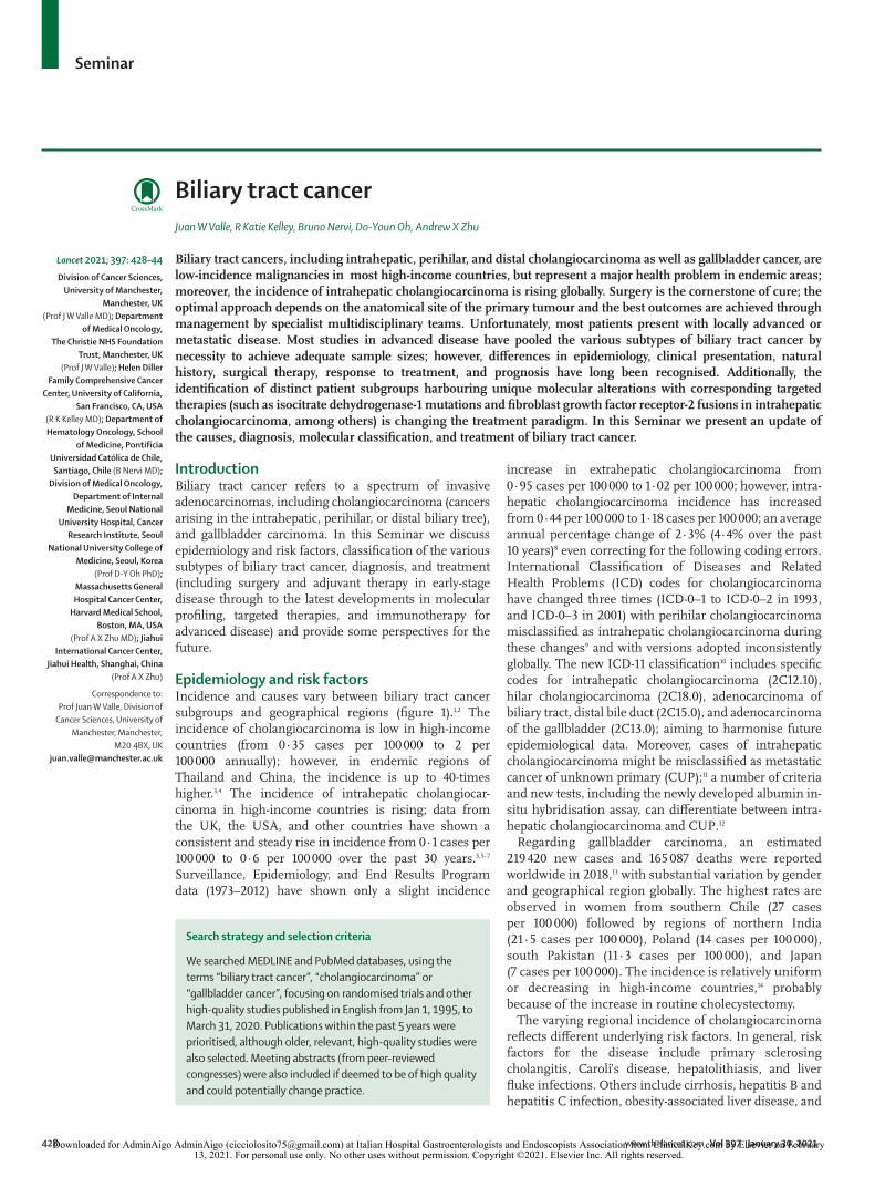
Figure 1: Global incidence of cholangiocarcinoma (A) and gallbladder cancer (B)Data for the global incidence of cholangiocarcinoma was reproduced from reference 1, by permission of Banales and colleagues. Data for the global incidence of gallbladder cancer was reproduced from reference 2.
A
B
2468101214
Cases per 100 000
02468101214
Incident cases per 100 000
Downloaded for AdminAigo AdminAigo ([email protected]) at Italian Hospital Gastroenterologists and Endoscopists Association from ClinicalKey.com by Elsevier on February 13, 2021. For personal use only. No other uses without permission. Copyright ©2021. Elsevier Inc. All rights reserved.

Seminar
430 www.thelancet.com Vol 397 January 30, 2021
sometimes included under the term biliary tract cancers; histologically, they can be pancreatobiliary, intestinal, or mixed and account for only 0·2% of gastrointestinal cancers.25 They have a distinct clinical course and man agement (diagnostic tests, surgery, and adjuvant treatment) although they have often been included in studies of chemotherapy for advanced disease given their infrequency, but they are not discussed in further detail in this Seminar.
Classification—anatomical and histopathologicalHistorically, biliary tract cancers are classified according to their anatomical primary site. Cancers arising from bile ducts proximal to the second-order ducts are classified as intrahepatic cholangiocarcinoma, those originating between the second-order ducts and the insertion of the cystic duct are perihilar cholangiocarcinoma, and those arising from epithelium distal to the insertion of the cystic duct are termed distal cholangiocarcinoma. The term extrahepatic cholangiocarcinoma is used to refer to perihilar (previously referred to as Klatskin tumours, although the use of this term is discouraged) and distal cholangiocarcinoma, collectively. Gallbladder cancers arise from the gallbladder itself or from the cystic duct (figure 2).
Three growth patterns have been described for intrahepatic cholangiocarcinoma: mass-forming (78% of cases) consists of a mass lesion within the liver parenchyma (these can be large and can have evidence of central necrosis or scarring as well as mucin production); periductal infiltrating (16% of cases) characterised by
infiltration along the bile ducts and portal tracts; and intraductal growing (6% of cases), which feature poly-poidal growth within the bile ducts.26 Perihilar and distal cholangiocarcinoma can be flat or nodular sclerosing (corresponding to features of periductal infiltrating; 73% of cases) or intraductal papillary type (27% of cases). Precursor lesions include biliary intra-epithelial neoplasia (graded 1–3 depending on degree of cellular and nuclear atypia), associated with periductal infiltrating type of intrahepatic cholangiocarcinoma and flat or nodular sclerosing type of perihilar or distal cholangiocarcinoma; and intraductal papillary neoplasms of the bile duct (IPNB), associated with intraductal growing (intrahepatic cholangio carcinoma) and intraductal papillary (perihilar and distal cholangiocarcinoma).27 However, the concept that cancers derived from IPNB are necessarily intraductal growing cholangiocarcinoma and intraductal papillary cholan giocarcinoma remains contentious.28 No precursor lesion has yet been shown for mass-forming intrahepatic cholan giocarcinoma.
Most biliary tract cancers are well differentiated, moderately differentiated, or poorly differentiated adeno-carcinomas; rare subtypes include squamous or adeno-squamous, mucinous or signet ring cell, clear cell; undifferentiated, and lymphoepithelial.26 Additionally, mixed tumours consisting of elements of both hepa-tocellular carcinoma and cholangiocarcinoma are well described, although only within the past 10 years has nomenclature been standardised setting the foundations for improved understanding of biliary tract cancer biology and clinicopathological behaviour.29
This anatomical and histopathological classification is complemented by the identification of patient subgroups harbouring discrete molecular aberrations, some of which have therapeutic implications described later in this Seminar.
Symptoms and diagnosis The presence and nature of symptoms depends on the anatomical location of the primary tumour and associated metastases, if present (figure 2). Symptoms arise as a result of direct compression (eg, biliary obstruction), can be constitutional or due to underlying pathology (eg, chronic liver disease). Because of their non-specificity, patients usually present with advanced stage disease. Patients can be asymptomatic, and malignancy is iden-tified incidentally, either through detection of deranged liver function tests or imaging undertaken for unrelated reasons. A medical history must include identification of risk factors and Eastern Cooperative Oncology Group (ECOG) performance status must be reported at physical examination. Liver function tests are essential with additional blood tests looking for evidence of infection, particularly in biliary obstruction (eg, raised white blood cell count, neutrophilia, elevated C-reactive protein and blood cultures). Cross-sectional imaging could involve an ultrasound scan as the first examination. This imaging
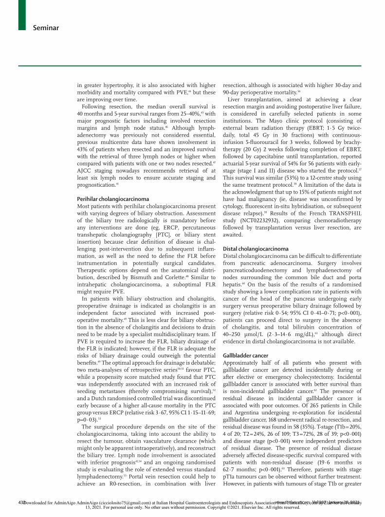
Figure 2: Clinical presentation of biliary tract cancerIntrahepatic cholangiocarcinoma arises proximal to the second-order bile ducts. Perihilar cholangiocarcinoma arises between the second-order ducts and the insertion of the cystic duct. Distal cholangiocarcinoma is distal to the insertion of the cystic duct. Gallbladder cancer arises from the gallbladder itself or from the cystic duct. Extrahepatic cholangiocarcinoma refers to perihilar cholangiocarcinoma and distal cholangiocarcinoma combined. *Biliary obstruction can occur from tumours arising in major bile ducts (perihilar cholangiocarcinoma or distal cholangiocarcinoma), or because of lymph node compression at the hilum.
Liver
Cystic duct
Gallbladder
Intrahepatic cholangiocarcinoma• Abdominal pain• Might be asymptomatic,
even if at advanced stage
Incidental gallbladder cancer• Found at routine
cholecystectomyNon-incidental gallbladder cancer• Right upper quadrant pain
Biliary obstruction*Obstructive jaundice• Yellow sclera• Dark urine• Pale stools• Pruritus
Ampulla of Vater
In patients with establishedchronic liver disease• Splenomegaly• Ascites• Spider naevi• Gynaecomastia• Caput medusae• Encephalopathy
Constitutional symptoms• Fatigue• Anorexia• Weight loss• Abdominal pain• Night sweats
Incidental findings• Deranged liver function
tests• Found on imaging for
other cause
Intrahepatic cholangiocarcinoma
Perihilar cholangiocarcinoma Distal cholangiocarcinoma
Common hepatic duct
Downloaded for AdminAigo AdminAigo ([email protected]) at Italian Hospital Gastroenterologists and Endoscopists Association from ClinicalKey.com by Elsevier on February 13, 2021. For personal use only. No other uses without permission. Copyright ©2021. Elsevier Inc. All rights reserved.

Seminar
www.thelancet.com Vol 397 January 30, 2021 431
technique excludes simple gallstone disease and identifies obstruction with upstream dilatation of the biliary tree, examines the gallbladder anatomy, and can identify space-occupying lesions within the liver. Ultrasound examination cannot be used for staging of malignancy.
CT scanning is the main modality for diagnosis and staging of biliary tract cancer. Intrahepatic cholangio-carcinoma can be present as a mass lesion, typically with rim enhancement (arterial phase), possible capsular retraction, and satellite nodules. Distal cholangiocar-cinomas show abrupt biliary tree cutoff due to an obstructing lesion (which might not be evident in very small tumours). Perihilar cholangiocarcinomas can be evident only by the presence of dilated segmental bile ducts; an obstructing lesion might be difficult to define, particularly if non-mass-forming.30 CT might show gallbladder cancer as a malignant, infiltrative mass centred on the gallbladder (possibly extending into the liver, bile duct, or hepatic artery) although small lesions might not be seen.31 Contrast-enhanced CT completes staging by assessing local invasion by the primary tumour (specifically portal vein and hepatic artery involvement, determining resectability), as well as identifying distant metastases.
MRI, particularly with hepato-specific contrast media and diffusion-weighted imaging, can provide detailed anatomical delineation of lesions in the liver, bile ducts, and gallbladder, as well as any vascular involvement. It can differentiate between cholangiocarcinoma and hepatocellular carcinoma, identify small lesions within the gallbladder, and is able to delineate the biliary tree, particularly in patients with a perihilar cholangiocar-cinoma.32 Image reconstruction can provide a magnetic resonance cholangio-pancreatogram to help with diag-nosis and treatment planning.
¹⁸F-fluorodeoxyglucose positron emission tomography can be used to complement CT and MRI. A previous systematic review and meta-analysis has shown that it is useful at identifying lymph node involvement, presence of distant metastases, and postoperative disease recurrence. However, because of low specificity it is not sufficient for the diagnosis of a primary lesion and cytological or histological confirmation is still required.33 Therefore, the technique is most useful when planning treatment, such as surgery, in which the identification of additional disease can alter an initial management plan.
Serum carbohydrate antigen 19–9, also known as sialylated Lewis-A antigen, lacks specificity for the diagnosis of biliary tract cancer and can be elevated in other malignancies, benign disease, and biliary obstruction, and is not elevated in Lewis antigen-negative patients.34 However, in the presence of an established diagnosis it can provide information on response to treatment in addition to prognostic information.35
Endoscopic retrograde cholangio-pancreatography (ERCP) is a well established modality for assessment of the biliary tree, with an imaging diagnostic sensi tivity of
74% and specificity of 70%;36 it enables biliary delineation and acquisition of brush cytology and biopsies. Endo-scopic ultrasound is very useful for assessment and diagnostic sampling of distal cholangiocarcinoma and regional lymph nodes.37 Peroral cholangioscopy allows direct visualisation of the biliary tract with the ability to accurately target abnormal lesions. A previous study reported 100% sensitivity and 89·5% specificity of visual impression at the time of cholangioscopy.38
Although brush cytology has high specificity, its low sensitivity is a major limitation (eg, 97% specificity and 43% sensitivity for detecting cholangiocarcinoma in patients with primary sclerosing cholangitis).39 A meta-analysis of fluorescence in-situ hybridisation has shown that this is highly specific (pooled 70%) but with limited sensitivity (68%) for identification of cholangiocarcinoma in patients with primary sclerosing cholangitis.40 Albumin RNA in-situ hybridisation has been shown as a sensitive and highly specific diagnostic tool for distinguishing intrahepatic cholangiocarcinoma from cancer of unknown primary.12
Upon completion of investigations, patients’ disease should be staged according to the American Joint Committee on Cancer (AJCC) Cancer Staging Handbook, 8th edition41 in order to plan treatment.
Treatment and surgery Given the complexity of biliary tract cancer, clinical management should be planned as part of a multi-disciplinary team considering patient-related factors (eg, ECOG performance status, comorbidities, prefer-ences), disease-related variables (eg, tumour stage, vascular involvement, presence of distant metastases), and availability of specialist expertise (high-volume centres are associated with a lower mortality rate for major liver resections).42 Surgery is the cornerstone of curative therapy and the approach depends on the primary site of disease.
Intrahepatic cholangiocarcinomaResection aims to achieve radical clearance with uninvolved margins, and because of the frequent absence of symptoms, only 22% of patients are able to undergo surgery.43 The feasibility of resection depends on the location of the primary tumour (and relationship to adjacent blood vessels) as well as assessment of the future liver remnant (FLR); an FLR volume of 25% or more is considered acceptable in the setting of an otherwise normal liver, although this increases to 40% or more in patients with background liver disease (eg, cirrhosis). A suboptimal FLR can be increased by portal vein embolisation (PVE), which induces hypertrophy of the left lobe over 4–6 weeks; the success of PVE can be limited by the presence of background liver disease. A previously introduced surgical procedure, Associating Liver Partition and Portal vein ligation for Staged hepatectomy, is used in some specialist centres. Although this procedure results
Downloaded for AdminAigo AdminAigo ([email protected]) at Italian Hospital Gastroenterologists and Endoscopists Association from ClinicalKey.com by Elsevier on February 13, 2021. For personal use only. No other uses without permission. Copyright ©2021. Elsevier Inc. All rights reserved.
Seminar
432 www.thelancet.com Vol 397 January 30, 2021
in greater hypertrophy, it is also associated with higher morbidity and mortality compared with PVE,44 but these are improving over time.
Following resection, the median overall survival is 40 months and 5-year survival ranges from 25–40%,45 with major prognostic factors including involved resection margins and lymph node status.46 Although lymph-adenectomy was previously not considered essential, previous multicentre data have shown involvement in 43% of patients when resected and an improved survival with the retrieval of three lymph nodes or higher when compared with patients with one or two nodes resected.47 AJCC staging nowadays recommends retrieval of at least six lymph nodes to ensure accurate staging and prognostication.41
Perihilar cholangiocarcinoma Most patients with perihilar cholangiocarcinoma present with varying degrees of biliary obstruction. Assessment of the biliary tree radiologically is mandatory before any interventions are done (eg, ERCP, percutaneous transhepatic cholangiography [PTC], or biliary stent inser tion) because clear definition of disease is chal-lenging post-intervention due to subsequent inflam-mation, as well as the need to define the FLR before instrumentation in potentially surgical candidates. Therapeutic options depend on the anatomical distri-bution, described by Bismuth and Corlette.48 Similar to intrahepatic cholangio carcinoma, a suboptimal FLR might require PVE.
In patients with biliary obstruction and cholangitis, preoperative drainage is indicated as cholangitis is an independent factor associated with increased post-operative mortality.49 This is less clear for biliary obstruc-tion in the absence of cholangitis and decisions to drain need to be made by a specialist multidisciplinary team. If PVE is required to increase the FLR, biliary drainage of the FLR is indicated; however, if the FLR is adequate the risks of biliary drainage could outweigh the potential benefits.49 The optimal approach for drainage is debatable; two meta-analyses of retrospective series50,51 favour PTC, while a propensity score matched study found that PTC was independently associated with an increased risk of seeding metastases (thereby compromising survival),52 and a Dutch randomised controlled trial was discontinued early because of a higher all-cause mortality in the PTC group versus ERCP (relative risk 3·67, 95% CI 1·15–11·69; p=0·03).53
The surgical procedure depends on the site of the cholangiocarcinoma, taking into account the ability to resect the tumour, obtain vasculature clearance (which might only be apparent intraoperatively), and reconstruct the biliary tree. Lymph node involvement is associated with inferior prognosis41,54 and an ongoing randomised study is evaluating the role of extended versus standard lymphadenectomy.55 Portal vein resection could help to achieve an R0-resection, in combination with liver
resection, although is associated with higher 30-day and 90-day perioperative mortality.56
Liver transplantation, aimed at achieving a clear resection margin and avoiding postoperative liver failure, is considered in carefully selected patients in some institutions. The Mayo clinic protocol (consisting of external beam radiation therapy (EBRT; 1·5 Gy twice-daily, total 45 Gy in 30 fractions) with continuous-infusion 5-fluorouracil for 3 weeks, followed by brachy-therapy (20 Gy) 2 weeks following completion of EBRT, followed by capecitabine until transplantation, reported actuarial 5-year survival of 54% for 56 patients with early-stage (stage I and II) disease who started the protocol.57 This survival was similar (53%) to a 12-centre study using the same treatment protocol.58 A limitation of the data is the acknowledgment that up to 15% of patients might not have had malignancy (ie, disease was unconfirmed by cytology, fluorescent in-situ hybridisation, or subsequent disease relapse).59 Results of the French TRANSPHIL study (NCT02232932), comparing chemoradiotherapy followed by transplantation versus liver resection, are awaited.
Distal cholangiocarcinomaDistal cholangiocarcinoma can be difficult to differentiate from pancreatic adenocarcinoma. Surgery involves pancreaticoduodenectomy and lymphadenectomy of nodes surrounding the common bile duct and porta hepatis.60 On the basis of the results of a randomised study showing a lower complication rate in patients with cancer of the head of the pancreas undergoing early surgery versus preoperative biliary drainage followed by surgery (relative risk 0·54; 95% CI 0·41–0·71; p<0·001), patients can proceed direct to surgery in the absence of cholangitis, and total bilirubin concentration of 40–250 μmol/L (2·3–14·6 mg/dL),61 although direct evidence in distal cholangiocarcinoma is not available.
Gallbladder cancer Approximately half of all patients who present with gallbladder cancer are detected incidentally during or after elective or emergency cholecystectomy. Incidental gallbladder cancer is associated with better survival than is non-incidental gallbladder cancer.62 The presence of residual disease in incidental gallbladder cancer is associated with poor outcomes. Of 265 patients in Chile and Argentina undergoing re-exploration for incidental gallbladder cancer, 168 underwent radical re-resection, and residual disease was found in 58 (35%). T-stage (T1b = 20%, 4 of 20; T2 = 24%, 26 of 109; T3 = 72%, 28 of 39; p<0·001) and disease stage (p<0·001) were inde pendent predictors of residual disease. The presence of residual disease adversely affected disease-specific survival compared with patients with non-residual disease (19·6 months vs 62·7 months; p<0·001).63 Therefore, patients with stage pT1a tumours can be observed without further treatment. However, in patients with tumours of stage T1b or greater
Downloaded for AdminAigo AdminAigo ([email protected]) at Italian Hospital Gastroenterologists and Endoscopists Association from ClinicalKey.com by Elsevier on February 13, 2021. For personal use only. No other uses without permission. Copyright ©2021. Elsevier Inc. All rights reserved.

Seminar
www.thelancet.com Vol 397 January 30, 2021 433
or involvement of the cystic duct, re-resection of the tumour bed to achieve negative margins (involving en bloc hepatic resection of segments IVB and V, portal lymphadenectomy, and possibly bile duct resection in a specialist centre) should be considered if imaging and staging laparoscopy confirms resectability. The use of chemotherapy between cholecystectomy and completion surgery remains investigational.
Fewer than 20% of patients with non-incidental gallbladder cancer are potential candidates for surgery.64 The AJCC 8th edition41 subclassifies stage T2 according to tumour location: peritoneal side tumours (T2a) and hepatic side tumours (T2b) because patients with tumours on the hepatic side have a higher incidence of nodal involvement and hepatic metastases65 leading to inferior 5-year survival and disease-free survival.66 Nodal involvement, an independent prognostic factor for survival, occurs frequently (62%) in stage T2; thus lymph node dissection is essential for curative resection in T2 disease.67 Recurrence occurs more frequently in the T2b group (32·9% vs 22·9%; p=0·007), systemic recurrence is more common than loco-regional recur-rence (71·0% vs 29·0%), and liver resection did not improve survival of patients with T2b gallbladder cancer in a Korean study.66 Therefore, in T2 gallbladder cancer, regardless of location, radical cholecystectomy including lymph node dissection for N-staging without liver resection could be a reasonable option.
Adjuvant therapy Historically, decisions regarding adjuvant therapy have been based on data from institutional retrospective series and phase 2 studies. On the basis of a meta-analysis of these data, chemotherapy was advocated for patients with lymph node-positive disease and radiotherapy advocated for patients with involved resection margins.68
Three previously published randomised controlled trials provide more robust data to inform practice. The French PRODIGE-12 study69 randomly assigned patients with resected biliary tract cancer (cholangiocarcinoma or gallbladder cancer) to surgery alone versus surgery followed by 6 months of gemcitabine and oxaliplatin. The study did not meet either of its coprimary endpoints: relapse-free survival (hazard ratio [HR] 0·88; 95% CI 0·62–1·25; p=0·48) and health-related quality of life (HR 1·28; 95% CI 0·73–2·26; log-rank p=0·39). In the Japanese Bile Duct Cancer Adjuvant Trial,70 patients with extrahepatic cholangiocarcinoma were randomised to surgery alone versus surgery followed by 6 months of gemcitabine. There was no difference in the primary endpoint of overall survival (HR 1·01; 95% CI 0·70–1·45; p=0·964). In the third study, BILCAP,71 patients with resected cholangiocarcinoma and gallbladder cancer were randomised to surgery alone versus surgery followed by a 6-month course of capecitabine. The study did not meet its primary endpoint of overall survival by intention-to-treat analysis (HR 0·81, 95% CI 0·63–1·04;
p=0·097). However, a prespecified sensitivity analysis adjusting for minimisation and prognostic factors (tumour grade, lymph node status, and gender) revealed a significant benefit in overall survival from the addition of capecitabine (HR 0·71, 95% CI 0·55–0·92; p=0·010). This improvement, supported by a significant improve-ment in relapse-free survival (by intention-to-treat analysis, a secondary endpoint); a clinically meaningful numerical improvement in the median overall survival (51·1 months [95% CI 34·6–59·1] vs 36·4 months [95% CI 29·7–44·5] in favour of capecitabine); and a likelihood that future studies against a no-chemotherapy arm would be challenging to perform from patient, clinician, and ethics review committee perspectives has led to capecitabine being recommended in the new American Society of Clinical Oncology (ASCO) guidelines.72
No phase 3 studies have yet evaluated the role of adjuvant radiotherapy. Findings from the SWOG-0809 study suggest this might be considered in selected patients; in this single-arm, phase 2 study, patients with extrahepatic cholangiocarcinoma or gallbladder cancer considered at high risk (stage pT2–4 or N+ or involved resection margins) received four cycles of gemcitabine and capecitabine (21-day regimen) followed by radio-therapy (45 Gy to regional lymph node and 54–59·4 Gy to the tumour bed) with concurrent capecitabine. The observed 2-year survival of 65% (95% CI 53–74); and 67% in R0-resected and 60% in R1-resected patients, exceeded the pre-specified threshold of activity to be considered effective (set at 2-year survival of >45%, R0 survival estimate of ≥65%, and R1 survival estimate >45%).73 This was reflected by inclusion in the ASCO guidelines.72
Ongoing studies include the Japanese (JCOG-1202) study (UMIN000011688) comparing surgery alone versus surgery followed by S1 (a composite drug including tegafur, gimeracil, and oteracil) in patients with cholangio-carcinoma, gallbladder cancer, and ampullary cancers; accrual is complete and results are awaited. In the ACTICCA-1 study, patients are randomised to standard-of-care treatment in the control group (originally surgery alone, amended to surgery followed by adjuvant capecita-bine post-BILCAP) versus cisplatin and gemcitabine chemotherapy; recruitment is ongoing (NCT02170090).74
Locoregional therapyLocoregional therapies can be considered in selected patients with localised unresectable disease, depending on availability and expertise, although none have been validated in randomised controlled trials.
Trans-arterial (chemo)-embolisation allows treatment delivery through the hepatic artery provided the portal vein is patent, supplying the normal liver parenchyma. In cholangiocarcinoma, different embolic agents, varying chemotherapies (cisplatin, doxorubicin, 5-fluorouracil, gemcitabine, irinotecan, mitomycin C, and oxaliplatin;
Downloaded for AdminAigo AdminAigo ([email protected]) at Italian Hospital Gastroenterologists and Endoscopists Association from ClinicalKey.com by Elsevier on February 13, 2021. For personal use only. No other uses without permission. Copyright ©2021. Elsevier Inc. All rights reserved.
Seminar
434 www.thelancet.com Vol 397 January 30, 2021
some via drug-eluting beads), and disparate treatment schedules have been tried. A systematic review found an average response rate (RR; by Response Evaluation Criteria in Solid Tumors) of 28·4%, median time to progression of 8·2 months, and overall survival of 13 months.75 59·5% of patients had bilobar disease, 35% had extrahepatic metastases, and 35% had previously received chemotherapy. Multiple lesions, ill-location and hypovascularity conferred a worse prog-nosis. Hetero geneity of patient selection, tumour types, regimens, schedules, and subsequent therapy precluded a meta-analysis; consequently, no single approach is recom mended and the need for randomised controlled trials is evident.
Hepatic Arterial Infusion (HAI) of chemotherapy enables differential dosing to the liver, and most data concerns colorectal liver metastases. A retrospective, single-centre series of floxuridine by HAI in patients with intrahepatic cholangiocarcinoma and a higher than 70% liver involvement found the addition of HAI to systemic chemotherapy (vs systemic chemotherapy alone) improved RR (47 [59%] of 79 vs 7 [39%] of 18; p=0·11), progression-free survival (12 months vs 7 months; p=0·20), and overall survival (30·8 months vs 18·4 months; p<0·001).76 However, there were differences in staging (HAI patients were staged operatively when fitting the HAI pump whereas non-HAI patients were only staged radiologically) and the systemic chemo-therapy was not standardised. In another study, HAI floxuridine in combination with systemic gemcitabine and oxaliplatin achieved a 6-month progression free survival of 84·1% (90% CI 74·8%–not reached) in unresectable intrahepatic cholangiocarcinoma. The RR was 58% (22 of 38 patients); four patients under-went resection and the median overall survival was 25·0 months (95% CI 20·6–not reached).77 The appli-cability in a multicentre setting, the optimal systemic regimen, and timing of HAI relative to other therapies are subject to evaluation in prospective controlled studies.
Radioembolisation, with β-emitting yttrium-90 micro-spheres, delivers high radiation doses to the liver. A systematic review showed a pooled RR of 28% with an additional 54% achieving stable disease.78 Notably, seven (10%) of 73 patients across three studies became surgically resectable. Although the overall survival (15·5 months) was promising, a post-hoc analysis from the Advanced Biliary tract Cancer (ABC)-01, ABC-02, and ABC-03 studies found that patients with intrahepatic cholangiocarcinoma have an improved survival compared with patients with other biliary tract cancers. Moreover, the median overall survival was 15·4 months (95% CI 11·1–17·1) in patients with intrahepatic cholangio-carcinoma with metastatic disease receiving cisplatin and gemcitabine chemotherapy, and 16·7 months (95% CI 8·7–20·0) in patients with intrahepatic cholangio-carcinoma confined to the liver.79 A randomised phase 2
study is evaluating the addition of radioembolisation to cisplatin and gemcitabine chemotherapy (SIRCCA study; NCT02807181).
Intraductal ablative procedures aim at restoring or maintaining biliary patency. Studies using radiofre-quency ablation are mostly retrospective in carefully selected patients. Although published outcomes appear promising,80 randomised studies are few. Despite the initially promising results from a small randomised study of photodynamic therapy (PDT) in patients with endoluminal tumours,81 a larger randomised controlled study was discontinued early as patients in the PDT group had an inferior survival.82 Consequently PDT-based approaches remain investigational.
Radiotherapy Approximately 25% of patients present with locally advanced disease and the role of radiotherapy is an active area of investigation in these patients. Studies are mostly retrospective series with some phase 1 and 2 studies evaluating conventionally fractionated radiotherapy and, more recently, intensity-modulated radiation therapy and stereotactic body radiation therapy. These have achieved local control rates in 45–100% of patients with 1-year survival of 58–81%.83 Outcomes are improved with increased doses of radiation delivered to the tumour, and hypofractionation with photons84 or protons85 can be considered for patients with intrahepatic cholangio-carcinoma in experienced centres. Despite these developments, there is no level-1 evidence to assess the incremental benefit of radiotherapy over established treatments. The ongoing prospective randomised ABC-07 study (EUDRACT 2014–003656–31) is comparing cisplatin and gemcitabine chemotherapy alone (eight cycles) or with stereotactic body radiotherapy (SBRT; six cycles followed by SBRT) in patients with intrahepatic or extrahepatic cholangiocarcinoma.
Management of advanced diseaseChemotherapyData from randomised controlled trials, including a no-chemotherapy control arm have shown that the median overall survival in patients with advanced disease is poor at between 2·5 months and 4·5 months.86,87 Early studies identified fluoropyrimidines, platinum, and gemcitabine as active agents in the treatment of advanced biliary tract cancer.88 The 410-patient UK ABC-02 study established cisplatin and gemcitabine as the reference regimen internationally, on the basis of an improved overall survival (11·7 months vs 8·1 months; HR 0·64, 95% CI 0·52–0·80; p<0·001) compared with gemcitabine mono-therapy (figure 3). Patients receiving the combination also had an improved progression-free survival (8·0 months vs 5·0 months, p<0·001) and tumour control rate (81·4% vs 71·8%, p=0·049).89 The BT22 study demon-strated that this magnitude of benefit was reproducible (median overall survival 11·2 months vs 7·7 months;
Downloaded for AdminAigo AdminAigo ([email protected]) at Italian Hospital Gastroenterologists and Endoscopists Association from ClinicalKey.com by Elsevier on February 13, 2021. For personal use only. No other uses without permission. Copyright ©2021. Elsevier Inc. All rights reserved.

Seminar
www.thelancet.com Vol 397 January 30, 2021 435
HR 0·69, 95% CI 0·42–1·13) and applicable to Japanese patients as well as UK patients.90 In a meta-analysis of these two studies, patients with cholan giocarcinoma (from all primary sites) and those with gallbladder cancer were shown to derive a similar magnitude of benefit in the exploratory subgroup analysis.103 Moreover, in a separate meta-analysis including studies from different geographical regions, the cisplatin and gemcitabine regimen was deemed applicable across a diverse range of countries and with different disease characteristics.104 Oxaliplatin is sometimes substituted for cisplatin although this combination has not been validated in a phase 3 study.105 In Japan, a randomised phase 3 study has shown that the combination of gemcitabine with S1 is non-inferior to cisplatin and gemcitabine (median overall survival 15·1 months for cisplatin and 13·4 months for gemcitabine; HR 0·95, 90% CI 0·78–1·15; p=0·046 for non-inferiority).92
A number of studies have sought to intensify therapy through the use of triple-agent chemotherapy regi-mens.106–108 A promising phase 2 combination is cisplatin, gemcitabine, and nab-paclitaxel with RR 45% and a median overall survival of 19·2 months (95% CI 13·2–not estimable);109 this regimen is currently being compared with cisplatin and gemcitabine in a randomised phase 3 study (SWOG-1815; NCT03768414). This trial, along with the results of another phase 3
study com paring cisplatin and gemcitabine versus FOLFIRINOX (oxaliplatin, irinotecan, and 5-fluorouracil) chemotherapy (NCT02591030), will determine whether intensifying chemotherapy is an appropriate strategy. In Japan, the cisplatin, gemcitabine, and S1 triplet could be a treatment option on the basis of an improved overall survival (13·5 months vs 12·6 months; HR 0·791, 90% CI 0·620–0·996, one-sided p value 0·046) compared with cisplatin and gemcitabine.91
Inevitably, patients develop disease progression fol-lowing first-line chemotherapy. There has been, until recently, a scarcity of evidence that further chemotherapy is of benefit. A systematic review of 14 phase 2 studies and nine retrospective studies, including 895 patients, was unable to identify an individual active treatment regimen.110 The only randomised controlled trial in this setting is the ABC-06 study (NCT01926236) which assessed the benefit of second-line oxaliplatin and 5-fluorouracil (FOLFOX) over Active Symptom Control (composed of proactive identification of biliary obstruc-tion, sepsis, and symptom management) in patients with disease progression following treatment with cisplatin and gemcitabine. The study met its primary endpoint with improved overall survival (HR 0·69, 95% CI 0·50–0·97, p=0·031). Although the median overall survival improvement was modest (6·2 months vs 5·3 months), a clinically meaningful 15% improvement
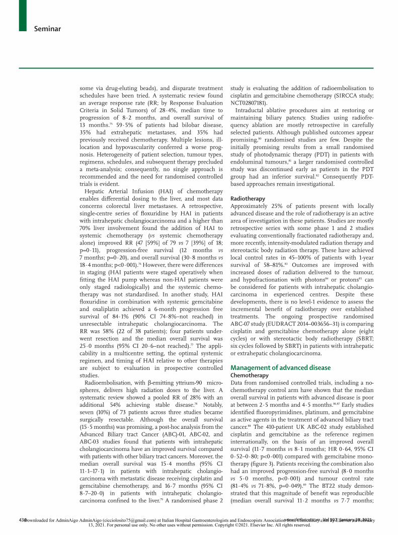
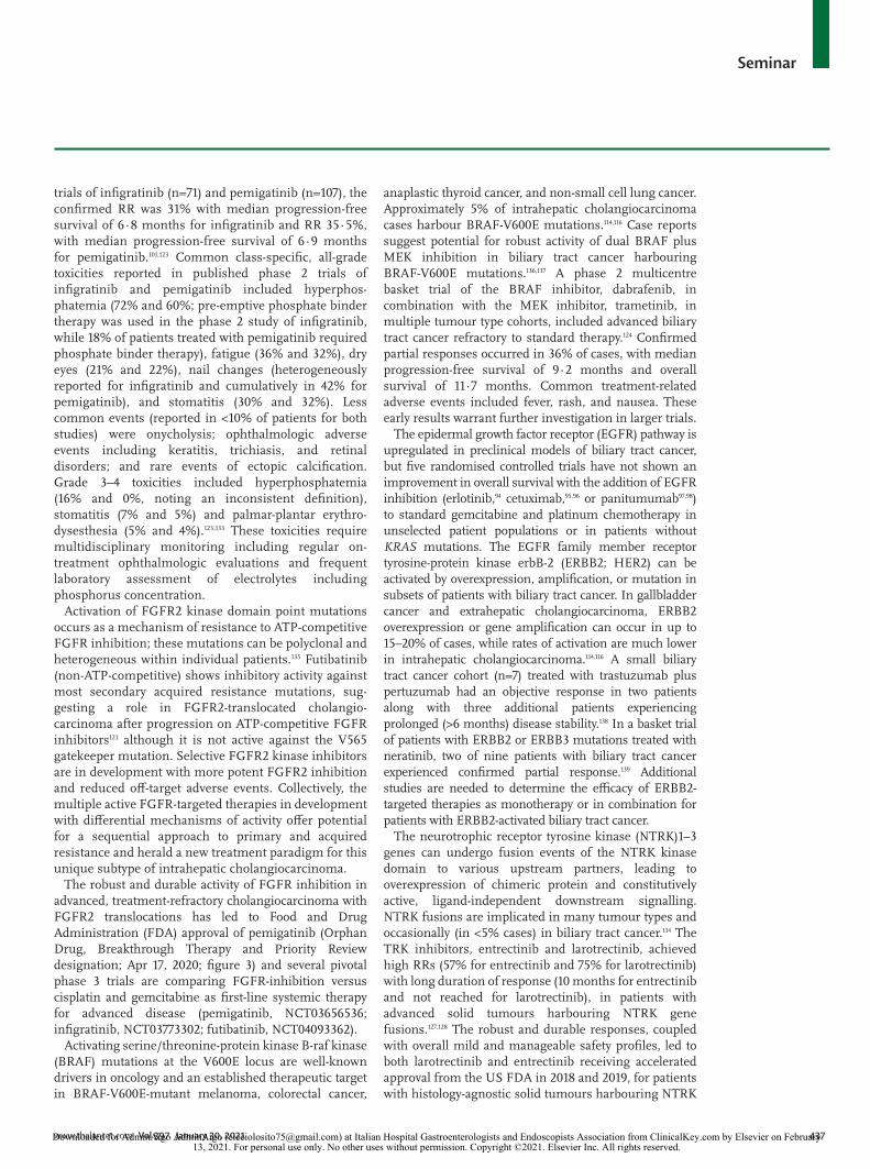
Figure 3: Timeline of developments in systemic therapy of biliary tract cancerRandomised controlled studies are presented, with randomised phase 3 studies in bold and randomised phase 2 in non-bold font. CisGem-S1 and ABC-06 have been presented as abstracts (final publication pending). Grey boxes signify licensed therapies. The timeline shows the year of final publication. ABC=Advanced Biliary tract Cancer. CisGem=cisplatin and gemcitabine. GemOx=gemcitabine and oxaliplatin. EGFR=epidermal growth factor receptor. VEGF=vascular endothelial growth factor. mIDH-1=mutated isocitrate dehydrogenase-1. FDA=Food and Drug Administration. FGFR=2=fibroblast growth factor receptor-2. *In prespecified sensitivity analysis (not by intention to treat). †One phase 3 study and four phase 2 studies. ‡Orphan drug, breakthrough therapy, and priority review designation (based on phase 2 study).
ABC-02: CisGem improves overall survival as a first-line therapy (UK)89
BT22: CisGem improves overall survival as a first-line therapy (Japan)90
FUGA-BT: Gem-S1 non-inferior to CisGem as a first-line therapy (Japan)92
CisGem-S1 improves overall survival vs CisGem as a first-line therapy (Japan)91
ABC-06: FOLFOX improves overall survival as a second-line therapy (UK)93
PRODIGE-12: no survival benefit from GemOx69
BCAT: no survival benefit from Gem70
BILCAP: survival benefit from capecitabine71*
No benefit of adding EGFR inhibition to chemotherapy (five studies†) in first line94–98
ABC-03: no benefit of adding VEGF inhibition to CisGem in first line35
No benefit from addition of ramucirumab or merestinib to CisGem in first line99
ClarIDHy: ivosidenib improves progression-free survival in mIDH-1 cholangiocarcinoma as a second-line or later-line therapy100
Pemigatinib approved by FDA for FGFR-2 fusions or rearrangements in second line or later line101‡
Pembrolizumab for microsatellite unstable or mismatch repair deficiency102
2010 2011 2012 2013 2014 2015 2016 2017 2018 2019 2020
Chem
othe
rapy
AdvancedAdjuvant
Advanced
Targ
eted
ther
apy
Immunotherapy
Downloaded for AdminAigo AdminAigo ([email protected]) at Italian Hospital Gastroenterologists and Endoscopists Association from ClinicalKey.com by Elsevier on February 13, 2021. For personal use only. No other uses without permission. Copyright ©2021. Elsevier Inc. All rights reserved.

Seminar
436 www.thelancet.com Vol 397 January 30, 2021
was observed in 6-month (50·6% vs 35·5%) and 12-month (25·9% vs 11·4%) survival (figure 3).93 There remains a clear unmet need to develop additional treatment options.
Molecular profiling and targeted therapies for advanced biliary tract cancersIn addition to anatomical heterogeneity, molecular profiling studies have demonstrated substantial molecular heterogeneity across intrahepatic cholangio-carcinoma, extrahepatic cholangiocarcinoma, and gall-bladder cancer.111–113 Certain molecular aberrations are associated with the anatomical subsite of tumour112,114–116 such as fibroblast growth factor receptor (FGFR) 2 gene trans locations and isocitrate dehydrogenase-1 (IDH1) mutations (which occur nearly exclusively in intrahepatic cholangiocarcinoma), and KRAS proto-oncogene (KRAS) mutations and receptor tyrosine-protein kinase erbB-2 (ERBB2) amplification, which are more common in extrahepatic cholangiocarcinoma and gallbladder cancer.112,114,116,117 Moreover, distinct gene signatures and epigenetic profiles have been identified in cases associated with history of fluke infection or primary sclerosing cholangitis.112,118–120 Despite this heterogeneity, recurring sub groups with driver mutations amenable to targeted therapy have been identified, which are generally mutually exclusive from one another (table 1). We discuss the most frequently occurring, targetable mutations in advanced biliary tract cancer.
The first notable mutation is the IDH1 mutation. Gain-of-function mutations in the coding region of IDH1 are
present in about 13% of cases of intrahepatic cholan-giocarcinoma (almost never in extrahepatic cholangio-carcinoma) based upon a systematic review including 5393 cases of cholangiocarcinoma.117 Overall, less than half of cases of cholangiocarcinoma occur in women; however, more than 60% of IDH1-mutant cases of cholangiocarcinoma occur in women, yet the mecha-nisms underlying this are unknown.117 The mutant-IDH1 protein catalyses produc tion of an oncometabolite, D-2-hydroxyglutarate (2-HG), via NADPH-dependent reduction.129,130 Accumulation of 2-HG impairs cellular differentiation through effects on chromatin structure and DNA methylation, leading to tumourigenesis.
Ivosidenib is a first-in-class, oral, selective, and reversible mutant-IDH1 inhibitor. In a phase 1 basket study of IDH1-mutated solid tumours, 73 patients with advanced cholangiocarcinoma refractory to standard therapies received ivosidenib.131 Although objective responses were uncommon (5%), the median progression-free survival (3·8 months) and overall survival (13·8 months) were longer than expected for standard chemotherapy in similar populations.93,132 The subsequent phase 3 trial enrolled 185 patients with advanced IDH1-mutant cholan gio-carcinoma after 1–2 lines of previous, unsuccessful systemic therapy, with 2:1 randomisation to ivosidenib versus placebo and allowance of crossover at progression for patients in the placebo group (NCT03173248).100 Ivosidenib improved progression-free survival (the primary endpoint): median 2·7 months for ivosidenib versus 1·4 months for placebo (HR 0·37, 95% CI 0·25–0·54, p<0·001), and 32% treated with ivosidenib were progression-free at 6 months (vs none in the placebo group). Ivosidenib was well tolerated with low rates of grade 3 or higher adverse events and only 1·7% requiring discontinuation for toxicity attributed to ivosidenib (figure 3). Regulatory approval is awaited for patients with advanced, IDH1-mutant cholan giocarcinoma after ineffective standard therapy.
Another notable mutation is the FGFR2 translocation. Activating translocation events (fusions or rearrange-ments) upstream of the coding region of the FGFR2 gene occur in about 20% of intrahepatic cholangio carcinoma cases121 and, like IDH1 mutations, are more common in women and are almost never found in extrahepatic biliary tract cancer. Translocations that relieve the FGFR2 gene of its upstream transcriptional regulation result in consti-tutively active growth factor pathway signalling, promoting cell proliferation, angiogenesis, and metastasis.
Multiple inhibitors of FGFR isoforms 1–3 have shown activity in advanced cholangiocarcinoma harbouring FGFR2 translocations, including several ATP-competitive, reversible inhibitors (erdafitinib,122 infigratinib,123 pemi-gatinib,133 and derazantinib134) as well as a non-ATP-competitive, covalent inhibitor, futibatinib.121 Early data from phase 1 and 2 trials of these agents consistently have shown tumour stabilisation or regression occurring in the majority of patients. In larger, phase 2
Frequency* Targeted agents Molecular test
IDH1 13% of intrahepatic cholangiocarcinoma cases100,117
Ivosidenib Tumour next-generation DNA sequencing or targeted sequencing for hotspot mutations in coding region of IDH1
FGFR pathway 20% of intrahepatic cholangiocarcinoma cases121
Erdafitinib;122 futibatinib;121 infigratinib;123 pemigatinib101
Tumour next-generation DNA sequencing including FGFR2 intronic region, targeted RNAseq, or FISH testing for FGFR2 translocation
BRAF 5% of intrahepatic cholangiocarcinoma cases114,116
Dabrafenib plus trametinib;124 vemurafenib125
Tumour next-generation DNA sequencing or targeted sequencing for hotspot mutations in coding region of BRAF
MSI-high or MMR deficiency
2% of biliary tract cancer cases126
Pembrolizumab126 Multiple testing modalities available: PCR, immunohistochemistry, or tumour next-generation DNA sequencing
ERBB2 (HER2) 15–20% gallbladder cancer and extrahepatic cholangiocarcinoma cases114,116
·· Multiple testing modalities available including immunohistochemistry and FISH for expression and amplification, tumour next-generation DNA sequencing for mutations
NTRK Rare Entrectinib;127 larotrectenib128
Tumour next-generation DNA sequencing including NTRK intronic region or targeted RNAseq, or FISH testing for NTRK translocation
IDH1=isocitrate dehydrogenase-1. FGFR=fibroblast growth factor receptor-2. FISH=fluorescent in-situ hybridisation. BRAF=activating serine threonine-protein kinase B-raf kinase. MSI=microsatellite instability. MMR=mismatch repair. ERBB2=receptor tyrosine-protein kinase erbB-2. NTRK=neurotrophic receptor tyrosine kinase. *All percentages are approximations.
Table 1: Therapeutic targets and approach to molecular profiling in biliary tract cancers
Downloaded for AdminAigo AdminAigo ([email protected]) at Italian Hospital Gastroenterologists and Endoscopists Association from ClinicalKey.com by Elsevier on February 13, 2021. For personal use only. No other uses without permission. Copyright ©2021. Elsevier Inc. All rights reserved.
Seminar
www.thelancet.com Vol 397 January 30, 2021 437
trials of infigratinib (n=71) and pemigatinib (n=107), the confirmed RR was 31% with median progression-free survival of 6·8 months for infigratinib and RR 35·5%, with median progression-free survival of 6·9 months for pemi gatinib.101,123 Common class-specific, all-grade toxicities reported in published phase 2 trials of infigratinib and pemigatinib included hyper phos-phatemia (72% and 60%; pre-emptive phosphate binder therapy was used in the phase 2 study of infigratinib, while 18% of patients treated with pemigatinib required phosphate binder therapy), fatigue (36% and 32%), dry eyes (21% and 22%), nail changes (heterogeneously reported for infigratinib and cumulatively in 42% for pemigatinib), and stomatitis (30% and 32%). Less common events (reported in <10% of patients for both studies) were onycholysis; ophthalmologic adverse events including keratitis, trichiasis, and retinal disorders; and rare events of ectopic calcification. Grade 3–4 toxicities included hyperphosphatemia (16% and 0%, noting an inconsistent definition), stomatitis (7% and 5%) and palmar-plantar erythro-dysesthesia (5% and 4%).123,133 These toxicities require multidisciplinary monitoring including regular on-treatment ophthalmologic evalua tions and frequent laboratory assessment of electrolytes including phosphorus concentration.
Activation of FGFR2 kinase domain point mutations occurs as a mechanism of resistance to ATP-competitive FGFR inhibition; these mutations can be polyclonal and heterogeneous within individual patients.135 Futibatinib (non-ATP-competitive) shows inhibitory activity against most secondary acquired resistance mutations, sug-gesting a role in FGFR2-translocated cholangio-carcinoma after progression on ATP-competitive FGFR inhibitors121 although it is not active against the V565 gatekeeper mutation. Selective FGFR2 kinase inhibitors are in development with more potent FGFR2 inhibition and reduced off-target adverse events. Collectively, the multiple active FGFR-targeted therapies in development with differential mechanisms of activity offer potential for a sequential approach to primary and acquired resistance and herald a new treatment paradigm for this unique subtype of intrahepatic cholangiocarcinoma.
The robust and durable activity of FGFR inhibition in advanced, treatment-refractory cholangiocarcinoma with FGFR2 translocations has led to Food and Drug Administration (FDA) approval of pemigatinib (Orphan Drug, Breakthrough Therapy and Priority Review designation; Apr 17, 2020; figure 3) and several pivotal phase 3 trials are comparing FGFR-inhibition versus cisplatin and gemcitabine as first-line systemic therapy for advanced disease (pemigatinib, NCT03656536; infigratinib, NCT03773302; futibatinib, NCT04093362).
Activating serine/threonine-protein kinase B-raf kinase (BRAF) mutations at the V600E locus are well-known drivers in oncology and an established therapeutic target in BRAF-V600E-mutant melanoma, colorectal cancer,
anaplastic thyroid cancer, and non-small cell lung cancer. Approximately 5% of intrahepatic cholangiocarcinoma cases harbour BRAF-V600E mutations.114,116 Case reports suggest potential for robust activity of dual BRAF plus MEK inhibition in biliary tract cancer harbouring BRAF-V600E mutations.136,137 A phase 2 multicentre basket trial of the BRAF inhibitor, dabrafenib, in combination with the MEK inhibitor, trametinib, in multiple tumour type cohorts, included advanced biliary tract cancer refractory to standard therapy.124 Confirmed partial responses occurred in 36% of cases, with median progression-free survival of 9·2 months and overall survival of 11·7 months. Common treatment-related adverse events included fever, rash, and nausea. These early results warrant further investigation in larger trials.
The epidermal growth factor receptor (EGFR) pathway is upregulated in preclinical models of biliary tract cancer, but five randomised controlled trials have not shown an improvement in overall survival with the addition of EGFR inhibition (erlotinib,94 cetuximab,95,96 or panitu mumab97,98) to standard gemcitabine and platinum chemotherapy in unselected patient populations or in patients without KRAS mutations. The EGFR family member receptor tyrosine-protein kinase erbB-2 (ERBB2; HER2) can be activated by over expression, amplification, or mutation in subsets of patients with biliary tract cancer. In gallbladder cancer and extrahepatic cholangiocarcinoma, ERBB2 overexpression or gene amplification can occur in up to 15–20% of cases, while rates of activation are much lower in intrahepatic cholangiocarcinoma.114,116 A small biliary tract cancer cohort (n=7) treated with trastuzumab plus pertuzumab had an objective response in two patients along with three additional patients experiencing prolonged (>6 months) disease stability.138 In a basket trial of patients with ERBB2 or ERBB3 mutations treated with neratinib, two of nine patients with biliary tract cancer experienced confirmed partial response.139 Additional studies are needed to determine the efficacy of ERBB2-targeted therapies as monotherapy or in combination for patients with ERBB2-activated biliary tract cancer.
The neurotrophic receptor tyrosine kinase (NTRK)1–3 genes can undergo fusion events of the NTRK kinase domain to various upstream partners, leading to overexpression of chimeric protein and constitutively active, ligand-independent downstream signalling. NTRK fusions are implicated in many tumour types and occasionally (in <5% cases) in biliary tract cancer.114 The TRK inhibitors, entrectinib and larotrectinib, achieved high RRs (57% for entrectinib and 75% for larotrectinib) with long duration of response (10 months for entrectinib and not reached for larotrectinib), in patients with advanced solid tumours harbouring NTRK gene fusions.127,128 The robust and durable responses, coupled with overall mild and manageable safety profiles, led to both larotrectinib and entrectinib receiving accelerated approval from the US FDA in 2018 and 2019, for patients with histology-agnostic solid tumours harbouring NTRK
Downloaded for AdminAigo AdminAigo ([email protected]) at Italian Hospital Gastroenterologists and Endoscopists Association from ClinicalKey.com by Elsevier on February 13, 2021. For personal use only. No other uses without permission. Copyright ©2021. Elsevier Inc. All rights reserved.

Seminar
438 www.thelancet.com Vol 397 January 30, 2021
fusions. Several patients with cholan giocarcinoma were included in the data leading to regulatory approval for both entrectinib and larotrectinib, supporting the role for NTRK fusion testing in cholangiocarcinoma, and treatment if present.
ImmunotherapyImmunotherapy in patients with biliary tract cancer is currently investigational. Early promise was seen in a patient with cholangiocarcinoma when whole exome sequencing of the tumour revealed 26 non-synonymous mutations, among them the ERBB2IP (erbb2 interacting
protein) mutation which was the neo-antigen recognised by the patient’s tumour-infiltrating lymphocytes. Adoptive transfer of tumour-infiltrating lymphocytes containing CD3+ ERBB2IP mutation-reactive T cells resulted in a substantial decrease of tumour burden. Regarding disease pro gression, 18 months after the first infusion, a further response was seen after a second T-cell infusion.140
Programmed death ligand 1 (PD-L1) is expressed in tumours from some patients with biliary tract cancer, suggesting a potential role for targeting the PD1/PDL1 pathway.141–143 The results of early clinical trials of checkpoint inhibitors reported to date are summarised:
Drug Setting n RR % (n) Time to response (months)
Duration of response (months)
Median progression-free survival, months (95% CI)
Median overall survival, months (95% CI)
Bang Y-J et al, 2019144 (KEYNOTE-028)
Pembrolizumab Second-line or later-line therapy; PS 0–1; PD-L1+ (100%)
24 13% (3/23) 3·5 21·5, ≥51·4, and ≥53·2 months for each responder
1·8 (1·4–3·1) 5·7 (3·1–9·8)
Ueno et al, 2018145 (KEYNOTE-158)
Pembrolizumab Second-line or later-line therapy; PS 0–1; PD-L1 unselected
104; PD-L1+ 61; PD-L1– 43
5·8% (6/104); PD-L1+ 6·6% (4/61); PD-L1– 2·9% (1/34)
2·2 Not reached 2·0 (1·9–2·1); PD-L1+
1·9 (1·8–2·0); PD-L1– 2·1 (1·9–2·6)
7·4 (5·5–9·6); PD-L1+
7·2 (3·7–10·8); PD-L1–9·3 (4·2–11·5)
Ueno et al, 2019146 Nivolumab Post prior chemotherapy; PS 0–1; PD-L1 unselected
30 3% (1/30) ·· ≥12·7 1·4 (1·4–1·4)* 5·2 (4·5–8·7)*
Ueno et al, 2019146 Nivolumab + CisGem
First-line; PS 0–1; PD-L1 unselected
30 37% (11/30) ·· 5·1 4·2 (2·8–5·6)* 15·4 (11·8–not reached)*
RR=response rate. PS=performance status. PD-L1=programmed death ligand 1. CisGem=cisplatin and gemcitabine. *90% CIs.
Table 2: Summary of reported studies of checkpoint inhibition in biliary tract cancer
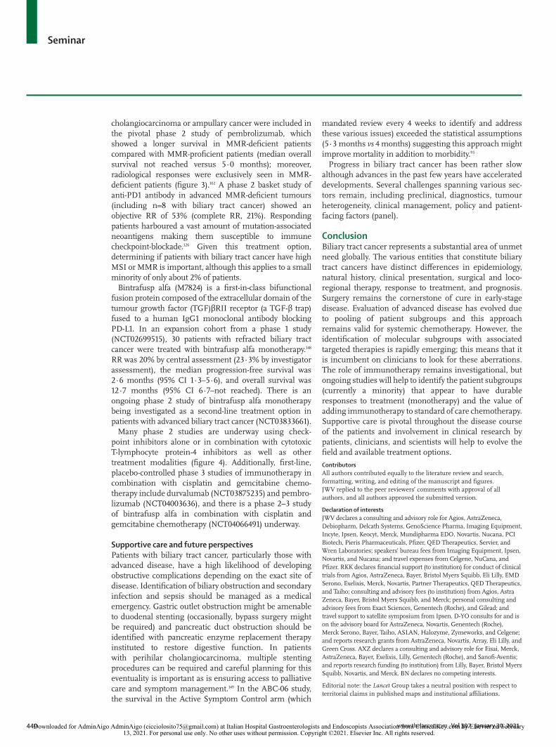
Figure 4: Clinical trial development of immunotherapy in biliary tract cancerCisGem=cisplatin and gemcitabine. GemOx=gemcitabine and oxaliplatin.
Phase 1
Phase 2 Phase 3
Monotherapy• KEYNOTE-028 (pembrolizumab)144
• Nivolumab146
• Bintrafusp (M7824)148
Combination therapy• Nivolumab + CisGem146
• Pembrolizumab + ramucirumab(NCT02443324)
Monotherapy• Pembrolizumab (KEYNOTE-158; NCT02628067)• Pembrolizumab (South Korea; NCT03110328)• Pembrolizumab (Spain; NCT03260712)• Pembrolizumab or nivolumab (NCT03695952)• Nivolumab (NCT02829918)• Bintrafusp (M7824; NCT03833661)
Combination therapy• Nivolumab + ipilimumab (NCT02834013)• Pembrolizumab + GM-CSF (NCT02703714)• Pembrolizumab + Peg-interferon α2b (NCT02982720)• Pembrolizumab + allogeneic natural killer cell (NCT03937895)• Pembrolizumab + CisGem (EORTC-1607 ABC-09; NCT03260712)• Pembrolizumab + capecitabine + oxaliplatin (NCT03111732)• Pembrolizumab + ramucirumab (NCT03260712)• Pembrolizumab + lenvatinib (LEAP-005; NCT03695952)• Durvalumab + tremelimumab + TACE/RFA/ablation (NCT02821754)• Durvalumab + tremelimumab + SIRT (NCT04238637)• Durvalumab + tremelimumab + radiotherapy (NCT03482102)• Durvalumab + tremelimumab + CisGem (NCT03046862)• Durvalumab + tremelimumab +/– paclitaxel (NCT03704480)• Durvalumab + AZD6738 (NCT04298008)• Camrelizumab + GemOx (NCT03486678)• Nivolumab + etinostat (NCT03250273)• Atezolizumab +/– cobimetinib (NCT03201458)• Neoadjuvant CisGem +/– durvalumab (DEBATE; NCT04308174)
First line• CisGem + durvalumab or placebo (TOPAZ-1; NCT03875235)• CisGem + pembrolizumab or placebo (KEYNOTE-966;
NCT04003636)• CisGem + bintrafusp or placebo (M7824; NCT04066491)
Downloaded for AdminAigo AdminAigo ([email protected]) at Italian Hospital Gastroenterologists and Endoscopists Association from ClinicalKey.com by Elsevier on February 13, 2021. For personal use only. No other uses without permission. Copyright ©2021. Elsevier Inc. All rights reserved.

Seminar
www.thelancet.com Vol 397 January 30, 2021 439
the KEYNOTE-028 (phase Ib) (NCT02054806)144 and KEYNOTE-158 (phase 2)145 trials with pembrolizumab, as well as the two arms of a phase 1 study of nivolumab alone or in combination with chemotherapy (table 2).146 From these data we discuss various emerging themes.
Firstly, activity is limited in monotherapy with objective RR (all partial responses), ranging between 3% and 13%; all patients were pre-treated with chemotherapy. Secondly, when present, responses are durable—in the minority of patients who respond, a sustained effect can be observed, extending to years in some instances. Another emerging theme was that the predictive value of PD-L1 testing remains uncertain. In KEYNOTE-028 PD-L1 positivity was defined as membranous PD-L1 expression in at least 1% of tumour and associated inflammatory cells, or positive staining in stroma (immunohistochemistry assay, QualTek, Goleta, CA, USA), and all patients were PD-L1 positive.144 KEYNOTE-158 did not select patients with PD-L1 expression; it was evaluated retrospectively (IHC 22C3 pharmDx assay, Agilent Technologies, Carpinteria, CA, USA). PD-L1-expressing tumours were those with a combined positive score of at least 1, calculated as the ratio of PD-L1-positive tumour cells, lymphocytes, and macrophages out of the total number of tumour cells × 100.145 In the nivolumab study, PD-L1 positivity was defined as expression in at least 1% of tumour cells, at least 1% of tumour-associated immune cells, or at least 1% of tumour cells or tumour-associated immune cells (or both; pharmDx 28–8 assay, Dako, Carpinteria, CA, USA).146 Responses appear to be more frequent in patients with PD-L1-positive tumours, although have also been documented in PD-L1-negative tumours, as shown in table 2. Whether this improved RR translates into improved survival with a longer overall survival in patients with PD-L1-negative tumours in KEYNOTE-158 remains unclear.
Furthermore, toxicities from immunotherapy are as expected from other studies. Few patients experienced grade 3 treatment-related adverse events (KEYNOTE-158, 13%145 and KEYNOTE-028, 17%144). The most common adverse events were fatigue (14%), rash (12%), and pruritus (9%) in KEYNOTE-158 and pyrexia (17%), nausea (13%), and pruritus (13%) in KEYNOTE-028. Immune-mediated adverse events and infusion reactions occurring in at least 5% of patients were hypothyroidism (8%) and pneu-monitis (6%) in KEYNOTE-158, and hypothyroidism (8%) in KEYNOTE-028. Although the majority were mild to moderate in severity, six patients (6%) in KEYNOTE-158 and two patients (8%) in KEYNOTE-028 had grade 3 immune-mediated adverse events and infusion reactions. No grade 4–5 immune-mediated adverse events or infusion reactions were recorded. The final theme we noted was that combinations with chemotherapy warrant further evaluation. In combination with cisplatin and gemcitabine, first-line nivolumab resulted in a median progression-free survival of 4·2 months, median overall survival
15·4 months, and RR 37% (all partial responses in 11 patients), by central review.146 The interplay between checkpoint inhibition and chemotherapy in biliary tract cancer needs to be elucidated.
Patients with high microsatellite instability (MSI) or mismatch repair (MMR) deficiency warrant specific discussion; pembrolizumab is FDA-approved for the treatment of patients with metastatic or inoperable solid tumours with these abnormalities. A genetic risk factor for biliary tract cancer includes Lynch syndrome, characterised by MSI and MMR deficiency.147 Four patients with
Panel: Future perspectives for improving understanding of biliary tract cancer
Preclinical• Enhance available preclinical models for forward and back translation into the clinic
Diagnostics• Improve tissue acquisition for diagnosis and research• Validate use of liquid biopsy (circulating tumour biomarkers such as DNA, RNA,
proteins, metabolites) for diagnosis and research• Improve diagnostic imaging (eg, for perihilar cholangiocarcinoma and detection of
peritoneal disease)• Improve imaging response assessment to treatments (eg, response assessment of
perihilar cholangiocarcinoma following treatment, or in the emerging setting of molecular therapies)
Heterogeneity• Ensure that research questions address anatomical subgroups• Integrate molecular subgroup evaluation understanding prognostic significance• Pool groups where appropriate, split subgroups if scientifically rational• Collaborative studies essential (in view of low incidence or prevalence)
Clinical management• Refine the role of liver transplantation in perihilar cholangiocarcinoma and
intrahepatic cholangiocarcinoma• Develop more effective adjuvant treatments• Evaluate effective therapies in the neoadjuvant setting• Critically assess the role of loco-regional therapies in intrahepatic cholangiocarcinoma
(stereotactic body radiotherapy, selective intra-arterial radiotherapy)• Develop more effective systemic treatment (first line and second line) in advanced
biliary tract cancer• Expand clinical experience and further develop molecularly targeted agents including
isocitrate dehydrogenase-1 and fibroblast growth factor inhibitors: companion diagnostics, combination strategies, new setting (eg, perioperative, adjuvant), novel targets, drug resistance mechanisms
• Define the role of immunotherapy in biliary tract cancer
Patient-facing• Ensure research and development is with patients as well as for patients• Increase patient advocacy, especially in unrepresented geographical regions• Incorporate patient-reported outcomes and experience measures
Policy• Increase awareness of biliary tract cancer as an area of unmet need with funding bodies• Ensure regulatory agencies are aware of biliary tract cancer (country-specific)• Educate the clinical community of developments in the field• Foster collaborations with industry in the search for novel therapies
Downloaded for AdminAigo AdminAigo ([email protected]) at Italian Hospital Gastroenterologists and Endoscopists Association from ClinicalKey.com by Elsevier on February 13, 2021. For personal use only. No other uses without permission. Copyright ©2021. Elsevier Inc. All rights reserved.
Seminar
440 www.thelancet.com Vol 397 January 30, 2021
cholangiocarcinoma or ampullary cancer were included in the pivotal phase 2 study of pembrolizumab, which showed a longer survival in MMR-deficient patients compared with MMR-proficient patients (median overall survival not reached versus 5·0 months); moreover, radiological responses were exclusively seen in MMR-deficient patients (figure 3).102 A phase 2 basket study of anti-PD1 antibody in advanced MMR-deficient tumours (including n=8 with biliary tract cancer) showed an objective RR of 53% (complete RR, 21%). Responding patients harboured a vast amount of mutation-associated neoan tigens making them susceptible to immune checkpoint-blockade.126 Given this treatment option, determining if patients with biliary tract cancer have high MSI or MMR is important, although this applies to a small minority of only about 2% of patients.
Bintrafusp alfa (M7824) is a first-in-class bifunctional fusion protein composed of the extracellular domain of the tumour growth factor (TGF)βRII receptor (a TGF-β trap) fused to a human IgG1 monoclonal antibody blocking PD-L1. In an expansion cohort from a phase 1 study (NCT02699515), 30 patients with refracted biliary tract cancer were treated with bintrafusp alfa monotherapy.148 RR was 20% by central assessment (23·3% by investigator assessment), the median progression-free survival was 2·6 months (95% CI 1·3–5·6), and overall survival was 12·7 months (95% CI 6·7–not reached). There is an ongoing phase 2 study of bintrafusp alfa monotherapy being investigated as a second-line treatment option in patients with advanced biliary tract cancer (NCT03833661).
Many phase 2 studies are underway using check-point inhibitors alone or in combination with cytotoxic T-lymphocyte protein-4 inhibitors as well as other treatment modalities (figure 4). Additionally, first-line, placebo-controlled phase 3 studies of immunotherapy in combination with cisplatin and gemcitabine chemo-therapy include durvalumab (NCT03875235) and pembro-lizumab (NCT04003636), and there is a phase 2–3 study of bintrafusp alfa in combination with cisplatin and gemcitabine chemotherapy (NCT04066491) underway.
Supportive care and future perspectivesPatients with biliary tract cancer, particularly those with advanced disease, have a high likelihood of developing obstructive complications depending on the exact site of disease. Identification of biliary obstruction and secondary infection and sepsis should be managed as a medical emergency. Gastric outlet obstruction might be amenable to duodenal stenting (occasionally, bypass surgery might be required) and pancreatic duct obstruction should be identified with pancreatic enzyme replacement therapy instituted to restore digestive function. In patients with perihilar cholangiocarcinoma, multiple stenting procedures can be required and careful planning for this eventuality is important as is ensuring access to palliative care and symptom management.149 In the ABC-06 study, the survival in the Active Symptom Control arm (which
mandated review every 4 weeks to identify and address these various issues) exceeded the statistical assumptions (5·3 months vs 4 months) suggesting this approach might improve mortality in addition to morbidity.93
Progress in biliary tract cancer has been rather slow although advances in the past few years have accelerated developments. Several challenges spanning various sec-tors remain, including preclinical, diagnostics, tumour heterogeneity, clinical management, policy and patient-facing factors (panel).
ConclusionBiliary tract cancer represents a substantial area of unmet need globally. The various entities that constitute biliary tract cancers have distinct differences in epidemiology, natural history, clinical presentation, surgical and loco-regional therapy, response to treatment, and prognosis. Surgery remains the cornerstone of cure in early-stage disease. Evaluation of advanced disease has evolved due to pooling of patient subgroups and this approach remains valid for systemic chemotherapy. However, the identification of molecular subgroups with associated targeted therapies is rapidly emerging; this means that it is incumbent on clinicians to look for these aberrations. The role of immunotherapy remains investigational, but ongoing studies will help to identify the patient subgroups (currently a minority) that appear to have durable responses to treatment (monotherapy) and the value of adding immunotherapy to standard of care chemotherapy. Supportive care is pivotal throughout the disease course of the patients and involvement in clinical research by patients, clinicians, and scientists will help to evolve the field and available treatment options.ContributorsAll authors contributed equally to the literature review and search, formatting, writing, and editing of the manuscript and figures. JWV replied to the peer reviewers’ comments with approval of all authors, and all authors approved the submitted version.
Declaration of interestsJWV declares a consulting and advisory role for Agios, AstraZeneca, Debiopharm, Delcath Systems, GenoScience Pharma, Imaging Equipment, Incyte, Ipsen, Keocyt, Merck, Mundipharma EDO, Novartis, Nucana, PCI Biotech, Pieris Pharmaceuticals, Pfizer, QED Therapeutics, Servier, and Wren Laboratories; speakers’ bureau fees from Imaging Equipment, Ipsen, Novartis, and Nucana; and travel expenses from Celgene, NuCana, and Pfizer. RKK declares financial support (to institution) for conduct of clinical trials from Agios, AstraZeneca, Bayer, Bristol Myers Squibb, Eli Lilly, EMD Serono, Exelixis, Merck, Novartis, Partner Therapeutics, QED Therapeutics, and Taiho; consulting and advisory fees (to institution) from Agios, Astra Zeneca, Bayer, Bristol Myers Squibb, and Merck; personal consulting and advisory fees from Exact Sciences, Genentech (Roche), and Gilead; and travel support to satellite symposium from Ipsen. D-YO consults for and is on the advisory board for AstraZeneca, Novartis, Genentech (Roche), Merck Serono, Bayer, Taiho, ASLAN, Halozyme, Zymeworks, and Celgene; and reports research grants from AstraZeneca, Novartis, Array, Eli Lilly, and Green Cross. AXZ declares a consulting and advisory role for Eisai, Merck, AstraZeneca, Bayer, Exelixis, Lilly, Genentech (Roche), and Sanofi-Aventis; and reports research funding (to institution) from Lilly, Bayer, Bristol Myers Squibb, Novartis, and Merck. BN declares no competing interests.
Editorial note: the Lancet Group takes a neutral position with respect to territorial claims in published maps and institutional affiliations.
Downloaded for AdminAigo AdminAigo ([email protected]) at Italian Hospital Gastroenterologists and Endoscopists Association from ClinicalKey.com by Elsevier on February 13, 2021. For personal use only. No other uses without permission. Copyright ©2021. Elsevier Inc. All rights reserved.

Seminar
www.thelancet.com Vol 397 January 30, 2021 441
References1 Banales JM, Cardinale V, Carpino G, et al. Cholangiocarcinoma:
current knowledge and future perspectives consensus statement from the European Network for the Study of Cholangiocarcinoma (ENS-CCA). Nat Rev Gastroenterol Hepatol 2016; 13: 261–80.
2 GLOBOCAN. Cancer incidence in 5 continents. Volume XI. International Agency for Research on Cancer, 2017.
3 Patel T. Increasing incidence and mortality of primary intrahepatic cholangiocarcinoma in the United States. Hepatology 2001; 33: 1353–57.
4 Sripa B, Kaewkes S, Sithithaworn P, et al. Liver fluke induces cholangiocarcinoma. PLoS Med 2007; 4: e201.
5 Shaib YH, Davila JA, McGlynn K, El-Serag HB. Rising incidence of intrahepatic cholangiocarcinoma in the United States: a true increase? J Hepatol 2004; 40: 472–77.
6 West J, Wood H, Logan RF, Quinn M, Aithal GP. Trends in the incidence of primary liver and biliary tract cancers in England and Wales 1971–2001. Br J Cancer 2006; 94: 1751–58.
7 Taylor-Robinson SD, Toledano MB, Arora S, et al. Increase in mortality rates from intrahepatic cholangiocarcinoma in England and Wales 1968–1998. Gut 2001; 48: 816–20.
8 Saha SK, Zhu AX, Fuchs CS, Brooks GA. Forty-year trends in cholangiocarcinoma incidence in the US: intrahepatic disease on the rise. Oncologist 2016; 21: 594–99.
9 Welzel TM, McGlynn KA, Hsing AW, O’Brien TR, Pfeiffer RM. Impact of classification of hilar cholangiocarcinomas (Klatskin tumors) on the incidence of intra- and extrahepatic cholangiocarcinoma in the United States. J Natl Cancer Inst 2006; 98: 873–75.
10 WHO. ICD-11 implementation or transition guide. Geneva: World Health Organization, 2019.
11 Hainsworth JD, Rubin MS, Spigel DR, et al. Molecular gene expression profiling to predict the tissue of origin and direct site-specific therapy in patients with carcinoma of unknown primary site: a prospective trial of the Sarah Cannon research institute. J Clin Oncol 2013; 31: 217–23.
12 Ferrone CR, Ting DT, Shahid M, et al. The ability to diagnose intrahepatic cholangiocarcinoma definitively using novel branched DNA-enhanced albumin RNA in situ hybridization technology. Ann Surg Oncol 2016; 23: 290–96.
13 Bray F, Ferlay J, Soerjomataram I, Siegel RL, Torre LA, Jemal A. Global cancer statistics 2018: GLOBOCAN estimates of incidence and mortality worldwide for 36 cancers in 185 countries. CA Cancer J Clin 2018; 68: 394–424.
14 Zatonski WA, Lowenfels AB, Boyle P, et al. Epidemiologic aspects of gallbladder cancer: a case-control study of the SEARCH Program of the International Agency for Research on Cancer. J Natl Cancer Inst 1997; 89: 1132–38.
15 Massarweh NN, El-Serag HB. Epidemiology of hepatocellular carcinoma and intrahepatic cholangiocarcinoma. Cancer Control 2017; 24: 1073274817729245.
16 Clements O, Eliahoo J, Kim JU, Taylor-Robinson SD, Khan SA. Risk factors for intrahepatic and extrahepatic cholangiocarcinoma: a systematic review and meta-analysis. J Hepatol 2020; 72: 95–103.
17 Sithithaworn P, Yongvanit P, Duenngai K, Kiatsopit N, Pairojkul C. Roles of liver fluke infection as risk factor for cholangiocarcinoma. J Hepatobiliary Pancreat Sci 2014; 21: 301–08.
18 Hsing AW, Bai Y, Andreotti G, et al. Family history of gallstones and the risk of biliary tract cancer and gallstones: a population-based study in Shanghai, China. Int J Cancer 2007; 121: 832–38.
19 Lewis JT, Talwalkar JA, Rosen CB, Smyrk TC, Abraham SC. Prevalence and risk factors for gallbladder neoplasia in patients with primary sclerosing cholangitis: evidence for a metaplasia-dysplasia-carcinoma sequence. Am J Surg Pathol 2007; 31: 907–13.
20 Chascsa DM, Lindor KD. Cancer risk, screening and surveillance in primary sclerosing cholangitis. Minerva Gastroenterol Dietol 2019; 65: 214–28.
21 Said K, Glaumann H, Bergquist A. Gallbladder disease in patients with primary sclerosing cholangitis. J Hepatol 2008; 48: 598–605.
22 Kamisawa T, Kaneko K, Itoi T, Ando H. Pancreaticobiliary maljunction and congenital biliary dilatation. Lancet Gastroenterol Hepatol 2017; 2: 610–18.
23 Koshiol J, Wozniak A, Cook P, et al. Salmonella enterica serovar Typhi and gallbladder cancer: a case-control study and meta-analysis. Cancer Med 2016; 5: 3310–235.
24 Li ZM, Wu ZX, Han B, et al. The association between BMI and gallbladder cancer risk: a meta-analysis. Oncotarget 2016; 7: 43669–79.
25 Ang DC, Shia J, Tang LH, Katabi N, Klimstra DS. The utility of immunohistochemistry in subtyping adenocarcinoma of the ampulla of vater. Am J Surg Pathol 2014; 38: 1371–79.
26 Nakanuma Y, Kakuda Y. Pathologic classification of cholangiocarcinoma: new concepts. Best Pract Res Clin Gastroenterol 2015; 29: 277–93.
27 Kendall T, Verheij J, Gaudio E, et al. Anatomical, histomorphological and molecular classification of cholangiocarcinoma. . Liver International 2019; 39: 7–18.
28 Nakanuma Y, Jang KT, Fukushima N, et al. A statement by the Japan-Korea expert pathologists for future clinicopathological and molecular analyses toward consensus building of intraductal papillary neoplasm of the bile duct through several opinions at the present stage. J Hepatobiliary Pancreat Sci 2018; 25: 181–87.
29 Brunt E, Aishima S, Clavien PA, et al. cHCC-CCA: consensus terminology for primary liver carcinomas with both hepatocytic and cholangiocytic differentation. Hepatology 2018; 68: 113–26.
30 Joo I, Lee JM, Yoon JH. Imaging diagnosis of intrahepatic and perihilar cholangiocarcinoma: recent advances and challenges. Radiology 2018; 288: 7–13.
31 Choi SY, Kim JH, Park HJ, Han JK. Preoperative CT findings for prediction of resectability in patients with gallbladder cancer. Eur Radiol 2019; 29: 6458–68.
32 Jhaveri KS, Hosseini-Nik H. MRI of cholangiocarcinoma. J Magn Reson Imaging 2015; 42: 1165–79.
33 Lamarca A, Barriuso J, Chander A, et al. (18)F-fluorodeoxyglucose positron emission tomography ((18)FDG-PET) for patients with biliary tract cancer: systematic review and meta-analysis. J Hepatol 2019; 71: 115–29.
34 Tsen A, Barbara M, Rosenkranz L. Dilemma of elevated CA 19–9 in biliary pathology. Pancreatology 2018; 18: 862–67.
35 Valle JW, Wasan H, Lopes A, et al. Cediranib or placebo in combination with cisplatin and gemcitabine chemotherapy for patients with advanced biliary tract cancer (ABC-03): a randomised phase 2 trial. Lancet Oncol 2015; 16: 967–78.
36 Park MS, Kim TK, Kim KW, et al. Differentiation of extrahepatic bile duct cholangiocarcinoma from benign stricture: findings at MRCP versus ERCP. Radiology 2004; 233: 234–40.
37 Rizvi S, Eaton J, Yang JD, Chandrasekhara V, Gores GJ. Emerging technologies for the diagnosis of perihilar cholangiocarcinoma. Semin Liver Dis 2018; 38: 160–69.
38 Pereira P, Santos S, Morais R, et al. Role of peroral cholangioscopy for diagnosis and staging of biliary tumors. Dig Dis 2020; 38: 431–39.
39 Trikudanathan G, Navaneethan U, Njei B, Vargo JJ, Parsi MA. Diagnostic yield of bile duct brushings for cholangiocarcinoma in primary sclerosing cholangitis: a systematic review and meta-analysis. Gastrointest Endosc 2014; 79: 783–89.
40 Navaneethan U, Njei B, Venkatesh PG, Vargo JJ, Parsi MA. Fluorescence in situ hybridization for diagnosis of cholangiocarcinoma in primary sclerosing cholangitis: a systematic review and meta-analysis. Gastrointest Endosc 2014; 79: 943–50.e3.
41 American Joint Committee on Cancer. AJCC Cancer Staging Manual, 8th edn. New York: Springer, 2017.
42 Filmann N, Walter D, Schadde E, et al. Mortality after liver surgery in Germany. Br J Surg 2019; 106: 1523–29.
43 Wu L, Tsilimigras DI, Paredes AZ, et al. Trends in the incidence, treatment and outcomes of patients with intrahepatic cholangiocarcinoma in the USA: facility type is associated with margin status, use of lymphadenectomy and overall survival. World J Surg 2019; 43: 1777–87.
44 Li J, Moustafa M, Linecker M, et al. ALPPS for locally advanced intrahepatic cholangiocarcinoma: did aggressive surgery lead to the oncological benefit? An international multi-center study. Ann Surg Oncol 2020; 27: 1372–84.
45 Mazzaferro V, Gorgen A, Roayaie S, Droz Dit Busset M, Sapisochin G. Liver resection and transplantation for intrahepatic cholangiocarcinoma. J Hepatol 2020; 72: 364–77.
46 DeOliveira ML, Cunningham SC, Cameron JL, et al. Cholangiocarcinoma: thirty-one-year experience with 564 patients at a single institution. Ann Surg 2007; 245: 755–62.
Downloaded for AdminAigo AdminAigo ([email protected]) at Italian Hospital Gastroenterologists and Endoscopists Association from ClinicalKey.com by Elsevier on February 13, 2021. For personal use only. No other uses without permission. Copyright ©2021. Elsevier Inc. All rights reserved.

Seminar
442 www.thelancet.com Vol 397 January 30, 2021
47 Sahara K, Tsilimigras DI, Merath K, et al. Therapeutic index associated with lymphadenectomy among patients with intrahepatic cholangiocarcinoma: which patients benefit the most from nodal evaluation? Ann Surg Oncol 2019; 26: 2959–68.
48 Bismuth H, Corlette MB. Intrahepatic cholangioenteric anastomosis in carcinoma of the hilus of the liver. Surg Gynecol Obstet 1975; 140: 170–78.
49 Wiggers JK, Groot Koerkamp B, Cieslak KP, et al. Postoperative mortality after liver resection for perihilar cholangiocarcinoma: development of a risk score and importance of biliary drainage of the future liver remnant. J Am Coll Surg 2016; 223: 321–31.e1.
50 Hameed A, Pang T, Chiou J, et al. Percutaneous vs. endoscopic pre-operative biliary drainage in hilar cholangiocarcinoma— a systematic review and meta-analysis. HPB (Oxford) 2016; 18: 400–10.
51 Al Mahjoub A, Menahem B, Fohlen A, et al. Preoperative biliary drainage in patients with resectable perihilar cholangiocarcinoma: is percutaneous transhepatic biliary drainage safer and more effective than endoscopic biliary drainage? A meta-analysis. J Vasc Interv Radiol 2017; 28: 576–82.
52 Komaya K, Ebata T, Yokoyama Y, et al. Verification of the oncologic inferiority of percutaneous biliary drainage to endoscopic drainage: a propensity score matching analysis of resectable perihilar cholangiocarcinoma. Surgery 2017; 161: 394–404.
53 Coelen RJS, Roos E, Wiggers JK, et al. Endoscopic versus percutaneous biliary drainage in patients with resectable perihilar cholangiocarcinoma: a multicentre, randomised controlled trial. Lancet Gastroenterol Hepatol 2018; 3: 681–90.
54 Aoba T, Ebata T, Yokoyama Y, et al. Assessment of nodal status for perihilar cholangiocarcinoma: location, number, or ratio of involved nodes. Ann Surg 2013; 257: 718–25.
55 He M, Xu X, Feng H, et al. Regional lymphadenectomy vs. extended lymphadenectomy for hilar cholangiocarcinoma (Relay-HC trial): study protocol for a prospective, multicenter, randomized controlled trial. Trials 2019; 20: 528.
56 de Jong MC, Marques H, Clary BM, et al. The impact of portal vein resection on outcomes for hilar cholangiocarcinoma: a multi-institutional analysis of 305 cases. Cancer 2012; 118: 4737–47.
57 Heimbach JK, Gores GJ, Haddock MG, et al. Liver transplantation for unresectable perihilar cholangiocarcinoma. Semin Liver Dis 2004; 24: 201–07.
58 Darwish Murad S, Kim WR, Harnois DM, et al. Efficacy of neoadjuvant chemoradiation, followed by liver transplantation, for perihilar cholangiocarcinoma at 12 US centers. Gastroenterology 2012; 143: 88–98.e3
59 Rosen CB, Darwish Murad S, Heimbach JK, Nyberg SL, Nagorney DM, Gores GJ. Neoadjuvant therapy and liver transplantation for hilar cholangiocarcinoma: is pretreatment pathological confirmation of diagnosis necessary? J Am Coll Surg 2012; 215: 31–38.
60 Dickson PV, Behrman SW. Distal cholangiocarcinoma. Surg Clin North Am 2014; 94: 325–42.
61 van der Gaag NA, Rauws EA, van Eijck CH, et al. Preoperative biliary drainage for cancer of the head of the pancreas. N Engl J Med 2010; 362: 129–37.
62 Ethun CG, Le N, Lopez-Aguiar AG, et al. Pathologic and prognostic implications of incidental versus nonincidental gallbladder cancer: a 10-institution study from the United States extrahepatic biliary malignancy consortium. Am Surg 2017; 83: 679–86.
63 Gil L, de Aretxabala X, Lendoire J, Duek F, Hepp J, Imventarza O. Incidental gallbladder cancer: how residual disease affects outcome in two referral hpb centers from South America. World J Surg 2019; 43: 214–20.
64 Boerma EJ. Towards an oncological resection of gall bladder cancer. Eur J Surg Oncol 1994; 20: 537–44.
65 Shindoh J, de Aretxabala X, Aloia TA, et al. Tumor location is a strong predictor of tumor progression and survival in T2 gallbladder cancer: an international multicenter study. Ann Surg 2015; 261: 733–39.
66 Park YJ, Hwang S, Kim KH, et al. Prognosis of patients with pT1b/T2 gallbladder carcinoma who have undergone laparoscopic cholecystectomy as an initial operation. Korean J Hepatobiliary Pancreat Surg 2013; 17: 113–17.
67 Cho JK, Lee W, Jang JY, et al. Validation of the oncologic effect of hepatic resection for T2 gallbladder cancer: a retrospective study. World J Surg Oncol 2019; 17: 8.
68 Horgan AM, Amir E, Walter T, Knox JJ. Adjuvant therapy in the treatment of biliary tract cancer: a systematic review and meta-analysis. J Clinical Oncol 2012; 30: 1934–40.
69 Edeline J, Benabdelghani M, Bertaut A, et al. Gemcitabine and oxaliplatin chemotherapy or surveillance in resected biliary tract cancer (PRODIGE 12-ACCORD 18-UNICANCER GI): a randomized phase III study. J Clinical Oncol 2019; 37: 658–67.
70 Ebata T, Hirano S, Konishi M, et al. Randomized clinical trial of adjuvant gemcitabine chemotherapy versus observation in resected bile duct cancer. Br J Surg 2018; 105: 192–202.
71 Primrose JN, Fox RP, Palmer DH, et al. Capecitabine compared with observation in resected biliary tract cancer (BILCAP): a randomised, controlled, multicentre, phase 3 study. Lancet Oncol 2019; 20: 663–73.
72 Shroff RT, Kennedy EB, Bachini M, et al. Adjuvant therapy for resected biliary tract cancer: ASCO clinical practice guideline. J Clinical Oncol 2019; 37: 1015–27.
73 Ben-Josef E, Guthrie KA, El-Khoueiry AB, et al. SWOG S0809: a phase ii intergroup trial of adjuvant capecitabine and gemcitabine followed by radiotherapy and concurrent capecitabine in extrahepatic cholangiocarcinoma and gallbladder carcinoma. J Clin Oncol 2015; 33: 2617–22.
74 Stein A, Arnold D, Bridgewater J, et al. Adjuvant chemotherapy with gemcitabine and cisplatin compared to observation after curative intent resection of cholangiocarcinoma and muscle invasive gallbladder carcinoma (ACTICCA-1 trial) - a randomized, multidisciplinary, multinational phase III trial. BMC Cancer 2015; 15: 564.
75 Yang L, Shan J, Shan L, Saxena A, Bester L, Morris DL. Trans-arterial embolisation therapies for unresectable intrahepatic cholangiocarcinoma: a systematic review. J Gastrointest Oncol 2015; 6: 570–88.
76 Konstantinidis IT, Groot Koerkamp B, Do RK, et al. Unresectable intrahepatic cholangiocarcinoma: systemic plus hepatic arterial infusion chemotherapy is associated with longer survival in comparison with systemic chemotherapy alone. Cancer 2016; 122: 758–65.
77 Cercek A, Boerner T, Tan BR, et al. Assessment of hepatic arterial infusion of floxuridine in combination with systemic gemcitabine and oxaliplatin in patients with unresectable intrahepatic cholangiocarcinoma: a phase 2 clinical trial. JAMA Oncol 2019; 6: 60–67.
78 Al-Adra DP, Gill RS, Axford SJ, Shi X, Kneteman N, Liau SS. Treatment of unresectable intrahepatic cholangiocarcinoma with yttrium-90 radioembolization: a systematic review and pooled analysis. Eur J Surg Oncol 2015; 41: 120–27.
79 Lamarca A, Ross P, Wasan HS, et al. Advanced intrahepatic cholangiocarcinoma: post hoc analysis of the ABC-01, -02, and -03 clinical trials. J Natl Cancer Inst 2020; 112: 200–10.
80 Satiya J, Schwartz I, Tabibian JH, Kumar V, Girotra M. Ablative therapies for hepatic and biliary tumors: endohepatology coming of age. Transl Gastroenterol Hepatol 2020; 5: 15.
81 Ortner ME, Caca K, Berr F, et al. Successful photodynamic therapy for nonresectable cholangiocarcinoma: a randomized prospective study. Gastroenterology 2003; 125: 1355–63.
82 Pereira SP, Jitlal M, Duggan M, et al. PHOTOSTENT-02: porfimer sodium photodynamic therapy plus stenting versus stenting alone in patients with locally advanced or metastatic biliary tract cancer. ESMO Open 2018; 3: e000379.
83 Keane FK, Zhu AX, Hong TS. Radiotherapy for biliary tract cancers. Semin Radiat Oncol 2018; 28: 342–50.
84 Tao R, Krishnan S, Bhosale PR, et al. Ablative radiotherapy doses lead to a substantial prolongation of survival in patients with inoperable intrahepatic cholangiocarcinoma: a retrospective dose response analysis. J Clin Oncol 2016; 34: 219–26.
85 Hong TS, Wo JY, Yeap BY, et al. Multi-institutional phase ii study of high-dose hypofractionated proton beam therapy in patients with localized, unresectable hepatocellular carcinoma and intrahepatic cholangiocarcinoma. J Clin Oncol 2016; 34: 460–68.
86 Glimelius B, Hoffman K, Sjoden PO, et al. Chemotherapy improves survival and quality of life in advanced pancreatic and biliary cancer. Ann Oncol 1996; 7: 593–600.
Downloaded for AdminAigo AdminAigo ([email protected]) at Italian Hospital Gastroenterologists and Endoscopists Association from ClinicalKey.com by Elsevier on February 13, 2021. For personal use only. No other uses without permission. Copyright ©2021. Elsevier Inc. All rights reserved.

Seminar
www.thelancet.com Vol 397 January 30, 2021 443
87 Sharma A, Dwary AD, Mohanti BK, et al. Best supportive care compared with chemotherapy for unresectable gall bladder cancer: a randomized controlled study. J Clin Oncol 2010; 28: 4581–86.
88 Eckel F, Schmid RM. Chemotherapy in advanced biliary tract carcinoma: a pooled analysis of clinical trials. Br J Cancer 2007; 96: 896–902.
89 Valle J, Wasan H, Palmer DH, et al. Cisplatin plus gemcitabine versus gemcitabine for biliary tract cancer. New Engl J Med 2010; 362: 1273–81.
90 Okusaka T, Nakachi K, Fukutomi A, et al. Gemcitabine alone or in combination with cisplatin in patients with biliary tract cancer: a comparative multicentre study in Japan. Br J Cancer 2010; 103: 469–74.
91 Sakai D, Kanai M, Kobayashi S, et al. Randomized phase III study of gemcitabine, cisplatin plus S-1 (GCS) versus gemcitabine, cisplatin (GC) for advanced biliary tract cancer (KHBO1401-MITSUBA). Annals of Oncology 2018; 29 (suppl 8): viii205–viii70.
92 Morizane C, Okusaka T, Mizusawa J, et al. Combination gemcitabine plus S-1 versus gemcitabine plus cisplatin for advanced/recurrent biliary tract cancer: the FUGA-BT (JCOG1113) randomized phase III clinical trial. Ann Oncol 2019; 30: 1950–58.
93 Lamarca A, Palmer DH, Wasan HS, et al. ABC-06. A randomised phase III, multi-centre, open-label study of active symptom control (ASC) alone or ASC with oxaliplatin / 5-FU chemotherapy (ASC+mFOLFOX) for patients (pts) with locally advanced / metastatic biliary tract cancers (ABC) previously-treated with cisplatin/gemcitabine (CisGem) chemotherapy. J Clin Oncol 2019; 37 (suppl 15): 4003.
94 Lee J, Park SH, Chang HM, et al. Gemcitabine and oxaliplatin with or without erlotinib in advanced biliary-tract cancer: a multicentre, open-label, randomised, phase 3 study. Lancet Oncol 2012; 13: 181–88.
95 Malka D, Cervera P, Foulon S, et al. Gemcitabine and oxaliplatin with or without cetuximab in advanced biliary-tract cancer (BINGO): a randomised, open-label, non-comparative phase 2 trial. Lancet Oncol 2014; 15: 819–28.
96 Chen JS, Hsu C, Chiang NJ, et al. A KRAS mutation status-stratified randomized phase II trial of gemcitabine and oxaliplatin alone or in combination with cetuximab in advanced biliary tract cancer. Ann Oncol 2015; 26: 943–49.
97 Leone F, Marino D, Cereda S, et al. Panitumumab in combination with gemcitabine and oxaliplatin does not prolong survival in wild-type KRAS advanced biliary tract cancer: a randomized phase 2 trial (Vecti-BIL study). Cancer 2016; 122: 574–81.
98 Vogel A, Kasper S, Bitzer M, et al. PICCA study: panitumumab in combination with cisplatin/gemcitabine chemotherapy in KRAS wild-type patients with biliary cancer-a randomised biomarker-driven clinical phase II AIO study. Eur J Cancer 2018; 92: 11–19.
99 Valle JW, Bai L-Y, Orlova R, et al. Ramucirumab (RAM) or merestinib (MER) or placebo (PL) plus gemcitabine (GEM) and cisplatin (CIS) as first-line treatment for advanced or metastatic biliary tract cancer (BTC): a randomized, double-blind, phase II study. J Clinical Oncol 2020; 38(4 suppl): 477.
100 Abou-Alfa GK, Macarulla T, Javle MM, et al. Ivosidenib in IDH1-mutant, chemotherapy-refractory cholangiocarcinoma (ClarIDHy): a multicentre, randomised, double-blind, placebo-controlled, phase 3 study. Lancet Oncol 2020; 21: 796–807.
101 Abou-Alfa GK, Sahai V, Hollebecque A, et al. Pemigatinib for previously treated, locally advanced or metastatic cholangiocarcinoma: a multicentre, open-label, phase 2 study. Lancet Oncol 2020; 21: 671–84.
102 Le DT, Uram JN, Wang H, et al. PD-1 blockade in tumors with mismatch-repair deficiency. N Engl J Med 2015; 372: 2509–20.
103 Valle JW, Furuse J, Jitlal M, et al. Cisplatin and gemcitabine for advanced biliary tract cancer: a meta-analysis of two randomised trials. Ann Oncol 2014; 25: 391–98.
104 Park JO, Oh DY, Hsu C, et al. Gemcitabine plus cisplatin for advanced biliary tract cancer: a systematic review. Cancer Res Treat 2015; 47: 343–61.
105 Fiteni F, Nguyen T, Vernerey D, et al. Cisplatin/gemcitabine or oxaliplatin/gemcitabine in the treatment of advanced biliary tract cancer: a systematic review. Cancer Med 2014; 3: 1502–11.
106 Wagner AD, Buechner-Steudel P, Moehler M, et al. Gemcitabine, oxaliplatin and 5-FU in advanced bile duct and gallbladder carcinoma: two parallel, multicentre phase-II trials. Br J Cancer 2009; 101: 1846–52.
107 Davis EJ, Griffith KA, Kim EJ, Ruch JM, McDonnell KJ, Zalupski MM. A phase II study of biweekly cisplatin, fixed-dose-rate gemcitabine and infusional 5-fluorouracil in patients with metastatic pancreatic and biliary cancers. Am J Clin Oncol 2018; 41: 128–32.
108 Sharma MR, Joshi SS, Karrison TG, et al. A UGT1A1 genotype-guided dosing study of modified FOLFIRINOX in previously untreated patients with advanced gastrointestinal malignancies. Cancer 2019; 125: 1629–36.
109 Shroff RT, Javle MM, Xiao L, et al. Gemcitabine, cisplatin, and nab-paclitaxel for the treatment of advanced biliary tract cancers: a phase 2 clinical trial. JAMA Oncol 2019; 5: 824–30.
110 Lamarca A, Hubner RA, David Ryder W, Valle JW. Second-line chemotherapy in advanced biliary cancer: a systematic review. Ann Oncol 2014; 25: 2328–38.
111 Andersen JB, Spee B, Blechacz BR, et al. Genomic and genetic characterization of cholangiocarcinoma identifies therapeutic targets for tyrosine kinase inhibitors. Gastroenterology 2012; 142: 1021–31.e15.
112 Jusakul A, Cutcutache I, Yong CH, et al. Whole-genome and epigenomic landscapes of etiologically distinct subtypes of cholangiocarcinoma. Cancer Discov 2017; 7: 1116–35.
113 Sia D, Hoshida Y, Villanueva A, et al. Integrative molecular analysis of intrahepatic cholangiocarcinoma reveals 2 classes that have different outcomes. Gastroenterology 2013; 144: 829–40.
114 Javle M, Bekaii-Saab T, Jain A, et al. Biliary cancer: utility of next-generation sequencing for clinical management. Cancer 2016; 122: 3838–47.
115 Zhu AX, Borger DR, Kim Y, et al. Genomic profiling of intrahepatic cholangiocarcinoma: refining prognosis and identifying therapeutic targets. Ann Surg Oncol 2014; 21: 3827–34.
116 Valle JW, Lamarca A, Goyal L, Barriuso J, Zhu AX. New horizons for precision medicine in biliary tract cancers. Cancer Discov 2017; 7: 943–62.
117 Boscoe AN, Rolland C, Kelley RK. Frequency and prognostic significance of isocitrate dehydrogenase 1 mutations in cholangiocarcinoma: a systematic literature review. J Gastrointest Oncol 2019; 10: 751–65.
118 Goeppert B, Folseraas T, Roessler S, et al. Genomic characterization of cholangiocarcinoma in primary sclerosing cholangitis reveals novel therapeutic opportunities. Hepatology 2020; 72: 1253–66.
119 Jinawath N, Chamgramol Y, Furukawa Y, et al. Comparison of gene expression profiles between Opisthorchis viverrini and non-Opisthorchis viverrini associated human intrahepatic cholangiocarcinoma. Hepatology 2006; 44: 1025–38.
120 Plieskatt J, Rinaldi G, Feng Y, et al. A microRNA profile associated with Opisthorchis viverrini-induced cholangiocarcinoma in tissue and plasma. BMC Cancer 2015; 15: 309.
121 Goyal L, Shi L, Liu LY, et al. TAS-120 overcomes resistance to atp-competitive fgfr inhibitors in patients with fgfr2 fusion-positive intrahepatic cholangiocarcinoma. Cancer Discov 2019; 9: 1064–79.
122 Bahleda R, Italiano A, Hierro C, et al. Multicenter phase i study of erdafitinib (JNJ-42756493), oral pan-fibroblast growth factor receptor inhibitor, in patients with advanced or refractory solid tumors. Clin Cancer Res 2019; 25: 4888–97.
123 Javle M, Lowery M, Shroff RT, et al. Phase II study of BGJ398 in patients with FGFR-altered advanced cholangiocarcinoma. J Clin Oncol 2018; 36: 276–82.
124 Subbiah V, Lassen UN, Élez E, et al. Efficacy and safety of dabrafenib and trametinib in patients with BRAF V600E–mutated biliary tract cancer: a multi-centre, open-label cohort of the ROAR basket trial. Lancet Oncology 2020; 21: 1234–43.
125 Subbiah V, Puzanov I, Blay JY, et al. Pan-cancer efficacy of vemurafenib in BRAF (V600)-mutant non-melanoma cancers. Cancer Discov 2020; 10: 657–63.
126 Le DT, Durham JN, Smith KN, et al. Mismatch repair deficiency predicts response of solid tumors to PD-1 blockade. Science 2017; 357: 409–13.
127 Doebele RC, Drilon A, Paz-Ares L, et al. Entrectinib in patients with advanced or metastatic NTRK fusion-positive solid tumours: integrated analysis of three phase 1–2 trials. Lancet Oncol 2020; 21: 271–82.
128 Drilon A, Laetsch TW, Kummar S, et al. Efficacy of larotrectinib in TRK fusion-positive cancers in adults and children. N Engl J Med 2018; 378: 731–39.
Downloaded for AdminAigo AdminAigo ([email protected]) at Italian Hospital Gastroenterologists and Endoscopists Association from ClinicalKey.com by Elsevier on February 13, 2021. For personal use only. No other uses without permission. Copyright ©2021. Elsevier Inc. All rights reserved.

Seminar
444 www.thelancet.com Vol 397 January 30, 2021
129 Dang L, White DW, Gross S, et al. Cancer-associated IDH1 mutations produce 2-hydroxyglutarate. Nature 2009; 462: 739–44.
130 Yang H, Ye D, Guan KL, Xiong Y. IDH1 and IDH2 mutations in tumorigenesis: mechanistic insights and clinical perspectives. Clin Cancer Res 2012; 18: 5562–71.
131 Lowery MA, Burris HA 3rd, Janku F, et al. Safety and activity of ivosidenib in patients with IDH1-mutant advanced cholangiocarcinoma: a phase 1 study. Lancet Gastroenterol Hepatol 2019; 4: 711–20.
132 Lowery MA, Goff LW, Keenan BP, et al. Second-line chemotherapy in advanced biliary cancers: a retrospective, multicenter analysis of outcomes. Cancer 2019; 125: 4426–34.
133 Abou-Alfa GK, Sahai V, Hollebecque A, et al. Pemigatinib for previously treated, locally advanced or metastatic cholangiocarcinoma: a multicentre, open-label, phase 2 study. Lancet Oncol 2020; 21: 671–84.
134 Mazzaferro V, El-Rayes BF, Droz Dit Busset M, et al. Derazantinib (ARQ 087) in advanced or inoperable FGFR2 gene fusion-positive intrahepatic cholangiocarcinoma. Br J Cancer 2019; 120: 165–71.
135 Goyal L, Saha SK, Liu LY, et al. Polyclonal secondary FGFR2 mutations drive acquired resistance to fgfr inhibition in patients with FGFR2 fusion-positive cholangiocarcinoma. Cancer Discov 2017; 7: 252–63.
136 Kocsis J, Arokszallasi A, Andras C, et al. Combined dabrafenib and trametinib treatment in a case of chemotherapy-refractory extrahepatic BRAF V600E mutant cholangiocarcinoma: dramatic clinical and radiological response with a confusing synchronic new liver lesion. J Gastrointest Oncol 2017; 8: E32–38.
137 Loaiza-Bonilla A, Clayton E, Furth E, O’Hara M, Morrissette J. Dramatic response to dabrafenib and trametinib combination in a BRAF V600E-mutated cholangiocarcinoma: implementation of a molecular tumour board and next-generation sequencing for personalized medicine. Ecancermedicalscience 2014; 8: 479.
138 Hainsworth JD, Meric-Bernstam F, Swanton C, et al. Targeted therapy for advanced solid tumors on the basis of molecular profiles: results from mypathway, an open-label, phase IIa multiple basket study. J Clinical Oncol 2018; 36: 536–42.
139 Hyman DM, Piha-Paul SA, Won H, et al. HER kinase inhibition in patients with HER2- and HER3-mutant cancers. Nature 2018; 554: 189–94.
140 Tran E, Turcotte S, Gros A, et al. Cancer immunotherapy based on mutation-specific CD4+ T cells in a patient with epithelial cancer. Science 2014; 344: 641–45.
141 Fontugne J, Augustin J, Pujals A, et al. PD-L1 expression in perihilar and intrahepatic cholangiocarcinoma. Oncotarget 2017; 8: 24644–51.
142 Kim R, Coppola D, Wang E, et al. Prognostic value of CD8CD45RO tumor infiltrating lymphocytes in patients with extrahepatic cholangiocarcinoma. Oncotarget 2018; 9: 23366–72.
143 Li M, Liu F, Zhang F, et al. Genomic ERBB2/ERBB3 mutations promote PD-L1-mediated immune escape in gallbladder cancer: a whole-exome sequencing analysis. Gut 2019; 68: 1024–33.
144 Bang Y-J, Ueno M, Malka D, et al. Pembrolizumab (pembro) for advanced biliary adenocarcinoma: results from the KEYNOTE-028 (KN028) and KEYNOTE-158 (KN158) basket studies. J Clin Oncol 2019; 37 (suppl 15): 4079.
145 Ueno M, Chung HC, Nagrial A, et al. Pembrolizumab for advanced biliary adenocarcinoma: results from the multicohort, phase 2 KEYNOTE-158 study. Annals of Oncology 2018; 29 (suppl 8): viii210.
146 Ueno M, Ikeda M, Morizane C, et al. Nivolumab alone or in combination with cisplatin plus gemcitabine in Japanese patients with unresectable or recurrent biliary tract cancer: a non-randomised, multicentre, open-label, phase 1 study. Lancet Gastroenterol Hepatol 2019; 4: 611–21.
147 Shigeyasu K, Tanakaya K, Nagasaka T, et al. Early detection of metachronous bile duct cancer in Lynch syndrome: report of a case. Surg Today 2014; 44: 1975–81.
148 Yoo C, Oh D, Choi HJ, et al. M7824 (MSB0011359C), a bifunctional fusion protein targeting PD-L1 AND TGF-β, in Asian patients with pretreated biliry tract cancer: preliminary results from a phase 1 trial. Annals of Oncology 2018; 29 (suppl 8): viii258–59.
149 Valle JW, Borbath I, Khan SA, et al. Biliary cancer: ESMO Clinical Practice Guidelines for diagnosis, treatment and follow-up. Ann Oncol 2016; 27 (suppl 5): v28–37.
© 2021 Elsevier Ltd. All rights reserved.
Downloaded for AdminAigo AdminAigo ([email protected]) at Italian Hospital Gastroenterologists and Endoscopists Association from ClinicalKey.com by Elsevier on February 13, 2021. For personal use only. No other uses without permission. Copyright ©2021. Elsevier Inc. All rights reserved.