Inhibition of Peptidylglycine .alpha.-Amidating Monooxygenase by N-Substituted Homocysteine Analogs
8
4430 J . Med. Chem. 1994,37, 4430-4437 Inhibition of Peptidylglycine a-AmidatingMonooxygenase by N-Substituted Homocysteine Analogs Mark D. Erion,* Jenny Tan, Mary Wong, and Arc0 Y. Jeng Research Department, Pharmaceuticals Division, CIBA-GEIGY Corporation, Summit, New Jersey 07901 Received September 8, 1994@ C-terminal amidation is a posttranslational modification found in many neuropeptides. Peptidylglycine a-amidating monooxygenase (PAM) catalyzes the synthesis of the biologically essential C-terminal amide from a glycine-extended precursor peptide. Reported herein are the first potent inhibitors of PAM. Dipeptides containing a C-terminal homocysteine and an N-acylated hydrophobic amino acid were found to inhibit PAM with IC~OS in the low nanomolar range. Inhibition potency was dependent on both the carboxylate and the thiolate functionalities of the homocysteine and on the hydrophobic groups of the second amino acid. The thiolate was postulated to produce high binding affinities through coordination with the active-site copper. The compound series also exhibited potent inhibition of PAM in rat dorsal root ganglion cells as demonstrated by a dose-dependent increase in the substance P-Glyhubstance P ratio. These results indicate that the compounds have sufficient potency and intracellular bioavail- ability to aid future studies focused on neuropeptide function and the contributions of neuropeptides to various disease processes. Introduction C-terminal amidation is a posttranslational modifica- tion present in nearly half of all endocrine and neu- roendocrine peptide h0rmones.l In most cases, the C-terminal amide is a prerequisite for full biological activity. Biosynthesis of the amidated peptide begins with proteolytic processing of a preprohormone peptide to generate a glycine-extendedprecursor peptide. Con- version of the glycine to the correspondingcarboxamide2 requires two sequential enzymatic steps (Scheme la).3 The first step is catalyzed by the enzyme peptidylglycine 2-hydroxylase (PGH, EC 1.14.17.3) and entails the oxidation of the glycine to the a-hydroxyglycine inter- mediate. The second step is catalyzed by the enzyme peptidylamidoglycolate lyase (PGL, EC 4.3.2.5) and results in the cleavage of the C-N bond to yield the amidated peptide and gly~xylate.~ Through alternative splicing, a single gene encodes for a monofunctional protein containing monooxygenase activity and for a membrane-associated bifunctional enzyme containing both the monooxygenase and lyase a~tivities.~ The bifunctional enzyme (peptidylglycine a-amidating mo- nooxygenase, PAM) has two catalytic domains that act in a sequential manner to generate the C-terminal amide. The monooxygenase activity exhibited by both PAM and PGH is dependent on copper, molecular oxygen, and ascorbate6 and involves the stereospecific abstraction of the pro-S hydrogen of glycine7by a hydroxy radical formed from a copper-oxo species.8 The resulting a-carbon-centered glycyl radical is then rapidly trapped by another hydroxy radical to form the a-hydroxyglycine peptide (Scheme lb). This mechanism is analogous to that proposed for dopamine B-hydroxyla~e.~ SAR stud- ies on PAM indicate that the monooxygenase activity is very specific for peptides with a C-terminal glycine.1° In contrast, a wide variety of amino acids are tolerated * Address correspondence to this author at Gensia, Inc., 9390 T o m e @ Abstract published in Advance ACS Abstracts, December 1, 1994. Centre Drive, San Diego, CA 92121. 0022-2623/94/1837-4430~04.50/0 in the penultimate position,ll suggesting either that the monooxygenase has very broad specificity or that there are isoforms of PAM expressed in a tissue-specific manner. As shown in Table 1, PAM activity has been found in all tissues expressing amidated peptides,12 including pituitary, thyroid, hypothalamus, serum, an- trum, cerebrospinal fluid, and the submandibular gland. One amidated peptide that has received considerable attention over the past decade is substance P (SP). Interest in SP is in part due to the finding that SP levels are elevated during inflammation and correlate with the degree of inflammation.13 SP may also contribute to the pathogenesis of rheumatoid arthritis as evidenced by its stimulation of prostaglandin E2 and collagenase release in ~ynoviocytesl~ and its enhanced release during passive movements of inflamed ankle joints relative to the control joint.15 Furthermore, infusion of SP into the rat knee increases the severity of experi- mental arthritis.16 Hence, an SP antagonist17 or an inhibitor of the amidating enzyme responsible for production of SP could be useful in the treatment of rheumatoid arthritis. Inhibition of peptide amidation was expected to yield higher levels of the biologically inactive glycine-extended precursor, which in turn would be released and de- graded by other enzymes. Several studies have shown that amidated peptide levels can be modulated. In one study, vitamin C deficient guinea pigs were shown to have elevated glycine-extended gastrin levels (30-fold) and a 2-fold lower gastrin level relative to the control.18 Since glycine-extendedgastrin is a good PAM substrate and since ascorbate serves as the natural cofactor responsible for supplying two-electron reducing equiva- lents per turnover, these effects were attributed to inhibition of PAM. A second study showed that the copper chelator Nfl-diethyldithiocarbamate (DDC) or its disulfide dimer disulfiram (Antabuse) were able to inhibit PAM in vitro and were able to decrease levels of the amidated pro-ACTWendorphin-derived peptides and a-melanotropin (aMSH) in mouse pituitary corti- cotropic tumor cells. Furthermore, disulfiram-treated 0 1994 American Chemical Society
-
Upload
independent -
Category
Documents
-
view
0 -
download
0
Transcript of Inhibition of Peptidylglycine .alpha.-Amidating Monooxygenase by N-Substituted Homocysteine Analogs

4430 J . Med. Chem. 1994,37, 4430-4437
Inhibition of Peptidylglycine a-Amidating Monooxygenase by N-Substituted Homocysteine Analogs
Mark D. Erion,* Jenny Tan, Mary Wong, and Arc0 Y. Jeng Research Department, Pharmaceuticals Division, CIBA-GEIGY Corporation, Summit, New Jersey 07901
Received September 8, 1994@
C-terminal amidation is a posttranslational modification found in many neuropeptides. Peptidylglycine a-amidating monooxygenase (PAM) catalyzes the synthesis of the biologically essential C-terminal amide from a glycine-extended precursor peptide. Reported herein are the first potent inhibitors of PAM. Dipeptides containing a C-terminal homocysteine and a n N-acylated hydrophobic amino acid were found to inhibit PAM with IC~OS in the low nanomolar range. Inhibition potency was dependent on both the carboxylate and the thiolate functionalities of the homocysteine and on the hydrophobic groups of the second amino acid. The thiolate was postulated to produce high binding affinities through coordination with the active-site copper. The compound series also exhibited potent inhibition of PAM in rat dorsal root ganglion cells as demonstrated by a dose-dependent increase in the substance P-Glyhubstance P ratio. These results indicate that the compounds have sufficient potency and intracellular bioavail- ability to aid future studies focused on neuropeptide function and the contributions of neuropeptides to various disease processes.
Introduction C-terminal amidation is a posttranslational modifica-
tion present in nearly half of all endocrine and neu- roendocrine peptide h0rmones.l In most cases, the C-terminal amide is a prerequisite for full biological activity. Biosynthesis of the amidated peptide begins with proteolytic processing of a preprohormone peptide to generate a glycine-extended precursor peptide. Con- version of the glycine to the corresponding carboxamide2 requires two sequential enzymatic steps (Scheme la).3 The first step is catalyzed by the enzyme peptidylglycine 2-hydroxylase (PGH, EC 1.14.17.3) and entails the oxidation of the glycine to the a-hydroxyglycine inter- mediate. The second step is catalyzed by the enzyme peptidylamidoglycolate lyase (PGL, EC 4.3.2.5) and results in the cleavage of the C-N bond to yield the amidated peptide and gly~xylate.~ Through alternative splicing, a single gene encodes for a monofunctional protein containing monooxygenase activity and for a membrane-associated bifunctional enzyme containing both the monooxygenase and lyase a~tivit ies.~ The bifunctional enzyme (peptidylglycine a-amidating mo- nooxygenase, PAM) has two catalytic domains that act in a sequential manner to generate the C-terminal amide.
The monooxygenase activity exhibited by both PAM and PGH is dependent on copper, molecular oxygen, and ascorbate6 and involves the stereospecific abstraction of the pro-S hydrogen of glycine7 by a hydroxy radical formed from a copper-oxo species.8 The resulting a-carbon-centered glycyl radical is then rapidly trapped by another hydroxy radical to form the a-hydroxyglycine peptide (Scheme lb). This mechanism is analogous to that proposed for dopamine B-hydroxyla~e.~ S A R stud- ies on PAM indicate that the monooxygenase activity is very specific for peptides with a C-terminal glycine.1° In contrast, a wide variety of amino acids are tolerated
* Address correspondence to this author at Gensia, Inc., 9390 Tome
@ Abstract published in Advance ACS Abstracts, December 1, 1994. Centre Drive, San Diego, CA 92121.
0022-2623/94/1837-4430~04.50/0
in the penultimate position,ll suggesting either that the monooxygenase has very broad specificity or that there are isoforms of PAM expressed in a tissue-specific manner. As shown in Table 1, PAM activity has been found in all tissues expressing amidated peptides,12 including pituitary, thyroid, hypothalamus, serum, an- trum, cerebrospinal fluid, and the submandibular gland.
One amidated peptide that has received considerable attention over the past decade is substance P (SP). Interest in SP is in part due to the finding that SP levels are elevated during inflammation and correlate with the degree of inflammation.13 SP may also contribute to the pathogenesis of rheumatoid arthritis as evidenced by its stimulation of prostaglandin E2 and collagenase release in ~ynoviocytesl~ and its enhanced release during passive movements of inflamed ankle joints relative to the control joint.15 Furthermore, infusion of SP into the rat knee increases the severity of experi- mental arthritis.16 Hence, an SP antagonist17 or an inhibitor of the amidating enzyme responsible for production of SP could be useful in the treatment of rheumatoid arthritis.
Inhibition of peptide amidation was expected to yield higher levels of the biologically inactive glycine-extended precursor, which in turn would be released and de- graded by other enzymes. Several studies have shown that amidated peptide levels can be modulated. In one study, vitamin C deficient guinea pigs were shown to have elevated glycine-extended gastrin levels (30-fold) and a 2-fold lower gastrin level relative to the control.18 Since glycine-extended gastrin is a good PAM substrate and since ascorbate serves as the natural cofactor responsible for supplying two-electron reducing equiva- lents per turnover, these effects were attributed to inhibition of PAM. A second study showed that the copper chelator Nfl-diethyldithiocarbamate (DDC) or its disulfide dimer disulfiram (Antabuse) were able to inhibit PAM in vitro and were able to decrease levels of the amidated pro-ACTWendorphin-derived peptides and a-melanotropin (aMSH) in mouse pituitary corti- cotropic tumor cells. Furthermore, disulfiram-treated
0 1994 American Chemical Society

Inhibition of PAM
Scheme 1
Journal of Medicinal Chemistry, 1994, Vol. 37, No. 26 4431
a. Peptide Amidation Sequence
b. Monooxygenase Mechanism
2 Ascorbate
2Cu(i) t 2H' 2 9 H t 2Cu(ll) 5- 2Cu(ll)
2 SemidehvdroaICorbsle
r 1
Table 1. Amidated Peptidea Characteristics ~~~
amidated peptide penultimate amino acid synthesis siteb possible function substance P Met EH, GI, HT inflammation mediator
AM, Brain, GI vasoconstrictor Brain, SN, P glycogen synthesis inhibitor
neuropeptide Y Tyr CGRP Phe
P glycogen synthesis inhibitor EH, GI, HT stomach acid secretion
amylin Tyr gastrin Phe cholecystokinin Phe EH, GI, HT stomach acid secretion vasopressin GlY AM, GO, GI, HT antidiuresis regulation oxytocin GlY HT, GO uterus contraction aMSH Val EH, IL melanocyte regulation calcitonin pro thyroid calcium homeostasis
a Only a fraction of known amidated peptide hormones. Abbreviations: CGRP, calcitonin gene-related peptide; aMSH, melanocyte stimulating hormone; AM, adrenal medulla; EH, extrahypothalamic brain areas; GI, gastrointestinal tract; GO, gonads; HT, hypothalamus; IL, intermediate lobe of pituitaly; P, pancreas; SN, sensory neurons.
rats (11 days at 4 mgkg) showed a dose-related increase ' in glycine-extended aMSH relative to the control (38% vs 5%).19 Both studies indicate that inhibition of peptide amidation will affect levels of amidated hormones. Unfortunately, the compounds used in these studies are inhibitors of cofactor binding and therefore are subject to potential specificity problems since a wide variety of enzymes use the same cofactors. Consequently, we initiated a program to find competitive inhibitors of PAM with the hope that these compounds could ulti- mately be tailored to inhibit specific PAM isoforms responsible for the synthesis of specific amidated pep- tides.
Only a few competitive inhibitors of PAM have been described in the literature. For example, a-keto acids such as acetopyruvate20 and [(4-methoxybenzoyl)oxyl- acetic acid21 are competitive inhibitors of PAM with Kis of 0.25 and 0.48 mM, respectively. Other competitive inhibitors of PAM include 4-phenyl-3-butenoic acid21 and peptides with a C-terminal vinylglycine,22 both of which are reported to produce time-dependent inactiva- tion. Considering the K,,, of the amidated peptides (e.g., substance P; K, = 2.2 pM), these compounds are relatively weak inhibitors and would not be expected to have an effect in vivo. Reported herein are the first potent competitive inhibitors of PAM and their effect on substance P synthesis in cell culture.
Chemistry The dipeptides shown in Table 2 were readily pre-
pared (Scheme 2) by either (i) acylating the N-terminal amino acid methyl ester with the corresponding car- boxylic acid followed by ester hydrolysis, coupling of the
carboxylate-protected C-terminal amino acid and ester hydrolysis (route A) or (ii) coupling the carboxylate- protected C-terminal amino acid with the corresponding BOC-protected amino acid followed by BOC deprotec- tion, acylation of the free amino, and carboxylate deprotection (route B). Tripeptide 1 was prepared by route A and the N-terminal BOC cleaved at the end of the synthesis with TFA. Compounds containing ho- mocysteine were prepared by hydrolysis of the cor- responding peptide containing D,L-homocysteine thio- lactone to give an inseparable 1:l mixture of diastereo- mers. Attempts to hydrolyze peptides containing L- homocysteine thiolactone under either acidic or basic conditions led to significant racemization (15-30%) as did procedures involving mercuric ion-assisted hydroly- is.^^ Since the extent of racemization was variable, compounds were prepared as a 1:l mixture of diaster- eomers and evaluated in the inhibition assay. Isolation of the single diastereomer was accomplished by pre- parative HPLC (compounds 22 and 23). C-terminal cysteine analogs (7 and 24) were prepared from L- cystine diethyl ester by following route A with the addition of a disulfide reduction step using zinc in acetic acid prior to the final ester hydrolysis. The presence of the thiol was confirmed in all cases by either NMR or by titration24 (see the Experimental Section).
Results and Discussion Previous substrate SAR studies indicate that PAM
activity requires a C-terminal glycine residue.lOJ1 Re- markably, replacement of the glycine with close analogs of glycine, e.g., ,!?-alanine, a-aminobutyric acid, sar- cosine, and L-alanine, leads to peptides that are neither
4432 Journal of Medicinal Chemistry, 1994, Vol. 37, No. 26
Scheme 2. General Synthesis of Homocysteine-Containing PAM Inhibitors
Erion et al.
1 . H2N Q , E E I CFh l.RCOOH,EDCl a 0
H2NA- 2.KOH 2. NaOH
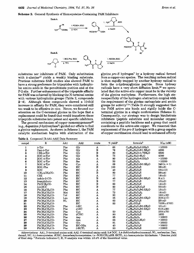
substrates nor inhibitors of PAM. Only substitution with D-alanine'O yields a weakly binding substrate. Previous substrate SAR studies also showed PAM to have a strong preference for tripeptides with hydropho- bic amino acids in the penultimate position and at the P-2 site. Further enhancement of the tripeptide affinity for PAM was achieved by blocking the N-terminal amine with various hydrophobic groups (Table 2, compounds 2-4). Although these compounds showed a 10-fold increase in affinity for PAM, they were considered still too weak to be effective in vivo. Hence, we focused our attention on the C-terminal glycine in the hope that a replacement could be found that would transform these tripeptide substrates into potent and specific inhibitors.
The general mechanism of copper monoo~ygenases~~ (e.g., dopamine @-hydroxylaseg) guided our efforts to find a glycine replacement. As s h o w in Scheme 1, the PAM catalytic mechanism begins with abstraction of the
glycine pro-S hydrogen7 by a hydroxy radical formed from a copper-oxo species. The resulting carbon radical is then rapidly trapped by another hydroxy radical to form the a-hydroxyglycine peptide. Since hydroxy radicals have a very short diffusion limit,26 we specu- lated that the active-site copper must be in the vicinity of the glycine methylene. Furthermore, the high ste- reospecificity of the hydrogen abstraction coupled with the requirement of the glycine carboxylate and amido groups for activity1la (Table 2) strongly suggested that the PAM active site binds and rigidly holds the C- terminal glycine in a single conformation (Scheme 3). Consequently, our strategy was to design bisubstrate inhibitors (peptide substrate and molecular oxygen) containing a peptidic backbone and a group that could coordinate to the active-site copper. We reasoned that replacement of the pro$ hydrogen with a group capable of copper coordination should lead to enhanced affinity
Table 2. Compound [R-AAl-AA2] Data Summary
compd R AA1 AA2 route % yield" formulab ICs0 (a) Phe GlY A 60 CzoHz3N30s.0.5HzO > 10000
B 76 1 D-Tyr 2 Dan-D-Tvr Phe Glv 3 4 5 6 7 8 9 10 11 12 13 14 15 16 17 18 19 20 21 22 23 24 25 26 27 28 29 30
CBZ-D-T;.~ B O C - D - T ~ ~ B O C - D - T ~ ~ B O C - D - T ~ ~ BOC-D-TJT BOC-Phe BOC
CBZ indole-240- 2-naphthoic-
(CH3)zCHzCO-
PhOCH2CO- 3,4-DCC PhCH2CH2CO- PhCHZCHzCO- PhCHZCHzCO- PhCHZCHzCO- PhCH2CHzCO- PhCH2CH2CO- PhCH2CH2CO- PhCHzCHZCO- PhCHzCHzCO- PhCHzCHzCO- PhCHzCHzCO- PhCHzCHzCO- PhCHzCHzCO- PhCHzCHZCO- PhCHzCHzCO-
Phe Phe Phe Phe Phe Phe Phe Phe Phe Phe Phe Phe Phe Phe Met Val Leu NL
Phe Phe Phe Phe Phe Phe Phe Phe Phe
Giy GlY Ala Ser CYS HC HC HC HC HC HC HC HC HC HC HC HC HC HC
CYS dCHC ASP A m Gln HC-OBn
1-HC d-HC
1-HCTL
B B A A A B A B A B B B B A A A A A A A A A A A A A A A
80 75 80 50 69
100 40 80 77 40 77 60 80 77 73 92 35 63 83
50 80 85 66 91 40 80
4200 3500 4000 > 10000 '10000 340 (n = 1) 39(f8) 53(f12) 29(f4) 18(f3) 8(fU lO(f3) 14(f3) 55(f5) 15(f3) 18(12) 160(f30) 300(f17) 23(&2) 7500(&1700) 10(&l) 610(&80) 330(f70) 1600 > 10000 > 10000 > 10000 > 10000 > 10000
Abbreviations: AA1, N-terminal amino acid; AA2, C-terminal amino acid; 3,4-DCC, 3,4-dichlorohydrocinnamoyl; NL, norleucine; Dan, dansyl; HC, D,L-homocysteine; dCHC, descarboxyhomocysteine, i.e -N(H)(CH&SH; HCTL, D,L-homocysteine thiolactone. " Denotes yield of final step. Formula indicates C, H, N analysis was within f0 .4% of the theoretical value.

Inhibition of PAM Journal of Medicinal Chemistry, 1994, Vol. 37, No. 26 4433
Scheme 3. Hypothetical Active-Site Structure of PAM
d
via both the increased number of favorable interactions and the chelation effect.27
Imidazoles, thioimidizoles, and thiolates are known to coordinate to cooper. Given the likelihood that the site surrounding the active-site copper was sterically encumbered, we chose to synthesize tripeptides contain- ing C-terminal alkylthiolates. Replacement of the C- terminal glycine with L-cysteine gave tripeptide 7, which was >lO-fold more potent than the corresponding gly- cine containing peptide 4. Evidence that the thiolate was responsible for the increased potency is apparent from comparing the IC50s for tripeptides 5-7. Replace- ment of the glycine pro-S hydrogen in tripeptide 4 with a methyl or hydroxymethyl (compounds 5 and 6, re- spectively) resulted in a complete loss in inhibitory activity (>go% remaining activity a t 10 pM concentra- tion) whereas replacement with the larger, more steri- cally-demanding thiomethyl group led to a 10-fold enhancement in potency. This result coupled with the known propensity of thiolates to coordinate with copper strongly suggests that tripeptide 7 interacts with the active-site copper.32
The S A R of 7 was further explored by synthesis of a series of analogs (Table 2). First, the tripeptide 8 was synthesized to determine the optimal length of the alkylthiolate sidechain. The finding that a C-terminal D,L-homocysteine residue gave another 10-fold increase in potency suggested that the distance between the glycine methylene and the copper ion was not ad- equately transversed by the thiomethyl group. Next, the SAR for the amino acid residues attached to the C-terminal homocysteine residue in tripeptide 8 was explored. Removal of one amino acid gave the N- blocked dipeptide 9, which resulted in no loss in potency whereas removal of both amino acids gave the N-blocked homocysteine analog 21, which resulted in a 500-fold loss in potency. Synthesis of a series of N-blocked dipeptides containing a C-terminal homocysteine indi- cated that aliphatic groups attached to the N-terminal end of the dipeptide were slightly worse than aryl groups (compounds 9-16). Furthermore, the aryl group could be directly attached to the amide (e.g., 12 and 13) or via a spacer (e.g., 14-16). SAR studies of the N-terminal amino acid side chain indicated a strong preference for hydrophobic residues. l1 A comparison of the hydrophobic residues showed that analogs with Phe (16) amd Met (17) were significantly more potent than analogs with branched aliphatic amino acids (e.g., Val
(18) and Leu (19)). The final area of SAR explored was the homocysteine residue. Similar to the SAR found for the tripeptide analogs, homocysteine was strongly pre- ferred over cysteine (22 vs 24). Furthermore, consistent with the predicted position of the active-site copper relative to the glycine methylene, the dipeptide contain- ing the L-isomer of homocysteine (22) was shown to be 60-fold more potent than the corresponding peptide containing the D-homocysteine isomer (23). The last piece of SAR uncovered in this study was that the carboxylate of the homocysteine residue is critical for inhibition potency as evidenced by the lack of activity found for the descarboxy analog 25, the benzyl ester 29, and the thiolactone 30.
Dipeptides containing a C-terminal homocysteine represent very potent inhibitors of the rat medulla PAM and a considerable improvement over the previous best known PAM inhibitors [ [(4-methoxybenzoyl)oxy]acetic acid; Ki = 480 pMZ1]. The magnitude of the inhibition constants were found to depend on whether the inhibitor was preincubated with the enzyme prior to initiation of the reaction. For example, without preincubation, compound 16 had an IC50 of 33 nM, whereas with a 20 min preincubation at room temperature the IC50 dropped to 15 nM. Importantly, preincubation of PAM with 0.3 pM 16 for 20 min followed by a 200-fold dilution led to total recovery of the starting activity over the following hour. Hence, the compounds bind reversibly with the highest affinity binding state apparently attained only through a kinetically slow process.28
Inhibition of Substance P Biosynthesis in Cell Culture
Substance P is a C-terminal amidated undecapeptide that is biosynthesized in the central and peripheral nervous systems. The possible role of substance P in neurogenic inflammation and potentially even in the pathogenesis of rheumatoid arthritis makes SP antago- nists and/or inhibitors of SP biosynthesis/release an attractive target. Since the dorsal root ganglion (DRG) cell represents the cell responsible for release of SP in neurogenic inflammation and also the site where SP is biosynthesized in the peripheral nervous system, we established a rat primary DRG cell culture in order to monitor the intracellular bioavailability of our PAM inhibitor^.^^
The effect of PAM inhibitors on SP biosynthesis in the DRG cell culture assay is shown in Table 3. All of the PAM inhibitors tested showed some inhibition of SP biosynthesis at 10 pM concentration. The apparent potency, however, was significantly less than the IC50 generated with the isolated enzyme, suggesting that the compounds had limited intracellular bioavailability. Since a free carboxylate can often limit cell penetration,
Table 3. PAM Inhibition in DRG Cells compd % I in DRG" compd % I in DRG"
8 34 f 7 15 37 f 15 10 10 f 4 16 28 f 7 11 40 f 7 20 7 f 4 12 42 f 8 22 25 f 4 13 67 f 5 30 37 f 9 14 22 f 12
a Denotes percent inhibition in DRG cells at 10 pM concentra- tion of the inhibitor.

4434 Journal of Medicinal Chemistry, 1994, Vol. 37, No. 26
we are preparing several proesters of the most active PAM inhibitors to optimize their intracellular bioavail- ability.
Erion et al.
Conclusions Dipeptides containing a C-terminal homocysteine and
an N-acylated hydrophobic amino acid were found to inhibit PAM with IC~OS in the low nanomolar range. Inhibition potency was dependent on both the carboxy- late and the thiolate functionalities of the homocysteine and on the hydrophobic groups of the second amino acid. The thiolate was postulated to produce high binding affinities through coordination with the active-site cop- per. The compound series also exhibited inhibition of PAM in rat dorsal root ganglion cells as demonstrated by a dose-dependent increase in the substance P-Gly/ substance P ratio. These results indicate that the compounds have sufficient potency and intracellular bioavailability to aid future studies focused on neu- ropeptide function and on the role neuropeptides play in various disease processes.
Experimental Section Melting points were determined in open capillary tubes with
a Thomas-Hoover Uni-Melt apparatus and are uncorrected. Proton NMR spectra were recorded on either a Brucker AC- 250, a Varian XL400, or a Varian XL300 spectrometer. Chemical shifts are reported in parts per million (ppm) downfield from tetramethylsilane. Coupling constants are given in hertz. Infrared (IR) spectra and MS spectra were measured on a Nicolet 5SXB FTIR spectrometer and a Hewlett-Packard GC/MS 5985B spectrometer, respectively. Microanalyses were carried out a t Robertson Laboratory, Inc., Madison, NJ. Optical rotations were recorded on a JASCO DIP-370 polarimeter. Thiol determinations were recorded on a Perkin-Elmer W-vis spectrophotometer according to the known p r o ~ e d u r e . ~ ~
Thin-layer chromatography was performed on Analtech silica gel GF plates. Visualization was accomplished by W illumination or by staining with a m n 0 4 solution. Column chromatography was performed with silica gel Kieselgel 60 (0.040-0.063 mm) from EM Scientific. High-performance liquid chromatography (HPLC) was accomplished with a Waters Delta prep 3000 and a 254 absorbance detector. Peak separations were achieved using a 250 mm x 30 cm YMC pack D.SIL-10-b s-10 120a silica gel column with a mobile phase of 1% acetic acid in ethyl acetatehexane (1:l) and a flow rate of 5.00 mumin.
General Amino Acid Coupling Procedure. To a solu- tion containing an N-protected amino acid (1 equiv, 0.1 M) in CHzClz was added an amino acid ester (1 equiv, usually as an HC1 salt) and 1-hydroxybenzotriazole (HOBT; 1-1.5 equiv) followed by triethylamine (TEA 1 equiv) and N-[3-(dimeth- y1amino)propyll-N'-ethylcarbodiimide hydrochloride (EDC1; 1.5-2 equiv). The mixture was stirred for 4 h at room temperature or until TLC analysis indicated that the reaction was complete. The reaction mixture was then diluted with ethyl acetate and washed with 1 N HC1(2x), saturated sodium bicarbonate (2 x 1, and brine. The organic layer was dried over MgSO4 and concentrated to dryness in vacuo. The resulting product was either directly used in the next step or purified further by flash chromatography.
General Deprotection Procedures. BOC-amino acids were deprotected by stirring a CHzC1z:trifluoroacetic acid (1: 1) solution at room temperature for 4 h. The solvent was removed in vacuo and the last traces of trifluoroacetic acid removed by repeated evaporations from dichloromethane. Methyl esters were deprotected t j l dissolving the compound in methanol at a concentration of about 0.1 M and slowly adding 1.2 equiv of 1 N NaOH. After completion of the reaction (1-4 h), the solvents were removed in vacuo and the
residue was dissolved in ethyl acetate. The organic layer was washed with 1 N HC1 and then water and dried over NazS04. Hydrocinnamoyl-cphenylalanyl+homocysteine Thio-
lactone (30). Route A. A solution of hydrocinnamic acid (15 g, 0.099 mol), L-phenylalanine methyl ester hydrochloride salt (20 g, 0.102 mol), 1-hydroxybenzotriazole (13.5 g, 0.099 mol), triethylamine (10.4 g, 0.102 mol), and N-[3-(dimethylamino)- propyl]-N'-ethylcarbodiimide hydrochloride (38.3 g, 0.2 mol) in 100 mL of methylene chloride was stirred at room tempera- ture for 4 h. The reaction mixture was diluted with 150 mL of ethyl acetate and washed with 1 N HC1 (2 x 50 mL) and saturated NaHC03 (2 x 50 mL). The organic layer was dried (MgS04) and concentrated to give the desired amide as a white solid (25.17 g, 81%): mp 60-62 "C; IR (KBr) 1754,1712,1647, 1535 cm-l; lH NMR (CDCl3) 2.42-2.47 (m, 2H), 2.78-2.83 (t, J = 7.1 Hz, 2H), 2.90 (dd, J = 8.5, 13.8 Hz, 1 H), 3.09 (dd, J = 5.8, 13.8 Hz, 1 H), 3.68 (s, 3H), 4.65 (dd, J = 5.7, 8.7 Hz, lH), 7.1-7.3 (m, 10H); MS (DCVCH4) 312 (M + 1). Anal. (CigHziN03) C, H, N.
The ester (25 g, 0.08 mol) was then treated with 250 mL of 2 N NaOH in MeOH for 1 h at room temperature. The reaction mixture was concentrated to half of the original volume and then diluted with 250 mL of water. The aqueous solution was washed with ether (2 x 60 mL) and acidified with 12 N HCl at 0 "C. The resulting suspension was extracted with ethyl acetate (3 x 100 mL), and the combined extracts were dried (Na2S04) and evapoarated to give hydrocinnamoyl- L-phenylalanine as a white solid (24 g, 100%): mp 158-159 "C; IR (KBr) 1730,1700,1633,1522 cm-l; 'H NMR (CD3OD) 2.44 (dd, J = 6.5, 8.6 Hz, 2H), 2.77-2.82 (m, 2H), 2.91 (dd, J = 8.9, 13.9 Hz, lH), 3.15 (dd, J = 5.1, 13.9 Hz, lH), 4.65 (dd, J = 5.1, 8.9 Hz, lH), 7.1-7.3 (m, 10H); MS (DCVCH4) 298 (M + 1). Anal. (ClsHleN03) C, H, N.
The synthesis was completed by stirring a solution contain- ing the acid (10 g, 0.034 mol), L-homocysteine thiolactone hydrochloride (5.2 g, 0.034 mol), 1-hydroxybenzotriazole (4.5 g, 0.034 mol), triethylamine (3.47 g, 0.034 mol), N434di- methy1amino)propyll-N'-ethylcarbodiimide hydrochloride (9.8 g, 0.051 mol), and CHzClz at room temperature. After 4 h, the reaction mixture was poured into ether (500 mL) and washed with 1N HC1 (2 x 200 mL), sat. NaHC03 (2 x 200 mL) and brine (1 x 100 mL). The organic layer was then dried (NazS04) and concentrated in vacuo. The resulting residue was chromatographed on silica gel (60% ethyl acetate in hexane) to give thiolactone 30 as a white solid (12.5 g, 93%): mp 168-169 "C; IR (KBr) 1705, 1639, 1540 cm-l; 'H NMR (CD30D) 1.86-2.00 (m, lH), 2.37-2.49 (m, 3H), 2.77-2.88 (m, 3H) 3.04 (dd, J = 6.7, 13.7 Hz, lH), 3.19-3.25 (m, lH), 3.29- 3.40(m,lH)4.54(dd,J=6.9, 13 .6Hz, lH) ,4 .63(dd,J=6.8 , 9.2 Hz, lH), 7.12-7.28 (m, 10H); MS (DCVCH4) 397 (M + 1); [aIz5~ = -11.42 (6.8 mg/mL in MeOH). Anal. (CzzH~4Nz03S) C, H, N. 2-Naphthoyl-~phenylalanyl-~,~homocysteine Thiolac-
tone. Route B. A solution of BOC-L-phenylalanine (2.0 g, 7.5 mmol), D,L-homocysteine thiolactone hydrochloride (1.16 g, 7.5 mmol), TEA(762 mg, 7.5 mmol), HOBT(l.O1 g, 7.5 mol), EDCl(2.88 g, 15 mmol), and CHzClz (100 mL) was stirred at room temperature. ARer 4 h, the reaction mixture was poured into 200 mL of ethyl acetate and washed with 1 N HCl(2 x 50 mL), saturated NaHC03 (2 x 50 mL), and brine (1 x 25 mL). The organic layer was dried (NazSOd) and evaporated to dryness to give the dipeptide as a white solid (2.5 g, 100%): mp 89-90 "C; IR (KBr) 3326, 1700, 1655, 1538, 1520, 1250, 1166 cm-l; NMR (CDCl3) 1.34 (s, 9H), 2.06-2.17 (m, lH), 2.53-2.61 (m, lH), 2.82 (dd, J = 9.3, 13.8 Hz, lH), 3.12 (dd, J = 4.9, 13.9 Hz, 1H) 3.39 (dd, J = 5.4, 11.7 Hz, lH), 3.43 (dd, J = 5.3, 11.6 Hz, lH), 7.14-7.26 (m, 5H); MS (CDVCH4) 365 (M + 1). Anal. ( C ~ B H ~ ~ N ~ O ~ S ) C, H, N.
The BOC-protected dipeptide (546 mg, 1.5 "01) was then dissolved in 50% TFMCHZC12 and stirred at room temperature. After 4 h, the reaction mixture was concentrated to dryness and redissolved in 10 mL of CHzClz. To this solution was added 2-naphthoic acid (275 mg, 1.6 mmol), TEA (163 mg, 1.6 mmol), HOBT (216 mg, 1.6 mmol), and EDCl (613 mg, 3.3 mmol). After the solution was stirred for 4 h at room temperature, it was poured into 60 mL of ethyl acetate and

Inhibition of PAM Journal of Medicinal Chemistry, 1994, Vol. 37, No. 26 4436
washed with 1 N HCl (2 x 20 mL), saturated NaHC03 (2 x 30 mL), and brine. The organic layer was then dried (Naz- S04) and concentrated in vacuo. The resulting residue was chromatographed on silica gel (50% ethyl acetate in hexane) to give the desired compound as a white solid (400 mg, 63.8%): mp 94-95 "C; IR (KBr) 3281,1806,1641,1531 cm-l; NMR (CDC13) 1.90-2.27 (m, lH), 2.43-2.69 (m, lH), 3.08- 3.15 (m, lH), 3.20-3.46 (m, 3H), 4.59-4.69 (m, lH), 4.90- 4.96 (m, lH), 7.16-7.37 (m, 5H), 7.51-7.60 (m, 2H), 7.74- 7.81 (m, lH), 7.81-7.95 (m, 3H), 8.26 (d, J = 10.9 Hz, 1H); MS (DCVCH4) 419 (M + 1). Anal. (Cz4HzzNz03S) C, H, N. Hydrocinnamoyl-L-phenylalanyl-L-homocysteine (22).
General Procedure for Thiolactone Hydrolysis. A 0 "C solution of hydrocinnamoyl-L-phenylalanyl-D,L-homocysteine thiolactone (2.0 g, 0.005 mol) and methanol (10 mL) was degassed by bubbling nitrogen through the solution for 5 min and then treated with 2 N NaOH (3 mL, 0.006 mol). m e r being stirred for 2 h at room temperature, the reaction mixture was acidified with 0 "C 12 N HCl and poured over EtOAc. The organic layer was separated and washed with 1 N HC1, saturated NaHC03, and brine. The mixture of diastereoiso- mers was separated by preparative HPLC using a Waters Delta prep 3000 and a 250 mm x 30 cm YMC pack DSIL- 10-b s-10 120 a silica gel column with a mobile phase of 1% acetic acid in ethyl acetatehexane (1:l) and a flow rate of 5.00 mumin. The L,L isomer (21) and the D,D isomer (22) had retention times of 25 and 38 min, respectively.
Compound 22 (129 mg): mp 117-119 "C; IR (KBr) 1720, 1632,1453 cm-l; NMR (CD30D) 1.76-2.02 (m, 3H), 2.41-2.56 (m,3H),2.75-2.89(m,3H),3.11(dd, J = 5 . 4 , 14Hz, lH),4.56 (dd, J = 4.7, 9.3 Hz, lH), 4.61-5.69 (m, lH), 7.1-7.52 (m,
C, H, N. Compound 23 (99 mg): mp 152-154 "C; NMR (CD30D)
2.06-2.18 (m, 3H), 2.21-2.89 (m, 3H), 2.75-2.89 (m, 3H), 3.0 (dd, J = 8.5,13.6 Hz, lH), 4.47 (dd, J= 4.7,9.3 Hz, lH), 4.61- 4.69 (m, lH), 7.10-7.52 (m, 10H); MS (DCVCH4) 415 (M + 1). Anal. (CZZHZ~NZO~S.~.~HZO) C, H, N. D-Tyrosyl-L-phenylalanylglycine (1): mp 185-187 "c; IR
(KBr) 2555, 1743, 1682, 1653 cm-l; 'H NMR (DzO) 6 2.81- 2.90 (m, 3H), 3.14 (dd, J = 4.3, 14.4 Hz, lH), 3.74 (9, 2H), 4.17 (t, J = 5.8 Hz, lH), 4.63-4.68 (m, lH), 6.80 (s,4H), 7.21- 7.23 (m, 2H), 7.31-7.39 (m, 3H); MS (DCVCHJ 546 (M + 1); [ a I z 5 ~ = -25.6 (8.1 mg/mL in MeOH). Anal.
Dansyl-D-tyrosyl-L-phenylalanylglycine (2): mp 174- 177 "C; NMR (CD30D) 6 2.30-2.46 (m, 2H), 2.72 (dd, J = 9, 14 Hz, lH), 2.83 (s, 6H), 3.09 (dd, J = 5.2, 13.9 Hz, lH), 3.70 (dd, J = 5.3, 9 Hz, lH), 3.86 (dd, J = 7.8, 5.2 Hz, 1 H), 4.56 (dd, 2H), 6.33 (dd, 4H), 7.10-7.21 (m, 6H), 7.38-7.44 (m, 2H), 8.05 (dd, J = 8.5, 16.5 Hz, 2H), 8.46 (d, 8.5 Hz, 1H); MS (FAB) (M + 1) 619. Anal. ( C ~ Z H ~ & O ~ S - ~ . ~ H Z O ) C, H, N. (Benzyloxycarbonyl)-D-tyrosyl-L-phenylalanylgly-
cine (3): mp 124-126 "C; IR (KBr) 1694, 1649, 1535, 1515 cm-1; 1H NMR (CD30D) 6 2.48-2.60 (m, lH), 2.61-2.83 (m, 2H), 3.05-3.18 (m, lH), 3.60-3.91 (m, 2H), 4.06-4.20 (m, lH), 4.59-4.65 (m, lH), 4.90-5.00 (m, 2H), 6.72 (dd, 4H), 7.12- 7.24 (m, 10H). Anal. (Cz8Hz~N30r1.75HzO) C, H, N. (tert-Butoxycarbonyl)-D-tyrosyl-L-phenylalanylgly-
cine (4): mp 150-151 "C; IR (KBr) 1738, 1633, 1646, 1614 cm-l; lH NMR (CD30D) 6 1.35 (s,9H), 2.51-2.58 (m, lH), 2.72 (dd, J = 6.8, 13 Hz, lH), 2.86 (dd, J = 9, 14 Hz, lH), 3.88 (dd, 2H), 4.11 (t, J = 7.0 Hz, lH), 4.60-4.63 (m, lH), 6.65 (dd, 4H), 7.12-7.23 (m, 5H); MS (DCVCH4) 486 (M + 1). Anal. (CZ~H~IN~OYHZO) C, H, N. (tert-Butoxycarbonyl)-D-tyrosyl-L-phenylalanylala-
nine (5): mp 117-119 "C; IR (Nujol) 1717, 1652, 1641, 1516 cm-l; lH NMR (CD30D) 6 1.29-1.50 (m, 12H), 2.51-2.60 (m, lH), 2.71-2.81 (m, lH), 2.82-2.93 (m, lH), 3.10-3.20 (m, lH), 4.13 (dd, J = 5.6, 8.6 Hz, lH), 4.39 (dd, J = 7.3, 14.5 Hz, lH), 4.56-4.80 (m, lH), 6.90 (dd, 4H), 7.13-7.25 (m, 5H); MS (DCV
(tert-Butoxycarbony1)-D-tyrosyl-L-phenylalanyl- serine (6): mp 123-124 "C; IR (KBr) 1720, 1650, 1514 cm-'; lH NMR (CDsOD) 6 1.35 (8, 9H), 2.57-2.60 (m, lH), 2.74- 2.80 (m, 1H), 2.89 (dd, J = 8.5, 13.8 Hz, lH), 3.76-3.89 (m,
10H); MS (DCVCH4) 415 (M + 1). Anal. (CzzHz&Tz04S.0.5HzO)
(CzoH23N305.0.5HzO) C, H, N.
CH4) 500 (M + 1). Anal. ( C Z ~ H ~ ~ N ~ O ~ . ~ H Z O ) C, H, N.
2H), 4.12-4.18 (m, lH), 4.46 (dd, J = 4.8, 10 Hz, lH), 4.58- 4.73 (m, lH), 6.64-6.67 (m, 2H), 6.87-6.91 (m, 2H), 7.18- 7.33 (m, 5H); MS (DCYCH4) 516 (M + 1). Anal. (Ch&3N308) C, H, N. (tert-Butoxycarbonyl)-D-tyrosyl-L-phenylalanylcys-
teine (7): mp 97-99 "C; IR (KBr) 2556,1743,1682,1653 cm-'; 1H NMR (CD30D) 6 1.35 (8, 9H), 2.03-2.08 (m, lH), 2.46- 2.63 (m, 2H), 2.73-2.91 (m, 2H), 3.06 (dd, lH), 3.30-3.45 (m, lH), 4.12 (t, lH), 4.39-4.51 (m, lH), 4.58-4.64 (m, 2H), 6.66 (d, 2H), 6.90 (d, 2H), 7.15-7.25 (m, 5H). Anal.
(tert-Butoxycarbonyl)-D-tyrosyl-L-phenyyl-D,L-ho- mocysteine (8): mp 92-94 "C; IR (KBr) 2554, 1716, 1690, 1650, 1516 cm-l; lH NMR (CD30D) 6 1.34 (9, 9H), 1.98-2.48 (m, 4H), 2.50-2.54 (m, lH), 2.60-2.78 (m, lH), 2.87-3.10 (m, 3H), 4.20-4.23 (m, lH), 4.47 (dd, 0.5H), 4.55 (dd, 0.5H), 4.65- 4.72 (m, 1H), 7.01-7.31 (m, 10H); MS (DCVCH4) 530 (M + 1). Anal. ( C Z ~ H ~ ~ N ~ O ~ S ) C, H, N. (tert-Butoxycarbonyl)-L-phenylalanyl-D,L-homocys-
teine (9): mp 60-62 "C; IR (KBr) 2556,1717,1698,1658,1525 cm-'; lH NMR (CD30D) 6 1.367 (s,4.5H), 1.37 (s, 4.5H), 1.85- 2.11 (m, 2H), 2.23-2.35 (m, lH), 2.53 (t, J= 7.3 Hz, lH), 2.78- 2.89 (m, lH), 3.05 (dd, 0.5H), 3.12 (dd, 0.5H), 4.28-4.33 (m, lH), 4.51 (dd, 0.5H), 4.59 (dd, 0.5H), 7.20-7.27 (m, 5H); MS
Isobutyryl-L-phenylalanyl-D,L-homocysteine (10): mp 67-68 "C; IR (KBr) 1640,1548,1719 cm-l; lH NMR (CD30D) 6 0.84-0.85 (m, 6H), 1.27-1.40 (m, 3H), 1.70-2.35 (m, 5H), 2.46-2.53 (m, lH), 2.82-2.93 (m, lH), 3.04-3.15 (m, lH), 4.51 (dd, 0.5H), 4.62 (dd, 0.5H), 4.67-4.74 (m, lH), 7.18-7.29 (m, 5H); MS (DCUCH4) 381 (M + 1). Anal. (CI~HZ~NZO~S) C, H, N.
benzyloxy carbonyl)-L-phenylalanyl-D,L-homocys- teine (11): mp 52-53 "C; IR (KBr) 2556, 1708, 1661, 1527 cm-1; lH NMR (CD30D) 6 1.82-2.31 (m, 3H), 2.42-2.59 (m, lH), 2.79-2.93 (m, lH), 3.04-3.17 (m, lH), 4.36-4.41 (m, lH), 4.44-4.60 (m, lH), 5.00-5.04 (m, 2H), 7.20-7.33 (m, 10H); MS (DCVCH4) 417 (M + 1). Anal. (CZIHZ~NZO~S) C, H, N. (Indol-2-ylcarbonyl)-~-tyrosyl-~-phenylalanyl-~,~-ho-
mocysteine (12): mp 176-179 "C; IR (KBr) 3297,1719,1624, 1565,1539,745 cm-'; 'H NMR (CD30D) 6 1.98-2.21 (m, 2H), 2.55 (t, J = 7 Hz, 2H), 3.07 (dd, J = 9.1, 13.9 Hz, lH), 3.25- 3.31 (m, lH), 4.60 (dd, J = 4.5, 9 Hz, lH), 4.80-4.9 (m, lH), 7.01-7.32 (m, 8H), 7.39 (d, J = 8.4 Hz, lH), 7.58 (d, J = 8 Hz, 1H); MS (DCUCH4) 408 (-HzO) (M + 1). Anal.
2-Naphthoyl-~-phenylalanyl-D,~-homocysteine (13): mp 102-103 "C; IR (KBr) 3295,1719,1638,1533,1539,1233 cm-l; lHNMR(CD30D)G 1.85-2.40(m,4H),2.58(t, J = 7 H z , lH), 3.06-3.16 (m, 0.5H), 3.22-3.33 (m, 0.5H), 4.56-4.63 (m, lH), 4.89-4.99 (m, lH), 7.16-7.36 (m, 5H), 7.51-7.59 (m, 2H), 7.73-7.96 (m, 3H), 8.27 (d, J = 15.7 Hz, 1H); MS (DCVCH4)
(Phenoxyacetoyl)-Lphenylalanyl-D+homocystdne (14): mp 68-71 "C; IR (KBr) 1722,1651,1538,1237 cm-'; 'H NMR (CD30D) 6 1.88-2.34 (m, 4H), 2.5-2.52 (m, lH), 2.94-3.21 (m, 2H), 4.39-4.59 (m, 4H), 6.85-7.0 (m, 4H), 7.21-7.30 (m, 6H); MS (DCVCHd 417 (M + 1). Anal. (CZ~HZ~NZO~S) C, H, N. (3,4-Dichlorobenzoyl)-~-phenylalanyl-~,~-homocys-
teine (15): mp 76-78 "C; IR (KBr) 1718,1644,1550 cm-l; 'H NMR (CD30D) 6 1.8-2.4 (m, 3H), 2.4-2.6 (m, 3H), 2.7-3.0 (m, 3H), 3.0-3.2 (m, lH), 4.49 (dd, J = 4.5,9.3 Hz, lH), 4.56- 4.70 (m, lH), 7.1-7.4 (m, 8H); MS (DCVCH4) 484 (weak), 466 (-HzO) (M + 1). Anal. (CzzHz4ClzNz04S) C, H, N.
HydrOcinnamoyl-bphenylalanyl-D,lhomocye (16): mp 65-67 "C; IR (KBr) 1720,1632,1453 cm-l; lH NMR (CD3- OD) 6 1.76-2.32 (m, 2H), 2.39-2.50 (m, 3H), 2.75-2.89 (m, 3H), 3.01 (dd, 0.5H), 3.11 (dd, 0.5H), 4.47 (dd, 0.5H), 4.56 (dd, 0.5H), 4.59-4.69 (m, lH), 7.10-7.29 (m, 10H); MS (DCVCH4) 415 (M + 1). Anal. (C~~HZ~NZO~S.O.~HZO) C, H, N. Hydrocinnamoyl-L-methionine-D,L-homocysteine (17):
mp 44-45 "C; lH NMR (CD30D) 6 1.79-1.89 (m, lH), 1.95- 2.13 (m, 4H), 2.04 (8, 3H), 2.32-2.38 (m, 2H), 2.39-2.58 (m, 4H), 2.88-2.93 (t, J = 6.8 Hz, 2H), 4.39-4.49 (m, lH), 4.52-
( C Z ~ H ~ ~ N ~ O ~ S . ~ . ~ H Z O ) C, H, N.
(DCUCH4) 383 (M + 1). Anal. (CI~HZ~NZO~S) C, H, N.
(CzzHz3N304S.0.5HzO) C, H, N.
419 (-HzO) (M + 1). Anal. (Cz4Hz4Nz04S) C, H, N.

4436 Journal of Medicinal Chemistry, 1994, Vol. 37, No. 26
4.60 (m, lH), 7.13-7.28 (m, 5H); MS (DCUCH4) 399 (M + 1). Anal. (Cl&&04Sz) c, H, N. Hydrocinnamoyl-L-valine-D,L-homocysteine (18): mp
60-62 "C; IR (KBr) 1721,1638,1546 cm-'; IH NMR (CD30D) 6 0.84-0.99 (m, 6H), 1.92-2.15 (m, 3H), 2.44-2.59 (m, 4H), 2.91 (t, J = 7.6 Hz, 2H), 4.09-4.23 (m, lH), 4.53-4.64 (m, lH), 7.12-7.27 (m, 5H); MS (DCUCHJ 367 (M + 1). Anal. (CieHzE"4S) c , H, N. Hydrocinnamoyl-L-leucine-D,L-homocysteine (19): mp
68-70 "C; IR (KBr) 1719,1638,1549 cm-l; lH NMR (CD30D) 6 0.875 (dd, J = 5.4 Hz, 6H), 1.45-1.51 (m, 3H), 1.96-2.14 (m, 2H), 2.44-2.58 (m, 4H), 2.91 (t, J = 7.0 Hz, 2H), 4.33- 4.39 (m, lH), 4.46-4.55 (m, lH), 7.13-7.27 (m, 5H); MS (DCU CHI) 381 (M + 1). Anal. (C19H2sNz04S.0.5HzO) C, H, N. Hydrocinnamoyl-L-norleucine-D,L-homocysteine (20):
mp 146-148 "C; IR (KBr) 1763, 1672, 1510, 1276 cm-'; 'H NMR (CD30D) 6 0.89 (8, J = 3.9 Hz, 3H), 1.17-1.49 (m, 4H), 1.51-1.62 (m, lH), 1.66-1.77 (m, lH), 1.91-2.15 (m, 2H), 2.42-2.57 (m, 4H), 2.85-3.0 (m, 2H), 4.23-4.33 (m, lH), 4.53- 4.60 (m, 1H), 7.12-7.27 (m, 5H); MS (DCUCH4) 381 (M + 1). Anal. (CigH28N204S) C, H, N. Hydrocinnamoyl-D,L-homocysteine (21): mp 96-97 "C;
IR (KBr) 1731, 1646, 1628, 1541 cm-l; 'H NMR (CD30D) 6 1.28-2.07 (m, 2H), 2.24-2.43 (m, 2H), 2.54 (t, J= 7.3 Hz, 2H), 2 .91( t ,J=7 .5Hz ,2H) ,4 .54 (dd ,J=4 .5 ,9 .5Hz , lH),7.13- 7.28 (m, 5H); MS (DCUCH4) 268 (M + 1). Anal. (C13H17N03S) C, H, N. Hydrocinnamoyl-L-phenylalanyl-L-cysteine (24). Hy-
drocinnamoyl-L-phenylalanine (200 mg, 0.67 "01) and cys- tine dimethyl ester (90 mg, 0.37 mmol) were coupled under normal conditions to yield after workup a yellow solid (230 mg, 80%): mp 73-75 "C; IH NMR (CDC13) 2.39-2.4 (m, 2H), 2.72-2.98(m, 5H), 3.05-3.13 (m, lH), 3.69(s, 3H), 4.57-4.71 (m, 2H) 7.02-7.3 (m, 10H). Zinc dust (50 mg) was added to a solution of the disulfide (220 mg, 0.27 mmol) in acetic acid (5 mL) and the reaction stirred overnight. After filtration and removal of the solvent, the residue was purified by flash chromatography on silica gel with 50% ethyl acetate as the eluent to afford the desired compound (200 mg, 91%): 'H NMR (CDCl3) 2.4-2.5 (m, 2H), 2.7-3.2 (m, 6H), 3.39 (s, 3H), 4.7- 4.82 (m, 2H), 7.02-7.3 (m, 10H). Hydrolysis of the methyl ester was accomplished by deoxygenating a methanol (5 mL) solution of the methyl ester (200 mg, 0.48 mmol), cooling to 0 "C, and adding 2 N KOH (0.36 mL, 0.73 mmol). After 1 h, the reaction was acidified with 1 N HC1 and extracted with ethyl acetate. The combined organic layers were washed with water and dried (Na2S04) to yield 97 mg of a white solid (50%): mp 73-75 "C; IR (KBr) 1724,1639,1532 cm-'; 'H NMR (CD30D) 6 2.42-2.49 (m, 2H), 2.76-3.06 (m, 5H), 3.04-3.16 (m, lH), 4.55-4.70 (m, 2H), 7.10-7.24 (m, 10H); MS (CDU CH4) 401 (M + 1); [ a ]25~ = -8.27" (4.0 mg/mL in MeOH). Anal. (CziHz4Nz04S) C, H, N. H y drocinnamoyl-L- phenylalanyldecarbolryhmocy s-
teine (25): mp 127-129 "C; IR (KBr) 3309,3285, 1643, 1537 cm-l; lH NMR (CD30D) 6 1.60-1.67 (m, 2H), 2.28-2.33 (t, J = 7.1 Hz, 2H), 2.44-2.49 (m, 2H), 2.78-2.86 (m, 3H), 2.96- 3.03 (dd, J = 7.3, 13.5 Hz, lH), 3.15-3.27 (m, 2H), 4.51 (t, J = 7.5 Hz, lH), 7.13-7.29 (m, 10H); MS (DCUCH4) 371 (M + 1). Anal. ( C Z ~ H ~ ~ N ~ O Z S ) c , H, N. Hydrocinnamoyl-L-phenylalmyl-L-aspartio acid (26):
mp 84-86 "C; IR (KBr) 1724,1669,1529 cm-l; lH NMR (CD3- OD) 6 2.39-2.47 (m, 2H), 2.70-2.93 (m, 5H), 3.06-3.18 (m, lH), 4.64-4.70 (m, 2H), 7.11-7.24 (m, 10H); MS (DCUCH4) 413 (M + 1). Anal. (C22H28206) C, H, N. Hydrocinnamoyl-L-phenylalanyl-casparagine (27): mp
113-114 "C; IR (KBr) 1729,1650,1538 cm-l; lH NMR (CD3- OD) 6 2.40-2.47 (m, 2H), 2.68 (dd, J = 5.5,10.8 Hz, lH), 2.73- 2.87 (m, 4H), 3.08-3.18 (m, lH), 4.63-4.72 (m, 2H), 7.10- 7.27 (m, 10H); MS (DCUCH4) 412 (M + 1). Anal. (CzzH25N305.HzO) C, H, N. Hydrocinnamoyl-L-phenylalanyl-L-glutamine (28): mp
78-79 "C; IR (KBr) 1718,1638,1544 cm-l; 'H NMR (CD30D) 6 1.75-2.23 (m, 3H), 2.34-2.49 (m, 3H), 2.74-2.89 (m, 3H), 3.03 (dd, J = 7.0, 13.6 Hz, 0.5H), 3.12 (dd, J = 5.3, 14 Hz, 0.5H),4.36(dd,J=4.3,8.8Hz,lH),4.4l(dd, J=4.8,9.1Hz,
Erion et al.
lH), 7.1-7.28 (m, 10H); MS (DCVCH4) 427 (M + 1). Anal.
Hyd"amoyl-cphenylalanyl-D&homocystehe Ben- zyl Ester (29). Benzyl alcohol (436 mg, 4.03 mmol) in anhydrous THF (5 mL) was treated with sodium metal (49 mg, 2.13 "01) at room temperature. After 2 h hydrocin- namoyl-L-phenylalanyl-D,L-homocysteine thiolactone (800 mg, 2.02 mmol) in anhydrous THF (10 mL) was added and the reaction stirred at room temperature. After 1 h, the reaction was quenched with 0.1 M HC1 and extracted with ethyl acetate. The combined organic layer were washed with saturated NaHC03 and brine and dried (MgS04). After removal of the solvents, the crude product was chromato- graphed on silica gel (EtOAdhexane, 3:l) to obtain a white solid (210 mg, 40%): mp 129-131 "C; IR (KBr) 1743, 1622 cm-'; lH NMR (CD3OD) 6 1.72-2.19 (m, 3H), 2.34-2.48 (m, 3H), 2.85-3.02 (m, 4H), 4.60-4.73 (m, 2H), 5.05-5.16 (m, 2H), 6.08-6.13 (m, 0.5H), 6.52-6.63 (m, 0.5H), 7.05-7.38(m, 15H);
PAM Enzyme Assay. PAM from conditioned medium derived from cultured rat medullary thyroid carcinoma CA- 77 cells (Unigene Laboratories; Fairfield, NJ) was partially purified using a DEAE cartridge followed by Sephacryl 300 SF column chr0matography.l' Reaction mixtures (100 pL) containing PAM (13.5 pU) and the inhibitor at the desired concentration were preincubated for 20 min at room tempera- ture in 150 mM TES buffer (pH = 7.0) containing 0.001% Triton-100. The substrate N-dansyl-D-Tyr-Phe-Gly (2.0 pM) and ascorbate (3 mM) were added to initiate the reaction. At the designated time, 10 pL of 0.1 M EDTA solution was added to quench the reaction. Separation and quantification (peak integration) of the product (Dan-D-Tyr-Phe-NHz) and starting tripeptide were performed on a CIS-reverse phase column with fluorometric detection.30 Each inhibitor was dissolved in DMSO (10 mM) and then diluted in 150 mM TES, pH = 7.0. A minimum of seven different inhibitor concentrations were tested in duplicate with each inhibitor tested three times. The percent inhibition for each compound was determined by comparing the reaction rates to the control and then construct- ing a log dose-response curve. Inhibitor IC50 determinations were obtained by least squares analyses of the linear portions of the log dose-response curves. DRG Cell Assay. Primary cultures of dorsal root ganglion
(DRG) neurons were prepared as previously described.2g In the absence of ascorbate, the ratio of substance P-Glyl substance P (SP-Gly/SP) changes from 0.05 on day one to 3.3 on day six. On the sixth day, the inhibitor was added followed by the addition of ascorbate (500 pM) 12 h later. After incubation overnight, the conversion of substance P-Gly to substance P was monitored by an RIA using an antibody that recognizes SP with an affinity 10 000-fold higher than SP- G l ~ . ~ l The SP-Gly level was determined by treating a dupli- cate sample with 3.5 pg of PAM followed by the RIA measure- ment of SP. The difference in SP measured between the two samples gave the level of SP-Gly. Percent inhibition was determined at a 10 pM inhibitor concentration.
(C23H2&2O6) C, H, N.
MS (CDVCH4) 505 (M + 1). Anal. ( C ~ ~ H ~ Z N Z O ~ S ) C, H, N.
Acknowledgment. We thank Michael Hatolski for performing the IR spectra and thiol titrations and Dr. J i m Stanton for many helpful discussions.
References (1) (a) Eipper, B. A.; Mains, R. E. Peptide a-Amidation. Annu. Rev.
Peptides: Chemistry, Biology, and Pharmacology. Adv. Phar-
(2) (a) Bradbury, A. F.; Finnie, M. D. A.; Smyth, D. G. Mechanism of C-Terminal Amide Formation by Pituitary Enzymes. Nature 1982,298, 686-688; (b) Bradbury, A. F.; Smyth, D. G. Peptide Amidation. Trends Bwl. Sci. 1991, 16, 112-115.
(3) Eipper, B. A.; Stoffers, D. A,; Mains, R. E. The Biosynthesis of Neuropeptides: Peptide a-Amidation. Annu. Rev. Neurosci.
P h p X 1988,50, 333-344. (b) Judd, A. K.; Schdnik , G. K.
m o l . 1990,21,221-285.
1992;15, 57-85. - (4) (a) Katopodie, A. G.; Ping, D.; May, S. W. A Novel Enzyme from
Bovine Neurointermediate Pituitarv Catalvzes Dealkvlation of a-Hydroxyglycine Derivatives, Thereby Funboning Se&entially with Peptidylglycine a-Amidating Monooxygenase in Peptide

Inhibition of PAM Journal of Medicinal Chemistry, 1994, Vol. 37, No. 26 4437
(14) Lotz, M.; Vaughan, J. H.; Carson, D. A. Effect of Neuropeptides on Production of Inflammatory Cytokines by Human Monocytes. Science 1988,241, 1218-1221.
(15) Oku, B.; Satoh, M.; Takagi, H. Release of substance P from the spinal dorsal horn is enhanced in polyarthritic rats. Neurosci.
(16) Levine, J. D.; Clark, R.; Devor, M.; Helms, C.; Moskowitz, M. A.; Basbaum, A. I. Intraneuronal Substance P Contributes to the Severity of Experimental Arthritis. Science 1984,226,547- 549.
(17) Lembeck, F.; Donnerer, J.; Tsuchiya, M.; Nagahisa, A. The non- peptide tachykinin antagonist, CP-96,345, is a potent inhibitor of neurogenic inflammation. Br. J. Pharmacol. 1992,105,527- 530.
(18) Hilsted, L.; Rehfeld, J. F.; Schwartz, T. W. Impaired a-Car- boxyamidation of gastrin in vitamin C-deficient Guinea Pigs.
(19) (a) Mains, R. E.; Park, L. P.; Eipper, B. A. Inhibition of Peptide Amidation by Disulfiram and Diethyldithiocarbamate. J. Biol. Chem. 1986,261,11938-11941. (b) Marchand, J. E.; Hershman, K; Kumar, M. S. A.; Thompson, M. L.; Kream, R. M. Disulfiram Administration Affects Substance P-like Immunoreactive and Monoaminergic Neural Systems in Rodent Brain. J. Bwl. Chem.
(20) Kizer, J. S.; Bateman, R. C., Jr.; Miller, C. R.; Humm, J.; Busby, W. H., Jr.; Youngblood, W. W. Purification and Characterization of a Peptidyl Glycine Monooxygenase from Porcine Pituitary. Endocrinology 1986,118, 2262-2267.
(21) Katopodis, A. G.; May, S. W. Novel Substrates and Inhibitors of Peptidylglycine a-Amidating Monooxygenase. Biochemistry 1990,
(22) Zabriskie, T. M.; Cheng, H.; Vederas, J . C. Mechanism-Based Inactivation of Peptidylglycine a-Hydroxylating Monooxygenase (PHM) by a Substrate Analogue, D-Phenylalanyl-L-Phenyl- alanyl-D-Vinylglycine: Inhibition of Formation of Peptide C- Terminal Amides. J. Am. Chem. SOC. 1992, 114, 2270-2272.
(23) Bos, L. B.; Arens, J. F. Chemistry of acetylenic ethers. WI I . The synthesis of a,B-unsaturated thiolo esters from carbonyl compounds, acetylenic ethers and boron trifluoride. Red. Z'rau. Chim. Pays-Bus 1963,82, 157-171.
(24) Ellman, G. L. A colorimetric method for determining low concentrations of mercaptans. Arch. Biochem. Biophys. 1988,
(25) Walsh, C. Enzymatic Reaction Mechanisms; Freeman: San Francisco, CA, 1979; pp 458-463.
(26) Hertzberg, R. P.; Dervan, P. B. Cleavage of DNA with Methidiumpropyl-EDTA-Iron(I1): Reaction Condition and Prod- uct Analyses. Biochemistry 1984,23, 3934-3945.
(27) Byers, L. D. Binding of Reactive Intermediate Analogs to Enzymes. J. Theor. Bid. 1978, 74, 501-512.
(28) Erion, M. D.; Walsh, C. T. 1-Aminocyclopropanephosphonate: Time-Dependent Inactivation of 1-Aminocyclopropanecarboxy- late Deaminase and Bacillus stearothermophilus Alanine Race- mase by Slow Dissociation Behavior. Biochemistry 1987, 26,
(29) Wong, M.; Jeng, A. Y. Posttranslational Modification of Glycine- Extended Substance P by an a-Amidating Enzyme in Cultured Sensory Neurons of Dorsal Root Ganglia. J. Neurosci. Res. 1994,
(30) Jones, B. N.; Tamburini, P. P.; Consalvo, A. P.; Young, S. D.; Lovato, S. J.; Gilligan, J. P.; Jeng, A. Y.; Wennogle, L. P. A Fluorometric Assay for Peptidyl a-Amidation Activity Using High-Performance Liquid Chromatography. Anal. Biochem.
(31) Jeng, A. Y.; Wong, M. Lovato, S. J.; Erion, M. D.; Gilligan, J. P. A Radioimmunoassay for Measuring a-Amidating Enzyme Ac- tivity. Anal. Biochem. 1990, 185, 213-219.
(32) Inhibition of peptide amidation was determined using the bifunctional enzyme isolated from CA-77 cells.30 Since the bifunctional enzyme contains the catalytic domains for both the monooxygenase and the lyase, these results alone do not prove unambiguously that inhibition occurs through inhibition of the monooxygenase activity. The fact that the intermediate hydrox- yglycine' was not detected, however, strongly suggests that the observed inhibition is not through inhibition of the lyase activity. Furthermore, the magnitude of the enhanced potency exhibited by the thiol-containing inhibitors is consistent with a thiolate- metal interaction and not a relatively weak thiolate hydrogen bond or hydrophobic interaction.
Lett. 1987, 74, 315-319.
FEBS Lett. 1986,196, 151-154.
1990,265, 264-273.
29,4541-4548.
74, 443-450.
3417-3425.
37,97-102.
1988,168,272-279.
Amidation. Biochemistry 1990,29,6115-6120. (b) Tajima, M.; Iida, T.; Yoshida, S.; Komatau, IC; Namba, R.; Yanagi, M.; Noguchi, M.; Okamoto, H. The Reaction Product of Peptidylg- lycine a-Amidating Enzyme is a Hydroxyl Derivative a t a-Car- bon of the Carboxyl-terminal Glycine. J. Biol. Chem. 1990,265,
( 5 ) Yun, H.-Y.; Keutmann, H. T.; Eipper, B. A. Alternative Splicing Governs Sulfation of Tyrosine or Oligosaccharide on Peptidylg- lycine a-Amidation Monooxygenase. J. Biol. Chem. 1994,269,
(6) Eipper, B. A.; Mains, R. E.; Glembotski, C. C. Identification in Pituitary Tissue of a Peptide a-Amidation Activity that acts on Glycine-Extended Peptides and Requires Molecular Oxygen, Copper and Ascorbic Acid. Proc. Natl. Acad. Sci. U.SA. 1983,
(7) (a) Young, S. D.; Tamburini, P. P. Enzymatic Peptidyl a-Ami- dation Proceeds through Formation of an a-Hydroxyglycine Intermediate. J. Am. Chem. SOC. 1989, 111, 1933-1934. (b) Ramer, S. E.; Cheng, H.; Palcic, M. M.; Vederas, J. C. Formation of Peptide Amides by Peptidylglycine a-Amidating Monooxy- genase: A New Assay and Stereochemistry of Hydrogen Loss. J. Am. Chem. SOC. 1988, 110, 8526-8532. (c) Ping, D.; Kato- podis, A.; May, S. W. Tandem Stereochemistry of Peptidylglycine a-Monooxygenase and Peptidylamidoglycolate Lyase, the Two Enzymes Involved in Peptide Amidation. J. Am. Chem. SOC.
(8) (a) Freeman, J. C.; Villafranca, J. J.; Merkler, D. J. Redox Cycling of Enzyme-Bound Copper during Peptide Amidation. J. Am. Chem. SOC. 1993, 115, 4923-4924. (b) Kulathila, R.; Consalvo, A. P.; Fitzpatrick, P. F.; Freeman, J. C.; Snyder, L. M.; Villafranca, J . J.; Merkler, D. J. Bifunctional Peptidylglycine a-Amidating Enzyme Requires Two Copper Atoms for Maximum Activity. Arch. Biochem. Biophys. 1994, 311, 191-194. (c) Merkler, D. J.; Kulathila, R.; Consalvo, A. P.; Young, S. D.; Ash, D. E. l80 Isotopic lsC NMR Shift as Proof that Bifunctional Peptidylglycine a-Amidation Enzyme is a Monooxygenase. Bio- chemistry 1992,31, 7282-7288.
(9) (a) Fitzpatrick, P. F.; Villafranca, J. J. Mechanism-Based Inhibi- tors of Dopamine B-Hydroxylase. Arch. Bwchem. Bwphys. 1987, 257, 231-250. (b) Kent, U. M.; Fleming, P. J. Purified Cyto- chrome b561 Catalyzes Transmembrane Electron Transfer for Dopamine B-Hydroxylase and Peptidyl Glycine a-Amidating Monooxygenase Activities in Reconstituted Systems. J. Biol. Chem. 1987,262,8174-8178.
(10) Landymore-Lim, A. E. N.; Bradbury, A. F.; Smyth, D. G. The Amidatine Enzvme in Pituitarv Will Accent a PeDtide with
9602-9605.
10946-10955.
80, 5144-5148.
1992,114,3998-4000.
C-Termin2 D-&nine as Substrate. BiocLm. Biiphys. Res. Commun. 1983,117, 289-293.
(11) (a)Tamburini, P. P.; Jones, B. N.; Consalvo,A. P.; Young, S. D.; Lovato, S. J.; Gilligan, J. P.; Wennogle, L. P.; Erion, M.; Jeng, A. Y. Structure-Activitv Relationshim for Glvcine-Extended Peptides and the a-Amidating Enzyme Derived &om Medullary Thyroid CA-77 Cells. Arch. Biochem. Bwphys. 1988,267,623- 631. (b) Mehta, N. M.; Gilligan, J. P.; Jones, B. N.; Bertelsen, A. H.; Roos, B. A.; Birnbaum, R. S. Purification of a Peptidyl- glycine a-Amidating Enzyme from Transplantable Rat Medul- lary Thyroid Carcinomas. Arch. Biochem. Biophys. 1988,261, 44-54. (c) Dickinson, C. J.; Daugherty, D.; Guo, Y. J.; Stadler, B.; Finniss, S.; Yamada, T. Substrate Specificity of the Gastrin- amidating Enzyme. J. Biol. Chem. 1993,268, 15929-15934.
(12) (a) Eipper, B. A.; Myers, A. C.; Mains, R. E. Peptidyl-Glycine a-Amidation Activity in Tissues and Serum of the Adult Rat. Endocrinology 1986,116, 2497-2504. (b) Vaeroy, H.; Nyberg, F.; Franzen, H.; Terenius, L. Characterization of a Substance P-Gly Amidating Enzyme in Human Cerebrospinal Fluid. Bio- chem. Biophys. Res. Commun. 1987,148,24-30. (c) Wand, G. S.; Ney, R. L.; Baylin, S.; Eipper, B.; Mains, R. E. Characteriza- tion of a Peptide a-Amidation Activity in Human Plasma and Tissues. Metabolism 1986, 34, 1044-1052.
(13) (a) Gilligan, J. P.; Lovato, S. J.; Erion, M. D.; Jeng, A. Y. Modulation of Carrageenan-Induced Hind Paw Edema by Sub- stance P. Inflammation 1994, 18, 285-292. (b) Tissot, M.; Pradelles, P.; Giroud, J. P. Substance P-Like Levels in Inflam- matory Exudates. Inflammation 1988,12,25-35. (c) Colpaert, F. C.; Donnerer, J.; Lembeck, F. Effects of Capsaicin on Inflam- mation and on the Substance P Content of Nervous Tissues in Rats with Adjuvant Arthritis. Life Sci. 1983, 32, 1827-1834. (d) Helme, R. D.; Andrews, P. V.; Watson, B. A. Neurogenic Inflammation Caused by Wool Fabric in the R a t Possible Mediation by Substance P. Neurosci. Lett. 1986, 66, 333-337. (e) Goetzl, E. J.; Chernov, T.; Renold, F. Neuropeptide Regulation of the Expression of Immediate Hypersensitivity. J. Zmmunol. 1986,135,802s-805s. (0 Perianin, A.; Snyderman, R.; Malfroy, B. Substance P F'rimes Human Neutrophil Activation: A Mechanism for Neurological Regulation of Inflammation. Bio- chem. Biophys. Res. Commun. 1989,161, 520-524.