Application of SIMSTEX to oligomerization of insulin analogs and mutants
9
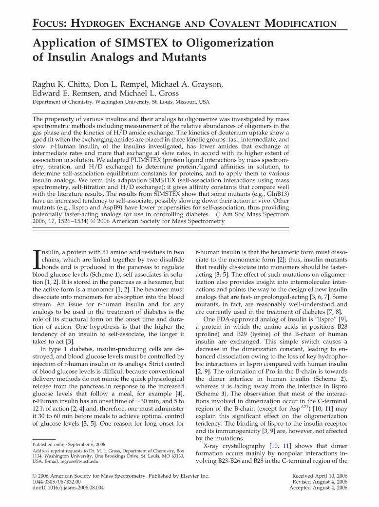
FOCUS:HYDROGEN EXCHANGE AND COVALENT MODIFICATION Application of SIMSTEX to Oligomerization of Insulin Analogs and Mutants Raghu K. Chitta, Don L. Rempel, Michael A. Grayson, Edward E. Remsen, and Michael L. Gross Department of Chemistry, Washington University, St. Louis, Missouri, USA The propensity of various insulins and their analogs to oligomerize was investigated by mass spectrometric methods including measurement of the relative abundances of oligomers in the gas phase and the kinetics of H/D amide exchange. The kinetics of deuterium uptake show a good fit when the exchanging amides are placed in three kinetic groups: fast, intermediate, and slow. r-Human insulin, of the insulins investigated, has fewer amides that exchange at intermediate rates and more that exchange at slow rates, in accord with its higher extent of association in solution. We adapted PLIMSTEX (protein ligand interactions by mass spectrom- etry, titration, and H/D exchange) to determine protein/ligand affinities in solution, to determine self-association equilibrium constants for proteins, and to apply them to various insulin analogs. We term this adaptation SIMSTEX (self-association interactions using mass spectrometry, self-titration and H/D exchange); it gives affinity constants that compare well with the literature results. The results from SIMSTEX show that some mutants (e.g., GlnB13) have an increased tendency to self-associate, possibly slowing down their action in vivo. Other mutants (e.g., lispro and AspB9) have lower propensities for self-association, thus providing potentially faster-acting analogs for use in controlling diabetes. (J Am Soc Mass Spectrom 2006, 17, 1526 –1534) © 2006 American Society for Mass Spectrometry I nsulin, a protein with 51 amino acid residues in two chains, which are linked together by two disulfide bonds and is produced in the pancreas to regulate blood glucose levels (Scheme 1), self-associates in solu- tion [1, 2]. It is stored in the pancreas as a hexamer, but the active form is a monomer [1, 2]. The hexamer must dissociate into monomers for absorption into the blood stream. An issue for r-human insulin and for any analogs to be used in the treatment of diabetes is the role of its structural form on the onset time and dura- tion of action. One hypothesis is that the higher the tendency of an insulin to self-associate, the longer it takes to act [3]. In type 1 diabetes, insulin-producing cells are de- stroyed, and blood glucose levels must be controlled by injection of r-human insulin or its analogs. Strict control of blood glucose levels is difficult because conventional delivery methods do not mimic the quick physiological release from the pancreas in response to the increased glucose levels that follow a meal, for example [4]. r-Human insulin has an onset time of 30 min, and 5 to 12 h of action [2, 4] and, therefore, one must administer it 30 to 60 min before meals to achieve optimal control of glucose levels [3, 5]. One reason for long onset for r-human insulin is that the hexameric form must disso- ciate to the monomeric form [2]; thus, insulin mutants that readily dissociate into monomers should be faster- acting [3, 5]. The effect of such mutations on oligomer- ization also provides insight into intermolecular inter- actions and points the way to the design of new insulin analogs that are fast- or prolonged-acting [3, 6, 7]. Some mutants, in fact, are reasonably well-understood and are currently used in the treatment of diabetes [7, 8]. One FDA-approved analog of insulin is “lispro” [9], a protein in which the amino acids in positions B28 (proline) and B29 (lysine) of the B-chain of human insulin are exchanged. This simple switch causes a decrease in the dimerization constant, leading to en- hanced dissociation owing to the loss of key hydropho- bic interactions in lispro compared with human insulin [2, 9]. The orientation of Pro in the B-chain is towards the dimer interface in human insulin (Scheme 2), whereas it is facing away from the interface in lispro (Scheme 3). The observation that most of the interac- tions involved in dimerization occur in the C-terminal region of the B-chain (except for Asp A21 ) [10, 11] may explain this significant effect on the oligomerization tendency. The binding of lispro to the insulin receptor and its immunogenicity [3, 9] are, however, not affected by the mutations. X-ray crystallography [10, 11] shows that dimer formation occurs mainly by nonpolar interactions in- volving B23-B26 and B28 in the C-terminal region of the Published online September 6, 2006 Address reprint requests to Dr. M. L. Gross, Department of Chemistry, Box 1134, Washington University, One Brookings Drive, St. Louis, MO 63130, USA. E-mail: [email protected] © 2006 American Society for Mass Spectrometry. Published by Elsevier Inc. Received April 10, 2006 1044-0305/06/$32.00 Revised August 4, 2006 doi:10.1016/j.jasms.2006.08.004 Accepted August 4, 2006
-
Upload
independent -
Category
Documents
-
view
1 -
download
0
Transcript of Application of SIMSTEX to oligomerization of insulin analogs and mutants
FOCUS: HYDROGEN EXCHANGE AND COVALENT MODIFICATION
Application of SIMSTEX to Oligomerizationof Insulin Analogs and Mutants
Raghu K. Chitta, Don L. Rempel, Michael A. Grayson,Edward E. Remsen, and Michael L. GrossDepartment of Chemistry, Washington University, St. Louis, Missouri, USA
The propensity of various insulins and their analogs to oligomerize was investigated by massspectrometric methods including measurement of the relative abundances of oligomers in thegas phase and the kinetics of H/D amide exchange. The kinetics of deuterium uptake show agood fit when the exchanging amides are placed in three kinetic groups: fast, intermediate, andslow. r-Human insulin, of the insulins investigated, has fewer amides that exchange atintermediate rates and more that exchange at slow rates, in accord with its higher extent ofassociation in solution. We adapted PLIMSTEX (protein ligand interactions by mass spectrom-etry, titration, and H/D exchange) to determine protein/ligand affinities in solution, todetermine self-association equilibrium constants for proteins, and to apply them to variousinsulin analogs. We term this adaptation SIMSTEX (self-association interactions using massspectrometry, self-titration and H/D exchange); it gives affinity constants that compare wellwith the literature results. The results from SIMSTEX show that some mutants (e.g., GlnB13)have an increased tendency to self-associate, possibly slowing down their action in vivo. Othermutants (e.g., lispro and AspB9) have lower propensities for self-association, thus providingpotentially faster-acting analogs for use in controlling diabetes. (J Am Soc Mass Spectrom2006, 17, 1526–1534) © 2006 American Society for Mass Spectrometry
Insulin, a protein with 51 amino acid residues in twochains, which are linked together by two disulfidebonds and is produced in the pancreas to regulate
blood glucose levels (Scheme 1), self-associates in solu-tion [1, 2]. It is stored in the pancreas as a hexamer, butthe active form is a monomer [1, 2]. The hexamer mustdissociate into monomers for absorption into the bloodstream. An issue for r-human insulin and for anyanalogs to be used in the treatment of diabetes is therole of its structural form on the onset time and dura-tion of action. One hypothesis is that the higher thetendency of an insulin to self-associate, the longer ittakes to act [3].
In type 1 diabetes, insulin-producing cells are de-stroyed, and blood glucose levels must be controlled byinjection of r-human insulin or its analogs. Strict controlof blood glucose levels is difficult because conventionaldelivery methods do not mimic the quick physiologicalrelease from the pancreas in response to the increasedglucose levels that follow a meal, for example [4].r-Human insulin has an onset time of �30 min, and 5 to12 h of action [2, 4] and, therefore, one must administerit 30 to 60 min before meals to achieve optimal controlof glucose levels [3, 5]. One reason for long onset for
Published online September 6, 2006Address reprint requests to Dr. M. L. Gross, Department of Chemistry, Box
1134, Washington University, One Brookings Drive, St. Louis, MO 63130,USA. E-mail: [email protected]© 2006 American Society for Mass Spectrometry. Published by Elsevie1044-0305/06/$32.00doi:10.1016/j.jasms.2006.08.004
r-human insulin is that the hexameric form must disso-ciate to the monomeric form [2]; thus, insulin mutantsthat readily dissociate into monomers should be faster-acting [3, 5]. The effect of such mutations on oligomer-ization also provides insight into intermolecular inter-actions and points the way to the design of new insulinanalogs that are fast- or prolonged-acting [3, 6, 7]. Somemutants, in fact, are reasonably well-understood andare currently used in the treatment of diabetes [7, 8].
One FDA-approved analog of insulin is “lispro” [9],a protein in which the amino acids in positions B28(proline) and B29 (lysine) of the B-chain of humaninsulin are exchanged. This simple switch causes adecrease in the dimerization constant, leading to en-hanced dissociation owing to the loss of key hydropho-bic interactions in lispro compared with human insulin[2, 9]. The orientation of Pro in the B-chain is towardsthe dimer interface in human insulin (Scheme 2),whereas it is facing away from the interface in lispro(Scheme 3). The observation that most of the interac-tions involved in dimerization occur in the C-terminalregion of the B-chain (except for AspA21) [10, 11] mayexplain this significant effect on the oligomerizationtendency. The binding of lispro to the insulin receptorand its immunogenicity [3, 9] are, however, not affectedby the mutations.
X-ray crystallography [10, 11] shows that dimerformation occurs mainly by nonpolar interactions in-
volving B23-B26 and B28 in the C-terminal region of ther Inc. Received April 10, 2006Revised August 4, 2006
Accepted August 4, 2006

1527J Am Soc Mass Spectrom 2006, 17, 1526–1534 OLIGOMERIZATION OF INSULIN
B chain and by hydrogen bonding involving residuesB24 and B26 [11]. In the A-chain, AsnA21 seems to be theonly amino acid that is involved in dimer formation.More evidence of the importance of the B chain inter-face in wild-type insulin (shown in Scheme 2) comesfrom the observation that insulin mutants withoutB26-B30 and B28-B30 regions do not dimerize [12].Formation of hexamers occurs by burial of both polarand nonpolar residues between the dimers leading tolooser packing than that of the dimer [2, 11, 13]. SixGluB13 are arranged in a circular pattern at the center ofthe hexamer, with charge repulsion counteracted byZn-binding [3]. When GluB13 is changed to Gln, aslow-acting insulin results [13] owing to a reduction ofcharge repulsion and an increase in hexamerization.
Our hypothesis is that mass spectrometry combinedwith H/D amide exchange and mathematical modelingcan reveal the effects of mutations on the oligomeriza-tion properties of proteins, and specifically on variousinsulins. We report here a mass spectrometric methodto determine the self-association properties of proteins,and we apply the method to insulin. We also wish to
Scheme 1. The sequence of human insulin. Insulin consists oftwo chains, A and B, having 21 and 30 amino acids, respectively.The amino acids that are changed to obtain various insulinmutants (the mutations are shown in Tables 1 and 2) are labeledwith the chain name, followed by the amino acid number in thechain.
Scheme 2. The dimer interface for the human insulin. A stron-ger, more compact dimer results because orientation of ProB28 istowards the interface forming a hydrophobic contact. Coordinates
were taken from RCSB Protein Data Bank, file 1ZNI.pdb.resolve issues with solubility of insulin in the earlierkinetic results reported in [14], a report in which thekinetics of amide exchange was used without modelingto distinguish various forms of insulin. We now fit thedata for r-human, lispro, porcine, and bovine insulins,whose sequences are shown in Table 1, to a kineticmodel similar to the one used in [15]. Exchange reac-tions for proteins and peptides were successfully mod-eled as pseudo first-order reactions [16], and threeexchange rate grouping models were previously ap-plied to the kinetics of exchange of other proteins [15].
H/D amide exchange is useful for studying thehigher order structure of proteins in solution [17, 18]. Itsuse in mapping protein/protein interfaces was pio-neered by Komives and coworkers [19, 20]. The premisefor their strategy is that amides buried in an interfacewill exchange more slowly in the complex than whenthe protein is free and not part of a complex. H/Dexchange can also provide thermodynamic informationabout protein/ligand binding [21]. Two H/D exchangemethods, SUPREX [21] and PLIMSTEX [22], make useof H/D exchange in a titration format to obtain thefree-energy and affinity for the binding. To adaptPLIMSTEX to self-association requires the mathematicalmodeling of PLIMSTEX to be changed (no modifica-tions are required for SUPREX). In this paper, wedescribe those modifications and apply the modifiedmethod, which we call SIMSTEX (self interactions bymass spectrometry self-titration and H/D exchange) to
Scheme 3. The dimer interface for the lispro dimer. An impor-tant hydrophobic contact in the dimer is eliminated because theProB29 is away from the interface. Coordinates were taken fromRCSB Protein Data Bank, file 1LPH.pdb.
Table 1. Sequence variations for insulin analogs studied herecompared to human insulin
Insulin type Sequence variation
Lispro KB28 and PB29 reversedBovine A in B30
Porcine A in B30, A in A8 and V in A10
1528 CHITTA ET AL. J Am Soc Mass Spectrom 2006, 17, 1526–1534
r-human insulin and to various insulin analogs toobtain affinity constants for self-association. The vari-ous insulins were designed by others to increase ordecrease hexamerization [2, 3] as can be seen by con-sidering the interface between two insulin molecules inthe dimer in Scheme 2.
Methods
Materials
r-Human Insulin was purchased from Sigma (Milwau-kee, WI). Porcine, bovine, and lispro insulins were giftsfrom the Food and Drug Administration of St. Louis.Other insulin mutants were donated by Dr. StephenBayne of NovoNordisk, Denmark, and were formed bymutating one or two residues at a time of human insulin[the list of mutants, their names (as used in this article),and the corresponding mutation(s) are shown in Table2]. All the insulin analogs were dissolved in 10-mMammonium acetate at pH 2.5. The pH was then raised to7.4 by slowly adding ammonia. The solution was thencentrifuged, and the supernatant supersaturated solu-tion was taken for study.
To obtain an independent assessment of the oli-gomerization state of insulin in solution, hydrodynamicdiameter distributions for the protein solutions weredetermined by dynamic light scattering (DLS) underboth pH conditions. A Brookhaven Instruments Co.(Holtsville, NY) DLS system consisting of a modelBI-200SM goniometer, a model EMI-9865 photomulti-plier, and a model 95-2 Ar ion laser (Lexel Corp., PaloAlto, CA) operated at 514.5 nm was employed. Allmeasurements were made at 20 � 1 °C. Before analysis,insulin solutions were centrifuged for 4 min at 8000 � gin a Brinkman model 5415 microfuge to sediment anyextraneous particles. Scattered light was collected at ascattering angle 90°. The system’s digital correlator wasoperated with 522 channels, ratio channel spacing,initial and final delay settings between 1.4 �s and 10 ms,respectively, and duration of 10 min. A photomultiplieraperture of 200 or 400 �m was used, and the incidentlaser intensity was adjusted to obtain a photon countingrate of 200 to 300 kcps. Only measurements for whichthe measured and calculated intensity autocorrelationfunction agreed to within 0.1% were used to calculatehydrodynamic diameters. Calculations of the hydrody-namic diameter distribution from measured intensity
Table 2. Insulin mutants studied in this work
Insulin mutant Mutation
A4A17 GluA4 and GluA17 to Gln and TyrB31
B27 ThrB27 to ArgA4B27 GluA4 to Gln and ThrB27 to ArgB9Asp SerB9 to AspB21 GluB21 to GlnB13B21 GluB13 and GluB21 to Gln
autocorrelation functions were performed with the
ISDA software package (Brookhaven Instruments Co.,Holtsville, NY), which employed single exponentialfitting, cumulant analysis, and non-negatively con-strained least-squares (NNLS), and continuous regular-ization (CONTIN) analysis routines. Reported hydro-dynamic diameter distribution averages are meanvalues of a minimum of three determinations.
For amide H/D exchange studies, the insulin inammonium acetate was diluted 10-fold in deuteratedammonium acetate. For kinetics measurements, theuptake of deuterium was monitored as a function of thetime for quenching the reaction in ice-cold water/acetonitrile/formic acid 50/49/1. To follow the deute-rium uptake as a function of concentration, the H/Dexchange was allowed to reach steady-state (�6 h). Theexchange was quenched, and the solution at a finalinsulin concentration of 10 �M was infused into the ESIsource for MS measurement. The quench caused theoligomers to dissociate into monomers. The deuteriumuptake was measured as the difference between themass centroids of the monomer distribution after andbefore exchange.
Mass Spectrometry
Mass spectra of the dimer in various solvents wereacquired with a Q-TOf-Ultima (Micromass, Manchester,UK), a tandem mass spectrometer consisting of a quad-rupole (Q) mass analyzer, a quadrupole collision cell,and a second-stage time-of-flight (TOF) analyzer. Posi-tive ions were formed by using a needle voltage of 3 kVand a cone voltage of 90 V. The temperatures of thesource block and for desolvation were 90 °C. A solutionflow rate of 10 �L/min was used for introduction. Allparameters (i.e., aperture to the TOF, transport voltage)were optimized to achieve maximum sensitivity and amass resolving power of 10,000 (full width at halfmaximum). The spectra were a sum of 2-sec scans over2 min, and the spectra were smoothed twice using athree-point Savitzky-Golay method before submittingthe data to the Mathcad (MathSoft Engineering &Education Inc., Cambridge, MA) program for calculat-ing the centroids.
Modeling
For kinetic modeling, the total number of active hydro-gens (sites) was divided into three groups. The numberof exchanging hydrogens in each group and the rateconstant for each group were varied in a process thatminimized the differences between the data and themodel at various time-points. The deuterium uptake isthe sum of number of exchangeable sites in each of thethree groups. Deuterium uptake at each site was mod-eled as a pseudo first-order reaction to calculate thefraction of molecules that were deuterated at this site[15]. From this, the mass shift responsible for the shift ofthe mass centroid of the isotopic pattern was calculated.
The process for calculating the affinity constants

1529J Am Soc Mass Spectrom 2006, 17, 1526 –1534 OLIGOMERIZATION OF INSULIN
starts with the model for PLIMSTEX [23]. Specifically,the modeling of the mass change based on the solutionconcentrations was carried over from PLIMSTEX, butthe equations for the oligomer solution concentrationswere modified to accommodate self-association as de-scribed in [24]. To allow the program to find a reliableminimum and avoid being trapped in an unreasonablelocal minimum, the search was started at “good-guess”values. Good-guess values were obtained by synthesiz-ing a model dataset on the basis of the start values, andthen comparing the outcome to the experimental data.The initial good-guess values were modified until agood graphical fit was obtained. The “minimize” func-tion in the program was then used to refine the sought-after solution by minimizing the square root of themean of the squares of the differences between the dataand the model over the concentration range of thetitration. The minimization is a nonlinear, quasi-New-tonian method �i and all the assumptions required for aNLLS regression are relevant [25].
The program started with guess values for the �i
involved in the association of insulin and the knowntotal protein (ligand) concentration. The model wasbased on the reactions shown in eq 1.
2Monomer¡K12
Dimer
2Dimer¡K24
Tetramer
Dimer � Tetramer¡K46
Hexamer
(1)
The unknown parameter �i for the system were taken tobe as those defined in eq 2
�0 � 1
�1 � K12
�3 � K24K122
�5 � K46K24K123
(2)
To give us consistent expressions for the followingequations, �2 and �4 were set to zero. Using �i to definethe oligomer concentrations was more economical forthe development of the equation. After solving for the�i, individual affinity constants were obtained as shownin eq 3
K12 � �1
K24 ��3
�12
K46 ��5
�3�1
(3)
Trial values for another model parameter, Di, were alsoprovided to the program; where Di is the number ofdeuteriums protected in the ith oligomer compared with
that in the monomer. For example, D0 is the number ofamide hydrogens protected in the monomer and D1 isthe number protected in the dimer compared to themonomer, and so on.
The concentration of free ligand in solution wascalculated by numerically integrating (using the “Rk-
adapt”) the functiond�Lig�
d�Lig�T
, which was calculated as a
reciprocal of thed�Lig�T
d�Lig�, where [Lig] is the free ligand
concentration in solution and [Lig]T is the total analyt-ical concentration of the ligand as described in [23].
The mass shift caused by D uptake, �D, at anyconcentration during the titration was defined as thenumber of exchangeable sites in the molecule for eacholigomer weighted by the corresponding fraction ofmonomers produced when the oligomer dissociates,subtracted from the number of exposed sites in themonomer. Equation 4 is the mathematical form of thisdescription. Here N is 5.
�D � f(Lig, �, D) � D0 ��j�1
N
Dj
(j � 1)�jLigj
�i�0
N
(i � 1)�iLigi(4)
Each term in eq 4 contained a solution fraction arisingfrom the oligomer before a common factor of [Lig] hadbeen used to divide both the denominator and thenumerator. The denominator is the total number ofprotein molecules from all oligomeric forms; that is, thenumber of molecules that would be detected in themass spectrometer after quenching the amide exchangereaction. This method assumes that each constituentmolecule has a characteristic number of exchangeablehydrogens. All the modeling and the plots were done inMathcad 2001i (MathSoft, Inc., Cambridge, MA).
3.0 3.5 4.0 4.5 5.0 5.5 6.0 6.5 7.00
20
40
60
80
100
Num
ber
%
Hydrodynamic Diameter (nm)
ba
Figure 1. Overlay of hydrodynamic diameter distributions de-termined by DLS for human insulin at (a) pH 2.5. The meandiameter of the particles is 3.64 nm, suggesting a mixture ofdimers and monomers in solution. (b) pH 7.4. The mean diameterof the particles is 5.43 nm, suggesting a mixture of higher
oligomers.
1530 CHITTA ET AL. J Am Soc Mass Spectrom 2006, 17, 1526–1534
Results and Discussion
Oligomers in the Gas Phase
The comparison of the molecular diameters obtainedby DLS (Figure 1) with the values reported [26]reveals that insulin exists mainly as a mixture ofmonomers and dimers at pH 2.5; at pH 7.4, themixture contains higher oligomers, mainly hexamers.To determine if ESI can introduce these oligomersinto the gas-phase, we electrosprayed r-human insu-lin in ammonium acetate solution (pH � 7.4). Theresulting mass spectrum (Figure 2) showed variousoligomers of characteristic charge-state distributions,similar to the observation made by Robinson andcoworkers [27] for bovine insulin. The resolvingpower of the Q-TOF did not permit us to distinguishthe 8� trimer from a 16� hexamer. To obtain evi-dence for existence of oligomers in solution, weelectrosprayed insulin solutions at different concen-
1162.42
1452.77
1660.28
1937(M + 5H)5+
(M + 4H)4+
(M +
(D + 7H)7+
00810001
100
Rel
ati v
eA
b und
ance
Re l
ati v
eA
b und
ance
249
2324.22
2178.91(Tr + 8H)8+
(D + 5H)5+
(Tr +
100
0002 2400
a
b
Figure 2. (a) Typical mass spectrum of r-human(b) Magnification of the region from 2000 to 3000The abbreviations used in the labeling are, D fpentamer.
1452.79(M + 4H)4+
100
100 1452.77
152014801440
Rel
ativ
e A
bund
ance
m/
a
b
Figure 3. Monomer (4�) and dimer (7�) regiondifference in the oligomerization properties in s
and D, respectively.trations to determine whether a variation in theconcentration of oligomers in solution produced acorresponding change in the oligomeric ions in thegas-phase. We found appreciable amount of oli-gomers in the mass spectrum only at the relativelyhigh concentrations of 100 �M and 1 mM (data notshown) of total protein. The abundance of dimer inthe spectrum is higher for the 1-mM solution, indi-cating that oligomerization increases with concentra-tion. The high concentrations may be necessary tocompensate for electrospray-induced dissociation ofthe oligomers, which are only held together by a fewnoncovalent interactions [2, 10, 11].
Investigating further, we electrosprayed lispro solu-tions from ammonium acetate at a pH of 7.4 (see Figure3 for the monomer and dimer regions of the massspectrum). We found that the abundance of lisprodimer in the gas phase was lower than that for r-human,suggesting that lispro self-associates to a lesser extent in
2324.22 2582.40178.89+ 8H)8+
(D + 5H)5+ (Tet + 9H)9+
(D + 4H)4+
m/z0062
2905.03
582.45
2641.17
et + 9H)9+
(P + 11H)11+ (P + 10H)10+
0082m/z
lin sprayed from ammonium acetate (pH � 7.4).om (a) showing the multiply charged oligomers.mer, Tr for trimer, Tet for tetramer, and P for
1660.36(D + 7H)7+
1660.28
1680164016000
(a) lispro, and (b) r-human insulins showing theon. Monomer and dimer are represented by M
.02
2
3H)3+
(Tr
2
0.117H)7+
(T
insuDa fror di
156z
s ofoluti

1531J Am Soc Mass Spectrom 2006, 17, 1526–1534 OLIGOMERIZATION OF INSULIN
solution than r-human insulin. This agrees qualitativelywith literature reports [9], indicating that ESI massspectrometry can screen insulin analogs for self-associ-ation in solution.
The oligomerization models reported in literature[28], however, do not consider that a trimer or pentamerexists in solution. Nevertheless, they are seen in the gasphase of the mass spectrometer. We suggest that thesegas-phase oligomers result from the dissociation ofsolution tetramers and hexamers during the process ofionization. Thus, the species seen in the gas phase maynot reflect those in solution. To obtain a quantitativeassessment of the oligomerization status of variousinsulin mutants in solution, we used H/D amide ex-change experiments, the results from which are de-scribed below.
Kinetics of Amide Exchange in Solution
To obtain more information on the self-association andto differentiate better the oligomerization properties ofvarious insulins, we followed the kinetics of amideH/D exchange by quenching the exchange at varioustimes and monitoring the deuterium uptake by ESI MS.The H/D exchange was initiated by diluting a stocksolution of insulin to the requisite concentration in adeuterated buffer. Dilution results in dissociation ofhigher oligomers (e.g., hexamers and tetramers), whichare first-order kinetic processes. Insulin follows an EX2mechanism of exchange as determined in 1968 [29]. Onthis basis, we assume that various steps in the reactiondiagram follow an EX2 mechanism of exchange; ex-change in which the rate-limiting step is the exchange ofthe exposed site.
0
5
10
15
20
25
30
35
0 100 200 300
Time (min)
No
. of
D's
Bov (Exp)
LP (Exp)
Por (Exp)
rH (Exp)
Bov (Th)
LP (Th)
Por (Th)
r-H (Th)
Figure 4. Time-dependent uptake of deuterium for various in-sulins in solution. The points are experimental data, and the solidcurves are the best theoretical fit.
Table 3. Number of amide hydrogens in the three kinetic group
Insulin analog No. of fast H’s (kf � 3 min�1) No. of int
Porcine 6 (� 1)Bovine 10 (� 0.5)r-Human 12 (� 1)
Lispro 9 (� 1) 2The rates of exchange for all the insulins studied herereach a constant value in �6 h (see Figure 4). Lisproinsulin exchanges 32 deuterium atoms after 6 h, com-pared with 27 by r-human insulin, and reaches �65% ofsteady-state value within 3 min after mixing, whereasr-human insulin reaches only 50% at that time. Thesecomparisons reveal that lispro insulin is less self-asso-ciated than r-human insulin. Kinetic studies of H/Dexchange for porcine and bovine insulins give similarresults to those of r-human insulin. Although porcineinsulin is most protected from exchange, the differencesbetween r-human, bovine, and porcine insulins aresmall. These results are similar to the ones obtainedearlier in this laboratory [14] under different solutionconditions.
On the basis of earlier work with insulin [29] and ourown work with calmodulin [15], we assume that kinet-ics for exchange can be classified in three groups: fast,intermediate, and slow. Applying the kinetic fit de-scribed in the Methods section allows us to obtain thenumber of hydrogens in each of the three groups (Table3). Lispro has 21 slow hydrogens compared with 24 forr-human. Although more amide hydrogens of r-humaninsulin exchange more rapidly than those of lispro,there are more amides that exchange at intermediaterates for lispro. Some of the slow-exchanging amidehydrogens of r-human insulin exchange at intermediaterates for lispro, providing more evidence that lisproinsulin is less self-associated in solution than is r-humaninsulin.
Similar to the results in [15], the number of hydro-gens in the slow group for r-human increases withbinding, indicating that binding protects amide hy-drogens. This could occur either by burial of activehydrogens in the oligomer interface(s) or by changesin stability (H-bonding). Although the kinetics ofexchange differentiate the self-association propertiesof various insulins, the difference between lispro andr-human is surprisingly small, given the large differ-ence in the solution oligomerization properties.The kinetic properties of H/D amide exchange ofthe various oligomers in solution may not be wellrepresented by the variation in the mass-centroidof the single insulin molecule whose mass we mea-sure. Furthermore, it is not possible to partition theglobal kinetic effects among the various oligomers bydoing an experiment at a single concentration. Atitration experiment, similar to the one carried out inthe PLIMSTEX [22] method, should be more informa-tive.
porcine, bovine, r-Human and lispro insulins
d. H’s (ki � 0.2 min�1) No. of slow H’s (ks � 0.001 min�1)
9 (� 0.5) 26 (� 0.5)6 (� 0.5) 25 (� 0.5)5 (� 0.5) 24 (� 0.5)
s for
erme
111
1 (� 1) 21 (� 0.5)
1532 CHITTA ET AL. J Am Soc Mass Spectrom 2006, 17, 1526–1534
Self-Association Interactions by Mass Spectrometry,Self-Titration, and H/D Exchange (SIMSTEX)
Adapting PLIMSTEX for self-association requires first ameasurement of deuterium uptake for monomeric in-sulin ions at steady-state exchange (e.g., 6 h as in Figure4) as a function of the total insulin concentration.Second, the adaptation requires changes in modelingthe titration curve to give the desired affinity constants.We call the adaptation of PLIMSTEX for the special caseof self-association, SIMSTEX (see “Modeling” in theMethods section for a description of the changes).
We measured the deuterium uptake for r-humaninsulin at various concentrations after 6 h of exchange.The deuterium uptake for r-human insulin decreasesgradually with increasing concentration of insulin (seethe points in Figure 5). As the concentration increases,more oligomerization occurs, resulting in more amidehydrogens being protected. Fitting the experiment datawith the SIMSTEX model gives, when the best fit isachieved, the theoretical curve in Figure 5.
As a starting point in applying SIMSTEX, we fol-lowed [28] the literature and assumed that the oli-gomerization of r-human insulin occurs by the process:monomer ^ dimer ^ tetramer ^ hexamer (i.e., D ^T ^ H). A small value of K24 (see first row of data inTable 4) means that there is no significant tetrameriza-tion, leaving the most likely steps in oligomerization tobe D ^ H (i.e., the reaction is monomer ^ dimer ^hexamer) for r-human insulin. We then fit the data to aD ^ H model to obtain more accurate values for theaffinity constants, as shown by the data in the row 2 ofTable 4. The Kas calculated for the D ^ H model agree
20
25
30
35
40
45
50
0 20 40 60 80 100
Conc (µM)
No
. of
D's
Figure 5. Plot of the uptake of deuterium as a function ofsolution concentration of r-human insulin. The points are theexperimental data and the curves are the theoretical fit.
Table 4. Results from SIMSTEX modeling on r-human and lisprtetramerization and hexamerization constants, respectively, and t
Insulin analog Model K12 (M�1) K24 (M�1)
r-Human D ^ T ^ H 5 � 105 2 � 10�3
r-Human D ^ H 7 � 2 � 105 –r-Human (Lit.) 7.5 � 105
[30]1.4 � 105
[31]
with literature values (from sedimentation equilibriumand CD) within a factor of 5 [30, 31]. The number ofamides protected upon dimerization and hexameriza-tion of r-human insulin are 18 and 25, respectively,compared with 51 exchangeable hydrogens in themonomer.
Application of SIMSTEX to Various InsulinMutants
We studied lispro and the six insulin mutants shown inTable 2 by measuring the extent of H/D exchange foreach as a function of their solution concentration (seeFigure 6 for four of the mutants). Qualitatively, thedeuterium uptakes and the shapes of the curves areindicators of the self-associating properties of the pro-teins. Insulin mutant, A4B27, for example, undergoesthe least exchange at the highest concentration studied(100 �M), suggesting a high extent of self-associationfor this mutant in solution. Another mutant, B21, hasthe smallest initial slope for the six mutants, suggestinga low extent of dimerization. To obtain quantitativeinformation on how these mutations affect the oli-gomerization properties, we applied SIMSTEX model-ing.
The best fits to the data (see the curves in Figure 6)afford a set of oligomerization equilibrium constants(Table 5). We found that some of the mutations affectdimerization and hexamerization constants by as much
27
30
33
36
39
42
45
0 20 40 60 80 100
Conc (µM)
No
. of
D's
Figure 6. Plot of the extent of H/D exchange studied as afunction of concentration for four insulin mutants. The points arethe experimental data, and the solid curves are the theoretical fitsto the data. The blue diamonds represent the data for lispro, thered squares for the mutant B13B21, the green triangles for themutant B9Asp, and the purple asterisks for A4B27.
ulins. K12, K24 and K26/K46 are the dimerization,i values are number of deuterium protected in the i-th oligomer
K46 or K26 D2 D4 D6 D0
� 1010 (M�1) 16 1 42 47� 0.2 � 109 (M�2) 18 � 3 – 25 � 2 51 � 1
� 108 (M�2) [31]
o inshe D
324

1533J Am Soc Mass Spectrom 2006, 17, 1526–1534 OLIGOMERIZATION OF INSULIN
as five orders of magnitude and that the effects varyfrom mutant to mutant.
We now consider the effect of each mutation and theagreement of the oligomerization equilibrium constantswith literature reports. The exchange of lispro showsthat a small initial decrease in exchange is quicklyreplaced by a nearly constant extent of exchange, sug-gesting either a cessation of the oligomerization or aformation of oligomers in which no additional amidehydrogens are protected. We obtained the affinity con-stants for lispro insulin by using a D^ T model (row 1,Table 5). This choice of model became obvious when wefound no appreciable hexamerization constant whenusing the D ^ T^ H model. The outcome shows thatthe equilibrium constant for lispro’s dimerizationagrees within a factor of twenty with that reported inliterature [7]. This discrepancy is probably due to thesmall protection afforded upon dimerization (D2 forlispro � 4; data not shown), giving rise to the nearlyhorizontal curve for lispro in Figure 6. SIMSTEX, simi-lar to PLIMSTEX, requires that there be a change in theextent of H/D exchange upon binding for the methodto work well. Accepting the values as estimates for theequilibrium constants, we see that lispro has a dimer-ization constant that is reduced by a factor of �20 timescompared to that of r-human insulin. Moreover, lisprodoes not hexamerize in the concentration range studied,further underscoring its lower tendency to form oli-gomers. The results are consistent with lispro being afaster-acting insulin than r-human, which is more per-sistent in vivo.
The equilibrium constants for dimerization and hexam-erization of the insulin mutant A4A17 are not stronglyaffected by the mutations compared to those for humaninsulin (Table 5). We found a small effect on the equilib-rium constants for the mutants A4 and A17, and this isconsistent with the view that these residues are notinvolved in the dimerization or hexamerization. Further-more, the mutant B27, an analog in which Thr at B27 isreplaced by Arg, does not undergo any significant dimer-ization but, instead, forms a tetramer and a hexamer to amuch higher extent than does human insulin. This insulinmutant has the property of prolonged action in vivopossibly because it has lower solubility (often precipitatesat the site of injection into a human) and absorbs slowly [2,3, 32]. The mutant A4B27 is more soluble, but its dimer-
Table 5. Oligomerization equilibrium constants for the six insulresidues in r-Human insulin, for which we determined K12 � 7 �
Insulin mutant K12 (M�1)
Lispro 3 � 2 � 104
A4A17 2 � 2 � 107
B27 –A4B27 2 � 1 � 105
B9Asp 6 � 3 � 104
B21 7 � 3 � 103
B13B21 5 � 1 � 104
ization constant is five times less than that of r-human
insulin. The equilibrium constant to give the hexamer isreduced by three orders of magnitude but that for forma-tion of the tetramer is increased. These variations arepossibly due to the change in pI caused by the replace-ment of Glu with Gln.
The three mutants, B9Asp, B21, and B13B21, havesmaller equilibrium constants for hexamerization thandoes r-human insulin. The Asp at B9 (in place of a Ser)introduces a charge site in the B-chain and decreasesself-association compared with that of r-human insulin[2, 3, 33, 34]. The replacement of GluB21 to Gln causessuch a significant decrease in self-association that noappreciable hexamer forms for B21. The mutation is ina �-turn that determines many of the secondary struc-tural features of the insulin molecule [35]. The equilib-rium constant for B13-B21 to give a hexamer is greaterthan that of B21, underscoring the role of GluB13 inhuman insulin in promoting oligomerization. Replace-ment of Glu with Gln is known to decrease chargerepulsion in the center of the hexamer and, thus,increase stability [13].
Conclusions
Application of SIMSTEX, a variant of PLIMSTEX, per-mits the determination of equilibrium constants foroligomerization of insulin and various insulin mutants.Changing amino acids B9, B13, B21, and B27 impactsignificantly the self-association of insulin. The mutantB13 forms hexamers more readily than r-human giventhe change of Glu to Gln, whereas mutants A4 and A17undergo little oligomerization of this type, and mutantB9 (Ser to Asp) shows decreased tendency to self-associate. These results are in good agreement withthose in the literature, motivating continued develop-ment and application of SIMSTEX not only for screen-ing newly designed insulin analogs but also for study-ing self-association of larger proteins. Nevertheless, theresults reveal a limitation of this approach, and ofPLIMSTEX; that is, there must be for self-association, orbinding, in general, a change in the number of amidehydrogens protected; evidence for this limitation is seenin the application of SIMSTEX to lispro.
There is also a serious question about this kineticapproach to equilibrium. Kinetics come into play in twosteps of the experiment: first in the reorganization of the
utants. The mutants were obtained by changing various(M�1) and K26 � 2 � 109 (M�2)
K24 (M�1) K26 or K46
3 � 2 � 102 –– 5.1 � 0.3 � 1010 (M�2)
1 � 1 � 1014 3 � 3 � 1011 (M�1)2.0 � 0.3 � 105 2 � 1 � 106 (M�1)
8 � 1 � 105 3 � 1 � 104 (M�1)2 � 2 � 106 –1 � 1 � 107 3 � 2 � 105 (M�1)
in m105
protein when its solution is diluted by adding D2O to

1534 CHITTA ET AL. J Am Soc Mass Spectrom 2006, 17, 1526–1534
start the exchange, and second in the actual kinetics ofH/D exchange. How do the kinetics of these steps affectthe SIMSTEX curves (and by analogy PLIMSTEXcurves)? We currently assume in their application thatthe reorganization of the oligomer steady-state occursrapidly when its solution is diluted with D2O at thestart of the exchange and that the H/D exchangefollows an EX2 mechanism. Preliminary simulations inour laboratory show that the dissociation/associationkinetics can distort the shape of the curve (i.e., theresponse that we observe as a mass shift versus insulinconcentration). We find that the distortion is reduced toa level of no practical consequence for the determina-tion of the oligomerization equilibrium constants if theH/D exchange kinetics are sufficiently slow comparedwith the dissociation/association kinetics. For insulinafter 6 h of exchange, those exchange reactions involv-ing amide hydrogens that exchange at fast and inter-mediate rates (Table 3) are essentially complete. Thiswould leave only the amide hydrogens counted as slowin Table 3 to contribute to the changes in deuteriumuptake as a function of insulin solution concentrationshown in Figures 5 and 6. Simulation also suggests thatthe hydrogen–deuterium exchange rate constants of�0.001 min�1 and less are sufficiently small to produceminimal distortion in the SIMSTEX curves. The reason-able agreement of the equilibrium constants with thosereported supports this conclusion, but future work isplanned to examine these questions in more detail.
AcknowledgmentsThe authors thank FDA, St. Louis for providing the lispro, bovine,and porcine insulins, Karen Wooley of the Department of Chem-istry for the use of the DLS instrument, and Stephen Bayne ofNovoNordisk for the insulin mutants. Funding was provided bythe National Center for Research Resources (NCRR) of the Na-tional Institutes of Health (grant no. 2P41RR00954).
References1. Brange, J.; Langkjoer, L. Insulin Structure and Stability. Pharm. Biotech.
1993, 5, 315–350.2. DeFelippis, M. R.; Chance, R. E.; Frank, B. H. Insulin Self-Association
and the Relationship to Pharmacokinetics and Pharmacodynamics. Crit.Rev. Therap. Drug. Carrier Sys. 2001, 18, 201–264.
3. Brange, J.; Volund, A. Insulin Analogs with Improved PharmacokineticProfiles. Adv. Drug Delivery Rev. 1999, 35, 307–335.
4. Kang, S.; Brange, J.; Burch, A.; Volund, A.; Owens, D. R. SubcutaneousInsulin Absorption Explained by Insulin’s Physicochemical Properties.Evidence from Absorption Studies of Soluble Human Insulin andInsulin Analogs in Humans. Diabetes Care 1991, 14, 942–948.
5. Holleman, F.; Hoekstra, J. B. L. Insulin Analogs: The Virtual Reality ofNormal Insulin Secretion. Nether. J. Med. 1995, 47, 95–98.
6. Markussen, J.; Diers, I.; Engesgaard, A.; Hansen, M. T.; Hougaard, P.;Langkjaer, L.; Norris, K.; Ribel, U.; Soerensen, A. R. Soluble, Prolonged-Acting Insulin Derivatives. II. Degree of Protraction and Crystallizabil-ity of Insulins Substituted in Positions A17, B8, B13, B27, and B30.Protein Eng. 1987, 1, 215–223.
7. Brems, D. N.; Alter, L. A.; Beckage, M. J.; Chance, R. E.; DiMarchi, R. D.;Green, L. K.; Long, H. B.; Pekar, A. H.; Shields, J. E.; Frank, B. H.Altering the Association Properties of Insulin by Amino Acid Replace-ment. Protein Eng. 1992, 5, 527–533.
8. Pillai, O.; Panchagnula, R. Insulin Therapies—Past, Present, and Future.Drug Discovery Today 2001, 6, 1056–1061.
9. Holleman, F.; Hoekstra, J. B. L. Insulin Lispro. N. Engl. J. Med. 1997, 337,176–183.
10. Blundell, T.; Dodson, G.; Hodgkin, D.; Mercola, D. Insulin Structure inthe Crystal and Its Reflection in Chemistry and Biology. Adv. ProteinChem. 1972, 26, 279–402.
11. Baker, E. N.; Blundell, T. L.; Cutfield, J. F.; Cutfield, S. M.; Dodson, E. J.;Dodson, G. G.; Hodgkin, D. M.; Hubbard, R. E.; Isaacs, N. W.; Reynolds,C. D. The Structure of 2Zn Pig Insulin Crystals at 1.5 A Resolution.Philosophical Transactions of the Royal Society of London 1988, 319, 369–456.
12. Slieker, L. J.; Brooke, G. S.; DiMarchi, R. D.; Flora, D. B.; Green, L. K.;Hoffmann, J. A.; Long, H. B.; Fan, L.; Schields, J. E.; Sundell, K. L.;Surface, O. L.; Chance, R. E. Modifications in the B10 and B26-30Regions of the B Chain of Human Insulin Alter Affinity for theHuman IGF-I Receptor More than for the Insulin Receptor. Diabeto-logia 1997, 40, S54 –S61.
13. Bentley, G. A.; Brange, J.; Derewenda, Z.; Dodson, E. J.; Dodson, G. G.;Markussen, J.; Wilkinson, A. J.; Wollmer, A.; Xiao, B. Role of B13 Glu inInsulin Assembly. The Hexamer Structure of Recombinant Mutant (B13Glu - � Gln) Insulin. J. Mol. Biol. 1992, 228, 1163–1176.
14. Ramanathan, R.; Gross, M. L.; Zielinski, W. L.; Layloff, T. P. MonitoringRecombinant Protein Drugs: A Study of Insulin by H/D Exchange andElectrospray Ionization Mass Spectrometry. Anal. Chem. 1997, 69, 5142–5145.
15. Zhu, M. M.; Rempel, D. L.; Zhao, J.; Giblin, D. E.; Gross, M. L. ProbingCa2� Induced Conformational Changes in Porcine Calmodulin by H/DExchange and ESI-MS: Effect of Cations and Ionic Strength. Biochemistry2003, 42, 15388–15397.
16. Zhang, Z.; Smith, D. L. Determination of Amide Hydrogen Exchange byMass Spectrometry: A New Tool for Protein Structure Elucidation.Protein Sci. 1993, 2, 522–531.
17. Engen, J. R.; Smith, D. L. Investigating Protein Structure and Dynamicsby Hydrogen Exchange MS. Anal. Chem. 2001, 73, 256A–265A.
18. Eyles, S. J.; Gumerov, D. R.; Gierasch, L. M.; Kaltashov, I. A. ProbingProtein Folding and Binding by Electrospray Ionization Fourier Trans-form Mass Spectrometry. Adv. Mass Spectrom. 2001, 15, 499–500.
19. Mandell, J. G.; Baerga-Ortiz, A.; Akashi, S.; Takio, K.; Komives, E. A.Solvent Accessibility of the Thrombin–Thrombomodulin Interface. J.Mol. Biol. 2001, 306, 575–589.
20. Komives, E. A. Protein–Protein Interaction Dynamics by Amide H/H-2Exchange Mass Spectrometry. Int. J. Mass Spectrom. 2005, 240, 285–290.
21. Powell, K. D.; Ghaemmaghami, S.; Wang, M. Z.; Ma, L.; Oas, T. G.;Fitzgerald, M. C. A General Mass Spectrometry-Based Assay for theQuantitation of Protein–Ligand Binding Interactions in Solution. J. Am.Chem. Soc. 2002, 124, 10256–10257.
22. Zhu, M. M.; Rempel, D. L.; Du, Z.; Gross, M. L. Quantification ofProtein–Ligand Interactions by Mass Spectrometry, Titration, and H/DExchange: PLIMSTEX. J. Am. Chem. Soc. 2003, 125, 5252–5253.
23. Zhu, M. M.; Rempel, D. L.; Gross, M. L. Modeling Data from Titration,Amide H/D Exchange, and Mass Spectrometry to Obtain Protein-Ligand Binding Constants. J. Am. Soc. Mass Spectrom. 2004, 15, 388–397.
24. Chitta, R. K.; Rempel, D. L.; Gross, M. L. Determination of AffinityConstants and Response Factors of the Noncovalent Dimer of Grami-cidin by Electrospray Ionization Mass Spectrometry and MathematicalModeling. J. Am. Soc. Mass Spectrom. 2005, 16, 1031–1038.
25. Johnson, M. L. Why, When, and How Biochemists Should Use LeastSquares. Anal. Biochem. 1992, 206, 215–225.
26. Kadima, W.; Oegendal, L.; Bauer, R.; Kaarsholm, N.; Brodersen, K.;Hansen, J. F.; Porting, P. The Influence of Ionic Strength and pH on theAggregation Properties of Zinc-Free Insulin Studied by Static andDynamic Laser Light Scattering. Biopolymers 1993, 33, 1643–1657.
27. Nettleton, E. J.; Tito, P.; Sunde, M.; Bouchard, M.; Dobson, C. M.;Robinson, C. V. Characterization of the Oligomeric States of Insulin inSelf-Assembly and Amyloid Fibril Formation by Mass Spectrometry.Biophys. J. 2000, 79, 1053–1065.
28. Pocker, Y.; Biswas, S. B. Self-Association of Insulin and the Role ofHydrophobic Bonding: A Thermodynamic Model of Insulin Dimeriza-tion. Biochemistry 1981, 20, 4354–4361.
29. Praissman, M.; Rupley, J. A. Comparison of Protein Structure in theCrystal and in Solution. II. Tritium-Hydrogen Exchange of Zinc-Freeand Zinc Insulin. Biochemistry 1968, 7, 2431–2445.
30. Pekar, A. H.; Frank, B. H. Conformation of Proinsulin. Comparison ofInsulin and Proinsulin Self-Association at Neutral pH. Biochemistry1972, 11, 4013–4016.
31. Holladay, L. A.; Ascoli, M.; Puett, D. Conformational Stability andSelf-Association of Zinc-Free Bovine Insulin at Neutral pH. Biochim.Biophys. Acta. 1977, 494, 245–254.
32. Markussen, J.; Diers, I.; Hougaard, P.; Langkjaer, L.; Norris, K.; Snel, L.;Soerensen, A. R.; Soerensen, E.; Voigt, H. O. Soluble, Prolonged-ActingInsulin Derivatives. II. Degree of Protraction, Crystallizability, andChemical Stability of Insulins Substituted in Positions A21, B13, B23,B27, and B30. Protein Eng. 1988, 2, 157–166.
33. Volund, A.; Brange, J.; Drejer, K.; Jensen, I.; Markussen, J.; Ribel, U.;Sorensen, A. R.; Schlichtkrull, J. In Vitro and in Vivo Potency of InsulinAnalogs Designed for Clinical Use. Diabetic Med. 1991, 8, 839–847.
34. Jorgensen, A. M.; Kristensen, S. M.; Led, J. J.; Balschmidt, P. Three-Dimensional Solution Structure of an Insulin Dimer. A Study of theB9(Asp) Mutant of Human Insulin Using Nuclear Magnetic Resonance,Distance Geometry, and Restrained Molecular Dynamics. J. Mol. Biol.1992, 227, 1146–1163.
35. Wang, S. H.; Hu, S. Q.; Burke, G. T.; Katsoyannis, P. G. Insulin Analogs
with Modifications in the �-Turn of the B-chain. J. Protein Chem. 1991,10, 313–324.