Prevention of Wrong Site Surgery, Retained Surgical Items, and Surgical Fires: A Systematic Review

The Rockefeller University Press, 0021-9525/2002/4/115/10 $5.00The Journal of Cell Biology, Volume 157, Number 1, April 1, 2002 115–124http://www.jcb.org/cgi/doi/10.1083/jcb.200108085
JCB
Article
115
The anti-apoptotic activity of XIAPis retained upon mutation of boththe caspase 3– and caspase 9–interacting sites
John Silke,
1
Christine J. Hawkins,
3,4
Paul G. Ekert,
1
Joanne Chew,
5
Catherine L. Day,
2
Miha Pakusch,
1
Anne M. Verhagen,
1
and David L. Vaux
1
1
The Walter and Eliza Hall Institute of Medical Research, Victoria 3050, Australia
2
Biochemistry Department, University of Otago, Dunedin, New Zealand
3
Department of Haematology and Oncology, Murdoch Children’s Research Institute, Royal Children’s Hospital, Parkville 3052 Australia.
4
Department of Paediatrics, University of Melbourne, Parkville 3050 Australia
5
Institute of Molecular and Cell Biology, National University of Singapore, Singapore 117609
he X-linked mammalian inhibitor of apoptosis protein(XIAP) has been shown to bind several partners.These partners include caspase 3, caspase 9, DIABLO/
Smac, HtrA2/Omi, TAB1, the bone morphogenetic proteinreceptor, and a presumptive E2 ubiquitin-conjugatingenzyme. In addition, we show here that XIAP can bind toitself. To determine which of these interactions are requiredfor it to inhibit apoptosis, we generated point mutant XIAPproteins and correlated their ability to bind other proteinswith their ability to inhibit apoptosis.
�
RING point mutants
T
of XIAP were as competent as their full-length counterpartsin inhibiting apoptosis, although impaired in their ability tooligomerize with full-length XIAP. Triple point mutants,unable to bind caspase 9, caspase 3, and DIABLO/HtrA2/Omi, were completely ineffectual in inhibiting apoptosis.However, point mutants that had lost the ability to inhibitcaspase 9 and caspase 3 but retained the ability to inhibitDIABLO were still able to inhibit apoptosis, demonstratingthat IAP antagonism is required for apoptosis to proceedfollowing UV irradiation.
Introduction
All IAPs* bear baculoviral IAP repeats (BIRs), zinc-bindingfolds of
�
70 amino acids (Deveraux and Reed, 1999; Silkeand Vaux, 2001). X-linked mammalian inhibitor of apopto-sis protein (XIAP)/hILP/MIHA, c-iap1/MIHB, and c-iap2/MIHC each bear three BIRs followed by a C-terminalRING finger. The BIRs and the regions that flank them arerequired for interaction of IAPs with tumor necrosis factor(TNF) receptor- associated factors (TRAFs) 1 and 2, TAB1,processed caspase 3, processed caspase 9, and processedDIABLO/smac (Rothe et al., 1995; Deveraux et al., 1997;
Yamaguchi et al., 1999; Chai et al., 2000; Ekert et al.,2001).
On the other hand, the RING finger domain of XIAP hasbeen shown to bind the bone morphogenetic protein recep-tor 1A (Yamaguchi et al., 1999). The RING finger of XIAPand c-iap1 has also been shown to function as an E3 ligase,promoting IAP ubiquitination (Yang et al., 2000). There-fore, the RING finger presumably binds to an as yet unidenti-fied E2 ubiquitin-conjugating enzyme. The RING finger hasbeen described to play an essential role in preventing apop-tosis because XIAP
�
RING mutants were less effective atblocking Fas-induced cell death (Suzuki et al., 2001).
Whereas mammalian IAPs have been shown to be able topotently inhibit caspases, it remained possible that otherprotein interactions were important for its ability to preventapoptosis, with the anti-caspase activity being redundant.For example, the very similar OpIAP is incapable of inhibit-ing viral- induced apoptosis when expressed without itsRING finger. Moreover, the
�
RING protein appears todominantly interfere with full-length OpIAPs anti-apoptotic
Address correspondence to John Silke, The Walter and Eliza Hall Insti-tute, c/o Royal Melbourne Hospital Post Office, Victoria 3050, Austra-lia. Tel.: 61-3-9345-2548. Fax: 61-3-9347-0852.E-mail: [email protected]
*Abbreviations used in this paper: IAP, inhibitor of apoptosis protein;BIR, baculoviral IAP repeat; TNF, tumor necrosis factor; TRAF, TNFreceptor associated factors; XIAP, X-linked mammalian inhibitor ofapoptosis protein.Key words: XIAP; caspase 9; caspase 3; DIABLO/smac; UV irradiation
on July 3, 2015jcb.rupress.org
Dow
nloaded from
Published April 1, 2002

116 The Journal of Cell Biology
|
Volume 157, Number 1, 2002
function even though it contains the BIRs that might medi-ate caspase inhibition (Hozak et al., 2000). Other studieshave linked XIAPs ability to inhibit cell death to jnk1 activa-tion (Sanna et al., 1998).
Many previous studies using IAPs have used mutants thatgrossly disrupt structure of the BIR domains or the RINGfinger, or alternatively utilized domains to assess the func-tions of IAPs (Takahashi et al., 1998; Deveraux et al.,1999). Data obtained from mutants that interfere withgross structure have to be interpreted with care, and analy-ses with domains have obvious drawbacks. Apart from thefact that normally domains function within the whole pro-tein and maybe regulated allosterically, it is difficult tocompare results between domains because of varying ex-pression levels. Also, domains cannot be assumed to foldcorrectly, and experiments with domains sometimes givecounter-intuitive results. For example, the BIR3 RING do-main of XIAP antagonises Fas-induced cell death, andwhen fused to GST the BIR3 RING domain binds the zy-mogen pro–caspase 9 rather than the active processed cas-pase 9 (Deveraux et al., 1999).
Although there is now a wealth of structural informationon the BIR domains (Sun et al., 1999, 2000; Liu et al.,2000; Wu et al., 2000; Chai et al., 2001; Huang et al.,2001; Riedl et al., 2001), IAP point mutants have not beenrigorously tested in their ability to inhibit apoptosis (Silkeet al., 2001; Suzuki et al., 2001). Using structurally soundmutants, it is possible to test in a very precise mannerwhether the ability of XIAP to inhibit apoptosis resides inits ability to inhibit processed caspases or whether it alsohas another anti-apoptotic activity distinct from its abilityto inhibit caspases.
Therefore, we constructed a panel of single point mu-tants of XIAP that no longer bind to caspase 3 or caspase9 or DIABLO, but maintain the structural integrity ofXIAP and its ability to oligomerize. To assess the impor-tance of the RING finger we also expressed some of thekey point mutants without the RING finger. The C-ter-minal end chosen for the
�
RING constructs gives rise to acorrectly folded BIR3 domain (Sun et al., 2000); there-fore, these particular deletion mutants should be correctlyfolded.
Using two different death stimuli, we show that XIAP’sability to inhibit cell death is unimpaired if either the caspase3 or caspase 9 binding activity is lost. Simultaneous disruptionof XIAP’s ability to bind caspase 3 and caspase 9 and DI-ABLO completely abrogated its ability to inhibit apoptosis,therefore XIAP requires only these three activities to inhibitUV/etoposide-induced cell death.
�
RING mutants whichwere severely impaired in their ability to oligomerize with full-length XIAP were as competent as full-length XIAP mutantsin inhibiting cell death, indicating that neither the RING, noroligomerization, are essential to XIAPs function. Interestingly,mutants that had lost the ability to inhibit caspase 9 and cas-pase 3 yet maintained their ability to bind DIABLO/smacwere able to inhibit cell death. These results indicate that en-dogenous XIAP is sufficient to prevent apoptosis provided it isnot antagonized by an IAP antagonist such as DIABLO/smac,revealing a requirement for IAP antagonism in this modeldeath system.
Results
In order to analyze the requirement for different protein in-teractions for XIAP to inhibit cell death, we generated alarge number of single point mutants of XIAP. These pointmutant proteins have been examined by nuclear magneticresonance and do not affect the folding of the BIR domains(Sun et al., 1999, 2000), and therefore should leave thestructure of the full length molecule intact (Fig. 1, top). Asthe point mutations were designed to disrupt particular in-teractions and were themselves based on domain analyses,they have rarely been tested against other partners but are as-sumed to specifically affect only the target partner (Fig. 1,top). Previously published in vitro data for binding and in-hibition of most of the mutants used in this study are tabu-lated with details described in the figure legend.
XIAP interaction with processed caspase 3
We and others have previously described analysis of singlepoint mutations within the linker region between the BIR1and BIR2 of XIAP including T143A, V146A, and D148A(Sun et al., 1999; Silke et al., 2001) (Fig. 1, top). Full-lengthXIAP bearing the D148A mutation to the linker N-terminalof BIR2 cannot bind processed caspase 3 in coimmunopre-cipitation analysis in vivo and its ability to inhibit caspase 3in vitro is reduced
�
500-fold (Silke et al., 2001).Therefore, to confirm that all D148A mutants described
here had lost the ability to inhibit caspase 3, we performed im-munoprecipitation analysis with FLAG-tagged XIAP in 293Tcells transfected with an autoactivating caspase 3-LacZ con-struct that is toxic to 293 cells. The autoactivating caspase 3LacZ is a convenient approach to obtain active processed cas-pase 3, the partner of XIAP. All XIAPs used in this study thatcarried the D148A mutation accumulated less caspase 3 be-cause of the poor survival of cells containing unbound anduninhibited caspase 3. All the other mutants without D148Awere able to bind active caspase 3 and accumulated more ac-tive caspase 3. These observations are consistent with the ideathat if active caspase 3 is not inhibited, small amounts are suf-ficient to kill the cell, leaving little time for processed caspase 3to accumulate. Note the similar lack of processed caspase 3 inthe cells transfected with TAB1.
Interestingly, although structural studies (Riedl et al.,2001) have shown that the processed N-terminus of the p10subunit of caspase 3 can make contact with BIR2 the D214Smutation to this region did not affect the ability of caspase 3to bind XIAP in cells.
XIAP interaction with processed caspase 9
E314 and W310 are residues that contribute to the forma-tion of a groove in the BIR3 of XIAP that is involved inbinding to the N terminus of DIABLO and processed cas-pase 9, and neither E314S nor W310A mutants bind or in-hibit processed caspase 9 in the context of the BIR3 alone(Fig. 1, top) (Sun et al., 2000). H343A mutants also do notinhibit caspase 9 in the context of the BIR3 but as H343 is notpresent in the groove, this residue must contact another im-portant surface of active caspase 9, perhaps acting in a man-ner analogous to the D148 residue in caspase 3 inhibition.To determine whether E314S, W310A, or H343A affects
on July 3, 2015jcb.rupress.org
Dow
nloaded from
Published April 1, 2002

XIAP interactions required to inhibit cell death |
Silke et al. 117
the ability of full-length XIAP to inhibit caspase 9, we tran-siently transfected 293T cells with constructs expressingXIAP mutants together with full-length caspase 9. Ectopicexpression of caspase 9 in 293T cells results in autoprocess-ing of caspase 9, presumably because the large over expres-sion drives dimerization and activation, and lysates containboth unprocessed and processed caspase 9. This active cas-pase 9 is toxic to 293T cells in a similar manner to the auto-activating caspase 3.
After transfection, XIAP mutants were immunoprecipi-tated and the presence of coimmunoprecipitated caspase 9was determined by Western blot analysis using anti–caspase9 antibodies. As observed previously (Ekert et al., 2001),wild-type XIAP bound only to processed caspase 9, and,
consistent with the results observed for the BIR3 domainalone (Sun et al., 2000), the E314S or W310A single pointmutants had greatly reduced ability to bind processed cas-pase 9 (Fig. 2). Whenever E314S, W310A, or H343A werecombined with another mutant affecting either DIABLObinding or caspase 3 inhibition, there was a significant re-duction in the amount of processed caspase 9 accumulatingbecause of the inability of the mutant XIAPs to prevent cas-pase 9 induced toxicity which, as expected, requires caspase3. Importantly, the mutant combinations that had unmu-tated W310, E314, or H343 were able to accumulate andbind processed caspase 9.
The analogous residue to G306S (G269S) has been de-scribed in the
Drosophila
DIAP1 to create a gain of function
Figure 1. The D148A mutation abolishes the ability of full length XIAP to interact with caspase 3 in cells. (top) ��, Schematic of XIAP showing the location of key residues and their role in binding to partner proteins; �, Strong contribution of residue to binding; �, Weak contribution of residue to binding; �, No contribution; ?, No interaction in crystal structure. An empty space indicates that this residue is not presumed to interact but has never been tested. The roles of particular residues in binding have only been tested in the context of domains of XIAP, the importance of these residues in the context of the full-length molecule, with the exception of D148A has not been looked at previously. The mutants described in this paper have been tested in vitro and Kis and KDs have been established and the relevant data is tabulated below the schematic. Caspase 3 inhibition is Ki data for mutant in the context of full-length XIAP (Silke et al., 2001), caspase 9 inhibition is IC50 data for mutant in the context of the BIR3 (Sun et al., 2000), DIABLO is KD data for a peptide of the NH2-terminal of DIABLO binding to mutant in the context of the BIR3 domain of XIAP (Liu et al., 2000). (bottom) 293T cells were transiently transfected with plasmids expressing FLAG XIAP, FLAG XIAP mutants, or FLAG TAB1 and caspase 3 LacZ. Cell lysates were immunoprecipitated with anti-FLAG beads, separated on SDS/PAGE gels, transferred to PVDF membranes, and immunoblotted with anti–caspase 3 and anti-FLAG.
on July 3, 2015jcb.rupress.org
Dow
nloaded from
Published April 1, 2002

118 The Journal of Cell Biology
|
Volume 157, Number 1, 2002
mutant (Goyal et al., 2000) that is a strong suppressor ofGrim and Reaper–induced cell death. Mammalian XIAPD148A G306S binds processed caspase 9 as weakly as anE314S mutant does in this immunoprecipitation assay. ABIR3 domain containing an H343A mutation in the con-text of the BIR3 alone fails to inhibit caspase 9, with anIC50 of
�
1
�
M (Fig. 1, top) (Sun et al., 2000). In the con-text of the full-length protein, H343A XIAP still appears tobe able to bind caspase 9, if due account is taken for the lev-els of XIAP protein in the immunoprecipitation (Fig. 2). How-ever, on three independent occasions we have observed thatnone of the H343A mutants accumulate to the same levelsas wild-type XIAP when coexpressed with caspase 9. Coex-
pression of caspase 3 or DIABLO does not affect levels ofH343A mutants, indicating that the reduced levels are mostlikely due to the inability of these XIAP mutants to inhibitcaspase 9 (Figs. 1 B and 3).
XIAP interaction with DIABLO/smac
Three regions of DIABLO have been implicated in interac-tions with XIAP. DIABLO has been shown to bind XIAPwith a preference for the BIR3 (Chai et al., 2000; Srinivasulaet al., 2000). The processed N-terminal finger of DIABLOcontacts the same residues of the BIR3 that the processedend of the p10 of caspase 9 does, namely E314 and W310(Liu et al., 2000; Wu et al., 2000). The middle portion of
Figure 2. The E314S, W310A, and G306S mutations significantly impair the ability of full length XIAP to interact with and inhibit processed caspase 9. 293T cells were transiently transfected with plasmids expressing FLAG-tagged XIAP, FLAG XIAP mutants, or FLAG TAB1 and caspase 9. Cell lysates were immunoprecipitated with anti-FLAG beads, separated on SDS/PAGE gels, transferred to PVDF membranes, and immunoblotted with anti–caspase 9 and anti-FLAG.
Figure 3. The E314S, W310A mutations impair, and the D214S E314S combination abolishes the ability of full-length XIAP to interact with processed DIABLO. 293T cells were transiently transfected with plasmids expressing FLAG XIAP, FLAG XIAP mutants, FLAG TAB1, or FLAG CrmA and HA-tagged DIABLO. Cell lysates were immunoprecipitated with anti-FLAG beads, separated on SDS/PAGE gels, transferred to PVDF membranes, and then immunoblotted with anti-HA and anti-FLAG.
on July 3, 2015jcb.rupress.org
Dow
nloaded from
Published April 1, 2002

XIAP interactions required to inhibit cell death |
Silke et al. 119
helices H2 and H3 in DIABLO also make contact with resi-dues N259, S261, and R258 in the linker region immedi-ately N-terminal to the BIR3 of XIAP (Wu et al., 2000). Fi-nally, it has been proposed that the finger of DIABLO mayalso contact D214 in the groove of BIR2, in an analogousfashion to its interactions with E314 in the groove of BIR3.To determine the importance of each of these contact pointsin full-length XIAP we performed cotransfection analysiswith the XIAP mutants and DIABLO followed by coimmu-noprecipitation and Western blotting (Fig. 3). Mutation ofeither N259 or D214 alone did not significantly impair theability of XIAP to bind processed DIABLO, confirming thatthe groove on BIR3 is the most important site. The N259DE314S double mutant was impaired in its interaction withDIABLO, but this would appear to be attributable to thepresence of the E314S mutation as similar impairment wasobserved for the E314S, W310A E314S mutants and forBIR2 linker E314S double mutants, V146A E314S andD148A E314S (Fig. 3). Mutation of the BIR2 linker regionalone has been previously shown to have no effect on DI-ABLO interaction (Silke et al., 2001). Although mutation ofE314 in XIAP significantly impairs interaction with DI-ABLO, it does not eliminate interaction, and the amount ofDIABLO immunoprecipitated by the E314S mutant variedbetween 5–50% depending upon the amount of processedDIABLO in the lysate. The residual interaction is presum-ably because DIABLO is also able to interact with the BIR2domain. Simultaneous mutation of the predicted BIR2binding site (D214S) and BIR3 binding site, E314S, com-pletely eliminates XIAP interaction with DIABLO (Fig. 3).These results indicate that the most important interaction ofDIABLO with XIAP is via the groove in BIR3, but there isalso a significant interaction with the analogous groove ofBIR2 and that the combination of D214S E314S eliminatesinteraction of XIAP with DIABLO. Similar data have beenobtained for HtrA2/Omi (Verhagen et al., 2001).
XIAP oligomerization
As OpIAP has been described to form oligomers (Hozak etal., 2000) we wanted to determine whether XIAP exists asdimers or higher order oligomers within cells. In a transienttransfection and immunoprecipitation analysis, wild-typeXIAP is able to bind to itself (Fig. 4). We also determinedwhether any of the mutations affected the ability of the mu-tant XIAPs to bind to wild-type XIAP. Consistent with thefact that none of the point mutations should affect the struc-ture of XIAP, all of the mutants tested behaved indistin-guishably from wild-type XIAP in their ability to bind wild-type XIAP (Fig. 4). This interaction is likely to be specificand not an overexpression artifact, as
�
RING c-iap1 did notinteract with XIAP even though expressed to higher levelsthan the full-length XIAP. Although it is possible that theobserved XIAP self-interaction is an indirect result of aggre-gation in the apoptosome, the inability of the mutantD148A D214S W310A E314S to bind caspase 3 (Fig. 1 B)and caspase 9 (Fig. 2) suggests that the interaction betweendifferent XIAP molecules is direct.
Ability of XIAP mutants to prevent apoptosis
The purpose of testing mutants in immunoprecipitationanalyses was to confirm within mammalian cells that theconstructs performed consistently. Nearly all of the muta-tions have been described and rigorously tested in vitro (Fig.1 A), and thus in most cases the assays were not designed totest interaction but control that the construct had the muta-tion in question. This was also the purpose behind includingseveral mutants that should behave analogously, thus the tri-ple mutant D148A D214S E314S should perform the sameas either D148A D214S W310A or D148A D214S W310AE314S as all target exactly the same interactions. Therefore,the three mutants can be considered to belong to a singleclass, and in Table I the mutants are grouped into classes sothat easy comparison of the results from the mutants is pos-
Figure 4. XIAP is able to oligomerize in cells, and neither a D148A, D214S, N259D, W310, E314S, or H343A mutation, nor any combination thereof affects the ability of full-length XIAP to interact with itself. 293T cells were transiently transfected with plasmids expressing FLAG XIAP, FLAG XIAP mutants, FLAG c-iap1 �RING, or FLAG CrmA, and HA-tagged XIAP wild-type. Cell lysates were immunoprecipitated with anti-FLAG beads, separated on SDS/PAGE gels, transferred to PVDF membranes, and immunoblotted with anti-HA and anti-FLAG.
on July 3, 2015jcb.rupress.org
Dow
nloaded from
Published April 1, 2002

120 The Journal of Cell Biology
|
Volume 157, Number 1, 2002
sible. In all cases, the mutants performed consistently andwe were thus in a position to assay these mutants in a physi-ological apoptosis setting.
To test whether loss of caspase 3, caspase 9, or DIABLObinding affected the ability of XIAP to inhibit apoptosis inmammalian cells, we expressed the mutants in NT2 terato-carcinoma cells and exposed them to UV radiation or etopo-side (Fig. 5). After 8 or 24 h (in the case of etoposide), allcells were collected and stained with Annexin V and analy-sed by flow cytometry, with transfected (GFP positive) cellsclassed as apoptotic or viable based on their annexin bind-ing. A representative sample of the data obtained is shown inFig. 5 A. These experiments were repeated on three indepen-dent occasions and the mean and standard deviation calcu-lated for each mutant (Fig. 5, B and C).
We have previously shown that loss of caspase 3 inhibitiondoes not affect XIAPs ability to prevent UV-induced celldeath (Silke et al., 2001) and in this study the double mu-tant D148A D214S is also unimpaired. Likewise, loss of theability to inhibit caspase 9 alone did not affect the ability of
the mutants to inhibit apoptosis, because neither E314SXIAP, H343A XIAP, nor the W310A E314S double mutantwere impaired in their ability to protect the cells. Loss ofability to bind DIABLO/caspase 9 did affect XIAP functionin inhibiting cell death because the D214S E314S doubleand the D214S W310A E314S, D214S N259D E314S tri-ple mutants while still able to inhibit cell death, were not ascompetent as the wild-type protein.
Any XIAP mutant that contained the combination of theD148A and E314S (or W310A) mutations, and thereforehad lost the ability to inhibit both caspase 3 and caspase 9,was impaired in its ability to inhibit cell death. Mutationswithin the BIR2 linker region that are not as compromisedin their ability to inhibit caspase 3 as D148A, i.e., V146Aand T143A (Silke et al., 2001), when combined withE314S, also reduced the ability of XIAP to inhibit cell death(Fig. 5, A and B). These double mutants provide a type ofdose-response and confirm in an independent fashion thatsimultaneous loss of the ability to inhibit caspase 3 and cas-pase 9 markedly reduces the potential of XIAP to inhibit cell
Table I.
Summary of the properties of XIAP mutants
Class of mutant
Average UV
death
Average Etoposide
deathDIABLO binding
Casp3 Inhibition
Casp9 Inhibition
XIAP binding
% %Vector 79 50 — — — —NT2 cells 69 52 — — — —
Wild-typeXIAP 11 5 10 10 10 10XIAP N259D 10 3 10 10 10 10
Caspase 9 onlyXIAP H343A 11 — 10 10 0 10
Part DIABLOXIAP D214S 9 3 9 10 10 10XIAP D214S N259D 11 3 9 10 10 10
Caspase 9 and part DIABLOXIAP E314S 13 6 5 10 0 10XIAP N259D E314S 14 5 5 10 0 10XIAP W310A E314S 16 6 4 10 0 —XIAP D214S H343A 11 — 8 10 0 10
Caspase 9 and DIABLOXIAP D214S E314S 23 10 0 10 0 10XIAP D214S N259D E314S 24 9 0 10 0 10XIAP D214S W310A E314S 33 18 0 10 0 10
Caspase 3, caspase 9, and part DIABLOXIAP T143A E314S 29 24 5 5 0 —XIAP V146A E314S 32 26 5 2 0 —XIAP D148A E314S 50 46 5 0 0 —XIAP D148A W310A E314S 49 56 4 0 0 —XIAP D148A D214S H343A 15 — 9 0 0 10XIAP D148A G306S 13 5 8 0 0 10
Caspase 3, caspase 9, and DIABLOXIAP D148A D214S E314S 67 52 0 0 0 10XIAP D148A D214S W310A 65 50 0 0 0 10XIAP D148A D214S W310A E314S 70 49 0 0 0 10
Caspase 3 and caspase 9XIAP 9 — 10 0 0 10
Mutants are grouped into classes to allow easy comparison of data between particular sets of mutants. N259D has been allocated as wild-type because ithad no apparent effect on DIABLO binding. Arbitrary values for XIAP binding or inhibition have been allocated based on consideration of previouslyunpublished in vitro inhibition data and binding data presented in this paper, with 10 representing 100%, and 0 representing 0%, relative to wild-type XIAP.The mean apoptosis is the data presented in bar graph format in Fig. 5, A and B.
on July 3, 2015jcb.rupress.org
Dow
nloaded from
Published April 1, 2002

XIAP interactions required to inhibit cell death |
Silke et al. 121
death. However, it is significant that the E314S/W310Amutant also interferes with XIAPs ability to bind DIABLO/smac, although it does not abrogate it entirely. Therefore,these mutants are grouped in the loss of caspase 3, caspase 9and part loss of DIABLO binding class in Table I.
Triple mutants that had completely lost their ability tobind not only caspase 3 and caspase 9 but also DIABLO/smac were completely ineffective in blocking either UV oretoposide induced apoptosis, confirming that the essentialpoints of XIAP inhibition of cell death are controlledthrough these three interactions.
Two types of mutants that have lost the ability to inhibitcaspase 9 but retain the ability to bind DIABLO (the D148G306S double mutant and the H343A double and triplemutants) were notable because they were still able to inhibitapoptosis as effectively as wild type XIAP.
RING finger function
To assess the function of the RING finger in XIAPs abilityto inhibit UV-induced apoptosis we created XIAP pointmutants without the RING finger. The
�
RING point mu-tants were assayed at the same time as the full-length XIAPmutants, and comparison of Figs. 5 B and 6 A shows thatthe full-length mutants gave the same levels of protectionas before. Altogether, the
�
RING mutants performed simi-larly to the full-length protein with single mutants protect-ing as well as wild-type XIAP, and the triple D148AD214S E314S mutant having lost the ability to inhi-bit UV-induced apoptosis (Fig. 6 A). Interestingly, theD148A E314S
�
RING protected better than the full-length protein. This was most likely due to the increasedstability/expression of the
�
RING mutants in NT2 cells(Fig. 6 B).
Figure 5. XIAP that is competent to bind DIABLO is able to inhibit cell death. (A) FACS analysis of transiently transfected NT2 cells stained with Annexin V 8 h after UV irradiation. (B) The ability of XIAP mutants to protect against UV induced death. NT2 cells were transiently transfected with FLAG XIAP (wt XIAP), FLAG XIAP mutants, empty vector (vector), DIABLO/smac (DIABLO), or were untransfected (cells). Cells were subsequently irradiated with UV, and stained with Annexin V 7–8 h later. The fraction of cells that were positive for GFP and Annexin V over total number of GFP positive cells were expressed as the percent of Annexin V positive cells, except in the case of un-transfected cells where the percent of Annexin V positive cells over the total number of cells were expressed as the percent of Annexin V positive cells. Error bars are standard deviation for threeindependent experiments. Different classes of mutants are indicated by different shading to enable easy comparison. (unfilled bars) Mutants that do not inhibit caspase 3, caspase 9 or DIABLO. (dark shaded bars) Mutants that affect caspase 3, caspase 9, and also affect DIABLO binding. (hatched bars) Mutants that affect caspase 3 andcaspase 9 only. (C) The ability of XIAP mutants to protect against etoposide induced death. As above for UV-induced death except NT2 cells were rather incubated with etoposide for 20–24 h and then stained with Annexin V. on July 3, 2015
jcb.rupress.orgD
ownloaded from
Published April 1, 2002
122 The Journal of Cell Biology
|
Volume 157, Number 1, 2002
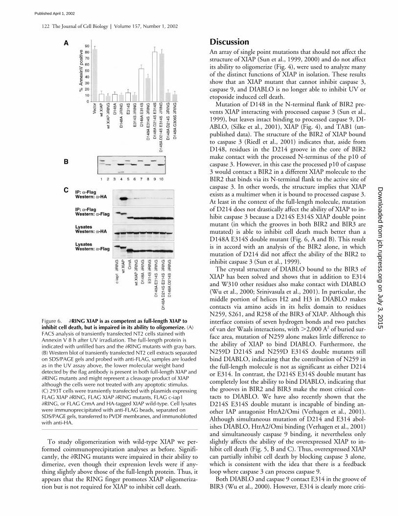
To study oligomerization with wild-type XIAP we per-formed coimmunoprecipitation analyses as before. Signifi-cantly, the
�
RING mutants were impaired in their ability todimerize, even though their expression levels were if any-thing slightly above those of the full-length protein. Thus, itappears that the RING finger promotes XIAP oligomeriza-tion but is not required for XIAP to inhibit cell death.
Discussion
An array of single point mutations that should not affect thestructure of XIAP (Sun et al., 1999, 2000) and do not affectits ability to oligomerize (Fig. 4), were used to analyze manyof the distinct functions of XIAP in isolation. These resultsshow that an XIAP mutant that cannot inhibit caspase 3,caspase 9, and DIABLO is no longer able to inhibit UV oretoposide induced cell death.
Mutation of D148 in the N-terminal flank of BIR2 pre-vents XIAP interacting with processed caspase 3 (Sun et al.,1999), but leaves intact binding to processed caspase 9, DI-ABLO, (Silke et al., 2001), XIAP (Fig. 4), and TAB1 (un-published data). The structure of the BIR2 of XIAP boundto caspase 3 (Riedl et al., 2001) indicates that, aside fromD148, residues in the D214 groove in the core of BIR2make contact with the processed N-terminus of the p10 ofcaspase 3. However, in this case the processed p10 of caspase3 would contact a BIR2 in a different XIAP molecule to theBIR2 that binds via its N-terminal flank to the active site ofcaspase 3. In other words, the structure implies that XIAPexists as a multimer when it is bound to processed caspase 3.At least in the context of the full-length molecule, mutationof D214 does not drastically affect the ability of XIAP to in-hibit caspase 3 because a D214S E314S XIAP double pointmutant (in which the grooves in both BIR2 and BIR3 aremutated) is able to inhibit cell death much better than aD148A E314S double mutant (Fig. 6, A and B). This resultis in accord with an analysis of the BIR2 alone, in whichmutation of D214 did not affect the ability of the BIR2 toinhibit caspase 3 (Sun et al., 1999).
The crystal structure of DIABLO bound to the BIR3 ofXIAP has been solved and shows that in addition to E314and W310 other residues also make contact with DIABLO(Wu et al., 2000; Srinivasula et al., 2001). In particular, themiddle portion of helices H2 and H3 in DIABLO makescontacts via amino acids in its helix domain to residuesN259, S261, and R258 of the BIR3 of XIAP. Although thisinterface consists of seven hydrogen bonds and two patchesof van der Waals interactions, with
�
2,000 A
2
of buried sur-face area, mutation of N259 alone makes little difference tothe ability of XIAP to bind DIABLO. Furthermore, theN259D D214S and N259D E314S double mutants stillbind DIABLO, indicating that the contribution of N259 inthe full-length molecule is not as significant as either D214or E314. In contrast, the D214S E314S double mutant hascompletely lost the ability to bind DIABLO, indicating thatthe grooves in BIR2 and BIR3 make the most critical con-tacts to DIABLO. We have also recently shown that theD214S E314S double mutant is incapable of binding an-other IAP antagonist HtrA2/Omi (Verhagen et al., 2001).Although simultaneous mutation of D214 and E314 abol-ishes DIABLO, HtrA2/Omi binding (Verhagen et al., 2001)and simultaneously caspase 9 binding, it nevertheless onlyslightly affects the ability of the overexpressed XIAP to in-hibit cell death (Fig. 5, B and C). Thus, overexpressed XIAPcan partially inhibit cell death by blocking caspase 3 alone,which is consistent with the idea that there is a feedbackloop where caspase 3 can process caspase 9.
Both DIABLO and caspase 9 contact E314 in the groove ofBIR3 (Wu et al., 2000). However, E314 is clearly more criti-
Figure 6. �RING XIAP is as competent as full-length XIAP toinhibit cell death, but is impaired in its ability to oligomerize. (A) FACS analysis of transiently transfected NT2 cells stained with Annexin V 8 h after UV irradiation. The full-length protein is indicated with unfilled bars and the �RING mutants with gray bars. (B) Western blot of transiently transfected NT2 cell extracts separated on SDS/PAGE gels and probed with anti-FLAG, samples are loaded as in the UV assay above, the lower molecular weight band detected by the flag antibody is present in both full-length XIAP and �RING mutants and might represent a cleavage product of XIAP although the cells were not treated with any apoptotic stimulus. (C) 293T cells were transiently transfected with plasmids expressing FLAG XIAP �RING, FLAG XIAP �RING mutants, FLAG c-iap1 �RING, or FLAG CrmA and HA-tagged XIAP wild-type. Cell lysates were immunoprecipitated with anti-FLAG beads, separated on SDS/PAGE gels, transferred to PVDF membranes, and immunoblotted with anti-HA.
on July 3, 2015jcb.rupress.org
Dow
nloaded from
Published April 1, 2002

XIAP interactions required to inhibit cell death |
Silke et al. 123
cal for caspase 9 binding because the E314S mutation alonecompletely abolishes the ability of XIAP to immunoprecipi-tate processed caspase 9, whereas it leaves some DIABLObinding intact (compare Figs. 2 and 3). G306 is also presentin the BIR3 groove and makes contacts to the fourth residue(Ile57) of the processed N-terminal of DIABLO (Liu et al.,2000; Wu et al., 2000). Mutation of Ile57 impairs DIABLON-terminal peptide binding to the BIR3 of XIAP, but is
�
100-fold less disruptive than mutation of the first residue ofprocessed DIABLO (Ala54). E314 and W310 of XIAP con-tact Ala54 of DIABLO, whereas G306 contacts Ile57, thus itis likely that the G306S XIAP binds DIABLO better than theE314S or W310A mutants (Liu et al., 2000). Furthermore,the G306S is likely to be more disruptive of the bulky Phe inthe fourth position of processed caspase 9 (Srinivasula et al.,2001) than the Ile of DIABLO or the Ala of HtrA2. H343Adoes not affect binding of the N-terminal peptide of DI-ABLO to the BIR3 of XIAP (Liu et al., 2000).
E314S and W310A mutations both simultaneously inter-fere with DIABLO and caspase 9 binding. In the UV deathassay, both the D148A E314S and D148A W310A E314Smutants still protect partially even though they have lost cas-pase 3 and caspase 9 binding and have reduced DIABLObinding. The protection the D148A E314S mutant affordscan be further increased by deleting the RING finger ofXIAP (Fig. 6 A) which increases the levels of XIAP in NT2cells (Fig. 6 B). This indicates that even residual DIABLObinding is sufficient to partially block apoptosis. The mu-tants G306S and H343A further confirm that DIABLObinding is sufficient for exogenous XIAP to block cell death.In these cases, either the double mutants D148A G306S,D148A H343A or the triple mutant D148A D214SH343A, which retain significant DIABLO binding, stillprotect NT2 cells as well as wild-type XIAP against UV-induced cell death.
Deletion of the RING finger has little effect on the abilityto inhibit UV-induced cell death. As with fly DIAP1 (Hay etal., 1995), removal of the RING finger makes XIAP a margin-ally more potent inhibitor of cell death, presumably due to itsincreased abundance. Contrary to OpIAP (Hozak et al.,2000), we find that the RING finger makes a strong contribu-tion to XIAPs ability to oligomerize and
�
RING c-iap1 doesnot interact at all. These results mean that oligomerization ofthe full-length protein is likely to be physiological, and at leastin the context of this assay, that the ability to oligomerize isnot required for XIAP to inhibit cell death.
Because an IAP that has lost the ability to bind caspase 3,caspase 9, DIABLO and HtrA2/Omi is ineffective at block-ing UV cell death, all of XIAPs anti-apoptotic activity mustreside in these functions. By selectively removing the abilityto inhibit caspase 3 and caspase 9, we have created severalXIAP mutants that can only bind IAP antagonists, and thesemutants still inhibit cell death as well as wild-type XIAP.The most likely mechanism for this inhibition is that exoge-nous expression of these mutants is sufficient to bind IAPantagonists that are released after a death stimulus such asDIABLO/smac and HtrA2/Omi, thereby allowing endoge-nous IAPs to block caspases without being antagonized. Asthere are several other IAP antagonists in addition to DI-ABLO and HtrA2/Omi (unpublished data), it might be dif-
ficult to prove the requirement for IAP antagonism byknock-out strategies alone. Therefore, these results demon-strate a requirement for IAP antagonism in cell death andindicate that the XIAP mutants described here will be usefultools in other similar analyses.
Materials and methods
Transfections and constructs
pEF expression constructs encoding C-terminal FLAG-tagged XIAP, N-ter-minally FLAG-tagged CrmA DQMD, caspase 3 LacZ, caspase 9, andpFLAG TAB1 have been previously described (Ekert et al., 1999; Verhagenet al., 2000; Silke et al., 2001). pEF TAB1 HA Tag was constructed by di-gesting the PCR product of primers 226 TAB1 5
�
cgggatccaccatggcggcgca-gaggaggag 3
�
and 227 TAB1 5
�
cgcgctagccggtgctgtcaccacgctctgct 3
�
with
Bam
H I,
Nhe
I and cloning into pEF HA Tag digested with
Bam
H I,
Nhe
I.pEF constructs encoding FLAG-tagged XIAP mutants were generated usingoverlap PCR technology. The D148A construct has already been described(Silke et al., 2001). The other mutants were generated with combina-tions of the primers 759
BstB
I 5
�
tgcttctttgttttgggccgga 3
�
, 760 E314S5
�
aactgattggaagcccagttccgacccttgggaacaacatgct 3
�
, 766 W310A 5
�
tggaggagggctaactgatgcgaagcccagtgaagacc 3
�
, 770
Eco
R V 5
�
ctgtctttctgag-cattcactagat 3
�
, 771 D214S 5
�
tgggaaccttgctcgagagcctggtcagaacacag 3
�
,and 772 N259D 5
�
tcaacaaatcttcctcgagatccatccatggcagatta 3
�
,
and in-serted into pEF FLAG XIAP digested with
Bst
B I and
Eco
R V. FLAG-taggedXIAP domain constructs were generated using PfuI polymerase PCR, andthe primers 743 BIR2 5
�
cgggatccaccatgcgtgatcattttgccttagacag 3
�
, 744BIR2 5
�
cgcgctagctggattcctaggaagatttgttgaatttggg 3
�
, 771 BIR1 5
�
cgggatccaccatgacttttaacagttttgaaggatcta 3
�
, 745 BIR1 5
�
ccgcc-ctaggggcaaaaagatctctgcttcccagatagtt 3
�
, 747 BIR3 5
�
ccgccctaggaatc-catccatggcagattat 3
�, 750 XIAP 5� cgtctagacataaaaattttttgcttgaa 3�, 761BIR3 5� cggaattcggatccaccatgtctgatgctgtgagttc 3�, and 762 BIR3 5�gcgttagctagcggagatctgagtagttcttaccagacact 3� and cloned into the vectorpEF c-term FLAG using sites compatible with BamH I and Nhe I. pEF XIAP�RING constructs were made using PCR primers 746 5� cgggatccaccat-gacttttaacagttttgaaggatcta 3� and 762 and cloned BamH I Nhe I into pEFC-terminal FLAG digested with BamH I Xba I. All constructs were verifiedby digest and sequencing.
Immunoprecipitations and Western blot analysisCells were lysed in a Triton X-100 based lysis buffer (1% Triton X-100,10% glycerol, 150 mM NaCl, 20 mM Tris, pH 7.5, 2 mM EDTA, 1 mMPMSF, 10 �g/ml aprotinin and 10 �g/ml leupeptin) for 1 h at 4�C and thedebris cleared by centrifugation. Immunoprecipitations were performedusing FLAG specific mAb M2 covalently coupled agarose beads (Sigma-Aldrich). The immunoprecipitates were washed five times in lysis bufferand proteins eluted with 100 mM glycine, pH 3, and then neutralized with1/10th vol of 1 M Tris, pH 8. Proteins were separated by SDS PAGE andimmunoprecipitates were examined by Western blot analysis after transferof proteins to Immobilon™-P membranes (Millipore). Antibodies used forWestern blots were anti-FLAG M2 (Sigma-Aldrich), anti-HA High Affinity3F10 (Boehringer Mannheim), anti–caspase 9, a gift of Y. Lazebnik, andanti–caspase 3 (Pharmingen). Proteins were visualized by ECL (AmershamPharmacia Biotech) after incubation of membranes with HRP-coupled sec-ondary antibodies.
Apoptosis assaysThe cell death assay is described in detail in Silke et al. (2001). In brief,NT2 cells were transfected with equal amounts of the mutant constructsand 1:20 (wt/wt) of pEGFP for 24 h. The cells were washed and induced toundergo apoptosis by irradiating with 25 J/m2 of UV and harvested 6–7 hlater, or treating with etoposide (100 �g/ml) and harvesting 20–24 h later.All cells, both adherent and nonadherent, were recovered and stained withrecombinant Annexin V-Biotin and streptavidin Tricolor (CalTag Laborato-ries) with appropriate washing steps. The stained cells were analysed witha FACs scan for FL1 fluorescence (EGFP positive) and FL3 fluorescence(Annexin V positive) and the proportion of cells that were EGFP and An-nexin V positive (transfected and apoptotic), to the total of EGFP positive(transfected) cells was determined.
SoftwareAn EPSON scanner was used to scan all autoradiographs and the scannedimages were imported directly into FreeHand9 and cropped withFreehand9.
on July 3, 2015jcb.rupress.org
Dow
nloaded from
Published April 1, 2002

124 The Journal of Cell Biology | Volume 157, Number 1, 2002
We wish to thank Kuni Matsumoto (Nagoya University, Nagoya, Japan) forthe gift of pFLAG TAB1, Yuri Lazebnik for the caspase 9 antibody, AndreasStrasser and David Huang (WEMI, Victoria, Australia) for helpful discus-sions and reagents, and Pascal Meier for discussions and proofreading.
This project was supported by the National Health and Medical Re-search Council (Regkey 973002). D.L. Vaux is a Scholar of the Leukaemiaand Lymphoma Society, and P.G. Ekert is funded by Murdoch Children’sResearch Institute.
Submitted: 17 August 2001Revised: 13 February 2002Accepted: 25 February 2002
ReferencesChai, J., C. Du, J.W. Wu, S. Kyin, X. Wang, and Y. Shi. 2000. Structural and bio-
chemical basis of apoptotic activation by Smac/DIABLO. Nature. 406:855–862.
Chai, J., E. Shiozaki, S.M. Srinivasula, Q. Wu, P. Dataa, E.S. Alnemri, and Y. Shi.2001. Structural basis of caspase-7 inhibition by XIAP. Cell. 104:769–780.
Deveraux, Q.L., E. Leo, H.R. Stennicke, K. Welsh, G.S. Salvesen, and J.C. Reed.1999. Cleavage of human inhibitor of apoptosis protein XIAP results infragments with distinct specificities for caspases. EMBO J. 18:5242–5251.
Deveraux, Q.L., and J.C. Reed. 1999. IAP family proteins—suppressors of apopto-sis. Genes Dev. 13:239–252.
Deveraux, Q.L., R. Takahashi, G.S. Salvesen, and J.C. Reed. 1997. X-linked IAP isa direct inhibitor of cell-death proteases. Nature. 388:300–304.
Ekert, P.G., J. Silke, and D.L. Vaux. 1999. Inhibition of apoptosis and clonogenicsurvival of cells expressing crmA variants: optimal caspase substrates are notnecessarily optimal inhibitors. EMBO J. 18:330–338.
Ekert, P.G., J. Silke, C.J. Hawkins, A.M. Verhagen, and D.L. Vaux. 2001. DI-ABLO promotes apoptosis by removing MIHA/XIAP from processed cas-pase 9. J. Cell Biol. 152:483–490.
Goyal, L., K. McCall, J. Agapite, E. Hartwieg, and H. Steller. 2000. Induction ofapoptosis by Drosophila Reaper, Hid and Grim through inhibition of IAPfunction. EMBO J. 19:589–597.
Hay, B.A., D.A. Wassarman, and G.M. Rubin. 1995. Drosophila homologs of bac-ulovirus inhibitor of apoptosis proteins function to block cell death. Cell. 83:1253–1262.
Hozak, R.R., G.A. Manji, and P.D. Friesen. 2000. The BIR motifs mediate domi-nant interference and oligomerization of inhibitor of apoptosis Op-IAP.Mol. Cell. Biol. 20:1877–1885.
Huang, Y., Y.C. Park, R.L. Rich, D. Segal, D.G. Myszka, and H. Wu. 2001.Structural basis of caspase inhibition by XIAP. Differential roles of the linkerversus the BIR domain. Cell. 104:781–790.
Liu, Z.H., C.H. Sun, E.T. Olejniczak, R.P. Meadows, S.F. Betz, T. Oost, J. Herr-mann, J.C. Wu, and S.W. Fesik. 2000. Structural basis for binding ofSmac/DIABLO to the XIAP BIR3 domain. Nature. 408:1004–1008.
Riedl, S.J., M. Renatus, R. Schwarzenbacher, Q. Zhou, C. Sun, S.W. Fesik, R.C.Liddington, and G.S. Salvesen. 2001. Structural basis for the inhibition ofcaspase-3 by XIAP. Cell. 104:791–800.
Rothe, M., M.G. Pan, W.J. Henzel, T.M. Ayres, and D.V. Goeddel. 1995. The
TNF-R2-TRAF signaling complex contains two novel proteins related tobaculoviral-inhibitor of apoptosis proteins. Cell. 83:1243–1252.
Sanna, M.G., C.S. Duckett, B. Richter, C.B. Thompson, and R.J. Ulevitch. 1998.Selective activation of jnk1 is necessary for the anti-apoptotic activity of hilp.Proc. Natl. Acad. Sci. USA. 95:6015–6020.
Silke, J., and D.L. Vaux. 2001. Two kinds of BIR-containing protein-inhibitors ofapoptosis, or required for mitosis. J. Cell Sci. 114:1821–1827.
Silke, J., P.G. Ekert, C.L. Day, C.J. Hawkins, M. Baca, J. Chew, M. Pakusch,A.M. Verhagen, and D.L. Vaux. 2001. Direct inhibition of caspase 3 is dis-pensable for the anti-apoptotic activity of XIAP. EMBO J. 20:3114–3123.
Srinivasula, S.M., P. Datta, X.J. Fan, T. Fernandes-Alnemri, Z. Huang, and E.S.Alnemri. 2000. Molecular determinants of the caspase-promoting activity ofSmac/DIABLO and its role in the death receptor pathway. J. Biol. Chem.275:36152–36157.
Srinivasula, S.M., R. Hegde, A. Saleh, P. Datta, E. Shiozaki, J. Chai, R.A. Lee,P.D. Robbins, T. Fernandes-Alnemri, Y. Shi, and E.S. Alnemri. 2001. Aconserved XIAP-interaction motif in caspase-9 and Smac/DIABLO regulatescaspase activity and apoptosis. Nature. 410:112–116.
Sun, C., M. Cai, A.H. Gunasekera, R.P. Meadows, H. Wang, J. Chen, H. Zhang,W. Wu, N. Xu, S.C. Ng, and S.W. Fesik. 1999. NMR structure and mu-tagenesis of the inhibitor-of-apoptosis protein XIAP. Nature. 401:818–821.
Sun, C., M. Cai, R.P. Meadows, N. Xu, A.H. Gunasekera, J. Herrmann, J.C. Wu,and S.W. Fesik. 2000. NMR structure and mutagenesis of the third BIR do-main of the inhibitor of apoptosis protein XIAP. J. Biol. Chem. 275:33777–33781.
Suzuki, Y., Y. Nakabayashi, and R. Takahashi. 2001. Ubiquitin-protein ligase ac-tivity of X-linked inhibitor of apoptosis protein promotes proteasomal deg-radation of caspase-3 and enhances its anti-apoptotic effect in Fas-inducedcell death. Proc. Natl. Acad. Sci. USA. 98:8662–8667.
Takahashi, R., Q. Deveraux, I. Tamm, K. Welsh, N. Assamunt, G.S. Salvesen, andJ.C. Reed. 1998. A single BIR domain of XIAP sufficient for inhibitingcaspases. J. Biol. Chem. 273:7787–7790.
Verhagen, A.M., P.G. Ekert, M. Pakusch, J. Silke, L.M. Connolly, G.E. Reid, R.L.Moritz, R.J. Simpson, and D.L. Vaux. 2000. Identification of DIABLO, amammalian protein that promotes apoptosis by binding to and antagonizingIAP proteins. Cell. 102:42–53.
Verhagen, A.M., J. Silke, P.G. Ekert, M. Pakusch, H. Kaufmann, L.M. Connolly,C.L. Day, A. Tikoo, R. Burke, C. Wrobel, et al. 2001. HtrA2 promotes celldeath through its serine protease activity and its ability to antagonize inhibi-tor of apoptosis proteins. J. Biol. Chem. 277:445–454.
Wu, G., J.J. Chai, T.L. Suber, J.W. Wu, C.Y. Du, X.D. Wang, and Y.G. Shi.2000. Structural basis of IAP recognition by Smac/DIABLO. Nature. 408:1008–1012.
Yamaguchi, K., S. Nagai, J. Ninomiya-Tsuji, M. Nishita, K. Tamai, K. Irie, N.Ueno, E. Nishida, H. Shibuya, and K. Matsumoto. 1999. XIAP, a cellularmember of the inhibitor of apoptosis protein family, links the receptors toTAB1-TAK1 in the bone morphogenetic protein signaling pathway. EMBOJ. 18:179–187.
Yang, Y., S. Fang, J.P. Jensen, A.M. Weissman, and J.D. Ashwell. 2000. Ubiquitinprotein ligase activity of IAPs and their degradation in proteasomes in re-sponse to apoptotic stimuli. Science. 288:874–877.
on July 3, 2015jcb.rupress.org
Dow
nloaded from
Published April 1, 2002
Copyright © 2022 FDOKUMEN