7-Bromoindirubin-3′-oxime induces caspase-independent cell death
15
ORIGINAL ARTICLE 7-Bromoindirubin-3 0 -oxime induces caspase-independent cell death J Ribas 1,2,6 , K Bettayeb 1,6 , Y Ferandin 1 , M Knockaert 1 , X Garrofe´-Ochoa 2 , F Totzke 3 , C Scha¨ chtele 3 , J Mester 4 , P Polychronopoulos 5 , P Magiatis 5 , A-L Skaltsounis 5 , J Boix 2 and L Meijer 1 1 C.N.R.S., Cell Cycle Group and UPS2682, Station Biologique, Bretagne, France; 2 Molecular Pharmacology Laboratory, DCMB, School of Medicine, University of Lleida, Lleida, Catalunya, Spain; 3 ProQinase GmbH, Freiburg, Germany; 4 INSERM U 482, 184, rue du Faubourg Saint Antoine, Paris Cedex, France and 5 Department of Pharmacy, Division of Pharmacognosy and Natural Products Chemistry, University of Athens, Panepistimiopolis Zografou, Athens, Greece Indirubin, an isomer of indigo, is a reported inhibitor of cyclin-dependent kinases (CDKs) and glycogen synthase kinase-3 (GSK-3) as well as an agonist of the aryl hydrocarbon receptor (AhR). Indirubin is the active ingredient of a traditional Chinese medicinal recipe used against chronic myelocytic leukemia. Numerous indirubin analogs have been synthesized to optimize this promising kinase inhibitor scaffold. We report here on the cellular effects of 7-bromoindirubin-3 0 -oxime (7BIO). In contrast to its 5-bromo- and 6-bromo- isomers, and to indirubin-3 0 - oxime, 7BIO has only a marginal inhibitory activity towards CDKs and GSK-3. Unexpectedly, 7BIO triggers a rapid cell death process distinct from apoptosis. 7-Bromoindirubin-3 0 -oxime induces the appearance of large pycnotic nuclei, without classical features of apoptosis such as chromatin condensation and nuclear fragmentation. 7-Bromoindirubin-3 0 -oxime-induced cell death is not accompanied by cytochrome c release neither by any measurable effector caspase activation. Furthermore, the death process is not altered either by the presence of Q-VD-OPh, a broad-spectrum caspase inhibitor, or the overexpression of Bcl-2 and Bcl-XL proteins. Neither AhR nor p53 is required during 7BIO- induced cell death. Thus, in contrast to previously described indirubins, 7BIO triggers the activation of non-apoptotic cell death, possibly through necroptosis or autophagy. Although their molecular targets remain to be identified, 7-substituted indirubins may constitute a new class of potential antitumor compounds that would retain their activity in cells refractory to apoptosis. Oncogene (2006) 25, 6304–6318. doi:10.1038/sj.onc.1209648; published online 15 May 2006 Keywords: indirubin; protein kinase; cell death; autophagy; caspase; cancer Introduction In man, 518 þ protein kinases and 80 þ protein phosphatases control the phosphorylation of enzymes and structural proteins. Phosphorylation/dephosphory- lation constitutes one of the most common yet complex post-translational cellular regulatory mechanism. Abnormal phosphorylation on specific proteins is observed in essentially all pathologies and this has stimulated an extraordinary interest in small molecular weight inhibitors of kinases and phosphatases. Protein kinases now constitute the second class of targets (after G-protein-coupled receptors) used in the drug screening efforts of the pharmaceutical industry (reviews in Cohen, 2002; Fischer, 2004; Weinmann and Metternich, 2005). These efforts have received considerable support from the remarkable success story of Gleevec, one of the first commercial kinase inhibitors. Currently, 55 kinase inhibitors are under clinical evaluation against diseases such as cancers, inflammation, diabetes, neurodegeneration, etc. Cyclin-dependent kinases (CDKs) play essential functions all along the cell cycle and there are multiple examples of dysfunctions of CDKs and their regulators in cancer (Vermeulen et al., 2003). Furthermore, CDKs are involved in various neurodegenerative diseases such as Alzheimer’s, Parkinson’s and Nieman-Pick’s diseases, ischemia and traumatic brain injury. A few academic groups, and most pharmaceutical companies, have thus invested in the search for pharmacological inhibitors of CDKs (reviews in Knockaert et al., 2002; Fischer et al., 2003; Benson et al., 2005; Fischer and Gianella- Borradori, 2005). Optimization of such inhibitors has been efficiently assisted by their co-crystallization with CDK2 (Noble et al., 2004), CDK5 (Mapelli et al., 2005) and CDK6 (Lu et al., 2005). The bis-indole alkaloid indirubin and its analogs (collectively referred to as indirubins) were among some of the early CDK inhibitors to be discovered (Hoessel et al., 1999). The red/purple indirubin is an isomer of the blue indigo. Both are derived from the spontaneous, non-enzymatic dimerization of isatin and indoxyl, two precursors found either free or conjugated to carbohy- drates in natural sources. Indirubins can indeed be extracted from various indigo dye-producing plants Received 7 December 2005; revised 27 March 2006; accepted 27 March 2006; published online 15 May 2006 Correspondence: Dr L Meijer, C.N.R.S., Cell Cycle Group and UPS2682, Station Biologique, B.P. 74, 29682 Roscoff cedex, Bretagne, France. E-mail: [email protected] 6 These two authors contributed equally to this work. Oncogene (2006) 25, 6304–6318 & 2006 Nature Publishing Group All rights reserved 0950-9232/06 $30.00 www.nature.com/onc
-
Upload
damiavericat -
Category
Documents
-
view
1 -
download
0
Transcript of 7-Bromoindirubin-3′-oxime induces caspase-independent cell death

ORIGINAL ARTICLE
7-Bromoindirubin-30-oxime induces caspase-independent cell death
J Ribas1,2,6, K Bettayeb1,6, Y Ferandin1, M Knockaert1, X Garrofe-Ochoa2, F Totzke3,C Schachtele3, J Mester4, P Polychronopoulos5, P Magiatis5, A-L Skaltsounis5, J Boix2 and LMeijer1
1C.N.R.S., Cell Cycle Group and UPS2682, Station Biologique, Bretagne, France; 2Molecular Pharmacology Laboratory, DCMB,School of Medicine, University of Lleida, Lleida, Catalunya, Spain; 3ProQinase GmbH, Freiburg, Germany; 4INSERM U 482, 184,rue du Faubourg Saint Antoine, Paris Cedex, France and 5Department of Pharmacy, Division of Pharmacognosy and NaturalProducts Chemistry, University of Athens, Panepistimiopolis Zografou, Athens, Greece
Indirubin, an isomer of indigo, is a reported inhibitor ofcyclin-dependent kinases (CDKs) and glycogen synthasekinase-3 (GSK-3) as well as an agonist of the arylhydrocarbon receptor (AhR). Indirubin is the activeingredient of a traditional Chinese medicinal recipe usedagainst chronic myelocytic leukemia. Numerous indirubinanalogs have been synthesized to optimize this promisingkinase inhibitor scaffold. We report here on the cellulareffects of 7-bromoindirubin-30-oxime (7BIO). In contrastto its 5-bromo- and 6-bromo- isomers, and to indirubin-30-oxime, 7BIO has only a marginal inhibitory activitytowards CDKs and GSK-3. Unexpectedly, 7BIO triggersa rapid cell death process distinct from apoptosis.7-Bromoindirubin-30-oxime induces the appearanceof large pycnotic nuclei, without classical features ofapoptosis such as chromatin condensation and nuclearfragmentation. 7-Bromoindirubin-30-oxime-induced celldeath is not accompanied by cytochrome c releaseneither by any measurable effector caspase activation.Furthermore, the death process is not altered either bythe presence of Q-VD-OPh, a broad-spectrum caspaseinhibitor, or the overexpression of Bcl-2 and Bcl-XLproteins. Neither AhR nor p53 is required during 7BIO-induced cell death. Thus, in contrast to previouslydescribed indirubins, 7BIO triggers the activation ofnon-apoptotic cell death, possibly through necroptosis orautophagy. Although their molecular targets remain to beidentified, 7-substituted indirubins may constitute a newclass of potential antitumor compounds that would retaintheir activity in cells refractory to apoptosis.Oncogene (2006) 25, 6304–6318. doi:10.1038/sj.onc.1209648;published online 15 May 2006
Keywords: indirubin; protein kinase; cell death;autophagy; caspase; cancer
Introduction
In man, 518þ protein kinases and 80þ proteinphosphatases control the phosphorylation of enzymesand structural proteins. Phosphorylation/dephosphory-lation constitutes one of the most common yet complexpost-translational cellular regulatory mechanism.Abnormal phosphorylation on specific proteins isobserved in essentially all pathologies and this hasstimulated an extraordinary interest in small molecularweight inhibitors of kinases and phosphatases. Proteinkinases now constitute the second class of targets (afterG-protein-coupled receptors) used in the drug screeningefforts of the pharmaceutical industry (reviews inCohen, 2002; Fischer, 2004; Weinmann and Metternich,2005). These efforts have received considerable supportfrom the remarkable success story of Gleevec, oneof the first commercial kinase inhibitors. Currently,55 kinase inhibitors are under clinical evaluationagainst diseases such as cancers, inflammation, diabetes,neurodegeneration, etc.Cyclin-dependent kinases (CDKs) play essential
functions all along the cell cycle and there are multipleexamples of dysfunctions of CDKs and their regulatorsin cancer (Vermeulen et al., 2003). Furthermore, CDKsare involved in various neurodegenerative diseases suchas Alzheimer’s, Parkinson’s and Nieman-Pick’s diseases,ischemia and traumatic brain injury. A few academicgroups, and most pharmaceutical companies, have thusinvested in the search for pharmacological inhibitorsof CDKs (reviews in Knockaert et al., 2002; Fischeret al., 2003; Benson et al., 2005; Fischer and Gianella-Borradori, 2005). Optimization of such inhibitors hasbeen efficiently assisted by their co-crystallization withCDK2 (Noble et al., 2004), CDK5 (Mapelli et al., 2005)and CDK6 (Lu et al., 2005).The bis-indole alkaloid indirubin and its analogs
(collectively referred to as indirubins) were among someof the early CDK inhibitors to be discovered (Hoesselet al., 1999). The red/purple indirubin is an isomer of theblue indigo. Both are derived from the spontaneous,non-enzymatic dimerization of isatin and indoxyl, twoprecursors found either free or conjugated to carbohy-drates in natural sources. Indirubins can indeed beextracted from various indigo dye-producing plants
Received 7 December 2005; revised 27 March 2006; accepted 27 March2006; published online 15 May 2006
Correspondence: Dr L Meijer, C.N.R.S., Cell Cycle Group andUPS2682, Station Biologique, B.P. 74, 29682 Roscoff cedex, Bretagne,France.E-mail: [email protected] two authors contributed equally to this work.
Oncogene (2006) 25, 6304–6318& 2006 Nature Publishing Group All rights reserved 0950-9232/06 $30.00
www.nature.com/onc
(200þ species) (Balfour-Paul, 1998). They are alsopresent in the historic ‘Tyrean purple’ dye extractedfrom various Muricidae mollusks (15þ species) (Meijeret al., 2003; review in Cooksey, 2001). They are alsoextracted from various wild-type and recombinantbacteria (Guengerich et al., 2004; Wu et al., 2005 andreferences therein). Finally, indirubin and indigo areoccasionally present in human urine (Adachi et al., 2001and references therein). Interestingly, indirubin is theactive ingredient of a traditional Chinese medicinerecipe, Danggui Longhui Wan, used to treat variousdiseases including chronic myelocytic leukemia (reviewin Xiao et al., 2002). Besides CDKs, indirubins werefound to target glycogen synthase kinase-3 (GSK-3)(Leclerc et al., 2001), glycogen phosphorylase b (Kos-mopoulou et al., 2004) and the aryl hydrocarbonreceptor (AhR), also known as the dioxin receptor(Adachi et al., 2001; Kawanishi et al., 2003). Arylhydrocarbon receptor mediates the effects of manyxenobiotics such as dioxin and indole-containing com-pounds (review in Denison and Nagy, 2003). Uponbinding to xenobiotic-responsive element, activatedAhR induces the transcription of numerous genes,including cytochrome P450 CYP1A1, p27kip1, myristoyl-transferase, etc. (review in Elferink, 2003). Evidencesuggests that the antiproliferative effects of indirubinsderive from their ability to inhibit CDKs (Damienset al., 2001; Marko et al., 2001). However, interactionwith AhR and the subsequent induction of p27kip1
contributes to a marked arrest in G1 (Knockaertet al., 2004). Finally it has been recently shown thatsome indirubins prevent the phosphorylation andsubsequent activation of the transcription factor signaltransducers and activators of transcription (STAT3),leading to a downregulation of survival factors such as
survivin and Mcl-1, and subsequent induction of celldeath (Nam et al., 2005).In this article, we report on a new subfamily of
indirubins, substituted on position 7. Unexpectedly,despite weak or insignificant inhibitory activityon various classical kinase targets of indirubins,7-bromoindirubin-30-oxime potently induces a rapid celldeath distinct from classical apoptosis. The possiblemechanisms of action of 7-substituted indirubins andtheir potential as antitumor agents will be discussed.
Results
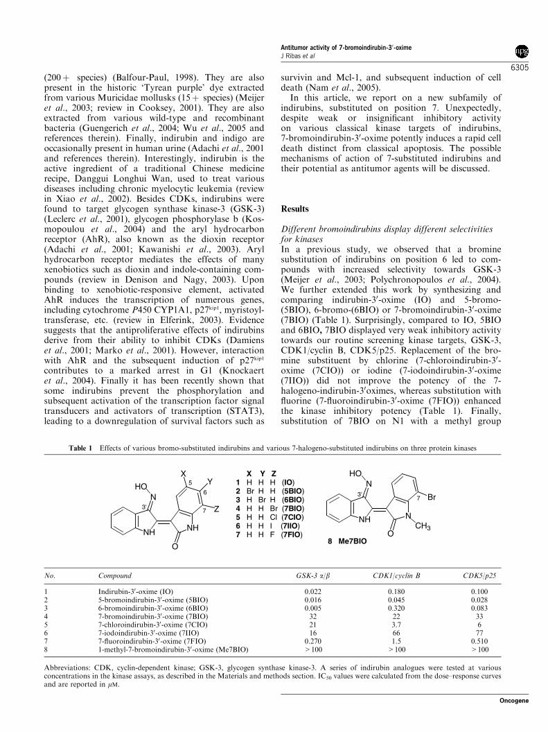
Different bromoindirubins display different selectivitiesfor kinasesIn a previous study, we observed that a brominesubstitution of indirubins on position 6 led to com-pounds with increased selectivity towards GSK-3(Meijer et al., 2003; Polychronopoulos et al., 2004).We further extended this work by synthesizing andcomparing indirubin-30-oxime (IO) and 5-bromo-(5BIO), 6-bromo-(6BIO) or 7-bromoindirubin-30-oxime(7BIO) (Table 1). Surprisingly, compared to IO, 5BIOand 6BIO, 7BIO displayed very weak inhibitory activitytowards our routine screening kinase targets, GSK-3,CDK1/cyclin B, CDK5/p25. Replacement of the bro-mine substituent by chlorine (7-chloroindirubin-30-oxime (7CIO)) or iodine (7-iodoindirubin-30-oxime(7IIO)) did not improve the potency of the 7-halogeno-indirubin-30oximes, whereas substitution withfluorine (7-fluoroindirubin-30-oxime (7FIO)) enhancedthe kinase inhibitory potency (Table 1). Finally,substitution of 7BIO on N1 with a methyl group
Table 1 Effects of various bromo-substituted indirubins and various 7-halogeno-substituted indirubins on three protein kinases
NH
N
NH
O
HO 5X Y Z
1 H H H (IO)2 Br H H (5BIO)3 H Br H (6BIO)4 H H Br (7BIO)5 H H Cl (7CIO)6 H H I (7IIO)7 H H F (7FIO)
3' Z
XY
6
7
NH
N
N
O
HO
8 Me7BIO
3'7 Br
CH3
No. Compound GSK-3 a/b CDK1/cyclin B CDK5/p25
1 Indirubin-30-oxime (IO) 0.022 0.180 0.1002 5-bromoindirubin-30-oxime (5BIO) 0.016 0.045 0.0283 6-bromoindirubin-30-oxime (6BIO) 0.005 0.320 0.0834 7-bromoindirubin-30-oxime (7BIO) 32 22 335 7-chloroindirubin-30-oxime (7CIO) 21 3.7 66 7-iodoindirubin-30-oxime (7IIO) 16 66 777 7-fluoroindirubin-30-oxime (7FIO) 0.270 1.5 0.5108 1-methyl-7-bromoindirubin-30-oxime (Me7BIO) >100 >100 >100
Abbreviations: CDK, cyclin-dependent kinase; GSK-3, glycogen synthase kinase-3. A series of indirubin analogues were tested at variousconcentrations in the kinase assays, as described in the Materials and methods section. IC50 values were calculated from the dose–response curvesand are reported in mM.
Antitumor activity of 7-bromoindirubin-30-oximeJ Ribas et al
6305
Oncogene

(1-methyl-7-bromoindirubin-30-oxime (Me7BIO)) led toa completely inactive compound, as observed previouslywith Me6BIO (Meijer et al., 2003).We next investigated the selectivity of IO, 5BIO, 6BIO
and 7BIO in the 85 kinase ProQinase selectivity panel(Table 2) (in contrast to the above-mentioned kinaseassays performed at a final 15 mM ATP concentration,the ProQinase assays are run in the absence of coldATP. As indirubins act by competing with ATP binding,the IC50 values are dependent on the assay ATPconcentration, therefore the values of Tables 1 and 2cannot be compared directly). This approach firstrevealed that Aurora A-C, FMS-like tyrosine kinase 3(FLT3), RET constitute new targets of IO, 5BIO and6BIO. Vascular endothelial growth factor receptor(VEGF-R) had been previously described as a targetfor indirubins (Jautelat et al., 2005). The selectivitypanel also revealed that, compared to the three otherindirubins, 7BIO is a poor kinase inhibitor. Only onekinase, FLT3, was inhibited by 7BIO with an IC50 below1 mM (Table 2) (15 kinases for IO, 11 for 5BIO, 19 for6BIO). 7-Br-, 7Cl- and 7I-substituted indirubin-30-oximes showed a significant inhibitory activity towardsAurora C, a modest activity on Aurora B and littleactivity, if any, on Aurora A (data not shown), whereas7FIO appeared to be equipotent on the three Auroraforms. Unexpectedly, Me7BIO was found to be ratheractive on Aurora C, but completely inactive on AuroraA and B (data not shown). FMS-like tyrosine kinase 3was found to be sensitive to all eight indirubins testedincluding Me7BIO. The effects of indirubins on Aurorakinases and FLT3 will be reported in detail elsewhere.
Induction of cell death by indirubinsWe next compared the four indirubins for their abilityto induce cell death in neuroblastoma SH-SY5Y cellsas measured with an 3-(4,5-dimethylthiazol-2-yl)-5-(3-carboxymethoxyphenyl)-2-(4-sulfophenyl)-2H-tetrazolium
KINASE IO 5BIO 6BIO 7BIO ABL1 >100 >100 3.4 >100 AKT1 >100 >100 >100 >100 AKT2 >100 >100 >100 >100 AKT3 n.t. n.t. >100 >100
Aurora-A 0.20 0.23 0.43 55 Aurora-B 1.4 35 1.5 4.7 Aurora-C 0.57 34 2.3 6.6
BRK 17 >100 4.6 33 CDK1/CycB 20 2.1 21 >100 CDK2/CycA 2.3 0.46 0.71 >100 CDK2/CycE 2.2 0.12 0.12 >100 CDK3/CycE 0.15 0.04 0.07 >100
CDK4/CycD1 0.56 1.33 0.80 >100 CDK6/CycD1 0.19 0.05 0.17 >100
CHK1 35 >100 12 >100 CK2 >100 >100 >100 >100 COT n.t. n.t. 75 49 CSK >100 >100 n.t. n.t.
DAPK1 >100 >100 n.t. n.t.EGF-R 102 >100 9.1 27 EPHA1 53 >100 38 44 EPHB1 56 >100 n.t. n.t.EPHB2 6.1 >100 5.0 33 EPHB3 >100 >100 34 28 EPHB4 8.6 >100 3.5 10.0 ERBB2 >100 >100 >100 26 ERBB4 >100 >100 >100 >100
FAK 0.99 >100 12 78 FGF-R1 0.74 0.83 0.43 36 FGF-R3 0.38 0.85 0.42 23 FGF-R4 13 65 1.2 17
FGR 1.6 >100 0.09 20 FLT3 0.07 0.02 0.20 0.34
GSK3-beta 2.5 0.07 0.21 >100 IGF1-R 5.3 5.6 79 26
IKK-beta >100 >100 >100 >100 INS-R 6.6 4.3 >100 >100 IRAK4 1.8 44 4.8 82 JAK2 33 >100 >100 >100 JNK3 25 >100 n.t. n.t.KIT 4.3 16 0.76 58 LCK 41 >100 3.1 >100 MET >100 >100 >100 >100 MST4 73 >100 n.t. n.t.MUSK 0.21 31 0.50 >100 NEK2 >100 >100 >100 >100 NEK6 >100 >100 >100 >100 NLK >100 >100 >100 >100 PAK1 13 11 n.t. n.t.PAK2 >100 >100 n.t. n.t.PAK4 >100 91 n.t. n.t.PBK >100 >100 >100 >100
PCTAIRE1 >100 >100 >100 >100 PCTAIRE2 n.t. n.t. 15 >100
PDGFR-alpha 4.0 9.5 0.52 47 PDGFR-beta 3.4 3.4 0.88 >100
PIM1 >100 >100 91 >100 PIM2 n.t. n.t. >100 >100
PKC-alpha >100 >100 >100 >100 PKC-beta1 >100 >100 >100 >100 PKC-beta2 >100 >100 >100 >100 PKC-delta >100 >100 >100 65
PKC-epsilon >100 >100 >100 >100 PKC-eta >100 >100 >100 >100
PKC-gamma >100 >100 >100 >100 PKC-iota >100 >100 >100 >100 PKC-mu >100 >100 >100 >100
PKC-theta >100 >100 86 >100 PKC-zeta >100 >100 >100 96
PLK1 >100 >100 >100 >100 PRK1 >100 >100 70 >100 RET 0.18 2.2 0.05 22 S6K 82 >100 6.1 >100
Table 2 ProQinase selectivity profile of IO, 5BIO, 6BIO and 7BIO KINASE IO 5BIO 6BIO 7BIO SGK1 0.14 1.6 n.t. n.t.SGK3 38 >100 5.8 >100 SNK >100 >100 >100 >100 SRC 1.5 17 0.13 27 SYK 3.0 >100 6.2 85 TIE2 11 68 13 27 TSF1 0.87 13 2.9 3.3 TSK2 >100 >100 >100 >100
VEGF-R1 1.6 1.6 3.2 >100 VEGF-R2 0.19 0.25 0.56 23 VEGF-R3 0.09 0.08 0.22 11
WEE1 n.t. n.t. >100 >100
Abbreviations: 5BIO, 5-bromoindirubin-30-oxime; 6BIO, 6-bromoin-dirubin-30-oxime; 7BIO, 7-bromoindirubin-30-oxime; CDK, cyclin-dependent kinase; IO, indirubin-30-oxime; NT, not tested. Thefour indirubins were tested at various concentrations in 85 kinaseassays, as described in the Materials and methods section. IC50values, calculated from the dose–response curves, are reportedin mM and underlined according to a gray scale code:
IC50 value (µM) < 0.1 0.1 - 1 1 - 10 10 - 100 > 100 .
Antitumor activity of 7-bromoindirubin-30-oximeJ Ribas et al
6306
Oncogene
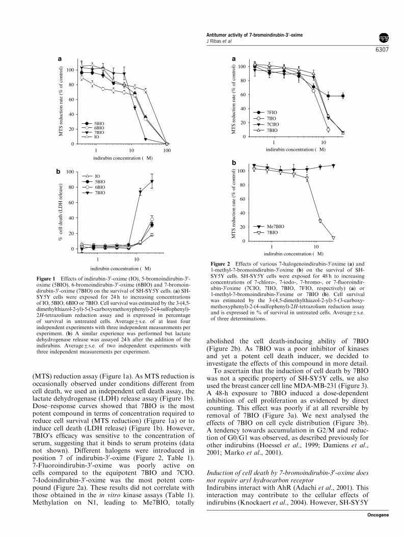
(MTS) reduction assay (Figure 1a). As MTS reduction isoccasionally observed under conditions different fromcell death, we used an independent cell death assay, thelactate dehydrogenase (LDH) release assay (Figure 1b).Dose–response curves showed that 7BIO is the mostpotent compound in terms of concentration required toreduce cell survival (MTS reduction) (Figure 1a) or toinduce cell death (LDH release) (Figure 1b). However,7BIO’s efficacy was sensitive to the concentration ofserum, suggesting that it binds to serum proteins (datanot shown). Different halogens were introduced inposition 7 of indirubin-30-oxime (Figure 2, Table 1).7-Fluoroindirubin-30-oxime was poorly active oncells compared to the equipotent 7BIO and 7CIO.7-Iodoindirubin-30-oxime was the most potent com-pound (Figure 2a). These results did not correlate withthose obtained in the in vitro kinase assays (Table 1).Methylation on N1, leading to Me7BIO, totally
abolished the cell death-inducing ability of 7BIO(Figure 2b). As 7BIO was a poor inhibitor of kinasesand yet a potent cell death inducer, we decided toinvestigate the effects of this compound in more detail.To ascertain that the induction of cell death by 7BIO
was not a specific property of SH-SY5Y cells, we alsoused the breast cancer cell line MDA-MB-231 (Figure 3).A 48-h exposure to 7BIO induced a dose-dependentinhibition of cell proliferation as evidenced by directcounting. This effect was poorly if at all reversible byremoval of 7BIO (Figure 3a). We next analysed theeffects of 7BIO on cell cycle distribution (Figure 3b).A tendency towards accumulation in G2/M and reduc-tion of G0/G1 was observed, as described previously forother indirubins (Hoessel et al., 1999; Damiens et al.,2001; Marko et al., 2001).
Induction of cell death by 7-bromoindirubin-30-oxime doesnot require aryl hydrocarbon receptorIndirubins interact with AhR (Adachi et al., 2001). Thisinteraction may contribute to the cellular effects ofindirubins (Knockaert et al., 2004). However, SH-SY5Y
indirubin concentration (µM)
1 10
% c
ell d
eath
(L
DH
rel
ease
)
0
20
40
60
80
100IO 5BIO 6BIO 7BIO
indirubin concentration (µM)
1 10 100
MT
S re
duct
ion
rate
(%
of
cont
rol)
0
20
40
60
80
100
5BIO6BIO 7BIO IO
a
b
Figure 1 Effects of indirubin-30-oxime (IO), 5-bromoindirubin-30-oxime (5BIO), 6-bromoindirubin-30-oxime (6BIO) and 7-bromoin-dirubin-30-oxime (7BIO) on the survival of SH-SY5Y cells. (a) SH-SY5Y cells were exposed for 24 h to increasing concentrationsof IO, 5BIO, 6BIO or 7BIO. Cell survival was estimated by the 3-(4,5-dimethylthiazol-2-yl)-5-(3-carboxymethoxyphenyl)-2-(4-sulfophenyl)-2H-tetrazolium reduction assay and is expressed in percentageof survival in untreated cells. Average7s.e. of at least fourindependent experiments with three independent measurements perexperiment. (b) A similar experience was performed but lactatedehydrogenase release was assayed 24 h after the addition of theindirubins. Average7s.e. of two independent experiments withthree independent measurements per experiment.
0
20
40
60
80
100
0
20
40
60
80
100
7FIO 7IIO 7ClIO 7BIO M
TS
redu
ctio
n ra
te (
% o
f co
ntro
l)M
TS
redu
ctio
n ra
te (
% o
f co
ntro
l)
1 10
Me7BIO 7BIO
indirubin concentration (µM)
1 10indirubin concentration (µM)
a
b
Figure 2 Effects of various 7-halogenoindirubin-30oxime (a) and1-methyl-7-bromoindirubin-30oxime (b) on the survival of SH-SY5Y cells. SH-SY5Y cells were exposed for 48 h to increasingconcentrations of 7-chloro-, 7-iodo-, 7-bromo-, or 7-fluoroindir-ubin-30oxime (7CIO, 7IIO, 7BIO, 7FIO, respectively) (a) or1-methyl-7-bromoindirubin-30oxime or 7BIO (b). Cell survivalwas estimated by the 3-(4,5-dimethylthiazol-2-yl)-5-(3-carboxy-methoxyphenyl)-2-(4-sulfophenyl)-2H-tetrazolium reduction assayand is expressed in % of survival in untreated cells. Average7s.e.of three determinations.
Antitumor activity of 7-bromoindirubin-30-oximeJ Ribas et al
6307
Oncogene

cells seem to be devoid of AhR (unpublished data). Toevaluate the contribution of AhR to the cell deatheffects of 7BIO, we made use of two hepatoma cell lines,5L (AhR þ /þ ) and its AhR-deficient subclone, BP8(AhR �/�) (Weiss et al., 1996; Kolluri et al., 1999;Knockaert et al., 2004). We first confirmed that, like2,3,7,8-tetrachlorodibenzo-p-dioxin (TCDD) (dioxin),both 7BIO and Me7BIO potently enhance the AhR-dependent expression of the CDK inhibitory proteinp27Kip1 (Figure 4a), as reported previously for IO and6BIO and their methylated counterparts, 1-methyl-indirubin-30-oxime (MeIO) and Me6BIO (Knockaertet al., 2004). No correlation is thus observed betweeninduction of p27Kip1 expression (Figure 4a) and induc-tion of cell death (Figure 2b). We next analysed theeffects of 7BIO and Me7BIO on cell cycle distribution ofAhR�/� and AhRþ /þ cells. As reported for otherindirubins, both 7BIO and Me7BIO induced a strikingAhR-dependent accumulation in G0/G1 (Figure 4b).Finally, cell death induction was estimated in both celllines following exposure to increased 7BIO concentra-tions. The dose–response curves were essentially thesame (Figure 4c). Altogether these results show thatAhR is not involved in the cell death inducing propertiesof 7BIO.
7-Bromoindirubin-30-oxime-induced cell death involvesneither p53 nor p21Cip1 nor STAT3 dephosphorylationWe next investigated the involvement of p53 and p21Cip1
in cell death induced by the four indirubins (Figure 5).P53 was strongly induced by 5BIO in a time-dependentmanner in SH-SY5Y cells (Figure 5a and b). Inductionof p53 was only modest in cells treated with 6BIO and
7BIO concentration (µM)
0 5 10 15 20
cell
cycl
e ph
ase
disr
ibut
ion
(% to
tal)
0
10
20
30
40
50
G0/G1
G2/M
sub G0/G1
S
b
Time (h)
-24 0 24 48 72 96
Cel
l num
ber
(x 1
000)
0
100
200
300
400
5 µM
0 µM
20 µM
10 µM
a
Figure 3 Effects of 7-bromoindirubin-30-oxime (7BIO) on cellproliferation and cell cycle distribution in MDA-MB-231 cells.(a) Cells were exposed at time 0 to various concentrations of 7BIOand cell numbers were determined at various times. At 48 h, theculture medium was replaced by fresh medium devoid of 7BIO.(b) Cells were exposed to various concentrations of 7BIO for 24 hand their distribution in the various cell cycle phases wasdetermined by fluorescence-activated cell sorter analysis.
0
20
40
60
80
100G0/G1SG2/M
5L BP8 5L BP8 5L BP8 5L BP8DMSO 7BIO Me7BIOcontrol
p27Kip1
actin
5LBP85LBP8
5LBP85LBP8
DMSOTCDD
7BIOMe7BIO
a
7BIO concentration (µM)
0 10 20 30 40 50 60
MT
T r
educ
tion
rate
(%
of
cont
rol)
0
20
40
60
80
100
5L (AhR +/+)BP8 (AhR -/-)
b
c
Figure 4 The cytotoxic effect of 7-bromoindirubin-30-oxime(7BIO) is independent of aryl hydrocarbon receptor (AhR).(a) Hepatocyte AhR�/� (BP8) and AhRþ /þ (5L) cells weretreated with 0.1mM 2,3,7,8-tetrachlorodibenzo-p-dioxin, or 10mM7BIO or 1-methyl-7-bromoindirubin-30-oxime (Me7BIO) for 24 hor with the vehicle dimethylsulfoxide (DMSO). The expressionlevel of p27KIP1 was determined by Western blotting using aspecific antibody. Actin Western blotting was used as a loadingcontrol. (b) Both 7BIO and Me7BIO induce an AhR-dependentaccumulation in G0/G1. 5L and BP8 cells were cultured in theabsence (control) or presence of DMSO or 10mM 7BIO or Me7BIOfor 24 h, and the cell cycle phase distribution was determinedby fluorescence-activated cell sorter analysis. (c) Both 5L and BP8cell lines were exposed for 24 h to increasing concentrations of7BIO or Me7BIO. Cell survival was estimated by the 3-[4,5]dimethylthiazol-2-5-diphenyltetrazolium bromide reduction assayand is expressed in % of survival in untreated cells. Average7s.e.of three determinations.
Antitumor activity of 7-bromoindirubin-30-oximeJ Ribas et al
6308
Oncogene

insignificant in cells treated with IO, 7BIO or Me7BIO(Figure 5a and b). As expected, analysis of p21Cip1
expression under the same conditions showed a time-dependent induction by 5BIO (Figure 5c). p21Cip1
expression occurred with some delay after p53 stabiliza-tion (Figure 5b). Indirubin-30-oxime, 5BIO and 6BIOwere roughly equipotent at inducing p21Cip1overexpres-sion, whereas 7BIO and Me7BIO had negligible effects(Figure 5a). Finally, we tested the effects of 7BIO onwild-type HCT-116 and a HCT-116 subclone deprivedof p53 (Figure 5e). The dose–response curves wereessentially the same. Altogether, these data suggest that7BIO-induced cell death does not induce p53 nor requireits contribution.Tyrosine phosphorylation and subsequent activation
of the transcription factor STAT3 were recently shownto be inhibited by some indirubins, leading to thedownregulation of survival factors and subsequentinduction of cell death (Nam et al., 2005). To examinewhether this mechanism is involved in the action of7BIO, we investigated the effect from IO, 5BIO, 6BIOand 7BIO on the level of tyrosine 705-phosphorylatedSTAT3 in MDA-MB-231 cells (Figure 6). As a positivecontrol, cells were also stimulated by interferon a(IFNa). Results show that the basal level of tyrosine705-phosphorylated STAT3 in MDA-MB-231 is very
a
p53
7BIOMe7BIO
Staurosporin
e
Etoposide
control
IO 5BIO6BIO
p21
actin
CIP1
b
p53
12 24 Et.240 3 6 9
5BIO
p537BIO
Time (h)
d
p21CIP1
p21CIP1
5BIO
7BIO
12 24 Et.240 3 6 9Time (h)c
actin
actin5BIO
7BIO
12 24 Et.240 3 6 9Time (h)
7BIO concentration (µM)
1 10
MT
S r
educ
tion
rat
e (%
of
cont
rol)
0
20
40
60
80
100
WT p53-/-
eFigure 5 7-Bromoindirubin-30-oxime (7BIO) does not induce norrequire p53 nor p21CIP1 expression. (a) SH-SY5Y cells were treatedwith 12.5mM indirubin-30-oxime (IO), 5-bromoindirubin-30-oxime(5BIO), 6-bromoindirubin-30-oxime (6BIO), 7-bromoindirubin-30-oxime (7BIO) or 1-methyl-7-bromoindirubin-30-oxime (Me7BIO),1mM staurosporine or 12.5mM etoposide for 12h. Cells were thenharvested and proteins were resolved by sodium dodecyl sulfate–polyacrylamide gel electrophoresis (SDS–PAGE) followed by Wes-tern blotting using antibodies directed against p53, p21CIP1 or actin(used as internal loading marker). (b–d) SH-SY5Y cells were treatedwith 12.5mM 5BIO or 7BIO or 12.5mM etoposide for various times.Cells were then harvested and proteins were resolved by SDS–PAGEfollowed by Western blotting using antibodies directed against p53(b), p21CIP1 (c) or actin (d). (e) Wild-type (�) and p53-deprived (J)HCT-116 cells were exposed for 48h to increasing concentrations of7BIO or Me7BIO. Cell survival was estimated by the 3-(4,5-dimethylthiazol-2-yl)-5-(3-carboxymethoxyphenyl)-2-(4-sulfophenyl)-2H-tetrazolium reduction assay and is expressed in % of survival inuntreated cells. Average7s.e. of three determinations.
P-STAT3
6BIO7BIO
IFN DMSO
IO 5BIO
actin
STAT3
Control
Figure 6 7-Bromoindirubin-30-oxime (7BIO) effects do notinvolve downregulation of signal transducers and activators oftranscription (STAT3) tyrosine phosphorylation. MDA-MB-231cells were either untreated or treated with 25mM indirubin-30-oxime(IO), 5-bromoindirubin-30-oxime (5BIO), 6-bromoindirubin-30-oxime (6BIO), 7-bromoindirubin-30-oxime (7BIO), or the dimethyl-sulfoxide carrier for 4 h, or with 100 ng/ml of interferon-a for 5min.Cellular proteins were resolved by sodium dodecyl sulfate–polyacrylamide gel electrophoresis followed by Western blottingusing antibodies directed total STAT3 and tyrosine-phosphory-lated STAT3. Western blotting with antiactin antibodies provideda loading marker.
Antitumor activity of 7-bromoindirubin-30-oximeJ Ribas et al
6309
Oncogene

low compared to the level reached by stimulation withIFNa, yet it is downregulated by IO, 5BIO and 6BIObut not by 7BIO. This suggests that the mechanism ofaction of 7BIO is not primarily due to an inactivation oftyrosine-phosphorylated STAT3.
Induction of cell death by 7-bromoindirubin-30-oximeis much faster than by other indirubinsA time course of SH-SY5Y cell death induction wasnext performed following exposure to 25 mM IO, 5BIO,6BIO, 7BIO or Me7BIO (Figure 7). Although 5BIO and6BIO required 36–48 h to induce 70% cell death, thislevel was reached by 12 h with 7BIO. Almost completecell death was obtained with 7BIO within 24 h (Figure 7).This much faster kinetics suggests that a differentmechanism of cell death is occurring in the case of7BIO compared to the other indirubins. Alternatively, asubpopulation of cells may respond to 5BIO and 6BIOas they do to 7BIO, whereas the vast majority undergoesapoptosis.
7-Bromoindirubin-30-oxime induces non-apoptotic celldeathTo investigate the mechanism of action of 7BIO, we firstexamined, under a fluorescence microscope, SH-SY5Ycells exposed to different indirubins following Bisbenzi-mide and propidium iodide (PI) staining (Figure 8).First of all, no PI staining was observed in control cellsand in Me7BIO-treated cells (Figure 8a and f),confirming the absence of cell death. Indirubin-30-oxime,5BIO and 6BIO all triggered nuclear fragmentationtypical of apoptosis, accompanied by secondary necrosis(Figure 8b–d). These figures were never observed in7BIO-treated cells that, in contrast, displayed numerous
large, unfragmented pycnotic nuclei (Figure 8e). Suchfigures were observed only occasionally with 5BIO and6BIO (Figure 8c and d). These morphological resultssuggest that 7BIO triggers an atypical cell deathdifferent from apoptosis.To challenge this possibility, the activity of caspases
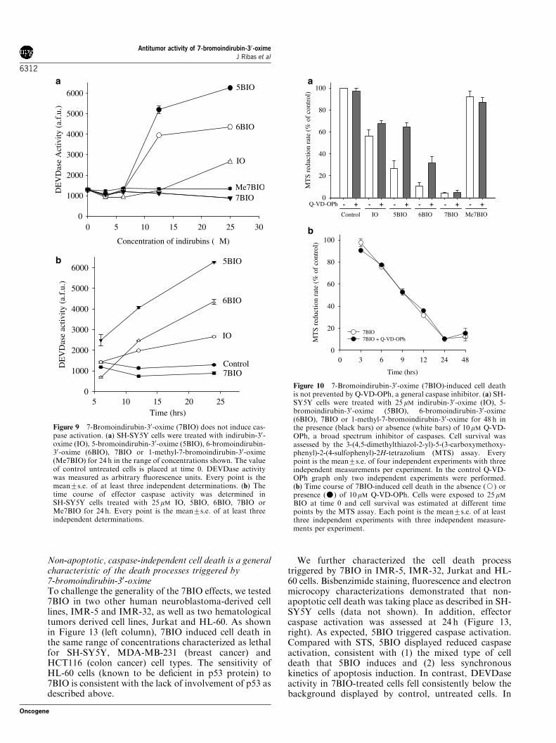
was assayed in SH-SY5Y cells exposed to various con-centrations of different indirubins (Figure 9). 5-bromo-indirubin-30-oxime and 6BIO and IO to a lesser extent,triggered a dose- (Figure 9a) and time- (Figure 9b)dependent activation of caspase. In sharp contrast,neither 7BIO nor Me7BIO induced any activation ofcaspases, which remained at the level of control,untreated cells. Furthermore, Q-VD-OPh, a generalcaspase inhibitor (Caserta et al., 2003), had no effect oncell death induced by 7BIO (Figure 10), whereas itreduced the level of cell death induced by 5BIO and6BIO, and IO, to a lesser extent (Figure 10a). The timecourse of 7BIO-induced cell death was unaffected byQ-VD-OPh (Figure 10b).Moreover, 7BIO triggered negligible release of cyto-
chrome c from mitochondria (Figure 11). Under thesame conditions IO, 5BIO and 6BIO induced the releaseof cytochrome c to levels similar to those reached bystandard apoptosis-inducing reagents like staurosporine(STS) and etoposide. We next examined DNA ladderingas a reflection of apoptotic cell death. The ladderingcaused by R-Roscovitine was consistent with thereported ability of this compound to induce apoptosis(Ribas and Boix, 2004). 5-Bromoindirubin-30-oxime andto a lesser extent 6BIO, also induced internuclesosomalfragmentation, whose intensity was proportional to theamount of apoptotic cells in the culture (see Bisbenzi-mide/PI staining in Figure 8). In 7BIO-treated cells, noladder was observed; however, most cells were dead. InMe7BIO, IO and dimethylsulfoxide (DMSO) treat-ments, cell death induction was negligible and noladdering was detected.Altogether these results show that 7BIO-induced cell
death does not induce cytochrome c release and does nottrigger nor require the activation of caspases, in sharpcontrast with cell death induced by IO, 5BIO and 6BIO.Thus 7BIO appears to induce a cell death pathwaywhich differs from the apoptosis induced by IO, 5BIOand 6BIO.
7-Bromoindirubin-30-oxime-induced cell death is notinhibited by cellular mechanisms able to protect cells fromapoptosisTo further explore the cell death process triggeredby 7BIO, we wondered whether proved mechanismsof resistance to apoptosis were able to protect cellsfrom 7BIO’s effects. SH-SY5Y cells can be differen-tiated in cell culture by retinoic acid (RA) and thisdifferentiation prevents apoptosis triggered by CDKinhibitors like olomoucine or roscovitine (Ribas andBoix, 2004). Similarly, differentiation renders SH-SY5Ycells refractory to STS. As shown in Figure 12,differentiation had negligible effect on the rates of7BIO-induced cell death.
Time (hrs)
0 10 20 30 40 50 60
MT
S re
duct
ion
rate
(%
of
cont
rol)
0
20
40
60
80
100
120
Me7BIO
IO
5BIO
6BIO7BIO
Figure 7 7-Bromoindirubin-30-oxime (7BIO) induces cell deathmuch faster than other indirubins. SH-SY5Y cells were treatedwith 25 mM indirubin-30-oxime (IO), 5-bromoindirubin-30-oxime(5BIO), 6-bromoindirubin-30-oxime (6BIO), 7BIO or 1-methyl-7-bromoindirubin-30-oxime (Me7BIO) for 6, 12, 24, 36 or 48 h. Cellsurvival was assessed by the 3-(4,5-dimethylthiazol-2-yl)-5-(3-carboxymethoxyphenyl)-2-(4-sulfophenyl)-2H-tetrazolium proce-dure. Every point is the mean7s.e. of two independent experimentswith at least three independent measurements per experiment.
Antitumor activity of 7-bromoindirubin-30-oximeJ Ribas et al
6310
Oncogene

Bcl-2 and Bcl-XL proteins are known for theirantiapoptotic effects. In addition, their cytoprotec-tive effects have been found to extend beyond apop-tosis (Kane et al., 1995). We previously describedthat Bcl-2 and Bcl-XL overexpression protectsSH-SY5Y cells from apoptosis triggered by STS(Yuste et al., 2002). As reported, Bcl-XL surpassed
Bcl-2 at inhibiting STS-induced apoptosis (Figure 12b).However, in a parallel time course experiment,neither Bcl-XL nor Bcl-2 overexpression provided anysignificant protection from 7BIO (Figure 12b). Takentogether, these results reinforce the action of 7BIOas an effective cell killer acting in an apoptosis-independent manner.
Figure 8 In contrast to indirubin-30-oxime (IO), 5-bromoindirubin-30-oxime (5BIO) and 6-bromoindirubin-30-oxime (6BIO),7-bromoindirubin-30-oxime (7BIO) induces non-apoptotic cell death in SH-SY5Y cells. SH-SY5Y cells were exposed for 24 h to 0.1%dimethylsulfoxide (control) (a), 25 mM IO (b), 25 mM 5BIO (c), 10mM 6BIO (d), 10 mM 7BIO (e) or 10 mM 1-methyl-7-bromoindirubin-30-oxime (Me7BIO) (f). Following double staining of DNA with Bisbenzimide and propidium iodide, cells were examined by fluorescencemicroscopy. Thick arrows: apoptosis (nuclear fragmentation); thin arrows: secondary necrosis; arrowheads: pycnotic nuclei. Scalebar: 20mm.
Antitumor activity of 7-bromoindirubin-30-oximeJ Ribas et al
6311
Oncogene
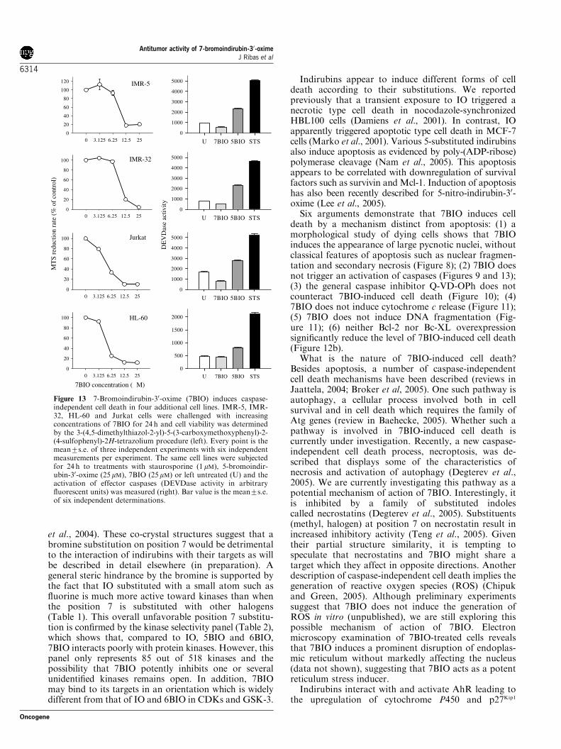
Non-apoptotic, caspase-independent cell death is a generalcharacteristic of the death processes triggered by7-bromoindirubin-30-oximeTo challenge the generality of the 7BIO effects, we tested7BIO in two other human neuroblastoma-derived celllines, IMR-5 and IMR-32, as well as two hematologicaltumors derived cell lines, Jurkat and HL-60. As shownin Figure 13 (left column), 7BIO induced cell death inthe same range of concentrations characterized as lethalfor SH-SY5Y, MDA-MB-231 (breast cancer) andHCT116 (colon cancer) cell types. The sensitivity ofHL-60 cells (known to be deficient in p53 protein) to7BIO is consistent with the lack of involvement of p53 asdescribed above.
We further characterized the cell death processtriggered by 7BIO in IMR-5, IMR-32, Jurkat and HL-60 cells. Bisbenzimide staining, fluorescence and electronmicrocopy characterizations demonstrated that non-apoptotic cell death was taking place as described in SH-SY5Y cells (data not shown). In addition, effectorcaspase activation was assessed at 24 h (Figure 13,right). As expected, 5BIO triggered caspase activation.Compared with STS, 5BIO displayed reduced caspaseactivation, consistent with (1) the mixed type of celldeath that 5BIO induces and (2) less synchronouskinetics of apoptosis induction. In contrast, DEVDaseactivity in 7BIO-treated cells fell consistently below thebackground displayed by control, untreated cells. In
Concentration of indirubins (µM)
0 5 10 15 20 25
5 10 15 20 25
30
DE
VD
ase
Act
ivity
(a.
f.u.
)
0
1000
2000
3000
4000
5000
6000
IO
5BIO
6BIO
7BIOMe7BIO
Time (hrs)
DE
VD
ase
activ
ity (
a.f.
u.)
0
1000
2000
3000
4000
5000
6000
Control
IO
5BIO
6BIO
7BIO
a
b
Figure 9 7-Bromoindirubin-30-oxime (7BIO) does not induce cas-pase activation. (a) SH-SY5Y cells were treated with indirubin-30-oxime (IO), 5-bromoindirubin-30-oxime (5BIO), 6-bromoindirubin-30-oxime (6BIO), 7BIO or 1-methyl-7-bromoindirubin-30-oxime(Me7BIO) for 24 h in the range of concentrations shown. The valueof control untreated cells is placed at time 0. DEVDase activitywas measured as arbitrary fluorescence units. Every point is themean7s.e. of at least three independent determinations. (b) Thetime course of effector caspase activity was determined inSH-SY5Y cells treated with 25 mM IO, 5BIO, 6BIO, 7BIO orMe7BIO for 24 h. Every point is the mean7s.e. of at least threeindependent determinations.
MT
S re
duct
ion
rate
(%
of
cont
rol)
0
20
40
60
80
100
Q-VD-OPh - + - + - + - + - + - +
Control IO 5BIO 6BIO 7BIO Me7BIO
Time (hrs)
3 6 9 12 24 48
MT
S re
duct
ion
rate
(%
of
cont
rol)
0
20
40
60
80
100
7BIO7BIO + Q-VD-OPh
0
a
b
Figure 10 7-Bromoindirubin-30-oxime (7BIO)-induced cell deathis not prevented by Q-VD-OPh, a general caspase inhibitor. (a) SH-SY5Y cells were treated with 25mM indirubin-30-oxime (IO), 5-bromoindirubin-30-oxime (5BIO), 6-bromoindirubin-30-oxime(6BIO), 7BIO or 1-methyl-7-bromoindirubin-30-oxime for 48 h inthe presence (black bars) or absence (white bars) of 10 mM Q-VD-OPh, a broad spectrum inhibitor of caspases. Cell survival wasassessed by the 3-(4,5-dimethylthiazol-2-yl)-5-(3-carboxymethoxy-phenyl)-2-(4-sulfophenyl)-2H-tetrazolium (MTS) assay. Everypoint is the mean7s.e. of four independent experiments with threeindependent measurements per experiment. In the control Q-VD-OPh graph only two independent experiments were performed.(b) Time course of 7BIO-induced cell death in the absence (J) orpresence (�) of 10mM Q-VD-OPh. Cells were exposed to 25mMBIO at time 0 and cell survival was estimated at different timepoints by the MTS assay. Each point is the mean7s.e. of at leastthree independent experiments with three independent measure-ments per experiment.
Antitumor activity of 7-bromoindirubin-30-oximeJ Ribas et al
6312
Oncogene

conclusion, the non-apoptotic, caspase-independenttype of cell death triggered by 7BIO appears to be anintrinsic property of the compound, independent of thecell model.
Discussion
Indirubins have been the object of many chemicalmodulations in order to improve their selectivity profile,kinase inhibitory efficacy and solubility. In particular,numerous modifications have been carried out onpositions 30, 50, 60 on one indole ring and 5 or 6 on theother indole ring (Hoessel et al., 1999; Leclerc et al.,2001; Meijer et al., 2003; Merz et al., 2004; Polychro-nopoulos et al., 2004; Jautelat et al., 2005). However, nomodifications have ever been reported on position 7.One reason might be that substitution at this position
almost annihilates inhibitory activity towards keytargets of indirubins, CDKs and GSK-3. The fast andpotent induction of cell death by 7BIO was totallyunexpected, but provided the impetus to investigate7-substituted indirubins further. Indirubins have beenco-crystallized with CDK2 (Hoessel et al., 1999),CDK2/cyclin A (Davies et al., 2001), CDK5 (Mapelliet al., 2005), PfPK5, the Plasmodium falciparum homo-log of CDK1 (Holton et al., 2003), GSK-3b (Bertrandet al., 2003; Meijer et al., 2003; Polychronopoulos et al.,2004), and glycogen phosphorylase b (Kosmopoulou
internalmarker
7BIOMe7BIO
Staurosporin
e
Etoposide
control
IO 5BIO6BIO
cytochrome C
7BIOMe7BIO
Roscovitin
e
DMSOIO 5BIO
6BIO
a
b
Figure 11 Indirubin-30-oxime (IO), 5-bromoindirubin-30-oxime(5BIO) and 6-bromoindirubin-30-oxime (6BIO), but not 7-bro-moindirubin-30-oxime (7BIO), induce cytochrome c release andDNA laddering. (a) SH-SY5Y cells were treated with 12.5mMIO, 5BIO, 6BIO, 7BIO or 1-methyl-7-bromoindirubin-30-oxime(Me7BIO), 0.25mM staurosporine or 12.5mM etoposide for 10 h.Cells were then harvested and fractionated into a nuclear pellet anda cytoplasmic supernatant. The latter was resolved by sodiumdodecyl sulfate–polyacrylamide gel electrophoresis followed byWestern blotting using an anticytochrome c antibody. Theantibody crossreacts with an irrelevant protein used as an internalloading marker. (b) SH-SY5Y cells were treated with dimethylsulf-oxide (0.25%), 25mM IO, 5BIO, 6BIO, 7BIO or Me7BIO, or 25 mM(R)-roscovitine for 24 h. Cells were then harvested and internu-cleosomal DNA fragmentation was analysed by electrophoresis in1.5% agarose gels.
0
20
40
60
MT
S re
duct
ion
rate
(% o
f co
ntro
l)
a
7BIOSTS Roscovitine
Bcl-2Bcl-XL
1 2 3 1 2 3
Tubulin Tubulin
c0 6 12 18 24
0
20
40
60
80
1000 6 12 18 24
0
20
40
60
80
100
MT
S re
duct
ion
rate
(%
of
cont
rol)
(hrs)
b
7BIO
STS
(hrs)
Bcl-XL
Bcl-XL
Bcl-2
Bcl-2control
control
Figure 12 In contrast to staurosporine-induced apoptosis,7-bromoindirubin-30-oxime (7BIO)-induced cell death is resistantto the protective effects of cell differentiation (a) or Bcl-2/Bcl-XLoverexpression (b, c). (a) SH-SY5Y cells were either treated withretinoic acid (RA) during 5 days to induce quiescence anddifferentiation (white bars) or kept proliferating (black bars). After24 h of treatment with STS (1mM), 7BIO (25mM) or racemicRoscovitine (50 mM), cell viability was determined by the 3-(4,5-dimethylthiazol-2-yl)-5-(3-carboxymethoxyphenyl)-2-(4-sulfophe-nyl)-2H-tetrazolium (MTS) procedure. Bar value is the mean7 s.e.of at least six independent determinations. (B) SH-SY5Y cells,permanently transfected with the vectors pcDNA3/Bcl-XL (�),pcDNA3/Bcl-2 (J) and empty pcDNA3 (’), were treated witheither STS (2 mM) or 7BIO (25mM). Cell viability was analysed bythe MTS procedure at 9 and 24 h of treatment. In the time courseplots, every point is the mean7s.e. of three independent experi-ments with six independent values per experiment. (c) The Bcl-XLand Bcl-2 content of pcDNA3/Bcl-2 (1), pcDNA3/Bcl-XL (2) andpcDNA3/empty (3) transfected SH-SY5Y cells was assessed duringthe viability determination experiments by Western blotting.Tubulin content was used to control for protein load.
Antitumor activity of 7-bromoindirubin-30-oximeJ Ribas et al
6313
Oncogene
et al., 2004). These co-crystal structures suggest that abromine substitution on position 7 would be detrimentalto the interaction of indirubins with their targets as willbe described in detail elsewhere (in preparation). Ageneral steric hindrance by the bromine is supported bythe fact that IO substituted with a small atom such asfluorine is much more active toward kinases than whenthe position 7 is substituted with other halogens(Table 1). This overall unfavorable position 7 substitu-tion is confirmed by the kinase selectivity panel (Table 2),which shows that, compared to IO, 5BIO and 6BIO,7BIO interacts poorly with protein kinases. However, thispanel only represents 85 out of 518 kinases and thepossibility that 7BIO potently inhibits one or severalunidentified kinases remains open. In addition, 7BIOmay bind to its targets in an orientation which is widelydifferent from that of IO and 6BIO in CDKs and GSK-3.
Indirubins appear to induce different forms of celldeath according to their substitutions. We reportedpreviously that a transient exposure to IO triggered anecrotic type cell death in nocodazole-synchronizedHBL100 cells (Damiens et al., 2001). In contrast, IOapparently triggered apoptotic type cell death in MCF-7cells (Marko et al., 2001). Various 5-substituted indirubinsalso induce apoptosis as evidenced by poly-(ADP-ribose)polymerase cleavage (Nam et al., 2005). This apoptosisappears to be correlated with downregulation of survivalfactors such as survivin and Mcl-1. Induction of apoptosishas also been recently described for 5-nitro-indirubin-30-oxime (Lee et al., 2005).Six arguments demonstrate that 7BIO induces cell
death by a mechanism distinct from apoptosis: (1) amorphological study of dying cells shows that 7BIOinduces the appearance of large pycnotic nuclei, withoutclassical features of apoptosis such as nuclear fragmen-tation and secondary necrosis (Figure 8); (2) 7BIO doesnot trigger an activation of caspases (Figures 9 and 13);(3) the general caspase inhibitor Q-VD-OPh does notcounteract 7BIO-induced cell death (Figure 10); (4)7BIO does not induce cytochrome c release (Figure 11);(5) 7BIO does not induce DNA fragmentation (Fig-ure 11); (6) neither Bcl-2 nor Bc-XL overexpressionsignificantly reduce the level of 7BIO-induced cell death(Figure 12b).What is the nature of 7BIO-induced cell death?
Besides apoptosis, a number of caspase-independentcell death mechanisms have been described (reviews inJaattela, 2004; Broker et al, 2005). One such pathway isautophagy, a cellular process involved both in cellsurvival and in cell death which requires the family ofAtg genes (review in Baehecke, 2005). Whether such apathway is involved in 7BIO-induced cell death iscurrently under investigation. Recently, a new caspase-independent cell death process, necroptosis, was de-scribed that displays some of the characteristics ofnecrosis and activation of autophagy (Degterev et al.,2005). We are currently investigating this pathway as apotential mechanism of action of 7BIO. Interestingly, itis inhibited by a family of substituted indolescalled necrostatins (Degterev et al., 2005). Substituents(methyl, halogen) at position 7 on necrostatin result inincreased inhibitory activity (Teng et al., 2005). Giventheir partial structure similarity, it is tempting tospeculate that necrostatins and 7BIO might share atarget which they affect in opposite directions. Anotherdescription of caspase-independent cell death implies thegeneration of reactive oxygen species (ROS) (Chipukand Green, 2005). Although preliminary experimentssuggest that 7BIO does not induce the generation ofROS in vitro (unpublished), we are still exploring thispossible mechanism of action of 7BIO. Electronmicroscopy examination of 7BIO-treated cells revealsthat 7BIO induces a prominent disruption of endoplas-mic reticulum without markedly affecting the nucleus(data not shown), suggesting that 7BIO acts as a potentreticulum stress inducer.Indirubins interact with and activate AhR leading to
the upregulation of cytochrome P450 and p27Kip1
U 7BIO 5BIO STS
0
1000
2000
3000
4000
5000
0 3.125 6.25 12.5 250
20
40
60
80
100
120 IMR-5
DE
VD
ase
activ
ity
MT
S re
duct
ion
rate
(%
of
cont
rol)
0 3.125 6.25 12.5 250
20
40
60
80
100
U 7BIO 5BIO STS
0
1000
2000
3000
4000
5000IMR-32
U 7BIO 5BIO STS
0
1000
2000
3000
4000
5000
0 3.125 6.25 12.5 250
20
40
60
80
100 Jurkat
U 7BIO 5BIO STS
0
500
1000
1500
2000
0 3.125 6.25 12.5 250
20
40
60
80
100
7BIO concentration (µM)
HL-60
Figure 13 7-Bromoindirubin-30-oxime (7BIO) induces caspase-independent cell death in four additional cell lines. IMR-5, IMR-32, HL-60 and Jurkat cells were challenged with increasingconcentrations of 7BIO for 24 h and cell viability was determinedby the 3-(4,5-dimethylthiazol-2-yl)-5-(3-carboxymethoxyphenyl)-2-(4-sulfophenyl)-2H-tetrazolium procedure (left). Every point is themean7s.e. of three independent experiments with six independentmeasurements per experiment. The same cell lines were subjectedfor 24 h to treatments with staurosporine (1mM), 5-bromoindir-ubin-30-oxime (25mM), 7BIO (25mM) or left untreated (U) and theactivation of effector caspases (DEVDase activity in arbitraryfluorescent units) was measured (right). Bar value is the mean7s.e.of six independent determinations.
Antitumor activity of 7-bromoindirubin-30-oximeJ Ribas et al
6314
Oncogene
(Adachi et al., 2001; Spink et al., 2003; Knockaert et al.,2004; Sugihara et al., 2004). 7-Bromoindirubin-30-oximeclearly activates AhR as observed in an in vitroluciferase reporter system (unpublished), and confirmedby the AhR-dependent expression of p27Kip1 and G1arrest (Figure 4). However, three pieces of evidencesuggest that AhR is not involved in the cell deathprocess induced by 7BIO: (1) 7BIO triggers cell deathequally well in AhR�/� and AhRþ /þ cells; (2)neuroblastoma SH-SY5Y cells express very little if anyAhR (unpublished); (3) Me7BIO, which is as potent as7BIO in activating AhR, does not induce cell death.7-Bromoindirubin-30-oxime inhibits Aurora C and
FLT-3 (Table 2 and unpublished data). Yet Me7BIO,which is as potent as 7BIO at inhibiting both kinases,does not trigger cell death (Figure 2b). 7-Bromoindir-ubin-30-oxime-induced cell death is therefore unlikely tobe a direct consequence of inhibition of these twokinases. We also believe that 7BIO action does notinvolve the downregulation of STAT3 tyrosine phos-phorylation and its subsequent inactivation as has beensuggested for other indirubins (Nam et al., 2005)(Figure 11).Although it is too early to speculate on the potential
clinical use of 7-substituted indirubins, this new familyof indirubins opens a promising research area. Firstly,these molecules seem to interact with much less kinasesthan previously described indirubins and thus appearto be more selective (Table 2). Identification of themolecular targets of 7BIO and 7-substituted indirubinsobviously constitutes a high priority. We plan to tacklethis using affinity chromatography on immobilized7BIO as described for other kinase inhibitors (Bachet al., 2006). Secondly, numerous cancer cells arecharacterized by the development of various mechan-isms of resistance to apoptosis (for instance over-expression of the Bcl-2 and Bcl-XL oncogenicproteins). Such tumor cells thus constitute putativetargets for 7-substituted indirubins such as 7BIO, whichinduce cell death through a non-apoptotic pathway andin a Bcl2/Bcl-XL-insensitive way. Such indirubins mightthus allow the killing of tumor cells resistant toconventional anticancer drugs.
Materials and methods
ChemistryGeneral chemistry experimental procedures All chemicals werepurchased from Aldrich Chemical Co. NMR spectra wererecorded on Bruker DRX 400; chemical shifts are expressed inppm downfield from TMS. The 1H-1H and the 1H-13C NMRexperiments were performed using standard Bruker micropro-grams. Chemical ionizaton-tandem-mass spectrometry (CI-MS)spectra were determined on a Finnigan GCQ Plus ion-trap massspectrometer using CH4 as the CI ionization reagent. Columnchomatographies were conducted using flash silica gel 60 Merck(40–63mm), with an overpressure of 300mbar. All the com-pounds gave satisfactory combustion analyses (C, H, N, within70.4% of calculated values).
Indirubin synthesis general procedures 5-Bromoindirubin(5BI), 7-bromoindirubin (7BI), 7-chloroindirubin (7CI),
7-iodoindirubin (7II), 7-fluoroindirubin (7FI) and 7-bromo-1-methylindirubin (Me7BI) were prepared from 5-bromoisa-tin, 7-bromoisatin, 7-chloroisatin, 7-iodoisatin, 7-fluoroisatin,7-bromo-1-methylisatin, respectively, and 3-acetoxyindol. Thesynthesis of the corresponding indirubins and isatins will bedescribed in details elsewhere (Polychronopoulos et al., 2004,in preparation).5-Bromoindirubin-30-oxime, 7BIO,7CIO, 7IIO 7FIO and
Me7BIO were prepared from the corresponding indirubins andhydroxylamine hydrochloride. Indirubin-30-oxime and 6BIOwere synthesized as described previously (Leclerc et al., 2001;Polychronopoulos et al., 2004).
General procedure for the preparation of the indirubin-oximes5BIO, 7BIO, 7CIO, 7IIO, 7FIO and Me7BIO The appro-priate indirubin derivative 5BI, 7BI, 7CI, 7II, 7FI or Me7BI(1mmol) was dissolved in pyridine (10ml). With magneticstirring, hydroxylamine hydrochloride (10 eq) was added andthe mixture was heated under reflux (1201C) for 1.5 h. Then thesolvent was evaporated under reduced pressure and the residuewas washed with water and cyclohexane to afford quantita-tively the corresponding 30-oxime.
5-Bromoindirubin-30-oxime 1H NMR (DMSO, 400MHz, dppm, J se Hz) 13.70 (1H, s, NOH), 11.83 (1H, s, N0-H), 10.87(1H, s, N-H), 8.76 (1H, d, J¼ 2.1Hz, H-4), 8.27 (1H, d,J¼ 7.9Hz, H-40), 7.44 (2H, m, H-60, 70), 7.28 (1H, dd, J¼ 8.2,2.0Hz, H-6), 7.06 (1H, td, J¼ 7.9, 2.0Hz, H-50), 6.85 (1H, d,J¼ 8.2Hz, H-7); CI-MS m/z 356, 358 (MþH)þ .
7-Bromoindirubin-30-oxime 1H NMR (DMSO, 400MHz, dppm, J seHz) 13.68 (1H, brs, NOH) 11.90 (1H, s, N0-H), 10.91(1H, s, N-H), 8.67 (1H, d, J¼ 7.8Hz, H-4), 8.23 (1H, d,J¼ 7.8, H-40), 7.42 (2H, m, H-60, 70), 7.29 (1H, d, J¼ 7.8Hz,H-6), 7.06 (1H, t, J¼ 7.8Hz, H-50), 6.90 (1H, t, J¼ 7.8Hz, H-5); CI-MS m/z 356, 358 (MþH)þ .
7-Chloroindirubin-30-oxime 1H NMR (DMSO, 400MHz, dppm, J se Hz) 13.70 (1H, brs, NOH) 11.86 (1H, s, N0-H), 11.09(1H, s, N-H), 8.62 (1H, d, J¼ 7.9Hz, H-4), 8.23 (1H, d,J¼ 7.6, H-40), 7.44 (2H, m, H-60, 70), 7.17 (1H, d, J¼ 7.9Hz,H-6), 7.06 (1H, t, J¼ 7.6Hz, H-50), 6.96 (1H, t, J¼ 7.8Hz, H-5); CI-MS m/z 312, 314 (MþH)þ .
7-Iodoindirubin-30-oxime 1H NMR (DMSO, 400MHz, dppm, J in Hz) 13.65 (1H, brs, NOH) 11.87 (1H, s, N0-H),10.63 (1H, s, N-H), 8.68 (1H, d, J¼ 7.8Hz, H-4), 8.23 (1H, d,J¼ 7.2, H-40), 7.47 (1H, d, J¼ 7.8Hz, H-6), 7.43 (2H, m, H-60,70), 7.06 (1H, t, J¼ 7.2Hz, H-50), 6.76 (1H, t, J¼ 7.8Hz, H-5);CI-MS m/z 404 (MþH)þ .
7-Fluoroindirubin-30-oxime 1H NMR (DMSO, 400MHz, dppm, J seHz) 13.61 (1H, brs, NOH) 11.85 (1H, s, N0-H), 11.19(1H, s, N-H), 8.44 (1H, d, J¼ 7.8Hz, H-4), 8.19 (1H, d,J¼ 7.5, H-40), 7.39 (2H, m, H-60, 70), 7.00 (2H, m, H-50, 6), 6.90(1H, m, H-5); CI-MS m/z 296 (MþH)þ .
1-Methyl-7-bromoindirubin-30-oxime 1H NMR (DMSO,400MHz, d ppm, J se Hz) 13.70 (1H, brs, NOH), 12.00 (1H,s, N0-H), 8.81 (1H, d, J¼ 7.9Hz, H-4), 8.23 (1H, d, J¼ 7.9Hz,H-40), 7.43 (2H, m, H-60, 70), 7.34 (1H, d, J¼ 7.9Hz, H-6), 7.07(1H, t, J¼ 7.9Hz, H-50), 6.93 (1H, t, J¼ 7.9Hz, H-5), 3.68(3H, s, N-CH3); CI-MS m/z 370, 372 (MþH)þ .
Antitumor activity of 7-bromoindirubin-30-oximeJ Ribas et al
6315
Oncogene
Protein kinase assaysBiochemical reagents Sodium ortho-vanadate, ethylenegly-coltetracetate (EGTA), ethylenediaminetetraacetate (EDTA),3,[N-morpholino] propane sulfonic acid (MOPS), b-glycero-phosphate, phenylphosphate, sodium fluoride, dithiothreitol(DTT), glutathione-agarose, glutathione, bovine serumalbumin (BSA), nitrophenylphosphate, leupeptin, aprotinin,pepstatin, soybean trypsin inhibitor, benzamidine, histoneH1 (type III-S) were obtained from Sigma Chemicals,St Louis, MO, USA. [g33-P]-ATP was obtained fromAmersham, Buckinghamshire, UK. The GS-1 peptide (YR-RAAVPPSPSLSRHSSPHQSpEDEEE) was synthesized bythe Peptide Synthesis Unit, Institute of Biomolecular Sciences(University of Southampton, Southampton, UK).
BuffersHomogenization buffer: 60mM b-glycerophosphate, 15mM p-nitrophenylphosphate, 25mM MOPS (pH 7.2), 15mM EGTA,15mMMgCl2, 1mM DTT, 1mM sodium vanadate, 1mM NaF,1mM phenylphosphate, 10mg leupeptin/ml, 10 mg aprotinin/ml, 10 mg soybean trypsin inhibitor/ml and 100 mM benzami-dine.
Buffer A: 10mM MgCl2, 1mM EGTA, 1mM DTT, 25mMTris-HCl pH 7.5, 50mg heparin/ml.
Buffer C: homogenization buffer but 5mM EGTA, no NaFand no protease inhibitors.
Kinase preparations and assays Kinase activities were assayedin buffer A or C, at 301C, at a final ATP concentration of15 mM. Blank values were subtracted and activities calculatedas picomoles of phosphate incorporated for a 10-min incuba-tion. The activities are usually expressed in % of the maximalactivity, that is, in the absence of inhibitors. Controls wereperformed with appropriate dilutions of dimethylsulfoxide.CDK1/cyclin B was extracted in homogenization buffer
from M-phase starfish (Marthasterias glacialis) oocytes andpurified by affinity chromatography on p9CKShs1-sepharosebeads, from which it was eluted by free p9CKShs1 as describedpreviously (Meijer et al., 1997). The kinase activity wasassayed in buffer C, with 1mg histone H1/ml, in the presenceof 15mM [g33-P] ATP (3000Ci/mmol; 10mCi/ml) in a finalvolume of 30ml. After 30min incubation at 301C, 25 ml aliquotsof supernatant were spotted onto 2.5� 3 cm pieces of What-man P81 phosphocellulose paper, and, 20 s later, the filterswere washed five times (for at least 5min each time) in asolution of 10ml phosphoric acid/litre of water. The wet filterswere counted in the presence of 1ml ACS (Amersham)scintillation fluid.
CDK5/p25: This was reconstituted by mixing equal amountsof recombinant mammalian CDK5 and p25 expressed inEscherichia coli as glutathione-S-transferase (GST) fusionproteins and purified by affinity chomatography on glu-tathione–agarose (vectors kindly provided by Dr JH Wang)(p25 is a truncated version of p35, the 35 kDa CDK5activator). Its activity was assayed with histone H1 in bufferC as described for CDK1/cyclin B.
Glycogen synthase kinase-3a/b: This was purified fromporcine brain by affinity chomatography on immobilized axin(Meijer et al., 2003). It was assayed, following a 1/100 dilutionin 1mg BSA/ml 10mM DTT, with 5 ml 4 mM GS-1 peptidesubstrate, in buffer A, in the presence of 15 mM [g33-P] ATP(3000Ci/mmol; 10mCi/ml) in a final volume of 30 ml. After30min incubation at 301C, 25ml aliquots of supernatant wereprocessed as described above.
ProQinase protein kinase assays: All protein kinases wereexpressed in Sf9 insect cells as human recombinant GST-fusion
proteins or His-tagged proteins by means of the baculovirusexpression system. Kinases were purified by affinity chomato-graphy using either GSH-agarose (Sigma) or Ni-NTH-agarose(Qiagen, Courtabeuf, France). The purity and identity of eachkinase was checked by sodium dodecyl sulfate–polyacrylamidegel electrophoresis (SDS–PAGE)/Coomassie staining and byWestern blot analysis. A proprietary protein kinase assay (33PanQinases Activity Assay) was used to assay the recombi-nant enzymes. All kinase assays were performed in 96-wellFlash Platest from Perkin Elmer/NEN (Boston, MA, USA) ina 50ml reaction volume using a BeckmanCoulter/Sagianrobotic system. The reaction cocktail was pipetted in foursteps in the following order: (i) 20ml of assay buffer, (ii) 5ml ofATP solution (in H2O), (iii) 5ml of test compound (in 10%DMSO) and (iv) 10 ml of substrate/10 ml of enzyme solution(premixed). The assays for all kinases (except for proteinkinase C (PKC), see below) contained 60mM 4-(2-hydro-xyethyl)-1-piperazineethanesulfonic acid (HEPES)-NaOH, pH7.5, 3mM MgCl2, 3mM MnCl2, 3 mM Na-orthovanadate,1.2mM DTT, 50 mg/ml PEG20000, 1 mM [g-33P]-ATP (approx5� 105 cpm per well). The final DMSO concentration was 1%in all assays. Protein kinase C assays contained 60mMHEPES–NaOH, pH 7.5, 1mM EDTA, 1.25mM EGTA,5mM MgCl2, 1.32mM CaCl2, 5mg/ml phosphatidylserine,1mg/ml 1.2 dioleyl–glycerol, 1.2mM DTT, 50mg/mlPEG20000, 1 mM [g33-P]-ATP (approx 5� 1005 cpm per well).The reaction cocktails were incubated at 301C for 80min Thereaction was stopped with 50ml of 2% (v/v) H3PO4, plateswere aspirated and washed two times with 200 ml H2O or 200ml0.9% (w/v) NaCl. Incorporation of 33Pi was determined with amicroplate scintillation counter (Microbeta, Wallac). With theresidual activities (in %) obtained for each concentration thecompound IC50 values were calculated using Prism 3.03 forWindows (Graphpad, San Diego, CA, USA). The model usedwas ‘sigmoidal response (variable slope)’ with parameters ‘top’fixed at 100% and ‘bottom’ at 0%.
Cell biologyChemicals and antibodies Bisbenzimide (Hoechst 33342) andPI were obtained from Sigma Chemicals. AcDEVDafc andQ-VD-OPh was purchased from MPbiomedicals (Vannes,France). Cell Titer 96s kit containing the MTS reagent waspurchased from Promega (Madison, WI, USA). 2,3,7,8-Tetrachlorodibenzo-p-dioxin was a kind gift from Dr SteveSafe (Veterinary Physiology and Pharmacology, Texas A&MUniversity, College Station, TX, USA). The protease inhibitorcocktail was from Roche, Penzberg, Germany. Interferon-awas obtained from R & D Systems, Lille, France and all-trans-RA, from Tocris (Bristol, UK). Unless otherwise stated, thenon-listed reagents were also from Sigma.Monoclonal antibodies against p21 WAF1/CIP1 and actin were
obtained from Oncogene Research Products (San Diego, CA,USA). Antibodies against p27 KIP1 and p53 were purchasedfrom Santa Cruz Biotechnology, Santa Cruz, CA, USA.Monoclonal antibody against cytochrome c and rabbitpolyclonal against Bcl-XL were provided by BD Biosciences,San Diego, CA, USA. AntiBcl-2 (clone 124) monoclonalantibody was purchased from Dako, Glostrup, Denmark.Anti-PhosphoTyr705-STAT3 and anti-STAT3 antibodies werefrom Cell Signalling, Denver, CO, USA. The antitubulinantibody was from Sigma.
Cell lines and culture conditions The mouse 5L hepatoma cellline (AhRþ /þ ) and BP8 (an AhR�/� subclone) were kindlyprovided by Dr M Goettlicher (Forschungszentrum Karlsruhe,Institute of Genetics, Karlsruhe, Germany). They were
Antitumor activity of 7-bromoindirubin-30-oximeJ Ribas et al
6316
Oncogene
cultured in Dulbecco’s modified Eagle’s medium (DMEM)(Biowhittaker, Verviers, Belgium) supplemented with 2mM L-glutamine (Eurobio), 10% fetal calf serum (FCS), andgentamycin (Gibco BRL, Paisley, Scotland, UK) at 371C inan atmosphere of 7% CO2. Indirubin or TCDD treatmentswere performed on 50–60% confluent cultures at the indicatedtime and concentrations. Control experiments were carried outusing appropriate dilutions of DMSO.SH-SY5Y, IMR-5 and IMR-32 human neuroblastoma cell
lines were grown in DMEM medium from (Biowhittaker) plus2mM L-glutamine from Eurobio (Courtaboeuf, France) orDMEM already supplemented with 2mM L-glutamine (Invi-trogen, Barcelona, Spain), plus antibiotics and a 10% volumeof FCS (Invitrogen, Cergy Pontoise, France or Barcelona,Spain). SH-SY5Y cell lines permanently transfected withpcDNA3/Bcl-2, pcDNA3/Bcl-XL and empty pcDNA3 vectorswere grown like their untransfected counterparts. However,Geneticin (G-418) selection was maintained in the growingcultures before the terminal experiments (Ribas and Boix,2004). In order to induce differentiation, SH-SY5Y cells werecultured on collagen coated plates and treated with 10 mM RAfor 5 days.HL-60 and Jurkat cells were grown in Rosewell Park
Memorial Institute media 1640 medium with 10% FCS andantibiotics from Invitrogen (Barcelona, Spain).HCT116 human adenocarcinoma cell line was kindly
provided by Dr Vogelstein (The Howard Hughes MedicalInstitute, Sidney Kimmel Comprehensive Cancer Center, TheJohns Hopkins School of Medicine, Baltimore, MD, USA).HCT116 cells were cultured in McCoy’s 5A (Biowhittaker)supplemented with antibiotics and 10% FCS. General cultureconditions were an atmosphere of 5% CO2 and a temperatureof 371C. Culture dishes and other plastic disposable tools weresupplied by Corning (Corning, NY, USA). Indirubin treat-ments were performed on exponentially growing cultures at theindicated time and concentrations. Control experiments werecarried also using appropriate dilutions of DMSO.MDA-MB-231 cells (derived from hormone-independent
breast cancer) were cultured in DMEM supplementedwith 10% FCS. For experiments, these cells were seeded in24-well boxes or in 35mm Petri dishes at appropriate den-sities (4.104 cells per well for cell growth experiments; 105 cellsper dish for cell cycle analysis) and exposed to indirubinsas indicated.
Cell proliferation and cell cycle analysis Propidium iodidestaining was performed as follows. First, SL, BP8 or MDA-MB-231 cells were harvested from the culture plates andwashed once with phosphate-buffered saline, pH 7.4. Second,1–2� 105 cells were incubated for 15min in 25 mg/ml PI, 10 mg/ml RNase A and 0.1% Triton X-100. Flow cytometry readingswere obtained by an EPICSs XL2 unit from Coulter (Beck-man, CA, USA). Data were processed by means of WinMDI(a free software from Joe Trotter) in order to obtainmonoparametric DNA histograms. Finally, these histogramswere analysed with the Multi-Cycle software.
Cell death and cell viability assessments Cell death character-ization based on nuclear morphology was assessed by doublestaining with 0.05mg/ml Bisbenzimide and 25 mg/ml PI.Cell viability was determined by means of the MTS method.Both procedures have been previously described in detail(Ribas and Boix, 2004). For evaluation of DNA laddering,cell DNA was extracted and electrophoresed in 1.5% agarosegels to evidence the internucleosomal fragmentation typicalof apoptosis.
Caspase assay The measurement of caspase activity is basedon determining the fluorescence released from the AcDEV-Dafc synthetic substrate after its direct addition to the culturemedium, detergent lysis, and incubation at 371C. This methodis devised to be applied to 96-multiwell plates. It allows kineticdeterminations of caspase activation and the characterizationof multiple drugs simultaneously (Ribas et al., 2005).
Electrophoresis and Western blotting Whole-cell extracts wereobtained in buffer containing 100mM Tris/HCl pH. 6.8, 1mMEDTA, 2% SDS. Following heat denaturation for 3min,proteins were separated by 10% SDS–PAGE (0.7mm thickgels) (p27Kip1) or by 10% NuPAGE precast Bis-Tris poly-acrylamide mini gel electrophoresis system (Invitrogen) withMOPS SDS (p53, p21Cip1, actin) or MES SDS (cytochrome c)running buffer depending on protein size. Proteins weretransferred to 0.45mM nitrocellulose filters (Schleicher andSchuell). These were blocked with 5% low-fat milk in Tris-buffered saline–Tween-20, incubated for 1 h with antibodies(anti-p27KIP1: 1:1000; anti-actin: 1:1000; anti-Bcl-2, 1:2000;anti-Bcl-XL, 1:5000; anti-tubulin, 1:4000; anti-STAT3: 1:1000)or overnight at 41C (anti-p53: 1:1000; p21Cip1: 1:1000;cytochrome c: 1:1000; anti-actin: 1:5000 (STAT3 experiment);anti-phosphoTyr705-STAT3: 1:1000) and analysed by en-hanced chemiluminescence (Amersham).To study expression of p53 and p21Cip1, cells were lysed for
30min at 41C in radioimmunoprecipition assay buffer (150mMNaCl, 1% NP40, 0.5% deoxycholate, 0.1% SDS and 50mMTris-HCl pH 8.0) supplemented with a protease inhibitorcocktail (Roche). After centrifugation (12 000 g for 10min), theprotein concentration was determined in the supernatants bythe Bradford protein assay (Bio-Rad, Marnes-la-Coquette,France). To study cytochrome c release from mitochondria, a0.05% digitonin cytosolic extraction was performed (Ribasand Boix, 2004).In the STAT3 study, cells were lysed in 30mM HEPES (pH
7.5), 10mM NaCl, 5mM MgCl2, 25mM NaF, 1mM EGTA,1% Triton X-100, 10% glycerol, 2mM sodium orthovanadate,6.4mg/ml p-nitrophenylphosphate and protease inhibitorcocktail (Roche). Total proteins (73 mg) was resolved on 10%NuPAGE with MOPS SDS running buffer.
Abbreviations
AhR, aryl hydrocarbon receptor; ARNT, aryl hydrocarbonreceptor nuclear translocator; BIO, bromoindirubin-30-oxime;CDK, cyclin-dependent kinase; FCS, fetal calf serum; FLT-3,FMS-like tyrosine kinase 3; GSK-3, glycogen synthase kinase-3; IFNa, interferon a; IO, indirubin-30-oxime; LDH, lactatedehydrogenase; MeBIO, 1-methyl-bromoindirubin-30-oxime;MeIO, 1-methyl-indirubin-30-oxime; MTS, 3-(4,5-dimethyl-thiazol-2-yl)-5-(3-carboxymethoxyphenyl)-2-(4-sulfophenyl)-2H-tetrazolium; RA, retinoic acid; ROS, reactive oxygenspecies; STS, staurosporine; TCDD, 2,3,7,8-tetrachlorodiben-zo-p-dioxin.
Acknowledgements
We thank our colleagues for providing reagents: Dr MartinGoettlicher, Dr Steve Safe, Dr Bert Vogelstein. This researchwas supported a grant from the EEC (FP6-2002-Life Sciences& Health, PRO-KINASE Research Project) (LM), and a‘Canceropole Grand-Ouest’ grant (LM). KB was supported bya fellowship from the ‘Ministere de la Recherche’. TheMolecular Pharmacology Group thank the ‘Instituto de SaludCarlos III’ (PI041488, 2005-2007) for financial support.
Antitumor activity of 7-bromoindirubin-30-oximeJ Ribas et al
6317
Oncogene

References
Adachi J, Mori Y, Matsui S, Takigami H, Fujino J, KitagawaH et al. (2001). J Biol Chem 276: 31475–31478.
Bach S, Blondel M, Meijer L. (2006). In: Yue E and Smith PJ(eds). Monographs on Enzyme Inhibitors. Vol. 2. CDKInhibitors and their Potential as Anti-Tumor Agents. CRCPress: Boca Raton, in press.
Baehecke EH. (2005). Nat Rev Mol Cell Biol 6: 505–510.Balfour-Paul J. (1998). Indigo. British Museum Press: London.Benson C, Kaye S, Workman P, Garrett M, Walton M, deBono J. (2005). Br J Cancer 92: 7–12.
Bertrand JA, Thieffine S, Vulpetti A, Cristiani C, Valsasina B,Knapp S et al. (2003). J Mol Biol 33: 393–407.
Broker LE, Kruyt FA, Giaccone G. (2005). Clin Cancer Res11: 3155–3162.
Caserta TM, Smith AN, Gultice AD, Reedy MA, Brown TL.(2003). Apoptosis 8: 345–352.
Chipuk JE, Green DR. (2005). Nat Rev Mol Cell Biol 6:268–275.
Cohen P. (2002). Nat Rev Drug Discov 1: 309–315.Cooksey CJ. (2001). Molecules 6: 736–769.Damiens E, Baratte B, Marie D, Eisenbrand G, Meijer L.(2001). Oncogene 20: 3786–3797.
Davies TG, Tunnah P, Meijer L, Marko D, Eisenbrand G,Endicott JA et al. (2001). Structure 9: 389–397.
Degterev A, Huang Z, Boyce M, Li Y, Jagtap P, Mizushima Net al. (2005). Nat Chem Biol 1: 112–119.
Denison MS, Nagy SR. (2003). Annu Rev Pharmacol Toxicol43: 309–334.
Elferink CJ. (2003) In: Meijer L, Jezequel A and Roberge M(eds). Progression Cell Cycle Research. Station Biologique deRoscoff: France, pp 261–267; Progr Cell Cycle Res, vol. 5,Life in Progress.
Fischer PM. (2004). Curr Med Chem 11: 1563–1583.Fischer PM, Endicott J, Meijer L. (2003) In: Meijer L, JezequelA, RobergeM (eds). Progression Cell Cycle Research. StationBiologique de Roscoff: France, pp 235–248; Progr Cell CycleRes, vol. 5, edns, Life in Progress.
Fischer PM, Gianella-Borradori A. (2005). Exp Opin InvestigDrugs 14: 457–477.
Guengerich FP, Sorrells JL, Schmitt S, Krauser JA, Aryal P,Meijer L. (2004). J Med Chem 47: 3236–3241.
Hoessel R, Leclerc S, Endicott J, Noble M, Lawrie A, TunnahP et al. (1999). Nat Cell Biol 1: 60–67.
Holton S, Merckx A, Burgess D, Doerig C, Noble M, EndicottJ. (2003). Structure 11: 1329–1337.
Jaattela M. (2004). Oncogene 23: 2746–2756.Jautelat R, Brumby T, Schafer M, Briem H, Eisenbrand G,Schwahn S et al. (2005). Chembiochem 6: 531–540.
Kane DJ, Ord T, Anton R, Bredesen DE. (1995). J NeurosciRes 40: 269–275.
Kawanishi M, Sakamoto M, Ito A, Kishi K, Yagi T. (2003).Mutat Res 540: 99–105.
Knockaert M, Blondel M, Bach S, Leost M, Elbi C, Hager Get al. (2004). Oncogene 23: 4400–4412.
Knockaert M, Greengard P, Meijer L. (2002). TrendsPharmacol Sci 23: 417–425.
Kolluri SK, Weiss C, Koff A, Gottlicher M. (1999). Genes Dev13: 1742–1753.
Kosmopoulou MN, Leonidas DD, Chysina ED, Bischler N,Eisenbrand G, Sakarellos CE et al. (2004). Eur J Biochem271: 2280–2290.
Leclerc S, Garnier M, Hoessel R, Marko D, Bibb JA, SnyderGL et al. (2001). J Biol Chem 276: 251–260.
Lee JW, Moon MJ, Min HY, Chung HJ, Park EJ, Park HJet al. (2005). Bioorg Med Chem Lett 15: 3948–3952.
Lu H, Chang DJ, Baratte B, Meijer L, Schulze-Gahmen U.(2005). J Med Chem 48: 737–743.
Mapelli M, Massimiliano L, Crovace C, Seeliger M, Tsai LH,Meijer L et al. (2005). J Med Chem 48: 671–679.
Marko D, Schatzle S, Friedel A, Genzlinger A, Zankl H,Meijer L et al. (2001). Br J Cancer 84: 283–289.
Meijer L, Borgne A, Mulner O, Chong JPJ, Blow JJ, InagakiN et al. (1997). Eur J Biochem 243: 527–536.
Meijer L, Skaltsounis AL, Magiatis P, PolychronopoulosP, Knockaert M, Leost M et al. (2003). Chem Biol 10:1255–1266.
Merz KH, Schwahn S, Hippe F, Muhlbeyer S, Jakobs S,Eisenbrand G. (2004). Int J Clin Pharmacol Ther 42:656–658.
Nam S, Buettner R, Turkson J, Kim D, Cheng JQ,Muehlbeyer S et al. (2005). Proc Natl Acad Sci USA 102:5998–6003.
Noble ME, Endicott JA, Johnson LN. (2004). Science 303:1800–1805.
Polychronopoulos P, Magiatis P, Skaltsounis L, Myriantho-poulos V, Mikros E, Tarricone A et al. (2004). J Med Chem47: 935–994.
Ribas J, Gomez-Arbones X, Boix J. (2005). Eur J Pharmacol524: 49–52.
Ribas J, Boix J. (2004). Exp Cell Res 295: 9–24.Spink BC, Hussain MM, Katz BH, Eisele L, Spink DC.(2003). Biochem Pharmacol 66: 2313–2321.
Sugihara K, Kitamura S, Yamada T, Okayama T, Ohta S,Yamashita K et al. (2004). Biochem Biophys Res Commun318: 571–578.
Teng X, Degterev A, Jagtap P, Xing X, Choi S, Denu R et al.(2005). Bioorg Med Chem Lett 15: 5039–5044.
Vermeulen K, Van Bockstaele DR, Berneman ZN. (2003). CellProlif 36: 131–149.
Weinmann H, Metternich R. (2005). Chem Biol Chem 6: 455–459.Weiss C, Kolluri SK, Kiefer F, Gottlicher M. (1996). Exp Cell
Res 226: 154–163.Wu ZL, Aryal P, Lozach O, Meijer L, Guengerich FP. (2005).
Chem Biodiv 2: 51–65.Xiao Z, Hao Y, Liu B, Qian L. (2002). Leukemia Lymphoma
43: 1763–1768.Yuste VJ, Sanchez-Lopez I, Sole C, Encinas M, Bayascas JR,Boix J et al. (2002). J Neurochem 80: 126–139.
Antitumor activity of 7-bromoindirubin-30-oximeJ Ribas et al
6318
Oncogene