Inactivating mutations of caspase-8 gene in colorectal carcinomas

MOL14712
1
Environmental Chemical-Induced Bone Marrow B Cell Apoptosis: Death Receptor-
Independent Activation of a Caspase-3 to Caspase-8 Pathway
Heui-Young Ryu1, Jessica K. Emberley1, Jennifer J. Schlezinger, Lenka L. Allan, Songqing Na,
and David H. Sherr
Department of Environmental Health, Boston University School of Public Health (H-Y.R., J.J.S.,
D.H.S.) and Department of Microbiology, Boston University School of Medicine (J.K.E.,
L.L.A.), Boston, MA, USA; Eli Lilly Co (S.N.) Indianapolis, IN, USA
Molecular Pharmacology Fast Forward. Published on July 12, 2005 as doi:10.1124/mol.105.014712
Copyright 2005 by the American Society for Pharmacology and Experimental Therapeutics.
This article has not been copyedited and formatted. The final version may differ from this version.Molecular Pharmacology Fast Forward. Published on July 12, 2005 as DOI: 10.1124/mol.105.014712
at ASPE
T Journals on July 23, 2015
molpharm
.aspetjournals.orgD
ownloaded from

MOL14712
2
Running Title: PAH-induced, death receptor-independent apoptosis
Correspondence:
David H Sherr, Ph.D.
Boston University School of Public Health
Dept. of Environmental Health
715 Albany Street, R-408
Boston, MA 02118
Telephone: 617-638-6464
Fax: 617-638-6463
Email: [email protected]
Manuscript information:
Text Pages: 35
Tables: 0
Figures: 7
References: 40
Abstract: 200 words
Introduction: 723 words
Discussion: 1,475 words
Abbreviations: DISC: death inducing signaling complex · DMBA: 7,12-
dimethylbenz[a]anthracene · FADD: fas-associated death domain · FBS: fetal bovine serum ·
PAH: polycyclic aromatic hydrocarbon
This article has not been copyedited and formatted. The final version may differ from this version.Molecular Pharmacology Fast Forward. Published on July 12, 2005 as DOI: 10.1124/mol.105.014712
at ASPE
T Journals on July 23, 2015
molpharm
.aspetjournals.orgD
ownloaded from

MOL14712
3
Abstract
Programmed cell death is a critical process in B lymphocyte development. Premature
apoptosis in developing B cells could affect the repertoire and number of mature B cells
produced. Of particular concern is the ability of environmentally ubiquitous polycyclic aromatic
hydrocarbons (PAH) to induce B cell apoptosis within the bone marrow microenvironment in a
clonally non-specific way. Here, models of bone marrow B cell development were employed to
assess the role of the “extrinsic” apoptosis pathway in PAH-induced apoptosis and to compare
PAH-induced apoptosis with that induced during clonal deletion. As previously demonstrated
with a non-transformed pro/pre-B cell line, primary pro-B cells cultured on bone marrow stromal
cells underwent apoptosis following exposure to a prototypic PAH, 7,12-
dimethylbenz[a]anthracene (DMBA). Apoptosis was preceded by cleavage of caspase-3 (4-6 h)
and caspase-8 (6-8 h) and their respective substrates, α-fodrin and Bid. Inhibition of caspase-3
blocked caspase-8 activation and apoptosis. Furthermore, a pan-caspase inhibitor blocked
apoptosis and activation of both caspases-3 and -8. Cells from mice defective in TNF-α, TNF-β,
LT-β or TNFR1, TNFR2, Fas, or DR6 were as susceptible to apoptosis signaling as wildtype
cells. These results suggest a complex death receptor-independent B cell apoptosis pathway in
which caspase-8 is activated downstream of caspase-3.
This article has not been copyedited and formatted. The final version may differ from this version.Molecular Pharmacology Fast Forward. Published on July 12, 2005 as DOI: 10.1124/mol.105.014712
at ASPE
T Journals on July 23, 2015
molpharm
.aspetjournals.orgD
ownloaded from

MOL14712
4
Introduction
Apoptosis is a critical event in the deletion of autoimmune B lymphocytes as they enter
the periphery from the bone marrow (Defrance et al., 2002). Some of the signaling pathway
leading to immature B cell death and clonal deletion has been mapped in model systems in which
transformed cells (e.g. WEHI-231) (Andjelic and Liou, 1998; Doi et al., 1999; Ruiz-Vela et al.,
1999; Wu et al., 1996a; Wu et al., 1998) or immature splenic B cells (Andjelic and Liou, 1998;
Tian et al., 2001) were induced to undergo apoptosis following immunoglobulin cross-linking. In
these systems, contributions of NF-κB and c-Myc down-regulation (Wu et al., 1996a; Wu et al.,
1996b), p53, p27Kip1 and p21WAF1 up-regulation (Wu et al., 1998), mitochondrial activation (Doi
et al., 1999) and protease (calpain, cathepsin and caspase) activation (Ruiz-Vela et al., 1999)
have begun to be defined. Previously, our laboratory investigated whether B lymphocytes earlier
in development are similarly susceptible to apoptosis (Mann et al., 1999; Mann et al., 2001; Ryu
et al., 2003; Yamaguchi et al., 1997a). Since pro- and pre-B cells do not express surface Ig,
prototypic polycyclic aromatic hydrocarbons (PAH) such as benzo[a]pyrene (B[a]P) or
dimethylbenz[a]anthracene (DMBA) were used to induce apoptosis in these early B cells.
Studies with PAH are particularly relevant since these ubiquitous environmental pollutants are
profoundly immunosuppressive (Dean et al., 1986; Thurmond et al., 1987) and much of their
immunotoxicity is directed toward B cells (Hardin et al., 1992; Page et al., 2003).
Using B cell/bone marrow stromal cell co-culture systems containing either primary pre-
B cells in Whitlock/Witte cultures or a non-transformed, stromal cell-dependent primary CD43+
pro/pre-B cell line (BU-11), it was shown that relatively low DMBA doses (>10 nM) rapidly
induce pre- or pro/pre-B cell apoptosis (Mann et al., 1999; Mann et al., 2001; Ryu et al., 2003;
Yamaguchi et al., 1997a). Like the clonal deletion pathway, down-regulation of NF-κB and c-
This article has not been copyedited and formatted. The final version may differ from this version.Molecular Pharmacology Fast Forward. Published on July 12, 2005 as DOI: 10.1124/mol.105.014712
at ASPE
T Journals on July 23, 2015
molpharm
.aspetjournals.orgD
ownloaded from

MOL14712
5
Myc and upregulation of p53 contribute to PAH-induced pro/pre-B cell death (Mann et al., 2001;
Ryu et al., 2003). However, unlike clonal deletion, upregulation of p27kip1 and p21waf1 plays no
role in PAH-induced apoptosis (Ryu et al., 2003).
Caspase activation is a hallmark of apoptosis in many cell types including immature B
lymphocytes undergoing clonal deletion (Ruiz-Vela et al., 1999). Caspases are grouped by
phylogenetic analysis into three major classes, inflammatory (caspases-1, -4, -5, -11, -12),
initiator (caspases-2, -8/10, and -9), and effector caspases (caspases-3, -6, -7). In many cases,
caspase cascades can be assigned to one of two non-mutually exclusive pathways based on the
initiator caspase activated and the contribution of death receptors in caspase activation (reviewed
in (Nicholson, 1999)). The “extrinsic pathway” is frequently induced by ligation of TNFR family
death receptors and requires early activation of caspase-8, the most proximal caspase in this
pathway (Medema et al., 1997). Downstream targets of caspase-8 include pro-caspase-3 and Bid,
the truncated form of which (tBid) translocates to and induces cytochrome c release from
mitochondria (Gross et al., 1999). TNFR family members also may be activated independently of
ligands resulting in caspase-8 activation (Aragane et al., 1998; Chen and Lai, 2001; Micheau et
al., 1999).
The “intrinsic pathway” is thought to be induced by stress (e.g. cytotoxic agents,
irradiation) rather than by specific extrinsic cytokines. This pathway involves caspase-8-
independent mitochondrial membrane potential depolarization (∆Ψm) and/or permeabilization
(reviewed in (Jiang and Wang, 2004)) and the formation of an “apoptosome,” a death complex
composed of cytochrome c, Apaf-1, and caspase-9. The apoptosome targets effector caspases-3
and -7. Importantly, caspase-6 may be activated by caspase-3 which in turn activates caspase-8
This article has not been copyedited and formatted. The final version may differ from this version.Molecular Pharmacology Fast Forward. Published on July 12, 2005 as DOI: 10.1124/mol.105.014712
at ASPE
T Journals on July 23, 2015
molpharm
.aspetjournals.orgD
ownloaded from

MOL14712
6
in an apoptosis amplification loop (Belka et al., 2000; Cowling and Downward, 2002; Murphy et
al., 2004; Slee et al., 1999; Wieder et al., 2001).
Given these models of caspase signaling, it was postulated that determination of a role for
caspase-8 in apoptosis and the signal through which it may be activated (i.e. death receptors
and/or caspase-3) would provide insight into whether and at what developmental stage
developing B cells are mature enough to have functional “extrinsic” or “intrinsic” apoptosis
pathways. Additionally, these studies could determine if the apoptotic pathway initiated during
clonal deletion is activated inappropriately by environmental chemicals. Consequently, studies
were designed to determine a putative role for TNFR family death receptors, caspase-8, and
caspase-3 in PAH-induced bone marrow stromal cell-dependent B cell apoptosis, using a
pro/pre-B cell line and primary pro-B cells.
This article has not been copyedited and formatted. The final version may differ from this version.Molecular Pharmacology Fast Forward. Published on July 12, 2005 as DOI: 10.1124/mol.105.014712
at ASPE
T Journals on July 23, 2015
molpharm
.aspetjournals.orgD
ownloaded from

MOL14712
7
Materials and methods
Cell Culture
Stromal cell-dependent, CD43+ (pro/pre-B) BU-11 cells expressing rearranged cytoplasmic Ig
heavy chains (Mann et al., 1999; Yamaguchi et al., 1997a) were co-cultured on cloned BMS2
bone marrow-derived stromal cells (kindly provided by Dr. P. Kincade, Oklahoma Medical
Research Foundation) in 50% RPMI and 50% DMEM (Mediatech, Washington, DC) containing
5% fetal bovine serum (FBS)(Hyclone, Logan, UT), 2 mM L-glutamine (Mediatech), 0.01 mM
2-mercaptoethanol (Sigma, St. Louis, MO), and 0.5 µg/ml of the anti-mycoplasma reagent
plasmocin (Invivogen, Carlsbad, CA) at 37oC in a humidified 5% CO2 incubator.
Primary bone marrow pro-B cell cultures were prepared from wildtype B6.129SF2/J and
age-matched B6.129S6-Tnftm1Gk1/J (TNF-α-/-) or B6.129S-Tnfrsf1atm1Imx/Tnfrsf1btm1Imx/J
(TNFR1-/-/TNFR2-/-) mice (Jackson Laboratories, Bar Harbor, ME), wildtype Balb/c and age-
matched Balb/c-lpr mice (the generous gifts of Dr. A. Marshak-Rothstein, Boston University
School of Medicine), B6.129-DR6-/- mice and their wildtype littermates (Schmidt et al., 2003), or
C57BL/6 mice essentially as described (Tze et al., 2000). Bone marrow was flushed from the
femurs of 4-6 week-old male mice. Red blood cells were lysed by incubation in 0.17 M NH4Cl,
10 mM KHCO3, and 1 mM EDTA at 37ºC for 5 m. The remaining cells were cultured for 5-7
days in RPMI containing 10% FBS, penicillin/streptomycin (Mediatech), L-glutamine, 2-
mercaptoethanol, and 16 ng/ml murine rIL-7 (RDI, Flanders, NJ). For isolation of stromal cells,
murine rIL-7 was not included in the media. B cells were stained with FITC-conjugated B220-
specific (Clone: RA3-6B2, Pharmingen, San Diego, CA) and PE-conjugated CD43-specific
(Clone: S7, Pharmingen) antibodies or with FITC-conjugated rat IgG2a and PE-conjugated rat
IgG2a (Clone: R35-95, Pharmingen) as controls, fixed in 1.5% paraformaldehyde, and analyzed
This article has not been copyedited and formatted. The final version may differ from this version.Molecular Pharmacology Fast Forward. Published on July 12, 2005 as DOI: 10.1124/mol.105.014712
at ASPE
T Journals on July 23, 2015
molpharm
.aspetjournals.orgD
ownloaded from

MOL14712
8
on a Becton/Dickinson FACScan flow cytometer. At least 95% of the cells express CD43 and
B220.
Experimental Treatment
BMS2 cells or primary bone marrow stromal cells were cultured for 24 h in 24-well plates or
T75 flasks in DMEM containing 5% FBS to form a monolayer which was approximately 75%
confluent. BU-11 cells or primary pro-B cells were added in RPMI containing 5% FBS and
allowed to associate with the stromal cells for 24 h. Stromal cell monolayers or B cell/stromal
cell co-cultures were treated in duplicate wells or flasks with vehicle (0.1% acetone) or DMBA
(1 µM)(Sigma) for 2-24 h. DMSO (0.1%), the pan caspase inhibitor VAD-FMK, the caspase-3
inhibitor DEVD-FMK, or a control peptide FA-FMK (15-30 µM)(Calbiochem, San Diego, CA)
was added to co-cultures 30 m prior to acetone (vehicle) or DMBA treatment.
Apoptosis Assays
BU-11 cells and primary pro-B cells were harvested and washed once with cold PBS containing
5% FBS and 0.01 M sodium azide (Sigma). For propidium iodide staining, cells were
resuspended in 0.15 ml hypotonic buffer containing 50 µg/ml propidium iodide (Sigma), 0.1%
sodium citrate and 0.1% Triton X-100 and analyzed by flow cytometry. Cells undergoing DNA
fragmentation (i.e. apoptosis) were shown to have a lower propidium iodide fluorescence than
those in the typical G0/G1 stages of cell cycle (Mann et al., 1999; Yamaguchi et al., 1997a). For
Annexin V staining, cells were resuspended in 0.2 ml Annexin V binding buffer containing 10
mM HEPES, pH 7.4, 140 mM NaCl and 2.5 mM CaCl2. 2.5 µl of Annexin V-PE (556422,
Pharmingen) were added. Cells were incubated for 15 m in the dark at room temperature and
analyzed by flow cytometry within an hour. Annexin V and propidium iodide staining yielded
equivalent results. Data from duplicates were averaged and used as a single representation of the
This article has not been copyedited and formatted. The final version may differ from this version.Molecular Pharmacology Fast Forward. Published on July 12, 2005 as DOI: 10.1124/mol.105.014712
at ASPE
T Journals on July 23, 2015
molpharm
.aspetjournals.orgD
ownloaded from

MOL14712
9
percentage of apoptotic cells for any given treatment. Experiments were performed with a
minimum of three mice.
Immunoblotting
BU-11 cells or primary pro-B cells were harvested and washed once in cold PBS. Cells were
resuspended in lysis buffer containing 50 mM Pipes/NaOH (pH 6.5), 2 mM EDTA, 0.1% Chaps,
5 mM DTT, and Protease Inhibitor Cocktail for Mammalian Cells (1:200 dilution; Sigma) and
incubated on ice for 15 m. The extracts were cleared by centrifugation at 14,000 rpm for 10 m at
4oC. Supernatants were collected, aliquoted, and frozen at -80oC until use. Protein concentrations
were determined using the Bradford assay.
Total proteins (50-80 µg) were resolved on 6% (α-fodrin) or 15% gels, transferred to a
0.2 µm nitrocellulose membrane, and incubated with primary antibody. Primary antibodies
included monoclonal mouse anti-α-fodrin (MAB1622, Chemicon International, Temecula, CA),
polyclonal rat anti-Bid (MAB860, R&D Systems. Miineapolis, MN), polyclonal rabbit anti-
cleaved caspase-3 (9661, Cell Signaling Technology, Beverly, MA), or polyclonal rat anti-
caspase-8 (ALX-804-447, Axxora, San Diego, CA). Immunoreactive bands were detected using
HRP-conjugated secondary antibodies (Biorad, Hercules, CA) followed by ECL. To control for
equal protein loading, blots were stripped and re-probed with a β-actin-specific antibody (A5441,
Sigma) or α-tubulin-specific antibody (CP06, EMD Biosciences, San Diego, CA) and analyzed
as above.
Caspase Activity Assays
Bone marrow B cells were harvested and washed once in cold PBS. Cytosolic proteins were
prepared according to the manufacturer’s instructions (Apoalert, Clontech, Palo Alto, CA).
Briefly, BU-11 cells were resuspended in 50 µl of chilled Cell Lysis Buffer, incubated on ice for
This article has not been copyedited and formatted. The final version may differ from this version.Molecular Pharmacology Fast Forward. Published on July 12, 2005 as DOI: 10.1124/mol.105.014712
at ASPE
T Journals on July 23, 2015
molpharm
.aspetjournals.orgD
ownloaded from

MOL14712
10
10 m, and centrifuged at 14,000 rpm for 10 m at 4oC. Supernatants were collected and caspase
activity determined immediately. Protein concentrations were determined using the Bradford
assay. Cytosolic proteins (50 µg) were incubated with Reaction Buffer containing 10 mM DTT
and chromophore p-nitroaniline-conjugated DEVD or IETD substrate (final concentration: 200
µM) at 37o C for 2 h. p-NA standard solution was diluted to a final concentration of 0–200 µM
with Cell Lysis Buffer to generate a standard curve. The concentration (µM) of free p-
nitroaniline released from caspase substrate was measured at 405 nm in a Bio-Tek Instruments
microplate reader.
Analysis of TNFR ligand expression
For RNA analysis, stromal cells were trypsinized, washed once in complete medium and once in
cold PBS. Immature dendritic cells were produced by culture of bone marrow cells with rGM-
CSF and rIL-4 for 7 days. These cells were treated with LPS (1µg/ml) for 6 h as a positive
control for TNF-α, TNF-β, and LT-β mRNA expression. Total RNA was isolated (RNAzol,
TEL-TEST, Friendswood, TX), and 5 µg were reverse transcribed (Superscript First Strand
Synthesis System for RT-PCR, Invitrogen, Carlsbad, CA). The cDNA was subjected to PCR
amplification with TNF-α, TNF-β, LT-β (36 cycles) and β-actin-specific (26 cycles) primers.
The primer sequences were as follows (Reddy et al., 2001):
TNF-α sense ATGAGCACAGAAAGCATGATCCGCGAC (700bp)
antisense TCACAGAGCAATGACTCCAAAGTAGACCTG
TNF-β sense CCCATGGCATCCTGAAAC (485bp)
antisense GGAGGCCTGGAATCCAAT
LT-β sense TCGGGTTGAGAAGATCATTGG (640bp)
antisense GCTCGTGTACCATAACGACC
This article has not been copyedited and formatted. The final version may differ from this version.Molecular Pharmacology Fast Forward. Published on July 12, 2005 as DOI: 10.1124/mol.105.014712
at ASPE
T Journals on July 23, 2015
molpharm
.aspetjournals.orgD
ownloaded from

MOL14712
11
β-actin sense GTCGTCGACAACGGCTCCGGCATGTG (256bp)
antisense CATTGTAGAAGGTGTGGTGCCAGATC
For analysis of membrane-bound TNF-α, stromal cells were trypsinized for 5 m, washed
once in complete medium and once in cold PBS. RAW 264.7 cells that were treated with LPS (1
µg/ml) for 4 h were included as a positive control. Cells were stained with anti-TNF-α-PE
(Clone: MP6-XT22, Pharmingen) or PE-conjugated rat IgG1 (Clone: R3-34, Pharmingen), fixed
in 1.5% paraformaldehyde and analyzed by flow cytometry. For analysis of secreted TNF-α,
cell-free supernatants were collected, and TNF-α production was determined by ELISA
(Pharmingen).
Statistics
Statistical analyses were performed with Statview (SAS Institute, Cary, NC). At least three
experiments were performed in each BU-11 cell protocol. Experiments with pro-B cells were
performed with a minimum of three mice and cells from each mouse were maintained separately.
Each treatment within an experiment using either BU-11 cells or primary pro-B cells was
performed in duplicate wells and each well was assayed independently. Results from duplicate
wells within each experiment were averaged prior to statistical analysis. Data from a minimum
of three experiments were averaged and are presented as means ± standard errors (SE). The
Student’s T-Test and one-factor ANOVAs were used to analyze the data. For ANOVAs, the
Dunnett’s or Scheffe’s multiple comparisons tests were used to determine significant differences.
This article has not been copyedited and formatted. The final version may differ from this version.Molecular Pharmacology Fast Forward. Published on July 12, 2005 as DOI: 10.1124/mol.105.014712
at ASPE
T Journals on July 23, 2015
molpharm
.aspetjournals.orgD
ownloaded from

MOL14712
12
Results
DMBA rapidly induces apoptosis in primary pro-B lymphocytes
Previous studies demonstrated that a non-transformed pro/pre-B cell line (BU-11), or primary
pre-B cells, co-cultured with bone marrow stromal cells, undergo apoptosis when the cultures are
exposed to DMBA (Mann et al., 1999; Mann et al., 2001; Ryu et al., 2003; Yamaguchi et al.,
1997a). To determine if earlier primary B cells, i.e. those at the pro-B cell stage, similarly
express an intact apoptosis signaling pathway, bone marrow-derived B220+/CD43+ B cell
populations were expanded in rIL-7. Culture of bone marrow cells with rIL-7 for at least 5 days
resulted in a highly enriched pro-B cell population, >95% of which expressed B220 and CD43
(Fig. 1A). These B cells loosely adhered to and, in some cultures, grew under the stromal cell
monolayer. Cultures of either primary pro-B or BU-11 cells on bone marrow stromal cell
(BMS2) monolayers were treated with vehicle (0.1% acetone) or DMBA (1 µM) for 2-18 h. This
dose of DMBA was chosen because it induces significant apoptosis that is completely AhR-
dependent (Mann et al., 1999). Apoptosis was quantified by propidium iodide staining and flow
cytometry.
Primary pro-B cells (Fig. 1B) and BU-11 cells (Fig. 1C) generally exhibited a relatively
low level of background apoptosis (<5%). Treatment with DMBA for 12 h consistently induced
apoptosis in a significant fraction of both bone marrow B cell types (Fig. 1, B and C). Time-
course experiments indicated a trend towards increased apoptosis 6-8 h after DMBA treatment of
BU-11 cultures that reached statistical significance 10 h after treatment (Fig. 1D). Similarly,
significant apoptosis was induced in primary pro-B cells within 10 h of treatment (Vh: 4.4 ±
1.3%, DMBA: 16.5 ± 2.5%, p<0.01, Student’s T test). These results demonstrate that bone
marrow B cells become responsive to DMBA-dependent death signals at an early stage of
This article has not been copyedited and formatted. The final version may differ from this version.Molecular Pharmacology Fast Forward. Published on July 12, 2005 as DOI: 10.1124/mol.105.014712
at ASPE
T Journals on July 23, 2015
molpharm
.aspetjournals.orgD
ownloaded from

MOL14712
13
development, i.e. at the pro-B cell stage. Furthermore, they support the use of primary pro-B
cells from mice deficient in apoptosis signaling components to map out the PAH-induced
apoptosis signaling pathway.
DMBA activates caspase-3 in developing B lymphocytes
Caspase-3 is considered to be the primary apoptosis executioner with the broadest substrate
repertoire of the effector caspases (Slee et al., 2001). Among the substrates for caspase-3 are
caspases-2 and -6, which may participate in an amplification loop leading to the activation of
what is otherwise considered to be an initiator caspase, i.e. caspase-8 (Cowling and Downward,
2002; Slee et al., 1999). To determine the role of caspase-3 in DMBA-induced pro/pre-B cell
apoptosis, BU-11/BMS2 co-cultures were treated with vehicle (0.1% acetone) or DMBA (1 µM)
for 2-18 h. B cells were analyzed for caspase-3 activation by immunblotting for cleaved
caspase-3, by a colorimetric assay for cleavage of the caspase-3 peptide substrate DEVD, and by
immunoblotting for endogenous cleaved α-fodrin, a specific caspase-3 substrate.
The appearance of the active 17 kDa caspase-3 fragment was detected in BU-11 cells 4-6
h after DMBA treatment (Fig. 2A). In the colorimetric assay, caspase-3-like activity increased
4-6 h after DMBA treatment and reached statistical significance 8 h after treatment (Fig. 2B). As
expected from these results, endogenous cleavage of α-fodrin, a caspase-3 substrate, was
observed after DMBA treatment (Fig. 2C, left panel). Similar data were obtained with primary
pro-B cells (e.g. Fig. 2C, right panel). α-Fodrin cleavage was chosen as a marker for caspase-3
activity since its cleavage is mediated solely by caspase-3. While PARP cleavage occurs in both
BU-11 cells and primary pro-B cells following DMBA treatment (data not shown), this cleavage
may occur as a results of either caspase-3 or caspase-7 activation (Slee et al., 2001).
If caspase-3 activity plays a causal role in DMBA-induced B cell death, it would be
This article has not been copyedited and formatted. The final version may differ from this version.Molecular Pharmacology Fast Forward. Published on July 12, 2005 as DOI: 10.1124/mol.105.014712
at ASPE
T Journals on July 23, 2015
molpharm
.aspetjournals.orgD
ownloaded from

MOL14712
14
predicted that a caspase-3 inhibitor, DEVD-FMK, would block apoptosis. To test this
prediction, BU-11/BMS2 or primary pro-B/BMS2 cell co-cultures were treated with vehicle
(0.1% DMSO) or DEVD-FMK (30 µM) for 30 m prior to treatment with acetone (0.1%) or
DMBA (1 µM). BU-11 cells were harvested 24 h later and analyzed for apoptosis by flow
cytometry.
DEVD-FMK reduced the level of DMBA-induced BU-11 cell death by 80% (Fig. 3A).
Similarly, DEVD-FMK suppressed DMBA-induced apoptosis 67% in primary pro-B cells (Fig.
3B). Interestingly, DEVD-FMK also appeared to suppress the spontaneous apoptosis seen in the
bone marrow B cell cultures (Fig. 3, A and B). FA-FMK, a peptide frequently used as a negative
control but which can suppress cathepsin B and caspases-2 and -9 at higher doses (Lopez-
Hernandez et al., 2003), had no effect on DMBA-induced apoptosis in BU-11 cells (data not
shown). FA-FMK was significantly toxic to the primary pro-B cells and therefore could not be
used as a control with these cells. These data are consistent with a role for caspase-3 in early
bone marrow B cell apoptosis induced with DMBA.
Caspase-8 is activated during DMBA-induced apoptosis
Typically, the apoptotic process is activated by initiator caspases such as caspase-8. However,
caspase-8 also can be activated by a caspase-3-dependent mechanism (Cowling and Downward,
2002; Slee et al., 1999). To determine if caspase-8 is involved in PAH-induced apoptosis in bone
marrow B cells, BU-11/BMS2 co-cultures were treated with vehicle (0.1% acetone) or DMBA (1
µM) for 2-18 h. Caspase-8 activation in the B cells then was determined by immunoblotting for
cleaved caspase-8, by a colorimetric assay for cleavage of the caspase-8 peptide substrate IETD,
and by immunoblotting for truncated Bid, an endogenous caspase-8 substrate.
An increase in the formation of 40 kDa cleaved caspase-8 fragments was evident 6-8 h
This article has not been copyedited and formatted. The final version may differ from this version.Molecular Pharmacology Fast Forward. Published on July 12, 2005 as DOI: 10.1124/mol.105.014712
at ASPE
T Journals on July 23, 2015
molpharm
.aspetjournals.orgD
ownloaded from

MOL14712
15
after DMBA treatment (Fig. 4A). In the colorimetric assay, an increase in caspase-8-like activity
began 6-8 h after DMBA treatment, reached statistical significance after 10 h, and continued to
increase through 20 h after treatment with DMBA (Fig. 4B). Furthermore, cleavage of Bid was
evident in both BU-11 cells and primary pro-B cells 10 h after DMBA treatment (Fig. 4C). These
data indicate that caspase-8 is activated after DMBA exposure.
Caspase-8 activation in bone marrow B cells is not mediated by TNF-α, TNF-β, LT-β, TNF
receptors (TNFR1, TNFR2), Fas, or death receptor 6 (DR6).
The caspase-8-dependent “extrinsic” apoptosis pathway most commonly is activated by TNF
family members through TNFR-like death receptors (Medema et al., 1997). BU-11 cells have a
functional extrinsic apoptotic response as they undergo apoptosis when exposed to FasL (data
not shown). To address the possible role of death receptors and their ligands in caspase-8
activation in specific and in DMBA-induced apoptosis in bone marrow B cells in general, the
contributions of TNF-α, TNF-β, and lymphotoxin-β (LT-β) from stromal cells and of TNFR1,
TNFR2, Fas, and DR6 on bone marrow B cells to PAH-induced apoptosis were investigated.
BMS2 cells were treated with vehicle (0.1% acetone) or DMBA (1 µM) for 1-16 h, and
steady-state levels of TNF-α, TNF-β, and LT-β mRNA were determined by RT-PCR. While
significant levels of TNF-α, TNF-β, and LT-β mRNA were readily detected in LPS-activated
primary murine dendritic cells, no signal was observed in vehicle or DMBA-treated BMS2
stromal cells (Fig. 5A). As would be expected from these results, TNF-α was detected on the
surface of LPS-activated RAW 264.7 cells but not on BMS2 or primary bone marrow stromal
cells (Fig. 5B). Similarly, DMBA did not induce TNF-α secretion, as measured by ELISA, in
either BMS2 (Fig. 5C) or primary bone marrow stromal cells (data not shown). Furthermore,
when BU-11 cells were co-cultured with primary bone marrow stromal cells isolated from
This article has not been copyedited and formatted. The final version may differ from this version.Molecular Pharmacology Fast Forward. Published on July 12, 2005 as DOI: 10.1124/mol.105.014712
at ASPE
T Journals on July 23, 2015
molpharm
.aspetjournals.orgD
ownloaded from

MOL14712
16
wildtype or TNF-α-/- mice and treated with DMBA (1 µM) for 24 h, there were no significant
differences in the ability of primary bone marrow stromal cells from wildtype or TNF-α-/- mice
to contribute to BU-11 cell apoptosis (Fig. 5D). Finally, a potential TNFR ligand autocrine
feedback loop described in other systems (Herr et al., 2000; Kasibhatla et al., 1998), appeared
not to be involved in DMBA-induced primary pro-B cell apoptosis since pro-B cells from TNF-
α-/- mice were as sensitive to DMBA-dependent death signals as pro-B cells from wildtype
controls (data not shown).
These results support the conclusion that these TNF family ligands do not play a role in
DMBA-induced pro- or pro/pre-B cell death. However, caspase-8 activation also may occur in
the absence of an exogenous death receptor ligand through FADD-dependent aggregation of
TNFR family members followed by autocatalysis of caspase-8 (Aragane et al., 1998; Chen and
Lai, 2001; Micheau et al., 1999). To determine the likelihood that such a mechanism contributes
to apoptosis in the current system, primary pro-B cells from wildtype, TNFR1-/-/TNFR2-/-, or
Balb/c-lpr mice were co-cultured with BMS2 stromal cells and treated with vehicle (0.1%
acetone) or DMBA (1 µM) for 24 h. B cells were stained with propidium iodide, and apoptosis
was quantified by flow cytometry.
DMBA induced a significant amount of apoptosis in age-matched wildtype B6.129SF2/J
and TNFR1-/-/R2-/- primary pro-B cells with no significant differences between the wildtype and
TNFR-/-/R2-/- primary pro-B cells (Fig. 6A). Similarly, DMBA induced significant levels of
apoptosis in Balb/c wildtype and Balb/c-lpr primary pro-B cells with no significant differences
observed between the wildtype and Balb/c-lpr primary pro-B cells (Fig. 6B).
Analysis of a potential role for DR6 was of particular interest since this recently
described TNFR-like death receptor is expressed on resting, mature B cells (Sheikh and Fornace,
This article has not been copyedited and formatted. The final version may differ from this version.Molecular Pharmacology Fast Forward. Published on July 12, 2005 as DOI: 10.1124/mol.105.014712
at ASPE
T Journals on July 23, 2015
molpharm
.aspetjournals.orgD
ownloaded from

MOL14712
17
2000) and since its genomic deletion results in increased mature B cell proliferation and reduced
apoptosis (Schmidt et al., 2003). Furthermore, our preliminary studies indicated an up-regulation
of DR6 on BU-11 cells following co-culture on BMS2 cells and treatment with DMBA (data not
shown). To test a possible role for DR6 in PAH-induced apoptosis, primary pro-B cells from
wildtype and DR6-/- littermates were co-cultured with BMS2 stromal cells, exposed to DMBA,
and assayed for apoptosis as above.
While the percentage of pro-B cells undergoing apoptosis was somewhat lower in this
series of experiments than was seen previously, a significant percentage of pro-B cells from both
wildtype and DR6-/- littermates underwent apoptosis following exposure to DMBA (Fig. 6C).
However, no differences were observed between the DMBA-treated wildtype and DR6-/-
littermate groups. In addition to the fact that caspase-3 appears to be activated prior to caspase-8
(Figs. 2 and 4), results here are consistent with the hypothesis that DMBA-induced apoptosis and
caspase-8 activation are not initiated by death signaling through TNFR1, TNFR2, Fas, or DR6.
Caspase-8 is not the initiator caspase in DMBA-induced pro/pre-B cell apoptosis
Caspase-8 also may be activated by other caspases, notably caspase-6 via caspase-3
(Belka et al., 2000; Cowling and Downward, 2002; Murphy et al., 2004; Slee et al., 1999;
Wieder et al., 2001). Since death receptors did not appear to be involved in caspase-8 activation,
the contribution of an alternative, caspase-3-dependent pathway was investigated. BU-11/BMS2
cell co-cultures were treated with vehicle (0.1% DMSO), FA-FMK (15 µM), as a putative
negative control, VAD-FMK (15 µM), a pan-caspase inhibitor, or DEVD-FMK (15 µM), a
caspase-3 inhibitor, 30 minutes prior to treatment with vehicle (0.1% acetone) or DMBA (1 µM).
Limiting inhibitor doses (15 µM; eg. the lowest dose of DEVD-FMK that completely suppressed
apoptosis at 10 hrs), which are significantly lower than those used in other publications (Andjelic
This article has not been copyedited and formatted. The final version may differ from this version.Molecular Pharmacology Fast Forward. Published on July 12, 2005 as DOI: 10.1124/mol.105.014712
at ASPE
T Journals on July 23, 2015
molpharm
.aspetjournals.orgD
ownloaded from

MOL14712
18
and Liou, 1998; Doi et al., 1999), were used to maximize inhibitor specificity. BU-11 cells were
harvested after a 10 h treatment with DMBA and analyzed for apoptosis by propidium iodide
staining and flow cytometry, for caspase activation by immunblotting for cleaved caspase-3 and -
8, and for caspase-8 activity by immunoblotting for truncated Bid.
A significant percentage of BU-11 cells underwent apoptosis following DMBA exposure
at this early time point (Fig. 7A). Apoptosis was blocked by treatment with either VAD-FMK or
DEVD-FMK but not with FA-FMK (Fig. 7A).
Formation of active caspase-3 fragments was reduced significantly in the presence of
VAD-FMK (Fig. 7B) suggesting that an upstream caspase is required for caspase-3 activation.
Since these peptide inhibitors block the activity and not the cleavage of caspases, DMBA-
induced cleavage of caspase-3 was not expected to be and was not inhibited by the caspase-3
inhibitor DEVD-FMK (Fig. 7B). Unexpectedly, the “control” FA-FMK peptide slightly, though
insignificantly, reduced caspase-3 formation without significantly reducing apoptosis (Fig. 7, A
and B). This effect on caspase signaling may be due to its ability to suppress caspases-2 and -9
or cathepsin B (Lopez-Hernandez et al., 2003).
If caspase-8 cleavage is dependent on caspase-3 activity, then it would be predicted that
inhibition of caspase-3 would decrease caspase-8 cleavage and activity. Indeed, 15 µM DEVD-
FMK completely blocked cleavage of caspase-8 (Fig. 7C) and formation of truncated Bid (Fig.
8D). The pan-caspase inhibitor VAD-FMK also inhibited both caspase-8 and Bid cleavage, again
supporting the hypothesis that upstream caspases appear to control both caspase-3 and caspase-8
activation. Consistent with its minimal inhibition of caspase-3 cleavage (Fig. 7B) and the
hypothesis that caspase-3 lies upstream of caspase-8, FA-FMK slightly though insignificantly
reduced cleavage of caspase-8 (Fig. 7C) and formation of truncated Bid (Fig. 7D). These results
This article has not been copyedited and formatted. The final version may differ from this version.Molecular Pharmacology Fast Forward. Published on July 12, 2005 as DOI: 10.1124/mol.105.014712
at ASPE
T Journals on July 23, 2015
molpharm
.aspetjournals.orgD
ownloaded from

MOL14712
19
support the hypothesis that DMBA-induced caspase-8 activation occurs as part of an
amplification loop rather than as an initiating event in the apoptotic process.
This article has not been copyedited and formatted. The final version may differ from this version.Molecular Pharmacology Fast Forward. Published on July 12, 2005 as DOI: 10.1124/mol.105.014712
at ASPE
T Journals on July 23, 2015
molpharm
.aspetjournals.orgD
ownloaded from

MOL14712
20
Discussion
Studies performed with transformed and primary immature, sIg+ B cells have begun
mapping apoptosis pathways invoked during clonal deletion (Andjelic and Liou, 1998; Doi et al.,
1999; Ruiz-Vela et al., 1999; Tian et al., 2001; Wu et al., 1996a; Wu et al., 1998). Our previous
studies with non-transformed, bone marrow stromal cell-dependent, primary pre-B cells and
pro/pre-B cell lines have shown that a similar but clearly distinct set of events leads to apoptosis
at an earlier stage of B cell development when cultures are exposed to immunosuppressive
environmental chemicals (Mann et al., 1999; Mann et al., 2001; Ryu et al., 2003; Yamaguchi et
al., 1997a). The work presented herein was designed to extend these studies by analyzing the
role of caspases in clonally non-restricted, PAH-induced apoptosis in bone marrow B cells.
These studies contribute to our understanding of when the capacity to undergo apoptosis is
acquired during B cell development and how environmental chemicals, represented by DMBA,
inappropriately activate apoptotic pathways leading to immunosuppression (Dean et al., 1986;
Thurmond et al., 1987).
To take advantage of mutant mouse strains defective in genes important to apoptosis, and
to study earlier stages in B cell development, studies were extended to primary pro-B cells
expanded from bone marrow. To model events taking place in the bone marrow
microenvironment, these primary pro-B cells were maintained on bone marrow stromal cells
during DMBA treatment. As with primary pre-B cells (Yamaguchi et al., 1997a) and the BU-11
pro/pre-B cell line (Mann et al., 1999), primary pro-B cells grown in rIL-7 in the absence of
stromal cells were completely resistant to PAH-induced apoptosis (data not shown). This result is
consistent with the hypothesis that stromal cells deliver a death signal to bone marrow B cells.
However, the exact nature of this signal is unknown. Attempts to identify a soluble stromal cell
This article has not been copyedited and formatted. The final version may differ from this version.Molecular Pharmacology Fast Forward. Published on July 12, 2005 as DOI: 10.1124/mol.105.014712
at ASPE
T Journals on July 23, 2015
molpharm
.aspetjournals.orgD
ownloaded from
MOL14712
21
“death factor” revealed what is likely to be DMBA metabolite-protein complexes in the
supernatant of DMBA-treated stromal cells that can act at a distance but still require stromal
cells to deliver a death signal to stromal cell-adherent B cells (Allan et al., 2003). This result,
together with the inability of DMBA-treated stromal cells to kill B cells separated by permeable
membranes (Yamaguchi et al., 1997b) suggests that either an as yet unidentified cytokine-like
factor or a toxic DMBA metabolite is delivered through cell-cell contact to bone marrow B cells.
While both of these possibilities are being considered, any putative death-inducing, membrane-
bound cytokine is not likely to be among the TNF family members studied herein (see below).
In addition to the demonstration of stromal cell dependence, the validity of the primary
pro-B cell system was supported further by similarities in the magnitude and kinetics of DMBA-
induced apoptosis as compared with what had been observed in the BU-11 cell system. Similar
caspase activation, shown by cleavage of endogenous caspase substrates and reduction in
apoptosis by caspase inhibitors, also occurred in primary pro-B cells.
Caspases have been assigned to either to the “extrinsic” or “intrinsic” pathway.
However, significant crossover can occur that leads to amplification of the apoptotic process.
For example, caspase-8 can participate in either pathway. Caspase-8, activated by the “extrinsic”
pathway through a death receptor, may directly activate caspase-3 or may activate Bid, leading to
activation of the “intrinsic” mitochondrial pathway (Gross et al., 1999). Furthermore, once the
“intrinsic” pathway has been activated, caspase-8 may be activated by a caspase-3-dependent
mechanism through caspase-6 (Cowling and Downward, 2002; Murphy et al., 2004; Slee et al.,
1999). The current studies, therefore, were centered on the possible activation of caspase-8 and
the role of caspase-3 and/or death receptors in that activation.
Caspase-8 was activated within 6-8 h of DMBA exposure as assessed by: 1) appearance
This article has not been copyedited and formatted. The final version may differ from this version.Molecular Pharmacology Fast Forward. Published on July 12, 2005 as DOI: 10.1124/mol.105.014712
at ASPE
T Journals on July 23, 2015
molpharm
.aspetjournals.orgD
ownloaded from

MOL14712
22
of a 40 kDa cleaved caspase-8 fragment, 2) cleavage of the caspase-8 peptide substrate IETD,
and 3) cleavage of Bid, an endogenous caspase-8 substrate. Interestingly, activation of caspase-3,
as assessed by similar criteria (i.e. appearance of the active caspase-3 fragment, cleavage of a
peptide substrate, and endogenous α-fodrin cleavage), preceded that of caspase-8. These results
suggest that caspase-8 activation occurs downstream of caspase-3 activation, presumably via the
intrinsic pathway. Our observations are reminiscent of those obtained with a diverse group of
toxicants including ionizing radiation (Belka et al., 2000), chemotherapeutic agents (Wieder et
al., 2001), and celecoxib (Jendrossek et al., 2003), all of which induce apoptosis through a
mitochondria-dependent process. As would be predicted if mitochondrial activation preceded
caspase-8 activation, inhibition of caspase-3 with DEVD-FMK blocked caspase-8 activation and
apoptosis. Inhibition of Bid cleavage with the caspase-3 inhibitor further suggests that caspase-8
may amplify a mitochondria-initiated “intrinsic” pathway through formation of tBid (Gross et al.,
1999).
These results may be contrasted with those obtained with a transformed, stromal cell-
independent pre-B cell line, 70Z3 (Page et al., 2002). In studies with these transformed pre-B
cells, DMBA induced a minimal and transient level of caspase-8 activity that preceded the
relatively late (approximately 15-20 h) induction of caspase-3 activity. Other characteristics,
including a longer period of time until apoptosis is evident (e.g. 15-20 h), a smaller percentage of
cells that undergo apoptosis (approximately 25% at 24 h), and the use of higher DMBA doses
(e.g. 3 µM) to induce apoptosis suggest that the transformed cells are more resistant to apoptosis
signals in general and that they may activate alternative pathways when exposed to PAH in
specific.
Studies in several systems suggest how the “extrinsic” mitochondrial pathway activates
This article has not been copyedited and formatted. The final version may differ from this version.Molecular Pharmacology Fast Forward. Published on July 12, 2005 as DOI: 10.1124/mol.105.014712
at ASPE
T Journals on July 23, 2015
molpharm
.aspetjournals.orgD
ownloaded from

MOL14712
23
caspase-8 through caspase-3. Most studies implicate caspase-6 as an intermediary. In cell-free
studies, caspases-6 and -8 were activated following cytochrome c treatment (Slee et al., 1999).
Expression of a catalytically inactive caspase-6 mutant prevented caspase-8 activation in COS-7
cells in response to serum starvation (Cowling and Downward, 2002). Caspase-8 activated by
caspase-6 is catalytically competent, despite the lack of a dimerization stimulus (Murphy et al.,
2004). Preliminary data obtained in the present system indicate that DMBA induces release of
cytochrome c from mitochondria followed by caspase-6 activation and cleavage of its
endogenous substrate lamin (data not shown). Studies are underway to determine if this putative
caspase-6 activation is causally linked to caspase-8 activity.
The likely contribution of caspase-3 to caspase-8 activation in and of itself does not rule
out a role for TNFR family members in DMBA-induced apoptosis. Indeed, in vivo studies with
DMBA suggest a role for TNFRs in the elimination of at least some hematopoietic cell types in
the bone marrow (Page et al., 2002). In the absence of specific analysis of the fate of bone
marrow B cells in particular (Page et al., 2002), it is difficult to tell if TNFRs were in fact
involved in DMBA-induced B cell death in vivo. However, several approaches described herein
failed to implicate TNFR family members in early B cell apoptosis: 1) TNF-α, TNF-β, and LT-β
mRNA were not detected by RT-PCR after DMBA treatment, 2) TNF-α, as assessed by ELISA
or surface expression, was not induced in DMBA-treated primary bone marrow stromal cells or
in BMS2 cells, 3) inhibitory TNF-Ig and Fas-Ig failed to block apoptosis (data not shown), 4)
bone marrow stromal cells from TNF-α-/- mice were as effective at inducing apoptosis as cells
from wildtype mice, and 5) pro-B cells from TNFR1-/-/TNFR2-/- double knock-out mice, Fas-
defective Balb/c-lpr mice, or DR6-/- mice were as susceptible to DMBA-induced apoptosis as
wildtype cells. Although these studies do not rule out the contribution of other, as yet
This article has not been copyedited and formatted. The final version may differ from this version.Molecular Pharmacology Fast Forward. Published on July 12, 2005 as DOI: 10.1124/mol.105.014712
at ASPE
T Journals on July 23, 2015
molpharm
.aspetjournals.orgD
ownloaded from
MOL14712
24
uncharacterized TNFR family members, they argue that at least the well-described death
receptors do not contribute to DMBA-induced apoptosis under these conditions. The apparent
disparity between the current studies and some in vivo studies (Page et al., 2002) could reflect the
lack of information on the effects of DMBA treatment on B cell subsets in vivo or on a systemic
stress response in vivo secondary to DMBA toxicity as measured relatively late (48 h) after
DMBA exposure.
In summary, the studies presented herein strongly suggest that PAH-induced apoptosis is
mediated primarily by activation of elements of the “intrinsic” pathway and not by death
receptors. Similarly, the apoptotic pathway activated during B cell clonal deletion involves
mitochondrial activation, followed by activation of caspases-3 and -9, PARP cleavage and DNA
fragmentation (Doi et al., 1999; Ruiz-Vela et al., 1999). Caspase-8 activation following DMBA
treatment appears to result from the activation of caspase-3, leading to the cleavage of Bid and
the activation of a positive feedback loop. Since a pan caspase inhibitor blocked activation of
both caspase-3 and caspase-8, it is postulated that an initiator caspase(s) upstream of caspase-3 is
required for DMBA-induced bone marrow B cell apoptosis. Finally, these data and preliminary
data indicating changes in cytochrome c release suggest that the apoptotic pathway induced by
DMBA shares key elements with the mitochondria-dependent pathway activated during clonal
deletion. Therefore, it appears as though immunosuppressive environmental chemicals activate
some, but not all of the elements of the apoptosis pathway which signal clonal deletion.
This article has not been copyedited and formatted. The final version may differ from this version.Molecular Pharmacology Fast Forward. Published on July 12, 2005 as DOI: 10.1124/mol.105.014712
at ASPE
T Journals on July 23, 2015
molpharm
.aspetjournals.orgD
ownloaded from
MOL14712
25
References
Allan LL, Mann KK, Matulka RA, Ryu HY, Schlezinger JJ and Sherr DH (2003) Bone marrow
stromal-B cell interactions in polycyclic aromatic hydrocarbon-induced pro/pre-B cell
apoptosis. Toxicol Sci 76(2):357-365.
Andjelic S and Liou HC (1998) Antigen receptor-induced B lymphocyte apoptosis mediated via
a protease of the caspase family. Eur J Immunol 28(2):570-581.
Aragane Y, Kulms D, Metze D, Wilkes G, Poppelmann B, Luger TA and Schwarz T (1998)
Ultraviolet light induces apoptosis via direct activation of CD95 (Fas/APO-1)
independently of its ligand CD95L. J Cell Biol 140(1):171-182.
Belka C, Rudner J, Wesselborg S, Stepczynska A, Marini P, Lepple-Wienhues A, Faltin H,
Bamberg M, Budach W and Schulze-Osthoff K (2000) Differential role of caspase-8 and
BID activation during radiation- and CD95-induced apoptosis. Oncogene 19(9):1181-
1190.
Chen Y and Lai MZ (2001) c-Jun NH2-terminal kinase activation leads to a FADD-dependent
but Fas ligand-independent cell death in Jurkat T cells. J Biol Chem 276(11):8350-8357.
Cowling V and Downward J (2002) Caspase-6 is the direct activator of caspase-8 in the
cytochrome c-induced apoptosis pathway: absolute requirement for removal of caspase-6
prodomain. Cell Death Differ 9(10):1046-1056.
Dean J, Ward E, Murray M, Lauer L, House R, Stillman W, Hamilton T and Adams D (1986)
Immunosuppression following 7,12-deimthylbenz[a]anthracene exposure in B6C3F1--II.
Altered cell-mediated immunity and tumore resistance. Int J Immunopharm 8:189-198.
Defrance T, Casamayor-Palleja M and Krammer PH (2002) The life and death of a B cell. Adv
Cancer Res 86:195-225.
This article has not been copyedited and formatted. The final version may differ from this version.Molecular Pharmacology Fast Forward. Published on July 12, 2005 as DOI: 10.1124/mol.105.014712
at ASPE
T Journals on July 23, 2015
molpharm
.aspetjournals.orgD
ownloaded from

MOL14712
26
Doi T, Motoyama N, Tokunaga A and Watanabe T (1999) Death signals from the B cell antigen
receptor target mitochondria, activating necrotic and apoptotic death cascades in a murine
B cell line, WEHI-231. Int Immunol 11(6):933-941.
Gross A, Yin XM, Wang K, Wei MC, Jockel J, Milliman C, Erdjument-Bromage H, Tempst P
and Korsmeyer SJ (1999) Caspase cleaved BID targets mitochondria and is required for
cytochrome c release, while BCL-XL prevents this release but not tumor necrosis factor-
R1/Fas death. J Biol Chem 274(2):1156-1163.
Hardin JA, Hinoshita F and Sherr DH (1992) Mechanisms by which benzo[a]pyrene, an
environmental carcinogen, suppresses B cell lymphopoiesis. Toxicol App Pharmacol
117:155-164.
Herr I, Posovszky C, Di Marzio LD, Cifone MG, Boehler T and Debatin KM (2000)
Autoamplification of apoptosis following ligation of CD95-L, TRAIL and TNF-alpha.
Oncogene 19(37):4255-4262.
Jendrossek V, Handrick R and Belka C (2003) Celecoxib activates a novel mitochondrial
apoptosis signaling pathway. Faseb J 17(11):1547-1549.
Jiang X and Wang X (2004) Cytochrome C-mediated apoptosis. Annu Rev Biochem 73:87-106.
Kasibhatla S, Brunner T, Genestier L, Echeverri F, Mahboubi A and Green DR (1998) DNA
damaging agents induce expression of Fas ligand and subsequent apoptosis in T
lymphocytes via the activation of NFκB and AP-1. Mol Cell 1(4):543-551.
Lopez-Hernandez FJ, Ortiz MA, Bayon Y and Piedrafita FJ (2003) Z-FA-fmk inhibits effector
caspases but not initiator caspases 8 and 10, and demonstrates that novel anticancer
retinoid-related molecules induce apoptosis via the intrinsic pathway. Mol Cancer Ther
2(3):255-263.
This article has not been copyedited and formatted. The final version may differ from this version.Molecular Pharmacology Fast Forward. Published on July 12, 2005 as DOI: 10.1124/mol.105.014712
at ASPE
T Journals on July 23, 2015
molpharm
.aspetjournals.orgD
ownloaded from

MOL14712
27
Mann K, Matulka R, Hahn M, Trombino A, Lawrence B, Kerkvliet N and Sherr D (1999) The
role of polycyclic aromatic hydrocarbon metabolism in dimethylbenz[a]anthracene-
induced pre-B lymphocyte apoptosis. Toxicol Appl Pharmacol 161:10-22.
Mann KK, Doerre S, Schlezinger JJ, Sherr DH and Quadri S (2001) The role of NF-κB as a
survival factor in environmental chemical-induced pre-B cell apoptosis. Mol Pharmacol
59(2):302-309.
Medema JP, Scaffidi C, Kischkel FC, Shevchenko A, Mann M, Krammer PH and Peter ME
(1997) FLICE is activated by association with the CD95 death-inducing signaling
complex (DISC). Embo J 16(10):2794-2804.
Micheau O, Solary E, Mammann A and Dimanche-Boitrel MT (1999) Fas ligand-indepedent,
FADD-mediated activation of the Fas death pathway by anticancer drugs. J Biol Chem
274:7987-7992.
Murphy BM, Creagh EM and Martin SJ (2004) Interchain proteolysis, in the absence of a
dimerization stimulus, can initiate apoptosis-associated caspase-8 activation. J Biol Chem
279(35):36916-36922.
Nicholson DW (1999) Caspase structure, proteolytic substrates, and function during apoptotic
cell death. Cell Death Differ 6(11):1028-1042.
Page TJ, O'Brien S, Holston K, MacWilliams PS, Jefcoate CR and Czuprynski CJ (2003) 7,12-
Dimethylbenz[a]anthracene Induced Bone Marrow Toxicity Is p53 Dependent. Toxicol
Sci 2:2.
Page TJ, O'Brien S, Jefcoate CR and Czuprynski CJ (2002) 7,12-Dimethylbenz[a]anthracene
induces apoptosis in murine pre-B cells through a caspase-8-dependent pathway. Mol
Pharmacol 62(2):313-319.
This article has not been copyedited and formatted. The final version may differ from this version.Molecular Pharmacology Fast Forward. Published on July 12, 2005 as DOI: 10.1124/mol.105.014712
at ASPE
T Journals on July 23, 2015
molpharm
.aspetjournals.orgD
ownloaded from

MOL14712
28
Reddy J, Chastagner P, Fiette L, Liu X and Theze J (2001) IL-2-induced tumor necrosis factor
(TNF)-beta expression: further analysis in the IL-2 knockout model, and comparison with
TNF-alpha, lymphotoxin-beta, TNFR1 and TNFR2 modulation. Int Immunol 13(2):135-
147.
Ruiz-Vela A, Gonzalez de Buitrago G and Martinez AC (1999) Implication of calpain in caspase
activation during B cell clonal deletion. Embo J 18(18):4988-4998.
Ryu H-Y, Mann KK, Schlezinger JJ, Jensen B and Sherr DH (2003) Environmental chemical-
induced pro/pre-B cell apoptosis: Analysis of c-Myc, p27Kip1, and p21WAF1 reveals a
death pathway distinct from clonal deletion. J Immunol 170:4897-4904.
Schmidt CS, Liu J, Zhang T, Song HY, Sandusky G, Mintze K, Benschop RJ, Glasebrook A,
Yang DD and Na S (2003) Enhanced B cell expansion, survival, and humoral responses
by targeting death receptor 6. J Exp Med 197(1):51-62.
Sheikh MS and Fornace AJ, Jr. (2000) Death and decoy receptors and p53-mediated apoptosis.
Leukemia 14(8):1509-1513.
Slee EA, Adrain C and Martin SJ (2001) Executioner caspase-3, -6, and -7 perform distinct, non-
redundant roles during the demolition phase of apoptosis. J Biol Chem 276(10):7320-
7326.
Slee EA, Harte MT, Kluck RM, Wolf BB, Casiano CA, Newmeyer DD, Wang HG, Reed JC,
Nicholson DW, Alnemri ES, Green DR and Martin SJ (1999) Ordering the cytochrome c-
initiated caspase cascade: hierarchical activation of caspases-2, -3, -6, -7, -8, and -10 in a
caspase-9-dependent manner. J Cell Biol 144(2):281-292.
This article has not been copyedited and formatted. The final version may differ from this version.Molecular Pharmacology Fast Forward. Published on July 12, 2005 as DOI: 10.1124/mol.105.014712
at ASPE
T Journals on July 23, 2015
molpharm
.aspetjournals.orgD
ownloaded from

MOL14712
29
Thurmond L, Lauer L, House R, Cook J and Dean J (1987) Immunosuppression following
exposure to 7,12-dimethylbenz[a]anthracene (DMBA) in Ah-responsive and Ah-
nonresponsive mice. Toxicology and Applied Pharmacology 91:450-460.
Tian MT, Chou CH and DeFranco AL (2001) Apoptosis induced by the antigen receptor and Fas
in a variant of the immature B cell line WEHI-231 and in splenic immature B cells. Int
Immunol 13(4):581-592.
Tze LE, Baness EA, Hippen KL and Behrens TW (2000) Ig light chain receptor editing in
anergic B cells. J Immunol 165:6796-6802.
Wieder T, Essmann F, Prokop A, Schmelz K, Schulze-Osthoff K, Beyaert R, Dorken B and
Daniel PT (2001) Activation of caspase-8 in drug-induced apoptosis of B-lymphoid cells
is independent of CD95/Fas receptor-ligand interaction and occurs downstream of
caspase-3. Blood 97(5):1378-1387.
Wu M, Arsura M, Bellas RE, Fitzgerald MJ, Lee H, Schauer SL, Sherr DH and Sonenshein GE
(1996a) Inhibition of c-myc epxression induces apoptosis of WEHI 231 murine B cells.
Mol Cell Biol 16(9):5015-5025.
Wu M, Arsura M, Lee H, Schauer S, Sherr DH and Sonenshein GE (1996b) Inhibition of NF-
κB/Rel induces apoptosis of murine B cells. EMBO J 15:101-107.
Wu M, Bellas RE, Shen J and Sonenshein GE (1998) Roles of the tumor suppressor p53 and the
cyclin-dependent kinase inhibitor p21WAF1/CIP1 in receptor-mediated apoptosis of
WEHI 231 B lymphoma cells. J Exp Med 187(10):1671-1679.
Yamaguchi K, Matulka RA, Schneider AM, Toselli P, Trombino AF, Yang S, Hafer LJ, Mann
KK, Tao X-J, Tilly JL, Near RI and Sherr DH (1997a) Induction of preB cell apoptosis
This article has not been copyedited and formatted. The final version may differ from this version.Molecular Pharmacology Fast Forward. Published on July 12, 2005 as DOI: 10.1124/mol.105.014712
at ASPE
T Journals on July 23, 2015
molpharm
.aspetjournals.orgD
ownloaded from

MOL14712
30
by 7,12-dimethylbenz[a]anthracene in long-term primary murine bone marrow cultures.
Toxicol Appl Pharmacol 147:190-203.
Yamaguchi K, Near RI, Matulka RA, Shneider A, Toselli P, Trombino AF and Sherr DH
(1997b) Activation of the aryl hydrocarbon receptor/transcription factor and bone marrow
stromal cell-dependent preB cell apoptosis. J Immunol 158(5):2165-2173.
This article has not been copyedited and formatted. The final version may differ from this version.Molecular Pharmacology Fast Forward. Published on July 12, 2005 as DOI: 10.1124/mol.105.014712
at ASPE
T Journals on July 23, 2015
molpharm
.aspetjournals.orgD
ownloaded from

MOL14712
31
Footnotes:
This work was supported by NIEHS Grants RO1-ES06086, P01-ES11624, PO1-HL68705, and
Superfund Basic Research Program Grant P42-ES07381.
1The first two authors contributed equally to this work.
This article has not been copyedited and formatted. The final version may differ from this version.Molecular Pharmacology Fast Forward. Published on July 12, 2005 as DOI: 10.1124/mol.105.014712
at ASPE
T Journals on July 23, 2015
molpharm
.aspetjournals.orgD
ownloaded from

MOL14712
32
Figure Legends
Fig. 1. DMBA induces apoptosis in primary bone marrow pro-B cells and in a non-transformed
pro/pre-B cell line (BU-11). (A) Bone marrow cells were cultured for 5 days with murine rIL-7
and then stained with FITC-conjugated rat anti-mouse B220 antibody and PE-conjugated rat
anti-mouse CD43 antibody. Data are representative of 15 experiments. Co-cultures of BMS2
with primary pro-B cells (B) or BU-11 cells (C) were treated with vehicle (0.1% acetone) or
DMBA (1 µM). B cells were harvested after 12 h, propidium iodide stained and analyzed for
apoptosis by flow cytometry. (D) BU-11/BMS2 co-cultures were treated with vehicle (0.1%
acetone) or DMBA (1 µM). BU-11 cells were harvested after 2-18 h and analyzed for apoptosis
by propidium iodide staining. Data are presented as means ± SE from at least 4 experiments.
*Statistically different from Vh (p<0.05, ANOVA, Dunnett’s).
Fig. 2. Caspase-3 is activated in bone marrow B cells after DMBA treatment. BU-11/BMS2 co-
cultures were treated with vehicle (0.1% acetone) or DMBA (1 µM), and BU-11 cells were
harvested after 2-18 h. (A) Total proteins were extracted and analyzed for active caspase-3
fragments (arrow) and for β-actin by immunoblotting. Data are representative of 4 experiments.
(B) Cytosolic proteins were extracted and caspase-3-like activity was measured using p-
nitroaniline-conjugated DEVD substrate. Data are presented as the average fold increase in
caspase-3-like activity relative to the activity in untreated cells ± SE from 3-4 experiments. The
activity in untreated cells was 22.9 ± 2.8 µM p-nitroaniline. *Statistically different from Vh
(p<0.05, ANOVA, Dunnett’s). (C) Primary bone marrow pro-B cells were isolated from bone
marrow from C57BL/6 mice by culturing with murine rIL-7 for 5 days. Primary pro-B cells or
BU-11 cells were co-cultured with BMS2 cells, treated with vehicle (0.1% acetone) or DMBA (1
This article has not been copyedited and formatted. The final version may differ from this version.Molecular Pharmacology Fast Forward. Published on July 12, 2005 as DOI: 10.1124/mol.105.014712
at ASPE
T Journals on July 23, 2015
molpharm
.aspetjournals.orgD
ownloaded from

MOL14712
33
µM), and were harvested after 10 h. Cytoplasmic proteins were extracted and analyzed for
cleaved α-fodrin and for α-tubulin by immunoblotting. Data are representative of 3 experiments.
Fig. 3. DEVD-FMK blocks DMBA-induced apoptosis in BU-11 cells (A) and in primary pro-B
cells (B). Primary bone marrow pro-B cells were isolated from bone marrow from C57BL/6
mice by culturing with murine rIL-7 for 5 days. Primary pro-B cells or BU-11 cells were co-
cultured with BMS2 cells, pre-treated for 30 m with either vehicle (0.1% DMSO) or DEVD-
FMK (30 µM) and treated with vehicle (0.1% acetone) or DMBA (1 µM). B cells were harvested
after 24 h and analyzed for apoptosis by propidium iodide staining. Data are presented as the
average percentage of cells undergoing apoptosis ± SE from 3-5 experiments. *Significantly
different from cultures pretreated with DMSO and then with DMBA (Student’s T test, p<0.05).
Fig. 4. Caspase-8 is activated in bone marrow B cells after DMBA treatment. BU-11/BMS2 co-
cultures were treated with vehicle (0.1% acetone) or DMBA (1 µM) and BU-11 cells were
harvested after 2-18 h. (A) Total proteins were extracted and analyzed for caspase-8 fragments
and β-actin by immunoblotting. Data are representative of 4 experiments. (B) Cytosolic proteins
were extracted and caspase-8-like activity was measured using p-nitroaniline-conjugated IETD
substrate. Data are presented as the average fold increase in caspase-8-like activity relative to the
activity in untreated cells ± SE from 4-6 experiments. The activity in untreated cells was 8.4 ±
1.3 µM p-nitroaniline. *Statistically different from Vh (p<0.05, ANOVA, Dunnett’s). (C)
Primary bone marrow pro-B cells were isolated from bone marrow from C57BL/6 mice by
culturing with murine rIL-7 for 5 days. Primary pro-B cells or BU-11 cells were co-cultured with
BMS2 cells, treated with vehicle (0.1% acetone) or DMBA (1 µM), and were harvested after 10
This article has not been copyedited and formatted. The final version may differ from this version.Molecular Pharmacology Fast Forward. Published on July 12, 2005 as DOI: 10.1124/mol.105.014712
at ASPE
T Journals on July 23, 2015
molpharm
.aspetjournals.orgD
ownloaded from
MOL14712
34
h. Cytoplasmic proteins were extracted and analyzed for truncated Bid (tBid) and β-actin by
immunoblotting. Data are representative of 3 experiments.
Fig. 5. TNF-α expression is not required for DMBA-induced bone marrow B cell apoptosis.
Primary stromal cell cultures were prepared from mice by culturing cells expunged from bone
marrow for 7 days. BMS2 cells or primary bone marrow stromal cells from C57BL/6 mice were
treated with vehicle (0.1% acetone) or DMBA (1 µM) for 1-16 h. Stromal cells were harvested at
the times indicated and analyzed for TNF-α, TNF-β, and LT-β mRNA by RT-PCR (A) or were
harvested at 16 h to assay for membrane-bound TNF-α by flow cytometry (B). Primary mouse
dendritic cells treated with LPS (1 µg/ml) for 6 h or RAW 264.7 cells treated with LPS for 4 h
were included as positive controls. The data are representative of 3 experiments. (C) BU-11
cells were co-cultured with primary bone marrow stromal cells from either TNF-α-/- or age-
matched B6129F2/J mice and treated with vehicle (0.1% acetone) or DMBA (1 µM). BU-11
cells were harvested after 24 h and analyzed for apoptosis by Annexin V staining. The data are
presented as the mean ± SE from 3-4 individual mice. **Significantly different from Vh-treated
controls (p<0.01, ANOVA, Scheffe’s). DMBA-induced apoptosis was not significantly different
in BU-11 cells cultured with TNF-α+/+ or TNF-α-/- primary stromal cells (p>0.5, ANOVA,
Scheffe’s).
Fig. 6. TNFR1/R2, Fas, and DR6 do not contribute to DMBA-induced B cell death. Primary
bone marrow pro-B cells were isolated from bone marrow aspirate from TNFR1/R2-/- and
wildtype, age-matched B6129F2/J wildtype mice, Balb/c-lpr and wild type, age matched Balb/c
mice or from DR6-/- or wildtype littermates by culturing with murine rIL-7 for 5 days. Primary
This article has not been copyedited and formatted. The final version may differ from this version.Molecular Pharmacology Fast Forward. Published on July 12, 2005 as DOI: 10.1124/mol.105.014712
at ASPE
T Journals on July 23, 2015
molpharm
.aspetjournals.orgD
ownloaded from

MOL14712
35
pro-B cell/BMS2 co-cultures were treated with vehicle (0.1% acetone) or DMBA (1µM) for 24 h
and analyzed for apoptosis by Annexin V staining. The data are presented as mean ± SE from 4-
10 individual mice. **Significantly different from Vh-treated controls (p<0.01, ANOVA,
Scheffe’s). DMBA-induced apoptosis was not significantly different in TNFR1/R2+/+ as
compared with TNFR1/R2-/- primary pro-B cells (p>0.4, ANOVA, Scheffe’s), in Balb/c-lpr as
compared with wildtype Balb/c primary pro-B cells (p>0.9, ANOVA, Scheffe’s), or in wildtype
littermate as compared with DR6-/- pro-B cells (p>0.9, ANOVA, Scheffe’s).
Fig. 7. Inhibition of caspase-3 activation blocks caspase-8 cleavage. BU-11/BMS2 co-cultures
were pre-treated for 30 m with vehicle (0.1% DMSO), or FA-FMK (15 µM, negative control
peptide), VAD-FMK (15 µM), or DEVD-FMK (15 µM) prior to treatment with vehicle (0.1%,
acetone) or DMBA (1 µM) for 10 h. An aliquot was analyzed for apoptosis by propidium iodide
staining (A). Data are presented as the mean ± SE of 3-4 experiments. *Significantly different
from Vh-treated controls (p<0.05, ANOVA, Dunnett’s). Total proteins were extracted from an
aliquot for analysis of caspase-3 cleavage (B), caspase-8 cleavage (C), or Bid cleavage (D), and
β-actin by immunoblotting. Data are representative of 3-4 experiments.
This article has not been copyedited and formatted. The final version may differ from this version.Molecular Pharmacology Fast Forward. Published on July 12, 2005 as DOI: 10.1124/mol.105.014712
at ASPE
T Journals on July 23, 2015
molpharm
.aspetjournals.orgD
ownloaded from
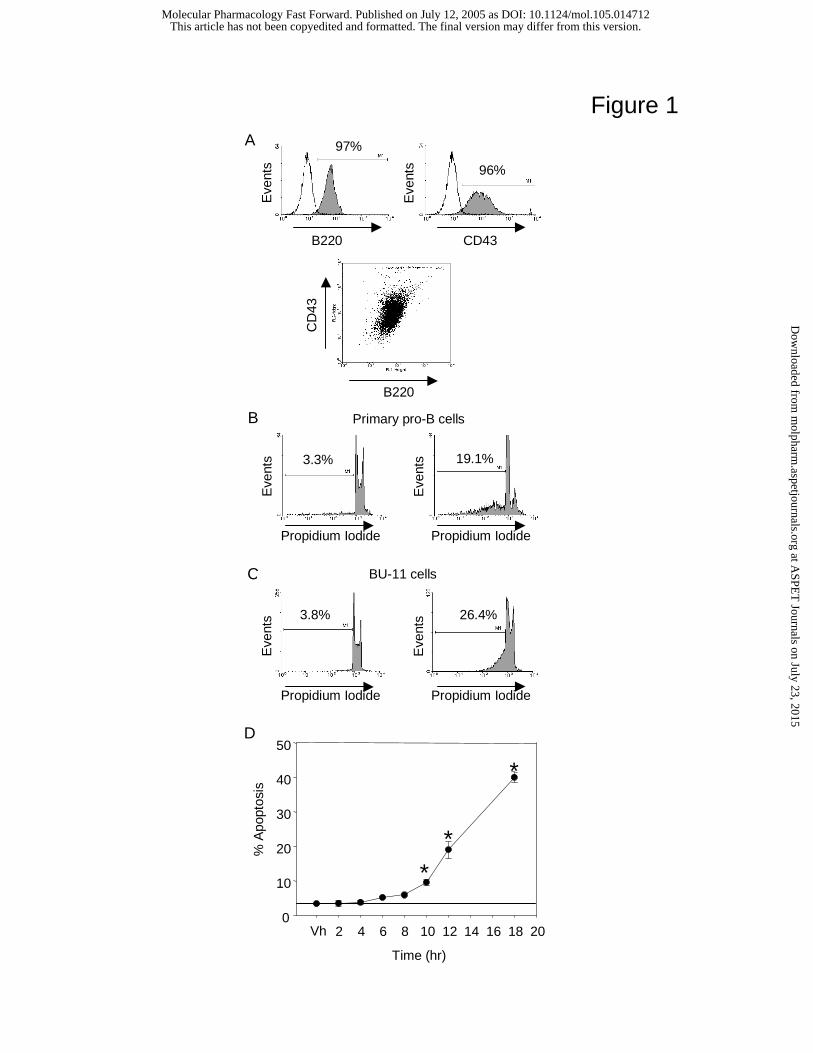
Figure 1
D
*
Time (hr)
Vh 2 4 6 8 10 12 14 16 18 20
% A
popt
osis
0
10
20
30
40
50
*
*
A
C
26.4%3.8%
BU-11 cells
Propidium Iodide Propidium Iodide
B220
CD
43
96%
97%
B220 CD43
Eve
nts
Eve
nts
B
3.3% 19.1%
Primary pro-B cells
Propidium Iodide Propidium Iodide
Eve
nts
Eve
nts
Eve
nts
Eve
nts
This article has not been copyedited and formatted. The final version may differ from this version.Molecular Pharmacology Fast Forward. Published on July 12, 2005 as DOI: 10.1124/mol.105.014712
at ASPE
T Journals on July 23, 2015
molpharm
.aspetjournals.orgD
ownloaded from

Figure 2
B
Time (hr)
Vh 2 4 6 8 10 12 14 16 18 20
Fol
d C
hang
e F
rom
Nai
ve
0
2
4
6
8
10
* *
*
*
20Cleaved
Caspase-3
β-actin
0 hr
2 hr
4 hr
6 hr
8 hr
10 h
r
Vh
DMBAA
C
Vh
DM
BA
BU-11cells
250
150
100
Cleavedα-Fodrin
α-Tubulin
Primarypro-B cells
Vh
DM
BA
This article has not been copyedited and formatted. The final version may differ from this version.Molecular Pharmacology Fast Forward. Published on July 12, 2005 as DOI: 10.1124/mol.105.014712
at ASPE
T Journals on July 23, 2015
molpharm
.aspetjournals.orgD
ownloaded from
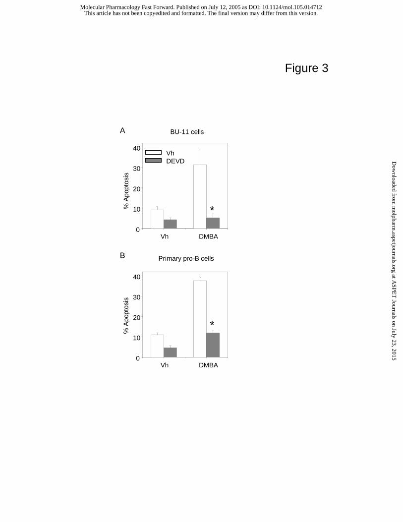
Figure 3
Vh DMBA
% A
popt
osis
0
10
20
30
40VhDEVD
BU-11 cellsA
Vh DMBA
% A
popt
osis
0
10
20
30
40
Primary pro-B cellsB
*
*
This article has not been copyedited and formatted. The final version may differ from this version.Molecular Pharmacology Fast Forward. Published on July 12, 2005 as DOI: 10.1124/mol.105.014712
at ASPE
T Journals on July 23, 2015
molpharm
.aspetjournals.orgD
ownloaded from

Figure 4
B
Time (hr)
Vh 2 4 6 8 10 12 14 16 18 20
Fol
d C
hang
e F
rom
Nai
ve
0
2
4
6
8
10
**
*
tBid15
β-actin
C
Vh
DM
BA
BU-11cells
Primarypro-B cells
Vh
DM
BA
37Cleaved
Caspase-8β-actin
0 hr
2 hr
4 hr
6 hr
8 hr
10 h
r
Vh
DMBAA
Full SizeCaspase-850
This article has not been copyedited and formatted. The final version may differ from this version.Molecular Pharmacology Fast Forward. Published on July 12, 2005 as DOI: 10.1124/mol.105.014712
at ASPE
T Journals on July 23, 2015
molpharm
.aspetjournals.orgD
ownloaded from

Figure 5
TNF-α
TNF-β
LT-β
β-actin
Vh
1 hr
2 hr
4 hr
8 hr
16 h
r
LPS
DC
DMBA A
Membrane bound TNF-α
BRAW 264.7+
LPSBMS2+DMBA
Primary Stroma+DMBA
Vh DMBA
TN
F-α
(pg/
ml)
0
20
40
60
80
C D
Vh DMBA
% A
popt
osis
0
10
20
30
40
50
60 ****
TNF-α WT Stromal cells TNF-α KO Stromal cells
Eve
nts
This article has not been copyedited and formatted. The final version may differ from this version.Molecular Pharmacology Fast Forward. Published on July 12, 2005 as DOI: 10.1124/mol.105.014712
at ASPE
T Journals on July 23, 2015
molpharm
.aspetjournals.orgD
ownloaded from

Figure 6
B
Vh DMBA
% A
popt
osis
0
10
20
30
40
50
60
****
A
Vh DMBA
% A
popt
osis
0
10
20
30
40
50
60
** **
TNFR1/R2 WT B cells TNFR1/R2 KO B cells
Fas WT B cells LPR B cells
C
Vh DMBA
% A
popt
osis
0
5
10
15
20
25
30
** **
DR6 WT B cells DR6 KO B cells
This article has not been copyedited and formatted. The final version may differ from this version.Molecular Pharmacology Fast Forward. Published on July 12, 2005 as DOI: 10.1124/mol.105.014712
at ASPE
T Journals on July 23, 2015
molpharm
.aspetjournals.orgD
ownloaded from

Figure 7A
DM
BA
+V
AD
+D
EV
D
% A
popt
osis
02468
1012141618
DMBA
* *
Vh
+F
A
DMBA
CleavedCaspase-3
20
β-actin
Vh
DM
BA
+F
A
+V
AD
+D
EV
DB
Nai
ve
D
tBid15
β-actin
C
CleavedCaspase-8
50
β-actin
Full SizeCaspase-8
37
This article has not been copyedited and formatted. The final version may differ from this version.Molecular Pharmacology Fast Forward. Published on July 12, 2005 as DOI: 10.1124/mol.105.014712
at ASPE
T Journals on July 23, 2015
molpharm
.aspetjournals.orgD
ownloaded from
Copyright © 2022 FDOKUMEN