Bone marrow stromal cells protect oligodendrocytes from oxygen-glucose deprivation injury
Upload
khangminh22Category
view
0download
0
�����������������
Citation: Laranjeira, P.; Pedrosa, M.;
Duarte, C.; Pedreiro, S.; Antunes, B.;
Ribeiro, T.; dos Santos, F.; Martinho,
A.; Fardilha, M.; Domingues, M.R.;
et al. Human Bone Marrow
Mesenchymal Stromal/Stem Cells
Regulate the Proinflammatory
Response of Monocytes and Myeloid
Dendritic Cells from Patients with
Rheumatoid Arthritis. Pharmaceutics
2022, 14, 404. https://doi.org/
10.3390/pharmaceutics14020404
Academic Editor:
Edorta Santos-Vizcaino
Received: 10 January 2022
Accepted: 10 February 2022
Published: 12 February 2022
Publisher’s Note: MDPI stays neutral
with regard to jurisdictional claims in
published maps and institutional affil-
iations.
Copyright: © 2022 by the authors.
Licensee MDPI, Basel, Switzerland.
This article is an open access article
distributed under the terms and
conditions of the Creative Commons
Attribution (CC BY) license (https://
creativecommons.org/licenses/by/
4.0/).
pharmaceutics
Article
Human Bone Marrow Mesenchymal Stromal/Stem CellsRegulate the Proinflammatory Response of Monocytesand Myeloid Dendritic Cells from Patients withRheumatoid ArthritisPaula Laranjeira 1,2,3,4,5,† , Mónia Pedrosa 2,6,7,† , Cátia Duarte 3,4,8, Susana Pedreiro 2, Brígida Antunes 9,Tânia Ribeiro 9, Francisco dos Santos 9,10, António Martinho 2, Margarida Fardilha 6,11 ,M. Rosário Domingues 12,13 , Manuel Abecasis 14,15, José António Pereira da Silva 3,4,8 and Artur Paiva 1,2,3,4,16,*
1 Flow Cytometry Unit, Department of Clinical Pathology, Centro Hospitalar e Universitário de Coimbra,Av. Bissaya Barreto, Bloco de Celas, 3000-075 Coimbra, Portugal; [email protected]
2 Centro do Sangue e da Transplantação de Coimbra, Instituto Português do Sangue e da Transplantação,Coimbra, Portugal, Quinta da Vinha Moura, São Martinho do Bispo, 3041-861 Coimbra, Portugal;[email protected] (M.P.); [email protected] (S.P.);[email protected] (A.M.)
3 Coimbra Institute for Clinical and Biomedical Research (iCBR), Faculty of Medicine, University of Coimbra,Pólo das Ciências da Saúde, Azinhaga de Santa Comba, 3000-548 Coimbra, Portugal;[email protected] (C.D.); [email protected] (J.A.P.d.S.)
4 Center for Innovative Biomedicine and Biotechnology (CIBB), University of Coimbra, Pólo das Ciências daSaúde, Azinhaga de Santa Comba, 3000-548 Coimbra, Portugal
5 Center for Neuroscience and Cell Biology (CNC), University of Coimbra, Polo 1, 1.◦ Piso, FMUC, Rua Larga,3004-504 Coimbra, Portugal
6 Signal Transduction Laboratory, Center of Cellular Biology, SACS and Department of Biology,University of Aveiro, Campus Universitário de Santiago, 3810-193 Aveiro, Portugal; [email protected]
7 Enzifarma—Diagnostica e Farmacêutica, S.A., Estrada da Luz, n.◦ 90, 2◦ F, 1600-160 Lisbon, Portugal8 Rheumatology Department, Hospitais da Universidade de Coimbra, Centro Hospitalar e Universitário de
Coimbra, Praceta Professor Mota Pinto, 3000-075 Coimbra, Portugal9 Cell2B Advanced Therapeutics, SA, Biocant Park, Núcleo 04, Lote 4A, 3060-197 Cantanhede, Portugal;
[email protected]; (B.A.); [email protected] (T.R.);[email protected] (F.d.S.)
10 Stemlab SA, Biocant Park, Núcleo 04, Lote 2, 3060-197 Cantanhede, Portugal11 Laboratory of Signal Transduction, Institute of Biomedicine—iBiMED, Department of Medical Sciences,
University of Aveiro, Campus Universitário de Santiago, 3810-193 Aveiro, Portugal12 Mass Spectrometry Centre, LAQV REQUIMTE, Department of Chemistry, University of Aveiro, Santiago
University Campus, 3810-193 Aveiro, Portugal; [email protected] Centre for Environmental and Marine Studies (CESAM), Department of Chemistry, University of Aveiro,
Santiago University Campus, 3810-193 Aveiro, Portugal14 Serviço de Transplantação de Progenitores Hematopoiéticos (UTM), Instituto Português de Oncologia de
Lisboa Francisco Gentil, Rua Professor Lima Basto, 1099-023 Lisbon, Portugal;[email protected]
15 Instituto Português do Sangue e da Transplantação—CEDACE, Alameda das Linhas de Torres, 117,1769-001 Lisboa, Portugal
16 Instituto Politécnico de Coimbra, ESTESC-Coimbra Health School, Ciências Biomédicas Laboratoriais,Rua 5 de Outubro, 3046-854 Coimbra, Portugal
* Correspondence: [email protected]; Tel.: +351-239-488-700† These authors contributed equally to this work.
Abstract: Rheumatoid arthritis (RA) is a disabling autoimmune disease whose treatment is ineffectivefor one-third of patients. Thus, the immunomodulatory potential of mesenchymal stromal/stemcells (MSCs) makes MSC-based therapy a promising approach to RA. This study aimed to explorethe immunomodulatory action of human bone marrow (BM)-MSCs on myeloid dendritic cells(mDCs) and monocytes, especially on cytokines/chemokines involved in RA physiopathology. Forthat, LPS plus IFNγ-stimulated peripheral blood mononuclear cells from RA patients (n = 12) andhealthy individuals (n = 6) were co-cultured with allogeneic BM-MSCs. TNF-α, CD83, CCR7 andMIP-1β protein levels were assessed in mDCs, classical, intermediate, and non-classical monocytes.
Pharmaceutics 2022, 14, 404. https://doi.org/10.3390/pharmaceutics14020404 https://www.mdpi.com/journal/pharmaceutics

Pharmaceutics 2022, 14, 404 2 of 25
mRNA expression of other cytokines/chemokines was also evaluated. BM-MSCs effectively reducedTNF-α, CD83, CCR7 and MIP-1β protein levels in mDCs and all monocyte subsets, in RA patients.The inhibition of TNF-α production was mainly achieved by the reduction of the percentage ofcellsproducing this cytokine. BM-MSCs exhibited a remarkable suppressive action over antigen-presenting cells from RA patients, potentially affecting their ability to stimulate the immune adaptiveresponse at different levels, by hampering their migration to the lymph node and the production ofproinflammatory cytokines and chemokines. Accordingly, MSC-based therapies can be a valuableapproach for RA treatment, especially for non-responder patients.
Keywords: mesenchymal stromal cells; mesenchymal stem cells; immunomodulation; rheumatoidarthritis; dendritic cells; monocytes; cytokines; chemokines
1. Introduction
Rheumatoid arthritis (RA) is an autoimmune disease, associated to Th1/Th17-mediatedchronic inflammation of the joints, whose etiology is still elusive. Symmetric polyarthritis,affecting especially the hands and feet, is a hallmark of this disease [1–3]. Several types ofimmune cells, namely monocytes/macrophages and dendritic cells (DCs), actively partici-pate in RA pathophysiology and, together with Th1 and Th17 cells, infiltrate the RA joint.There, they produce inflammatory mediators—namely interleukin (IL)-1, IL-6, tumor necro-sis factor (TNF)-α, extracellular matrix-degrading enzymes, and free radicals—leading tochronic joint inflammation, with consequent cartilage destruction and bone erosion [1,3–6].
The introduction of disease-modifying anti-rheumatic drugs (DMARDs), namelybiological agents targeting cytokines implicated in RA physiopathology, had definitelychanged the clinical course of RA. Notwithstanding, DMARDs are not an effective treatmentfor all patients [2] and have been associated to an augmented risk of infections [7,8], whichconstitutes an important limitation. In this scenario, mesenchymal stromal/stem cells(MSCs) emerge as an alternative treatment for RA. A recent clinical trial in RA, reported thatthe administration of umbilical cord blood MSCs resulted in the reduction DAS28 score andperipheral inflammatory parameters, including TNF-α, IL-1β, IL-6 and IL-8, accompaniedby the increase of IL-10 expression. These data suggest MSC therapy influences the courseof RA with evident clinical improvement [9]. Encouraging results had also been describedin other autoimmune diseases, like systemic lupus erythematosus [10–12] and multiplesclerosis [13,14]. Notwithstanding, clinical trials point out that multiple MSCs infusions atdifferent time points will be probably needed and, in this scenario, immune sensitizationagainst allogeneic MSCs may be a limitation for their use [15,16].
In the preclinical setting, MSC treatment of mice with collagen-induced arthritis (CIA)delayed the disease onset and abolished arthritis progression. Mice infused with MSCsalso displayed decreased paw swelling, decreased immune infiltrate into the joints, andreduction of proinflammatory cytokines levels, along with increased IL-10 expression andregulatory T cells percentage [5,17,18].
Our research group had previously reported that human bone marrow (BM)-derivedMSCs, co-cultured with peripheral blood mononuclear cells (PBMCs) from healthy in-dividuals, resulted in the inhibition of inflammatory mediators production by myeloiddendritic cells (mDCs) and all the monocyte subpopulations identified in the peripheralblood (PB) (classical, intermediate and non-classical monocytes) [19], as well as differentCD4+ and CD8+ T cell subsets, namely Th1, Th17, and Th9 [20]. Recently, we demon-strated MSC-mediated immunomodulation was maintained for T cell subsets from RApatients [21].
Here, we explore if this regulatory action is simultaneously exerted on RA antigen-presenting cells (APCs). For that, PBMCs (from the same RA patients and control groupenrolled in our former study [21]) were cultured alone or in the presence of allogeneicBM-MSCs, in order to investigate the influence of BM-MSCs on the protein levels of

Pharmaceutics 2022, 14, 404 3 of 25
TNF-α, CD83, CCR7, and CCL4 (or macrophage inflammatory protein, MIP-1β) bymDCs and monocyte subpopulations (classical, intermediate, and non-classical mono-cytes). Furthermore, mRNA levels of IL-1β, CXCL9, CXCL10, CCL3 and CCL5, weremeasured in fluorescence-activated cell sorting (FACS)-purified mDCs, classical, andnon-classical monocytes.
This study demonstrated that BM-MSCs exert a significant inhibitory action over allmonocyte subsets and mDCs from RA patients, potentially affecting their ability to stimu-late the immune adaptive response at different levels: BM-MSCs hinder CCR7 upregulationupon cell activation, and this can potentially hamper APCs’ migration into the lymph node;simultaneously, BM-MSCs inhibit the proinflammatory cytokines/chemokines’ produc-tion by mDCs and monocytes, which can potentially hamper T cell polarization towardsTh1/Th17 and migration to inflamed tissue. Accordingly, MSC therapies can be valuablefor RA treatment, especially for non-responder patients.
2. Materials and Methods2.1. Collection of Peripheral Blood and Gradient Density Separation of PBMCs
The collection of PB samples in heparin was carried out at the Centro Hospitalar eUniversitário de Coimbra (CHUC, Rheumatology Unit), Portugal, and Instituto Portuguêsdo Sangue e da Transplantação (Centro do Sangue e da Transplantação de Coimbra),Portugal. The study enrolled six healthy donors (five females and one male; mean age:44 ± 7 years, range: 22–51 years old) and 12 RA patients (eight females and four males;mean age: 53 ± 9 years, range: 38–71 years old).
Five patients were classified as patients with inactive RA (DAS28-CRP3v = 1.9 ± 0.8)and seven as patients with active disease (DAS28-CRP3v = 4.6 ± 0.7), according to thedisease activity score 28 using CRP level (DAS28-CRP; 3-variable). Diagnosis of rheumatoidarthritis was made in accordance with the American College of Rheumatology 1987 Criteriaor ACR/EULAR criteria 2010. Clinical and demographic data about the individuals en-rolled in this study are detailed in Appendix A Table A1. RA patients treated with biologics(such as rituximab, tocilizumab, or anti-TNF), with other autoimmune or inflammatorydiseases, previous cancer, infection or other acute or chronic diseases, were excluded fromthis study. PBMCs isolation were performed by gradient density centrifugation, using Lym-phoprep (Stemcell Technologies, Vancouver, BC, Canada), and by centrifuging at 800× gfor 20 min. Then, HBSS (Gibco, Life Technologies, Paisley, UK) was used to wash PBMCs,which were finally resuspended in RPMI 1640 medium supplemented with GlutaMax(Invitrogen, Life Technologies, Waltham, MA, USA) and antibiotic-antimycotic (Gibco,at a final concentration of 100 units/mL of penicillin, 100 µg/mL of streptomycin, and0.25 µg/mL of Gibco amphotericin B).
The analysis of protein and mRNA expression by mDCs and monocytes was carriedout in PBMCs cultured with or without allogeneic BM-MSCs, in the presence/absenceof the stimulation agents lipopolysaccharide (LPS) and interferon (IFN)γ, as follows:(1) 106 PBMCs + 500 µL RPMI (negative control); (2) 106 PBMCs + 0.5 × 10⁶ MSCs (nega-tive control); (3) 106 PBMCs + LPS + IFNγ (positive control); (4) 106 PBMCs + 0.5 × 10⁶
MSCs + LPS + IFNγ; (5) 106 PBMCs + 0.5 × 10⁶ MSCs, with subsequent BM-MSCs depletionand, after that, LPS plus IFNγ stimulation. The experimental protocols used here had beendescribed in a previous work from our group [19] and are detailed in the following sections.
2.2. Isolation of Human BM-MSCs
BM-MSCs were isolated from eight healthy BM donors admitted to the Instituto Por-tuguês de Oncologia de Lisboa Francisco Gentil (Serviço de Transplantação de ProgenitoresHematopoiéticos, UTM), Portugal.. Sepax S-100 system (Biosafe, Eysins, Switzerland) wasused to isolate PBMCs from BM samples, by following the manufacturer’s instructions.Trypan Blue (Gibco) exclusion method were performed to determine cell count and cell via-bility. BM PBMCs, plated in 10% qualified fetal bovine serum (FBS, Sigma, Madrid, Spain)supplemented DMEM (Gibco), at a 2 × 10⁵ cells/cm2 density, were incubated for 3 days, at

Pharmaceutics 2022, 14, 404 4 of 25
37 ◦C, in 5% CO2 sterile and humidified atmosphere. After discarding the non-adherent cellfraction, the adherent cells were maintained in culture with a complete medium renewalevery 3 to 4 days, until reach a 70–80% confluency. At that point, a 7 min incubation withTrypLE (Life Technologies) was performed to detach the cells, following their replating at adensity of 3000 cells/cm2. BM-MSCs between passage 3 and 5 were used in the presentstudy, and their identity was confirmed by immunophenotype characterization, fluorescentmorphological analysis, and osteogenic, adipogenic, and chondrogenic mesodermal differ-entiation assays, in accordance to the Mesenchymal and Tissue Stem Cell Committee of theInternational Society for Cellular Therapy [22]. Protein levels of CD271 were also evaluatedby flow cytometry. The Ethics Committee of Centro Hospitalar e Universitário de Coimbraapproved this study (CHUC-086-16), and written informed consent were obtained fromall participants.
2.3. Co-Culture of PBMCs and BM-MSCs
The co-culture system used here is described in a former study from our group [19]:106 PBMCs alone, or 106 PBMCs + 0.5 × 10⁶ allogeneic MSCs (ratio PBMCs:MSCs = 2:1)were placed in tissue culture plates (Falcon, Becton Dickinson Biosciences (BD), San Jose,CA, USA) in a total volume of 1 mL of RPMI 1640 medium supplemented with GlutaMax(Invitrogen) and antibiotic/antimycotic (Gibco, at a final concentration of 100 units/mL ofpenicillin, 100 µg/mL of streptomycin, and 0.25 µg/mL of Gibco amphotericin B). After20 h of incubation at 37 ◦C, in humidified and sterile atmosphere, containing 5% CO2, weproceeded to MSCs’ depletion, in part of PBMCs + MSCs co-cultures, with the EasySepHuman CD271 Selection kit (Stemcell Technologies, Vancouver, BC, Canada), following theinstructions of the manufacturer.
Lipopolysaccharide (LPS, 100 ng/mL) and interferon (IFN)γ (100 U/mL) were usedto stimulate PBMCs. In addition, 10 µg/mL of brefeldin A, from Penicillium brefeldianum(Sigma), was added to cell cultures devoted to the study of TNF-α and MIP-1β proteinproduction, by flow cytometry. Brefeldin A blocks protein transport to the Golgi complex,leading to the accumulation of proteins in the endoplasmic reticulum, whose levels can beassessed by flow cytometry, using an intracellular staining protocol. No brefeldin A wasadded to the cell cultures that would be used for the evaluation of CD83 and CCR7 proteinlevels (using flow cytometry), nor for mRNA levels of cytokines, in purified monocytesand mDCs. This was followed by 6 h of incubation in the same conditions. In sum, thestudy of the proteins’ and mRNA expression was systematically performed in each one ofthe following culture conditions: (1) PBMCs; (2) PBMCs + MSCs; (3) PBMCs + LPS + IFNγ;(4) PBMCs + MSCs + LPS + IFNγ; (5) PBMCs + MSCs + MSCs’ depletion + LPS + IFNγ.
2.4. Immunophenotypic Study of Monocyte Subsets and mDCs2.4.1. Staining Protocol
As described previously by our group [19], a 10 min incubation with TrypLE (Gibco)was performed to detached cells from tissue culture plates, which were then transferred to a12 mm × 75 mm cytometer tube, and centrifuged at 540× g for 5 min. The supernatant wasdiscarded. An eight-color monoclonal antibody (mAb) combinations’ panel was used to thephenotypic study of PB monocytes and mDCs (Table 1). To study CD83 and CCR7 proteinlevels (Table 1, tube 1), a stain-lyse-and-then-wash protocol was used [19]: cells werestained with mAbs and incubated for 10 min, in the dark, at room temperature; then 2 mLof FACSLysing Solution (BD) was added, followed by a 10 min period of incubation; finally,cells were washed with 1 mL of PBS (540× g, 5 min), the cell pellet was resuspended in500 µL and immediately acquired in a FACSCanto II (BD) flow cytometer. To evaluate TNF-α and MIP-1β protein production (Table 1, tube 2), cells were stained for surface proteinantigens, in a first step, followed by a 10 min incubation in the dark, at room temperature;after that, cells were washed with PBS; and then stained for the intracellular proteinantigens, using Fix&Perm (Caltag, Hamburg, Germany), and following the manufacturer’sinstructions [19]. After incubating for 15 min, in the dark, at room temperature, with the

Pharmaceutics 2022, 14, 404 5 of 25
mAbs against the intracellular antigens, cells were washed twice with 1 mL of PBS (540× g,5 min), the cell pellet was resuspended in 500 µL of PBS and immediately acquired in aFACSCanto II (BD) flow cytometer.
Table 1. Panel of monoclonal antibodies used for immune cells’ phenotypic characterization, indicat-ing the commercial source and clone.
Fluorochromes
Tubes PB PO FITC PE PerCP-Cy5.5 PE-Cy7 APC APC-H7
1
CD16BD
Pharmingen3G8
CD45Invitrogen
HI30
CD83BeckmanCoulterHB15a
CCR7BD
Pharmingen3D12
CD14BD
PharmingenM5E2
CD33BeckmanCoulter
D3HL60.251
CD300eImmunostepSL UP-H2
HLA-DRBD
L243
2
CD16BD
Pharmingen3G8
CD45Invitrogen
HI30
cyTNF-αBD
Pharmin-gen
MP6-XT22
cyMIP-1βBD
PharmingenD21-1351
CD14BD
PharmingenM5E2
CD33BeckmanCoulter
D3HL60.251
CD300e Im-munostepSL UP-H2
HLA-DRBD
L243
3
CD16BD
Pharmingen3G8
CD123Beckman
Coulter SSDCLY107D2
CD14BD
PharmingenM5E2
CD33BeckmanCoulter
D3HL60.251
CD300e Im-munostepSL UP-H2
HLA-DRBD
L243
Abbreviations: APC, allophycocyanin; APC-H7, allophycocyanin-hilite 7; FITC, fluorescein isothiocyanate;PB, pacific blue; PE, phycoerythrin; PE-Cy7, phycoerythrin-cyanine 7; PerCP-Cy5.5, peridinin chlorophyll protein-cyanine 5.5; PO, pacific orange. Commercial sources: BD (Becton Dickinson Biosciences, San Jose, CA, USA); BDPharmingen (San Diego, CA, USA); Beckman Coulter (Miami, FL, USA); Immunostep S.L (Salamanca, Spain);Invitrogen, Life Technologies (Carlsbad, CA, USA).
2.4.2. Data Acquisition and Analysis
Data were acquired in a FACSCanto II (BD) flow cytometer equipped with the FACS-Diva software (v6.1.2; BD, San Jose, CA, USA). For all samples, the number of events storedwas always above 0.5 × 10⁶. Data analysis was performed using the Infinicyt software(version 1.7; Cytognos SL, Salamanca, Spain).
2.4.3. Identification and Characterization of PB mDCs and Classical, Intermediate, andNon-Classical Monocytes, by Flow Cytometry
For the identification of classical, intermediate, and non-classical monocytes, andmDCs, we applied the gating strategy reported in our previous study [19] and depicted inFigure 1. In short, classical monocytes display high levels of CD14, HLA-DR and CD33,are positive for IREM-2 (CD300e), and negative for CD16; intermediate monocytes arepositive for CD14, with an increasing CD16 levels, and lower levels of CD33, in comparisonto classical monocytes; lastly, non-classical monocytes are positive for CD16, displayinglow to negative reactivity for CD14, and presenting the lowest CD33 levels and the highestCD45 levels, amongst the three PB monocyte subpopulations; in turn, mDCs have lowerside-scatter (SSC) and lower levels of CD45, compared to monocytes, along with high levelsof HLA-DR and CD33, and are negative for CD14, CD300e and CD16.
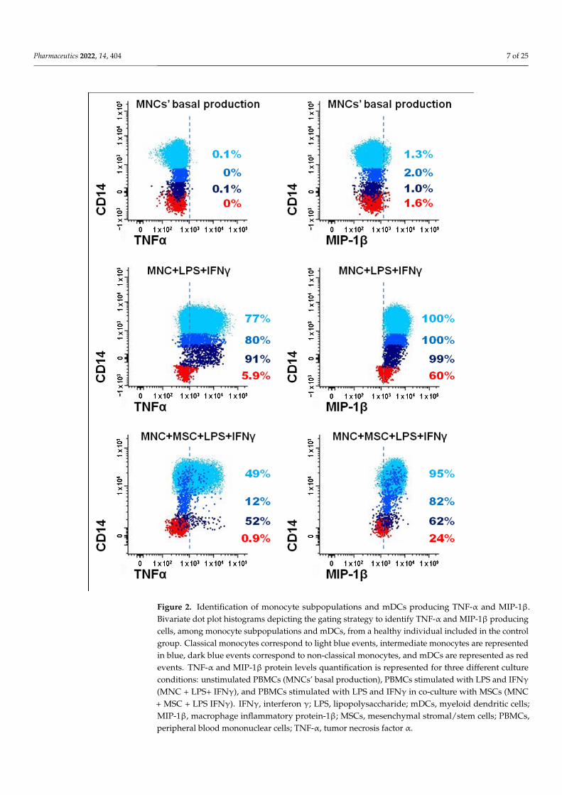
The strategy used to identify monocytes and mDCs producing TNF-α and MIP-1βis illustrated in Figure 2. To evaluate the levels of these two proteins by flow cytometry,brefeldin A was added to the cell cultures in order to block the transport of the newlysynthesized proteins at intracellular level. Then, an intracellular staining protocol wasused to evaluate, at intracellular level, and within each one of the immune cell populationsunder study, the protein levels of TNF-α and MIP-1β.

Pharmaceutics 2022, 14, 404 6 of 25Pharmaceutics 2022, 14, x FOR PEER REVIEW 7 of 27
Figure 1. Gating strategy to identify peripheral blood monocyte subsets (classical, intermediate and non-classical) and mDCs. Classical monocytes (light blue events) were identified as CD14+CD16−, with high levels of CD33, CD300e (or IREM-2), CD45 and HLA-DR; the blue events correspond to intermediate monocytes which are CD14+CD16−/+, with high reactivity for CD300e; non-classical monocytes (dark blue events) were identified as CD14+/−CD16+, with high levels of CD45 and CD300e, and low CD33 levels; mDCs (red events) are phenotypically characterized as CD14-CD16-
CD300e-, with low levels of CD45 and low SSC light dispersion properties, presenting higher levels of CD33 and HLA-DR than monocytes. Grey events correspond to the remaining PBMC populations: lymphocytes, plasmacytoid dendritic cells and basophils. mDCs, myeloid dendritic cells; PBMCs, peripheral blood mononuclear cells.
Figure 1. Gating strategy to identify peripheral blood monocyte subsets (classical, intermediate andnon-classical) and mDCs. Classical monocytes (light blue events) were identified as CD14+CD16−,with high levels of CD33, CD300e (or IREM-2), CD45 and HLA-DR; the blue events correspond to inter-mediate monocytes which are CD14+CD16−/+, with high reactivity for CD300e; non-classical mono-cytes (dark blue events) were identified as CD14+/−CD16+, with high levels of CD45 and CD300e,and low CD33 levels; mDCs (red events) are phenotypically characterized as CD14-CD16-CD300e-,with low levels of CD45 and low SSC light dispersion properties, presenting higher levels of CD33 andHLA-DR than monocytes. Grey events correspond to the remaining PBMC populations: lymphocytes,plasmacytoid dendritic cells and basophils. mDCs, myeloid dendritic cells; PBMCs, peripheral bloodmononuclear cells.
Pharmaceutics 2022, 14, 404 7 of 25Pharmaceutics 2022, 14, x FOR PEER REVIEW 8 of 27
Figure 2. Identification of monocyte subpopulations and mDCs producing TNF-α and MIP-1β. Bivariate dot plot histograms depicting the gating strategy to identify TNF-α and MIP-1β producing cells, among monocyte subpopulations and mDCs, from a healthy individual included in the control group. Classical monocytes correspond to light blue events, intermediate monocytes are represented in blue, dark blue events correspond to non-classical monocytes, and mDCs are represented as red events. TNF-α and MIP-1β protein levels quantification is represented for three different culture conditions: unstimulated PBMCs (MNCs’ basal production), PBMCs stimulated with LPS and IFNγ (MNC + LPS+ IFNγ), and PBMCs stimulated with LPS and IFNγ in co-culture with MSCs (MNC + MSC + LPS IFNγ). IFNγ, interferon γ; LPS, lipopolysaccharide; mDCs, myeloid dendritic cells; MIP-1β, macrophage inflammatory protein-1β; MSCs, mesenchymal stromal/stem cells; PBMCs, peripheral blood mononuclear cells; TNF-α, tumor necrosis factor α.
Figure 2. Identification of monocyte subpopulations and mDCs producing TNF-α and MIP-1β.Bivariate dot plot histograms depicting the gating strategy to identify TNF-α and MIP-1β producingcells, among monocyte subpopulations and mDCs, from a healthy individual included in the controlgroup. Classical monocytes correspond to light blue events, intermediate monocytes are representedin blue, dark blue events correspond to non-classical monocytes, and mDCs are represented as redevents. TNF-α and MIP-1β protein levels quantification is represented for three different cultureconditions: unstimulated PBMCs (MNCs’ basal production), PBMCs stimulated with LPS and IFNγ
(MNC + LPS+ IFNγ), and PBMCs stimulated with LPS and IFNγ in co-culture with MSCs (MNC+ MSC + LPS IFNγ). IFNγ, interferon γ; LPS, lipopolysaccharide; mDCs, myeloid dendritic cells;MIP-1β, macrophage inflammatory protein-1β; MSCs, mesenchymal stromal/stem cells; PBMCs,peripheral blood mononuclear cells; TNF-α, tumor necrosis factor α.

Pharmaceutics 2022, 14, 404 8 of 25
2.5. Cell Purification of Classical Monocytes, Non-Classical Monocytes and mDCs
A FACSAria II flow cytometer (BD) was used to purify classical monocytes, non-classical monocytes and mDCs. The combination of mAbs described in Table 1 (tube 3)enabled classical monocytes (CD14++ CD16− HLA-DR+ CD33+ CD300e+), non-classicalmonocytes (CD14dim/− CD16+ HLA-DR+ CD33+ CD300e+), and mDCs (CD14− CD16−
HLA-DR++ CD33++ CD300e−) identification. Studies of mRNA expression were subse-quently performed in the purified cell subsets.
2.6. mRNA Expression in the Purified Cell Populations
The experimental protocol for the study of mRNA expression by purified classicalmonocytes, non-classical monocytes and mDCs had been previously described by ourgroup [19]. Purified cells were resuspended in RLT Lysis Buffer (Qiagen, Hilden, Ger-many). Total RNA were extracted, using RNeasy Micro kit (Qiagen), as per manufacturerrecommendations, and eluted in RNase-free water, in a final volume of 20 µL. After re-verse transcription with Tetra cDNA Synthesis® (Bioline, London, UK) we preformedreal-time (RT) PCR, using the LightCycler 480 II (Roche Diagnostics, Rotkreuz, Switzer-land), for the relative quantification of gene expression. RT-PCR reactions were performedwith QuantiTect SYBR Green PCR Master Mix (Qiagen), and QuantiTect Primer Assay(CXCL9: QT00013461; CXCL10: QT01003065; CCL3: QT01008063; CCL5: QT00090083;IL-1β: QT00021385) (Qiagen), in a final volume of 10 µL, and all samples were run induplicate. We used the thermal profile previously described by our group [19] for thepolymerase chain reactions: 95 ◦C for 10 min (1 cycle), then 50 cycles of 95 ◦C for 10 s,55 ◦C for 20 s, 72 ◦C for 30 s, 1 cycle of 95 ◦C for 5 s, 65 ◦C for 60 s, and continuous at97 ◦C; at last, 1 cycle of 21 ◦C for 10 s. The analysis of RT-PCR results was performedusing the LightCycler software (Roche Diagnostics). Reference genes selection and datanormalization were performed in GeNorm software (PrimerDesign Ltd., Southampton,UK). As reference genes for classical monocytes, we selected topoisomerase DNA I (TOP1)and glyceraldehyde-3-phosphate dehydrogenase (GAPDH); while the reference genes fornon-classical monocytes and mDCs were β-2 microglobulin (B2M) and GAPDH. The delta-Ct method was used to calculate the normalized expression levels of the genes of interest.The mRNA expression of CCL3, CCL5, CXCL9, CXCL10 and IL-1β was determined inpurified classical monocytes and non-classical monocytes, whereas CXCL10 and IL-1βmRNA expression was measured in purified mDCs.
2.7. Statistical Analyses
Data were presented as the mean values ± standard deviation. The Wilcoxon, Fried-man, and Mann-Whitney non-parametric tests were applied to determine the significanceof the differences between the different experimental conditions, as appropriate, with theStatistical Package for Social Sciences (IBM SPSS, version 17.0, Armonk, NY, USA) software.Differences were considered statistically significant when p < 0.05.
3. Results
To investigate how allogeneic BM-MSCs regulate the immune function of PB mono-cytes and mDCs from RA patients, we evaluated the protein levels or mRNA expressionof proinflammatory cytokines (TNF-α and IL-1β), proteins involved in cell migration(CCL3 or MIP-1α, CCL4 or MIP-1β, CCL5 or RANTES, CXCL9 or MIG, CXCL10 or IP-10,and CCR7), and the maturation marker CD83, in the presence/absence of BM-MSCs andstimulating agents (LPS plus IFNγ). RA patients with inactive and active disease wereconsidered together because the immunomodulatory behavior of BM-MSCs was similarfor both groups of patients, as statistically tested. Likewise, no differences were foundwhen comparing female vs. male RA patients, therefore, they were included in the samegroup. More detailed data, discriminating inactive and active RA patients, can be found inAppendix A Table A2; and data on female vs. male patients in Appendix A Figure A1.

Pharmaceutics 2022, 14, 404 9 of 25
3.1. BM-MSCs Hamper the Production of TNF-α and MIP-1β by Monocytes and mDCs from RAPatients and Healthy Individuals
In order to assess the effect of BM-MSCs over TNFα and MIP-1β protein production,we used flow cytometry to measure the intracellular levels of these cytokines in monocytesand mDCs, using PBMCs cultured in the absence or in the presence of BM-MSCs.
The presence of BM-MSCs in the cell culture resulted in an inhibitory effect over TNFαand MIP-1β protein production, transversal to all monocyte subsets (p < 0.05) and mDCs(p < 0.05), from both RA patients and healthy group (HG), as illustrated in Figures 3–5.For TNFα, this inhibitory effect was mainly caused by the decreased percentage of cellsproducing cytokines (Figure 3); while for MIP-1β, it was achieved not only by the decrementof the percentage of MIP-1β+ cells, but also by the reduction of the amount of cytokineproduced per cell, quantified as mean fluorescence intensity (MFI), as shown in Figure 4.Interestingly, the inhibition of MIP-1β was more pronounced in non-classical monocytesthan in the remaining monocyte subpopulations. Concerning TNF-α, the inhibitory effectof BM-MSCs were stronger over mDCs than monocytes (Figure 5). Interestingly, theinhibitory effect of BM-MSCs was verified even in the assays where BM-MSCs weredepleted immediately before PBMCs’ stimulation. Of note, BM-MSCs depletion prior to cellstimulation partially restored TNF-α production by classical and intermediate monocytes,but only from HG (p < 0.05).
Finally, we observed that unstimulated monocytes and mDCs from RA patientsshowed a basal production of TNF-α and MIP-1β, not detected among the HG (p < 0.05).On the other hand, monocytes from HG displayed a higher response to cell stimulationthan those from RA patients (p < 0.05), (Figures 3–5).
3.2. Effect of BM-MSCs on mRNA Levels of CXCL9, CXCL10, CCL3, CCL5, and IL-1β, inMonocytes and mDCs from RA Patients and Healthy Individuals
In the same line, LPS plus IFNγ stimulation of PBMCs from HG showed a tendency toincrease mRNA levels of all cytokines/chemokines under study, which was not verified forRA patients; as result, in RA patients, the cytokine’s mRNA expression upon cell activationwas significantly lower as compared to HG (p < 0.05), except for IL-1β in non-classicalmonocytes (Figure 6). As observed for TNF-α and MIP-1β, at protein level, a prior contactof 20 h of PBMCs with BM-MSCs sufficed for an inhibitory effect over mRNA cytokine andchemokine expression, not being necessary the presence of BM-MSCs during the PBMCsactivation period.
Overall, our results suggest that BM-MSCs have propensity to inhibit the expression ofthe analyzed cytokines/chemokines at mRNA level in HG (with no statistical significance);while in RA patients this inhibitory trend is only verified for IL-1β in classical (p < 0.05) andnon-classical monocytes, and for CCL3 in non-classical monocytes (p > 0.05), as showed inFigure 6.
3.3. BM-MSCs Reduce the Percentage of CCR7+ and CD83+ Monocytes and mDCs in RA Patients
Under our experimental settings, the protein levels of CCR7 and CD83 were upreg-ulated by both BM-MSCs or LPS plus IFNγ stimulation; also, CCR7 induction upon cellstimulation was stronger for RA in comparison to HG (p < 0.05). It is interesting to noticethat, in the HG, the intermediate monocytes stand out in relation to the remaining mono-cytes by displaying the highest upregulation of CCR7, upon LPS plus IFNγ stimulation. Inturn, in RA patients, LPS plus IFNγ stimulation resulted in similar percentages of CCR7+
cells for all monocyte subpopulations, which were higher than those observed for HG.Besides, a marked difference, among HG and RA patients, was verified for the percentageof CCR7+ cells in classical and non-classical monocytes (Figure 7).

Pharmaceutics 2022, 14, 404 10 of 25Pharmaceutics 2022, 14, x FOR PEER REVIEW 11 of 27
Figure 3. TNF-α protein levels in monocytes and mDCs from RA patients and healthy individuals. Percentage (mean ± standard deviation) of cells producing TNF-α, among monocyte subpopulations (classical, intermediate and non-classical) and mDCs, ; amount of protein (MFI) produced per cell (mean ± standard deviation), measured in the following experimental conditions: unstimulated PBMCs (MNC), non-stimulated PBMCs in co-culture with MSCs (MNC + MSC), PBMCs stimulated with LPS plus IFNγ (MNC + LPS + IFNγ), PBMCs in co-culture with MSCs and stimulated with LPS plus IFNγ in the presence of MSCs (MNC + MSC + LPS + IFNγ), PBMCs in co-culture with MSCs
Figure 3. TNF-α protein levels in monocytes and mDCs from RA patients and healthy individuals.Percentage (mean ± standard deviation) of cells producing TNF-α, among monocyte subpopulations(classical, intermediate and non-classical) and mDCs, ; amount of protein (MFI) produced per cell(mean ± standard deviation), measured in the following experimental conditions: unstimulatedPBMCs (MNC), non-stimulated PBMCs in co-culture with MSCs (MNC + MSC), PBMCs stimulatedwith LPS plus IFNγ (MNC + LPS + IFNγ), PBMCs in co-culture with MSCs and stimulated with LPS

Pharmaceutics 2022, 14, 404 11 of 25
plus IFNγ in the presence of MSCs (MNC + MSC + LPS + IFNγ), PBMCs in co-culture with MSCsand stimulated with LPS plus IFNγ immediately after the depletion of MSCs from the co-culture(MNC + MSC + Depletion + LPS + IFNγ). p values of less than 0.05 were considered as statisticallysignificant for Mann-Whitney test: * vs. HG, in the same culture conditions; and for Friedman’spaired-sample test: � between the groups indicated in the graph. HG, healthy group; IFNγ, interferonγ; LPS, lipopolysaccharide; mDCs, myeloid dendritic cells; MFI, mean fluorescence intensity; MSCs,mesenchymal stromal/stem cells; PBMCs, peripheral blood mononuclear cells; RA, rheumatoidarthritis; TNF-α, tumor necrosis factor α.
Pharmaceutics 2022, 14, x FOR PEER REVIEW 12 of 27
and stimulated with LPS plus IFNγ immediately after the depletion of MSCs from the co-culture (MNC + MSC + Depletion + LPS + IFNγ). p values of less than 0.05 were considered as statistically significant for Mann-Whitney test: * vs. HG, in the same culture conditions; and for Friedman’s paired-sample test: between the groups indicated in the graph. HG, healthy group; IFNγ, interferon γ; LPS, lipopolysaccharide; mDCs, myeloid dendritic cells; MFI, mean fluorescence intensity; MSCs, mesenchymal stromal/stem cells; PBMCs, peripheral blood mononuclear cells; RA, rheumatoid arthritis; TNF-α, tumor necrosis factor α.
Figure 4. MIP-1β protein levels in monocytes and mDCs from RA patients and healthy individuals.

Pharmaceutics 2022, 14, 404 12 of 25
Percentage (mean ± standard deviation) of cells producing MIP-1β, among monocyte subpopulations(classical, intermediate and non-classical) and mDCs; amount of protein (MFI) produced per cell(mean ± standard deviation), measured in the following experimental conditions: unstimulatedPBMCs (MNC), non-stimulated PBMCs in co-culture with MSCs (MNC + BM-MSC), PBMCs stim-ulated with LPS plus IFNγ (MNC + LPS + IFNγ), PBMCs in co-culture with MSCs and stimulatedwith LPS plus IFNγ in the presence of MSCs (MNC + MSC + LPS + IFNγ), PBMCs in co-culturewith MSCs and stimulated with LPS plus IFNγ, immediately after the MSCs were depleted fromthe cell culture (MNC + MSC + Depletion + LPS + IFNγ). Differences were considered statisticallysignificant for p < 0.05 for Mann-Whitney test: * vs. HG, in the same culture conditions; and forFriedman’s paired-sample test: � between the groups indicated in the graph. HG, healthy group;IFNγ, interferon γ; LPS, lipopolysaccharide; mDCs, myeloid dendritic cells; MFI, mean fluorescenceintensity; MIP-1β, macrophage inflammatory protein-1β; MSCs, mesenchymal stromal/stem cells;PBMCs, peripheral blood mononuclear cells; RA, rheumatoid arthritis.
Pharmaceutics 2022, 14, x FOR PEER REVIEW 13 of 27
Figure 4. MIP-1β protein levels in monocytes and mDCs from RA patients and healthy individuals. Percentage (mean ± standard deviation) of cells producing MIP-1β, among monocyte subpopulations (classical, intermediate and non-classical) and mDCs; amount of protein (MFI) produced per cell (mean ± standard deviation), measured in the following experimental conditions: unstimulated PBMCs (MNC), non-stimulated PBMCs in co-culture with MSCs (MNC + BM-MSC), PBMCs stimulated with LPS plus IFNγ (MNC + LPS + IFNγ), PBMCs in co-culture with MSCs and stimulated with LPS plus IFNγ in the presence of MSCs (MNC + MSC + LPS + IFNγ), PBMCs in co-culture with MSCs and stimulated with LPS plus IFNγ, immediately after the MSCs were depleted from the cell culture (MNC + MSC + Depletion + LPS + IFNγ). Differences were considered statistically significant for p < 0.05 for Mann-Whitney test: * vs. HG, in the same culture conditions; and for Friedman’s paired-sample test: between the groups indicated in the graph. HG, healthy group; IFNγ, interferon γ; LPS, lipopolysaccharide; mDCs, myeloid dendritic cells; MFI, mean fluorescence intensity; MIP-1β, macrophage inflammatory protein-1β; MSCs, mesenchymal stromal/stem cells; PBMCs, peripheral blood mononuclear cells; RA, rheumatoid arthritis.
Figure 5. Percentage of BM-MSC-derived inhibition of TNF-α and MIP-1β protein production, by monocytes and mDCs, from RA patients and healthy individuals. Percentage of the inhibition (mean ± standard deviation), mediated by BM-MSCs, on the percentage of TNF-α- and MIP-1β-producing monocytes and mDCs, evaluated either when BM-MSCs were in the cell culture during PBMCs’ activation with LPS and IFNγ, or when BM-MSCs were removed from the cell culture immediately
Figure 5. Percentage of BM-MSC-derived inhibition of TNF-α and MIP-1β protein production, bymonocytes and mDCs, from RA patients and healthy individuals. Percentage of the inhibition (mean± standard deviation), mediated by BM-MSCs, on the percentage of TNF-α- and MIP-1β-producing

Pharmaceutics 2022, 14, 404 13 of 25
monocytes and mDCs, evaluated either when BM-MSCs were in the cell culture during PBMCs’ acti-vation with LPS and IFNγ, or when BM-MSCs were removed from the cell culture immediately beforePBMCs’ activation. p values of less than 0.05 were considered as statistically significant for Mann-Whitney test: * vs. HG, in the same cell population and the same culture condition; and for Wilcoxonpaired-sample test: � between the groups indicated in the graph; (a) vs. classical monocytes, in thesame culture condition, and within the same group of individuals; (b) vs. intermediate monocytes, inthe same culture condition and within the same group of individuals; (c) vs. non-classical monocytes,in the same culture condition and within the same group of individuals. Percentage of inhibition wascalculated as follows, for TNF-α: [[(percentage of TNF-α+ cells in PBMC + LPS + IFNγ) − (percentageof TNF-α+ cells in PBMC + MSC + LPS + IFNγ)]/(percentage of TNF-α+ cells in PBMC + LPS + IFNγ)]* 100. The same formula was applied to calculate the percentage of inhibition for MIP-1β. Accordingly,a percentage of inhibition of 100% corresponds to a complete suppression of the cytokine production,whereas a percentage of inhibition of 0% means that BM-MSCs had no effect on cytokine productionby immune cells. HG, healthy group; IFNγ, interferon γ; LPS, lipopolysaccharide; mDCs, myeloiddendritic cells; MIP-1β, macrophage inflammatory protein-1β; MSCs, mesenchymal stromal/stemcells; PBMCs, peripheral blood mononuclear cells; RA, rheumatoid arthritis; TNF-α, tumor necrosisfactor-α.

Pharmaceutics 2022, 14, 404 14 of 25Pharmaceutics 2022, 14, x FOR PEER REVIEW 15 of 27
Figure 6. Effects of BM-MSCs over cytokines and chemokines mRNA expression by monocytes and mDCs from healthy individuals and RA patients. Semi-quantitative determination of CCL3, CCL5, CXCL9, CXCL10 and IL-1β mRNA expression in FACS-purified classical monocytes and non-classical monocytes, and of CXCL10 and IL-1β in mDCs. mRNA levels were measured under the following culture conditions: non-stimulated PBMCs (MNC), non-stimulated PBMCs in co-culture with MSCs (MNC + BM-MSC), PBMCs stimulated with LPS plus IFNγ (MNC + LPS + IFNγ), PBMCs in co-culture with MSCs and stimulated with LPS plus IFNγ in the presence of MSCs (MNC + MSC + LPS + IFNγ), PBMCs in co-culture with MSCs and stimulated with LPS plus IFNγ immediately after the depletion of MSCs from the culture system (MNC + MSC + Depletion + LPS + IFNγ). The normalized expression levels of the genes were calculated by using the delta-Ct method. Statistically significant differences were considered for p < 0.05 for Mann-Whitney test: * vs. HG, in the same culture condition and in the same cell population; and for Friedman’s paired-sample test: between the groups indicated in the graph. FACS, fluorescence-activated cell sorting; HG, healthy group; IFNγ, interferon γ; IL, interleukin; LPS, lipopolysaccharide; mDCs, myeloid dendritic cells; MSCs, mesenchymal stromal/stem cells; PBMCs, peripheral blood mononuclear cells; RA, rheumatoid arthritis.
Figure 6. Effects of BM-MSCs over cytokines and chemokines mRNA expression by mono-cytes and mDCs from healthy individuals and RA patients. Semi-quantitative determination ofCCL3, CCL5, CXCL9, CXCL10 and IL-1β mRNA expression in FACS-purified classical mono-cytes and non-classical monocytes, and of CXCL10 and IL-1β in mDCs. mRNA levels were mea-sured under the following culture conditions: non-stimulated PBMCs (MNC), non-stimulatedPBMCs in co-culture with MSCs (MNC + BM-MSC), PBMCs stimulated with LPS plus IFNγ
(MNC + LPS + IFNγ), PBMCs in co-culture with MSCs and stimulated with LPS plus IFNγ inthe presence of MSCs (MNC + MSC + LPS + IFNγ), PBMCs in co-culture with MSCs and stim-ulated with LPS plus IFNγ immediately after the depletion of MSCs from the culture system(MNC + MSC + Depletion + LPS + IFNγ). The normalized expression levels of the genes were calcu-lated by using the delta-Ct method. Statistically significant differences were considered for p < 0.05for Mann-Whitney test: * vs. HG, in the same culture condition and in the same cell population; andfor Friedman’s paired-sample test: � between the groups indicated in the graph. FACS, fluorescence-activated cell sorting; HG, healthy group; IFNγ, interferon γ; IL, interleukin; LPS, lipopolysaccharide;mDCs, myeloid dendritic cells; MSCs, mesenchymal stromal/stem cells; PBMCs, peripheral bloodmononuclear cells; RA, rheumatoid arthritis.

Pharmaceutics 2022, 14, 404 15 of 25Pharmaceutics 2022, 14, x FOR PEER REVIEW 17 of 27
Figure 7. Influence of BM-MSCs over the protein levels of CCR7 and CD83 by monocyte subsets and mDCs from RA patients and healthy individuals. Percentage (mean ± standard deviation) of CCR7+ and CD83+ cells, among monocyte subpopulations (classical, intermediate and non-classical)
Figure 7. Influence of BM-MSCs over the protein levels of CCR7 and CD83 by monocyte subsets andmDCs from RA patients and healthy individuals. Percentage (mean ± standard deviation) of
Pharmaceutics 2022, 14, 404 16 of 25
CCR7+ and CD83+ cells, among monocyte subpopulations (classical, intermediate and non-classical)and mDCs measured in the following experimental conditions: unstimulated PBMCs (MNC), non-stimulated PBMCs in co-culture with BM-MSCs (MNC + MSC), PBMCs stimulated with LPS plusIFNγ (MNC + LPS + IFNγ), PBMCs in co-culture with BM-MSCs and stimulated with LPS plus IFNγ
in the presence of MSCs (MNC + MSC + LPS + IFNγ), PBMCs in co-culture with BM-MSCs andstimulated with LPS plus IFNγ immediately after the depletion of BM-MSCs from the cell culture(MNC + MSC + Depletion + LPS + IFNγ). p values of less than 0.05 were regarded as statisticallysignificant for Mann-Whitney test: * vs. HG, in the same culture condition; and for Friedman’s paired-sample test: � between the groups indicated in the graph. HG, healthy group; IFNγ, interferon γ;LPS, lipopolysaccharide; mDCs, myeloid dendritic cells; MSCs, mesenchymal stromal/stem cells;PBMCs, peripheral blood mononuclear cells; RA, rheumatoid arthritis.
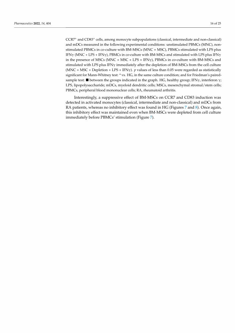
Interestingly, a suppressive effect of BM-MSCs on CCR7 and CD83 induction wasdetected in activated monocytes (classical, intermediate and non-classical) and mDCs fromRA patients, whereas no inhibitory effect was found in HG (Figures 7 and 8). Once again,this inhibitory effect was maintained even when BM-MSCs were depleted from cell cultureimmediately before PBMCs’ stimulation (Figure 7).

Pharmaceutics 2022, 14, 404 17 of 25
Pharmaceutics 2022, 14, x FOR PEER REVIEW 18 of 27
and mDCs measured in the following experimental conditions: unstimulated PBMCs (MNC), non-stimulated PBMCs in co-culture with BM-MSCs (MNC + MSC), PBMCs stimulated with LPS plus IFNγ (MNC + LPS + IFNγ), PBMCs in co-culture with BM-MSCs and stimulated with LPS plus IFNγ in the presence of MSCs (MNC + MSC + LPS + IFNγ), PBMCs in co-culture with BM-MSCs and stimulated with LPS plus IFNγ immediately after the depletion of BM-MSCs from the cell culture (MNC + MSC + Depletion + LPS + IFNγ). p values of less than 0.05 were regarded as statistically significant for Mann-Whitney test: * vs. HG, in the same culture condition; and for Friedman’s paired-sample test: between the groups indicated in the graph. HG, healthy group; IFNγ, interferon γ; LPS, lipopolysaccharide; mDCs, myeloid dendritic cells; MSCs, mesenchymal stromal/stem cells; PBMCs, peripheral blood mononuclear cells; RA, rheumatoid arthritis.
Figure 8. Percentage of CCR7+ and CD83+ cells, among classical, intermediate, and non-classicalmonocytes and mDCs. Bivariate dot plot histograms depicting the gating strategy to identify CCR7+
and CD83+ cells, among monocyte subpopulations and mDCs, from a RA patient. Classical monocytescorrespond to light blue events, intermediate monocytes are represented in blue, dark blue eventscorrespond to non-classical monocytes, and mDCs are represented as red events. The percentageof CCR7+ and CD83+ cells was evaluated for the three different culture conditions: unstimulatedPBMCs (MNC), PBMCs stimulated with LPS and IFNγ (MNC + LPS+ IFNγ), and PBMCs stimulatedwith LPS and IFNγ in co-culture with MSCs (MNC + MSC + LPS+ IFNγ). IFNγ, interferon γ; LPS,lipopolysaccharide; mDCs, myeloid dendritic cells; MSCs, mesenchymal stromal/stem cells; PBMCs,peripheral blood mononuclear cells.

Pharmaceutics 2022, 14, 404 18 of 25
4. Discussion
Though Th1 and Th17 cells constitute the main cell subsets implicated in thechronic inflammation of the joints observed in RA patients, other immune cells (namelymonocytes/macrophages) have been increasingly recognized to play an active role inRA pathophysiology.
Lately, much attention has been paid to PB monocyte subpopulations, becomingclear they possess important functional differences, namely in what concerns to cytokineand chemokine expression profile, phagocytic activity, patrolling behavior in vivo, abilityto stimulate and influence T cells’ polarization, propensity to migrate into normal orinflamed tissues, and ability to undergo differentiation into macrophages, dendritic cells,and osteoclasts [23–28]. With this background, it becomes evident the importance ofstudying the effect of BM-MSCs on each individual PB monocyte subset.
All these monocyte subpopulations are capable of infiltrating the synovium in RApatients [29,30], wherein they play an active role in joint damage [23]. In fact, the directinfluence of monocytes on RA bone erosion became evident with a recent study demon-strating that synovial fluid mononuclear cells from RA patients spontaneously undergodifferentiation into functional osteoclasts in vitro, without any additional preparation orstimulation [31]; accordingly, the number of macrophages in the synovium has been re-ported to correlate with joint damage [32]. Interestingly, pathological conditions are likelyto modify the functional abilities of monocyte subpopulations, being reported that, inpatients with psoriatic arthritis, the CD16+ monocytes are those with the highest abilityto undergo differentiation into osteoclasts, whereas in healthy individuals the classicalmonocytes are the ones showing more propensity to do it [26].
The altered distribution of monocyte subsets described in RA patients further supportsthe involvement of these cells in the pathophysiology of the disease. In fact, the propor-tion of CD14+CD16+ intermediate monocytes is increased in RA synovial fluid [29] andat peripheral level [27,29,33,34]; it is also correlated to disease activity [33], and predictsthe response to methotrexate therapy [23,35]. In turn, the absolute number of circulatingclassical and intermediate monocytes is increased in treatment-refractory patients [23]. Like-wise, an increased proportion of intermediate monocytes is found among non-responderpatients [23,35] and is positively correlated to disease activity [33].
The increasing evidence of the contribution of monocytes to RA pathophysiology, andthe specific role of each monocyte subpopulation, highlight the relevance of our resultsdemonstrating the ability of BM-MSCs to inhibit transversally all monocyte subsets fromRA patients, ex vivo. Here, we found a consistent BM-MSC-derived suppression of TNF-α and MIP-1β (CCL4) production at protein level, by circulating monocytes. Notably,the presence of TNF-α in RA patients’ synovium leads T cells, as well as synoviocytes, topromote osteoclast activation and maturation, which can be in the basis of bone erosion [1,4].This is further supported by studies demonstrating that anti-TNF drugs can slow or evenprevent the progression of cartilage and bone damage in patients with RA [4,8]. In turn,the inhibition of MIP-1β production, mediated by BM-MSCs, was more pronounced innon-classical monocytes, a cell population composed by patrolling monocytes, in bothhuman and mice [25,26,36], which actively migrates into injured joints and initiate jointinflammation in a murine model of RA [36]. BM-MSCs suppression of MIP-1β productionby classical and non-classical monocytes, and mDCs, is a relevant finding because thesecell populations can infiltrate into RA synovium [29,37] and attract Th1 cells. In fact,MIP-1β attracts CCR5+ cells [38], and Th1 cells infiltrating RA synovium do present CCR5and CXCR3 at their surface [4]; intermediate monocytes also express CCR5 [27]. Thus,according to our results, MSC-based therapies may potentially hamper the recruitment ofmonocytes and Th1 cells into the RA inflamed joints, reduce osteoclast activity, and preventbone destruction.
Though little information is available concerning the role of mDCs in RA, an increasedpercentage of mDCs [39] and upregulated levels of CD86 activation marker and CCR7chemokine receptor [40], in the PB of RA patients have been reported; also, mDCs infil-

Pharmaceutics 2022, 14, 404 19 of 25
trating the synovium express the proinflammatory cytokines IL-12p70, IL-15, IL-18, IL-23,and IFN-β [37]. Our results demonstrate that BM-MSCs can inhibit the proinflammatorycytokines production by mDCs and, at the same time, downregulate the surface levels ofCD83 maturation marker and CCR7, in mDCs. Both CD83 and CCR7 are upregulated uponDC maturation and, while CCR7 is essential for DCs migration to lymph nodes, where theystimulate T cells, CD83 is implicated in the upregulation of both MHC class II and CD86costimulatory molecule. Therefore, adequate levels of CD83 and CCR7 are essential forthe development of an adaptive immune response [41,42]. Accordingly, our results showBM-MSCs can potentially restrain the deleterious effect of mDCs in the RA context.
Monocytes and DCs are important players in the activation of the adaptive immunesystem in RA: synovial monocytes from RA patients induce autologous peripheral CD4+
memory T cell polarization into Th1 and Th17cells, in vitro [29], an effect shared with RAPB monocyte-derived DCs [39]. Besides, the percentage of PB intermediate monocytescorrelates positively with the percentage of Th17 cells in RA [27]; and this latter, in turn,correlates positively to DAS28 [4,43].
Our study demonstrates that BM-MSCs hinder the production of the proinflammatorycytokines implicated in the RA pathophysiology. TNF-α in the RA synovium is mainlyproduced by infiltrating macrophages and has the ability to activate endothelial cellsand upregulate chemokine expression, promoting the infiltration of immune cells intothe synovium. TNF-α is also responsible for amplifying the proinflammatory response,activating fibroblasts, chondrocytes and osteoclasts, with subsequent upregulation ofmatrix metalloproteinases (MMPs) expression, ultimately leading to the destruction ofcartilage and bone [6,44]. Notably, by upregulating monocyte expression of IL-1β and IL-6,TNF-α promotes Th17 cell polarization [45]. All these cytokines, together with IFNγ, aredetected in the synovium from RA patients and their role in cartilage destruction and boneresorption is well established [1,4,44,46–48]. In fact, biological drugs that block TNF-α andIL-6 undoubtedly demonstrate the critical role these cytokines play in RA. TNF-α inhibitorsdecrease leukocyte infiltration into the joint, reduce joint swelling, synovial vascularity,and DAS28 [3,6,49]. Furthermore, both anti-TNF and anti-IL-6 biologics stop joint damageprogression in RA patients [8,48].
Remarkably, BM-MSCs possess the capability to inhibit TNF-α production (reportedhere) and, simultaneously, impair IL-6 expression and Th1 and Th17 cells’ function inhealthy individuals [20] and RA patients [21].
Our work demonstrates BM-MSCs possess the ability to regulate monocyte and mDC’sfunction at different levels. And each of these levels can impact the adaptive immunesystem, especially the activity of Th1 and Th17 cells. BM-MSCs impair the production ofproinflammatory cytokines by monocytes and mDCs; this may compromise the capabilityof these antigen-presenting cells to induce Th1/Th17 polarization. BM-MSCs inhibitthe upregulation of CCR7, a chemokine receptor essential for cell migration to lymphnodes; this may hinder antigen-presentation. Finally, BM-MSCs downregulate chemokineexpression by monocytes and mDCs, which can hamper the infiltration of immune cellsinto inflamed synovium, namely Th1 cells.
It has been demonstrated that IL-10 [50,51] and PGE2 [52,53], produced by MSCs,mediate the inhibition of DCs’ maturation and production of proinflammatory cytokines.In turn, the monocytes’ proinflammatory function can be hampered by CD200, expressedon MSC surface [54].
At the present moment, there are 16 phase I, II and/or phase III clinical trials, registeredat www.clinicaltrials.gov (accessed on 6 February 2022), investigating MSCs-based cell ther-apies for RA, five of them are completed, but none of them has the results publicly availableon clinicaltrials.gov. Despite that, there are several studies in small cohorts of patientsreporting the safety and clinical benefits of MSCs administration to RA patients, namelyin visual analog scale (VAS 100 mm) pain score, erythrocyte sedimentation rate (ESR),and 28-joint disease activity score (DAS-28) [9,55,56]. Besides, reduction in the levels ofproinflammatory cytokines and Th17 cells was also observed, accompanied by an increase

Pharmaceutics 2022, 14, 404 20 of 25
in Treg cells [9,57]. Clinical studies using MSCs-based cell therapy for RA patients wererecently reviewed by Lopez-Santalla and colleagues [58]. RA patients’ immune cells exhibita proinflammatory phenotype, consequently increased basal levels of TNF-α and MIP-1βin monocytes and mDCs, as well as other proinflammatory cytokines in T cells, are ob-served [21]. According to the current knowledge, this inflammatory environment improvesthe immunoregulatory function of MSCs [59], explaining the stronger immunomodulatoryeffect of BM-MSCs over immune cells from RA patients, in comparison to HG, as observedin the present work.
5. Conclusions
Recent research in RA patients and animal models pointed monocytes/macrophagesas active players in RA pathophysiology. These studies have confirmed that allogeneic BM-MSCs can suppress the proinflammatory function of monocytes and mDCs, particularly tohamper TNF-α production. Our previous work, carried out in this same set of RA patientsand at the same time, demonstrated allogeneic BM-MSCs also impair Th1 and Th17 cells,among others CD4+ and CD8+ T cell subsets, inhibiting the production of proinflammatorycytokines, while increasing IL-10 and TGF-β mRNA expression [21]. Altogether, our resultssuggest that BM-MSCs suppress the inflammatory response in RA at different levels, as theyare able to hamper simultaneously antigen-presenting cells’ and T cells’ immune functions.Our findings, together with the promising results obtained after MSCs administration toRA animal models, reinforces the assumption that MSC-based therapies can be a valuableapproach for RA treatment, especially for non-responder patients.
Author Contributions: Conceptualization, A.P.; methodology, M.P., S.P., P.L., A.M., B.A., T.R., F.d.S.and M.A.; validation, P.L., F.d.S., A.M., M.A. and A.P.; formal analysis, P.L. and M.P.; investigation,P.L., F.d.S., J.A.P.d.S., A.M. and A.P.; resources, C.D., J.A.P.d.S., F.S, M.A. and A.P.; data curation, P.L.and A.P.; writing—original draft preparation, P.L. and M.P.; writing—review and editing, F.d.S., M.F.,M.R.D., A.M., M.A., J.A.P.d.S. and A.P.; visualization, M.P., P.L. and A.P.; supervision, A.P.; projectadministration, A.P.; funding acquisition, A.P. All authors have read and agreed to the publishedversion of the manuscript.
Funding: This research received no external funding.
Institutional Review Board Statement: The study was conducted according to the guidelines of theDeclaration of Helsinki, and approved by the Ethics Committee of Centro Hospitalar e Universitáriode Coimbra (CHUC-086-16, 13-03-2016).
Informed Consent Statement: Informed consent was obtained from all subjects involved in the study.
Data Availability Statement: The data presented in this study are available on request from the corre-sponding author. The data are not publicly available due to applicable ethical or privacy regulations.
Acknowledgments: We thank Enzifarma—Diagnostica e Farmacêutica, S.A. (Lisbon, Portugal) andStemcell Technologies Inc. (Vancouver, BC, Canada) for providing reagents.
Conflicts of Interest: Brígida Antunes, Tânia Ribeiro & Francisco dos Santos were employees ofCell2B Advanced Therapeutics SA and may own stock in the company. The remaining authorsdeclare no conflict of interest.

Pharmaceutics 2022, 14, 404 21 of 25
Appendix A
Table A1. Demographic data and clinical characteristics of rheumatoid arthritis patients and healthy group.
GenderAge DAS28-
CRP3vCRP ESR TJC
(0–28)SJC
(0–28)
Medication
ComorbiditiesMTX HCQ Sulfasalazine PrednisoloneNSAIDs
(Years) (mg/dL) (mm/h) (mg/Week) (mg/Day) (mg/Day) (mg/Day)
Patients with inactive RA *
# 1 F 61 1.81 0.08 4 1 0 15 400 2000 5 yes Osteoporosis# 2 M 50 2.39 0.47 19 0 0 25 0 2000 5 no None# 3 M 47 1.21 0.12 4 0 0 15 0 0 2.5 yes None# 4 F 52 1.68 0.1 5 0 1 15 0 0 1.25 no None# 5 F 42 2.56 0.12 24 0 0 0 0 0 0 no None# 6 M 38 0.68 0.2 2 0 0 10 0 2000 0 no None# 7 M 55 2.5 0.34 15 0 1 0 400 2000 5 yes Dyslipidemia
Patients with active RA *
# 8 F 60 4.65 1.92 30 6 8 25 400 3000 5 no None# 9 F 41 5.63 5.4 70 9 14 0 0 3000 7.5 yes Depression
# 10 F 53 4.31 0.33 48 3 5 15 400 0 5 yes Obesity
# 11 F 71 3.91 4.25 40 0 12 25 400 2000 5 yes Hypertension,osteoporosis
# 12 F 59 6.08 1.15 72 11 5 0 0 0 5 yes Depression,dyslipidemia
HG
# 1 M 50 None# 2 F 50 None# 3 F 50 None# 4 F 48 None# 5 F 35 None# 6 F 34 None
* Disease activity was assessed using DAS28, and classified as inactive RA if DAS28 < 2.6, and as active RA if DAS28 ≥ 2.6; CRP, C-reactive protein; DAS28-CRP3v, disease activity score28 using CRP level (DAS28-CRP; 3-variable); ESR, erythrocyte sedimentation rate; F, female; HCQ, hydroxychloroquine; HG, healthy group; M, male; MTX, methotrexate; NSAIDs,nonsteroidal antiinflammatory drugs; RA, rheumatoid arthritis; SJC, swollen joint count; TJC, tender joint count.

Pharmaceutics 2022, 14, 404 22 of 25
Table A2. Effect of MSCs on the percentage (%) of cells producing cytokines (mean ± standarddeviation), upon stimulation with LPS and IFNγ.
Cytokine Cell Type Culture Conditions
HealthyIndividuals Inactive RA * Active RA * Total RA
Patients
(n = 6) (n = 5) (n = 7) (n = 12)
TNF-α Classicalmonocytes
MNC +LPS + IFNγ
85 ± 12 50 ± 9.4 68 ± 6.0 57 ± 12
MNC + MSC +LPS + IFNγ
39 ± 23 31 ± 15 42 ± 8.6 35 ± 14
Intermediatemonocytes
MNC +LPS + IFNγ
81 ± 13 46 ± 11 66 ± 3.8 54 ± 13
MNC + MSC +LPS + IFNγ
31 ± 14 26 ± 8.5 31 ± 7.3 28 ± 8.1
Non-classicalmonocytes
MNC +LPS + IFNγ
87 ± 7.5 47 ± 15 59 ± 19 52 ± 17
MNC + MSC +LPS + IFNγ
61 ± 23 20 ± 10 35 ± 17 26 ± 15
mDCs MNC +LPS + IFNγ
16 ± 7.4 9.8 ± 7.2 15 ± 6.7 12 ± 7.2
MNC + MSC +LPS + IFNγ
4.2 ± 1.9 5.5 ± 2.6 8.6 ± 3.8 6.7 ± 3.3
MIP-1β Classicalmonocytes
MNC +LPS + IFNγ
99 ± 0.8 94 ± 8.4 93 ± 4.2 94 ± 6.8
MNC + MSC +LPS + IFNγ
94 ± 3.7 81 ± 14 81 ± 9.1 81 ± 12
Intermediatemonocytes
MNC +LPS + IFNγ
99 ± 0.9 90 ± 10 88 ± 8.7 89 ± 9.2
MNC + MSC +LPS + IFNγ
80 ± 12 70 ± 15 65 ± 14 68 ± 14
Non-classicalmonocytes
MNC +LPS + IFNγ
92 ± 9.8 82 ± 18 73 ± 7.6 78 ± 15
MNC + MSC +LPS + IFNγ
73 ± 17 57 ± 23 45 ± 20 52 ± 22
mDCs MNC +LPS + IFNγ
17 ± 8.4 22 ± 11 13 ± 4.6 18 ± 9.5
MNC + MSC +LPS + IFNγ
9.6 ± 5.8 12 ± 3.7 7.6 ± 1.6 10 ± 3.7
* Disease activity was assessed using DAS28, and classified as inactive RA if DAS28 < 2.6, and as active RA ifDAS28 ≥ 2.6; IFNγ, interferon γ; LPS, lipopolysaccharide; mDC, myeloid dendritic cell; MNC, mononuclear cells;MSC, mesenchymal stromal/stem cells; RA, rheumatoid arthritis.

Pharmaceutics 2022, 14, 404 23 of 25Pharmaceutics 2022, 14, x FOR PEER REVIEW 24 of 27
Figure A1. Comparison between male and female RA patients, concerning the protein production of TNF-α and MIP-1β by monocytes and mDCs. Percentage (mean ± standard deviation) of mono-cyte subsets (classical, intermediate and non-classical) and mDCs, producing TNF-α and MIP-1β,
Figure A1. Comparison between male and female RA patients, concerning the protein production ofTNF-α and MIP-1β by monocytes and mDCs. Percentage (mean ± standard deviation) of monocyte

Pharmaceutics 2022, 14, 404 24 of 25
subsets (classical, intermediate and non-classical) and mDCs, producing TNF-α and MIP-1β, underthe following culture conditions: non-stimulated PBMCs (MNC), non-stimulated PBMCs in co-culturewith MSCs (MNC + MSC), PBMCs stimulated with LPS plus IFNγ (MNC + LPS + IFNγ), PBMCs inco-culture with MSCs and stimulated with LPS plus IFNγ in the presence of MSCs (MNC + MSC+ LPS + IFNγ), PBMCs in co-culture with MSCs and stimulated with LPS plus IFNγ immediatelyafter the depletion of MSCs from the culture system (MNC + MSC + Depletion + LPS + IFNγ). Nostatistically significant differences were found between female and male RA patients, using theMann-Whitney test. HG, healthy group; IFNγ, interferon γ; LPS, lipopolysaccharide; mDCs, myeloiddendritic cells; MFI, mean fluorescence intensity; MSCs, mesenchymal stromal/stem cells; PBMCs,peripheral blood mononuclear cells; RA, rheumatoid arthritis; TNF-α, tumor necrosis factor α.
References1. Smolen, J.S.; Aletaha, D.; McInnes, I.B. Rheumatoid arthritis. Lancet 2016, 388, 2023–2038. [CrossRef]2. Boissier, M.C.; Semerano, L.; Challal, S.; Saidenberg-Kermanac’h, N.; Falgarone, G. Rheumatoid arthritis: From autoimmunity to
synovitis and joint destruction. J. Autoimmun. 2012, 39, 222–228. [CrossRef]3. Buckley, C.D.; McGettrick, H.M. Leukocyte trafficking between stromal compartments: Lessons from rheumatoid arthritis. Nat.
Rev. Rheumatol. 2018, 14, 476–487. [CrossRef] [PubMed]4. Firestein, G.S. Evolving concepts of rheumatoid arthritis. Nature 2003, 423, 356–361. [CrossRef] [PubMed]5. Gonzalez, M.A.; Gonzalez-Rey, E.; Rico, L.; Buscher, D.; Delgado, M. Treatment of experimental arthritis by inducing immune
tolerance with human adipose-derived mesenchymal stem cells. Arthritis Rheumatol. 2009, 60, 1006–1019. [CrossRef]6. Noack, M.; Miossec, P. Selected cytokine pathways in rheumatoid arthritis. Semin. Immunopathol. 2017, 39, 365–383. [CrossRef]
[PubMed]7. Minozzi, S.; Bonovas, S.; Lytras, T.; Pecoraro, V.; Gonzalez-Lorenzo, M.; Bastiampillai, A.J.; Gabrielli, E.M.; Lonati, A.C.; Moja, L.;
Cinquini, M.; et al. Risk of infections using anti-TNF agents in rheumatoid arthritis, psoriatic arthritis, and ankylosing spondylitis:A systematic review and meta-analysis. Expert Opin. Drug Saf. 2016, 15, 11–34. [CrossRef]
8. Lipsky, P.E.; van der Heijde, D.M.; St Clair, E.W.; Furst, D.E.; Breedveld, F.C.; Kalden, J.R.; Smolen, J.S.; Weisman, M.; Emery, P.;Feldmann, M.; et al. Infliximab and methotrexate in the treatment of rheumatoid arthritis. Anti-Tumor Necrosis Factor Trial inRheumatoid Arthritis with Concomitant Therapy Study Group. N. Engl. J. Med. 2000, 343, 1594–1602. [CrossRef]
9. Park, E.H.; Lim, H.S.; Lee, S.; Roh, K.; Seo, K.W.; Kang, K.S.; Shin, K. Intravenous Infusion of Umbilical Cord Blood-DerivedMesenchymal Stem Cells in Rheumatoid Arthritis: A Phase Ia Clinical Trial. Stem Cells Transl. Med. 2018, 7, 636–642. [CrossRef]
10. Liang, J.; Zhang, H.; Hua, B.; Wang, H.; Lu, L.; Shi, S.; Hou, Y.; Zeng, X.; Gilkeson, G.S.; Sun, L. Allogenic mesenchymal stemcells transplantation in refractory systemic lupus erythematosus: A pilot clinical study. Ann. Rheum. Dis. 2010, 69, 1423–1429.[CrossRef]
11. Li, X.; Wang, D.; Liang, J.; Zhang, H.; Sun, L. Mesenchymal SCT ameliorates refractory cytopenia in patients with systemic lupuserythematosus. Bone Marrow Transpl. 2013, 48, 544–550. [CrossRef] [PubMed]
12. Wang, D.; Akiyama, K.; Zhang, H.; Yamaza, T.; Li, X.; Feng, X.; Wang, H.; Hua, B.; Liu, B.; Xu, H.; et al. Double allogenicmesenchymal stem cells transplantations could not enhance therapeutic effect compared with single transplantation in systemiclupus erythematosus. Clin. Dev. Immunol. 2012, 2012, 273291. [CrossRef]
13. Karussis, D.; Karageorgiou, C.; Vaknin-Dembinsky, A.; Gowda-Kurkalli, B.; Gomori, J.M.; Kassis, I.; Bulte, J.W.; Petrou, P.;Ben-Hur, T.; Abramsky, O.; et al. Safety and immunological effects of mesenchymal stem cell transplantation in patients withmultiple sclerosis and amyotrophic lateral sclerosis. Arch. Neurol. 2010, 67, 1187–1194. [CrossRef]
14. Connick, P.; Kolappan, M.; Crawley, C.; Webber, D.J.; Patani, R.; Michell, A.W.; Du, M.Q.; Luan, S.L.; Altmann, D.R.; Thompson,A.J.; et al. Autologous mesenchymal stem cells for the treatment of secondary progressive multiple sclerosis: An open-label phase2a proof-of-concept study. Lancet Neurol. 2012, 11, 150–156. [CrossRef]
15. Alvaro-Gracia, J.M.; Jover, J.A.; Garcia-Vicuna, R.; Carreno, L.; Alonso, A.; Marsal, S.; Blanco, F.; Martinez-Taboada, V.M.; Taylor,P.; Martin-Martin, C.; et al. Intravenous administration of expanded allogeneic adipose-derived mesenchymal stem cells inrefractory rheumatoid arthritis (Cx611): Results of a multicentre, dose escalation, randomised, single-blind, placebo-controlledphase Ib/IIa clinical trial. Ann. Rheum. Dis. 2017, 76, 196–202. [CrossRef]
16. Ansboro, S.; Roelofs, A.J.; De Bari, C. Mesenchymal stem cells for the management of rheumatoid arthritis: Immune modulation,repair or both? Curr. Opin. Rheumatol. 2017, 29, 201–207. [CrossRef]
17. Bouffi, C.; Bony, C.; Courties, G.; Jorgensen, C.; Noel, D. IL-6-dependent PGE2 secretion by mesenchymal stem cells inhibits localinflammation in experimental arthritis. PLoS ONE 2010, 5, e14247. [CrossRef] [PubMed]
18. Abdelmawgoud, H.; Saleh, A. Anti-Inflammatory and antioxidant effects of mesenchymal and hematopoietic stem cells in arheumatoid arthritis rat model. Adv. Clin. Exp. Med. 2018, 27, 873–880. [CrossRef] [PubMed]
19. Laranjeira, P.; Gomes, J.; Pedreiro, S.; Pedrosa, M.; Martinho, A.; Antunes, B.; Ribeiro, T.; Santos, F.; Domingues, R.; Abecasis, M.;et al. Human Bone Marrow-Derived Mesenchymal Stromal Cells Differentially Inhibit Cytokine Production by Peripheral BloodMonocytes Subpopulations and Myeloid Dendritic Cells. Stem Cells Int. 2015, 2015, 819084. [CrossRef]

Pharmaceutics 2022, 14, 404 25 of 25
20. Laranjeira, P.; Pedrosa, M.; Pedreiro, S.; Gomes, J.; Martinho, A.; Antunes, B.; Ribeiro, T.; Santos, F.; Trindade, H.; Paiva, A. Effectof human bone marrow mesenchymal stromal cells on cytokine production by peripheral blood naive, memory, and effector Tcells. Stem Cell Res. Ther. 2015, 6, 3. [CrossRef]
21. Pedrosa, M.; Gomes, J.; Laranjeira, P.; Duarte, C.; Pedreiro, S.; Antunes, B.; Ribeiro, T.; Santos, F.; Martinho, A.; Fardilha, M.; et al.Immunomodulatory effect of human bone marrow-derived mesenchymal stromal/stem cells on peripheral blood T cells fromrheumatoid arthritis patients. J. Tissue Eng. Regen. Med. 2020, 14, 16–28. [CrossRef]
22. Dominici, M.; Le Blanc, K.; Mueller, I.; Slaper-Cortenbach, I.; Marini, F.; Krause, D.; Deans, R.; Keating, A.; Prockop, D.; Horwitz,E. Minimal criteria for defining multipotent mesenchymal stromal cells. The International Society for Cellular Therapy positionstatement. Cytotherapy 2006, 8, 315–317. [CrossRef] [PubMed]
23. Chara, L.; Sanchez-Atrio, A.; Perez, A.; Cuende, E.; Albarran, F.; Turrion, A.; Chevarria, J.; del Barco, A.A.; Sanchez, M.A.;Monserrat, J.; et al. The number of circulating monocytes as biomarkers of the clinical response to methotrexate in untreatedpatients with rheumatoid arthritis. J. Transl. Med. 2015, 13, 2. [CrossRef] [PubMed]
24. Ziegler-Heitbrock, L.; Hofer, T.P. Toward a refined definition of monocyte subsets. Front. Immunol. 2013, 4, 23. [CrossRef]25. Thomas, G.; Tacke, R.; Hedrick, C.C.; Hanna, R.N. Nonclassical patrolling monocyte function in the vasculature. Arter. Thromb.
Vasc. Biol. 2015, 35, 1306–1316. [CrossRef]26. Wong, K.L.; Yeap, W.H.; Tai, J.J.; Ong, S.M.; Dang, T.M.; Wong, S.C. The three human monocyte subsets: Implications for health
and disease. Immunol. Res. 2012, 53, 41–57. [CrossRef] [PubMed]27. Rossol, M.; Kraus, S.; Pierer, M.; Baerwald, C.; Wagner, U. The CD14(bright) CD16+ monocyte subset is expanded in rheumatoid
arthritis and promotes expansion of the Th17 cell population. Arthritis Rheumatol. 2012, 64, 671–677. [CrossRef]28. Traunecker, E.; Gardner, R.; Fonseca, J.E.; Polido-Pereira, J.; Seitz, M.; Villiger, P.M.; Iezzi, G.; Padovan, E. Blocking of LFA-1
enhances expansion of Th17 cells induced by human CD14(+) CD16(++) nonclassical monocytes. Eur. J. Immunol. 2015, 45,1414–1425. [CrossRef]
29. Yoon, B.R.; Yoo, S.J.; Choi, Y.; Chung, Y.H.; Kim, J.; Yoo, I.S.; Kang, S.W.; Lee, W.W. Functional phenotype of synovial monocytesmodulating inflammatory T-cell responses in rheumatoid arthritis (RA). PLoS ONE 2014, 9, e109775. [CrossRef]
30. Calabresi, E.; Petrelli, F.; Bonifacio, A.F.; Puxeddu, I.; Alunno, A. One year in review 2018: Pathogenesis of rheumatoid arthritis.Clin. Exp. Rheumatol. 2018, 36, 175–184.
31. Greisen, S.R.; Einarsson, H.B.; Hvid, M.; Hauge, E.M.; Deleuran, B.; Kragstrup, T.W. Spontaneous generation of functionalosteoclasts from synovial fluid mononuclear cells as a model of inflammatory osteoclastogenesis. APMIS 2015, 123, 779–786.[CrossRef]
32. Mulherin, D.; Fitzgerald, O.; Bresnihan, B. Synovial tissue macrophage populations and articular damage in rheumatoid arthritis.Arthritis Rheumatol. 1996, 39, 115–124. [CrossRef]
33. Tsukamoto, M.; Seta, N.; Yoshimoto, K.; Suzuki, K.; Yamaoka, K.; Takeuchi, T. CD14brightCD16+ intermediate monocytes areinduced by interleukin-10 and positively correlate with disease activity in rheumatoid arthritis. Arthritis Res. Ther. 2017, 19, 28.[CrossRef]
34. Luo, Q.; Xiao, P.; Li, X.; Deng, Z.; Qing, C.; Su, R.; Xu, J.; Guo, Y.; Huang, Z.; Li, J. Overexpression of CD64 on CD14(++)CD16(-)and CD14(++)CD16(+) monocytes of rheumatoid arthritis patients correlates with disease activity. Exp. Ther. Med. 2018, 16,2703–2711. [CrossRef]
35. Cooper, D.L.; Martin, S.G.; Robinson, J.I.; Mackie, S.L.; Charles, C.J.; Nam, J.; Isaacs, J.D.; Emery, P.; Morgan, A.W. FcgammaRIIIaexpression on monocytes in rheumatoid arthritis: Role in immune-complex stimulated TNF production and non-response tomethotrexate therapy. PLoS ONE 2012, 7, e28918. [CrossRef] [PubMed]
36. Misharin, A.V.; Cuda, C.M.; Saber, R.; Turner, J.D.; Gierut, A.K.; Haines, G.K., 3rd; Berdnikovs, S.; Filer, A.; Clark, A.R.; Buckley,C.D.; et al. Nonclassical Ly6C(-) monocytes drive the development of inflammatory arthritis in mice. Cell Rep. 2014, 9, 591–604.[CrossRef] [PubMed]
37. Lebre, M.C.; Jongbloed, S.L.; Tas, S.W.; Smeets, T.J.; McInnes, I.B.; Tak, P.P. Rheumatoid arthritis synovium contains two subsets ofCD83-DC-LAMP- dendritic cells with distinct cytokine profiles. Am. J. Pathol. 2008, 172, 940–950. [CrossRef] [PubMed]
38. Charo, I.F.; Ransohoff, R.M. The many roles of chemokines and chemokine receptors in inflammation. N. Engl. J. Med. 2006, 354,610–621. [CrossRef] [PubMed]
39. Estrada-Capetillo, L.; Hernandez-Castro, B.; Monsivais-Urenda, A.; Alvarez-Quiroga, C.; Layseca-Espinosa, E.; Abud-Mendoza,C.; Baranda, L.; Urzainqui, A.; Sanchez-Madrid, F.; Gonzalez-Amaro, R. Induction of Th17 lymphocytes and Treg cells bymonocyte-derived dendritic cells in patients with rheumatoid arthritis and systemic lupus erythematosus. Clin. Dev. Immunol.2013, 2013, 584303. [CrossRef]
40. Cooles, F.A.H.; Anderson, A.E.; Skelton, A.; Pratt, A.G.; Kurowska-Stolarska, M.S.; McInnes, I.; Hilkens, C.M.U.; Isaacs, J.D.Phenotypic and Transcriptomic Analysis of Peripheral Blood Plasmacytoid and Conventional Dendritic Cells in Early Drug NaiveRheumatoid Arthritis. Front. Immunol. 2018, 9, 755. [CrossRef]
41. Rodriguez-Fernandez, J.L.; Criado-Garcia, O. The Chemokine Receptor CCR7 Uses Distinct Signaling Modules with BiasedFunctionality to Regulate Dendritic Cells. Front. Immunol. 2020, 11, 528. [CrossRef]
42. Li, Z.; Ju, X.; Silveira, P.A.; Abadir, E.; Hsu, W.H.; Hart, D.N.J.; Clark, G.J. CD83: Activation Marker for Antigen Presenting Cellsand Its Therapeutic Potential. Front. Immunol. 2019, 10, 1312. [CrossRef] [PubMed]

Pharmaceutics 2022, 14, 404 26 of 25
43. Kim, J.; Kang, S.; Kwon, G.; Koo, S. Elevated levels of T helper 17 cells are associated with disease activity in patients withrheumatoid arthritis. Ann. Lab. Med. 2013, 33, 52–59. [CrossRef] [PubMed]
44. Brennan, F.M.; McInnes, I.B. Evidence that cytokines play a role in rheumatoid arthritis. J. Clin. Investig. 2008, 118, 3537–3545.[CrossRef]
45. Zheng, Y.; Sun, L.; Jiang, T.; Zhang, D.; He, D.; Nie, H. TNF-α promotes Th17 cell differentiation through IL-6 and IL-1β producedby monocytes in rheumatoid arthritis. J. Immunol Res. 2014, 2014, 385352. [CrossRef] [PubMed]
46. Sato, K.; Suematsu, A.; Okamoto, K.; Yamaguchi, A.; Morishita, Y.; Kadono, Y.; Tanaka, S.; Kodama, T.; Akira, S.; Iwakura, Y.; et al.Th17 functions as an osteoclastogenic helper T cell subset that links T cell activation and bone destruction. J. Exp. Med. 2006, 203,2673–2682. [CrossRef]
47. Hot, A.; Miossec, P. Effects of interleukin (IL)-17A and IL-17F in human rheumatoid arthritis synoviocytes. Ann. Rheum. Dis.2011, 70, 727–732. [CrossRef]
48. Md Yusof, M.Y.; Emery, P. Targeting interleukin-6 in rheumatoid arthritis. Drugs 2013, 73, 341–356. [CrossRef]49. Hull, D.N.; Cooksley, H.; Chokshi, S.; Williams, R.O.; Abraham, S.; Taylor, P.C. Increase in circulating Th17 cells during anti-TNF
therapy is associated with ultrasonographic improvement of synovitis in rheumatoid arthritis. Arthritis Res. Ther. 2016, 18, 303.[CrossRef] [PubMed]
50. Griffin, M.D.; Ritter, T.; Mahon, B.P. Immunological aspects of allogeneic mesenchymal stem cell therapies. Hum. Gene Ther. 2010,21, 1641–1655. [CrossRef]
51. Beyth, S.; Borovsky, Z.; Mevorach, D.; Liebergall, M.; Gazit, Z.; Aslan, H.; Galun, E.; Rachmilewitz, J. Human mesenchymal stemcells alter antigen-presenting cell maturation and induce T-cell unresponsiveness. Blood 2005, 105, 2214–2219. [CrossRef]
52. Aggarwal, S.; Pittenger, M.F. Human mesenchymal stem cells modulate allogeneic immune cell responses. Blood 2005, 105,1815–1822. [CrossRef] [PubMed]
53. Wehner, R.; Wehrum, D.; Bornhauser, M.; Zhao, S.; Schakel, K.; Bachmann, M.P.; Platzbecker, U.; Ehninger, G.; Rieber, E.P.; Schmitz,M. Mesenchymal stem cells efficiently inhibit the proinflammatory properties of 6-sulfo LacNAc dendritic cells. Haematologica2009, 94, 1151–1156. [CrossRef]
54. Pietila, M.; Lehtonen, S.; Tuovinen, E.; Lahteenmaki, K.; Laitinen, S.; Leskela, H.V.; Natynki, A.; Pesala, J.; Nordstrom, K.;Lehenkari, P. CD200 positive human mesenchymal stem cells suppress TNF-α secretion from CD200 receptor positive macrophage-like cells. PLoS ONE 2012, 7, e31671. [CrossRef]
55. Liang, J.; Li, X.; Zhang, H.; Wang, D.; Feng, X.; Wang, H.; Hua, B.; Liu, B.; Sun, L. Allogeneic mesenchymal stem cellstransplantation in patients with refractory RA. Clin. Rheumatol. 2012, 31, 157–161. [CrossRef] [PubMed]
56. Wang, L.; Huang, S.; Li, S.; Li, M.; Shi, J.; Bai, W.; Wang, Q.; Zheng, L.; Liu, Y. Efficacy and Safety of Umbilical Cord MesenchymalStem Cell Therapy for Rheumatoid Arthritis Patients: A Prospective Phase I/II Study. Drug Des. Dev. Ther. 2019, 13, 4331–4340.[CrossRef] [PubMed]
57. Ghoryani, M.; Shariati-Sarabi, Z.; Tavakkol-Afshari, J.; Ghasemi, A.; Poursamimi, J.; Mohammadi, M. Amelioration of clin-ical symptoms of patients with refractory rheumatoid arthritis following treatment with autologous bone marrow-derivedmesenchymal stem cells: A successful clinical trial in Iran. Biomed. Pharm. 2019, 109, 1834–1840. [CrossRef] [PubMed]
58. Lopez-Santalla, M.; Fernandez-Perez, R.; Garin, M.I. Mesenchymal Stem/Stromal Cells for Rheumatoid Arthritis Treatment: AnUpdate on Clinical Applications. Cells 2020, 9, 1852. [CrossRef] [PubMed]
59. Krampera, M. Mesenchymal stromal cell ‘licensing’: A multistep process. Leukemia 2011, 25, 1408–1414. [CrossRef] [PubMed]
Copyright © 2022 FDOKUMEN