Preparation method and growth factor content of platelet concentrate influence the osteogenic...
10
Preparation method and growth factor content of platelet concentrate influence the osteogenic differentiation of bone marrow stromal cells FRANCESCA PERUT 1 , GIUSEPPE FILARDO 2 , ERMINIA MARIANI 3,4 , ANNARITA CENACCHI 5 , LOREDANA PRATELLI 6 , VALENTINA DEVESCOVI 1 , ELIZAVETA KON 2 , MAURILIO MARCACCI 2 , ANDREA FACCHINI 3,4 , NICOLA BALDINI 1,7 & DONATELLA GRANCHI 1 1 Laboratory for Orthopaedic Pathophysiology and Regenerative Medicine, 2 Laboratory of Biomechanics and Technology Innovation, 3 Laboratory of Immunorheumatology and Tissue Regeneration, and 6 Clinical Pathology Unit, Rizzoli Orthopedic Institute, Bologna, Italy, 5 Immunohematology and Transfusion Medicine and Cell and Musculoskeletal Tissue Bank, Bologna, Italy, and 4 Department of Clinical Medicine and 7 Department of Biomedical and Neuromotion Sciences, University of Bologna, Bologna, Italy Abstract Background aims. An extensive debate about the clinical benefits of autologous platelet concentrates used as a treatment option for patients with orthopedic injuries is ongoing. The aim of this study was to determine whether different compo- sitions of platelet concentrates may affect the osteogenic differentiation of bone marrow stromal cells (BMSC). Methods. Pure platelet-rich plasma (P-PRP) and leukocyte-PRP (L-PRP) were characterized for platelet and leukocyte content. As an indicative marker of the delivery of growth factors (GFs), the release of basic fibroblast growth factor (bFGF) from platelet gel (PG) was measured at 1, 18, 48 and 72 h and at 7 d. The ability of different PGs to induce proliferation and differ- entiation of BMSC was evaluated by using bioactivity assays. Results. The platelet recovery was significantly higher in L-PRP, either fresh or frozen. PGs derived from L-PRP and P-PRP showed significant differences in terms of bFGF release and biological activity. bFGF release was faster both in fresh and frozen L-PRP preparations. Moreover, L-PRP samples were able to induce a significantly higher proliferation of BMSC compared with P-PRP or PPP samples. Even though all PG preparations allowed the deposition of mineral nodules in BMSC cultures, the mineralization activity correlated significantly with bFGF levels. Conclusions. The biological activity of platelet concentrates differs according to preparation technique, which affects platelet and leukocyte content and GF availability. Because GF levels are not always optimal in subjects with defective bone healing, composition and bioactivity of PRP should be analyzed to test the reliability and potential effec- tiveness of the regenerative treatment. Key Words: bone marrow stromal cells, osteogenesis, platelet gel, platelet-rich plasma Introduction Bone regeneration is a process in which a complex series of integrated events, which ultimately lead to bone healing, mostly depend on cells resident in bone and their surrounding microenvironment (1). Syner- gistic effects between mesenchymal stromal cells, growth factors (GFs), vascular structures, and extra- cellular matrix have been extensively demonstrated (2,3). On the basis of this concept, one of the most studied approaches in bone regenerative medicine combines the use of GFs and osteogenic precur- sors. Bone marrowederived stromal cells (BMSC) represent the most common source of osteogenic precursors used in bone-related clinical studies (4e6). Autologous blood concentrates, such as platelet-rich plasma (PRP), have been gaining popularity among orthopedic surgeons as a treatment modality for a variety of musculoskeletal injuries. It has been pro- jected that the PRP market in the United States will be worth more than $120 million by 2016 (7). The rationale for the use of PRP on bone lesions, which are hardly able to heal autonomously, is based on the significant reduction of endogenous GFs observed in nonunion sites (8). PRP may be used as a special source of GFs (9), mirroring what occurs physiologi- cally after any bone injury, when platelets are Correspondence: Francesca Perut, MSc, PhD, Laboratory for Orthopaedic Pathophysiology and Regenerative Medicine, Rizzoli Orthopedic Institute, Via di Barbiano 1/10, 40136 Bologna, Italy. E-mail: [email protected] Cytotherapy, 2013; 15: 830e839 (Received 20 September 2012; accepted 29 January 2013) ISSN 1465-3249 Copyright Ó 2013, International Society for Cellular Therapy. Published by Elsevier Inc. All rights reserved. http://dx.doi.org/10.1016/j.jcyt.2013.01.220
Transcript of Preparation method and growth factor content of platelet concentrate influence the osteogenic...

Cytotherapy, 2013; 15: 830e839
Preparation method and growth factor content of platelet concentrateinfluence the osteogenic differentiation of bone marrow stromal cells
FRANCESCA PERUT1, GIUSEPPE FILARDO2, ERMINIA MARIANI3,4,ANNARITA CENACCHI5, LOREDANA PRATELLI6, VALENTINA DEVESCOVI1,ELIZAVETA KON2, MAURILIO MARCACCI2, ANDREA FACCHINI3,4,NICOLA BALDINI1,7 & DONATELLA GRANCHI1
1Laboratory for Orthopaedic Pathophysiology and Regenerative Medicine, 2Laboratory of Biomechanics and TechnologyInnovation, 3Laboratory of Immunorheumatology and Tissue Regeneration, and 6Clinical Pathology Unit, RizzoliOrthopedic Institute, Bologna, Italy, 5Immunohematology and Transfusion Medicine and Cell and MusculoskeletalTissue Bank, Bologna, Italy, and 4Department of Clinical Medicine and 7Department of Biomedical and NeuromotionSciences, University of Bologna, Bologna, Italy
AbstractBackground aims. An extensive debate about the clinical benefits of autologous platelet concentrates used as a treatmentoption for patients with orthopedic injuries is ongoing. The aim of this study was to determine whether different compo-sitions of platelet concentrates may affect the osteogenic differentiation of bone marrow stromal cells (BMSC).Methods. Pureplatelet-rich plasma (P-PRP) and leukocyte-PRP (L-PRP) were characterized for platelet and leukocyte content. As anindicative marker of the delivery of growth factors (GFs), the release of basic fibroblast growth factor (bFGF) from plateletgel (PG) was measured at 1, 18, 48 and 72 h and at 7 d. The ability of different PGs to induce proliferation and differ-entiation of BMSC was evaluated by using bioactivity assays. Results. The platelet recovery was significantly higher in L-PRP,either fresh or frozen. PGs derived from L-PRP and P-PRP showed significant differences in terms of bFGF release andbiological activity. bFGF release was faster both in fresh and frozen L-PRP preparations. Moreover, L-PRP samples wereable to induce a significantly higher proliferation of BMSC compared with P-PRP or PPP samples. Even though all PGpreparations allowed the deposition of mineral nodules in BMSC cultures, the mineralization activity correlated significantlywith bFGF levels. Conclusions. The biological activity of platelet concentrates differs according to preparation technique,which affects platelet and leukocyte content and GF availability. Because GF levels are not always optimal in subjects withdefective bone healing, composition and bioactivity of PRP should be analyzed to test the reliability and potential effec-tiveness of the regenerative treatment.
Key Words: bone marrow stromal cells, osteogenesis, platelet gel, platelet-rich plasma
Introduction
Bone regeneration is a process in which a complexseries of integrated events, which ultimately lead tobone healing, mostly depend on cells resident in boneand their surrounding microenvironment (1). Syner-gistic effects between mesenchymal stromal cells,growth factors (GFs), vascular structures, and extra-cellular matrix have been extensively demonstrated(2,3). On the basis of this concept, one of the moststudied approaches in bone regenerative medicinecombines the use of GFs and osteogenic precur-sors. Bone marrowederived stromal cells (BMSC)represent the most common source of osteogenic
Correspondence: Francesca Perut, MSc, PhD, Laboratory for Orthopaedic PathBarbiano 1/10, 40136 Bologna, Italy. E-mail: [email protected]
(Received 20 September 2012; accepted 29 January 2013)
ISSN 1465-3249 Copyright � 2013, International Society for Cellular Therapy. Phttp://dx.doi.org/10.1016/j.jcyt.2013.01.220
precursors used in bone-related clinical studies (4e6).Autologous blood concentrates, such as platelet-richplasma (PRP), have been gaining popularity amongorthopedic surgeons as a treatment modality fora variety of musculoskeletal injuries. It has been pro-jected that the PRPmarket in the United States will beworth more than $120 million by 2016 (7). Therationale for the use of PRP on bone lesions, which arehardly able to heal autonomously, is based on thesignificant reduction of endogenous GFs observed innonunion sites (8). PRP may be used as a specialsource of GFs (9), mirroring what occurs physiologi-cally after any bone injury, when platelets are
ophysiology and Regenerative Medicine, Rizzoli Orthopedic Institute, Via di
ublished by Elsevier Inc. All rights reserved.

Platelet concentrates and osteoblast differentiation 831
entrapped in the native fracture hematoma. To beapplied at the surgical site, PRP must be activated toinduce platelet degranulation and fibrin polymeriza-tion, thus obtaining a clot usually called platelet gel(PG). PRP displays the unique characteristics of amixture of bioactive molecules, and the regenerativepotency of PRP probably depends on GF levels(10,11). Pro-osteogenic factors, such as platelet-derived growth factor, transforming growth factor-b,epidermal growth factor, vascular endothelial growthfactor, insulin-like growth factors I and II and basicfibroblast growth factor (bFGF), play a pivotal role inbone regeneration, either alone or in combination withBMSC (12e15). However, despite two decades ofclinical study, the efficacy of PG in bone healing is stillunder discussion because no accepted standard for itspreparation is available (7,16e18). The plateletconcentration procedure is mainly based on bloodcentrifugation protocols (19), but the preparationtechnique and storage may influence composition andbiological activity of PRP (20,21).
The aim of this study was to investigate whetherPRPs that differ in platelet concentration and in thepresence or not of white blood cells may affect theosteogenic differentiation of BMSC. PRPs wereprepared by two different techniques, namely a one-step procedure that is based on a clinically usedcommercial device (22) and a two-step procedure thatis based on a previously described method (23). Thesetwo techniques are widely used in clinical practice anddiffer with regard to the resulting platelet concentra-tion and leukocyte content. PRPs were characterizedfor platelet and white blood cell content. To track theGF delivery, as a representative marker we tested thebFGF released from PGs at different time points. Weevaluated the relationship among the above parametersand the ability of PRP to induce the differentiation ofosteogenic precursors in mature and functioningosteoblasts. Moreover, we evaluated whether or notthe damage of leukocytes and platelets, which wasinduced by freezing and thawing of the concentrates,affected the osteogenic properties of PRP obtainedwith the two-step method (24).
Methods
Subjects
Ten healthymen (age range, 26e38 years), enrolled ona voluntary basis, underwent blood sample collection(200 mL per subject). Subjects did not presentsystemic disorders, infection, smoking habit, nonste-roidal anti-inflammatory drug use during the 5 d beforeblood donation, hemoglobin values <11 g/dL orplatelet values <150 � 103/mL. A code number wasassigned to each sample to ensure subject anonymity.
The study was approved by the local ethicscommittee, and an informed written consent formwas signed by each donor.
Preparation of platelet concentrates
For the pure platelet-rich plasma (P-PRP) one stepmethod, a 45-mL venous blood sample was dividedinto five tubes containing 1 mL of sodium citratesolution (3.8%) and was then centrifuged (460g for8 min); 1 mL per tube of the platelet-rich superna-tant on the red blood cell pellet was then collected,with care taken to avoid leukocyte harvesting (22).
For the platelet leukocyte-rich plasma (L-PRP)two-stepmethod, a 150-mLvenous blood sample (25),collected in a bag containing 21 mL sodium citrate,was centrifuged (730g for 15 min). The resultingplasma and buffy-coat were transferred in a separatebag through a closed circuit, and red blood cells wereeliminated. After centrifugation (3800g for 10 min),the supernatant was collected to produce L-PRP.During the second centrifugation step, poor plateletplasma (PPP) was collected and used as control.A half-volume of the L-PRP obtained was stored ate30�C for 2 h and then thawed in a dry thermostat at37�C for 30 min (fL-PRP) just before use. P-PRP,L-PRP, fL-PRP and PPP were activated with 10%CaCl2 (228 mmol/L) to produce PGs.
Platelet concentration and leukocyte number
The platelet concentration and the white blood cellnumber were determined by blood cell count(COULTER LH 750 Hematology Analyzer; Beck-man Coulter s.r.l, Milan, Italy). Linearity was5e1000 � 103/mL for platelet count and 0.1e100 �103/mL for white blood cell count.
Evaluation of bFGF released from PG
Each sample of PRP and PPP preparations was di-vided into five aliquots, activated with 10% CaCl2and incubated for 1, 18, 48 and 72 h and 7 d at 37�Cin 5% CO2. The first time point of bFGF release wasperformed in agreement with the therapeuticschedule, taking into account that in clinical practice,the PRP injection is performed within 1 h fromCaCl2 activation. The time course of bFGF releasewas aimed at evaluating the relationship with thefunctional results.
After centrifugation (15 min at 4000 rpm at 20�C),the supernatant was collected and frozen at e30�Cuntil use. Samples were assayed in duplicate andbFGF concentration was evaluated with the use ofcommercially available bead-based sandwich immuno-assay kits (Bio-Rad Laboratories, Hercules, CA, USA)
832 F. Perut et al.
(26). Standard curve range was 0.07e18.366 pg/mL,with an intra- and inter-assay coefficient of variability of8%. The immunocomplexes formed on distinct beadswere quantified by use of the Bio-Plex Protein ArraySystem (Bio-Rad Laboratories). Data were analyzed byuse of the Bio-Plex Manager software version 6.0 (Bio-Rad Laboratories). Standard levels between 70e130%of the expected values were considered accurate andwere used. At least eight standards were accepted andused to establish standard curves, following a five-parameter logistic regression model.
BMSC cultures
After approval by internal review boards, bonemarrow samples were retrieved from discardedmaterial collected during reconstructive hip surgery,and the resulting mononuclear cells were obtained aspreviously described (27). Isolated mononuclear cellswere maintained in a-minimum essential mediumsupplemented with 100 U/mL penicillin, 0.1 mg/mLstreptomycin, and 100 mmol/L ascorbic acid-2 phos-phate (Sigma-Aldrich, Milan, Italy) and 10% fetalbovine serum (Lonza, Milan, Italy). After 4 d,nonadherent cells were removed and the culturewas re-fed with fresh medium supplemented with10e8 mol/L dexamethasone (Sigma) (differentiationmedium). At first confluence, cells expressed thetypical surface antigens of BMSC (CD44, CD90,
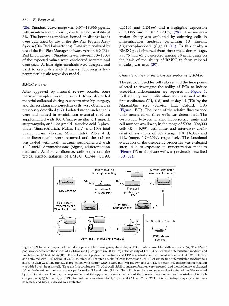
Figure 1. Schematic diagram of the culture protocol for investigating thepool was seeded onto the inserts of a 24-transwell plate (pore size, 0.45 mmincubated for 24 h at 37�C; (B) 108 mL of different platelet concentratesand activated with 10% vol/vol of CaCl2 solution; (C, D) after 1 h, the PGadded to each well. The transwells pre-loaded with human MSCS werewas added over the transwell; (E) at the first confluence (T1, 6 d), cell via(F) while the mineralization assay was performed at T2 end point (14 d).by the PG, at days 1 and 7, the supernatants of the upper and lowercompartment; (J) for each type of PG, five vials were incubated for 1, 18,collected, and bFGF released was evaluated.
CD105 and CD166) and a negligible expressionof CD45 and CD117 (<1%) (28). The mineral-ization ability was evaluated by culturing cells inmineralization medium containing 10 mmol/Lb-glycerophosphate (Sigma) (13). In this study, aBMSC pool obtained from three male donors (age,55, 75 and 65 y), selected among 20 individuals onthe basis of the ability of BMSC to form mineralnodules, was used (29).
Characterization of the osteogenic properties of BMSC
The protocol used for cell cultures and the time pointsselected to investigate the ability of PGs to induceosteoblast differentiation are reported in Figure 1.Cell viability and proliferation were assessed at thefirst confluence (T1, 6 d) and at day 14 (T2) by theAlamarBlue test (Serotec Ltd, Oxford, UK)(Figure 1E,F). The mean of the relative fluorescenceunits measured on three wells was determined. Thecorrelation between relative fluorescence units andcell number was linear, in the range of 5000e200,000cells (R ¼ 0.99), with intra- and inter-assay coeffi-cient of variations of 8% (range, 1.8e16.3%) and11% (range, 0.7e20%), respectively. The functionalevaluation of the osteogenic properties was evaluatedafter 14 d of exposure to mineralization medium(Figure 1F) on duplicate wells, as previously described(30e32).
ability of PG to induce osteoblast differentiation. (A) The BMSC) at the density of 1 � 104 cells/well in differentiation medium andand PPP as control were distributed in each well of a 24/well platewas formed and 480 mL of serum-free differentiation medium was
put over the PG, and 200 mL of serum-free differentiation mediumbility and proliferation were assessed, and the medium was changed(GeI) To favor the homogeneous distribution of the GFs releasedchambers of the transwell were mixed and redistributed in each48 and 72 h and 7 d at 37�C. After centrifugation, supernatant was

Platelet concentrates and osteoblast differentiation 833
Statistical analysis
Statistical analysis was performed by use of MedCalcSoftware 7.5.0.0 (Mariakerke, Belgium). Summarystatistics were used to calculate mean, standard errorof themean,median andminimum-maximum values.The D0Agostino-Pearson test was used to verify thenormal distribution of analyzed parameters in thedifferent subgroups. A nonparametric analysis ofvariance (Kruskal-Wallis test) was applied to detectthe effects of the different platelet concentrates on themeasured variables; the Mann-Whitney U test wasapplied as a post hoc test if the P value of the Kruskal-Wallis test was <0.05. The Wilcoxon signed rank testwas used in a paired analysis to verify the differencesin bFGF release from PG at the different time points.The Spearman rank correlation coefficient (R) wasused to analyze the degree of association among themeasured variables. All P values <0.05 were consid-ered statistically significant.
Results
Platelet recovery differs according to the preparationmethod of PRP
The number of platelets was significantly higher in allPRP preparations compared with PPP (Table I). Thehighest platelet recovery was obtained in the L-PRPpreparation (P < 0.0001 versus fL-PRP, P-PRP andPPP). The ratio between platelet number before andafter concentration is significantly higher in L-PRP.Even though the preparation was the same as thatused for L-PRP, in fL-PRP the platelet number wassignificantly reduced up to 65% (range, 31e89%).
Table I. Platelet concentration and leukocyte count in different PG pre
Platelet number, 103/mLRatio between platele
and platelet nu
Native blood 202 � 55(163e258)
199L-PRP 920 � 491
(555e1114)912
P < 0.0001 vs fL-PRP, PRP, PPP
4
P < 0.0001fL-PRP 308 � 325
(107e686)238
P < 0.0001 vs PPP
1
P <
P-PRP 260 � 37(83e738)
194P < 0.0001 vs PPP
1
P <
PPP 6.2 � 6.3(3e16)
5
0.0(
Data are expressed as mean � standard error of the mean (minimum-m
The two-step procedure determined effective plateletenrichment in all samples despite a wide variability,whereas the increment in platelet number was notalways ensured with the one-step technique. Calciumchloride addition determined the gel formation in allsamples, including PPP, demonstrated that plateletactivation has been induced even if the platelet num-ber was under the detection limit, as for PPP prepara-tion. White blood cells were not found in P-PRP andPPP, whereas in L-PRP the mean number of leuko-cytes was comparable to the one of native blood(Table I). In L-PRP, the percentages of lymphocytesand polymorphonuclear cells (PMN) were 77.1% �1.9% (range, 60e80%; median, 81%) and 21.9% �1.9% (range, 12e40%; median, 18.5%), respectively.As observed for platelets, the leukocyte amount wassignificantly reduced after thawing of fL-PRP, thusproving that the freezing procedure was destructivefor the cells. The number of leukocytes and plateletsshowed a significant correlation in both L-PRPpreparations (L-PRP: R ¼ 0.63 and P < 0.0001; fL-PRP: R ¼ 0.90 and P < 0.0001).
Progressive release of bFGF from PG varies according tothe composition of PRP
The amount of bFGF increased significantly from 1 hto the final time point (7 d) in all samples (L-PRP,P ¼ 0.005; fL-PRP, P ¼ 0.04; P-PRP, P < 0.0004;PPP, P < 0.001) (Figure 2). A progressive increasewas observed in P-PRP, which showed significantchanges at each time point in comparison to theprevious one, whereas the increase was more variablein L-PRP preparations, either fresh or thawed. The
parations.
t number after concentrationmber in native blood Leukocyte number, 103/mL
NA 6.4 � 2.3(4.8e8.8)
5.9.7 � 0.3(3.0e8.8)
4.3vs fL-PRP, PRP, PPP
6.4 � 6.5(1.8e14.4)
5.7P < 0.0001 vs fL-PRP, PRP, PPP
.5 � 0.1(0.6e3.1)
1.20.0001 vs PPP
0.4 � 1.2(0.0e1.8)
0.0
.2 � 0.2(0.5e3.3)
1.00.0001 vs PPP
0.0
3 � 0.0030.02e0.07)
0.03
0.0
aximum values) and median.

Figure 2. Graph shows the amount of bFGF (pg/mL) released byPG derived from different PRP preparations. (a,b) Significantdifference in comparison to PPP and P-PRP, respectively (P <
0.05, Mann-Whitney U test); (c) significant difference incomparison to the previous time point; (d) significant change from1 h to 7 d (P < 0.05 , Wilcoxon signed rank test).
834 F. Perut et al.
bFGF release from both L-PRPs was faster, becauseafter 1 h and 18 h, the concentration was significantlyhigher in comparison to P-PRP. Even though thebFGF amount was higher in PRP than in PPP, afterday 7, the differences disappeared because a consider-able amount of this GF was found also in PPP. Aninverse correlation was found between bFGF releasedfrom PG prepared with L-PRPs, either fresh or frozen,and platelet number at all time points (Table II). Theinverse relationship was significant up to 18 h and at72 h. On the contrary, in P-PRP samples, the corre-lation between the platelet number and bFGF wasalways positive and became significant at 48 h up to7 d. A positive correlation was also found in PPPsamples, and it was significant from 18 h forward. InPG obtained from L-PRP, the correlation betweenbFGF release and leukocyte number was also evalu-ated. An inverse relationship was found at all the timepoints, and it was significant at 1 h for fL-PRP and at
Table II. Correlation between bFGF released from PG and number of
1 h 18 h
L-PRPNo. of platelets �0.50 (0.009) �0.61 (0.0006)No. of leukocytes �0.13 (0.489) �0.20 (0.261)No. of PMN �0.44 (0.009) �0.42 (0.0057)
fL-PRPNo. of platelets �0.60 (0.0009) �0.45 (0.012)No. of leukocytes �0.39 (0.028) �0.23 (0.197)
P-PRPNo. of platelets 0.30 (0.090) 0.30 (0.097)
PPPNo. of platelets 0.29 (0.102) 0.63 (0.0005)
Spearman R and P values are shown in parentheses.
day 7 for L-PRP. Moreover, at all the time points, thenumber of PMN found in L-PRP correlated inverselywith bFGF (Table II), whereas no significant corre-lation was found between bFGF and lymphocytenumber (data not shown).
BMSC proliferation is influenced by PRP compositionand is significantly associated with bFGF release
As expected, the proliferation of BMSC increasedfrom the seeding to the end point, with a log phasefrom T0eT1, and a stationary phase from T1eT2(Figure 3). The type of PRPs influenced the yield ofBMSC (Kruskal-Wallis P value, T1: 0.002; T2:0.001).The post hoc analysis revealed that L-PRPsweremore effective than P-PRP in favoring cell prolifera-tion, and a significant difference was found at T2(L-PRP versus P-PRP, P ¼ 0.014; fL-PRP versusP-PRP, P ¼ 0.027). The proliferation rate induced byPPP was similar to that observed with PRPs, but thecell number at T1 and T2 was significantly lower incomparison to L-PRPs (L-PRP versus PPP, P¼ 0.004at T1 and P ¼ 0.001 at T2; fL-PRP versus PPP, P ¼0.0003 at T1 and P ¼ 0.002 at T2).
We analyzed the association between cell prolif-eration in the exponential phase of growth, forexample, up to T1, and concentration of the bloodcomponents in different PGs (Table III). A signifi-cant, positive correlation was found between BMSCproliferation and number of platelets in culturesexposed to PG derived from P-PRP and PPP; on thecontrary, a significant, negative correlation wasfound when BMSC were cultured with fresh L-PRP.The BMSC proliferation was significantly correlatedwith the amount of bFGF released from all PGsamples during 7 d.
As expected, BMSC proliferation in the steadystate of growth, that is, up to T2, was poorly influ-enced by leukocyte and platelet number present inboth L-PRPs (data not shown), whereas the positive
platelets and leukocytes after concentration procedure.
bFGF
48 h 72 h 7 d
�0.46 (0.011) �0.49 (0.006) �0.22 (0.238)�0.15 (0.405) �0.25 (0.179) �0.59 (0.002)�0.38 (0.0009) �0.37 (0.0014) 0.59 (0.0003)
�0.25 (0.164) �0.46 (0.010) �0.17 (0.370)�0.09 (0.606) �0.13 (0.463) �0.18 (0.352)
0.37 (0.030) 0.38 (0.033) 0.37 (0.039)
0.62 (0.0006) 0.66 (0.0003) 0.52 (0.004)

Figure 3. Number of BMSC retrieved after 6 (T1) and 14 (T2) d ofculture with different PG preparation. A significant difference incomparison to PPP (a) and to P-PRP (b) was found (P < 0.05,Mann-Whitney U test).
Platelet concentrates and osteoblast differentiation 835
and significant correlation with P-PRP was main-tained (R ¼ 0.52, P ¼ 0.004). No correlationwas found between bFGF amount and cell recoveryat T2.
Mineralization ability of BMSC is significantlyassociated with bFGF release
Cell mineralization was always detectable irrespectiveof the source of PG, including that obtained fromPPP. The number and the size of calcium phosphatedeposits obtained with the use of PG from differentindividuals were variable. A qualitative analysis ofmineral nodule formation revealed a more effectiveability of L-PRP and fL-PRP to induce osteoblastdifferentiation, compared with the other samples(Figure 4). However, the Alizarin red stain quantifi-cation did not show any significant difference betweenPG preparation and/or PPP samples (Figure 5).
In L-PRP, either fresh or frozen, the ability ofBMSC to mineralize was not influenced by plateletnumber, whereas a positive significant correlationwas found with the platelet amount recovered inP-PRP and PPP samples (Table IV). In all cases,the bFGF released during the first 72 h showed
Table III. Correlation between BMSC proliferation at T1 in cultures ex(first column).
L-PRP f
No. of platelets in PRP �0.71 (<0.0001) �0.32No. of leukocytes in PRP �0.02 (0.880) �0.38bFGF 1 h 0.75 (<0.0001) 0.70bFGF 18 h 0.85 (<0.0001) 0.84bFGF 48 h 0.84 (<0.0001) 0.82bFGF 72 h 0.84 (<0.0001) 0.78bFGF 7 d 0.84 (<0.0001) 0.78
Spearman R and P values are shown in parentheses.
a significant positive correlation with the minerali-zation ability of BMSC. After 7 d, the positive rela-tionship persisted only with fL-PRP and P-PRP. Inaddition, a strong association was found betweenmineralization ability and number of BMSC recov-ered at T1 (L-PRP, R ¼ 0.71; fL-PRP, R ¼ 0.73;P-PRP, R ¼ 0.65; PPP, R ¼ 0.70; P < 0.0001 for allsamples).
Discussion
In this study, we demonstrate that the ability ofdifferent PGs to induce proliferation and osteogenicdifferentiation of BMSC is related to PRP features,including platelet and leukocyte content and GFavailability.
The highest platelet yield was obtained in freshL-PRP samples, and the difference in terms of plateletrecovery between L-PRP and fL-PRP or P-PRP washighly significant. It is reasonable to assume thata more selective process aimed to remove leukocytes,for example, the procedure to obtain P-PRP, trans-lates into a lower platelet recovery. It is noteworthythat the platelet enrichment was always obtained whenthe L-PRP method was used, even with a wide vari-ability among donors, whereas the platelet enrichmentwas not guaranteed with the P-PRP technique. That isnot surprising because the two-step method allows therecovery of platelets that have not been fully concen-trated after the first centrifugation. Other authorsobserved that some individuals can have a completefailure to concentrate platelets with one method butare successful in concentrating platelets with alterna-tive systems (19). Previous investigators have definedthe efficacy of PRP according to platelet concentra-tion. Generally, a concentration ranging from200e1000 � 103 platelets/mL is considered effectivefor tissue healing, whereas higher counts appear to bebiologically unfavorable (19). In our hands, plateletamounts>1000� 103/mL were obtained in two casestreated with the two-step procedure, whereas in five of10 P-PRP, the platelet recovery was <200 � 103/mL.
It has been demonstrated that the composition ofPRP influences the release of the most relevant GFs,
posed to different PG (first row) and variables measured in PRPs
L-PRP P-PRP PPP
(0.080) 0.79 (<0.0001) 0.37 (0.039)(0.033) NA NA(<0.0001) 0.51 (0.004) 0.19 (0.283)(<0.0001) 0.44 (0.010) 0.46 (0.010)(<0.0001) 0.60 (0.0009) 0.76 (<0.0001)(<0.0001) 0.50 (0.005) 0.86 (<0.0001)(<0.0001) 0.50 (0.005) 0.86 (<0.0001)

Figure 4. Mineralization assay. PGs from different individuals (lab code at top left) were able to induce the formation of mineral nodulesirrespective of the preparation method of PRP (magnification �4).
836 F. Perut et al.
but no data are available on the optimal amount ofosteogenic factors contained in PGs and on theirmutual relationship in terms of efficacy (10,33).Among the variety of GFs released from PG, asa representative marker we chose to test bFGF. It isobvious that bFGF cannot be considered the uniquefactor to explain the potency of different PG prepara-tions but could be an adequate reference to predict theability of autologous PRP to induce osteoblast differ-entiation. Indeed, a close interconnection between therelease of bFGF and other osteogenic GFs has beendemonstrated (19,34). bFGF is actively involved inosteogenesis and angiogenesis (35e37). RecombinantbFGF has been used for supporting BMSC expansion,and it is able to induce an abundant calciumdeposition(38). Preclinical investigations suggest that bFGFmaybe useful to promote bone healing (39,40). The use ofrecombinant bFGF after nailing of tibial fractures
Figure 5. Mineralization assay. Histograms show quantitativeanalysis of the Alizarin red staining (mean � standard error of themean) obtained with the different platelet preparations.
significantly favors fracture healing (41), and, recently,we demonstrated that bFGF circulating level predictsthe outcome of a severe bone lesion (31). The absoluteconcentration of bFGF increased significantly from1 h to 7 d in all samples. Both L-PRPs exhibiteda variable and discontinuous release of GFs. Never-theless, the release was more prompt because at 1-hand 18-h time points, bFGF levels were significantlyhigher than that measured in P-PRP. Interestingly,after 7 d, the bFGF release from the clot obtained withPPP was similar to that observed in the other samples.GFs released from PG basically derive from activatedplatelets but also from plasma, for example, circulatingGFs. We may reasonably assume that the amount ofbFGF detected in PPP corresponds mainly to theplasma circulating GF (42), because the number ofplatelets detected in the PPP preparation was very low,that is, close to or under the detection limit.
On the contrary, bFGF measured in PRPs deriveslargely from activated platelets and strictly depends ontheir number, and the circulating level of GFs repre-sents only a marginal part of the total amount.According to this hypothesis, at the early time pointswe found that bFGF was significantly higher insamples with a higher platelet content, for example,L-PRPs, even though these differences disappearedafter 7 d. A positive correlation between plateletnumber and bFGF amount was found only in P-PRPand in PPP, even if in the latter the platelet numberwasvery low. Unexpectedly, despite the higher plateletcontent, a negative correlation between plateletnumber and bFGF amount was detected in L-PRPsamples, either fresh or thawed. One possible expla-nation for this discrepancy may be the presence ofleukocytes, that is, the more numerous they are, themore the bFGF release could be hindered even if thebasal amount is very high. Other authors have shown

Table IV. Correlation between Alizarin red staining in cultures exposed to different PG (first row) and variables measured in PRP (firstcolumn).
L-PRP fL-PRP P-PRP PPP
No. of platelets in PRP �0.32 (0.074) �0.29 (0.105) 0.50 (0.005) 0.53 (0.004)No. of leukocytes in PRP �0.21 (0.240) �0.22 (0.216) NA NAbFGF 1 h 0.56 (0.003) 0.72 (<0.0001) 0.62 (0.0005) 0.17 (0.350)bFGF 18 h 0.70 (<0.0001) 0.81 (<0.0001) 0.57 (0.002) 0.38 (0.036)bFGF 48 h 0.64 (0.0004) 0.78 (<0.0001) 0.65 (0.0003) 0.61 (0.0008)bFGF 72 h 0.63 (0.0005) 0.76 (<0.0001) 0.61 (0.0006) 0.75 (<0.0001)bFGF 7 d 0.22 (0.217) 0.67 (0.004) 0.71 (<0.0001) 0.08 (0.662)
Spearman R and P values are shown in parentheses.
Platelet concentrates and osteoblast differentiation 837
that the GF release varies according to PRP prepara-tion and is influenced by the presence of white bloodcells and the architecture of the fibrin network (33).Because P-PRP and L-PRP exhibit a similar pattern offibrin organization, we can hypothesize that leukocytesplay a crucial role inGF spreading. It has been recentlydemonstrated that PMN from healthy individualsexpress different levels of FGF receptors in theircytosol (FGFR-1 and FGFR-4) and cytoplasmicmembrane (FGFR-2) (43). These receptors maycapture bFGF, therefore explaining the inverse rela-tion between bFGF and PMN number in L-PRPsamples at all time points. In addition, PRP concen-trates containing lymphocytes can sustain theproduction of new bFGF after the initial release (44).The effect of leukocytes is still controversial and couldbe either positive or negative, depending on tissue and/or underlying disease. Some authors have demon-strated that the presence of PMN can result in proin-flammatory cell signaling and local tissue catabolism(45e47), whereas others have shown that macro-phages are essential for debridement of damaged liga-mentous tissue and for cytokine release mediating therepair process (48). For this reason, the manufacturingprocedure of PG and the relative presence of leuko-cytes must be thoroughly investigated.
To better understand the PG “potency,” the pro-osteogenic activity may be evaluated by means ofa bioactivity assay. We previously demonstrated thatthe in vitro mineralization assay is a reliable tool toevaluate the activity of the whole GF system in itscomplexity more effectively than other biochemical,functional and molecular assays (30). Moreover, thecorrelation between in vitro mineralization test andregenerative ability has been demonstrated in otherexperimental models (49).
In the log phase, proliferation of BMSC is signif-icantly different, according to the composition ofPRP. The highest number of platelets and the mostabundant release of bFGF positively influence BMSCproliferation. In fact, it is higher in cells treated withL-PRP or fL-PRP, thus suggesting that the presenceof leukocytes does not affect proliferation of targetcells. The positive correlation that was found between
bFGF and cell number at T1 confirms that GFavailability plays a pivotal role in supporting theexpansion of osteogenic precursors (37,50). AlthoughPPP could promote cell proliferation when it is acti-vated, the effect was lower than that of PRPs in thisstudy and some other studies (10).
The mineralization ability of different PGs wassimilar irrespective of the preparation method of PRPand was observed also in cells cultured with PPP.L-PRP appeared to be more active in inducing osteo-blast differentiation, even though the quantitativeanalysis of the mineral matrix deposition did not showsignificant differences. Once again, the bFGF releasedfrom PG was strictly related to the mineralizationability of BMSC, and this direct relationship wasobserved with all the preparations, including the onederived from PPP. Because bFGF detected in PPPsamples is reasonably derived from circulating bFGF,we can hypothesize that GF availability in the serumcould influence the PG bioactivity and that this aspectshould be carefully considered. In fact, it has beendemonstrated that autologous serum with low bFGFlevels (<4 pg/mL) is not able to induce the minerali-zation of BMSC, whereas deposition of mineralnodules occurs if cells are treated with homologoussera containing high levels of bFGF (>10 pg/mL) (31).
We performed freezing and thawing of L-PRPsamples to evaluate whether the damage of leuko-cytes and platelets could affect the ability of L-PRPto induce osteoblast differentiation. Repeated short-term freezing and thawing are sometimes used todamage platelet membranes and to cause extensivedegranulation to increase GF content in PRP (51).In our hands, the procedure reduced platelet andleukocyte number but did not affect the GFs release,as shown by comparable levels of bFGF detected inL-PRP and fL-PRP samples. Accordingly, prolifer-ation of BMSC and mineralization were similar incells treated with L-PRP or fL-PRP.
In conclusion, we found that the method of PRPpreparation influences not only platelet and leuko-cyte content but also GF release and the ability ofPRP to induce osteoblast differentiation. Experi-mental evidence shows that L-PRP is more effective

838 F. Perut et al.
to reach a high bFGF concentrations and stimulatethe proliferation of osteogenic precursors. Fresh andfrozen L-PRP show similar biological activities, thussuggesting that PG cryopreservation should beregarded as a safe procedure that does not affect thefinal properties of platelet concentrate and allows anadequate quality control of the product. Themineralization ability of osteogenic precursors isrelated to GF levels. Because the GF levels might notalways be optimal in subjects with defective bonehealing, composition and bioactivity of PRP shouldbe analyzed every time, so that a clinician may havean additional assurance on the reliability andpotential effectiveness of the regenerative treatment.
Acknowledgments
This study was supported by grants from the ItalianMinistry of the Health, “5 per mille” 2009 (DG, NBand EM) and partially supported by grants fromBologna University, RFO fund (EM and AF); EUProject ADIPOA, contract No. 241719 (EM andAF); FIRB RBAP10KCNS fund (EM and AF); andItalian Health Ministry: “Platelet Rich Plasma, FromClinical Application to Research and Back: A NewNon-Surgical Bioactive Treatment for Knee Carti-lage Degeneration and Osteoarthritis” (EK).
Disclosure of interest: The authors have nocommercial, proprietary, or financial interest in theproducts or companies described in this article.
References
1. Arvidson K, Abdallah BM, Applegate LA, Baldini N,Cenni E, Gomez-Barrena E, et al. Bone regeneration andstem cells. J Cell Mol Med. 2011;15:718e46.
2. Augello A, Kurth TB, De Bari C. Mesenchymal stem cells:a perspective from in vitro cultures to in vivo migration andniches. Eur Cell Mater. 2010;20:121e33.
3. Kim SH, Turnbull J, Guimond S. Extracellular matrix andcell signalling: the dynamic cooperation of integrin, proteo-glycan and growth factor receptor. J Endocrinol. 2011;209:139e51.
4. Hernigou P, Poignard A, Manicom O, Mathieu G, Rouard H.The use of percutaneous autologous bone marrow trans-plantation in nonunion and avascular necrosis of bone. J BoneJoint Surg Br. 2005;87:896e902.
5. Jäger M, Jelinek EM, Wess KM, Scharfstädt A, Jacobson M,Kevy SV, et al. Bone marrow concentrate: a novel strategy forbone defect treatment. Curr Stem Cell Res Ther. 2009;4:34e43.
6. Chanda D, Kumar S, Ponnazhagan S. Therapeutic potentialof adult bone marrow-derived mesenchymal stem cells indiseases of the skeleton. J Cell Biochem. 2010;111:249e57.
7. Sheth U, Simunovic N, Klein G, Fu F, Einhorn TA,Schemitsch E, et al. Efficacy of autologous platelet-rich plasmause for orthopaedic indications: a meta-analysis. J Bone JointSurg Am. 2012;94:298e307.
8. Gandhi A, Bibbo C, Pinzur M, Lin SS. The role of platelet-rich plasma in foot and ankle surgery. Foot Ankle Clin. 2005;10:621e37.
9. Anitua E, Andia I, Ardanza B, Nurden P, Nurden AT.Autologous platelets as a source of proteins for healing andtissue regeneration. Thromb Haemost. 2004;91:4e15.
10. Boswell SG, Cole BJ, Sundman EA, Karas V, Fortier LA.Platelet-rich plasma: a milieu of bioactive factors. Arthros-copy. 2012;28:429e39.
11. Cho HS, Song IH, Park SY, Sung MC, Ahn MW, Song KE.Individual variation in growth factor concentrations inplatelet-rich plasma and its influence on human mesenchymalstem cells. Korean J Lab Med. 2011;31:212e8.
12. Intini G. The use of platelet-rich plasma in bone reconstruc-tion therapy. Biomaterials. 2009;30:4956e66.
13. Cenni E, Perut F, Ciapetti G, Savarino L, Dallari D,Cenacchi A, et al. In vitro evaluation of freeze-dried boneallografts combined with platelet rich plasma and human bonemarrow stromal cells for tissue engineering. J Mater Sci MaterMed. 2009;20:45e50.
14. Janicki P, Schmidmaier G. What should be the characteristicsof the ideal bone graft substitute? Combining scaffolds withgrowth factors and/or stem cells. Injury. 2011;42:S77e81.
15. Cenni E, Savarino L, Perut F, Fotia C, Avnet S, Sabbioni G.Background and rationale of platelet gel in orthopaedicsurgery. Musculoskel Surg. 2010;94:1e8.
16. Bielecki T, Gazdzik TS, Szczepanski T. Benefit of percuta-neous injection of autologous platelet-leukocyte-rich gel inpatients with delayed union and nonunion. Eur Surg Res.2008;40:289e96.
17. Kawasumi M, Kitoh H, Siwicka KA, Ishiguro N. The effectof the platelet concentration in platelet-rich plasma gel onthe regeneration of bone. J Bone Joint Surg Br. 2008;90:966e72.
18. Kazakos K, Lyras DN, Thomaidis V, Agrogiannis G,Botaitis S, Drosos G, et al. Application of PRP gel alone or incombination with guided bone regeneration does not enhancebone healing process: an experimental study in rabbits.J Craniomaxillofac Surg. 2011;39:49e53.
19. Mazzocca AD, McCarthy MB, Chowaniec DM, Cote MP,Romeo AA, Bradley JP, et al. Platelet-rich plasma differsaccording to preparationmethod and human variability. J BoneJoint Surg Am. 2012;94:308e16.
20. Dohan Ehrenfest DM, Rasmusson L, Albrektsson T. Classi-fication of platelet concentrates: from pure platelet-richplasma (P-PRP) to leucocyte- and platelet-rich fibrin (L-PRF). Trends Biotechnol. 2009;27:158e67.
21. Tschon M, Fini M, Giardino R, Filardo G, Dallari D,Torricelli P, et al. Lights and shadows concerning plateletproducts for musculoskeletal regeneration. Front Biosci (EliteEd). 2011;3:96e107.
22. Anitua E, Orive G, Aguirre JJ, Andía I. Five-year clinicalevaluation of short dental implants placed in the posteriorareas: a retrospective study. J Periodontal. 2008;79:42e8.
23. Kon E, Mandelbaum B, Buda R, Filardo G, Delcogliano M,Timoncini A, et al. Platelet-rich plasma intra-articular injectionversus hyaluronic acid viscosupplementation as treatments forcartilage pathology: from early degeneration to osteoarthritis.Arthroscopy. 2011;27:1490e501.
24. Takahashi T, Hammett MF, Cho MS. Multifaceted freezinginjury in human polymorphonuclear cells at high subfreezingtemperatures. Cryobiology. 1985;22:215e36.
25. Filardo G, Kon E, Buda R, Timoncini A, Di Martino A,Cenacchi A, Fornasari PM, Giannini S, Marcacci M. Platelet-rich plasma intra-articular knee injections for the treatment ofdegenerative cartilage lesions and osteoarthritis. Knee SurgSports Traumatol Arthrosc. 2011;4:528e35.

Platelet concentrates and osteoblast differentiation 839
26. Mariani E, Cattini L, Neri S, Malavolta M, Mocchegiani E,Ravaglia G, et al. Simultaneous evaluation of circulatingchemokine and cytokine profiles in elderly subjects by multi-plex technology: relationship with zinc status. Biogerontology.2006;7:449e59.
27. Ciapetti G, Ambrosio L, Marletta G, Baldini N, Giunti A.Human bone marrow stromal cells: in vitro expansion anddifferentiation for bone engineering. Biomaterials. 2006;27:6150e60.
28. Dominici M, Le Blanc K, Mueller I, Slaper-Cortenbach I,Marini F, Krause D, et al. Minimal criteria for definingmultipotent mesenchymal stromal cells: the InternationalSociety for Cellular Therapy position statement. Cytotherapy.2006;8:315e7.
29. Leonardi E, Devescovi V, Perut F, Ciapetti G, Giunti A.Isolation, characterisation and osteogenic potential of humanbone marrow stromal cells derived from the medullary cavityof the femur. Chir Organi Mov. 2008;92:97e103.
30. Granchi D, DeVescovi V, Baglio SR, Magnani M, Donzelli O,Baldini N. Regenerative approach for bone repair in congen-ital pseudarthrosis: correlation between laboratory findingsand clinical outcome. Cytotherapy. 2012;14:306e14.
31. Granchi D, Devescovi V, Pratelli L, Verri E, Magnani M,Donzelli O, et al. Serum levels of fibroblast growth factor 2 inchildren with orthopedic diseases: potential role in predictingbone healing. J Orthop Res. 2013;31:249e56.
32. Lohmann M, Walenda G, Hemeda H, Joussen S,Drescher W, Jockenhoevel S, et al. Donor age of humanplatelet lysate affects proliferation and differentiation ofmesenchymal stem cells. PLoS One. 2012;7:e37839.
33. Dohan Ehrenfest DM, Bielecki T, Jimbo R, Barb�e G, DelCorso M, Inchingolo F, et al. Do the fibrin architecture andleukocyte content influence the growth factor release ofplatelet concentrates? An evidence-based answer comparinga pure platelet-rich plasma (P-PRP) gel and a leukocyte- andplatelet-rich fibrin (L-PRF). Curr Pharm Biotechnol. 2012;13:1145e52.
34. Weiss S, Zimmermann G, Pufe T, Varoga D, Henle P. Thesystemic angiogenic response during bone healing. ArchOrthop Trauma Surg. 2009;129:989e97.
35. Montero A, Okada Y, Tomita M, Ito M, Tsurukami H,Nakamura T, et al. Disruption of the fibroblast growth factor-2 gene results in decreased bone mass and bone formation.J Clin Invest. 2000;105:1085e93.
36. Presta M, Dell’Era P, Mitola S, Moroni E, Ronca R,Rusnati M. Fibroblast growth factor/fibroblast growth factorreceptor system in angiogenesis. Cytokine Growth FactorRev. 2006;16:159e78.
37. Hughes-Fulford M, Li CF. The role of bFGF and BMP-2in regulation of gene induction, cell proliferation and miner-alization. J Orthop Surg Res. 2011;9:6e8.
38. Martin I, Muraglia A, Campanile G, Cancedda R, Quarto R.Fibroblast growth factor-2 supports ex vivo expansion andmaintenance of osteogenic precursors from human bonemarrow. Endocrinology. 1997;138:4456e62.
39. Chen WJ, Jingushi S, Aoyama I, Anzai J, Hirata G,Tamura M, et al. Effects of FGF-2 on metaphyseal fracture
repair in rabbit tibiae. J Bone Miner Metab. 2004;22:303e9.
40. RadomskyML,AufdemorteTB,SwainLD,FoxWC,SpiroRC,Poser JW. A novel formulation of FGF-2 in a hyaluronan gelaccelerates fracture healing in non-human primates. J OrthopRes. 1999;17:607e14.
41. Kawaguchi H, Oka H, Jingushi S, Izumi T, Fukunaga M,Sato K, et al. A local application of recombinant humanfibroblast growth factor 2 for tibial shaft fractures: a random-ized, placebo-controlled trial. J Bone Miner Res. 2010;25:2735e43.
42. Clendenen TV, Arslan AA, Lokshin AE, Idahl A,Hallmans G, Koenig KL, et al. Temporal reliability of cyto-kines and growth factors in EDTA plasma. BMC Res Notes.2010;3:302e11.
43. Haddad LE, Khzam LB, Hajjar F, Merhi Y, Sirois MG.Characterization of FGF receptor expression in humanneutrophils and their contribution to chemotaxis. Am JPhysiol Cell Physiol. 2011;301:C1036e45.
44. Blotnick S, Peoples GE, Freeman MR, Eberlein TJ,Klagsbrun M. T lymphocytes synthesize and export heparin-binding epidermal growth factor-like growth factor and basicfibroblast growth factor, mitogens for vascular cells andfibroblasts: differential production and release by CD4þ andCD8þ T cells. Proc Natl Acad Sci U S A. 1994;91:2890e4.
45. Jacobsen LC, Sørensen OE, Cowland JB, Borregaard N,Theilgaard-Mönch K. The secretory leukocyte proteaseinhibitor (SLPI) and the secondary granule protein lacto-ferrin are synthesized in myelocytes, colocalize in subcellularfractions of neutrophils, and are coreleased by activatedneutrophils. J Leukoc Biol. 2008;83:1155e64.
46. McCarrel T, Fortier L. Temporal growth factor release fromplatelet-rich plasma, trehalose lyophilized platelets, and bonemarrow aspirate and their effect on tendon and ligament geneexpression. J Orthop Res. 2009;27:1033e42.
47. Sundman EA, Cole BJ, Fortier LA. Growth factor and cata-bolic cytokine concentrations are influenced by the cellularcomposition of platelet-rich plasma. Am J Sports Med. 2011;39:2135e40.
48. Chamberlain CS, Leiferman EM, Frisch KE. The influence ofmacrophage depletion on ligament healing. Connect TissueRes. 2011;52:203e11.
49. De Bari C, Dell’Accio F, Karystinou A, Guillot PV, Fisk NM,Jones EA, et al. A biomarker-based mathematical model topredict bone-forming potency of human synovial and perios-teal mesenchymal stem cells. Arthritis Rheum. 2008;58:240e50.
50. Yamachika E, Tsujigiwa H, Matsubara M, Hirata Y, Kita K,Takabatake K, et al. Basic fibroblast growth factor supportsexpansion of mouse compact bone-derived mesenchymalstem cells (MSCs) and regeneration of bone from MSC invivo. J Mol Histol. 2012;43:223e33.
51. Zimmermann R, Arnold D, Strasser E, Ringwald J,Schlegel A, Wiltfang J, et al. Sample preparation techniqueand white cell content influence the detectable levels ofgrowth factors in platelet concentrates. Vox Sang. 2003;85:283e9.