Apoptosis of Multiple Myeloma
15
Apoptosis of Multiple Myeloma Marcela Oancea a,d , Aruna Mani b , Mohamad A Hussein b , and Alexandru Almasan a,c a Department of Cancer Biology, Lerner Research Institute, b Myeloma Program, and c Department of Radiation Oncology, Taussig Cancer Center, Cleveland Clinic Foundation; d Department of Chemistry, Cleveland State University, Cleveland, Ohio, USA Abstract Multiple myeloma (MM) is a malignancy of terminally differentiated plasma cells. MM cells localize to the bone marrow, where cell adhesion–mediated autocrine or paracrine activation of various cytokines, such as interleukin 6, insulin-like growth factor 1, and interferon α, results in their accumulation mainly because of loss of critical apoptotic controls. Resistance to apoptosis, a genetically regulated cell death process, may play a critical role in both pathogenesis and resistance to treatment of MM. Abnormalities in regulation and execution of apoptosis can contribute to tumor initiation, progression, as well as to tumor resistance to various therapeutic agents. Apoptosis is executed via 2 main pathways that lead to activation of caspases: the death receptor (extrinsic) pathway and the mitochondrial (intrinsic) pathway. Ionizing radiation and chemotherapeutic agents act primarily through the intrinsic pathway, in which mitochondria play the central role. Various therapeutic modalities that are effective in MM modulate levels of the proapoptotic and antiapoptotic Bcl-2 family of proteins and of inhibitors of apoptosis, expression of which is primarily regulated by p53, nuclear factor κB, and STAT (signal transducers and activators of transcription) factors. This review focuses on the key concepts and some of the most recent studies of signaling pathways regulated in MM and summarizes what is known about the clinical role of these pathways. Keywords Multiple myeloma; Apoptosis; Bcl-2 family; TNF ligand and receptor; Ionizing radiation 1. Introduction Multiple myeloma (MM) is a malignancy of terminally differentiated B-lymphocytes, also known as plasma cells. MM is characterized by accumulation of a monotypic plasma cell population in the bone marrow (BM), serum and/or urine monoclonal immunoglobulin, and osteolytic lesions. Because MM cells are postgerminal, further mutation does not occur [1]. MM constitutes 10% of the hematopoietic malignancies and ranks just behind non-Hodgkin’s lymphoma as the second most common of these diseases in the United States [2]. MM is a malignancy characterized by very slow proliferation of malignant plasma cells, which leads to accumulation of these cells within the bone marrow. The existence of this phenomenon suggests that resistance to apoptosis may play a critical role in both pathogenesis and treatment resistance of MM. Moreover, inducers of apoptosis not only may have a lethal effect but also may support their immortalization in BM. Apoptosis is a morphologically and biochemically distinct form of eukaryotic cell death that occurs under a variety of physiological and pathological conditions [3]. Apoptosis is executed Correspondence and reprint requests: Alex Almasan, PhD, Departments of Cancer Biology and Radiation Oncology, NB40, Cleveland Clinic Foundation, Cleveland, OH 44195, USA; 1-216-444-9970; fax: 1-216-445-6269 (e-mail: [email protected]).. NIH Public Access Author Manuscript Int J Hematol. Author manuscript; available in PMC 2005 August 29. Published in final edited form as: Int J Hematol. 2004 October ; 80(3): 224–231. NIH-PA Author Manuscript NIH-PA Author Manuscript NIH-PA Author Manuscript
-
Upload
independent -
Category
Documents
-
view
1 -
download
0
Transcript of Apoptosis of Multiple Myeloma

Apoptosis of Multiple Myeloma
Marcela Oanceaa,d, Aruna Manib, Mohamad A Husseinb, and Alexandru Almasana,c
aDepartment of Cancer Biology, Lerner Research Institute, bMyeloma Program, and cDepartment ofRadiation Oncology, Taussig Cancer Center, Cleveland Clinic Foundation; dDepartment of Chemistry,Cleveland State University, Cleveland, Ohio, USA
AbstractMultiple myeloma (MM) is a malignancy of terminally differentiated plasma cells. MM cells localizeto the bone marrow, where cell adhesion–mediated autocrine or paracrine activation of variouscytokines, such as interleukin 6, insulin-like growth factor 1, and interferon α, results in theiraccumulation mainly because of loss of critical apoptotic controls. Resistance to apoptosis, agenetically regulated cell death process, may play a critical role in both pathogenesis and resistanceto treatment of MM. Abnormalities in regulation and execution of apoptosis can contribute to tumorinitiation, progression, as well as to tumor resistance to various therapeutic agents. Apoptosis isexecuted via 2 main pathways that lead to activation of caspases: the death receptor (extrinsic)pathway and the mitochondrial (intrinsic) pathway. Ionizing radiation and chemotherapeutic agentsact primarily through the intrinsic pathway, in which mitochondria play the central role. Varioustherapeutic modalities that are effective in MM modulate levels of the proapoptotic and antiapoptoticBcl-2 family of proteins and of inhibitors of apoptosis, expression of which is primarily regulatedby p53, nuclear factor κB, and STAT (signal transducers and activators of transcription) factors. Thisreview focuses on the key concepts and some of the most recent studies of signaling pathwaysregulated in MM and summarizes what is known about the clinical role of these pathways.
KeywordsMultiple myeloma; Apoptosis; Bcl-2 family; TNF ligand and receptor; Ionizing radiation
1. IntroductionMultiple myeloma (MM) is a malignancy of terminally differentiated B-lymphocytes, alsoknown as plasma cells. MM is characterized by accumulation of a monotypic plasma cellpopulation in the bone marrow (BM), serum and/or urine monoclonal immunoglobulin, andosteolytic lesions. Because MM cells are postgerminal, further mutation does not occur [1].MM constitutes 10% of the hematopoietic malignancies and ranks just behind non-Hodgkin’slymphoma as the second most common of these diseases in the United States [2]. MM is amalignancy characterized by very slow proliferation of malignant plasma cells, which leads toaccumulation of these cells within the bone marrow. The existence of this phenomenonsuggests that resistance to apoptosis may play a critical role in both pathogenesis and treatmentresistance of MM. Moreover, inducers of apoptosis not only may have a lethal effect but alsomay support their immortalization in BM.
Apoptosis is a morphologically and biochemically distinct form of eukaryotic cell death thatoccurs under a variety of physiological and pathological conditions [3]. Apoptosis is executed
Correspondence and reprint requests: Alex Almasan, PhD, Departments of Cancer Biology and Radiation Oncology, NB40, ClevelandClinic Foundation, Cleveland, OH 44195, USA; 1-216-444-9970; fax: 1-216-445-6269 (e-mail: [email protected])..
NIH Public AccessAuthor ManuscriptInt J Hematol. Author manuscript; available in PMC 2005 August 29.
Published in final edited form as:Int J Hematol. 2004 October ; 80(3): 224–231.
NIH
-PA Author Manuscript
NIH
-PA Author Manuscript
NIH
-PA Author Manuscript

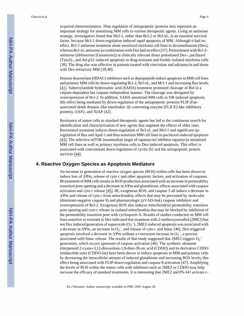
via 2 main pathways that lead to the activation of caspases: the death receptor (DR) pathwayand the mitochondrial pathway [4]. Caspases are members of a cysteine protease family thatare synthesized as inactive zymogens. They are responsible for activation of various cellularproteases and endonucleases. The result of activation is cleavage of structural and regulatorycellular proteins and of nuclear DNA. This process leads to morphologic and biochemicalcellular changes that are characteristic of apoptosis. The DR pathway is activated by ligationof members of the tumor necrosis factor (TNF) family, such as Fas ligand (FasL; also calledApo1), TNF-α, and Apo2 ligand (Apo2L; also called TNF-related apoptosis-inducing factor[TRAIL]), to DR on the plasma membrane. The receptors of these ligands—Fas, TNFR1 (TNFreceptor 1), and DR4/DR5—are members of the TNF receptor superfamily, which ischaracterized by similar, cysteine-rich extracellular domains and homologous cytoplasmicdeath domains [5]. Binding of these DRs by their respective ligands recruits the adaptor proteinsFas-associated death domain (FADD) and/or TNFR1-associated death domain (TRADD) andcaspase 8 to the death-inducing signaling complex, triggering the proteolytic activation ofcaspase 8. Caspase 8 in turn activates the effector caspases 3 and 7 [6] (Figure 1). The resultis proteolytic targeting of key apoptotic or cell cycle regulatory proteins, such as Bcl-2 [7] andcyclin E [8].
The mitochondrial pathway is regulated primarily by members of the Bcl-2 family, whichcomprises both proapoptotic and antiapoptotic proteins. The ratio between these subsetsdetermines the susceptibility of cells to various death signals [4]. All contain at least 1 of the4 conserved Bcl-2 homology (BH) domains [9]. The antiapoptotic members include Bcl-2,Bcl-xL, Bcl-w, Mcl-1, and Bfl-1/A1, which are characterized by the presence of all 4 BHdomains (BH1-BH4). Most members also contain a C-terminal hydrophobic tail, which cantarget these proteins predominantly to the mitochondria and/or endoplasmic reticulum. Theseproteins prevent cell death by binding and sequestering proapoptotic proteins. The deathpromoter members are subdivided according to their function and biochemical structure intomultidomain proteins, such as Bax and Bak, that closely resemble Bcl-2 but lack the N-terminalBH4 domain. The other subgroup is represented by BH3-only members that possess the BH3domain required for binding to other members and for death-promoting activity. The BH3-only proteins include Bik, Bid, Puma/BBC3, and Noxa/APR, each thought to be responsiblefor transducing a specific cell death signal [10].
After their activation, Bax and Bak undergo conformational changes that contribute toincreased permeability of the outer mitochondrial membrane through formation of pores. Baxand Bak also facilitate the release of cytochrome c (cyto c) and sequential activation of thecaspase cascade [11]. Cytosolic cyto c binds the adaptor molecule APAF-1 (apoptotic proteaseactivating factor 1) and caspase 9, forming a macromolecular complex, the apoptosome. Thisprocess leads to activation of caspase 9 [12]. Activated caspase 9 cleaves caspase 3, whichcleaves and activates the other effector caspases (caspases 6 and 7). Additional moleculesreleased from the mitochondria include apoptosis-inducing factor [13] and endonuclease G[14], which exert their apoptotic activity on nuclei. Smac/Diablo [15–17] and Omi/HtrA2[18,19] facilitate caspase activation by antagonizing molecules belonging to the inhibitor ofapoptosis (IAP) family.
A number of recent reviews describe the biology of apoptosis and the clinical applications inMM [1,20–23]. We focus on the key concepts and some of the most recent studies thatsummarize the field of MM apoptosis.
2. Role of the Mitochondrial Pathway in ApoptosisIonizing radiation (IR) and chemotherapeutic agents act primarily through the intrinsicpathway, in which mitochondria play the central role. DNA-damaging agents signal cell death
Oancea et al. Page 2
Int J Hematol. Author manuscript; available in PMC 2005 August 29.
NIH
-PA Author Manuscript
NIH
-PA Author Manuscript
NIH
-PA Author Manuscript

by altering the mitochondrial transmembrane potential (ΔΨm), activating Bcl-2 familymembers with subsequent cyto c release, and activating the caspase family of proteins [7,21].Bcl-2, frequently expressed in follicular lymphomas bearing the t(14;18) chromosomaltranslocation, is also widely expressed in many other B- and T-cell lymphomas withoutinvolvement of a bcl-2 rearrangement [9,21].
The effect of genotoxic agents, such as IR and chemotherapeutic drugs, is directed through themitochondrial pathway in a p53-dependent manner. BH3-only members of the Bcl-2 family—Bik, Puma, and Noxa—are believed to play a central role in p53-activated cell death [24,25]. We observed that after IR the BH3-only genes puma, noxa, and bik were up-regulated inMM and lymphoma tumor cells. Eight and 16 hours after IR, ribonuclease protection assaysindicated dramatic transcriptional induction of Bik, and there were similar changes in proteinlevels. In contrast, an increase in Noxa messenger RNA (mRNA) levels was observed as earlyas 0.5 hours after IR, and Puma levels had increased by 4 hours after IR. The differences inkinetics of induction of these BH3-only proteins indicated their distinct role in apoptosisactivation in MM cells (M.O., A.A., unpublished data). Because Bid is not activated by IR[7], the identity of the Bax- and/or Bak-activating BH3-only protein is of great interest.
3. Bcl-2 Proteins Are Key Targets of TherapeuticsImbalances in expression levels of the Bcl-2 family members result in defects in programmedcell death associated with chemoresistance, malignancy, and aggressiveness of tumors. Theexpression pattern of the Bcl-2 family of proapoptotic and antiapoptotic genes in MM havebeen the subject of multiple studies in which the investigators found increased levels ofexpression of Bcl-2, Bcl-xL, and Mcl-1 are linked to MM cell survival and resistance tochemotherapeutic agents [21,26-28]. The expression pattern of the Bcl-2 family separates themalignant phenotype of MM from normal plasma cells. In MM there is higher expression ofthe antiapoptotic Bcl-2 and Mcl-1 but not of Bcl-xL, and there is a lower level of expressionof Bax [29]. On the other hand, targeted overexpression of Bcl-xL and c-Myc in B-lymphoidcells in mice resulted in lymphoproliferative disease and plasma cell malignancies. Thesefindings were evidence that Bcl-xL can contribute to plasmacytomagenesis [30]. Bcl-xLexpression is also associated with drug resistance in MM patients [31].
Chemotherapeutic agents, such as doxorubicin (Dox) induce apoptosis by causing cyto crelease from mitochondria and subsequent activation of caspases, which are blocked byoverexpression of Bcl-2. Treatment of U266 cells with Dox increased activation of Bax andBak as well as of the BH3-only proteins Bid and Bik [32]. Arsenic trioxide (ATO) has beenshown to induce apoptosis in MM cells [33] by directly inducing cyto c release frommitochondria via the mitochondrial permeability transition pore. The voltage-dependent anionchannel was identified as a biological target of ATO [34]. Recent studies showed 2 distinctpathways for ATO-induced death in MM, depending on their p53 status. ATO treatment ofcells with mutated p53 resulted in G2/M cell-cycle phase block. In contrast, cells with wild-type p53 were blocked in G1. Moreover, apoptosis may be activated differentially by ATO,with cells having mutated p53 engaging the extrinsic pathway and those having functional p53engaging the intrinsic pathway. Finally, ATO treatment led to up-regulation of Apo2L (TRAIL)receptors and down-regulation of decoy receptors, observations that help explain thesynergistic effect of ATO with Apo2L [35]. Recent published data from a phase 2 study showedthat ATO as monotherapy has therapeutic efficacy in relapsed or refractory MM and that thisagent was well tolerated with manageable adverse effects [36].
Overexpression of the antiapoptotic members has been linked to resistance to variouschemotherapeutic agents. Increased expression of these proteins after exposure tochemotherapeutic agents of MM cell lines suggested that these agents might contribute to
Oancea et al. Page 3
Int J Hematol. Author manuscript; available in PMC 2005 August 29.
NIH
-PA Author Manuscript
NIH
-PA Author Manuscript
NIH
-PA Author Manuscript
acquired chemoresistance. Thus regulation of antiapoptotic proteins may represent animportant strategy for sensitizing MM cells to various therapeutic agents. Using an antisensestrategy, investigators found that Mcl-1, rather than Bcl-2 or Bcl-xL, is an essential survivalfactor, because Mcl-1 down-regulation induced rapid apoptosis of MM. Although it had noeffect, Bcl-2 antisense treatment alone sensitized myeloma cell lines to dexamethasone (Dex),whereas Bcl-xL antisense in combination with Dex had no effect [37]. Pretreatment with Bcl-2–antisense (oblimersen [Genasense]) at clinically relevant doses potentiated Dex-, paclitaxel(Taxol)-, and Ad-p53–induced apoptosis in drug-resistant and freshly isolated myeloma cells[38]. The drug also was effective in patients treated with vincristine and adriamycin and thosewith Dex-refractory MM [39,40].
Histone deacetylase (HDAC) inhibitors such as depsipeptide induce apoptosis in MM cell linesand primary MM cells by down-regulating Bcl-2, Bcl-xL, and Mcl-1 and increasing Bax levels[41]. Suberoylanilide hydroxamic acid (SAHA) treatment promoted cleavage of Bid in acalpain-dependent but caspase-independent manner. The cleavage was abrogated byoverexpression of Bcl-2. In addition, SAHA sensitized MM cells to DR-induced apoptosis,this effect being mediated by down-regulation of the antiapoptotic proteins FLIP (Fas-associated death domain–like interleukin 1β–converting enzyme [FLICE]-like inhibitoryprotein), cIAP2, and XIAP [42].
Resistance of tumor cells to standard therapeutic agents has led to the continuous search foridentification and characterization of new agents that augment the effects of older ones.Resveratrol treatment induces down-regulation of Bcl-xL and Mcl-1 and significant up-regulation of Bax and Apaf-1 and thus sensitizes MM cell lines to paclitaxel-induced apoptosis[43]. The selective mTOR (mammalian target of rapamycin) inhibitor rapamycin sensitizedMM cell lines as well as primary myeloma cells to Dex-induced apoptosis. This effect isassociated with concomitant down-regulation of cyclin D2 and the antiapoptotic proteinsurvivin [44].
4. Reactive Oxygen Species as Apoptosis MediatorsAn increase in generation of reactive oxygen species (ROS) within cells has been shown toinduce loss of ΔΨm, release of cyto c and other apoptotic factors, and activation of caspases.IR treatment of MM cells results in ROS production associated with an increase in permeabilitytransition pore opening and a decrease in ΔΨm and glutathione, effects associated with caspaseactivation and cyto c release [45]. IR, exogenous ROS, and caspase 3 all induce a decrease inΔΨm and release of cyto c from mitochondria, effects that may be prevented by molecular(dominant-negative caspase 9) and pharmacologic (zVAD-fmk) caspase inhibitors andoverexpression of Bcl-2. Exogenous ROS also induces mitochondrial permeability transitionpore opening and cyto c release in isolated mitochondria that may be blocked by inhibition ofthe permeability transition pore with cyclosporin A. Results of studies conducted on MM celllines sensitive or resistant to Dex indicated that treatment with 2-methoxyestradiol (2ME2) butnot Dex induced generation of superoxide (O2
−). 2ME2-induced apoptosis was associated witha decrease in ΔΨm, an increase in O2
−, and release of cyto c and Smac [46]. Dex-triggeredapoptosis involved a decrease in ΔΨm without a concurrent increase in O2
−, a processassociated with Smac release. The results of that study suggested that 2ME2 triggers O2
−
generation, which occurs upstream of caspase activation [46]. The synthetic oleananetriterpenoid 2-cyano-3,12-dioxoolean-1,9-dien-28-oic acid (CDDO) and its derivative CDDOimidazolide ester (CDDO-Im) have been shown to induce apoptosis in MM and primary cellsby decreasing the intracellular amount of reduced glutathione and increasing ROS levels, thiseffect being associated with FLIP down-regulation and caspase 8 activation [47]. Amplifyingthe levels of ROS within the tumor cells with inhibitors such as 2ME2 or CDDO may helpincrease the efficacy of standard treatments. It is interesting that 2ME2 and PS-341 activate c-
Oancea et al. Page 4
Int J Hematol. Author manuscript; available in PMC 2005 August 29.
NIH
-PA Author Manuscript
NIH
-PA Author Manuscript
NIH
-PA Author Manuscript

jun NH2-terminal kinase (JNK), which translocates to mitochondria [48]. JNK recently wasshown to activate a novel apoptotic pathway by releasing Smac rather than cyto c during TNF-induced apoptosis [49], a process similar to Dex-induced apoptosis in MM [23].
5. Role of the DR Pathway in ApoptosisActivation of cell surface DRs, such as DR4 and DR5, by the Apo2L ligand induces a signalingcascade culminating in activation of caspases [5] (Figure 2). In addition to being tumor specific,Apo2L-based therapy is attractive because it is not dependent on p53 and therefore should beeffective in p53-deficient tumors. Apo2L potently induces apoptosis of MM cells from patientsand the majority of MM cell lines, including cells sensitive or resistant to Dex, Dox, melphalan,and mitoxantrone. Apo2L also has overcome the survival effect of interleukin 6 (IL-6). A recentreport indicated that Apo2L is expressed in plasma cells and may be responsible for apoptosisof plasma cells in a caspase-independent manner after antibody secretion [50].
Interferons (IFNs) have been used as effective pharmacological agents in treating a variety ofcancers and viral diseases in the last 30 years. Our studies have shown that type I but not typeII IFNs induce apoptosis through activation of the Apo2L pathway and modulation of the Bcl-2family of proteins in MM cell lines and patient-derived primary cells [6,21]. IFNs play animportant role in the immune system and may influence expression of a number of genesassociated with both apoptosis and cell cycle progression [51]. One mechanism by which IFNsmay achieve their role in immune surveillance may be through regulation of other cytokines.Our data provided evidence that IFNs exert their profound effect by inducing apoptosis in MM.The data also suggested that Apo2L may be an important mediator of these effects. We haveidentified and isolated the promoter region of Apo2L and found that it contains IFN-stimulatedregulatory elements that can be regulated in MM [52]. Cytokines suppress apoptosis bypreventing Apo2L expression through modulation of the forkhead FOXO3a transcriptionfactor [53]. These findings support a rationale for exploring Apo2L as well as the therapiesthat modulate its expression in the management of MM, especially in instances in which IFN-α and IFN-β may be effective, such as in advanced MM. Given the lack of cytotoxicity ofApo2L toward most other types of blood cells, as well as other cell types in mice and nonhumanprimates, it would be of interest to examine whether Apo2L, which is expected to enter clinicaltrials, could be used for treating MM patients.
Apo2L induction was at least partially necessary for proteolytic cleavage-dependent activationof apoptotic activity of Bcl-2 and Bid (Figure 2). In addition, we observed that IFN-α inducesdown-regulation of Bcl-xL, which is expressed at high levels in these cells (A.A., Q. Chen,unpublished data, 2002). Other reports have indicated that the effect of several therapeuticagents could be mediated by down-regulation of Bcl-2 family protein expression [21].Expression of these proteins also may be regulated by the myeloma survival factors IL-6, IFNs,and insulin-like growth factor 1 (IGF-1) [54].
Recent studies have further demonstrated the role of the mitochondrial pathway in IFN-α–induced apoptosis in MM by showing that IFNs activate sequentially the proapoptotic Bcl-2family members Bak and Bax [32]. IFN-α–induced apoptosis can be blocked through inhibitionof the phosphatidylinositol 3-kinase (PI3K)/mTOR pathway. Because many tumor types areresistant to IFN treatment, these findings are an aid to understanding the variability of cellularresponse to IFN treatment and emphasize the importance of knowledge of the signalingpathways responsible for apoptosis induction [6,55].
6. Initiation and Amplification Stages in ApoptosisThe link between the extrinsic and intrinsic pathways of apoptosis through engagement ofmitochondria by truncated Bid provides amplification of the apoptotic signal and further
Oancea et al. Page 5
Int J Hematol. Author manuscript; available in PMC 2005 August 29.
NIH
-PA Author Manuscript
NIH
-PA Author Manuscript
NIH
-PA Author Manuscript

supports the critical role of mitochondria in apoptosis. In contrast, genotoxic agents such as IRand IFNs cause MM apoptosis by 2 distinct stages of cyto c release and a positive feedbackloop linking caspase activation to cyto c release and mitochondrial dysfunction [6,7]. Inaddition, we found that in MM and all other hematopoietic tumor cells examined, a caspase3–generated p18-kd proteolytic fragment of cyclin E produced during genotoxic stress-inducedapoptosis [8] will further amplify the process by activating Bax. Bax is sequestered tocytoplasmic proteins, such as the DNA repair protein Ku70, and is released after p18 cyclin Ebinding to Ku70 (A.A., S. Mazumder, unpublished data, 2004) or acetylation [56].Radiotherapeutics and chemotherapeutics also induce apoptosis through increased expressionof cyclin E [57], perhaps by providing more substrate for genesis of p18 cyclin E.
7. Growth and Survival Factors Prevent ApoptosisMM cells accumulate in the BM, where they acquire growth and survival properties conferredby the BM microenvironment [22]. BM stromal cells (BMSCs) secrete the chemokine stromalcell–derived factor 1α (SDF-1α). This chemokine plays a major role in homing, because MMcells express the SDF-1α receptor, CXCR4 (Figure 3). Once in the BM, integrin α4B1 (VLA-4)mediates attachment of MM cells to the stroma [58], and this process confers the cells a survivaladvantage. Adhesion of MM cells to fibronectin (FN) is known to protect the cells from drug-induced apoptosis [23]. Although nearly all myeloma cells respond to growth and survivalfactors, such as IL-6, only some require them, and only some produce them. Those that do notproduce IL-6 and require it rely on its production by BMSCs [1,59]. The pathophysiology ofMM has been attributed to dysregulation of autocrine growth loops (including IL-6 and IL-1)and various survival pathways, including NF-κB/IκB (nuclear factor κB/inhibitory unit of NF-κB), JAK2/STAT3 (Janus kinase 2/signal transducers and activators of transcription 3), PI3K/Akt, and Ras/Raf/MAPK (Ras/Raf/mitogen-activated protein kinase) [23,60], which actthrough targets such as Bcl-2 or IAPs to prevent their apoptotic demise.
NF-κB refers to a group of dimeric transcription factors regulating various genes the functionsof which include cell growth, angiogenesis, cell adhesion, and protection from apoptosis. NF-κB exists in the cytoplasm in an inactive form bound to IκBα, one of its interacting negativeregulators, which has to be phosphorylated to mark it for degradation by the 26S proteasome.This process leaves NF-κB free to translocate to the nucleus [61], where it binds to its targets,such as cyclin D1 [61], Bcl-xL, and IAPs [62]. NF-κB, because of its striking importance inMM, continues to be a compelling target for drug development. Dex, thalidomide, PS-341,and curcumin all inhibit NF-κB [23,63]. Combined with the Chk1 abrogator UCN-01, Bay11-7082, an irreversible inhibitor of IκBα phosphorylation, and the NF-κB inhibitor SN50effectively induce apoptosis [60]. NF-κB is responsible for up-regulation of IAPs and Bcl-2[62] to control cell survival and for expression of ICAM1 and VCAM1 (intercellular andvascular adhesion molecules 1) on MM cells and BMSCs [1]. Adhesion molecules such asthese and LFA-1 (lymphocyte function–associated antigen 1) facilitate the BMSC-MM cellinteraction [20,64].
Posttranslational modifications involve ubiquitination and proteasome degradation.Proteasome inhibitors block accumulation of IκB in the cytoplasm, inhibiting the NF-κB cellsurvival pathway and leading to apoptosis. A transcriptional profile of MM.1S cells treatedwith the bortezomib/proteasome inhibitor PS-341 (Velcade) revealed distinct patterns ofcoordinated changes in a range of transcripts. The changes included down-regulation of growthand antiapoptotic transcripts and induction of apoptotic members, such as the Fas ligand andreceptor. The effect of the agent was blocked by a dominant-negative Fas [65]. Moreover,coadministration of proteasome and HDAC inhibitors in MM cell lines sensitive or resistantto established cytotoxic agents resulted in a synergistic increase in mitochondrial injury,caspase activation, and apoptosis [66]. Furthermore, IL-6–induced growth was associated with
Oancea et al. Page 6
Int J Hematol. Author manuscript; available in PMC 2005 August 29.
NIH
-PA Author Manuscript
NIH
-PA Author Manuscript
NIH
-PA Author Manuscript

high levels of CDC34 (ubiquitin-conjugating enzyme [UBC3]), IL-6–mediated protectionfrom Dex-induced apoptosis being dampened by blocking of CDC34. CDC34 is responsiblefor ubiquitination of various proteins, including IκBα. MM cells express high levels of CDC34at both the mRNA and the protein levels. Dex, 2ME2, and PS-341 treatments are associatedwith decreased CDC34 expression. Blocking CDC34 augments sensitivity to these therapies[67]. Targeting mitochondria may be an effective way to overcome conventional resistanceand Velcade resistance in MM [68].
MM cell adhesion to BMSCs up-regulates secretion of vascular endothelial growth factor(VEGF) [64] and IL-6 by BMSCs. IL-6 secretion by BMSCs induces MM cells to producetheir own IL-6. Secretion by MM cells in turn increases IL-6 secretion by BMSCs. VEGF isproduced and secreted by both MM cells and BMSCs [64]. FLT1, a high-affinity VEGFreceptor on MM cells, when phosphorylated by VEGF activates p42/p44/MAPK, leading toMM cell proliferation. VEGF is known to stimulate angiogenesis but also induces up-regulation of MCL-1 (myeloid cell leukemia 1), thus protecting MM cells against apoptosis[69]. In a PI3K/Akt/PKC (protein kinase C)-dependent manner, VEGF promotes migration ofMM cells from BM [1]. IGF-1 mediates migration through PI3K as well [70]. CD40, a TNFfamily transmembrane protein expressed in most MM cells, may have a role in MM cell homingand migration by induction of VEGF [71].
IL-6, a major survival factor for MM tumor cells, induces signaling through the STAT proteins.STAT3 is constitutively activated in BM mononuclear cells from patients with MM and in theIL-6–dependent human MM cell line U266 [27]. IL-6–induced Dex resistance occurs in partthrough JAK/STAT3 signaling [1,31]. MM cells from patients have been shown to expressconstitutively active forms of NF-κB and STAT3, whereas cells from healthy subjects havenot [31]. Up-regulation of Bcl-xL and Mcl-1 via retroviral insertion of activated STAT3 andSTAT5A, targets of both IL-6 and IGF-1, causes cell growth, oncogenesis, and cytokineindependence [59]. Fibroblast growth factor receptor 3 (FGFR3), frequently overexpressedowing to a t(4:14) translocation, also confers a growth and survival advantage, possibly byactivating STAT3 and resulting in Bcl-xL expression [72]. Bcl-xL expression can be inhibitedby blocking IL-6 receptor signaling from JAKs to the STAT3 protein, demonstrating thatSTAT3 signaling is essential for the survival of MM tumor cells [27]. IL-6, via Shp2/RAFTK(related adhesion focal tyrosine kinase) prevents a Dex-induced increase in Smac, thuspreventing apoptosis. Dex, in addition to triggering apoptosis, has the effect of transientlyincreasing expression of IL-6 receptor and transforming growth factor β receptor II andincreasing production and release of IL-6 [22].
IL-6 and IGF-1 afford protection against Dex-induced apoptosis by up-regulation of Bcl-xL,and Mcl-1 (through STATs) and activation of the MAPK and PI3K/Akt pathways, whichcontribute to inactivation of caspase 9 [61,64]. IGF-I has been reported to have multiplefunctions that lead to sustained activation of NF-κB and Akt, phosphorylation of FKHRL-1(forkhead homolog of rhabdomyosarcoma receptor ligand 1), up-regulation of FLIP, survivin,cIAP-2, Bfl-1, and XIAP, and a decrease in Apo2L sensitivity [23,73]. More recently it hasbeen shown that, via extracellular signal–regulated kinase (ERK), IGF-1 also induces VEGFsecretion [70]. IL-6 and IGF-1 have separate receptors but have substantial downstream overlapof pathways. Simvastatin (a 3-hydroxy-3-methylglutaryl coenzyme A inhibitor) inducesapoptosis in vitro despite protective influences of IL-6, IGF-1, and VLA-4 (which allows MMcell adhesion to FN). This finding suggests a common downstream pathway for IL-6, IGF-1,and VLA-4, possibly through PI3K [74].
Oancea et al. Page 7
Int J Hematol. Author manuscript; available in PMC 2005 August 29.
NIH
-PA Author Manuscript
NIH
-PA Author Manuscript
NIH
-PA Author Manuscript
8. Future DirectionsNovel therapeutic targets in MM emerge from proteins identified through microarray [1,75]and proteomics strategies [20]. Gene expression profiling provides essential information onexpression and regulation of many genes in MM, including cell DRs (eg, DR4/5, BAFFR [B-cell–activating factor receptor], BCMA [B-cell maturation antigen], TACI [transmembraneactivator and calcium modulator and cyclophilin ligand interactor]) and their ligands (Apo2L/TRAIL, APRIL [a proliferation-inducing ligand], BAFF, and CD40) [75] (M.O., A.A.,unpublished data). Exogenous cytokine-dependent MM cells were reported to overexpressreceptors for APRIL and BAFF. All 3 BAFF receptors—TACI, BCMA, and BAFFR—activateNF-κB. APRIL induces up-regulation of Mcl-1 and Bcl-2, and IL-6 up-regulates Mcl-1 only.Both BAFF and APRIL have been shown to be as effective as IL-6 in preventing Dex-inducedapoptosis. IL-6 has induced phosphorylation of STAT3, MAPK, and Akt (activating the JAK/STAT, MAPK, and PI3K/Akt cascades, respectively) [2]. IL-6–producing MM cells, viaactivated STAT3, are resistant to Fas [59]. IGF-1 has been shown to activate MAPK and PI3K/Akt. BAFF and APRIL have induced phosphorylation of only MAPK and Akt (activating theERK1/2 and PI3K/Akt cascades, respectively) [76]. Establishing the function of thesemolecules in MM and their validation as targets in clinical therapy will broaden ourunderstanding of the molecular determinants of the disease and provide additional tools thatmay help to cure it.
Acknowledgements
Supported in part by research grants CA81504 and CA82858 to A.A. from the National Cancer Institute.
References1. Hideshima T, Anderson KC. Molecular mechanisms of novel therapeutic approaches for multiple
myeloma. Nat Rev Cancer 2002;2:927–937. [PubMed: 12459731]2. Hussein MA, Juturi JV, Lieberman I. Multiple myeloma: present and future. Curr Opin Oncol
2002;14:31–35. [PubMed: 11790977]3. Arends MJ, Wyllie AH. Apoptosis: mechanisms and roles in pathology. Int Rev Exp Pathol
1991;32:223–254. [PubMed: 1677933]4. Danial NN, Korsmeyer SJ. Cell death: critical control points. Cell 2004;116:205–219. [PubMed:
14744432]5. Almasan A, Ashkenazi A. Apo2L/TRAIL: apoptosis signaling, biology, and potential for cancer
therapy. Cytokine Growth Factor Rev 2003;14:337–348. [PubMed: 12787570]6. Chen Q, Gong B, Mahmoud-Ahmed A, et al. Apo2L/TRAIL and Bcl-2–related proteins regulate type
I interferon-induced apoptosis in multiple myeloma. Blood 2001;98:2183–2192. [PubMed: 11568006]7. Chen Q, Gong B, Almasan A. Distinct stages of cytochrome c release from mitochondria: evidence
for a feedback amplification loop linking caspase activation to mitochondrial dysfunction in genotoxicstress induced apoptosis. Cell Death Differ 2000;7:227–233. [PubMed: 10713737]
8. Mazumder S, Chen Q, Gong B, Drazba JA, Buchsbaum JC, Almasan A. Proteolytic cleavage of cyclinE leads to inactivation of associated kinase activity and amplification of apoptosis in hematopoieticcells. Mol Cell Biol 2002;22:2398–2409. [PubMed: 11884622]
9. Cory S, Adams JM. The bcl2 family: regulators of the cellular life-or-death switch. Nat Rev Cancer2002;2:647–656. [PubMed: 12209154]
10. Huang DC, Strasser A. BH3-only proteins: essential initiators of apoptotic cell death. Cell2000;103:839–842. [PubMed: 11136969]
11. Desagher S, Martinou JC. Mitochondria as the central control point of apoptosis. Trends Cell Biol2000;10:369–377. [PubMed: 10932094]
12. Strasser A, O’Connor L, Dixit VM. Apoptosis signaling. Annu Rev Biochem 2000;69:217–245.[PubMed: 10966458]
Oancea et al. Page 8
Int J Hematol. Author manuscript; available in PMC 2005 August 29.
NIH
-PA Author Manuscript
NIH
-PA Author Manuscript
NIH
-PA Author Manuscript

13. Joza N, Susin SA, Daugas E, et al. Essential role of the mitochondrial apoptosis-inducing factor inprogrammed cell death. Nature 2001;410:549–554. [PubMed: 11279485]
14. Li LY, Luo X, Wang X. Endonuclease G is an apoptotic DNase when released from mitochondria.Nature 2001;412:95–99. [PubMed: 11452314]
15. Du C, Fang M, Li Y, Li L, Wang X. Smac, a mitochondrial protein that promotes cytochrome c-dependent caspase activation by eliminating IAP inhibition. Cell 2000;102:33–42. [PubMed:10929711]
16. Verhagen AM, Ekert PG, Pakusch M, et al. Identification of DIABLO, a mammalian protein thatpromotes apoptosis by binding to and antagonizing IAP proteins. Cell 2000;102:43–53. [PubMed:10929712]
17. Srinivasula SM, Hegde R, Saleh A, et al. A conserved XIAP-interaction motif in caspase-9 and Smac/DIABLO regulates caspase activity and apoptosis. Nature 2001;410:112–116. [PubMed: 11242052]
18. Hegde R, Srinivasula SM, Zhang Z, et al. Identification of Omi/HtrA2 as a mitochondrial apoptoticserine protease that disrupts inhibitor of apoptosis protein-caspase interaction. J Biol Chem2002;277:432–438. [PubMed: 11606597]
19. Martins LM, Iaccarino I, Tenev T, et al. The serine protease Omi/HtrA2 regulates apoptosis by bindingXIAP through a reaper-like motif. J Biol Chem 2002;277:439–444. [PubMed: 11602612]
20. Hideshima T, Bergsagel PL, Kuehl WM, Anderson KC. Advances in biology of multiple myeloma:clinical applications. Blood 2004;104:607–618. [PubMed: 15090448]
21. Chen Q, Ray S, Hussein MA, Srkalovic G, Almasan A. Role of Apo2L/TRAIL and Bcl-2-familyproteins in apoptosis of multiple myeloma. Leuk Lymphoma 2003;44:1209–1214. [PubMed:12916874]
22. Chauhan D, Anderson KC. Mechanisms of cell death and survival in multiple myeloma (MM):therapeutic implications. Apoptosis 2003;8:337–343. [PubMed: 12815276]
23. Chauhan D, Hideshima T, Anderson KC. Apoptotic signaling in multiple myeloma: therapeuticimplications. Int J Hematol 2003;78:114–120. [PubMed: 12953804]
24. Yu J, Zhang L, Hwang PM, Kinzler KW, Vogelstein B. PUMA induces the rapid apoptosis ofcolorectal cancer cells. Mol Cell 2001;7:673–682. [PubMed: 11463391]
25. Oda E, Ohki R, Murasawa H, et al. Noxa, a BH3-only member of the Bcl-2 family and candidatemediator of p53-induced apoptosis. Science 2000;288:1053–1058. [PubMed: 10807576]
26. Zhang B, Gojo I, Fenton RG. Myeloid cell factor-1 is a critical survival factor for multiple myeloma.Blood 2002;99:1885–1893. [PubMed: 11877256]
27. Catlett-Falcone R, Landowski TH, Oshiro MM, et al. Constitutive activation of Stat3 signaling confersresistance to apoptosis in human U266 myeloma cells. Immunity 1999;10:105–115. [PubMed:10023775]
28. Tu Y, Renner S, Xu F, et al. BCL-X expression in multiple myeloma: possible indicator ofchemoresistance. Cancer Res 1998;58:256–262. [PubMed: 9443402]
29. Spets H, Stromberg T, Georgii-Hemming P, Siljason J, Nilsson K, Jernberg-Wiklund H. Expressionof the bcl-2 family of pro- and anti-apoptotic genes in multiple myeloma and normal plasma cells:regulation during interleukin-6 (IL-6)-induced growth and survival. Eur J Haematol 2002;69:76–89.[PubMed: 12366710]
30. Linden M, Kirchhof N, Carlson C, Van Ness B. Targeted overexpression of Bcl-XL in B-lymphoidcells results in lymphoproliferative disease and plasma cell malignancies. Blood 2004;103:2779–2786. [PubMed: 14656874]
31. Bharti AC, Shishodia S, Reuben JM, et al. Nuclear factor-kappaB and STAT3 are constitutively activein CD138+ cells derived from multiple myeloma patients, and suppression of these transcriptionfactors leads to apoptosis. Blood 2004;103:3175–3184. [PubMed: 15070700]
32. Panaretakis T, Pokrovskaja K, Shoshan MC, Grander D. Interferon-alpha–induced apoptosis in U266cells is associated with activation of the proapoptotic Bcl-2 family members Bak and Bax. Oncogene2003;22:4543–4556. [PubMed: 12881711]
33. Park WH, Seol JG, Kim ES, et al. Arsenic trioxide-mediated growth inhibition in MC/CAR myelomacells via cell cycle arrest in association with induction of cyclin-dependent kinase inhibitor, p21, andapoptosis. Cancer Res 2000;60:3065–3071. [PubMed: 10850458]
Oancea et al. Page 9
Int J Hematol. Author manuscript; available in PMC 2005 August 29.
NIH
-PA Author Manuscript
NIH
-PA Author Manuscript
NIH
-PA Author Manuscript

34. Zheng Y, Shi Y, Tian C, et al. Essential role of the voltage-dependent anion channel (VDAC) inmitochondrial permeability transition pore opening and cytochrome c release induced by arsenictrioxide. Oncogene 2004;23:1239–1247. [PubMed: 14647451]
35. Liu Q, Hilsenbeck S, Gazitt Y. Arsenic trioxide-induced apoptosis in myeloma cells: p53-dependentG1 or G2/M cell cycle arrest, activation of caspase-8 or caspase-9, and synergy with APO2/TRAIL.Blood 2003;101:4078–4087. [PubMed: 12531793]
36. Hussein MA, Saleh M, Ravandi F, Mason J, Rifkin RM, Ellison R. Phase 2 study of arsenic trioxidein patients with relapsed or refractory multiple myeloma. Br J Haematol 2004;125:470–476.[PubMed: 15142117]
37. Derenne S, Monia B, Dean NM, et al. Antisense strategy shows that Mcl-1 rather than Bcl-2 or Bcl-x(L) is an essential survival protein of human myeloma cells. Blood 2002;100:194–199. [PubMed:12070027]
38. Liu Q, Gazitt Y. Potentiation of dexamethasone-, paclitaxel-, and Ad-p53–induced apoptosis by Bcl-2antisense oligodeoxynucleotides in drug-resistant multiple myeloma cells. Blood 2003;101:4105–4114. [PubMed: 12521996]
39. van de Donk NW, Kamphuis MM, van Dijk M, Borst HP, Bloem AC, Lokhorst HM.Chemosensitization of myeloma plasma cells by an antisense-mediated downregulation of Bcl-2protein. Leukemia 2003;17:211–219. [PubMed: 12529680]
40. van de Donk NW, de Weerdt O, Veth G, et al. G3139, a Bcl-2 antisense oligodeoxynucleotide, inducesclinical responses in VAD refractory myeloma. Leukemia 2004;18:1078–1084. [PubMed:15085157]
41. Khan SB, Maududi T, Barton K, Ayers J, Alkan S. Analysis of histone deacetylase inhibitor,depsipeptide (FR901228), effect on multiple myeloma. Br J Haematol 2004;125:156–161. [PubMed:15059137]
42. Mitsiades N, Mitsiades CS, Richardson PG, et al. Molecular sequelae of histone deacetylase inhibitionin human malignant B cells. Blood 2003;101:4055–4062. [PubMed: 12531799]
43. Jazirehi AR, Bonavida B. Resveratrol modifies the expression of apoptotic regulatory proteins andsensitizes non-Hodgkin’s lymphoma and multiple myeloma cell lines to paclitaxel-inducedapoptosis. Mol Cancer Ther 2004;3:71–84. [PubMed: 14749477]
44. Stromberg T, Dimberg A, Hammarberg A, et al. Rapamycin sensitizes multiple myeloma cells toapoptosis induced by dexamethasone. Blood 2004;103:3138–3147. [PubMed: 15070696]
45. Chen Q, Chai Y-C, Mazumder S, et al. The late increase in intracellular free radical oxygen speciesduring apoptosis is associated with cytochrome c release, caspase activation, and mitochondrialdysfunction. Cell Death Differ 2003;10:323–334. [PubMed: 12700632]
46. Chauhan D, Li G, Sattler M, et al. Superoxide-dependent and - independent mitochondrial signalingduring apoptosis in multiple myeloma cells. Oncogene 2003;22:6296–6300. [PubMed: 13679868]
47. Ikeda T, Nakata Y, Kimura F, et al. Induction of redox imbalance and apoptosis in multiple myelomacells by the novel triterpenoid 2-cyano-3,12-dioxoolean-1,9-dien-28-oic acid. Mol Cancer Ther2004;3:39–45. [PubMed: 14749474]
48. Chauhan D, Li G, Hideshima T, et al. JNK-dependent release of mitochondrial protein, Smac, duringapoptosis in multiple myeloma (MM) cells. J Biol Chem 2003;278:17593–17596. [PubMed:12665525]
49. Deng Y, Ren X, Yang L, Lin Y, Wu XA. JNK-dependent pathway is required for TNFalpha-inducedapoptosis. Cell 2003;115:61–70. [PubMed: 14532003]
50. Ursini-Siegel J, Zhang W, Altmeyer A, et al. TRAIL/Apo-2 ligand induces primary plasma cellapoptosis. J Immunol 2002;169:5505–5513. [PubMed: 12421926]
51. Sangfelt O, Erickson S, Castro J, Heiden T, Einhorn S, Grander D. Induction of apoptosis andinhibition of cell growth are independent responses to interferon-alpha in hematopoietic cell lines.Cell Growth Differ 1997;8:343–352. [PubMed: 9056677]
52. Gong B, Almasan A. Genomic organization and transcriptional regulation of the human Apo2L/TRAIL gene. Biochem Biophys Res Commun 2000;278:747–752. [PubMed: 11095979]
53. Ghaffari S, Jagani Z, Kitidis C, Lodish HF, Khosravi-Far R. Cytokines and BCR-ABL mediatesuppression of TRAIL-induced apoptosis through inhibition of forkhead FOXO3a transcriptionfactor. Proc Natl Acad Sci U S A 2003;100:6523–6528. [PubMed: 12750477]
Oancea et al. Page 10
Int J Hematol. Author manuscript; available in PMC 2005 August 29.
NIH
-PA Author Manuscript
NIH
-PA Author Manuscript
NIH
-PA Author Manuscript

54. Jourdan M, Veyrune JL, Vos JD, Redal N, Couderc G, Klein B. A major role for Mcl-1 antiapoptoticprotein in the IL-6–induced survival of human myeloma cells. Oncogene 2003;22:2950–2959.[PubMed: 12771946]
55. Thyrell L, Hjortsberg L, Arulampalam V, et al. Interferon alpha-induced apoptosis in tumor cells ismediated through the phosphoinositide 3-kinase/mammalian target of rapamycin signaling pathway.J Biol Chem 2004;279:24152–24162. [PubMed: 15056668]
56. Cohen H, Lavu S, Bitterman K, et al. Acetylation of the C terminus of Ku70 by CBP and PCAFcontrols Bax-mediated apoptosis. Mol Cell 2004;13:627–638. [PubMed: 15023334]
57. Mazumder S, Gong B, Almasan A. Cyclin E induction by genotoxic stress leads to apoptosis ofhematopoietic cells. Oncogene 2000;19:2828–2835. [PubMed: 10851086]
58. Parmo-Cabanas M, Bartolome RA, Wright N, Hidalgo A, Drager AM, Teixido J. Integrin alpha4beta1involvement in stromal cell-derived factor-1alpha-promoted myeloma cell transendothelialmigration and adhesion: role of cAMP and the actin cytoskeleton in adhesion. Exp Cell Res2004;294:571–580. [PubMed: 15023543]
59. Hodge DR, Xiao W, Wang LH, Li D, Farrar WL. Activating mutations in STAT3 and STAT5differentially affect cellular proliferation and apoptotic resistance in multiple myeloma cells. CancerBiol Ther 2004;3:188–194. [PubMed: 14726660]
60. Dai Y, Pei XY, Rahmani M, Conrad DH, Dent P, Grant S. Interruption of the NF-kappaB pathwayby Bay 11-7082 promotes UCN-01-mediated mitochondrial dysfunction and apoptosis in humanmultiple myeloma cells. Blood 2004;103:2761–2770. [PubMed: 14645003]
61. Panwalkar A, Verstovsek S, Giles F. Nuclear factor-kappaB modulation as a therapeutic approach inhematologic malignancies. Cancer 2004;100:1578–1589. [PubMed: 15073843]
62. Mitsiades N, Mitsiades CS, Poulaki V, et al. Biologic sequelae of nuclear factor-kappaB blockade inmultiple myeloma: therapeutic applications. Blood 2002;99:4079–4086. [PubMed: 12010810]
63. Bharti AC, Donato N, Singh S, Aggarwal BB. Curcumin (diferuloylmethane) down-regulates theconstitutive activation of nuclear factor-kappa B and IkappaBalpha kinase in human multiplemyeloma cells, leading to suppression of proliferation and induction of apoptosis. Blood2003;101:1053–1062. [PubMed: 12393461]
64. Greenstein S, Krett NL, Kurosawa Y, et al. Characterization of the MM. 1 human multiple myeloma(MM) cell lines: a model system to elucidate the characteristics, behavior, and signaling of steroid-sensitive and -resistant MM cells. Exp Hematol 2003;31:271–282. [PubMed: 12691914]
65. Mitsiades N, Mitsiades CS, Poulaki V, et al. Molecular sequelae of proteasome inhibition in humanmultiple myeloma cells. Proc Natl Acad Sci U S A 2002;99:14374–14379. [PubMed: 12391322]
66. Pei XY, Dai Y, Grant S. Synergistic induction of oxidative injury and apoptosis in human multiplemyeloma cells by the proteasome inhibitor bortezomib and histone deacetylase inhibitors. ClinCancer Res 2004;10:3839–3852. [PubMed: 15173093]
67. Chauhan D, Li G, Hideshima T, et al. Blockade of ubiquitin-conjugating enzyme CDC34 enhancesanti-myeloma activity of bortezomib/proteasome inhibitor PS-341. Oncogene 2004;23:3597–3602.[PubMed: 15094775]
68. Chauhan D, Li G, Podar K, et al. Targeting mitochondria to overcome conventional and bortezomib/proteasome inhibitor PS-341 resistance in multiple myeloma (MM) cells. Blood [Epub ahead ofprint]. June 24, 2004.
69. Le Gouill S, Podar K, Amiot M, et al. VEGF induces MCL-1 upregulation and protects multiplemyeloma cells against apoptosis. Blood [Epub ahead of print]. June 24, 2004.
70. Menu E, Kooijman R, Van Valckenborgh E, et al. Specific roles for the PI3K and the MEK-ERKpathway in IGF-1-stimulated chemotaxis, VEGF secretion and proliferation of multiple myelomacells: study in the 5T33MM model. Br J Cancer 2004;90:1076–1083. [PubMed: 14997210]
71. Tai YT, Catley LP, Mitsiades CS, et al. Mechanisms by which SGN-40, a humanized anti-CD40antibody, induces cytotoxicity in human multiple myeloma cells: clinical implications. Cancer Res2004;64:2846–2852. [PubMed: 15087402]
72. Paterson JL, Li Z, Wen XY, et al. Preclinical studies of fibroblast growth factor receptor 3 as atherapeutic target in multiple myeloma. Br J Haematol 2004;124:595–603. [PubMed: 14871245]
Oancea et al. Page 11
Int J Hematol. Author manuscript; available in PMC 2005 August 29.
NIH
-PA Author Manuscript
NIH
-PA Author Manuscript
NIH
-PA Author Manuscript

73. Tarte K, Jourdan M, Veyrune JL, et al. The Bcl-2 family member Bfl-1/A1 is strongly repressed innormal and malignant plasma cells but is a potent anti-apoptotic factor for myeloma cells. Br JHaematol 2004;125:373–382. [PubMed: 15086420]
74. Osadchy A, Drucker L, Radnay J, Shapira H, Lishner M. Microenvironment factors do not affordmyeloma cell lines protection from simvastatin. Eur J Haematol 2004;73:1–8. [PubMed: 15182331]
75. Ray S, Hissong JG, Oancea M, Almasan A. Expression and regulation of death receptors in multiplemyeloma and prostate carcinoma. In: El-Deiry WS, ed. Death Receptors in Cancer Therapy. Totowa,NJ: Humana Press; 2004:281–296.
76. Moreaux J, Legouffe E, Jourdan E, et al. BAFF and APRIL protect myeloma cells from apoptosisinduced by interleukin 6 deprivation and dexamethasone. Blood 2004;103:3148–3157. [PubMed:15070697]
Oancea et al. Page 12
Int J Hematol. Author manuscript; available in PMC 2005 August 29.
NIH
-PA Author Manuscript
NIH
-PA Author Manuscript
NIH
-PA Author Manuscript

Figure 1.Apoptotic pathways. The death receptor (DR) pathway is activated by ligation of the deathligands (eg, Apo2L/TRAIL [tumor necrosis factor–related apoptosis-inducing factor ligand]and FasL) to their cognate receptors on the cell surface. This process results in sequentialbinding of Fas-associated death domain (FADD) and pro–caspase 8. Active caspase 8 cleavesand activates caspase 3. In addition, caspase 8 cleaves Bid, and truncated Bid translocates tothe mitochondria to promote the release of cytochrome c (Cyto c). The mitochondrial pathwayis activated by a number of stimuli, including chemotherapeutic drugs and ionizing radiation(IR). All these stimuli result in activation and oligomerization of Bax and Bak. These changescontribute to pore formation in the outer mitochondrial membrane and the release of cyto cand other apoptogenic factors. Cyto c promotes activation of caspase 9 and of the effectorcaspases. DNA-damaging agents such as IR induce activation of the p53 that promotestranscription of proapoptotic Bcl-2 family members such as the multidomain Bax and the Bcl-2homology 3 (BH3)-only proteins Puma, Noxa, and Bik. Activation of the BH3-only moleculeseither directly or indirectly results in activation of Bax and Bak. AIF indicates apoptosis-inducing factor.
Oancea et al. Page 13
Int J Hematol. Author manuscript; available in PMC 2005 August 29.
NIH
-PA Author Manuscript
NIH
-PA Author Manuscript
NIH
-PA Author Manuscript

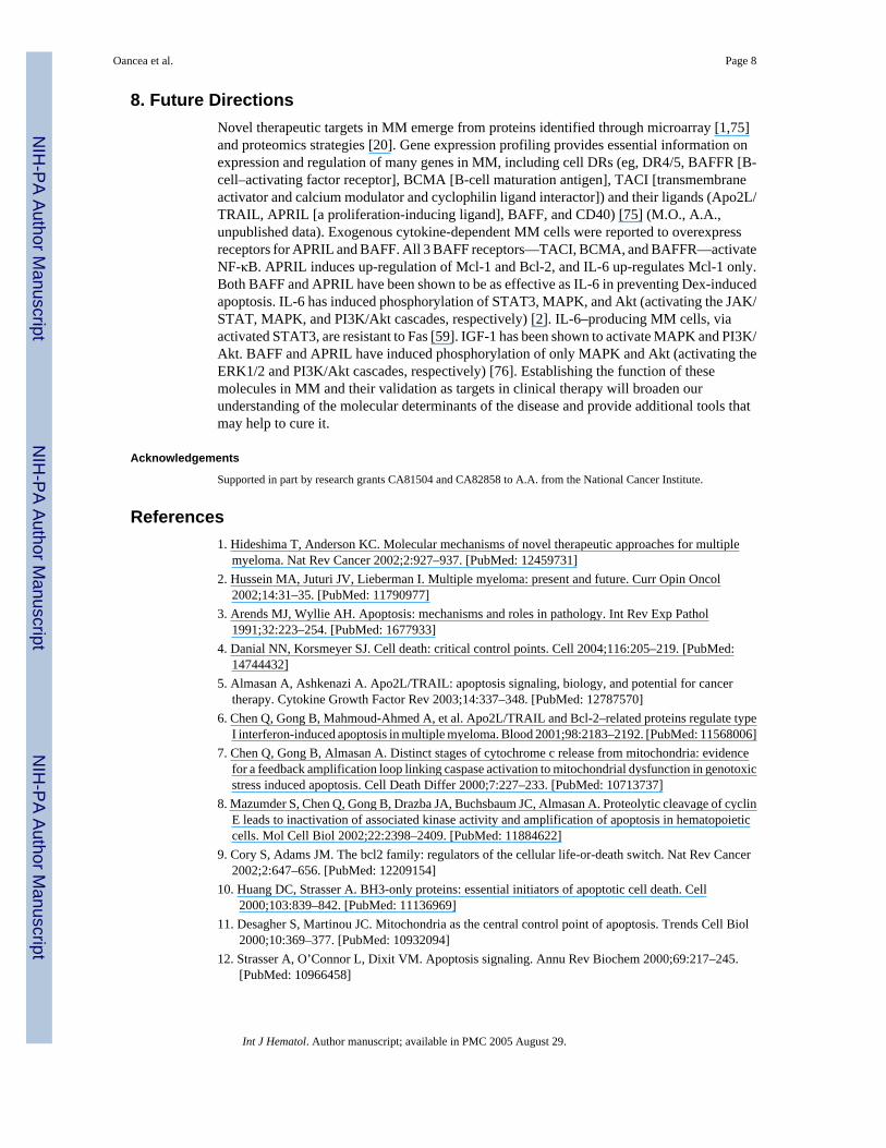
Figure 2.Model for activation of apoptosis in multiple myeloma by interferons (IFNs). Aftertranscriptional induction by IFNs, Apo2 ligand (Apo2L) engages its death receptor 5 (DR5)or DR4 and through an adaptor Fas-associated death domain (FADD) recruits caspase 8 to thecell membrane, which can be blocked by a dominant-negative DR5Δ. After caspase 8 activationby proteolysis, Bid is cleaved and translocates to mitochondria, causing release of low levelsof cytochrome c (cyto c) into the cytosol, a process that leads to caspase 9 and 3 activation.This process results in attack of the antiapoptotic protein Bcl-2 on the mitochondrialmembranes, producing a truncated Bcl-2Δ protein, which causes release of more cyto c, caspaseactivation, and apoptosis. Bcl-xL or Mcl-1 transcriptional down-regulation mediated by signaltransducers and activators of transcription 3 (Stat 3) is an additional mechanism by which IFNsmay decrease levels of antiapoptotic proteins and shift the balance toward a proapoptotic state.(Modified from [6], with permission.) TRAIL indicates tumor necrosis factor–relatedapoptosis-inducing factor ligand; DD, death domain.
Oancea et al. Page 14
Int J Hematol. Author manuscript; available in PMC 2005 August 29.
NIH
-PA Author Manuscript
NIH
-PA Author Manuscript
NIH
-PA Author Manuscript

Figure 3.Growth and survival pathways. Receptors for a proliferation-inducing ligand (APRIL) and B-cell–activating factor (BAFF) include B-cell maturation antigen (BCMA), transmembraneactivator and calcium modulator and cyclophilin ligand interactor (TACI), and BAFF receptor(BAFFR). FLT1 is a high-affinity vascular endothelial growth factor (VEGF) receptor. CXCR4is a receptor for stromal cell–derived factor (SDF1α). These receptors, along with interleukin6 receptor (IL6-R), insulin-like growth factor 1 receptor (IGF1-R), and CD40, mediatedownstream activation of the various pathways. These pathways include nuclear factor κB(NF-κB), Janus kinase/signal transducers and activators of transcription (JAK/STAT),phosphatidylinositol 3-kinase (PI3K)/Akt, and Ras/Raf/mitogen-activated protein kinase(MAPK). These pathways ultimately lead to cytokine production (IL-6, IGF-1, VEGF),resistance to apoptosis (Bcl-2, inhibitors of apoptosis [IAPs]), cell proliferation, and migrationof multiple myeloma (MM) cells. The bone marrow stromal cell (BMSC)/MM cell interactionis mediated by the surface receptors lymphocyte function–associated antigen 1 (LFA1), mucin,(MUC1) integrin α4β1 (VLA4), intracellular adhesion molecule 1 (ICAM1), vascular adhesionmolecule 1 (VCAM1), and fibronectin (FN). Activation of ICAM by cell-cell interaction leadsto secretion of various cytokines from the BMSCs. FGFR3 indicates fibroblast growth factorreceptor 3; MEK, mitogen-activated protein/ERK kinase; SHP2, Src homology region 2domain-containing phosphatase 2; gp130, glycoprotein 130; PKC, protein kinase C; BAD,Bcl-2 antagonist of cell death; FKHR, forkhead homolog of rhabdomyosarcoma; Casp9,caspase 9; mTOR, mammalian target of rapamycin; CycD, cyclin D; ERK, extracellular signal–regulated kinase; nuc, nucleus.
Oancea et al. Page 15
Int J Hematol. Author manuscript; available in PMC 2005 August 29.
NIH
-PA Author Manuscript
NIH
-PA Author Manuscript
NIH
-PA Author Manuscript