The Effect of Beliefs on Writing Synthesis from Multiple Texts
Upload
khangminh22Category
view
0download
0
University of Nebraska Medical Center University of Nebraska Medical Center
DigitalCommons@UNMC DigitalCommons@UNMC
Theses & Dissertations Graduate Studies
Fall 12-16-2016
Chemosensitization Effect of SP1017 on Multiple Myeloma Chemosensitization Effect of SP1017 on Multiple Myeloma
Hangting Hu University of Nebraska Medical Center
Follow this and additional works at: https://digitalcommons.unmc.edu/etd
Part of the Other Pharmacy and Pharmaceutical Sciences Commons
Recommended Citation Recommended Citation Hu, Hangting, "Chemosensitization Effect of SP1017 on Multiple Myeloma" (2016). Theses & Dissertations. 209. https://digitalcommons.unmc.edu/etd/209
This Dissertation is brought to you for free and open access by the Graduate Studies at DigitalCommons@UNMC. It has been accepted for inclusion in Theses & Dissertations by an authorized administrator of DigitalCommons@UNMC. For more information, please contact [email protected].

The CHEMOSENSITIZATION EFFECT OF SP1017 ON MULTIPLE
MYELOMA (MM)
By
Hangting Hu
A DISSERTATION
Presented to the Faculty of
The Graduate College in the University of Nebraska
In partial Fulfillment of the Requirements
For the Degree of Doctor of Philosophy
Department of Pharmaceutical Sciences
Under the Supervision of Professor Tatiana K. Bronich
Medical Center
Omaha, Nebraska
September, 2016

I
ACKNOWLEDGMENTS………………………………………………….............................IV
ABSTRACT…………………………………………………................................................VI
LIST OF FIGURES…………………………………………………................................... VIII
LIST OF TABLES…………………………………………………......................................XIII
LIST OF ABBREVIATIONS…………………………………………………......................XIV
LIST OF CONTRIBUTIONS…………………………………………………....................XVIII
CHAPTER I: INTRODUCTION
1.1. Mutlidrug resistant (MDR) tumors …………………………………………………....1
1.1.1 Pump resistance………………………...…………………...…………………1
1.1.2 Non-pump resistance ……………………...…………………...……………..4
1.2 Strageties to overcome MDR in cancer cells ……...………….……...…………....6
1.2.1 Anticancer drugs capable of overcoming MDR …………………................6
1.2.2 MDR modulator …………………................................................................7
1.2.3 Gene silencing by RNA interference (RNAi) or antisense oligonucleotides
(ASOs) to overcome MDR…....................................................................10
1.2.4 Multifunctional nanoparticles for chemotherapy.......................................13
1.2.5 Combinatorial nanoparticles against MDR in cancer...............................19
1.2.5.1 Combinations of MDR modulators with chemotherapeutics..............19
1.2.5.2 Combinations of MDR-targeted siRNA with chemotherapeutics.......21
1.3 Pluronic® block copolymers for overcoming MDR in cancer...............................22
1.3.1 Pluronic® block copolymers as micellar nanocarriers for drug delivery…24
1.3.1.1 Pluronic® block copolymers structure ………………………………….24
1.3.1.2 Micellization and solubilization ………………………………………….25
1.3.1.3 Stability and drug release of Pluronic® micelles……………………….29

II
1.3.1.4 Pluronic®-based drug delivery systems………………………………...32
1.3.2 Pluronic® block copolymers acting as chemosensitizer for MDR cells….34
1.3.2.1 Structure-functional relationship of Pluronic® block copolymers…….35
1.3.2.2 Inhibition of ABC transporter activity by Pluronic® block copolymers:
role of Pluronic-membrane interaction………………………………….39
1.3.2.3 Effect of Pluronic® block copolymers on cancer cells’ metabolism …41
1.3.2.4 Effect of Pluronic® block copolymers on proapoptotic signaling ...….43
1.3.2.5 Pluronic® block copolymers prevent development of MDR...………..44
1.3.2.6 Pluronic® block copolymers suppress cancer stem cells (CSCs) …..45
1.4 Other applications of Pluronic® block copolymers in cancer treatment ...………47
1.5 Statement of purpose………………………………………………………………...50
1.6 References………………………………………………………………...................53
CHAPTER II: SP1017 SENSITIZE BOTH SENSITIZE AND RESISTANT MM CELLS
BUT NOT NORMAL HEMATOLOGICAL CELLS OR NON-HEMATOLOGICAL
CANCER CELLS
2.1 Introduction………………………………………………………………...................77
2.2 Materials and methods……………………………………………….......................80
2.3 Results and discussion………………………………………………......................86
2.4 Conclusions…………………………………………….........................................101
2.5 References……………………………………………..........................................103
CHAPTER III: THE MOLECULAR MECHANISM OF THE SENSITIZATION EFFECT
OF THE COMBINATION THERAPY TO MM.
3.1 Introduction………………………………………………………….……................111
3.2 Materials and methods……………………………………………….....................122
3.3 Results and discussion………………………………………………....................130

III
3.4 Conclusions…………………………………………….........................................160
3.5 References……………………………………………..........................................162
CHAPTER IV: THE ANTI-TUMOR ACTIVITY OF THE BTZ+SP1017 COMBINATION
THERAPY IN HUMAN MM/SCID MODELS.
4.1 Introduction……………………………………………………………….................172
4.2 Materials and methods……………………………………………….....................174
4.3 Results and discussion………………………………………………....................179
4.4 Conclusions…………………………………………….........................................189
4.5 References……………………………………………..........................................191
CHAPTER V: SUMMARY……………………………………...........................................194
IV
ACKNOWLEDGEMENTS
First and foremost, I would like to extend my sincere appreciation to my
supervisor, Dr. Tatiana K. Bronich. During the past 5 years, she has been always there
willing to listen and help me understand the complicated scientific problems. I have been
constantly inspired and encouraged by her hard work, broad knowledge, critical thinking,
and perpetual energy in scientific research. I appreciate all her contributions of time,
ideas, and funding to support my doctoral study. With her as a mentor, I have learned
many things not only professionally but also personally. Being as a great scientist, she
still stays humble and keeps on learning and exploring new scientific knowledge. Her
positive attitude and great passion in science really motivate and inspire me and I think I
will keep learning in my future professional life. It is my great pleasure to work under her
supervision and I am heartily thankful for her excellent mentoring.
I also would like to thank our collaborators, Dr. Edward A. Faber, Dr. Armen
Petrosyan, Dr. Alexander V. Kabanov and Dr. Daria Y. Alakhova for their discussions
and advices in the project. Dr. Edward A. Faber had the bi-weekly or monthly meeting
with us to follow the progress of my project and also gave valuable ideas and comments.
Dr. Armen Petrosyan was involved in the study of translocation of Cy5-L61 to ER and
Golgi apparatus and Golgi fragmentation. I really appreciate their contributions to my
project.
I would extend my gratitude to the members of my advisory committee which
include Dr. Natalia A. Osna, Dr. Vimla Band and Dr. Jered C. Garrison for their constant
support, advice and guidance throughout my study. They reviewed my work and gave
me their insightful comments in every committee meeting, which are valuable for my
project. Dr. Natalia A. Osna also advised and helped me in the study of proteasome
inhibition and caspases activations.

V
I would like to thank all past and present members of the laboratory. In particular,
I would like to thank Dr Swapnil S. Desale for teaching and helping with the tumor
implantation of the mice model, Dr. Fan Lei for helping with the purification of Cy5-L61,
Dr. Shaheen Ahmed for helping with the animal study of biodistribution of Cy5-L61, Kurti
Soni for helping me with English editing and presentation and for all her
encouragements in my bad situations, Xinyuan Xi, Dr. Svetlana Romanova, Tong Liu,
Dr. Chantey Morris, Dr. Anya Brynskikh Boyum, Dr. Hardeep Singh Oberoi, Dr Jinjin
Zhang and many others for their help in the lab and support. Many thanks to the
administrative staff (Christine Allmon, Jamie Arbaugh, Keith Sutton, and Katina Winters)
for their administration support. I would like to acknowledge the technical assistance
from UNMC core facilities in my research and financial support from NIH, China
Scholarship Council and UNMC Graduate Assistantship.
Lastly, I would like to thank my parents and all my friends in Omaha for their love,
encouragement and support in the past five years.

VI
THE CHEMOSENSITIZATION EFFECT OF SP1017 ON MULTIPLE MYELOMA (MM)
Hangting Hu, Ph.D.
University of Nebraska Medical Center, 2016
Advisor: Tatiana K. Bronich, Ph.D.
Multiple myeloma (MM) is a hematological malignancy of plasma cells that are
predominantly located in bone marrow (BM). Despite recent improvements in MM
treatment by introduction of several novel agents including immunomodulatory drugs,
proteasome inhibitors and the use of the drug combinations, MM remains incurable and
almost all patients eventually relapse or become refractory to the current treatment
regimens. So the current challenge of MM treatments is to maintain treatment response,
prevent relapse and eventually prolong survival.
Here we demonstrated that Pluronic block copolymers ((Pluronic L61: Pluronic
F127 = 1:8 w/w, SP1017) significantly increase cytotoxicity of proteasome inhibitors
(Bortezomib, BTZ or Carfilzomib, CFZ) in a panel of MM cells. Specifically, SP1017
(0.005%) co-treatment triggered 2-fold increase in drug cytotoxicity to MM cells. Lower
concentrations of SP1017 (0.002%) showed lower, but still significant cytotoxicity when
combined with proteasome inhibitors. The mechanistic basis for sensitization effect of
SP1017 was multifactorial and included: 1) augmented inhibition of chymotrypsin-like
proteolytic activity, concomitant with increased accumulation of poly-ubiquitinated
proteins and proteotoxic stress; 2) translocation of Pluronic L61 into the endoplasmic
reticulum (ER) and increase of ER stress response; 3) translocation of Pluronic L61 into
the Golgi apparatus and increase of rate of Golgi fragmentation, concomitant with
reduction of secretion of paraprotein; 4) enhanced depletion of reduced glutathione, that
is essential for mitigation of drug-induced oxidative stress; 5) enhanced pro-apoptotic

VII
activity of the proteasome inhibitors and; 6) decreased anti-apoptotic defense in MM
cells. Importantly, SP1017 co-treatments restore drug sensitivity in BTZ/CFZ-resistant
MM cells. Further studies have revealed that SP1017 co-treatment could also sensitize
MM cells in co-culture models of the BM microenvironment, which triggers cytokine- and
adhesion-mediated MM drug resistance. These results provide support for the design of
therapeutic strategies aimed to counteract the drug resistance mechanisms in MM. We
also demonstrated that combination of BTZ and SP1017 exerted enhanced antitumor
efficacy in human MM/SCID mice model compared to BTZ alone, delaying the disease
progression without causing systemic toxicity or hematological toxicity. Moreover, we
observed the accumulation of the Cy5-L61 in the BM, which was proved by both
fluorescence imaging and flow cytometry, indicating that Pluronic L61 could target and
accumulate within the BM, thus playing an important role in sensitizing MM cells in the
bone microenvironment to proteasome inhibitors.

VIII
LIST OF FIGURES
1.1 Mechanisms of multidrug resistance………………………..……………..……...….2
1.2 Pluronic® block copolymers contain two hydrophilic EO blocks and a hydrophobic
PO block………………………..……………………………………………………....26
1.3 Multiple effects of Pluronic® block copolymers in MDR cells……………………...36
1.4 (A) Effects of Pluronic® block copolymers on cytotoxicity of DOX with respect to
MDR cells. (B) Efficacy of Pluronic® block copolymer composition in MDR KBv
cells…………………………………………………………………………………......38
1.5 (A) Time course of the development of drug resistance in MCF7 cell lines
cultured with Dox either alone or in combination with 0.001% P85. (B) Western
blot data for expression of Pgp in MCF7 parental cells, and selected MCF7/ADR
cells…………………………………………………………………………………...…46
2.1 (A) Cytotoxicity of SP1017 with different concentrations to RPMI 8226 MM cells
after 24h treatment (B) Increased expression of cleaved caspase 9 and caspase
8 in RPMI 8226 MM cells after 24h treatment of different concentrations of
SP1017…………..……………………………………………………………………87
2.2 Percentage of overall cell apoptosis (early and late apoptosis) of RPMI 8225 MM
cells treated with BTZ± 0.005% SP1017 for (A) 8h and (B) 24h by flow cytometric
analysis with annexin V and PI staining ………. …………………………………..90
2.3 Pgp expression by western blot of Rh123 accumulation of sensitive MM cell lines
and BTZ/CFZ-resistant MM cell lines………………………………………………..94
2.4 Pgp functional activity of Rh123 accumulation of (A) RPMI 8226 MM cell line, (B)
ARH-77 MM cell line, (C) BTZ-resistant RPMI 8226 MM cell line and (D) CFZ-
resistant RPMI 8226 MM cell line………………………………………………...….95

IX
2.5 PBMC and RPMI 8226 MM cells were exposed to 10nM BTZ ± 0.005% SP1017
for 20h and assessed for apoptosis by flow cytometric analysis using annexin V
and PI staining………………………………………………………...……………….99
2.6 Cytotoxicity of BTZ± 0.005% SP1017 in (A) RPMI 8226 human MM cell line, (B)
MCF-7 human breast adenocarcinoma cell line, (C) Hela human cervix
adenocarcinoma cell line, and (D) HepG2 human liver carcinoma cell line……100
3.1 The ubiquitin-proteasome pathway..………………………………………...…….113
3.2 (A) The 26S proteasome consists of the 20S core capped with the 19S regulatory
complexes that recognize ubiquitinated protein substrates designated for
proteolysis. (B) One of the constitutive 20S proteasome core β rings which has
seven nonidentical subunits…………………………………………….…………..114
3.3 Intrinsic and extrinsic apoptotic pathways…………………………………………118
3.4 The binding of MM cells to the BMSCs triggers adhesion- and cytokine-mediated
MM cell growth, survival and migration…………………………………………....121
3.5 (A) Chymotrypsin-like proteasome activity of RPMI 8226 MM cells treated with
BTZ± 0.005% SP1017 for 1h, 2h and 4h. (B) Ubiquitinated proteins of RPMI 8226
MM cells treated with BTZ± 0.005% SP1017 for 4h, 8h and 24h……………….131
3.6 (A) HSP70 and (B) CHOP expressions of RPMI 8226 MM cells treated with BTZ±
0.005% SP1017 for 8h, 12h and 24h. (C) GRP 78/BiP expression of RPMI 8226
MM cells treated with BTZ± 0.005% SP1017 for 4h and 8h…………………….133
3.7 Translocation of Cy5-L61 (0.00055% L61 equivalent) into (A) the Golgi apparatus
(Giantin as the Golgi marker) and (B) ER (Calreticulin as the ER marker) of RPMI
8226 MM cells after 8h incubation. Nuclei were counterstained with DAPI (blue).
Quantification of Pearson’s coefficient in cells presented in (C)……………. …135
3.8 Golgi fragmentation of RPMI 8226 MM cells treated with BTZ± 0.005% SP1017
for 8h, as measured by the number of Golgi fragments stained with Giantin….136

X
3.9 Paraprotein (human lambda) level from the supernatant of RPMI 8226 MM cells
treated with BTZ± 0.005% SP1017 for 8h…………………………………………138
3.10 GSH level of (A) sensitive, (B) BTZ- resistant RPMI 8226 MM and (C) CFZ-
resistant RPMI 8226 MM cells treated with drug ± 0.005% SP1017 for 12h….140
3.11 Cell survival of sensitive RPMI 8226 MM cells treated with BTZ± 0.005% SP1017
in presence of antioxidant 10mM NAC……………………………………....…….142
3.12 Co-localization of Cy 5-L61 with mitochondria marker (MitoTracker-Red) in RPMI
8226 MM cell after 20min exposure………………………………………….….....143
3.13 Loss of mitochondrial membrane potential (green positive %) of cells treated with
BTZ± 0.005% SP1017 for (a)12h and (b) 24h, CCCP used as positive control; (c)
Cytochrome c release into cytoplasm of cells were treated with BTZ± 0.005%
SP1017 for 12h……………………………………………………………………….144
3.14 (A) caspase 9 activation, (B) caspase 8 activation and (C) Caspase 3 activation
of cells treated with BTZ± 0.005% SP1017 for 24h…………………………..…..146
3.15 Western blot of cleaved caspase 9, cleaved Caspase 8 and cleaved caspase 3 of
cells treated with BTZ± 0.005% SP1017 for 8h and 24h……………...…………147
3.16 Western blot of cleaved caspase 9, cleaved Caspase 8 and cleaved caspase 3 of
cells treated with CFZ ± 0.005% SP1017 for 24h………………………………...149
3.17 Western blot of anti-apoptotic proteins (Survivin, XIAP, BCL-xl) of cells treated
with BTZ± 0.005% SP1017 for 12h and 24h……………………………….……..150
3.18 Cell survival of sensitive RPMI 8226 MM cell line treated with BTZ± 0.005%
SP1017 for 24h…………………………………………………..……….………….151
3.19 Stromal cells protect multiple myeloma cells by direct contact in Adhesion
Model……………………………………………………………………………….....153
3.20 Cytotoxicity of BTZ ± 0.005%SP1017 to OMA-AD stromal cells after 24h
treatment…………………………………………………………………..………….154

XI
3.21 IL-6 level in the supernatant of stromal cells and stromal cells co-cultured with
MM cells in Adhersion Model after (A) 4h and (B) 24h adhesion, followed by
another 24h drug treatments……..……………………………………………..…..155
3.22 Cell survival of (A) sensitive RPMI 8226 MM cell line and (B) ARH 77 cell line
treated with DEX with different doses for 24h………………………….……..…..157
3.23 Cell survival of (A) sensitive RPMI 8226 MM cell line and (B) ARH 77 cell line
treated with 3-drug combination (BTZ+SP1017+Dex) for 24h…………………..158
3.24 Expression of XIAP of sensitive RPMI 8226 MM cell line treated with 3-drug
combination for 24h…………………………………….…………………………....159
4.1 GFP+/Luc+ RPMI 8226 MM cells in the femur of (a) one control mouse and (b)
one tumor-bearing NSG mouse……………………………………………...……..180
4.2 In vivo antitumor efficacy of BTZ+SP1017 combination therapy in RPMI 8226/Luc
human MM xenograft-bearing NSG mice. (A) Relative paraprotein levels (Pt/P0)
and (B) changes in tumor volume measured by means of BLI over time following
IV administration of 1) 0.9% saline; 2) 0.1% SP1017 (100ul); 3) 0.5mg/kg BTZ; 4)
0.5mg/kg BTZ+0.1% SP1017 (100ul) ……………………………………………..182
4.3 BLI images on the third day after all the 8 injections (Day 28) of four groups of
NSG mice treated with of 1) 0.9% saline; 2) 0.1% SP1017 (100ul); 3) 0.5mg/kg
BTZ; 4) 0.5mg/kg BTZ+0.1% SP1017 (100ul)………………………. …………..183
4.4 Body weight of tumor-bearing NSG mice which are received treatments of 1)
0.9% saline; 2) 0.1% SP1017 (100ul); 3) 0.5mg/kg BTZ; 4) 0.5mg/kg BTZ+0.1%
SP1017 (100ul)………………………………………………………………...….....184
4.5 The fluorescence (Cy5) images of mice skeleton of both (A) tumor-bearing mice
and (B) control mice without tumor after 24h of injection of 0.011% Cy5-
L61detected by IVIS with Ex=640nm and Em=680nm…………………………..186
XII
4.6 The fluorescence (Cy5) images of mice organs (liver, heart, lung, kidney, spleen)
of both (A) tumor-bearing mice and (B) control mice without tumor after 24h of
injection of Cy5-L61 (0.011% L61 equivalent) detected by IVIS with Ex=640nm
and Em=680nm …………………………………………………………………..…187
4.7 (A) Cy5+ cells in BM extracted from both tumor-bearing mice and control mice
with or without injection of Cy5-L61 (0.011% L61 equivalent). (B) Cy5+ cells in
CD45+hematopoietic cells and CD11b+ cells (macrophage/monocyte) in BM
extracted from control mice with injection of Cy5-L61 (0.011% L61 equivalent).
………...…………………………………………………………………………..…..188

XIII
LIST OF TABLES
1.1 Physicochemical characteristics of Pluronic® block copolymers…………..……..27
2.1 Table 2.1 (A) IC50 of BTZ ± Pluronics in different human MM cell lines. (B) IC50 of
CFZ ± Pluronics in different human MM cell lines. ……………………………..…88
2.2 IC50 of BTZ ± Pluronics in RPMI 8226 human MM cell line with different
treatment schedules……………………………………….…………………………..91
2.3 Cross-resistance profile in BTZ/CFZ-resistant cells to BTZ/CFZ ± 0.005%
SP1017 for 24h…………………………………………………………….…………..93
4.1 Blood cell counting after 4 injections and 8 injections of 1) 0.9% saline; 2) 0.1%
SP1017 (100ul); 3) 0.5mg/kg BTZ; 4) 0.5mg/kg BTZ+0.1% SP1017 (100ul)… 185
XIV
LIST OF ABBREVIATIONS
ABC transporters ATP binding cassette transporters
ADH adipic dihydrazide
AIF apoptosis-inducing factor
APAR-1 apoptosis protease activating factor-1
ASOs antisense oligonucleotides
ATF6 activating transcription factor 6
ATP adenosine triphosphate
BBB blood brain barrier
BBMECs microviscosity in bovine brain microvessel endothelial cells
Bcl-2 B-cell CLL/lymphoma 2
Bcl-XL BCL2-like 1
BCRP breast cancer resistance protein
BID domain death agoinst
BLI bioluminescence imaging
BM bone marrow
BMSCs bone marrow stromal cells
BTZ Bortezomib
CCK-8 Cell counting kit-8
CFZ Carfilzomib
CHOP CCAAT/enhancer-binding homologous protein
ChT-L chymotrypsin-like
CM Conditioned medium
CMC critical micelle concentration
CMT critical micelle temperature
CNS central nervous system

XV
c(RGDyK) cyclic RGD [arginine-glycine-aspartic acid] peptide
CSCs cancer stem cells
CyA cyclosporine A
Cy5-L61 Cyanine 5-labeled Pluronic®L61
DEX Dexamethasone
DISC death-inducing signaling complex
DMF Dimethylformamide
DOX doxorubicin
DSF disulfiram
dsRNA double strandard RNA
ECL enhanced chemiluminescence
ECM extracellular matrix
EDTA ethylenediaminetetraacetic acid
ELISA Enzyme-linked Immunosorbent Assay
EPL ɛ-polylysine
EPR enhanced permeability and retention
ER endoplasmic reticulum
FACS Fluorescence-activated Cell Sorting
FADD FAS-associated death domain
FDA Food and Drug Administration
GADD153 growth arrest- and damage-inducible gene 153
GR glucocorticoid receptor
GSH glutathione
GSSG glutathione disulfide or oxidized glutathione
GST glutathione-S-transferases
HBSS Hank’s Balanced Salt Solution

XVI
HIF1 hypoxia inducible factor 1
HLB hydrophilic–lipophilic balance
HSPs heat-shock proteins
IL-6 interleukin-6
IMIDs immunomodulatory drugs
IRE1α inositol-requiring protein 1α
LRP/MVP lung resistance-related protein /major vault protein
Luc Luciferase
MAPK mitogen-activated protein kinase
MDR mutlidrug resistant
Δψm mitochondrial membrane potential
MM multiple myeloma
MPA metaphosphoric acid
mRNA messenger RNA
MRP1 multidrug resistance-associated protein 1
MTT 3-(4,5-Dimethylthiazol-2-yl)-2,5-diphenyltetrazolium bromide
NAC N-acetyl-L-cysteine
NBDs nucleotide-binding domains
NFκB nuclear factor kappa B
PBMC Peripheral Blood Mononuclear Cells
PBS phosphate buffered saline
PEO-b-PMA poly (ethylene oxide)-b-poly(methacrylic acid)
PEG poly (ethylene glycol)
PEG-PLL-PLLeu poly (ethylene glycol)-b-poly(L-lysine)-b-poly(L-leucine)
PERK protein kinase RNA-like ER kinase
PEI polyethyleneimine

XVII
PFA paraformaldehyde
P-gp P-glycoprotein
PHis-PLA poly(L-histidine)-poly (D,L-lactide)
PI propidium iodide
PLGA poly(lactide-co-glycolide)
PPO poly (propylene oxide)
PSMB5 β5-subunit of proteasome
RES/ MPS reticuloendothelial system/ mononuclear phagocyte system
RISC RNA-induced silencing complexes
RNAi RNA interference
ROS reactive oxygen species
SDS-PAGE sulfate-polyacrylamide gel electrophoresis
shRNAs short-hairpin RNAs
siRNAs small interfering RNA
TBS Tris-buffered saline
tBID truncated domain death agoinst
TMDs transmembrane domains
Topo II Type II topoisomerase
TPGS D-α-tocopheryl polyethylene glycol 1000 succinate
UPP ubiquitin-proteasome pathway
UPR unfolded protein response
XIAP X-linked inhibitor of apoptosis
XVIII
LIST OF CONTRIBUTIONS
1. Chapter III— Dr. Armen Petrosyan made a major contribution into the studies of
translocation of Cy5-L61 to ER and Golgi apparatus as presented in Fig 3.7 and
Golgi fragmentation in Fig 3.8.
2. Chapter III and Chapter IV—Dr. Lei Fan assisted in purification of Cy5-L61,
which was used to study intracellular colocalization of block copolymer.
3. Chapter IV—Dr. Shaheen Ahmed assisted in the animal study of biodistribution
of Cy5-L61.
4. All experiments were mainly performed by Hangting Hu. The overall project was
designed under the guidance of Dr. Tatiana K. Bronich. Dr. Edward A. Faber also
provided valuable direction to this project. Dr. Tatiana K. Bronich and Dr. Armen
Petrosyan provide important contributions in preparation of the manuscripts.
5. This work was supported by NIH grants to Dr Bronich (COBRE, Nebraska Center
for Nanomedicine- 5P20GM103480).

1
CHAPTER I: INTRODUCTION
1.1 Mutlidrug resistant (MDR) tumors
Chemotherapy currently remains the main treatment option for cancers.
However, patients suffer from its severe side effects, systemic toxicity and limited
efficacy due to poor delivery of drug to cancer cells and/or intracellular targets. Multidrug
resistance (MDR) to chemotherapy, as a result of intrinsic or acquired drug resistance of
the tumor to chemotherapeutic agents, is a major obstacle in the treatment of cancer
patients. Many primary tumors and metastatic lesions often relapse and develop drug
resistance though they respond well to the chemotherapeutic treatment. The resistance
of tumors occurs not only to a single cytotoxic drug used, but also occurs as a cross-
resistance to a whole range of drugs with different mechanisms of action and cellular
targets. The most investigated mechanisms with known clinical significance are: pump
resistance and non-pump resistance (Fig.1.1).
1.1.1 Pump resistance
Pump resistance depends on membrane-bound active drug efflux pumps that
expel anticancer drugs out of the cells. The major efflux pumps belong to mammalian
adenosine triphosphate (ATP)-binding cassette (ABC) transporters, a large member of
functionally diverse transmembrane proteins that are associated with the plasma
membrane of cells. There are 48 known human ABC transporters, classified into seven
distinct subfamilies of proteins (ABC-A through ABC-G) on the basis of their sequence
homology and domain organization [1]. The ABC transporters associated with MDR
include (a) P-glycoprotein (P-gp, ABCB1), multidrug resistance-associated protein 1

2
Fig.1.1 Mechanisms of multidrug resistance. (Adapted from [4])

3
(MRP1, ABCC1), and breast cancer resistance protein (BCRP, ABCG2). The most
typical efflux pump in the cell membrane is P-gp, with a molecular weight of 170kDa due
to the amplification of ABCB1 (MDR1) gene. P-gp has a typical structure for ABC
transporter, glycosylated at the first extracellular loop and composed of 12
transmembrane domains (TMDs) and 2 intracellular nucleotide-binding domains (NBDs),
which bind and hydrolyze ATP providing energy for transmembrane movement of the
drugs [2]. The drug-binding pocket of P-gp is localized in the TM domain of the protein.
The inward-open conformation of P-gp allows for substrate access both from cytoplasm
and from the inner leaflet of the membrane but not from the upper leaflet or extracellular
space. After the drug enters P-gp’s binding site from the inner leaflet of the membrane, it
stimulates the binding of the two molecules of ATP by NBDs followed by their
dimerization, which causes the major conformational change in the protein and formation
of the outward-facing structure, open to the extracellular space. The drug is released
due to the change of the affinity of the protein to it or is facilitated by ATP hydrolysis,
which brings the protein back to the initial state [3]. MRP1, a 190-kDa protein, belong to
the C group of the ABC transporters. MRP1 is consist of 17 TMDs having P-gp like cores
and acts as an ATP-dependent glutathione S-conjugate export pump, thus its MDR
mechanism is entirely different from that of P-gp mediated resistance. However, many of
the anticancer drugs are the substrates for both P-gp and MRP1 except taxanes which
are poor substrates for MPR1 [2]. BCRP, also called the mitoxantrone resistance
associated protein (MXR) or ABC transporter in placenta (ABC-P), belongs to a member
of the G subfamily of ABC transporters. It is a half transporter consisting of six TMDs
and an ATP-binding site and functions as either homodimers or homomultimers bridged
by disulfide bonds [2, 4].

4
A 110 kDa protein originally named as lung resistance-related protein /major
vault protein (LRP/MVP) is not an ABC transporter. It is a complex ribonucleoprotein
located in the cytoplasm membrane, nuclear membrane and nuclear pore complex, not
on the cell membrane like P-gp and MRP. It is involved in intracellular transport
processes, the bidirectional distribution of compounds between the nucleus and the
cytoplasm, which might confer MDR by transporting drugs away from their intracellular
targets and by the sequestration of drugs [5]. It has been demonstrated that LRP is
involved in resistance to DNA interacting drugs such as doxorubicin (DOX) by the
redistribution of DOX from the nucleus to the cytoplasm [6].
1.1.2 Non-pump resistance
In addition to enhanced drug efflux by different drug transporters associated with
tumor cells, several mechanisms of drug resistance exist that are independent from drug
efflux transporters, and are classified as non-pump resistance mechanisms. Such
mechanisms decrease the ability of anticancer drugs to induce cells death without
interfering with the entry and accumulation of drugs in tumor cells. The mechanisms
include but are not limited to the following classes: (1) altered drug targets such as
decreased topoisomerase II levels and activity; (2) increased drug metabolism by
detoxification systems, such as glutathione-medicated reduction; (3) alterations in cell
cycle checkpoints as a result of mutated cell cycle proteins such as p53; (4) increased
DNA repair capacity; (5) reduced ability to undergo apoptosis (6) chromosomal
abnormalities in cancer cells lead to over-expression of anti-apoptotic genes; (7) altered
signal transduction pathways in cancer cells governed via integrin receptors, growth
factor receptors etc. leads to blockage of apoptosis and expression of MDR-linked genes

5
those involved in DNA repair and drug-efflux pumps; (8) hypoxia up-regulated
expression of MDR-linked genes through activation of transcription factor hypoxia
inducible factor 1 (HIF1) [7, 8, 9, 10].
Drug detoxification/inactivation is one of the contributing factors, where the toxic
effect of cytotoxic drugs that gain entry inside the cells are successfully reduced with
drug-metabolizing enzymes that are overexpressed in cancer cells such as isoforms of
aldehyde dehydrogenase ( ALDH1A1 and ALDH3A1) [11], glutathione-S-transferases
(GST) [12], and cytochrome P4503A [13]. Enhanced DNA damage repair efficiency also
pays a major role in MDR resistance. A widely used anticancer drug DOX exhibit
topoisomerases (Topo II) inhibitory effect, which is a critical enzyme that is involved in
DNA replication and repair. DOX stabilizes the Topo II complex preventing the release of
DNA double helix and stopping the process of replication. However, resistant cancer
cells can activate Topo II in response to DOX treatment to compensate for the damage
and thereby increase the resistance against the treatment [ 14 , 15 ]. Antiapoptotic
defense, involving a huge complex of proteins, represents another important mechanism
of MDR resistance to prevent transformation of a damage induced by anticancer drugs
into apoptotic cell death. The anti-apoptotic members of this family, such as B-cell
CLL/lymphoma 2 (Bcl-2) and BCL2-like 1 (Bcl-XL), prevent apoptosis either by
sequestering proforms of death-driving cysteine proteases called caspases (a complex
called the apoptosome) or by preventing the release of mitochondrial apoptogenic
factors such as cytochrome c and apoptosis-inducing factor (AIF) into the cytoplasm.
After entering the cytoplasm, cytochrome c and AIF directly activate caspases that
cleave a set of cellular proteins to cause apoptotic changes [16]. It is evident that Bcl-2
overexpression results in the resistance of cells to different drugs, including DOX [17],
paclitaxel [18], cisplatin [19], mitoxantrone [20], etc. Solid tumors are found within a

6
tumor microenvironment that is comprised of cancer cells and stromal cells (including
fibroblasts, immune and inflammatory cells, etc.), embedded in an extracellular matrix
[21].The tumor microenvironment is characterized not only by marked gradients in drug
concentration but also by gradients in the rate of cell proliferation and by regions of
hypoxia and acidity, all of which can influence tumor cell sensitivity to drug treatment
[ 22 ]. The hypoxic situation which results from the reduced blood flow caused by
abnormal angiogenesis or the compression/closing of blood vessels, can lead to the
activation of genes such as ABC transporters, Bcl-2 family genes, glutathione, etc that
contribute to a drug-resistant phenotype [23, 24, 25, 26].
1.2 Strageties to overcome MDR in cancer cells
Extensive studies have been conducted during the last several decades to
enhance the efficacy of chemotherapy by suppressing or evading these MDR
mechanisms including the use of new anticancer drugs that could escape from the efflux
reaction, MDR modulators, chemosensitizers, multifunctional nanocarriers, and RNA
interference (RNAi) therapy[27].
1.2.1 Anticancer drugs capable of overcoming MDR
Anticancer drugs that are not substrates of drug efflux transporters may offer
improved therapeutic outcomes for patients by circumventing MDR. For instance,
alkylating drugs (cyclophosphamide), antimetabolites (5-fluorouracil), and the
anthracycline modified drugs (annamycin and doxorubicin-peptide) are among this
category of anticancer drugs [28, 29, 30]. Novel camptothecin analogs with low polarity
can circumvent ABCG2-associated drug resistance in ABCG2-overexpressing human
7
cell lines [ 31 ]. Taxanes, such as paclitaxel and docetaxel, are widely prescribed
chemotherapeutic drugs to treat many forms of cancer including breast, ovarian and lung
cancers [32]. Both paclitaxel and docetaxel are substrates for the ATP binding cassette
multidrug transporters [33]. The novel taxane analog Tesetaxel (DJ-927) exhibited
stronger cytotoxicity than paclitaxel and docetaxel in various tumor cells, especially
against P-gp-expressing cells. The cytoxicity of DJ-927 was not affected by the P-gp
expression level in tumor cells, or by the co-presence of a P-gp modulator [ 34 ].
Cabazitaxel (Jevtana), a novel tubulin-binding taxane with poor affinity for P-glycoprotein,
was approved by US Food and Drug Administration (FDA) in June 2010 for second line
use in advanced hormone refractory prostate cancer in docetaxel-pretreated men and is
the first clinically approved taxane that can circumvent resistance caused by the
overexpression of the P-gp drug efflux pump [35]. Other novel taxane analogs such as
BMS-184476, RPR109881A, Ortataxel, Trabectedin-ET-743 are evaluated in clinical
trials for their broad spectrum activity in sensitive and resistant tumor cell lines to
overcome MDR [36].
1.2.2 MDR modulator
MDR modulators, also named as MDR inhibitors or chemosensitizor, are the
compounds that have the ability to reverse the resistance against anticancer drugs by
inhibiting ABC transporters but are not cytotoxic themselves [2]. Modulators targeting P-
gp directed MDR belong to a number of chemical classes and have been classified as
first, second and third generation MDR modulators based on their affinity for transporter
proteins and their relative side effects [37]. First geneartion modulators includes drugs
that were not specifically developed for inhibiting MDR but were used for other

8
pharmacological activities, such as calcium channel blockers (eg, verapamil),
immunosuppressants (eg, cyclosporin A), antibiotics (eg,erythromycin), antimalarials
(eg, quinine), psychotropic phenothiazines and indole alkaloids (eg, fluphenazine and
reserpine), steroid hormones and anti-steroids (eg, progesterone and tamoxifen) and
detergents (eg, cremophor EL) [ 38 ]. However, since their low affinity for ABC
transporters required high doses to achieve the desired effect, first-geneartion
modulators caused unacceptable high toxicity which limit their application. In addition,
many of these chemosensitizers are substrates for other transporters and enzyme
systems, resulting in unpredictable or adverse pharmacokinetic interactions in the
presence of chemotherapy agents [39,40]. Clinical trials with first-generation MDR drugs
failed for various reasons, often due to side effects [39, 41, 42]. The second-generation
modulators include valspodar (PSC 833), dexverapamil, dexniguldipine, and biricodar
(VX-710), which are more potent and less toxic than their predecessors [40]. A novel
nonimmunosuppressive cyclosporin A, PSC-833, is shown to be a highly potent
resistance modifier, being 7-10-fold more potent than the parent compound cyclosporin
A, whilst approximately equal to cyclosporin A in the growth inhibitory effects of
compound alone [ 43 ]. Valspodar has been studied in numerous clinical trials in
combination with cytotoxic agents such as mitoxantrone, paclitaxel, dexamethasone,
etoposide, cytarabine, in patients with various refractory carcinomas [ 44 , 45 ].
Dexverapamil is a stereoisomer of racemic verapamil and has approxiamately 25% of
the cardiac activity of the racemic mixture, but appears to be equally potent in reversing
MDR [46]. Second-generation P-gp modulators have a better pharmacologic profile than
the first generation compounds, but they also retain some characteristics that limit their
clinical usefulness. In particular, these compounds significantly inhibit the metabolism
and excretion of cytotoxic agents due to the effect on cytochrome P450 isoenzyme 3A4-
9
mediated drug metabolism, thus leading to unacceptable toxicity in clinical trials [39]. For
example, the combination of dexverapamil and etoposide, prednisone, vincristine,
cyclophosphamide, and doxorubicin (EPOCH) produced more hematologic toxicity
compared with EPOCH alone [47]. The third generation modulator is highly specific for
MDR efflux pumps sparing any interaction with drug-metabolizing enzymes that do not
alter pharmacokinetic interaction with other therapeutic agents. Tariquidar (XR9576),
zosuquidar (LY335976), ONX-093 (OC144-093) and laniquidar (R101933) are among
the third generation MDR modulators with increased specificity, potency, and fewer
pharmacokinetic interactions [27]. LY335979 significantly enhanced the survival of mice
implanted with P-gp-expressing murine leukemia (P388/ADR) when administered in
combination with either daunorubicin or DOX or etoposide without any significant effect
on the pharmacokinetics of these anticancer agents [48]. OC144-093 increased the life
span of doxorubicin-treated mice engrafted with MDR P388 leukemia cells by >100%
and significantly enhanced the in vivo antitumor activity of paclitaxel in MDR human
breast and colon carcinoma xenograft models, without a significant increase in DOX or
paclitaxel toxicity [49]. Clinical trials with these new third generation agents are ongoing,
but none of them has found a general clinical use so far [27]. Recently, the fourth
generation MDR modulator have been developed originating from natural resources
such as plants, fungi and even marine organisms to lower the toxicity for better tolerance
by the human body [50]. Curcumin is a common term used for a mixture of curcuminoids
that are purified from the Indian spice turmeric powder, are known to have many
biological activities, including anti-inflammatory, anti-cancer, and anti-viral properties
[50]. In addition, both curcumin and its major metabolite tertrahydrocurcumin were found
to restore drug sensitivity in cancer cells overexpressing the MDR-linked ABC
transporters P-gp, MRP1 and ABCG2 by directly inhibiting their functions [51]. More

10
recently, curcumin was found to have the modulatory effect on ABCG2 efflux activity in
vivo [52]. Flavonoids, the main group of polyphenolic compounds present in plants as
well as marine bio source, has been studied and characterized extensively by numerous
research groups to determine their ability to inhibit P-gp-, MRP1- and ABCG2-mediated
efflux and restore drug sensitivity in MDR cancer cells [50]. Flavonoids was found to be
bi-functional in reversing the MDR not only by competitively binding to the substrate-
binding sites of transporters but also inhibiting the ATPases activity involved in drug
efflux, thus enhancing the therapeutic index [53]. However, further studies are needed to
elucidate the potential usage of natural products as chemosensitizers in MDR cancer
chemotherapy.
1.2.3 Gene silencing by RNA interference (RNAi) or antisense oligonucleotides
(ASOs) to overcome MDR
RNAi and ASOs are the two most widely used strategies for silencing gene
expression. RNAi is a biological process that cells use to inhibit or silence specific gene
expression through the destruction of specific mRNA molecules triggered by RNA
molecules. Typically, RNAi can be achieved through two different pathways: 1) a RNA-
based approach where effector small interfering RNA (siRNAs) are delivered to the
target cells; 2) a DNA-based approach in which effector siRNAs are generated by the
intracellular processing of RNA hairpin transcipts [27]. When a double strandard RNA
(dsRNA) molecule is introduced into a cell, it is recognized and cleaved by the enzyme
Dicer, a member of the RNaseIII family of dsRNA-specific endonucleases into short
fragments of 21-23 nucleotides. These small fragments, referred to as siRNA, bind to
proteins from a special family: the Argonaute proteins and form RNA-induced silencing
complexes (RISC). Within the RISC, one strand of the dsRNA is removed, leaving the

11
remaining strand available to bind to messenger RNA (mRNA) target sequences
according to the rules of base pairing: A binds U, G binds C, and vice versa. Once
bound, the Argonaute protein can cleave and destroy the mRNA, thereby preventing it
from being used as a translational template and silencing the expression of the gene
from which the mRNA was transcribed [54]. The latter approach is primarily based on
nuclear synthesis of short-hairpin RNAs (shRNAs) using gene expressing vectors, which
are transcribed in the nucleus and transported to the cytoplasm via the miRNA export
pathway and are processed into siRNAs by Dicer [55]. ASOs share the fundamental
principle with RNAi: a single-stranded oligonucleotide binds a target RNA through
Watson-Crick base pairing, then cleaves and destroys the target RNA [56].
RNAi has been extensively investigated in down-regulating the expression of
MDR genes to restore drug sensitivity in cancer cells. Several effective sequences of
siRNA and ASOs targeted to major drug efflux transporters (e.g. P-gp, MRP, BCRP,
LRP proteins) were developed and successfully used in order to inhibit the expression of
main drug efflux transporters. Nieth et al designed siRNA duplexes against two regions
of the P-gp-encoding mRNA for disruption of P-gp-mediated drug extrusion and re-
sensitization of gastrointestinal tumor cells to treatment with the antineoplastic agent
daunorubicin [57]. Pichelr et al demonstrated two tested shRNAi constructs targeted
against human MDR1 mRNA inhibited expression of P-gp by >90%, whereas control
shRNAi had no effect [58]. Lv et al proved stable shRNA-medicated RNAi increased the
sensitivity to mitoxantrone of anti-BCRP shRNA treated MCF-7/BCRP cells about 14.6-
fold compared with the control [59]. Pan et al developed pSUPER-shRNA/mdr1 vector
system to achieve knockdown of P-gp by RNAi in malignant cells and animals to restore
their sensitivity to Adriamycin [60]. As unmodified, naked siRNA are relatively unstable in
blood are rapidly degraded by endo-and exonucleases and they can hardly cross the cell

12
membrane due to their polyanionic and hydrophilic nature and relatively large molecular
weight [61]. Typically, chemical modifications can be introduced into the RNA duplex
structure so as to enhance biological stability without adversely affecting gene-silencing
activity. Alternatively, they can be formulated with a delivery system that could enhance
biological stability, facilitate intracellular uptake and specifically target to the tumor site of
action [61]. Non-viral vectors are more promising because of their low toxicity,
biodegradability, biocompatibility as well as availability as viral vectors (e.g. retrovirus,
lentivirus, adenovirus, adeno-associated virus, and herpes simplex virus) due to their
inflammatory and immunogenic effects [62]. Chemical non-viral vectors are broadly
classified into inorganic particles, lipid based, polymer based and peptide based
[ 63 ]. Nanocarriers designed for gene delivery can be synthesized from variety of
materials including polymers, dendrimers, liposomes, carbon nanotubes, metal and
metal-oxide nanoparticles [64]. For instance, novel biocompatible, lipid-modified dextran-
based polymeric nanoparticles were used as the platform for MDR1 siRNA delivery,
which efficiently suppresses P-gp expression in the drug resistant osteosarcoma cell
lines [65]. The cationic polymers, namely lipid-substituted low molecular weight (2 kDa)
polyethyleneimine (PEI) was used as a carrier for siRNA-mediated BCRP down-
regulation, which sensitized the drug-resistant cells to cytotoxic effect of mitoxantrone by
a ∼14-fold decrease in the IC50 value [66].
RNAi and ASOs can be also used for suppression of proteins mainly responsible
for nonpump resistance in order to sensitize MDR tumor cells. The siRNA-based
therapeutics can induce apoptosis of cancer cells by targeting anti-apoptotic factors,
such as survivin, Bcl-2, X-linked inhibitor of apoptosis (XIAP), and HIF1α [67]. siRNA-
based silencing of the survivin splice variant 2B inhibits cell growth in vitro and reduces
tumor growth in orthotopic models of taxane-resistant ovarian cancer [68]. Bcl-2 siRNA

13
and Bcl-xl siRNA in transfected HepG2 cells blocked the target gene, induced apoptosis
in HepG2 cells and increased the sensitivity to chemotherapeutic drugs 5-fluorouracil
and 10-hydroxycamptothecin [69].XIAP gene silencing enhanced chemosensitivity of P-
gp expressing chondrosarcoma cells to DOX in comparison to nonsilencing control
group [70]. Silencing the HIF-1α gene reversed MDR in colon cancer cells by increasing
the sensitivities to Adriamycin, vincristine, 5-fluorouracil and inducing a marked increase
in the apoptotic level of each corresponding drug [71].
1.2.4 Multifunctional nanoparticles for chemotherapy
Nanoparticles such as polymeric nanoparticles, solid lipid nanoparticles,
magnetic nanoparticles, dendrimers, liposomes, micelles, quantum dots, etc. are
extensively explored for cancer diagnosis, treatment, imaging, and as ideal vectors to
overcome drug resistance by diverting ABC-transporter mediated drug efflux
mechanisms [72]. Nanoparticles enhance the therapeutic efficacy of anticancer agents
at the target site of action due to their passive and active tumor targeting abilities, which
can reduce systemic toxicity and potentially circumvent the problem of drug resistance
[27]. Passive targeting takes advantage of the unique pathophysiological characteristics
of tumor vessels, enabling nanodrugs to accumulate in tumor tissues. Typically, tumor
vessels are highly disorganized and dilated with a high number of pores, resulting in
enlarged gap junctions between endothelial cells and compromised lymphatic drainage.
The 'leaky' vascularization, which refers to the enhanced permeability and retention
(EPR) effect, allows migration of macromolecules up to 400 nm in diameter into the
surrounding tumor region [73]. Long circulation times will allow for effective transport of
the nanoparticles to the tumor site through the EPR effect, which can be modulated by

14
the interactions of the nanoparticles with the environment and can be modified by
changing the nanoparticles’ size, particle shape, and surface characteristics [74]. Well-
designed nanoparticles in the 10–100 nm size range and with a surface charge either
slightly positive or slightly negative should have accessibility to and within disseminated
tumors when dosed into the circulatory system [74]. The lower bound is on the basis of
sieving coefficients for the glomerular capillary wall, as it is estimated that the diameter
threshold for first-pass elimination by the kidneys is 10nm [75]. Moreover, researchers
determined that the nanoparticle's capacity to navigate between the tumor interstitium
after extravasation increased with decreasing size. By contrast, larger nanoparticles
(diameter>100 nm) do not extravasate far beyond the blood vessel because they remain
trapped in the extracellular matrix between cells [76]. When nanoparticles enter the
bloodstream, the particle surface may experience nonspecific protein adsorption
(opsonization), thereby making them more visible to phagocytic cells. After opsonization,
nanoparticles, especially the large ones (diameter>200 nm [77]) could be rapidly cleared
through phagocytosis by macrophages of the reticuloendothelial system (RES; also
known as the mononuclear phagocyte system (MPS)) in the liver and spleen. The
surface of nanoparticles can be modified by hydrophilic polymers that results in
decreased clearance by the RES/ MPS system, thus increased circulation time. When
attached to the surface of nanoparticles, the hydrophilic polymer poly (ethylene glycol)
PEG improves the solubility and stability of the nanocarriers in an aqueous solution and
imparts stealth characteristics by shielding the nanoparticles from opsonin adsorption
and subsequent clearance by the RES/ MPS, leading to longer blood circulation times
and improved pharmacokinetic properties [78]. However, targeting cancer cells using the
EPR effect is not feasible in all tumors as the degree of tumor vascularization and
porosity of tumor vessels can vary with the tumor type and status [79]. To overcome the

15
limitation of passive targeting, nanoparticles with active targeting were developed by
conjugation of peripherally conjugated targeting moieties (folate, transferrin, aptamers,
antibodies, peptides, or other small molecules that only bind to specific receptors on the
cell surface) for increase endocytosis of the nanoparticles [80]. The folate receptor, a
glycosylphosphatidylinositol anchored cell surface receptor with extremely high affinity
binding to folate molecule, is overexpressed on a variety of tumors such as epithelial,
ovarian, cervical, breast, lung, kidney, colorectal, and etc. , while its expression is limited
in healthy tissues and organs [81]. Therefore, modification of nanocarrier with folate
molecules has been extensively investigated for targeted drug delivery
systems. Poly(lactide-co-glycolide) (PLGA)–PEG micelles with folate conjugated at the
PEG terminal end of PEG–PLGA di-block copolymer, entrapping a high loading amount
of DOX demonstrated superior cellular uptake over DOX and DOX micelles against a
folate-receptor positive cell line. The enhanced cellular uptake was caused by a folate-
receptor mediated endocytosis process, which also resulted in increased cytotoxicity.
The decrease of cardiotoxicity indicates that the targeting moiety was able to
differentiate between healthy and tumor tissue with greater specificity than untargeted
DOX [82 ].Studies from our group also demonstrated a tumor-specific delivery and
superior antitumor effect in vivo of an anti-cancer drug using diblock copolymer
poly(ethylene oxide)-b-poly(methacrylic acid) (PEO-b-PMA) to form nanogels decorated
with folate targeting groups for ovarian cancer treatment [ 83 ]. Folate-decorated
polypeptide-based nanogels formed by biodegradable amphiphilic block copolymers,
poly(ethylene glycol)-b-poly(L-glutamic acid)-b-poly(L-phenylalanine) significantly
suppressed the growth of intraperitoneal ovarian tumor xenografts outperforming their
nontargeted counterparts without extending their cytotoxicity to the normal tissues [84].

16
Moreover, nanoparticles can bypass drug efflux by ABC transporter as they are
internalized via either non-specific or specific endocytosis which results in a higher
intracellular accumulation of the drug. Endocytosis involves multiple steps. First, the
cargo is engulfed in membrane invaginations that are pinched off to form membrane-
bound vesicles, also known as endosomes (or phagosomes in case of
phagocytosis). Second, the endosomes deliver the cargo to various specialized
vesicular structures, which enables sorting of cargo towards different
destinations. Finally, the cargo is delivered to various intracellular compartments,
recycled to the extracellular milieu or delivered across cells [85]. There are four main
mechanisms of endocytosis: clathrin-mediated endocytosis, caveolae-mediated
endocytosis, micropinocytosis and other clathrin-and caveolea-independent endocytosis
[ 86 ]. Clathrin- and caveolae-mediated endocytosis indicates receptor-mediated
endocytosis. Many types of cells use the clathrin- and caveolae-mediated endocytosis
pathways to internalize nanoscale materials, including viruses and nanoparticles [87].
Mechanistically, Clathrin-mediated endocytosis involves engulfment of receptors (such
as transferrin receptor, low density lipoprotein receptor, epidermal growth factor
receptors, human epidermal growth factor receptor 2 and etc.) associated with their
ligands to a coated pit, which forms due to polymerization of a cytosolic protein called
clathrin-1 and assembly proteins like AP180 and AP-2. The assembled vesicle (ca. 120
nm) is pinched off from the plasma membrane and the vesicles fuse with the early
endosomes where they are sorted to late endosomes/lysosomes, to trans-Golgi network
or to the recycling endosomes to be transported back to plasma membrane [85].
Clathrin-mediated endocytosis appears to be defined as the most prominent mechanism
for the PEG-PLA nanoparticles, PLGA nanoparticles, silica-based nanomaterials,
chitosan nanoparticles, Pluronic® micelles and surface modified nanoparticles with the

17
surface ligand such as transferrin [85, 88]. Caveole-mediated endocytosis is the most
common type of clathrin-independent receptor-mediated endocytosis. After binding to
the cell membrane, nanocarrier move along the membrane to the caveolar invagination
and form caveolar endocytic vesicles. The released caveolar vesicle can fuse with early
endosome or caveosome, an endosomal compartment with neutral pH. Several
nanomaterials are reported to enter cells via caveolae, including polymeric micelles with
cross-linked anionic core, DOXIL®, polysiolxane nanoparticles, quantum dots,
Abraxane®, Pluronic® unimers and surface-modified nanoparticles with the surface
ligands include folic acid, albumin, and cholesterol [85, 88].
The majority of anticancer agents including paclitaxel, etoposide and
docetaxel are highly hydrophobic, which leads to poor aqueous solubility and low
bioavailability at the target site and limits the use of intravenous (IV) administration. The
use of nanocarriers such as lipid- or phospholipid-based nanocarriers, polymer-or
dendrimer-based nanocarriers and albumin-based nanoformulation can enhance the
solubility of hydrophobic drugs in aqueous solutions, prevent drugs from premature in
vivo degradation and provide sustained and controlled drug release over a long period of
time [ 89 ]. Acidic extracellular pH is a major feature of tumor tissue, extracellular
acidification being primarily considered to be due to lactate secretion from anaerobic
glycolysis [90]. Much research effort has been directed to the development of pH-
sensitive polymeric nanoparticles for intracellular drug delivery in that there exist natural
pH gradients in the tumor microenvironment (pH 6.5–7.2) and in the
endosomal/lysosomal compartments of tumor cells (pH 4.0–6.5). Taking advantage of
acidic extracellular pH (6.5–7.2) in the tumor compared with the normal tissues, pH-
sensitive nanoparticles have been developed to achieve accelerated drug release at the
tumor site. Moreover, polymeric nanoparticles are usually internalized by cancer cells via

18
endocytosis. Following endocytosis, rapid endosomal acidification occurs due to a
vacuolar proton ATPase-mediated proton influx, which leads to a drop of pH levels in the
endosomes to approximately 5.0–6.5 and 4.0–5.0 in the lysosomes [91]. In the past few
years, acid-sensitive nanoparticles that are prone to swelling, dissolution or degradation
at endosomal/lysosomal pH (4.0–6.5) have been devised to obtain fast intracellular drug
release in tumor cells in order to enhance the therapeutic efficacy, reverse multidrug
resistance in tumors and resolve the extracellular stability and intracellular drug release
dilemma. One type of pH-sensitive nanoparticles has been designed based on polymers
containing protonable amine groups, such as primary, secondary and tertiary amines
[88]. For example, pH-sensitive nanoparticles of poly(ethylene glycol)-poly(L-histidine)-
poly(L-lactide) (PEG45-PHis45-PLLA82) triblock copolymers had higher release rate at pH
5.0 compared to pH 7.4 due to protonation of the imidazole groups in the PHis block,
inducing a high antitumor effect in HepG2 cells [92]. DOX-loaded PEGylated nanogels
containing a pH-sensitive polyamine core exhibited superior antitumor activity against
drug-resistant human hepatoma HuH-7 cells compared with their free DOX and
the DOX-loaded, pH-insensitive, PEGylated nanogel. Using fluorescence microscopy,
pH-sensitive PEGylated nanogel in HuH-7 cells was found to be initially localized within
the endosome and/or lysosome, with subsequent release of DOX from the nanogel in
response to the endosomal pH, and ultimately, diffusion via the cytoplasm into the cell
nucleus [93]. Our lab also has developed the DOX-loaded PEO-b-PMA polymer micelles
that exhibited noticeable pH-sensitive behavior with accelerated release of DOX in acidic
environment due to the protonation of carboxylic groups in the cores of the micelles,
resulting in a potent cytotoxicity against human A2780 ovarian carcinoma cells [94].
Another type of pH-sensitive nanoparticles has also been developed by incorporating
acid-cleavable bonds such as hydrazone, acetal, imine and oxime bonds onto polymer
19
main or side chains [91]. For example, DOX was conjugated via pH-sensitive hydrazone
linkage along with PEG to a biodegradable, non-toxic and non-immunogenic
nanoconjugate platform: poly (β-L-malic acid) (PMLA). DOX-nanoconjugates were found
stable under physiological conditions. The majority of DOX (>80%) was released from
the PMLA-platform under acidic pH prevalent in late endosome and lysosomes [95].
Benzoinc imine bond was used cholate grafted poly (L-lysine), (PLL-CA)/PEG-DOX
vesicle, resulting in pH respnisive permeability and dissociation of the nanoparticles
following an environmental pH dropping from 7.4 to 5.0 [96].
1.2.5 Combinatorial nanoparticles against MDR in cancer
Many combinatorial nanoparticle formulations have been developed by co-
delivering combination of chemosensitizing agents and chemotherapy agents to reverse
MDR in in vitro and in vivo cancer models, including combinations of MDR modulators
with chemotherapeutics and combination of MDR-targeted siRNA with
chemotherapeutics [97].
1.2.5.1 Combinations of MDR modulators with chemotherapeutics
The combination of a chemotherapeutic drug with a MDR modulator has
emerged as a promising strategy to restore the sensitivity of tumor cells and enhance
the therapeutic efficacy of cancer treatment both in vitro and in vivo. The first attempt to
co-deliver a MDR modulator with chemotherapeutics was
polyalkylcyanoacrylate nanoparticles loaded with P-gp inhibitor cyclosporine A (CyA)
and DOX. The incorporation of DOX and CyA in the same nanoparticle formulation
elicited the most effective growth rate inhibition to multidrug-resistant
murine P388/ADR leukemia cell line compared to other alternative approaches such as

20
mixed solution of DOX + CyA, DOX-only nanoparticles or DOX-nanoparticles with free
CyA, probably as a result of a synergistic effect due to the rapid release of a high
amount of CyA at the surface of the cell membrane allowing a facilitated intracellular
diffusion of DOX [ 98 ]. Paclitaxel and tariquidar (P-gp inhibitor) loaded PLGA
nanoparticles showed significantly higher cytotoxicity in vitro than nanoparticles loaded
with paclitaxel alone, which could be correlated with increased accumulation of paclitaxel
in drug-resistant adenocarcinoma cells. Paclitaxel and tariquidar (P-gp inhibitor) biotin-
functionalized loaded PLGA nanoparticles showed significantly greater anti-tumor
efficacy and higher overall survival rate in a mouse model of drug-resistant tumor at a
paclitaxel dose that was ineffective in the absence of tariquidar [99]. To ensure two
drugs could be simultaneously delivered to tumor region at the optimum ratio, and the
MDR modulator could be released earlier and faster than the chemotherapeutic drug to
inactivate P-gp and subsequently inhibit the pumping out of the chemotherapeutic drug,
a smart pH-sensitive polymeric micelles system loaded with DOX and disulfiram (DSF)
(P-gp inhibitor) was developed. DOX was conjugated to poly (styrene-co-maleic
anhydride) (SMA) derivative with adipic dihydrazide (ADH) through an acid-cleavable
hydrazone bond, and then DSF was encapsulated into the micelles formed by the self-
assembly of SMA-ADH-DOX conjugate. The co-delivery system enabled a temporal
release of two drugs: encapsulated DSF was released fast to inhibit the activity of P-gp
and restore cell apoptotic signaling pathways, while the conjugated DOX was released in
a sustained and pH-dependent manner and highly accumulated in cancer cells to exert
therapeutic effect. It was proved that the co-delivery system exhibited superior anti-
tumor activity high tumor accumulation and excellent antitumor effect in MDR tumor with
low systemic toxicity in MCF-7/ADR tumor-bearing mice [100]. Except for being as
inhibitors of drug efflux transporters, some compounds also sensitize the MDR tumor by

21
repairing the dysfunctional apoptotic associated with MDR. Curcumin, an inhibitor of
nuclear factor kappa B (NFκB) as well as a potent down-regulator of ABC transporters,
was encapsulated in flaxseed oil containing nanoemulsion formulations with paclitaxel.
The coadministration system increased accumulation of paclitaxel and enhanced
apoptosis within MDR-1 positive SKOV3TR ovarian adenocarcinoma cells [ 101 ].
Curcumin was observed to not only inhibit the nuclear efflux of DOX but also
downregulate the gradual expression of MDR1 and BCL-2 at the mRNA level, leading
higher cytotoxicity of K562 human leukemia cells treated with DOX /curcumin loaded
PLGA nanoparticles. Both the drugs induced a number of apoptotic pathways, leading to
a higher expression of a number of apoptotic proteins [ 102 ]. Curcumin was also
demonstrated to reverse cis-platin resistance and trigger apoptotic death of human lung
adenocarcinoma A549/DDP cells by promoting HIF-1α degradation and activating
caspase-3, respectively [103].
1.2.5.2 Combinations of MDR-targeted siRNA with chemotherapeutics
Coadministration of anticancer drugs and siRNAs offer another very promising
strategy to enhance the therapeutic efficacy in various MDR tumor models. As discussed
above, siRNAs can down-regulate the expression of MDR proteins to overcome both
pump resistance and nonpump resistance. For example, PEI-coated mesoporous silica
nanoparticles loaded with P-gp siRNA and DOX restore the DOX sensitivity in drug-
resistant KB-V1 squamous carcinoma cell line where the delivered siRNA silenced the
expression of P-gp which consequently increased the intracellular concentration of DOX
[104]. In vivo studies demonstrated significantly greater inhibition of tumor growth in a
drug-resistant tumor bearing mouse model following treatment with biotin-functionalized

22
PLGA-PEI nanoparticles encapsulating both paclitaxel and P-gp targeted siRNA at a
paclitaxel dose that was ineffective in the absence of gene silencing [105]. siRNAs that
overcome nonpump resistance of MDR have been encapsulated in nanocarriers
combined with anti-cancer therapeutics. Zheng et al. reported the co-delivery of Bcl-2
siRNA and docetaxel using polymer micelles formed by poly (ethylene glycol)-b-poly(L-
lysine)-b-poly(L-leucine) (PEG-PLL-PLLeu) triblock copolymers. The hydrophobic
PLLeu core entrapped with anticancer drugs, while the PLL polypeptide cationic
backbone allowed for electrostatic interaction with the negatively charged siRNA. The
co-delivery system downregulated the anti-apoptotic Bcl-2 gene and enhanced antitumor
activity with a smaller dose of DTX, resulting in the significantly inhibited tumor growth of
MCF-7 xenograft murine model as compared to the individual siRNA and only DTX
treatments [106]. Wang et al. developed a new nanocarrier platform E-NP, composed of
amphiphilic block copolymer of methoxy poly(ethylene glycol)–poly(lactide-co-glycolide)
(mPEG–PLGA) and ɛ-polylysine (EPL), simultaneously delivering both hydrophilic and
hydrophobic chemotherapeutics along with siRNA. Dox was packaged into the
hydrophilic core of the nanoemulsion, while paclitaxel was encapsulated into the
hydrophobic layer. Survivin siRNA was complexed onto the surface of the nanoemulsion
through electrostatic interactions with EPL. Survivin siRNA, which was adsorbed on the
outmost layer of the NPs and released first before both DOX and TAX could reach the
tumor cells, sensitized and expanded the current chemotherapeutic regimen of DOX and
paclitaxel [107].
1.3 Pluronic® block copolymers for overcoming MDR in cancer

23
Polymer-based nanotechnology became one of the fastest growing areas of
pharmaceutical research and attracted tremendous attention during the last two
decades. As discussed above, the roles polymer nanoformulation play included: 1)
increase of drug solubility and stability by drug encapsulation, which alone exhibit poor
solubility, undesired pharmacokinetics and low stability in a physiological environment.;
2) passive or active targeting of drug into tumor sites by the EPR) effect or molecular
targeting moieties modified on the surface of the nanoformulation; 3) increased cellular
uptake by either “passive” endocytosis or receptor-mediated endocytosis, thus
bypassing drug efflux transporters on the plasma member; 4) controlled and sustained
release of the drug at the tumor site in response to specific tumor conditions, such as pH
or presence of particular enzymes, thus reducing systemic toxicity; 5) simultaneous
delivery of several cytotoxic drugs or cytotoxic drugs with MDR modulator/siRNA to
achieve synergetic anti-tumor effect. Additionally, polymeric carriers can exhibit
biological activity of their own, acting as a biological response modifier that potentiates
the drug cytotoxic effect in tumor cells [ 108 ]. One of the promising examples of
amphiphilic block copolymers which benefit the above properties is Pluronic® block
copolymers, which are listed in the U.S. and British Pharmacopoeia under the name
“poloxamers” as excipients and are widely used in a variety of clinical applications [109,
110]. Previous studies demonstrated that Pluronic® block copolymers sensitize MDR
cancer cells resulting in increased cytotoxic activity of Dox, paclitaxel, and other drugs
by 2-3 orders of magnitude [111, 112]. A lot of efforts have been spent on overcoming
drug resistance of Pluronic® block copolymers working as drug nanocarriers as well as
chemosensitizer for MDR cells.

24
1.3.1 Pluronic® block copolymers as micellar nanocarriers for drug delivery
Pluronic® block copolymers consist of hydrophilic poly (ethylene oxide) (PEO)
and hydrophobic poly (propylene oxide) (PPO) blocks arranged in A-B-A triblock
structure: PEO-PPO-PEO, which is non-ionic in nature. Due to the amphiphilic nature of
these block copolymers, Pluronic® block copolymers are an important class of
surfactants and widely used in pharmaceutical systems as suspending, adjuvants,
adhesives, emulsifying agents and coating material for controlled and site specific drug
delivery systems [113]. Because of large solubility differences between hydrophobic and
hydrophilic moieties, in aqueous medium they are able to self-assemble into polymeric
micelles consist of water-insoluble cores and water-soluble shells. Depending on blocks
length, core can assemble into various supramolecular structures characterized by
different morphologies above a certain concentration or temperature [113]. Due to this
characteristic, the core–shell (core of PPO and shell of PEO) micelles of Pluronic® can
be used to solubilize poorly water soluble drugs and can function as effective drug
carriers [114, 115].
1.3.1.1 Pluronic® block copolymers structure
Pluronic® block copolymers, PEOx-PPOy-PEOx, is an amphiphilic copolymer, in
which the number of hydrophilic PEO and hydrophobic PPO units can be altered. The
physical and chemical properties of Pluronic® block copolymers can be finely tuned by
modifying the molar mass ration between the PEO and PPO blocks (from 1:9 to 8:2).
The structure formula of Pluronic® block copolymers is shown in Fig. 1. 2. Table
1.1 presents a list of selected Pluronic® copolymers which are commercial available from

25
BASF Corp. Copolymers with various x and y values result in variable
hydrophilicity/hydrophobicity, which are characterized by distinct hydrophilic–lipophilic
balance (HLB) [115]. The trade names of the Pluronic® block copolymers are also known
such as L61, P85 and F127. The First letter L, P or F refers to the liquid, paste or solid
form of the block copolymers. The Last digit multiplied by the gives the mass percent of
the PEO block while the first one or two digits refer to the Pluronic® grid and provide
1/300 of the molar mass of the PPO block. For example, L61 is liquid with 10% of molar
mass percentage of PEO per individual block copolymer molecule (unimer) and PPO
molecular mass of 1800 g/mol. F127 is paste with 70% of molar mass percentage of
PEO per unimer and PPO molecular mass of 3600 g/mol [116].
1.3.1.2 Micellization and solubilization
The solubility of Pluronic® block copolymers in water depends on their structure as well
as the temperature. Below room temperature, both types of blocks within a
Pluronic® molecule are hydrated and are relatively soluble in water. Increase of the
temperature promotes dehydration of firstly, PPO block and secondly, PEO blocks, thus
decreasing the solubility of the block copolymers. At physiological temperature, 37°C,
PPO chains are water-insoluble while PEO chains are well-hydrated and water-soluble.
Below critical micelle concentration (CMC), the amphiphilic molecules have a strong
tendency to be absorbed at the air/water interface. With the increase of the
concentration of block copolymers, a point, defined as CMC, is reached when both the
interface and the bulk of the solvent (water) become saturated with monomeric
copolymers. The unimer molecules aggregate to form micelles through the process
called “micellization” at the concentrations of block copolymer above CMC in water by

26
Fig. 1.2 Pluronic® block copolymers available from BASF (Wyandotte, MI), contain two
hydrophilic EO blocks and a hydrophobic PO block. (Adapted from [184])
27
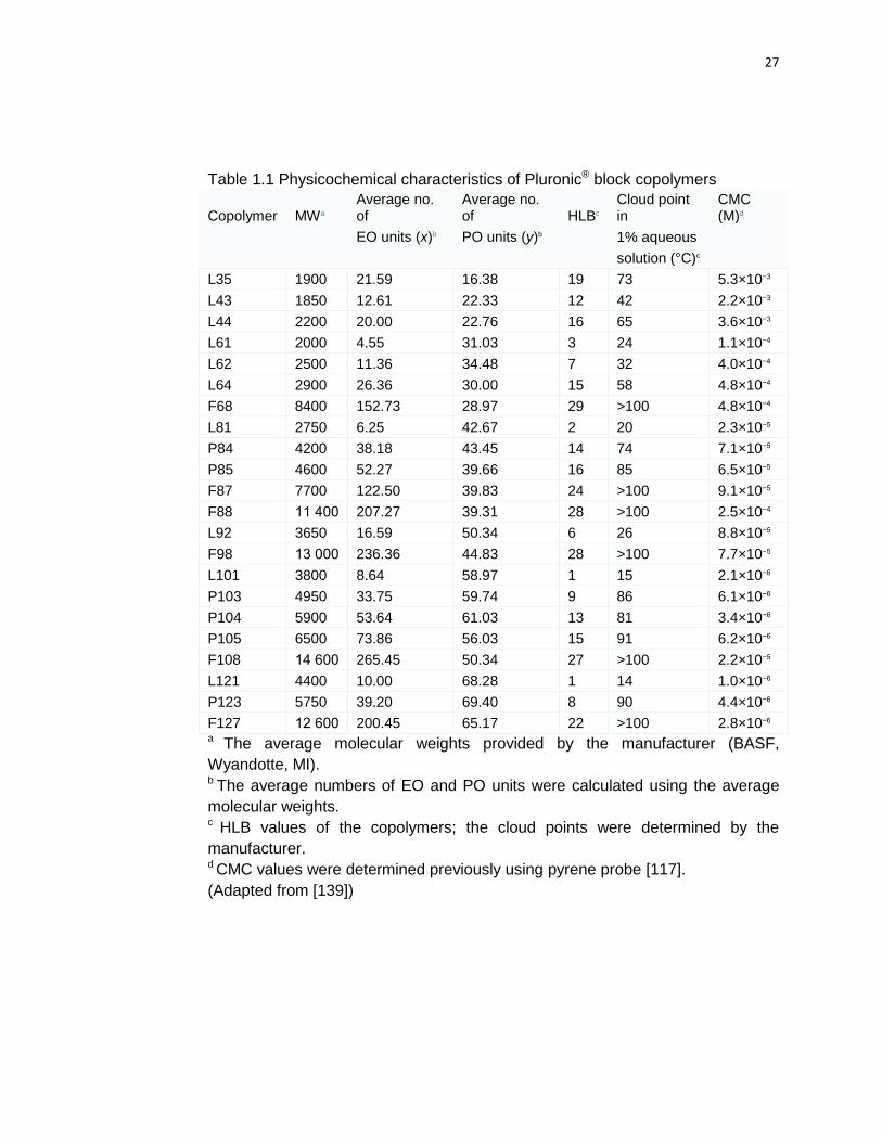
Table 1.1 Physicochemical characteristics of Pluronic® block copolymers
Copolymer MWa Average no. of
Average no. of HLBc
Cloud point in
CMC (M)d
EO units (x)b PO units (y)b
1% aqueous
solution (°C)c
L35 1900 21.59 16.38 19 73 5.3×10⁻³
L43 1850 12.61 22.33 12 42 2.2×10⁻³
L44 2200 20.00 22.76 16 65 3.6×10⁻³
L61 2000 4.55 31.03 3 24 1.1×10⁻⁴
L62 2500 11.36 34.48 7 32 4.0×10⁻⁴
L64 2900 26.36 30.00 15 58 4.8×10⁻⁴
F68 8400 152.73 28.97 29 >100 4.8×10⁻⁴
L81 2750 6.25 42.67 2 20 2.3×10⁻⁵
P84 4200 38.18 43.45 14 74 7.1×10⁻⁵
P85 4600 52.27 39.66 16 85 6.5×10⁻⁵
F87 7700 122.50 39.83 24 >100 9.1×10⁻⁵
F88 11 400 207.27 39.31 28 >100 2.5×10⁻⁴
L92 3650 16.59 50.34 6 26 8.8×10⁻⁵
F98 13 000 236.36 44.83 28 >100 7.7×10⁻⁵
L101 3800 8.64 58.97 1 15 2.1×10⁻⁶
P103 4950 33.75 59.74 9 86 6.1×10⁻⁶
P104 5900 53.64 61.03 13 81 3.4×10⁻⁶
P105 6500 73.86 56.03 15 91 6.2×10⁻⁶
F108 14 600 265.45 50.34 27 >100 2.2×10⁻⁵
L121 4400 10.00 68.28 1 14 1.0×10⁻⁶
P123 5750 39.20 69.40 8 90 4.4×10⁻⁶
F127 12 600 200.45 65.17 22 >100 2.8×10⁻⁶ a The average molecular weights provided by the manufacturer (BASF,
Wyandotte, MI). b The average numbers of EO and PO units were calculated using the average
molecular weights.
c HLB values of the copolymers; the cloud points were determined by the
manufacturer. d CMC values were determined previously using pyrene probe [117].
(Adapted from [139])

28
the driving force of the hydrophobic interactions of the PPO blocks [118].The CMC of the
block copolymers is strongly dependent on the lengths of the blocks. An increase in the
length of the hydrophobic PPO block elevates the net hydrophobicity of the
Pluronic® molecule and favors the segregation of the PPO chains into the micelle core,
resulting in a CMC decrease. In contrary, an increase in the lengths of the PEO blocks
elevates the probability of contacts of the PPO units with the PEO units within the core of
the micelles, which decreases the hydrophobicity of the core and results in
destabilization of the micelle. Therefore, the CMC increases as the hydrophilic PEO
block length is increased [118].
Pluronic® micelles are commonly pictured as spheres with a PPO inner core and
a hydrophilic PEO corona. The micelle size, aggregation number and morphology of the
Pluronic® block copolymers aggregates strongly depend on the block copolymer
composition, specifically, the lengths of PEO and PPO units, the block copolymer
concentration and environmental parameters such as temperature and the quality of the
solvent [ 119 , 120 ]. Spherical micelles commonly have an average hydrodynamic
diameter ranging from about 20 to 80nm, within the preferred nanoparticles size range
10–100 nm discussed above and aggregation numbers ranging from 10 to several
dozen [120]. Except for the spherical structure, additional micelle morphologies include
lamella and rods (cylinders). Nagarajan predicted the formation of cylindrical and
lamellar structures by modeling the framework of a simple molecular theory of
solubilization. It was found that solubilization is shown to induce a transition in aggregate
shapes from spheres to cylinders and then to lamellae [120]. For example, the lamellar
aggregates are favored when the ratio of PEO: PPO is small while spherical aggregates
are favored when the PEO: PPO ratio is large. Typically, for block copolymers containing
40 or more weight percent PEO, only spherical aggregates form at 25°C. For block
29
copolymers containing 30 wt% PEO, cylindrical aggregates are possible. Block
copolymers containing 20 or less weight percent PEO generate lamellae [120]. It has
been demonstrated that increase of the temperature, which induced the decrease of
solubility, led to the transition into cylindrical or lamellar geometries [121].
The hydrophobic core can serve as a microenvironment that can enhance the
loading efficiency of hydrophobic drugs which alone exhibit poor solubility, undesired
pharmacokinetics and low stability in a physiological environment. This process is
referred to as “solubilization”. The outer hydrophilic shell severs as a stabilizing interface
between the hydrophobic core and the aqueous medium to ensure the micelles remain
in a dispersed state and decrease undesirable drug interactions with cells and proteins
[115]. The extent of drug incorporation in Pluronic® micelles is dependent on factors
including the molecular volume of the solubilizate, the interfacial tension of the
solubilizate against waster, length of the core and shell-forming blocks in the copolymer
and the polymer and solubilizate concentrations [122]. It seems reasonable that longer
PPO blocks would produce larger hydrophobic inner core, suggesting hydrophobic core
of micelles as locus of solubilization [123]. Moreover, Nagarajan demonstrated that the
amount of the incorporated solubilizate increases as the molecular volume of the
solubilizate decreases [120]. Furthermore, the solubilization capacity is increased when
high compatibility exsists between the micellar core and the solubilizate and the
solubilizate-water interfacial tension is lower [122].
1.3.1.3 Stability and drug release of Pluronic® micelles
The CMC and the partition coefficient are the major thermodynamic constants
determining the stability of the micellar carrier and the drug release in equilibrium

30
condition. The dilution of the micelles, for example, in the body fluids, leads to the
release of the encapsulated drug. If the system is diluted below the CMC, the micelles
are completely disintegrated and the drug is prematurely release the drug into blood
following intravenous injection [118]. The low stability of Pluronic® micelles results in
failure of increase in drug circulation time and delivery of the drug to the tumor site by
the EPR effect, which limits the practical use of systemic delivery of drugs. In order to
overcome the poor stability and low drug entrapment efficiency of Pluronic® micelles,
several methods have been developed.
Mixed micelles composed different Pluronic® block copolymers were explored to
encapsulate hydrophobic drug with high drug loading capacity as well as improving the
stability of the micellar systems. Mixed micelles composed of relatively hydrophobic
Pluronics® L61/P123/P105 and relatively hydrophilic Pluronics® F127 incorporating
insoluble anticancer therapeutics have been evaluated in vitro and in vivo [124, 125,
126, 127]. The relatively hydrophobic block copolymers with a greater portion of PPO,
was able to load satisfactory hydrophobic drug content. However, due to the rather short
PEO length, the stability of micelles is not desirable. Owing to the high PEO-PPO ratio,
F127 possessed a lower CMC value, creating a stable and effective delivery system.
Mixed micelles of ionic and nonionic surfactants show advantageous solubilization
behavior and an expanded stability when compared with the pure nonionic system. The
phospholipids mixed micelles had lower CMC than that of polymeric micelles and
overcame the thermodynamic instability present in polymeric micelles [128]. Vitamin E
succinate modified pluronic® micelles showed enhanced encapsulation efficiency and
better stability as compared to the non-modified controls [129].
Another method to increase the stability of Pluronic® micelles is the formation of
crosslinks within their outer shells. To form crosslinks, the two terminal alcohols on L121

31
were first chemically converted into aldehydes (L121-CHO) using the Dess–Martin
periodinane. Diamine compounds were then used to bridge the converted aldehyde
termini on L121-CHO via conjugated Schiff bases. After crosslinking, the morphology of
the L121 micelles remained spherical in shape, the sizes were comparable before and
after crosslinking and the micelle stability was greatly enhanced [130]. The stabilization
of Pluronic® micelles composed of Pluronic® P94 and F127 was obtained through
incorporation of a hydrophobic cross-linking agent and subsequent photo-
polymerization, which prevented micelle disruption below CMC or the critical micelle
temperature (CMT), the temperature below which only single chains are present, while
maintaining the integrity of the PEO corona and the hydrophobic properties of the PPO
core [131].
Another attractive strategy to eliminate the burst and premature release of the
drug could be forming polymeric micelles by conjugating drugs to the polymer backbone
via covalent bonds, which can hold the drug firmly in the micelles during circulation in the
blood. To form the DOX- Pluronic® F68 conjugate, the end group of pluronic® F68 was
modified to a carboxyl group and then then chemically conjugated with DOX via an
amide. Drug release profile showed that the DOX- Pluronic® F68 micelles maintained a
sustained DOX release at pH 7.4. The release of DOX from the micelles was
accelerated when the PH was reduced to 6.0 comparing with PH 7.4, due to the
protonation of carboxylic groups at the acidic PH [132]. Another novel polymer-drug
conjugate was prepared by the chemical reaction between the copolymer Pluronic®
P123 and the docetaxel via a pH sensitive hydrazone bond. With a low CMC, these pH
sensitive P123- docetaxel conjugates could self-assemble into nano-size polymeric
micelles in aqueous solution. With the introduction of the covalent hydrazone bonds,
these pH sensitive polymeric conjugate micelles were proved to exhibit their stability

32
against the rat plasma and show a pH dependent drug release, showing the potential to
antitumor drug delivery [133].
1.3.1.4 Pluronic®-based drug delivery systems
Pluronic® micelles containing water insoluble or poorly water-soluble drugs [134],
polypeptides [135], plasmid DNA [136], oligonucleotides [137] as well as diagnostic
imaging agents [138] have been investigated as potential drug delivery systems in
multiple applications. For example, Pluronic® micelles were utilized for delivery of central
nervous system (CNS) drugs across the blood brain barrier (BBB) [139, 140, 141], oral
delivery of drugs to improve oral bioavailability [142, 143], pulmonary delivery of drugs
by inhalation [144] and especially tumor-specific delivery of antineoplastic agents [145,
146]. Following are the examples of Pluronic®-based drug delivery systems targeted to
the MDR tumors.
SP1049C, which contained DOX in the mixed micelles of Pluronics L61 and F127,
was the first in class polymeric micelle drug to advance to clinical stage [124] and has
successfully completed phase II clinical trial which showed high objective response rates
(47%) in the evaluable population and increased median survival (10 months) and
progression free survival (6.6 months) in patients with inoperable metastatic
adenocarcinoma of the esophagus and gastro esophageal junction with toxicity profile
similar to that of Dox at equivalent dose and administration schedule [147].SP1049C
also have been shown recently to decrease the tumorigenicity and aggressiveness of
cancer cells in vivo, suppress BCRP overexpression and the Wnt--catenin signaling
activation observed with Dox alone, significantly alter the DNA methylation profiles, as
well as depletes CD 133+ cells populations, which displayed cancer stem cells (CSCs)-

33
like properties and were much more tumorigenic compared to CD133- cells in murine
leukemia model [148]. Other systems paclitaxel, methotrexate, or docetaxel loaded
polymeric micelles mixed with Pluronic® P105 and F127 has been evaluated in vitro and
in vivo [125,127,127]. All the systems indicated that drug loaded Pluronic® micelles
displayed higher anti-tumor efficacy than free drug in both MDR cancerous cell lines and
MDR tumor xenografts with higher blood circulation time and less systemic toxicity in
comparison with drug alone injection. Moreover, docetaxel-loaded P105/F127 mixed
micelles displayed a higher cytotoxicity towards human lung adenocarcinoma A549-
Taxol resistant cells than Taxotere®, the commercial preparation that approved by the
FDA in 2004 . Longer mean residence time in circulation, larger area under the plasma
concentration-time curve and higher tumor inhibition rate were observed with docetaxel-
loaded P105/F127 mixed micelles compared to Taxotere®, indicating the potential of
P105/F127 mixed micelle as a drug drug delivery system to overcome multidrug
resistance in lung cancer [127].
Targeting moieties- conjugated Pluronic® block copolymers have been developed
to form micelles for active targeting. cyclic RGD [arginine-glycine-aspartic acid] peptide
(c(RGDyK)) that can bind to the integrin protein richly expressed at the site of tumor
vascular endothelial cells and tumor cells with high affinity and specificity, was
conjugated to the N-hydroxysuccinimide-activated PEO terminus of the Pluronic F127
block copolymer. c(RGDyK)-decorated Pluronic F127 and P105 mixed micelles are
developed in this study to form a dual-functional drug delivery system for the
antiangiogenesis and modulation of drug resistance [149]. Folic acid and its conjugate
were widely used for selective delivery of anti-cancer agents to cells with folate
receptors. Folic acid functionalized Pluronic P123/F127 mixed micelles was efficiently
internalized into the cells through by folate receptor-mediated endocytosis, leading to

34
faster and larger extent of drug delivery by folic acid-conjugated micelles compared with
that by non-targeted micelles [125].
Pluronic®-based drug delivery systems were also explored for gene delivery. A
I Belenkov et al evaluated PEI grafted with nonionic amphiphilic block copolymer,
Pluronic® P85 (P85-g-PEI) as a carrier for Ku86 ASO delivery [150]. Compared with
PEI/DNA complex, P85-g-PEI/DNA formulations significantly reduced aggregation due to
the effect of nonionic chains that are soluble in aqueous dispersions and prolonged the
circulation time after systemic administration while the high transfection activity was
remained. Jianan She and other authors developed and investigated a new co-delivery
system, P85-PEI/ D-α-tocopheryl polyethylene glycol 1000 succinate (TPGS) complex
nanoparticle, conveying chemotherapeutic agent and RNA for reversing drug resistance
[151]. TPGS was used to improve micelle stability and drug loading; P85 was used for
preparing micelles and inhibit GST activity, which may contributed to the direct decrease
of the metabolism of drug and potentiate the drug-induced cell apoptosis and PEI was
used to bind shRNA. The IC50 of formulation against PTX resistant human lung
adenocarcinoma epithelial (A549/T cells) was 360-fold lower than that of free drug and
better antitumor efficacy of formulation was observed in vivo.
1.3.2 Pluronic® block copolymers acting as chemosensitizer for MDR cells
Besides the ability to self-assemble into micelles, Relatively hydrophobic
Pluronic® block copolymers at concentrations below the Critical Micelle Concentration
(CMC) with intermediate length of PEO units from 30 to 60 and HLB <20 [152, 153] can
sensitize MDR cancer cells mainly by ABC transporters inhibition and selective ATP
depletion, which is accompanied with generation of reactive oxygen species (ROS) and

35
simultaneously inhibition of glutathione/glutathione S-transferase (GSH/GST)
detoxification system [ 154 , 155 , 156 ]. On the other hand, the Pluronic-caused
impairment of respiration in mitochondria decreased the mitochondrial membrane
potential, promoted release of cytochrome c and overall enhanced pro-apoptotic
signaling (Bax, p53, APAF1, caspase 9 and caspase 3) and mitigated anti-apoptotic
cellular defense (Bcl-2 and Bcl-xl) of MDR cells (Fig.1.3) [157].
1.3.2.1 Structure-functional relationship of Pluronic® block copolymers
The relationship between composition of Pluronic® block copolymers and their biological
response-modifying effects has been investigated. Batrakova et al demonstrated that the
MDR sensitizing effects were achieved by Pluronic® block copolymers at the
concentration below the CMC. When the concentration below the CMC, the resistance
reversion indexes (i.e., ratio of the IC50 of the drug in the assay buffer and Pluronic
solution (IC50.0/IC50)) rapidly increased with increasing copolymer concentration and
reached 400-fold (KBv) and 3400-fold (MCF-7/ADR) as copolymer concentration
approached the CMC values. At concentration above the CMC, the leveling off of
cytotoxicity was observed and the drug cytotoxicity decreased as concentration
continued to rise (Fig.1.4A) [152]. Thus, the sensitizing effect on MDR cells is mediated
by the copolymer signal chain unimers, rather than the micelles.
The specific effects on transporter activity profiles and the critical factors that
influence transporter activity (i.e., membrane fluidization, energy depletion) in both
sensitive and drug resistant cells has been conducted with a series of Pluronic® block
copolymers with a wide range of HLB [152, 153]. Results of R123 (the fluorescent dye,

36
Fig.1.3 Multiple effects of Pluronic® block copolymers in MDR cells: 1) incorporation of
Pluronic molecules into membranes and decrease of the membrane microviscosity; 2)
induction of ATP depletion; 3) inhibition of drug efflux transporters; 4) release of
cytochrome c from mitochondria and increase in ROS levels in cytoplasm; 5) increase of
pro-apoptotic signaling and decrease of anti-apoptotic defense in MDR cells; 6) inhibition
of the glutathione/glutathione S-transferase detoxification system; and 7) abolishment of
drug sequestration within cytoplasmic vesicles. (Adapted from [119])

37
P-gp substrate) accumulation studies suggested that the sensitizing effects of
Pluronic® block copolymers in MDR cells depend on the copolymer hydrophobicity. The
entire set of the hydrophilic Pluronic® block copolymers with HLB varying from 20-29,
presented in Table.1.1, at concentrations below the CMC, had no or little effect on R123
accumulation. Except for the HLB values, the length of PPO block also plays an
important role in the inhibiting the P-gp transporters. It was found a bell-shaped
dependency of the length of hydrophobic PPO segment in inducing R123 uptake. The
maximal effects on the drug uptake were characteristic of Pluronic® block copolymers
with intermediate length of PPO block ranging from 30 to 60 (Fig.1.4B) [153]. The same
bell-shaped curves were observed of the total microviscosity factor values (value of total
cellular membrane microviscosity in the control group in the absence of the
Pluronic® block copolymers versus those observed in the presence of the
Pluronic® block copolymers) or ATP depletion factor (ATP intracellular levels in the
absence of the Pluronic® block copolymers versus those observed in the presence of the
Pluronic® block copolymers ) corresponding to the length of PPO blocks. Lipophilic
copolymers with intermediate PPO block decreased the microviscosity in bovine brain
microvessel endothelial cells (BBMECs), indicating their incorporation into the lipid
bilayer and subsequent increase in membrane fluidization and reduced the intracellular
ATP level at significantly high extent. Very lipophilic block copolymers with long PPO
blocks, the most membranotropic block copolymers, anchor in the plasma membranes
and remain there for an extended period of time. As a result, although they are potent
inhibitors of P-gp ATPase, they are less efficiently transported into the cell and cause
less ATP depletion and, consequently, have less effect on P-gp efflux system compared
with the intermediate block copolymers. In contrast, the block copolymers displaying
intermediate lipophilicity transport across the membranes, and spread throughout

38
A B
Fig.1.4 (A) Effects of Pluronic® block copolymers on cytotoxicity of DOX with respect to
MDR cells: curve 1, MCF7/ADR cells were treated with doxorubicin–L61 compositions;
curve 2, KBv cells were treated with doxorubicin–P85 compositions containing varying
concentrations of the block copolymer. Resistance reversion indexes (ratio of IC50 of
DOX in the assay buffer and Pluronic® solution) are plotted as functions of the
concentration of the block copolymers. (B) Efficacy of Pluronic® block copolymer
composition in MDR KBv cells. R123 enhancement factors are defined here as the ratios
of R123 accumulation in the cells in the presence of the block copolymer to R123
accumulation in the assay buffer. (Adapted from [152])

39
the cytoplasm and reach cellular organelles and to some extent the nuclei. Another
factor of low sensitization effect of very lipophilic block copolymers is due to their low
CMC. They tend to form micelles at low concentrations of the copolymers in aqueous
solutions and decrease the inhibition effect of Pluronic® molecules on ABC transporters
of MDR cells. Overall, the most efficacious block copolymers are those with intermediate
lengths of PPO block and relatively hydrophobic structure (HLB <20), such as Pluronic®
P85 or Pluronic® L61.
1.3.2.2 Inhibition of ABC transporter activity by Pluronic® block copolymers: role
of Pluronic-membrane interaction
ABC transporters are one of the distinctive characteristics of MDR tumors as
discussed before. The Pluronic® copolymer-cell interaction starts from the cell
membrane, where the ABC transporters are located. Pluronic® block copolymers binding
to cell membrane is associated with two steps: 1) hydrophilic interactions of PEO chain
blocks with the polar groups of the lipids at the membrane surface (absorption at the
membrane) and the hydrophobic interactions of PPO chain blocks with the fatty acid
residues in the lipid bilayer (insertion in the membrane) [9]. Relatively hydrophilic
Pluronic® block copolymers could only absorb on the membrane without penetrating into
the lipid bilayer while relatively hydrophobic copolymers could insert into the membrane
below the polar head groups, loosen the lipid packaging and translocate through the
membrane. The immersed Pluronic® block copolymers disrupt the plasma membrane
integrity, which results in decreased membrane microviscosity, accompanied with
conformational changes in the transport proteins and inhibition of P-gp or MRP ATPase
activity in the cells overexpressing these transporters [158, 159]. Moreover, ATPase
40
activity inhibition by Pluronic® block copolymer may be attributed to sterically hindered
protein-drug interaction in the appropriate binding sites, supported by the observation of
the decreases in the maximal reaction rates and increases in apparent Michaelis
constants for these transporters in the presence of Pluronic® P85 [159].
Pluronic® P85 was found to have more pronounced inhibition effects on P-gp
ATPase activity compared with the effects on MRP1 and MRP2 [159]. One reason could
be the structure of MRPs is more robust and less vulnerable to the conformational
changes caused by P85 than the structure of P-gp. The other reason is due to the high
affinity drug-binding sites of MRPs, making less accessible for the hindrance by the
block copolymer than similar sites of P-gp since Pluronic unimers can selectively localize
in particular domains of cellular membrane called caveolae /lipid rafts and employ
caveolae-mediated endocytosis pathway for cellular entry [88], where P-gp is believed to
localize and properties of rafts are essential for the transporter’s proper function while
MRP may have different localization [119].
Inhibition of drug efflux transporters and enhancement of the drug accumulation
in MDR tumors in the presence of Pluronic® block copolymers was demonstrated in vitro
and in vivo [110, 112]. Moreover, Pluronic® block copolymers attributed to overcome the
BBB and achieved the brain delivery of a wide range of therapeutic agents in vitro and in
vivo [ 160 , 161 , 162 ]. Pluronic® P85 block copolymer increases apical to basolateral
transport of a broad spectrum of drugs in the polarized monolayers of BBMECs as in
vitro models of the blood brain barrier. The coadministration of 1% P85 with radiolabeled
digoxin, a P-gp substrate, in wild-type mice increased the brain penetration of digoxin 3-
fold and the digoxin level in the P85-treated wild-type mice was similar to that observed
in the P-glycoprotein-deficient animals, indicating Pluronic® P85 can enhance the
delivery of digoxin to the brain through the inhibition of the P-gp-mediated efflux

41
mechanism. Modification of a BBB-impermeable polypeptide, horseradish peroxidase,
with Pluronic® P85 moieties via biodegradable disulfide links enhance the transport of
this polypeptide across the BBB and accumulation of the polypeptide in the brain in vitro
and in vivo, providing the potential of the brain delivery of many therapeutic polypeptides
that severely restricted by the BBB [163].
1.3.2.3 Effect of Pluronic® block copolymers on cancer cells’ metabolism
One of the factors contributing to sensitization effects on MDR by Pluronic® block
copolymers is the increased ATP depletion, which is crucial for the supporting of energy
for ABC transporter activity and other life processes [158]. Pluronic® unimers translocate
across the plasma membrane via a caveolae-mediated endocytosis pathway [88] and
rapidly (15min after exposure to the cells) colocalizes with mitochondria [156]. It has
been proved that Pluronic® P85 first inhibited the cytochrome c oxidase (complex IV) and
then the NADH-ubiquinone oxidoreductase (complex I) of the mitochondria respiratory
chain at relatively low doses (0.001% and 0.01% for complexes IV and I, respectively),
decreased oxygen consumption and caused ATP depletion in MDR cells, while non-
MDR cells require significantly higher doses of Pluronic® copolymers to achieve similar
functions [156]. Moreover, i.v. injections of DOX formulated with 0.2% Pluronic P85 in
tumor bearing mice caused significant reduction in ATP levels in P388/ADR tumors,
while P388 tumors are not affected. Similar effects were observed with P85 alone
although Dox/P85 is considerably more effective in ATP depletion. The selectivity of
Pluronic® copolymers to MDR phenotype may attribute to innate metabolic and
physiological differences between MDR and non-MDR cells. Notably, MDR cells are
associated with a drastic change in cellular metabolism by using a different major fuel

42
source in energy production. Specifically, the MDR cells predominantly utilize fatty acid
as a source of electrons entering the respiration chain while the non-MDR cells
predominantly employ glucose metabolism for this purpose. Since the PPO blocks of
Pluronic® molecules may interact with fatty acids even at low (unimers) concentrations
and effectively immobilize fatty molecules inside micelle cores at higher concentrations,
it may affect the respiration mechanism at higher extent in MDR cells. Furthermore,
MDR cells also have lower mitochondria membrane potential and more uncoupled
respiration due to the mitochondria membrane leakage and higher level of mitochondrial
uncoupling protein 2, resulting in inefficient ATP synthesis and thus making the
mitochondria of MDR cells even more vulnerable to the depolarization effects compared
with that of the sensitive cells [164]. It was observed at about an orders of magnitude
lower concentration of P85 necessary for 50% decrease of mitochondria membrane
potential in the MCF7/ADR cells compared to MCF7 cells [156].
Interestingly, higher responsiveness of the respiratory chain to Pluronic
copolymers appears to be at least partially associated with P-gp expression, since
increased inhibition of oxygen consumption was observe not only in drug selected MDR
cells, but also in MDCKII/MDR1 P-gp-transfected cells [155, 156]. P-gp plays multiple
roles in cells, such as inducing alteration in the membrane lipid composition [165],
increasing the synthesis of glycolipid, globotriaosylceramide [166] and pumping the
substrates inside mitochondria [167], leading to drastic effect on membrane structure,
cell signaling and mitochondrial activities and thus contributing to the increased
responsiveness of molecular “targets” of Pluronic copolymers.
1.3.2.4 Effect of Pluronic® block copolymers on proapoptotic signaling

43
Oxidative stress is a condition of excessive ROS formation or a diminished ability
of cells to scavenge ROS by antioxidant networks. The formed peroxides and free
radicals will damage various cellular components including proteins, lipids and DNA that
can culminate in cell death [168]. GSH, the ubiquitous tripeptide, is the major cellular
antioxidant that protects the cells against ROS, toxins and drugs. GSH has a sulfhydryl
(SH) group on the cysteinyl portion, which accounts for its strong electron-donating
character. As electrons are lost, the molecule becomes oxidized, and two such
molecules become linked (dimerized) by a disulfide bridge to form glutathione disulfide
or oxidized glutathione (GSSG). In healthy cells, more than 90% of the
total glutathione pool is in the reduced form (GSH). When cells are exposed to increased
levels of oxidative stress, GSSG accumulates and the ratio of GSSG to GSH increases
[ 169 ]. An increased ratio of GSSG-to-GSH is an indication of oxidative stress.
Additionally, GST, the major cellular detoxifying enzyme performs a range of functional
roles using GSH as a cosubstrate or coenzyme. Researches indicated that MDR is
associated with elevated GSH level and GST catalytic activity [170, 171]. Batrakova EV
et al demonstrated Pluronic® P85 inhibited GSH/GST detoxification system by depleting
levels of GSH and inactivating GST in MDR cancer cells [154]. Moreover, as discussed
above, Pluronic® block copolymers quickly reach mitochondrial and inhibit complex I and
IV of mitochondria electron transfer chain in MDR cells, stimulating the production of
ROS in the resistant, but not in the sensitive cells, where it remained at the basal level
[156].
Pluronic® P85 was observed to localize in mitochondria after 15min of the cell
exposure by incorporation of P85 into mitochondria membranes followed by alteration of
the membrane structure, permeabilization of outer mitochondrial membrane, decrease of
mitochondria membrane potential and release of cytochrome c, which are the early signs

44
of mitochondrial apoptotic pathway [156]. In cytoplasm, cytochrome c binds to apoptosis
protease activating factor (APAR-1) and forms apoptosome, which cleaves and activates
the procaspase-9 and forms caspase 9. The activated caspase 9 further activates the
effector caspases, such as caspase 3, which all together contribute to the completion of
apoptosis. Indeed, Formulation of DOX with P85 increased the expression levels of the
proapoptotic genes (BAX, P53, APAF1, Caspase 9 and Caspase 3)
while simultaneously downregulating the intracellular levels of antiapoptotic genes
(BCL2 and BCLXL) compared to free DOX in MDR cells [Error! Bookmark not
defined.]. Furthermore, the effect of DOX/P85 formulations on apoptosis was evaluated
in vivo. Data indicated that i.v. injection of DOX/P85 into drug-resistant tumor (3LL-M27)
-bearing mice formulations increased the level pro-apoptotic signals of caspase 8 and
caspase 9, promoting tumor apoptosis compared to injection of DOX alone [110].
1.3.2.5 Pluronic® block copolymers prevent development of MDR
In addition to the chemosensitization effects listed above, Pluronic® block
copolymers have been proved to prevent the development of MDR upon selection with
cytotoxic drugs in vitro and in vivo [172, 173]. Specifically, the human breast carcinoma
MCF7 cell line was cultured in increasing concentration of either DOX alone or DOX
formulated with 0.001% P85 (below the CMC) in the medium. After 305 days of
escalating the drug exposure, the cells cultured in the presence of Pluronic® P85 were
unable to stably grow in concentrations of Dox that exceeded 10ng Dox/ml of culture
media. However, MCF7 cells cultured in the absence of the block copolymer resulted in
the selection and stable growth of cells that tolerated 1000 times higher concentration of
the drug (10,000ng Dox/ml culture media) (Fig.1.5A). Detailed characterization of the
isolated sublines demonstrated that those cells selected in the DOX-P85 formulation did

45
not show amplification of the MDR1 gene while cells selected with Dox alone showed an
elevated level in the expression of the MDR1 gene along with a corresponding increase
in the expression level of the drug efflux transporter, P-gp, contributing to the high
resistance of the cells to Dox (Fig.1.5B). Moreover, it was demonstrated that selection of
cells with DOX and DOX-P85 formulation resulted in very different changes in the gene
expression pattern in the cells, which may potentially enhance therapeutic outcomes of
DOX-P85 formulation [172]. Similar results were observed in murine lymphocytic
leukemia cells (P388) when selected for DOX resistance with or without P85. Cells
selected with DOX-P85 formulation in vitro and in vivo exhibited some increases in
IC(50) values compared to parental cells, but these values were much less than IC50 in
respective cells selected with DOX alone. In addition to MDR1, P85 abolished alterations
of genes implicated in apoptosis, drug metabolism, stress response, molecular transport
and tumorigenesis [173]. Overall, if resistance is intrinsic, Pluronic sensitizes the tumor,
whereas if resistance is acquired, MDR cells no longer have a selective advantage as
Pluronic prevents development of MDR.
1.3.2.6 Pluronic® block copolymers suppress cancer stem cells (CSCs)
Tumors are complex heterogeneous tissues comprising phenotypically and functionally
different cancer cells. One theory suggests that the heterogeneity of tumor cells arises
as a result of differentiation of small cell subpopulation within the tumor, called tumor-
initiating cells or CSCs, with indefinite potential for self-renewal that drive tumorigenesis
[174]. CSCs are intrinsically drug resistant to chemotherapeutics by overexpressing drug
efflux transporters such as P-gp and BCRP, having extensive survival and anti-oxidant
networks and having most of their time in the G0 nondividing cell cycle state [175].

46
A B
Fig.1.5 (A) Time course of the development of drug resistance in MCF7 cell lines
cultured with Dox either alone (filled diamonds) or in combination with 0.001% P85
(open diamonds). [Dox] is the concentration of Dox in the growth medium. (B) Western
blot data for expression of P-gp in MCF7 parental cells, and selected MCF7/ADR cells
tolerating: 10ng/ml Dox; 200ng/ml Dox; 1000ng/ml Dox; 5000ng/ml Dox; 10,000ng/ml
Dox; and 10ng/ml Dox with 0.001% P85. The cells were harvested at different points of
selection as presented in (a) arrows 1-7. (Adapted from [172])

47
Since CSCs share certain characteristics of MDR cells such as overexpressing drug
efflux transporters and altered metabolic pathways, Pluronics have been applied to
sensitize CSCs to chemotherapeutic drugs. A recent study demonstrated that SP1049C
treatment reduced tumor aggressiveness, in vivo tumor formation frequency in mice
bearing the leukemia ascetic cells and in vitro clonogenic potential of the leukemia cells
derived from the treated animals compared to drug, saline and polymer controls.
SP1049C also prevented overexpression of BCRP and activation of Wnt-β-catenin
signaling observed with DOX alone significantly altered the DNA methylation profiles of
the cells and decreased CD133+ P388 cells populations, which displayed CSC-like
properties and were more tumorigenic compared to CD133− cells [148].
Overall, capacities of forming micelles and encapsulating water-insoluble
anticancer drugs, MDR sensitization by multiple mechanisms, prevention of MDR
development, depletion of tumorigenic cell subpopulations, and decrease of
tumorigenicity and tumor aggressiveness offer significant advantages for the
development of Pluronics formations of approved and/or experimental therapeutics.
1.4 Other applications of Pluronic® block copolymers in cancer treatment
Doxil®, the first nanoformulated drug approved by the US FDA, was designed to
reduce cardiotoxicity of DOX by encapsulating it in PEGylated (stealth) liposome.
However, the therapeutic effectiveness of Doxil® is limited by its penetration to the tumor
and poor release of the active drug from the liposome at the tumor site. In clinical
studies, Doxil® produced less cardiotoxicity but did not show significant improvement of
progression-free survival compared with free DOX for breast cancer patients [176].
Thus, combination of Doxil® therapy with the regimens that enhance permeability of

48
tumor vasculature is necessary to improve the therapeutic effect. Pluronic® P85 was
proved to increase the process of solute diffusion across planar lipid and liposomal
membranes without affecting the overall integrity of the membranes [177]. Zhao Y et al
demonstrated enhancement of drug release from the liposomes and increase of
therapeutic efficacy of Doxil® by administering Pluronic® P85 once the liposomal drug
accumulates in the tumor sites [178]. Specifically, DOX was observed to rapidly entered
cells (15min) and then accumulated in the nucleus (60min) while little if any drug was
seen in the intracellular vesicles and nucleus even after 24h exposures of the cells to the
highest dose of Doxil® (200 µg/mL). Anti-tumor growth and distribution of drug were
evaluated when Pluronic P85 was i.v. injected 1h, 48h, or 96h after the
Doxil® administration in A2780 human ovarian cancer xenografts. The greatest anti-
tumor effect of single injection of Doxil® was achieved when Pluronic P85 was
administered 48h after Doxil®, when the liposomal drug was already distributed to the
tumor sites .The Confocal tile scanning images of tumor section showed that copolymer
treatment induced the release of the drug in the tumors from the vessels regions to the
bulk of the tumor. Therefore, systemic application of the Pluronic® block copolymers may
be a useful adjuvant to the Doxil® -based chemotherapy regimens.
Another application could be intraceullar delivery of the unimers of Pluronic® P85
to MDR cells by mixed micellar system based mainly on the pH-sensitive copolymer of
poly(L-histidine)-poly (D,L-lactide)-polyethyleneglycol-poly (D,L-lactide)-poly (L-histidine)
(PHis-PLA-PEG-PLA-PHis) and Pluronic® F127, which overcomes the poor micellar
stability of plain puronics micelles [ 179 ]. The micelles with inserted Pluronic® P85
unimers demonstrated more efficient intracellular delivery of Pluronic® P85 unimers to
the cytosol of the MDR cells than the triple-component mixed micelles and plain
Pluronic® micelles, which was confirmed by the subcellular colocalization of Pluronic®

49
P85 unimers with mitochondria and led to enhanced MDR reversal effect. In addition, the
potential of such system has been confirmed by the higher antitumor efficiency against
MCF-7/ADR tumors and lower cellular ATP levels in the isolated tumors. The idea of
incorporation of Pluronic®P85 unimers and drug together into the mixed micelles may
contribute to more efficient MDR reversal by Pluronic® block copolymers.
Besides acting as chemosensitzer for MDR cells, Pluronic® block copolymers
were also shown to improve the outcome of the radiofrequency ablation induced
hyperthermia in solid and tumors and sensitize cancer cells to hyperthermia in vitro and
in vivo [180]. Consistent with the structure-functional relationships as chemosensitzer for
MDR cells, the thermosensitzing activity of Pluronic® block copolymers was associated
with copolymer structure properties including molecular weight, HLB values and PO
length. Pluronics with lower molecular weight between 1100 to 3200 Da and a HLB <8
demonstrated the highest thermossensitizing ability with Pluronics L31, L61, L62, L10
and L64 among the most effective copolymers for hyperthermia sensitization under
tested conditions. Most encouraging, L61 in synergy with hyperthermia significantly
reduced tumor growth progression in vivo compared to tumors treated with hyperthermia
alone [ 181 ]. The thermosensitization ability of Pluronic® block copolymers may be
directly linked to its ability to interact with plasma membrane and induce membrane
fluidization as cells become more sensitive to treatment when hyperthermia induced
increase of membrane fluidity [ 182 ]. Another factor contributing to the
thermosensitization effect is that the combination of low-grade hyperthermia
and Pluronic treatment induced depletion of ATP levels and decreased expression of
heat shock protein 70 (HSP 70), the chaperone molecules that assist in protein folding in
the active confirmation and are expressed rapidly as a result of cellular stresses such as
hyperthermia to increase the thermoresistance of the tumors [183].

50
Pluronic® block copolymers also demonstrated exciting opportunities for
development of novel gene therapies [184]. It was discovered SP1017 (0.25% Pluronic
L61 and 2% Pluronic F127) significantly increases intramuscular expression of plasmid
DNA by 5- to 20- fold compared to naked DNA [185]. Pluronic® P85 enhanced levels of
expression of a reporter gene (luciferase) by 17 to 19-fold in immunocompetent C57Bl/6
and Balb/c mice by activating the p53 and NFκB signaling pathways. High levels of
expression of a reporter gene (luciferase) were sustained for at least 40 days and the
area under the gene expression curve increased by at least 10 times compared to the
DNA alone in mice. In addition Pluronic increased the number of DNA copies and thus
affected initial stages of gene transfer in a promoter selective manner [ 186 ].
Pluronic® P85 was also shown to enhance gene transfection in HepG2 cells under
ultrasound irradiation in vitro. The transfection efficacy in ulstrasound+P85 group was
three times higher than in single ultrasound group [ 187 ]. Thus, Pluronic® block
copolymers could be a good candidate applicable in multiple gene therapy protocols
involving intramuscular injection of plasmid DNA by 1) enhancing the gene transfer and
2) activating the transcription of the genes.
1.5 Statement of purpose
MM is the second most common hematologic malignancy characterized by
infiltration in the bone marrow (BM) of malignant plasma cells with production of
monoclonal proteins. In the last decade, the introduction of immunomodulatory drugs
(IMIDs), and of new front-line agents such as the proteasome inhibitors (Bortezomib,
BTZ or Carfilzomib, CFZ) have significantly improved overall survival [188]. In spite of
current efficient therapeutic regimens for MM patients, like almost all anti-cancer

51
drugs, drug resistance becomes a major concern and almost all patients eventually
relapse or become refractory to current treatment regimens [189, 190]. For instance,
BTZ (PS-341, Velcade), the first proteasome inhibitor approved by U.S. FDA, used as a
first-in-class drug in MM by reversibly inhibiting the chymotrypsin-like activity at the β5-
subunit and to a lesser extent inhibit the trypsin-like activity at the β1-subunit. However,
many patients may be intrinsically resistant to it or develop resistance in the course of
treatment [191]. Although real mechanisms of resistance to BTZ in MM patients are not
yet deciphered, mutation in β5-subunit of proteasome (PSMB5) which impair BTZ
binding, alterations of gene and protein expression in stress response, cell survival and
antiapoptotic pathways and MDR have been indicated to be involved [192]. The BM
microenvironment supported by fibroblast-like BM stromal cells, osteoblasts, and
osteoclasts which secrete soluble factors and extracellular matrix proteins can facilitate
the survival, differentiation, and proliferation of MM cells and offer protection from
chemotherapeutic agents, thereby increases the probability for the development of
drug resistance [193, 194]. According to the chemosensitization effect on MDR cells by
Pluronic® block copolymers discussed above, we propose to use the combination of
proteasome inhibitors together with mixture of Pluronics (SP1017) for MM treatment to
increase the drug sensitivity of cancer cells. Moreover, almost all of the previous studies
on sensitizing effect indicated that Pluronic® block copolymers only sensitize MDR cells
but have little effects on sensitive cells [155, 156]. Here our study mainly focuses on the
effect of Pluronic® block copolymers on sensitive MM cells and the related targets of the
sensitization effect.
Our hypothesis of this research is that SP1017 will sensitize multiple myeloma in
vitro and in vivo by targeting multiple molecular mechanisms. To test this hypothesis, the
following specific aims will be addressed.

52
Specific aim 1: To determine the chemosensitization effect of SP1017 on both sensitive
and drug-resistant MM cell lines combined with proteasome inhibitors.
Specific aim 2: To investigate the related molecular mechanisms of the sensitization
effect of SP1017 combined with proteasome inhibitors.
Specific aim 3: To evaluate the treatment efficacy of the combination of SP1017 with
proteasome inhibitor BTZ in human MM/SCID mice model.

53
1 Linton KJ. Structure and Function of ABC Transporters. Physiology (Bethesda). 2007
Apr;22:122-130.
2 Ozben T. Mechanisms and strategies to overcome multiple drug resistance in cancer.
FEBS Lett. 2006 May 22;580(12):2903-2909.
3 Aller SG, Yu J, Ward A, Weng Y, Chittaboina S, Zhuo R, Harrell PM, Trinh YT, Zhang
Q, Urbatsch IL, Chang G.. Structure of P-glycoprotein reveals a molecular basis for poly-
specific drug binding. Science. 2009 Mar 27; 323(5922):1718-1722.
4 Kibria G, Hatakeyama H, Harashima H. Cancer multidrug resistance: mechanisms
involved and strategies for circumvention using a drug delivery system. Arch Pharm
Res. 2014 Jan; 37(1):4-15.
5 Dalton WS, Scheper RJ. Lung Resistance-Related Protein: Determining Its Role in
Multidrug Resistance. J Natl Cancer Inst. 1999 Oct 6;91(19):1604-1605.
6 Kitazono M, Sumizawa T, Takebayashi Y, Chen ZS, Furukawa T, Nagayama S, Tani
A, Takao S, Aikou T, Akiyama S. Multidrug Resistance and the Lung Resistance-Related
Protein in Human Colon Carcinoma SW-620 Cells. J Natl Cancer Inst. 1999 Oct
6;91(19):1647-1653.
7 Stavrovskaya AA. Cellular mechanisms of multidrug resistance of tumor cells.
Biochemistry (Mosc). 2000 Jan;65(1):95-106.
8 Cerqueira BB, Lasham A, Shelling AN, Al-Kassas R. Nanoparticle therapeutics:
Technologies and methods for overcoming cancer. Eur J Pharm Biopharm. 2015
Nov;97(Pt A):140-151.
9 Broxterman HJ, Lankelma J, Hoekman K. Resistance to cytotoxic and anti-angiogenic
anticancer agents: similarities and differences. Drug Resist Updat. 2003 Jun;6(3):111-
127.

54
10 Alakhova DY, Kabanov AV. Pluronics and MDR reversal: an update. Mol Pharm. 2014
Aug 4;11(8):2566-2578.
11 Sládek NE, Kollander R, Sreerama L, Kiang DT. Cellular levels of aldehyde
dehydrogenases (ALDH1A1 and ALDH3A1) as predictors of therapeutic responses to
cyclophosphamide-based chemotherapy of breast cancer: a retrospective study.
Rational individualization of oxazaphosphorine- based cancer chemotherapeutic
regimens. Cancer Chemother Pharmacol. 2002;49:309–321.
12 Wang K, Ramji S, Bhathena A, Lee C, Riddick DS. Glutathione S-transferases in wild-
type and doxorubicin-resistant MCF-7 human breast cancer cell lines.Xenobiotica. 1999
Feb;29(2):155-170.
13 Bedard PL, Di Leo A, Piccart-Gebhart M. Taxanes: optimizing adjuvant chemotherapy
for early-stage breast cancer. Nat Rev Clin Oncol. 2010;7:22–36.
14 Beck WT, Danks MK, Wolverton JS, Granzen B, Chen M, Schmidt CA, Bugg
BY, Friche E, Suttle DP. Altered DNA topoisomerase II in multidrug resistance.
Cytotechnology. 1993; 11(2): 115-119.
15 Eijdems EW, de Haas M, Timmerman AJ, Van der Schans GP, Kamst E, de Nooij
J, Astaldi Ricotti GC, Borst P, Baas F. Reduced topoisomerase II activity in multidrug-
resistant human non-small cell lung cancer cell lines. Br J Cancer. 1995 Jan;71(1):40-7.
16 Tsujimoto Y. Role of Bcl-2 family proteins in apoptosis: apoptosomes or mitochondria?
Genes Cells. 1998 Nov;3(11):697-707.
17 Davis JM, Navolanic PM, Weinstein-Oppenheimer CR, Steelman LS, Hu
W, Konopleva M, Blagosklonny MV, McCubrey JA. Raf-1 and Bcl-2 induce distinct and
common pathways that contribute to breast cancer drug resistance.
Clin Cancer Res. 2003 Mar;9(3):1161-1170.
55
18 Srivastava RK, Sasaki CY, Hardwick JM, Longo DL. Bcl-2-mediated drug resistance:
inhibition of apoptosis by blocking nuclear factor of activatred T lymphocytes (NFAT)-
induced Fas ligand transcription. J Exp Med. 1999 Jul 19;190(2):253-265.
19 Yu L, Wang Z. Difference in expression of Bcl-2 and Bcl-xl genes in cisplatin-sensitive
and cisplatin-resistant human in ovarian cancer cell lines. J Huazhong Univ Sci
Technolog Med Sci. 2004;24(2):151-153.
20 Hermanson DL, Das SG, Li Y, Xing C. Overexpression of Mcl-1 Confers Multidrug
Resistance, Whereas Topoisomerase IIβ Downregulation Introduces Mitoxantrone-
Specific Drug Resistance in Acute Myeloid Leukemia. Mol Pharmacol. 2013
Aug;84(2):236-243.
21 Chen F, Zhuang X, Lin L, Yu P, Wang Y, Shi Y, Hu G, Sun Y. New horizons in tumor
microenvironment biology: challenges and opportunities. BMC Med. 2015 Mar 5;13:45.
22 Trédan O, Galmarini CM, Patel K, Tannock IF. Drug Resistance and the Solid Tumor
Microenvironment. J Natl Cancer Inst. 2007 Oct 3;99(19):1441-1454.
23 Comerford KM, Wallace TJ, Karhausen J, Louis NA, Montalto MC, Colgan SP.
Hypoxia-inducible Factor-1-dependent Regulation of the Multidrug Resistance (MDR1) Gene.
Cancer Res. 2002 Jun 15;62(12):3387-3394.
24 Scotto KW. Transcriptional regulation of ABC drug transporters. Oncogene. 2003 Oct
20;22(47):7496-7511.
25 Wang JH, Wu QD, Bouchier-Hayes D, Redmond HP. Hypoxia upregulates Bcl-2
expression and suppresses interferon-gamma induced antiangiogenic activity in human
tumor derived endothelial cells. Cancer. 2002 May 15;94(10):2745-2755.

56
26 Lu H, Samanta D, Xiang L, Zhang H, Hu H, Chen I, Bullen JW, Semenza GL.
Chemotherapy triggers HIF-1–dependent glutathione synthesis and copper chelation
that induces the breast cancer stem cell phenotype. Proc Natl Acad Sci U S A. 2015 Aug
18;112(33):E4600-4609.
27 Saraswathy M1, Gong S.. Different stragegies to overcome multidrug resistance in
cancer. Biotechnol Adv. 2013 Dec;31(8):1397-1407.
28 Consoli U, Priebe W, Ling YH, Mahadevia R, Griffin M, Zhao S, Perez-Soler R,
Andreeff M. The novel anthracycline annamycin is not affected by P-glycoprotein-related
multidrug resistance: comparison with idarubicin and doxorubicin in HL-60 leukemia cell
lines. Blood. 1996 Jul 15;88(2):633-44.
29 Perez-Soler R, Neamati N, Zou Y, Schneider E, Doyle LA, Andreeff M, Priebe W, Ling
YH. Annamycin circumvents resistance mediated by the multidrug resistance-associated
protein (MRP) in breast MCF-7 and small-cell lung UMCC-1 cancer cell lines selected for
resistance to etoposide. Int J Cancer. 1997 Mar 28;71(1):35-41.
30 Mazel M, Clair P, Rousselle C, Vidal P, Scherrmann JM, Mathieu D, Temsamani J.
Doxorubicin-peptide conjugates overcome multidrug resistance. Anticancer Drugs. 2001
Feb;12(2):107-116.
31 Yoshikawa M, Ikegami Y, Hayasaka S, Ishii K, Ito A, Sano K, Suzuki T, Togawa T,
Yoshida H, Soda H, Oka M, Kohno S, Sawada S, Ishikawa T, Tanabe S. Novel
camptothecin analogues that circumvent ABCG2-associated drug resistance in human
tumor cells. Int J Cancer. 2004 Jul 20;110(6):921-927.
32 Jordan MA, Wilson L. Microtubules as a target for anticancer drugs. Nat Rev
Cancer. 2004 Apr;4(4):253-265.
33 Oshiro C, Marsh S, McLeod H, Carrillo MW, Klein T, Altman R. Taxane Pathway.
Pharmacogenet Genomics. 2009 Dec;19(12):979-983.

57
34 Shionoya M, Jimbo T, Kitagawa M, Soga T, Tohgo A. DJ-927, a novel oral taxane,
overcomes P-glycoprotein-mediated multidrug resistance in vitro and in vivo. Cancer
Sci. 2003 May;94(5):459-466.
35Abidi A. Cabazitaxel: A novel taxane for metastatic castration-resistant prostate cancer-
current implications and future prospects. J Pharmacol Pharmacother. 2013
Oct;4(4):230-237.
36 Dong X, Mumper RJ. Nanomedicinal strategies to treat multidrug-resistant tumors:
current progress. Nanomedicine (Lond). 2010 Jun;5(4):597-615.
37 Ullah MF.Cancer Multidrug Resistance (MDR): A Major Impediment to Effective
Chemotherapy. Asian Pac J Cancer Prev. 2008 Jan-Mar;9(1):1-6.
38 Liscovitch M, Lavie Y. Cancer multidrug resistance: A review of recent drug discovery
research. IDrugs. 2002 Apr;5(4):349-355.
39 Thomas H, Coley HM. Overcoming Multidrug Resistance in Cancer: An Update on the
Clinical Strategy of Inhibiting P-Glycoprotein. Cancer Control. 2003 Mar-Apr;10(2):159-
165.
40 Krishna R, Mayer LD. Multidrug resistance (MDR) in cancer: Mechanisms, reversal
using modulators of MDR and the role of MDR modulators in influencing the
pharmacokinetics of anticancer drugs. Eur J Pharm Sci. 2000 Oct;11(4):265-283.
41 Ferry DR, Traunecker H, Kerr DJ. Clinical trials of p-glycoprotein reversal in solid
tumor. Eur J Cancer. 1996 Jun;32A(6):1070-1081.
42 Theis JG, Chan HS, Greenberg ML, Malkin D, Karaskov V, Moncica I, Koren G, Doyle
J. Assessment of systemic toxicity in children receiving chemotherapy with cyclosporine
for sarcoma. Med Pediatr Oncol. 2000 Apr;34(4):242-249.
43 Twentyman PR, Bleehen NM. Resistance modification by PSC-833, a novel non-
immunosuppressive cyclosporin [corrected]. Eur J Cancer. 1991;27(12):1639-1642.

58
44 Visani G, Milligan D, Leoni F, Chang J, Kelsey S, Marcus R, Powles R, Schey
S, Covelli A, Isidori A, Litchman M, Piccaluga PP, Mayer H, Malagola M,Pfister C.
Combined action of PSC 833 (Valspodar), a novel MDR reversing agent, with
mitoxantrone, etoposide and cytarabine in poor-prognosis acute myeloid leukemia.
Leukemia. 2001 May;15(5):764-771.
45 Fracasso PM, Brady MF, Moore DH, Walker JL, Rose PG, Letvak L, Grogan
TM, McGuire WP. Phase II Study of Paclitaxel and Valspodar (PSC 833) in Refractory
Ovarian Carcinoma: A Gynecologic Oncology Group Study. J Clin Oncol. 2001 Jun
15;19(12):2975-2982.
46 Gruber A, Peterson C, Reizenstein P. D-verapamil and L-verapamil are equally
effective in increasing vincristine accumulation in leukemic cells in vitro. Int J
Cancer. 1988 Feb 15;41(2):224-226.
47 Wilson WH, Bates SE, Fojo A, Bryant G, Zhan Z, Regis J, Wittes RE, Jaffe
ES, Steinberg SM, Herdt J. Controlled trial of dexverapamil, a modulator of multidrug
resistance, in lymphomas refractory to EPOCH chemotherapy. J Clin Oncol. 1995
Aug;13(8):1995-2004.
48 Dantzig AH, Law KL, Cao J, Starling JJ. Reversal of multidrug resistance by the P-
glycoprotein modulator, LY335979, from the bench to the clinic. Curr Med Chem. 2001
Jan;8(1):39-50.
49 Newman MJ, Rodarte JC, Benbatoul KD, Romano SJ, Zhang C, Krane S, Moran
EJ, Uyeda RT, Dixon R, Guns ES, Mayer LD. Discovery and characterization of OC144-
093, a novel inhibitor of P-glycoprotein-mediated multidrug resistance. Cancer
Res. 2000 Jun 1;60(11):2964-2972.

59
50 Wu CP, Ohnuma S, Ambudkar SV. Discovering Natural Product Modulators to
Overcome Multidrug Resistance in Cancer Chemotherapy. Curr Pharm Biotechnol. 2011
Apr;12(4):609-620.
51 Limtrakul P, Chearwae W, Shukla S, Phisalphong C, Ambudkar SV. Modulation of
function of three ABC drug transporters, P-glycoprotein (ABCB1), mitoxantrone
resistance protein (ABCG2) and multidrug resistance protein 1 (ABCC1) by
tetrahydrocurcumin, a major metabolite of curcumin. Mol Cell Biochem. 2007 Feb;296(1-
2):85-95.
52 Shukla S, Zaher H, Hartz A, Bauer B, Ware JA, Ambudkar SV. Curcumin inhibits the
activity of ABCG2/BCRP1, a multidrug resistance-linked ABC drug transporter in mice.
Pharm Res. 2009 Feb;26(2):480-487.
53 Conseil G, Baubichon-Cortay H, Dayan G, Jault JM, Barron D, Di Pietro A.
Flavonoids: a class of modulators with bifunctional interactions at vicinal ATPand
steroid-binding sites on mouse P-glycoprotein. Proc Natl Acad Sci U S A. 1998 Aug
18;95(17):9831-9836.
54 Agrawal N, Dasaradhi PV, Mohmmed A, Malhotra P, Bhatnagar RK, Mukherjee
SK.RNA interference: biology, mechanism, and applications. Microbiol Mol Biol
Rev. 2003 Dec;67(4):657-685.
55 Kim D, Rossi J. RNAi mechanisms and applications. Biotechniques. 2008
Apr;44(5):613-616.
56 Watts J and Corey D*Gene silencing by siRNAs and antisense oligonucleotides in the
laboratory and the clinic. J Pathol. 2012 Jan; 226(2): 365–379.
57 Nieth C, Priebsch A, Stege A, Lage H. Modulation of the classical multidrug resistance
(MDR) phenotype by RNA interference (RNAi). FEBS Lett. 2003 Jun 19;545(2-3):144-
150.

60
58 Pichler A, Zelcer N, Prior JL, Kuil AJ, Piwnica-Worms D. In vivo RNA Interference–
Mediated Ablation of MDR1 P-Glycoprotein. Clin Cancer Res. 2005 Jun 15;11(12):4487-
4494.
59 Lv H, He Z, Liu X, Yuan J, Yu Y, Chen Z. Reversal of BCRP-mediated multidrug
resistance by stable expression of small interfering RNAs. J Cell Biochem. 2007 Sep
1;102(1):75-81.
60 Pan GD, Yang JQ, Yan LN, Chu GP, Liu Q, Xiao Y, Yuan L. Reversal of multi-drug
resistance by pSUPER-shRNA-mdr1 in vivo and in vitro. World J Gastroenterol. 2009
Jan 28;15(4):431-440.
61 Xu CF, Wang J. Delivery systems for siRNA drug development in cancer therapy.
Asian J. Pharm. Sci., 10 (2015), pp. 1–12
62 Nayerossadat N, Maedeh T, Ali PA. Viral and nonviral delivery systems for gene
delivery. Adv Biomed Res. 2012;1:27.
63 Ramamoorth M, Narvekar A. Non Viral Vectors in Gene Therapy- An Overview. J Clin
Diagn Res. 2015 Jan;9(1):GE01-6.
64 Singh S.Nanomaterials as Non-viral siRNA Delivery Agents for Cancer Therapy.
Bioimpacts. 2013;3(2):53-65.
65 Susa M, Iyer AK, Ryu K, Choy E, Hornicek FJ, Mankin H, Milane L, Amiji MM, Duan Z.
Inhibition of ABCB1 (MDR1) Expression by an siRNA Nanoparticulate Delivery System
to Overcome Drug Resistance in Osteosarcoma. PLoS One. 2010 May 24;5(5):e10764.
66 Aliabadi HM, Landry B, Mahdipoor P, Hsu CY, Uludağ H. Effective down-regulation of
Breast Cancer Resistance Protein (BCRP) by siRNA delivery using lipid-substituted
aliphatic polymers. Eur J Pharm Biopharm. 2012 May;81(1):33-42.

61
67 Guo P, Coban O, Snead NM, Trebley J, Hoeprich S, Guo S, Shu Y. Engineering RNA
for Targeted siRNA Delivery and Medical Application. Adv Drug Deliv Rev. 2010 Apr
30;62(6):650-666.
68 Vivas-Mejia PE, Rodriguez-Aguayo C, Han HD, Shahzad MM, Valiyeva F, Shibayama
M, Chavez-Reyes A, Sood AK, Lopez Berestein G. Silencing surviving splice variant 2B
leads to antitumor activity in taxane-resistant ovarian cancer. Clin Cancer Res. 2011 Jun
1;17(11):3716-3726.
69 Lei XY, Zhong M, Feng LF, Zhu BY, Tang SS, Liao DF. SiRNA-mediated Bcl-2 and
Bclxl gene silencing sensitizes human hepatoblastoma cells to chemotherapeutic drugs.
Clin Exp Pharmacol Physiol. 2007 May-Jun;34(5-6):450-456.
70 Kim DW, Kim KO, Shin MJ, Ha JH, Seo SW, Yang J, Lee FY. siRNA-based targeting
of antiapoptotic genes can reverse chemoresistance in P-glycoprotein expressing
chondrosarcoma cells. Mol Cancer. 2009 May 15;8:28.
71 Chen J, Ding Z, Peng Y, Pan F, Li J, Zou L, Zhang Y, Liang H. HIF-1α Inhibition
Reverses Multidrug Resistance in Colon Cancer Cells via Downregulation of MDR1/P-
Glycoprotein. PLoS One. 2014 Jun 5;9(6):e98882.
72 Kapse-Mistry S, Govender T, Srivastava R, Yergeri M. Nanodrug delivery in reversing
multidrug resistance in cancer cells. Front Pharmacol. 2014 Jul 10;5:159.
73 Bamrungsap S, Zhao Z, Chen T, Wang L, Li C, Fu T, Tan W. Nanotechnology in
Therapeutics A Focus on Nanoparticles as a Drug Delivery System. Nanomedicine
(Lond). 2012 Aug;7(8):1253-1271.
74 Davis ME, Chen ZG, Shin DM. Nanoparticle therapeutics: an emerging treatment
modality for cancer. Nat Rev Drug Discov. 2008 Sep;7(9):771-782
75 Chen ZG. Small-molecule delivery by nanoparticles for anticancer therapy. Trends Mol
Med. 2010 Dec;16(12):594-602.

62
76 Albanese A, Tang PS, Chan WC. The Effect of Nanoparticle Size, Shape, and Surface
Chemistry on Biological Systems. Annu Rev Biomed Eng. 2012;14:1-16.
77 Shubayev VI, Pisanic TR 2nd, Jin S. Magnetic nanoparticles for theragnostics. Adv
Drug Deliv Rev. 2009 Jun 21;61(6):467-77.
78 Ruoslahti E, Bhatia SN, and Sailor MJ. Targeting of drugs and nanoparticles to
tumors. J Cell Biol. 2010 Mar 22; 188(6): 759–768.
79 Yu MK, Park J, Jon S. Targeting Strategies for Multifunctional Nanoparticles in Cancer
Imaging and Therapy. Theranostics. 2012;2(1):3-44.
80 Steichen SD, Caldorera-Moore M2, Peppas NA3. A review of current nanoparticle and
targeting moieties for the delivery of cancer therapeutics. Eur J Pharm Sci. 2013 Feb
14;48(3):416-427.
81 Yoo H, Park T. Folate-receptor-targeted delivery of doxorubicin nano-aggregates
stabilized by doxorubicin–peg–folate conjugate. J Control Release. 2004 Nov
24;100(2):247-256.
82 Yoo HS, Park TG. Folate receptor targeted biodegradable polymeric doxorubicin
micelles. J Control Release. 2004 Apr 28;96(2):273-283.
83 Nukolova NV, Oberoi HS, Cohen SM, Kabanov AV, Bronich TK. Folate-Decorated
Nanogels for Targeted Therapy of Ovarian Cancer. Biomaterials. 2011 Aug;32(23):5417-
5426.
84 Desale SS, Soni KS, Romanova S, Cohen SM, Bronich TK.Targeted delivery of
platinum-taxane combination therapy in ovarian cancer. J Control Release. 2015 Dec
28;220(Pt B):651-659.
85 Sahay G, Alakhova DY, Kabanov AV. Endocytosis of Nanomedicines. J Control
Release. 2010 Aug 3;145(3):182-195

63
86 Hillaireau H, Couvreur P. Nanocarriers’ entry into the cell: relevance to drug
delivery. Cell Mol Life Sci. 2009 Sep;66(17):2873-2896.
87 Oh N, Park JH. Endocytosis and exocytosis of nanoparticles in mammalian cells. Int J
Nanomedicine. 2014 May 6;9 Suppl 1:51-63.
88 Sahay G, Batrakova EV, Kabanov AV. Different internalization pathways of polymeric
micelles and unimers and their effects on vesicular transport. Bioconjug Chem. 2008
Oct;19(10):2023-9.
89 Narvekar M, Xue HY, Eoh JY, Wong HL. Nanocarrier for Poorly Water-Soluble
Anticancer Drugs—Barriers of Translation and Solutions. AAPS PharmSciTech. 2014
Aug;15(4):822-833.
90 Kato Y, Ozawa S, Miyamoto C, Maehata Y, Suzuki A, Maeda T, Baba Y. Acidic
extracellular microenvironment and cancer. Cancer Cell Int. 2013 Sep 3;13(1):89.
91 Meng F, Zhong Y, Cheng R, Deng C, Zhong Z. pH-Sensitive Polymeric Nanoparticles
for Tumor-Targeting Doxorubicin Delivery: Concept and Recent Advances.
Nanomedicine (Lond). 2014 Mar;9(3):487-499.
92 Liu R, Li D, He B, Xu X, Sheng M, Lai Y, Wang G, Gu Z. Anti-tumor drug delivery of
pH-sensitive poly(ethylene glycol)-poly(L-histidine-)-poly(L-lactide) nanoparticles. J
Control Release. 2011 May 30;152(1):49-56.
93 Oishi M, Hayashi H, Iijima M and Nagasaki Y. Endosomal release and
intracellular delivery of anticancer drugs using pH-sensitive PEGylated nanogels. J.
Mater. Chem. 2007,17, 3720-3725
94Kim JO, Kabanov AV, Bronich TK. Polymer micelles with cross-linked polyanion core
for delivery of a cationic drug doxorubicin. J Control Release. 2009 Sep 15;138(3):197-
204.

64
95 Patil R, Portilla-Arias J, Ding H, Konda B, Rekechenetskiy A, Inoue S, Black KL, Holler
E, Ljubimova JY. Cellular Delivery of Doxorubicin via pH-Controlled Hydrazone Linkage
Using Multifunctional Nano Vehicle Based on Poly(β-L-Malic Acid). Int J Mol
Sci. 2012;13(9):11681-11693.
96 Zhu L, Zhao L, Qu X, Yang Z. pH-sensitive polymeric vesicles from coassembly of
amphiphilic cholate grafted poly(L-lysine) and acid-cleavable polymer-drug conjugate.
Langmuir. 2012 Aug 21;28(33):11988-96.
97 Hu CM, Zhang L.Nanoparticle-based combination therapy toward overcoming dug
resistance in cancer. Biochem Pharmacol. 2012 Apr 15;83(8):1104-1111.
98 Soma CE, Dubernet C, Bentolila D, Benita S, Couvreur P. Reversion of multidrug
resistance by co-encapsulation of doxorubicin and cyclosporin A in
polyalkylcyanoacrylate nanoparticles. Biomaterials. 2000 Jan;21(1):1-7.
99 Patil Y, Sadhukha T, Ma L, Panyam J. Nanoparticle-mediated simultaneous and
targeted delivery of paclitaxel and tariquidar overcomes tumor drug resistance. J Control
Release. 2009 May 21;136(1):21-9.
100 Duan X, Xiao J, Yin Q, Zhang Z, Yu H, Mao S, Li Y. Smart pH-Sensitive and
TemporalControlled Polymeric Micelles for Effective Combination Therapy of
Doxorubicin and Disulfiram. ACS Nano. 2013 Jul 23;7(7):5858-5869
101 Ganta S, Amiji M. Coadministration of Paclitaxel and Curcumin in Nanoemulsion
Formulations To Overcome Multidrug Resistance in Tumor Cells. Mol Pharm. 2009 May-
Jun;6(3):928-939.
102 Misra R, Sahoo SK. Coformulation of Doxorubicin and Curcumin in Poly(D,L-lactide-
co-glycolide) Nanoparticles Suppresses the Development of Multidrug Resistance in
K562 Cells. Mol Pharm. 2011 Jun 6;8(3):852-866.

65
103 Ye MX, Zhao YL, Li Y, Miao Q, Li ZK, Ren XL, Song LQ, Yin H, Zhang J. Curcumin
reverses cis-platin resistance and promotes human lung adenocarcinoma A549/DDP cell
apoptosis through HIF-1α and caspase-3 mechanisms. Phytomedicine. 2012 Jun
15;19(8-9):779-787.
104 Meng H, Liong M, Xia T, Li Z, Ji Z, Zink JI, Nel AE. Engineered Design of
Mesoporous Silica Nanoparticles to Deliver Doxorubicin and Pgp siRNA to Overcome
Drug Resistance in a Cancer Cell Line. ACS Nano. 2010 Aug 24;4(8):4539-50.
105 Patil YB, Swaminathan SK, Sadhukha T, Ma L, Panyam J. The use of nanoparticle-
mediated targeted gene silencing and drug delivery to overcome tumor drug resistance.
Biomaterials. 2010 Jan;31(2):358-365.
106 Zheng C, Zheng M, Gong P, Deng J, Yi H, Zhang P, Zhang Y, Liu P, Ma Y, Cai L.
Polypeptide cationic micelles mediated co-delivery of docetaxel and siRNA for
synergistic tumor therapy. Biomaterials. 2013 Apr;34(13):3431-8.
107 Wang H, Wu Y, Zhao R, Nie G. Engineering the Assemblies of Biomaterial
Nanocarriers for Delivery of Multiple Theranostic Agents with Enhanced Antitumor
Efficacy. Adv Mater. 2013 Mar 20;25(11):1616-22.
108 Alakhova DY, Kabanov AV. Pluronic and MDR reversal; an update. Mol Pharm. 2014
Aug 4;11(8):2566-2578.
109 I.R. Schmolka, Poloxamers in the Pharmaceutical Industry, CRC Press, Boca Ratin,
FL, 1991.
110 Batrakova EV, Li S, Brynskikh AM, Sharma AK, Li Y, Boska M, Gong N, Mosley
RL, Alakhov VY, Gendelman HE, Kabanov AV. Effects of pluronic and doxorubicin on
drug uptake, cellular metabolism, apoptosis and tumor inhibition in animal models of
MDR cancers. J Control Release. 2010 May 10;143(3):290-301.

66
111 Alakhov VYu, Moskaleva EYu, Batrakova EV, Kabanov AV.. Hypersensitization of
multidrug resistant human ovarian carcinoma cells by Pluronic P85 block copolymer.
Bioconjug Chem. 1996 Mar-Apr;7(2):209-216.
112 Venne A, Li S, Mandeville R, Kabanov A, Alakhov V. Hypersensitizing Effect of
Pluronic L61 on Cytotoxic Activity, Transport, and Subcellular Distribution of Doxorubicin
in Multiple Drug-resistant Cells. Cancer Res. 1996 Aug 15;56(16):3626-3629.
113 Kamboj VK and Verma PK. Poloxamers based nanocarriers for drug delivery system.
Der Pharmacia Lettre, 2015, 7 (2):264-269
114 Kadam Y, Yerramilli U, Bahadur A. Solubilization of poorly water-soluble drug
carbamezapine in pluronic micelles: effect of molecular characteristics, temperature and
added salt on the solubilizing capacity. Colloids Surf B Biointerfaces. 2009 Aug
1;72(1):141-147.
115 Kabanov AV, Batrakova EV, Alakhov VY. Pluronic block copolymers for overcoming
drug resistance in cancer. Adv Drug Deliv Rev. 2002 Sep 13; 54(5):759-79.
116 Pitto-Barry, A.; Barry, N. P. E. Pluronic® block-copolymers in medicine: from
chemical and biological versatility to rationalisation and clinical advances. Polym.Chem.
2014, 5, 3291-3297.
117 Kabanov AV, Nazarova IR, Astafieva IV, Batrakova EV, Alakhov VW, Yaroslavov
AA, Kabanov VA. Micelle Formation and Solubilization of Fluorescent Probes in
Poly(oxyethylene-b-oxypropylene-b-oxyethylene) Solutions. Macromolecules, 1995, 28
(7), 2303–2314
118 Kabanov AV, Batrakova EV, Alakhov VY. Pluronic block copolymers as novel
polymer therapeutics for drug and gene delivery. J Control Release. 2002 Aug 21; 82(2-
3):189-212.

67
119 Batrakova EV, Kabanov AV. Pluronics block copolymers: evolution of drug delivery
concept from inert nanocarriers to biological response modifiers. J Control Release.
2008 Sep 10;130(2):98-106.
120 Nagarajan R.Solubilization of hydrocarbons and resulting aggregate shape
transitions in aqueous solutions of Pluronic (PEO-PPO-PEO) block copolymers. Colloids
Surf B Biointerfaces, 1999 Nov, 16: 55–72
121 Foster B, Cosgrove T, Hammouda B. Pluronic Triblock Copolymer Systems and Their
Interactions with Ibuprofen. Langmuir. 2009 Jun 16;25(12):6760-6766.
122 Lavasanifar A1, Samuel J, Kwon GS. Poly(ethylene oxide)-block-poly(L-amino acid)
micelles for drug delivery. Adv Drug Deliv Rev. 2002 Feb 21;54(2):169-190.
123 Wang L, Peng M, Zhu Y, Tong SS, Cao X, Xu XM, Yu JN. Preparation of
Pluronic/Bile salt/Phospholipid Mixed Micelles as Drug Solubility Enhancer and Study
the Effect of the PPO Block Size on the Solubility of Pyrene. Iran J Pharm Res. 2014
Fall;13(4):1157-1163.
124 Danson S, Ferry D, Alakhov V, Margison J, Kerr D, Jowle D, Brampton M, Halbert
G, Ranson M. Phase I dose escalation and pharmacokinetic study of pluronic polymer-
bound doxorubicin (SP1049C) in patients with advanced cancer. Br J Cancer. 2004 Jun
1;90(11):2085-91.
125 Zhang W, Shi Y, Chen Y, Ye J, Sha X, Fang X. Multifunctional Pluronic P123/F127
mixed polymeric micelles loaded with paclitaxel for the treatment of multidrug resistant
tumors. Biomaterials. 2011 Apr;32(11):2894-906.
126 Chen, Y, Sha, X, Zhang, W, Zhong, W, Fan, Z, Ren, Q, Chen, L, Fang, X. Pluronic
mixed micelles overcoming methotrexate multidrug resistance: in vitro and in vivo
evaluation. Int. J. Nanomed. 2013, 8, 1463−1476.

68
127 Chen, L, Sha, X, Jiang, X, Chen, Y, Ren, Q, Fang, X. Pluronic P105/F127 mixed
micelles for the delivery of docetaxel against Taxol-resistant non-small cell lung cancer:
optimization and in vitro, in vivo evaluation. Int. J. Nanomed. 2013, 8, 73−84.
128Wang L, Peng M, Zhu Y, Tong SS, Cao X, Xu XM, Yu JN. Preparation of Pluronic/Bile
salt/Phospholipid Mixed Micelles as Drug Solubility Enhancer and Study the Effect of the
PPO Block Size on the Solubility of Pyrene. Iran J Pharm Res. 2014 Fall;13(4):1157-
1163.
129 Tao Y, Han J, Wang X, Dou H. Nano-formulation of paclitaxel by vitamin E succinate
functionalized pluronic micelles for enhanced encapsulation, stability and cytotoxicity.
Colloids Surf B Biointerfaces. 2013 Feb 1;102:604-610.
130 Yang TF, Chen CN, Chen MC, Lai CH, Liang HF, Sung HW. Shell-crosslinked
Pluronic L121 micelles as a drug delivery vehicle. Biomaterials. 2007 Feb;28(4):725-34.
131 Arranja A, Schroder AP, Schmutz M, Waton G, Schosseler F, Mendes E. Cytotoxicity
and internalization of Pluronic micelles stabilized by core cross-linking. J Control
Release. 2014 Dec 28;196:87-95.
132 Sun CZ, Lu CT, Zhao YZ, Guo P, Tian JL, Zhang L, Li XK, Lv HF, Dai DD, Li X.
Characterization of the Doxorubicin-Pluronic F68 Conjugate Micelles and Their Effect on
Doxorubicin Resistant Human Erythroleukemic Cancer Cells. J Nanomedic Nanotechnol
2:114.
133 Liang YC, Su ZH, Yao Y, Zhang N. Preparation of pH Sensitive Pluronic-Docetaxel
Conjugate Micelles to Balance the Stability and Controlled Release Issues.
Materials 2015, 8(2), 379-391.
134 Alakhov V, Klinski E, Li S, Pietrzynski G, Venne A, Batrakova EV, Bronitch T,
Kabanov A. Block copolymer-based formulation of Doxorubicin. From cell screen to
clinical trials. Colloids Surf. B: Biointerfaces 1999;16:113–134.

69
135 Ron ES, Bromberg LE.Temperature-responsive gels and thermogelling polymer
matrices for protein and peptide delivery. Adv Drug Deliv Rev. 1998 May 4;31(3):197-
221.
136 Wang W, Liu Z, Sun P, Fang C, Fang H, Wang Y, Ji J, Chen J. RGD Peptides-
Conjugated Pluronic Triblock Copolymers Encapsulated with AP-2α Expression Plasmid
for Targeting Gastric Cancer Therapy in Vitro and in Vivo. Int J Mol Sci. 2015 Jul
17;16(7):16263-16274.
137 Wang M, Wu B, Lu P, Tucker JD, Milazi S, Shah SN, Lu QL.Pluronic-PEI copolymers
enhance exon-skipping of 2'-O-methyl phosphorothioate oligonucleotide in cell culture
and dystrophic mdx mice. Gene Ther. 2014 Jan;21(1):52-59.
138 Simon T, Potara M, Gabudean AM, Licarete E, Banciu M, Astilean S. Designing
Theranostic Agents Based on Pluronic Stabilized Gold Nanoaggregates Loaded with
Methylene Blue for Multimodal Cell Imaging and Enhanced Photodynamic Therapy. ACS
Appl Mater Interfaces. 2015 Aug 5;7(30):16191-16201.
139 Kabanov AV, Batrakova EV, Miller DW. Pluronic block copolymers as modulators of
drug efflux transporter activity in the blood-brain barrier. Adv Drug Deliv Rev. 2003 Jan
21;55(1):151-164.
140 Spitzenberger TJ, Heilman D, Diekmann C, Batrakova EV, Kabanov AV, Gendelman
HE, Elmquist WF, Persidsky Y. Novel delivery system enhances efficacy of antiretroviral
therapy in animal model for HIV-1 encephalitis. J Cereb Blood Flow Metab. 2007
May;27(5):1033-1042.
141 Huang Y, Liu W, Gao F, Fang X, Chen Y. c(RGDyK)-decorated Pluronic micelles for
enhanced doxorubicin and paclitaxel delivery to brain glioma. Int J Nanomedicine. 2016
Apr 19;11:1629-1641

70
142 Batrakova EV, Han HY, Alakhov VYu, Miller DW, Kabanov AV. Effects of pluronic
block copolymers on drug absorption in Caco-2 cell monolayers. Pharm Res. 1998
Jun;15(6):850-855.
143 Kwon SH, Kim SY, Ha KW, Kang MJ, Huh JS, Im TJ, Kim YM, Park YM, Kang
KH, Lee S, Chang JY, Lee J, Choi YW. Pharmaceutical evaluation of genistein-loaded
pluronic micelles for oral delivery. Arch Pharm Res. 2007 Sep;30(9):1138-1143.
144 Andrade F, das Neves J, Gener P, Schwartz S Jr, Ferreira D, Oliva M, Sarmento B.
Biological assessment of self-assembled polymeric micelles for pulmonary
administration of insulin. Nanomedicine. 2015 Oct;11(7):1621-1631.
145 Nguyen DH, Bae JW, Choi JH, Lee JS, Park KD. Bioreducible cross-linked Pluronic
micelles: pH-triggered release of doxorubicin and folate-mediated cellular uptake. J.
Bioact. Compat. Polym., 2013 July; 28: 341–354
146 Wei Z, Hao J, Yuan S, Li Y, Juan W, Sha X, Fang X. Paclitaxel-loaded Pluronic
P123/F127 mixed polymeric micelles: formulation, optimization and in vitro
characterization. Int J Pharm. 2009 Jul 6;376(1-2):176-185
147 Valle JW, Armstrong A, Newman C, Alakhov V, Pietrzynski G, Brewer J, Campbell
S, Corrie P, Rowinsky EK, Ranson M. A phase 2 study of SP1049C, doxorubicin in
Pglycoprotein-
targeting Pluronics, in patients with advanced adenocarcinoma of the esophagus and
gastroesophageal junction. Invest New Drugs. 2011 Oct;29(5):1029-1037.
148 Alakhova DY, Zhao Y, Li S, Kabanov AV. Effect of Doxorubicin/Pluronic SP1049C on
tumorigenicity, aggressiveness, DNA methylation and stem cell markers in murine
leukemia, PLoS One. 2013 Aug 19;8(8):e72238.

71
149 Chen Y, Zhang W, Huang Y, Gao F, Fang X. Dual-functional c(RGDyK)-decorated
Pluronic micelles designed for antiangiogenesis and the treatment of drug-resistant
tumor. Int J Nanomedicine. 2015 Jul 30;10: 4863-4881.
150 Belenkov AI, Alakhov VY, Kabanov AV, Vinogradov SV, Panasci LC, Monia
BP, Chow TY. Polyethyleneimine grafted with pluronic P85 enhances Ku86 antisense
delivery and the ionizing radiation treatment efficacy in vivo. Gene Ther. 2004
Nov;11(22):1665-1672.
151 Shen J, Yin Q, Chen L, Zhang Z, Li Y. Co-delivery of paclitaxel and survivin shRNA
by pluronic P85-PEI/TPGS complex nanoparticles to overcome drug resistance in lung
cancer. Biomaterials. 2012 Nov;33(33):8613-8624.
152 Batrakova E, Lee S, Li S, Venne A, Alakhov V, Kabanov A. Fundamental
Relationships Between the Composition of Pluronic Block Copolymers and Their
Hypersensitization Effect in MDR Cancer Cells. Pharm Res. 1999 Sep;16(9):1373-1379.
153 Batrakova EV, Li S, Alakhov VY, Miller DW, Kabanov AV. Optimal Structure
Requirements for Pluronic Block Copolymers in Modifying P glycoprotein Drug Efflux
Transporter Activity in Bovine Brian Microvessel Endothelial Cells. J Pharmacol Exp
Ther. 2003 Feb;304(2):845-854.
154Batrakova EV, Li S, Alakhov VY, Elmquist WF, Miller DW, Kabanov AV. Sensitization
of cells overexpressing multidrug-resistant proteins by pluronic p85. Pharm Res. 2003
Oct;20(10):1581-1590.
155 Batrakova EV, Li S, Elmquist WF, Miller DW, Alakhov VY, Kabanov AV. Mechanism
of sensitization of MDR cancer cells by Pluronic block copolymers: Selective energy
depletion Br J Cancer. 2001 Dec 14;85(12):1987-1997.
156 Alakhova DY, Rapoport NY, Batrakova EV, Timoshin AA, Li S, Nicholls D, Alakhov
VY, Kabanov AV. Differential metabolic responses to pluronic in MDR and non-MDR

72
cells: A novel pathway for chemosensitization of drug resistant cancers. J Control
Release. 2010 Feb 25;142(1):89-100.
157 Minko T, Batrakova EV, Li S, Li Y, Pakunlu RI, Alakhov VY, Kabanov AV. Pluronic
block copolymers alter apoptotic signal transduction of doxorubicin in drug-resistant
cancer cells. J Control Release. 2005 Jul 20;105(3):269-278.
158 Batrakova EV, Li S, Vinogradov SV, Alakhov VY, Miller DW, Kabanov AV.
Mechanism of Pluronic Effect on P-Glycoprotein Efflux System in Blood-Brain Barrier:
Contributions of Energy Depletion and Membrane Fluidization. J Pharmacol Exp
Ther. 2001 Nov;299(2):483-493.
159 Batrakova EV, Li S, Li Y, Alakhov VY, Kabanov AV. Effect of pluronic P85 on ATPase
activity of drug efflux transporters. Pharm Res. 2004 Dec;21(12):2226-2233.
160Batrakova EV, Li S, Miller DW, Kabanov AV. Pluronic P85 Increases Permeability
of a Broad Spectrum of Drugs in Polarized BBMEC and Caco-2 Cell Monolayers.
Pharm Res. 1999 Sep;16(9):1366-1372.
161 Batrakova EV, Han HY, Miller DW, Kabanov AV. Effects of pluronic P85 unimers and
micelle on drug permeability in polarized BBMEC and Caco-2 cells. Pharm Res. 1998
Oct;15(10):1525-1532.
162 Batrakova EV, Miller DW, Li S, Alakhov VY, Kabanov AV, Elmquist WF. Pluronic P85
enhances the delivery of digoxin to the brain: in vitro and in vivo studies. J Pharmacol
Exp Ther. 2001 Feb;296(2):551-557.
163 Batrakova EV, Vinogradov SV, Robinson SM, Niehoff ML, Banks WA, Kabanov AV.
Polypeptide point modifications with fatty acid and amphiphilic block copolymers for
enhanced brain delivery. Bioconjug Chem. 2005 Jul-Aug;16(4):793-802.

73
164 Harper ME, Antoniou A, Villalobos-Menuey E, Russo A, Trauger R, Vendemelio
M, George A, Bartholomew R, Carlo D, Shaikh A, Kupperman J, Newell EW,Bespalov
IA, Wallace SS, Liu Y, Rogers JR, Gibbs GL, Leahy JL, Camley RE, Melamede
R, Newell MK. Characterization of a novel metabolic strategy used by drug-resistant
tumor cells. FASEB J. 2002 Oct;16(12):1550-1557.
165 Meyer dos Santos S1, Weber CC, Franke C, Müller WE, Eckert GP.
Cholesterol: Coupling between membrane microenvironment and ABC
transporter activity. Biochem Biophys Res Commun. 2007 Mar 2;354(1):216-221.
166 Lala P, Ito S, Lingwood CA. Retroviral transfection of Madin-Darby canine kidney
cells with human MDR1 results in a major increase in globotriaosylceramide and 10(5)-
to 10(6)-fold increased cell sensitivity to verocytotoxin. Role of p-glycoprotein in
glycolipid synthesis. J Biol Chem. 2000 Mar 3;275(9):6246-6251.
167 Munteanu E, Verdier M, Grandjean-Forestier F, Stenger C, Jayat-Vignoles C, Huet
S, Robert J, Ratinaud MH. Mitochondrial localization and activity of P-glycoprotein in
doxorubicin-resistant K562 cells. Biochem Pharmacol. 2006 Apr 14;71(8):1162-1174.
168 Poljsak B, Šuput D, Milisav I.Achieving the balance between ROS and antioxidants:
when to use the synthetic antioxidants. Oxid Med Cell Longev. 2013;2013:956792.
169 Circu ML, Aw TY. Glutathione and modulation of cell apoptosis. Biochim Biophys
Acta. 2012 Oct;1823(10):1767-1777.
170 Chao CC, Huang YT, Ma CM, Chou WY, Lin-Chao S. Overexpression
of glutathione S-transferase and elevation of thiol pools in a multidrug-resistant human
colon cancer cell line. Mol Pharmacol. 1992 Jan;41(1):69-75.
171 Ruiz-Gómez MJ, Souviron A, Martínez-Morillo M, Gil L.P-glycoprotein, glutathione
and glutathione S-transferase increase in a colon carcinoma cell line by colchicine. J
Physiol Biochem. 2000 Dec;56(4):307-312.

74
172 Batrakova EV, Kelly DL, Li S, Li Y, Yang Z, Xiao L, Alakhova DY, Sherman
S, Alakhov VY, Kabanov AV. Alteration of genomic responses to doxorubicin and
prevention of MDR in breast cancer cells by a polymer excipient: pluronic P85. Mol
Pharm. 2006 Mar-Apr;3(2):113-123.
173 Sharma AK, Zhang L, Li S, Kelly DL, Alakhov VY, Batrakova EV, Kabanov AV.
Prevention of MDR development in leukemia cells by micelle-forming polymeric
surfactant. J Control Release. 2008 Nov 12;131(3):220-227.
174 Tang DG. Understanding cancer stem cell heterogeneity and plasticity. Cell
Res. 2012 Mar;22(3):457-472.
175 Singh A, Settleman J. EMT, cancer stem cells and drug resistance: an emerging axis
of evil in the war on cancer. Oncogene. 2010 Aug 26;29(34):4741-4751.
176 Harris L, Batist G, Belt R, Rovira D, Navari R, Azarnia N, Welles L, Winer E; TLC D-
99 Study Group. Liposome-encapsulated doxorubicin compared with conventional
doxorubicin in a randomized multicenter trial as first-line therapy of metastatic breast
carcinoma. Cancer. 2002 Jan 1;94(1):25-36.
177Erukova VY, Krylova OO, Antonenko YN, Melik-Nubarov NS. Effect of ethylene oxide
and propylene oxide block copolymers on the permeability of bilayer lipid membranes to
small solutes including doxorubicin. Biochim Biophys Acta. 2000 Sep 29;1468(1-2):73-
86.
178 Zhao Y, Alakhova DY, Kim JO, Bronich TK, Kabanov AV. A simple way to enhance
Doxil® therapy: drug release from liposomes at the tumor site by amphiphilic block
copolymer. J Control Release. 2013 May 28;168(1):61-69.
179 Hong W, Chen D, Zhang X, Zeng J, Hu H, Zhao X, Qiao M. Reversing multidrug
resistance by intracellular delivery of Pluronic® P85 unimers. Biomaterials. 2013
Dec;34(37):9602-14.

75
180 Weinberg BD, Krupka TM, Haaga JR, Exner AA.Combination of sensitizing
pretreatment and radiofrequency tumor ablation: evaluation in rat model.Radiology. 2008
Mar;246(3):796-803.
181 Krupka TM, Exner AA. Structural parameters governing activity of Pluronic triblock
copolymers in hyperthermia cancer therapy. Int J Hyperthermia. 2011;27(7):663-671.
182 Dynlacht JR, Fox MH. The effect of 45 degrees C hyperthermia on the membrane
fluidity of cells of several lines. Radiat Res. 1992 Apr;130(1):55-60.
183 Reshani H. Perera, Tianyi M. Krupka, Hanping Wu, Bryan Traughber, David
Dremann, Ann-Marie Broome, and Agata A. Exner Role of Pluronic block copolymers in
modulation of heat shock protein 70 expression Int J Hyperthermia. 2011; 27(7): 672–
681.
184 Kabanov AV, Lemieux P, Vinogradov S, Alakhov V. Pluronic block copolymers: novel
functional molecules for gene therapy. Adv Drug Deliv Rev. 2002 Feb 21;54(2):223-233.
185 Lemieux P, Guérin N, Paradis G, Proulx R, Chistyakova L, Kabanov A, Alakhov V. A
combination of poloxamers increases gene expression of plasmid DNA in skeletal
muscle. Gene Ther. 2000 Jun;7(11):986-991.
186 Yang Z, Zhu J, Sriadibhatla S, Gebhart C, Alakhov V, Kabanov A. Promoter- and
strain-selective enhancement of gene expression in a mouse skeletal muscle by a
polymer excipient Pluronic P85. J Control Release. 2005 Nov 28;108(2-3):496-512.
187 Wang F, Li K, Chen Y. Gene transfection medicated by ultrasound and Pluronic P85
in HepG2 cells. J Huazhong Univ Sci Technolog Med Sci. 2007 Dec;27(6):700-2.
188 Meletios A. Dimopoulos, et al. Emerging therapies for the treatment of relapsed or
refractory multiple myeloma. Eur J Haematol 2011,86(1):1-15.

76
189 Roberto Castelli et al. Current and Emerging Treatment Options for Patients with
Relapsed Myeloma. Clinical Medicine Insights: Oncology 2013,7: 209-219.
190 Andrzej Jakubowiak. Management Strategies for Relapsed/Refractory Multiple
Myeloma: Current Clinical Perspectives. Seminars in Hematology.2012, 3(49): S16-S32.
191 Buac D, Shen M, Schmitt S, Kona FR, Deshmukh R, Zhang Z, Neslund-Dudas
C, Mitra B, Dou QP. From Bortezomib to other Inhibitors of the Proteasome and Beyond.
Curr Pharm Des. 2013; 19(22): 4025–4038.
192 Lü S, Wang J. The resistance mechanisms of proteasome inhibitor bortezomib.
Biomark Res. 2013 Mar 1;1(1):13.
193 Meads MB, Hazlehurst LA, Dalton WS. The bone marrow microenvironment as a
tumor sanctuary and contributor to drug resistance. Clin Cancer Res. 2008 May
1;14(9):2519-2526.
194 Abdi J, Chen G, Chang H. Drug resistance in multiple myeloma: latest findings and
new concepts on molecular mechanisms. Oncotarget. 2013 Dec;4(12):2186-2207.

77
CHAPTER II: SP1017 SENSITIZE BOTH SENSITIZE AND RESISTANT MM CELLS
BUT NOT NORMAL HEMATOLOGICAL CELLS OR NON-HEMATOLOGICAL
CANCER CELLS
2.1 Introduction
MM is a complex and heterogeneous neoplastic disorder due to the
proliferation of clonal plasma cells in BM with production of monoclonal protein, which
is associated with a spectrum of clinical symptoms including bone destruction, anemia,
hypercalcemia, and renal failure [1]. It has a prevalence of 70,000 patients in the
United States, occurring in approximately 20,000 new individuals each year [2], with
the median overall survival of patients estimated at 5 to 7 years [3] and with median
age at diagnosis being 66 years [4]. In the last decade, the introduction of IMIDs, and
of new front-line agents such as the proteasome inhibitors have significantly improved
overall survival [5]. Among the proteasome inhibitors, BTZ, a reversible inhibitor with
the boronic acid group binding and forming a complex with the active site of threonine
hydroxyl group in the β5-subunit, is the first proteasome inhibitor approved by
U.S.FDA for treatment of relapsed/refractory MM in 2003 [6, 7]. However, not all
patients respond to BTZ -based therapies and relapse occurs in many patients who
initially responded. Only about 30 – 40% of mantle cell lymphoma patients and 35%
multiple myeloma patients are sensitive to BTZ, indicating that more than half of
patients possess intrinsic resistance to proteasome inhibition, which is a critical

78
limitation of BTZ therapy [8, 9,10]. Several clinical studies showed that the median
duration of response to BTZ treatment ranged from half a year to a year, supporting
the theory that in most of cases of continuous BTZ treatment, MM eventually acquired
BTZ resistance within a year [11]. Although real mechanisms of acquired resistance to
BTZ in MM patients are not yet deciphered, mutation in PSMB5 which impair BTZ
binding, alterations of gene and protein expression in stress response, cell survival
and antiapoptotic pathways and MDR have been indicated to be involved [12].
Moreover, BTZ treatment is often limited by BTZ-induced, dose-limiting side effects,
mostly consequent to peripheral neuropathy [13 ]. CFZ, the second generation
epoxyketone-based irreversible proteasome inhibitor, was approved by U.S. FDA in
2012 for patients with MM who had relapsed and were refractory to BTZ and at least
one thalidomide derivative, associated with less peripheral neuropathy compared to
BTZ [14]. CFZ However, acquired CFZ-resistance has been observed by increased
resistance to CFZ in sensitive MM cells following drug selection, which was
associated with up-regulation of P-glycoprotein, suggesting that drug transport may
contribute to resistance [15]. Due to the development of drug resistance, MM remains
an incurable disease and almost all patients eventually relapse or become refractory
to current treatment regimens [16,17]. So the current challenge of MM treatments is to
maintain treatment response (or even improve it), to prevent relapse and eventually to
prolong survival [18]. In the clinical, BTZ is combined with several other classes of
drugs used in the treatment of MM, including alkylating agents, anthracyclines and
immunomodulatory drugs to achieve synergistic anti-myeloma activity by targeting

79
different apoptotic pathways and/or alleviate side effects [14, 19] .
Previous studies demonstrated that Pluronic® block copolymer
(PEO-PPO-PEO) sensitize MDR cancer cells resulting in increased cytotoxic activity
of Dox, paclitaxel, and other drugs by 2-3 orders of magnitude [20, 21]. Improved
treatment of drug resistant and metastatic cancers with Dox/Pluronic formulations was
also reported using several in vivo tumor models [22,23].SP1049C, a solution of Dox
with a mixture of SP1017 (0.25% Pluronic L61 and 2% Pluronic F127) in isotonic
buffered saline, has completed Phase II clinical trial and demonstrated safety and
efficacy in patients with advanced adenocarcinoma of the esophagus and
gastroesophageal junction [24]. Relatively hydrophobic Pluronics at concentrations
below the CMC with intermediate length of PEO units from 30 to 60 and HLB <20 [25,
26] can sensitize MDR cancer cells mainly by Pgp inhibition and selective ATP
depletion, which is accompanied with generation of ROS and simultaneously
inhibition of GSH/GST detoxification [ 27 , 28 , 29 ]. On the other hand, the
Pluronic-caused impairment of respiration in mitochondria decreased the
mitochondrial membrane potential, promoted release of cytochrome c and overall
enhanced pro-apoptotic signaling (Bax, p53, APAF1, caspase 9 and caspase 3) and
mitigated anti-apoptotic cellular defense (Bcl-2 and Bcl-xl) of MDR cells [30].
Based on the promising previous results of Pluronic® block copolymers in
MDR cells, we explored the synergistic effect of SP1017 and proteasome inhibitors on
both BTZ/CFZ-resistant MM cell lines which are selected by drug exposure for a
period of time. In addition, previous studies indicated that Pluronic® block copolymers

80
did not have a pronounced sensitization effect in sensitive cells compared to resistant
cells or non-MDR cells require significantly higher doses of Pluronic® copolymers to
achieve similar functions [27, 29]. So here our studies also examined the effect of
SP1017 at the inert concentration on different sensitive MM cells to explore the way
SP1017 works on non-MDR cells. Furthermore, we examined the cytotoxicity of the
combination of BTZ with SP1017 in normal hematological cells (PBMC) to ensure the
safety and in several non-hematological cancer cells to explore its specificity.
2.2 Materials and Methods
2.2.1 Cell culture
RPMI 8226 human MM cell line and ARH-77 human MM cell line were
generous gifts from Dr. Sagar Lonial (Emory University, Atlanta, GA, USA). U266
human MM cell line and NCI-H929 human MM cell line were generous gifts from Dr.
Adriana Zingone (Multiple Myeloma Section/Metabolism Branch, NCI/NIH, Bethesda,
MD, USA). BTZ/CFZ-resistant RPMI 8226 MM cell lines were selected and
established by exposure to sensitive RPMI 8226 MM cells of gradually increasing
concentrations of BTZ /CFZ starting at 0.2nM. Consequently, the BTZ/CFZ-resistant
RPMI 8226 cell lines stably growing in the presence of 40nM of BTZ or CFZ within 6
months. Peripheral Blood Mononuclear Cells (PBMC) were provided by the Elutriation
Core Facility in the Department of Pharmacology and Experimental Neuroscience
(UNMC, Omaha, NE, USA). MCF-7 human breast adenocarcinoma cell line,

81
ADR-resistant cancer cell line MCF-7/ADR, HepG2 human liver carcinoma cell line
and Hela human cervix adenocarcinoma cell line were from our own lab. All MM cell
lines were grown in Roswell Park Memorial Institute (RPMI)-1640 medium with 100
I.U/ml penicillin, 100ug/ml streptomycin and 10% of fetal bovine serum (FBS). MCF-7,
HepG2 and Hela cells were grown in Dulbecco's Modified Eagle Medium (DMEM)
with 100 I.U/ml penicillin, 100ug/ml streptomycin and 10% of FBS. MCF-7/ADR cells
was grown in DMEM with 100 I.U/ml penicillin, 100ug/ml streptomycin and 10% of
FBS in the presence of free DOX at 1 μg/ml.
2.2.2 Materials
Bortezomib was purchased from Selleck Chemicals (Houston, TX, USA).
Carfizomib was purchased from Chemie Tek (Indianapolis, IN, USA). SP1017 was
kindly provided by Supratek Pharma. Inc (Montreal, Canada). Cell counting kit-8
(CCK-8) was purchased from Dojindo Molecular Technologies (Kumamoto, Japan).
MTT reagent (3-(4,5-Dimethylthiazol-2-yl)-2,5-diphenyltetrazolium bromide) was
purchased from Research Products International (Prospect, IL). Annexin V-FITC/
propidium iodide (PI) Apoptosis Kit was purchased from Biovision (Milpitas, CA, USA).
Rhodamine 123 was purchased from Fisher Scientific (Waltham, MA). Verapamil and
Dulbecco's phosphate buffered saline solution (PBS) was purchased from
Sigma-Aldrich (St. Louis, MO). Anti-P-Glycoprotein mouse mAb (C219) was
purchased from EMD Millipore (Darmstadt, Germany). Cleaved caspase-8 and -9
rabbit antibodies, anti-rabbit IgG HRP-linked antibody, cell Lysis Buffer (10×) and
protease inhibitor cocktail (100×) were purchased from cell signaling technology

82
(Danvers, MA, USA). Anti-mouse IgG HRP-linked antibody was purchased from
Santa Cruz Biotechnology (Santa Cruz, CA, USA). FBS, RPMI 1640 medium, DMEM
medium, penicillin, streptomycin, Trypsin–ethylenediaminetetraacetic acid (EDTA)
(0.5% trypsin, 5.3 mM EDTA tetra-sodium) were purchased from Invitrogen (Carlsbad,
CA, USA).
2.2.3 Cytotoxicity in human MM cell lines by CCK-8 assay
Cytotoxicity of SP1017 alone was assessed in sensitive RPMI 8226 human
MM cell line and cytotoxicity of BTZ/ CFZ ± 0.005% SP1017 was assessed in
sensitive RPMI 8226 human MM cell line, ARH-77 human MM cell line, H929 human
MM cell line and BTZ/CFZ-resistant RPMI 8226 cell lines by a standard CCK-8 assay.
Briefly, cells were seeded in a 96-well microtiter plates with 104 of cells per well and
exposed to various doses of SP1017 alone or BTZ/CFZ± 0.005% SP1017 for 24h at
37 ℃ in a humidified, 5% CO2 atmosphere. 10ul of CCK-8 was added to each well
and the cells were incubated for 1-4 h (when the absorption value ranges from 0.2-0.8,
which shows better linearity) at 37 ℃ in the dark. Absorption was measured at 450nm
in a microplate reader (SpectraMax M5, Molecular Devices Co., Sunnyvale, CA, USA)
using wells without cells as blanks. The net absorbance was taken as index of cell
viability. The reading taken from the wells with cells cultured with control medium was
used as 100% viability value. The cell viability was calculated as Asample/Acontrol ×100%.
Each concentration point was determined from samples from eight separate
wells. Based on the results of the test, the IC50 values (the concentration which kill 50%

83
of cells) were calculated by using GraphPad Prism Software (Version 5.0, GraphPad
Software, San Diego, CA, USA).
2.2.3 Overall apoptosis in human MM cell lines by Annexin V/PI assay
Cytotoxicity of BTZ ± 0.005% SP1017 was accessed in normal hematologic
cells (PBMC) by Annexin V/PI assay. Briefly, 2×10⁵ of PBMC and RPMI 8226 MM
cells were seeded in 24-well plate per well and treated with Media as control, 0.005%
SP1017, 10nM of BTZ and 10nM of BTZ+ 0.005% SP1017 for 20h. After treatment,
cells were washed twice with PBS and treated with Annexin V-FITC and PI in 1X
Binding Buffer according to the manufacturer’s instructions. The labeled cells were
analyzed using Becton Dickinson FACSCalibur™ flow cytometer in Fluorescence
Activated Cell Sorting (FACS) cell analysis core facility and FACSDiva software
(Version 8.0, Becton Dickinson, San Jose, CA).
2.2.4 Rhodamine 123 (Rh 123) uptake in both sensitive and resistant MM
cell lines
Determination of the function of Pgp was assessed by the efflux of Rh123, the
Pgp substrate. Briefly, 105 of RPMI 8226 human MM cells, ARH 77 human MM cells,
H929 human MM cell line BTZ/CFZ-resistant RPMI 8226 human MM cells were
exposed to 3.2 uM Rh123, 3.2uM Rh123+5uM Verapamil (Pgp inhibitor), 3.2uM
Rh123+0.005%SP1049 for1h, 2h, 4h respectively. After the incubation, the
suspension cells were centrifuged, washed three times with PBS and solubilized in
100 ul M-PER® Mammalian Protein Extraction Reagent. After 5-10mins, 70ul of lysed
cell solutions were taken for determination of the cellular dye content using

84
fluorescent spectrophotometer with λex=505nm and λem=540nm. The results were
normalized by protein content determined by BCA assay.
2.2.5 Western blot
Determination of Pgp expression levels, cleaved caspase 8 and cleaved
caspase 9 were assessed by immunoblot technique. Briefly, 1.5×10⁶ of RPMI 8226
human MM cells, ARH 77 human MM cells, BTZ/CFZ-resistant RPMI 8226 human
MM cells were lysed with 100ul of Cell Lysis Buffer supplemented with protease
inhibitor cocktail for Pgp detection. 1.5×10⁶ of RPMI 8226 human MM cells treated
with different concentrations of SP1017 for 24h were lysed in lysis buffer for caspases
detection. Supernatant were collected after removing cell debris by centrifugation
(14000 rpm for 10 min) and protein content was determined by BCA assay. 30μg of
proteins were resolved by 10% sodium dodecyl sulfate-polyacrylamide gel
electrophoresis (SDS-PAGE) and transferred onto PVDF membranes. The
membranes were blocked by incubation in 5% dry milk in TBST (0.1% Tween-20 in
Tris-buffered saline, TBS) and probed with the relative monoclonal antibodies were
used at 1:100 dilutions. The secondary horseradish peroxide anti-mouse Ig antibody
was used at 1:5000 dilutions. Blots were then visualized by enhanced
chemiluminescence (ECL) substrates. To correct for loading differences, the levels of
protein expression were normalized to a constitutively expressed GAPDH. Intensity of
the bands was quantified by Image J software.
2.2.6 Cytotoxicity in non-hematological cancer cells by MTT assay

85
Cytotoxicity of BTZ ± 0.005% SP1017 was accessed in different
non-hematologic tumor cells by MTT assay. Briefly, 104 of MCF-7 human breast
adenocarcinoma cells, HepG2 human liver carcinoma cells, Hela human cervix
adenocarcinoma cells per well were seeded in a 96-well microtiter plates, respectively.
After 24h of attachment, cells were treated with various doses of BTZ ± 0.005%
SP1017 for 24h at 37 ℃ in a humidified, 5% CO2 atmosphere. Following treatment,
the medium was removed and the cells were rinsed three times by PBS. 20ul of MTT
(5mg/ml) was added to each well and the cells were incubated for 2h at 37℃ in the
dark to form insoluble formazan, which was then solubilized by 100ul of 20% SDS in
50% Dimethylformamide (DMF), and the absorbance was determined by at 570 nm
using a microplate reader (SpectraMax M5, Molecular Devices Co., Sunnyvale, CA,
USA) using wells without cells as blanks. The net absorbance was taken as index of
cell viability. The reading taken from the wells with cells cultured with control medium
was used as 100% viability value. The cell viability was calculated as Asample/Acontrol
×100%. Each concentration point was determined from samples from eight separate
wells. Based on the results of the test, IC50 values were calculated by using
GraphPad Prism 5 Software (GraphPad Software, San Diego, CA).
2.2.7 Cytotoxicity in normal hematological cells by Annexin V/PI assay
Cytotoxicity of BTZ ± 0.005% SP1017 was accessed in normal hematologic
cells (PBMC) by Annexin V/PI assay. Briefly, 2×10⁵ of PBMC and RPMI 8226 MM
cells were seeded in 24-well plate per well and treated with Media as control, 0.005%
SP1017, 10nM of BTZ and 10nM of BTZ+ 0.005% SP1017 for 20h. After treatment,

86
cells were washed twice with PBS and treated with Annexin V-FITC and PI in 1X
Binding Buffer according to the manufacturer’s instructions. The labeled cells were
analyzed using Becton Dickinson FACSCalibur™ flow cytometer and FACSDiva
software (Version 8.0, Becton Dickinson, San Jose, CA).
2.2.8 Statistical analysis
Statistical comparisons between two groups for in vitro studies were evaluated
using Student’s t-test. Two-tailed P values less than 0.05 were considered significant.
2.3 Results and discussion
SP1017 alone showed dose-dependent cytotoxicity in RPMI 8226 human MM
cells (Figure 2.1A). 0.005% was the maximum concentration of SP1017 which
showed no cytotoxicity in RPMI 8226 MM cells after 24h exposure. The
concentrations above 0.005% of SP1017 induced cell death associated with
apoptosis-associated caspase 9 and caspase 8 activations (Fig 2.1B). We chose
0.005% as the concentration of SP1017 (below the CMC of Pluronic® L61) in the
combination therapy to investigate the sensitization effect of SP1017 on MM when
combined with proteasome inhibitors.
The responses to the combination of proteasome inhibitors+SP1017 in
different MM cell lines (RPMI 8226 human MM cell line, ARH-99 human MM cell line
and H929 human MM cell line) were evaluated. As seen in Table.2.1, 0.005% SP1017
triggered 2-fold of increase of cytotoxicity in three different MM cell lines when
combined with the reversible proteasome inhibitor BTZ or the irreversible proteasome

87
A
0.0001 0.001 0.01 0.10
50
100
150
SP1017 (%)
Su
rviv
al (%
of
the
co
ntr
ol)
B
Fig.2.1. (A) Cytotoxicity of SP1017 with different concentrations to RPMI 8226 MM
cells after 24h treatment. Data presented as mean ± SD. N=3. (B) Increased
expression of cleaved caspase 9 and caspase 8 in RPMI 8226 MM cells after 24h
treatment of different concentrations of SP1017.

88
Table 2.1 (A) IC50 of BTZ ± Pluronics in different human MM cell lines.
IC50±SD
(nM)
BTZ BTZ+
0.005%SP1017
BTZ+
0.0006%L61
BTZ+
0.0044%F127
BTZ+
0.002% SP1017
RPMI 8226 14.6 ± 3.0a 8.1 ± 1.6 a *** 9.7 ± 1.5 b ** 15.7 ± 2.2 b 10.5 ± 0.5 b *
ARH-77 17.1 ± 1.7 9.7 ± 2.3*
H929 4.3 ± 0.6 2.4 ± 0.2**
Table 2.1 (B) IC50 of CFZ ± Pluronics in different human MM cell lines.
IC50±SD
(nM)
CFZ CFZ+
0.005%SP1017
CFZ+
0.0006% L61
CFZ+
0.0044%F127
CFZ+
0.002% SP1017
RPMI 8226 13.9 ± 2.4c 6.3 ± 1.2c *** 6.3 ± 1.0** 12.2 ± 2.0 8.0 ± 1.6**
ARH-77 11.7 ± 0.8 5.7 ± 1.0** 5.1 ± 2.3** 9.3 ± 2.0
H929 16.5 ± 2.4 6.1 ± 1.0**
Cells were treated for 24h. Data presented as mean ± SD. N=3
aN=10; bN=4; cN=5; *p<0.05,**p<0.01,***p<0.001;compared to drug alone.

89
inhibitor CFZ after 24h treatment. Lower concentration of SP1017 (0.002%) showed
lower, but still significant increased cytotoxicity when combined with proteasome
inhibitors compared to drugs alone. To confirm whether the decrease of MM cell
viability in response to BTZ + SP1017 combination was due to apoptosis, we
performed Annexin V/ PI assay by flow cytometry. BTZ+SP1017 combination showed
much more early and late apoptosis compared with BTZ alone in both time- and
dose-dependent manner (Fig.2.2), which further confirming the
chemosensitizationeffect of SP1017 on MM cells when combined with proteasome
inhibitors. Pluronic® L61 (HLB=3; PPO units=30; CMC=0.02%), which is more
hydrophobic than Pluronic® F127 (HLB=22, PPO units=65; CMC=0.0035%) [31],
played the major role in the chemosensitization effect (Table 2.1). This could be
explained by previous studies which demonstrated the hydrophobic PPO chains with
intermediate length of PEO units from 30 to 60 and HLB <20 [25, 26] of Pluronic®
unimers immerse into the membrane hydrophobic areas, resulting in alterations of the
membrane structure, and triggering subsequent biological effects [32]. Different drug
treatment schedules were investigated by treating RPMI 8226 MM cells with
BTZ+SP1017 concomitantly or sequentially. Maximal anti-MM activity was noted
when SP1017 and BTZ were given concomitantly than in other order drug treatment
schedules (Table2.2).
Acquired BTZ and CFZ resistance have been observed in MM cells as well as
clinical patients, which makes proteasome inhibitors unappealing for long-term
administration and limit the efficacy to be used as anti-MM drugs [33]. According to
current researches, BTZ-resistance is mainly due to the impaired BTZ binding to
mutant β5 subunits or the overexpression of β5 subunits while CFZ-resistance is
associated with overexpression of Pgp protein [15,34]. Our lab developed the
BTZ/CFZ resistant RPMI 8226 cell lines by the stepwise increase of BTZ/CFZ

90
A
B
Fig.2.2 Percentage of overall cell apoptosis (early and late apoptosis) of RPMI 8226
MM cells treated with BTZ± 0.005% SP1017 for (A) 8h and (B) 24h by flow cytometric
analysis using annexin V and PI staining. Data presented as mean ± SD. N=3
(*p<0.05, **p<0.01, ***p<0.001).

91
Table.2.2. IC50 of BTZ ± Pluronics in RPMI 8226 human MM cell line with different
treatment schedules
IC50±SD
(nM) [BFZ] 24h
[BTZ+0.005%SP101
7] 24h
[0.005% SP1017]
24h+[BTZ]24h
RPMI 8226 14.6 ± 3.0a 8.1 ± 1.63 a *** 12.43 ± 1.33
Data presented as mean ± SD. N=3 (a N=10; ***p<0.001, compared to [BTZ] 24h).

92
concentration to the final concentration to 40nM by 6 months [35]. BTZ-resistant
(9-fold) RPMI 8226 cells gained a marked but less pronounced cross-resistance to
CFZ (5-fold) and CFZ-resistant (23-fold) RPMI 8226 cells also exhibited less
cross-resistant to BTZ (6-fold). The results was in consist with previous findings that
THP1 myeloid sublines with acquired resistance to BTZ (54- to 235-fold) caused by
mutations in the PSMB5 gene displayed less pronounced cross-resistance to CFZ (9-
to 32-fold) while MDR lymphoid CEM/VLB cells with Pgp overexpression exhibited
substantial resistance to carfilzomib (114-fold) whereas less resistance to BTZ
(4.5-fold) was observed [36]. Around 2.8-fold of sensitizing effect of SP1017 could be
observed in BTZ-resistant cell lines when combined with BTZ or CFZ, which was
comparable to that on sensitive cells. However, interestingly, we observed the
significant increase of sensitizing factor (6.61±1.07) of SP1017 in CFZ resistant cell
line when combined with CFZ rather than BTZ (Table.2.3). The reason of higher
sensitizing factor of SP1017 in CFZ- resistant cell line could be explained by the Pgp
expression in CFZ-resistant cell line rather than sensitive or BTZ-resistant MM cell
lines, which is confirmed by western blot (Fig.2.3) and functional analysis, Rh123
accumulation (Fig.2.4) as Pluronics inhibiting Pgp drug efflux system of MDR
phenotype has been already proved [37, 38]. We found Pgp expression in selected
BTZ-resistant RPMI 8226 MM cells rather than sensitive RPMI 8226 MM cells, H929
MM cells, ARH-77 MM cells or BTZ-resistant RPMI 8226 MM cells (Fig.2.4), though
the expression was lower than that of the positive control, MCF-7/ADR cells.
Moreover, the uptake of Rh123, the Pgp substrate in sensitive RPMI 8226 MM cells,

93
Table.2.3. IC50 of BTZ/CFZ± 0.005%SP1017 in BTZ/CFZ-resistant RPMI 8226 MM cell
lines (RPMI 8226/BTZ cell line and RPMI 8226/CFZ cell line)
Cells were treated for 24h. Data presented as mean ± SD. N=3 (aN=10,bN=5,
p<0.01,***p<0.001, compared to relative drug alone). aSensitization factor= [IC50of
drug] / [IC50 of drug+SP].
IC50±SD
(nM)
BTZ BTZ+SP Sensitization
factora
CFZ CFZ+SP Sensitization
factora
Sensitive
RPMI 8226 14.6 ± 3.0a 8.1 ± 1.6 a *** 1.8 ± 0.2 13.9 ± 2.4b 6.3 ± 1.2b *** 2.3 ± 0.2
RPMI
8226/BTZ 127.7 ± 29.2b 50.2 ± 17.1b*** 2.7 ± 0.7 64.2 ± 20.9b 24.7 ± 5.9b** 2.6 ± 0.1
RPMI
8226/CFZ 86.0 ± 9.3 29.1 ± 8.6** 3.0 ± 0.7 324.1 ± 77.1b 49.4 ± 7.3b*** 6.6 ± 1.1
94
Fig 2.3 Pgp expression by western blot of Rh123 accumulation of sensitive MM cell
lines and BTZ/CFZ-resistant MM cell lines.

95
A B
1h 2h 4h
0
5
10
15
20R123
R123+SP1017
R123+Verapamil
Rh
123 (
nM
)/u
g p
rote
in
1h 2h 4h
0
5
10
15
20R123
R123+SP1017
R123+Verapamil
Rh
123 (
nM
)/u
g p
rote
in
C D
1h 2h 4h
0
5
10
15
20R123
R123+SP1017
R123+Verapamil
Rh
123 (
nM
)/u
g p
rote
in
1h 2h 4h
0
5
10
15
20R123
R123+SP1017
R123+Verapamil
** *
**
******
Rh
123 (
nM
)/u
g p
rote
in
Fig.2.4 Pgp functional activity of Rh123 accumulation of (A) RPMI 8226 MM cell line,
(B) ARH-77 MM cell line, (C) BTZ-resistant RPMI 8226 MM cell line and (D)
CFZ-resistant RPMI 8226 MM cell line. Data presented as mean ± SD. N=3 (*P <0.05,
**P<0.01, ***p<0.001).

96
ARH-77 MM cells and BTZ-resistant RPMI 8226 MM cells was higher than that in
CFZ-resistant RPMI 8226 MM cells and was not increased in the presence of SP1017
or the Pgp inhibitor, verapamil. However, the intracellular Rh123 was much higher in
CFZ-resistant RPMI 8226 MM cells in the presence of SP1017 or verapamil in a time
dependent manner, indicating the inhibition of Pgp in CFZ-resistant MM cells
significantly increase the uptake of Pgp substrate. Therefore, Pgp upregulation plays
a major role in the development of CFZ resistance in MM exposed to prolonged CFZ
therapy. Since CFZ received U.S.FDA approval in 2012, the Pluronics have greater
potential to work in a combination regimen in clinical for treatment of patients with MM
who were refractory to CFZ. The sensitization effect of SP1017 in CFZ-resistant MM
cells when combined with BTZ was comparable to that in BTZ-resistant MM cells.
Though previous studies indicated that BTZ is a substrate for Pgp and the most
synergy of BTZ combined with Pgp inhibitor, elacridar was found the cells with highest
Pgp level [39], BTZ exhibited as a poor Pgp substrate and that Pgp upregulation may
be not the overwhelming mechanism of BTZ resistance, which was mainly related
with point mutation or overexpression of PSMB5 gene and alterations of gene and
protein expression in stress response, cell survival and antiapoptotic pathways[12].
For example, Pgp-expressing leukemic cells (CEM/VLB) are markedly resistant to
CFZ (114-fold increase in IC50) yet only a slight (4.5-fold) increase in IC50 was
observed for BTZ. Furthermore, the Pgp inhibtor completely reversed carfilzomib
resistance with little to no effect on BTZ sensitivity [36]. Despite of the
chemosensitization effects with anticancer drugs, Pluronic® block copolymer was

97
reported to prevent the development of multidrug resistance in the cells exposed to
drug in the selection as MDR cells selected in the presence of Pluronic® P85 tolerated
a 1000 times less concentration of the drug compared with that selected in the
absence of Pluronic® P85 [40]. Thus, the co-administration of SP1017 with anti-MM
drugs might prevent or delay the emergence of acquired resistance, showed the great
potential the combination of Pluronic® block copolymers with anticancer drugs. The
detailed molecular mechanisms of sensitization effect of SP1017 on both BTZ and
CFZ resistant MM cells need further investigation and elucidation.
Hematologic toxicity is commonly encountered in patients with MM owing to
the nature of the disease and the adverse effects related to myeloma treatment. BTZ
has been proved to be associated with increased rates of anemia, neutropenia, and
thrombocytopenia, as well as greater incidences of infection caused by associated
immunosuppression [41]. To ensure the safety of the BTZ+SP1017 combination, the
cytotoxicity of the combined therapy to normal hematological cells (PBMC) was
examined by Annexin V/PI assay. Though previous studies indicated that normal
PBMC cell viability was relatively less affected by proteasome inhibitors as compared
with that of myeloma cells [42,43], we found 10nM BTZ induced similar toxicity to
RPMI 8226 MM cells and normal hematological cells after 20h treatment. However,
the combination was not more toxic than single drug in PBMC while significant
increase of the overall apoptosis of RPMI 8226 MM cells was induced by BTZ in the
presence of SP1017 (Fig.2.5). Therefore, SP1017 showed its potential to be used as
a sensitizer in the combination therapy.
The ubiquitin-proteasome pathway (UPP) plays a critical role of intracellular
protein degradation and cellular homeostasis maintenance. More than 80% of cellular
proteins are degraded through this pathway including those involved in a broad array

98
of processes such as cell cycle, apoptosis, transcription, DNA repair, protein quality
control and antigen presentation [44]. As the essential role of the proteasome in cell
function (up-regulation of cell proliferation and down-regulation of cell death in human
cancer cells), proteasome becomes the drug target for anti-cancer agents. There are
two types of proteasomes, the constitutive form (20S), expressed in most cell types,
and the immunoproteasome (i20S), a unique target found in lymphoid-derived cells
[45]. Both BTZ and CFZ have been validated to target both the constitutive and
immunoproteasomes indiscriminately in myeloma and other model systems [46]. By
selectively inhibiting the i20S, it may be possible to maintain anti-myeloma and
anti-lymphoma efficacy while reducing these toxicities, thereby increasing the
therapeutic index. Researches also indicated cell lines representing hematologic
cancers were more sensitive to immunoproteasome-specific inhibitors, which retained
its preferential i20S activity in cytotoxicity assays, than a panel of solid tumor cell lines
that were tested [46]. Clinical trials of BTZ also indicated the unsatisfied efficacy in
treatment of solid tumors such as metastatic breast cancer, metastatic
neuroendocrine tumors and advanced renal cell carcinoma [7]. For instance, from
their clinical trials on 12 enrolled metastatic breast cancer patients, no objective
responses were observed. One patient had stable disease, and 11 others
experienced disease progression. The median survival time was only 4.3 months
[47].To ensure the specificity of the BTZ+SP1017 combination to MM, we also
examined the cytotoxicity of the BTZ+/-SP1017 to other non-hematologic tumor cells,
such as MCF-7 human breast adenocarcinoma cells, HepG2
human liver carcinoma cells and Hela human cervix adenocarcinoma cells. BTZ alone
giving less than encouraging results in non-hematologic tumor cells compared with
that to RPMI 8226 MM cells and SP1017 did not showed the significant sensitization
effect on non-hematological tumor cells as what showed in MM cells (Fig.2.6).
Therefore, the chemosensitization effect of SP1017 was proved to be specific to MM
cells rather than non-hematologic tumor cells, showing the promising potential of

99
Fig.2.5. PBMC and RPMI 8226 MM cells were exposed to 10nM BTZ ± 0.005%
SP1017 for 20h and assessed for apoptosis by flow cytometric analysis using annexin
V and PI staining. Data presented as mean ± SD. N=6. (*p<0.05, **p<0.01, NS=no
significant difference).

100
A B
1 10 100 1000 100000
50
100
BTZ
BTZ+SP1017
BTZ (nM)
Su
rviv
al (%
of
the
co
ntr
ol)
1 10 100 1000 100000
50
100
BTZ
BTZ+SP1017
BTZ (nM)
Su
rviv
al (%
of
the
co
ntr
ol)
C D
1 10 100 1000 100000
50
100
BTZ
BTZ+SP1017
BTZ (nM)
Su
rviv
al (%
of
the
co
ntr
ol)
1 10 100 1000 100000
50
100
BTZ
BTZ+SP1017
BTZ (nM)
Su
rviv
al (%
of
the
co
ntr
ol)
Fig.2.6. Cytotoxicity of BTZ± 0.005% SP1017 in (A) RPMI 8226 human MM cell line, (B)
MCF-7 human breast adenocarcinoma cell line, (C) Hela human cervix
adenocarcinoma cell line, and (D) HepG2 human liver carcinoma cell line. Cells were
treated for 24h. Data presented as mean ± SD. N=6.

101
using the combination of BTZ+SP1017 in hematologic malignancies.
2.4 Conclusions
SP1017 is the mixed micelles formed by Pluronic® L61 which exhibited the biological
effects due to its relative hydrophobicity and Pluronic® F127 as a stabilizer due to its
relative hydrophilicity. 0.005% SP1017 (below the CMC of Pluronic® L61) potentiated
2-fold of both the reversible proteasome inhibitor (BTZ)- and irreversible proteasome
inhibitor (CFZ)- induced cell cytotoxicity of three different MM cell lines. The increased
cytotoxicity in response to BTZ + SP1017 combination was due to apoptosis analyzed
by annexin V/ PI assay. Lower concentration of SP1017 (0.002%) showed lower, but
still significant sensitization effect on RPMI 8226 MM cells when combined with
proteasome inhibitors compared to drugs alone. BTZ-resistant RPMI 8226 cells
gained a marked but less pronounced cross-resistance to CFZ and CFZ-resistant
RPMI 8226 cells also exhibited less cross-resistant to BTZ. SP1017 remained the
comparable sensitization effect in BTZ-resistant RPMI 8226 cells while gained
significant higher sensitization factor in CFZ-resistant RPMI 8226 cells. The reason of
higher sensitization effect in CFZ-resistant RPMI 8226 cell line was attributed to the
Pgp expression in CFZ-resistant MM cells, which indicated that Pgp upregulation
plays a major role in the development of CFZ resistance in multiple myeloma exposed
to prolonged CFZ therapy while not contribute to BTZ resistance. Interestingly,
SP1017 did not sensitize BTZ-induced apoptosis of normal hematological cell
(PBMC), which indicates the potential of maintaining the anti-MM efficacy of the

102
combination therapy while reducing hematological toxicities. Moreover, BTZ alone
exhibiting less than encouraging results in non-hematologic tumor cells compared
with that to RPMI 8226 MM cells. The BTZ-induced cytotoxicity of non-hematological
cells was not increase in the presence of SP1017, indicating the specificity of the
combination therapy in dealing with hematologic malignancies.

103
2.5 References
1 Laubach JP, Voorhees PM, Hassoun H, Jakubowiak A, Lonial S, Richardson PG.
Current strategies for treatment of relapsed/refractory multiple myeloma. Expert Rev
Hematol. 2014 Feb;7(1):97-111.
2Howlader N,et,al. SEER Cancer Statistics Review, 1975-2010, National Cancer
Institute. Bethesda, MD, http://seer.cancer.gov/csr/1975_2010/, based on November
2012 SEER data submission, posted to the SEER web site, April 2014.
3 Raab MS, et al, Multiple myeloma. Lancet 2009: 324–39.
4 Kyle RA, et al. Review of 1027 patients with newly diagnosed multiple myeloma.
Mayo Clin Proc 2003,78:21-33.
5 Meletios A. Dimopoulos, et al. Emerging therapies for the treatment of relapsed or
refractory multiple myeloma. Eur J Haematol 2011,86(1):1-15.
6 Twombly R. First proteasome inhibitor approved for multiple myeloma. J Natl
Cancer Inst. 2003 Jun 18;95(12):845.
7 Chen D, Frezza M, Schmitt S, Kanwar J, Dou QP. Bortezomib as
the first proteasome inhibitor anticancer drug: current status and future perspectives.
Curr Cancer Drug Targets. 2011 Mar;11(3):239-253.

104
8 Fisher RI, Bernstein SH, Kahl BS, Djulbegovic B, Robertson MJ, de Vos S, Epner
E, Krishnan A, Leonard JP, Lonial S, Stadtmauer EA, O'Connor OA, Shi H,Boral
AL, Goy A. Multicenter phase II study of bortezomib in patients with relapsed or
refractory mantle cell lymphoma. J Clin Oncol. 2006 Oct 20;24(30):4867-4874.
9 Richardson PG, Barlogie B, Berenson J, Singhal S, Jagannath S, Irwin
D, Rajkumar SV, Srkalovic G, Alsina M, Alexanian R, Siegel D, Orlowski RZ, Kuter
D,Limentani SA, Lee S, Hideshima T, Esseltine DL, Kauffman M, Adams
J, Schenkein DP, Anderson KC. A phase 2 study of bortezomib in relapsed, refractory
myeloma. N Engl J Med. 2003 Jun 26;348(26):2609-2617.
10 Goy A, Younes A, McLaughlin P, Pro B, Romaguera JE, Hagemeister F, Fayad
L, Dang NH, Samaniego F, Wang M, Broglio K, Samuels B, Gilles F, Sarris AH, Hart
S, Trehu E, Schenkein D, Cabanillas F, Rodriguez AM. Phase II study of proteasome
inhibitor bortezomib in relapsed or refractory B-cell non-Hodgkin's lymphoma. J Clin
Oncol. 2005 Feb 1;23(4):667-675.
11 Jagannath S, Barlogie B, Berenson J, Siegel D, Irwin D, Richardson PG, Niesvizky
R, Alexanian R, Limentani SA, Alsina M, Adams J, Kauffman M,Esseltine
DL, Schenkein DP, Anderson KC. A phase 2 study of two doses of bortezomib in
relapsed or refractory myeloma. Br J Haematol. 2004 Oct;127(2):165-172.
12 Lü S, Wang J. The resistance mechanisms of proteasome inhibitor bortezomib.
Biomark Res. 2013 Mar 1;1(1):13.
13 Meregalli C. An overview of bortezomib-induced neurotoxicity Toxics 2015 July 27,
3, 294-303.

105
14 Dou QP, Zonder JA. Overview of proteasome inhibitor-based anti-cancer therapies:
perspective on bortezomib and second generation proteasome inhibitors versus
future generation inhibitors of ubiquitin-proteasome system. Curr Cancer Drug
Targets. 2014;14(6):517-536.
15 Hawley TS, Riz I, Yang W, Wakabayashi Y, Depalma L, Chang YT, Peng W, Zhu
J, Hawley RG. Identification of an ABCB1 (P-glycoprotein)-positive carfilzomib-
resistant myeloma subpopulation by the pluripotent stem cell fluorescent dye CDy1.
Am J Hematol. 2013 Apr;88(4):265-272.
16 Castelli R, Gualtierotti R, Orofino N, Losurdo A, Gandolfi S, Cugno M. Current and
Emerging Treatment Options for Patients with Relapsed Myeloma. Clin Med Insights
Oncol. 2013 Aug 19;7:209-219.
17 Jakubowiak A.. Management Strategies for Relapsed/Refractory Multiple Myeloma:
Current Clinical Perspectives. Semin Hematol. 2012 Jul;49 Suppl 1:S16-32.
18 Vande Broek I, Jacobs P. Continuous treatment in multiple myeloma: The future?
Transfus Apher Sci. 2013 Oct;49(2):147-50.
19 Kapoor P, Ramakrishnan V, Rajkumar SV. Bortezomib combination
therapy in multiple myeloma. Semin Hematol. 2012 Jul;49(3):228-242.
20 Alakhov VYu, Moskaleva EYu, Batrakova EV, Kabanov AV.. Hypersensitization of
multidrug resistant human ovarian carcinoma cells by Pluronic P85 block copolymer.
Bioconjug Chem. 1996 Mar-Apr;7(2):209-16.
21 Venne A, Li S, Mandeville R, Kabanov A, Alakhov V. Hypersensitizing Effect of
Pluronic L61 on Cytotoxic Activity, Transport, and Subcellular Distribution of

106
Doxorubicin in Multiple Drug-resistant Cells. Cancer Res. 1996 Aug
15;56(16):3626-3629.
22 Batrakova EV, Dorodnych TY, Klinskii EY, Kliushnenkova EN, Shemchukova
OB, Goncharova ON, Arjakov SA, Alakhov VY, Kabanov AV. Anthracycline antibiotics
non-covalently incorporated into the block copolymer micelles: in vivo evaluation of
anti-cancer activity. Br J Cancer. 1996 Nov;74(10):1545-52.
23 Alakhov V, Klinski E, Li S, Pietrzynski G, Venne A, Batrakova EV, Bronitch T,
Kabanov A. Block copolymer-based formulation of Doxorubicin. From cell screen to
clinical trials. Colloids Surf. B: Biointerfaces 1999;16:113–134.
24 Valle JW, Armstrong A, Newman C, Alakhov V, Pietrzynski G, Brewer J, Campbell
S, Corrie P, Rowinsky EK, Ranson M. A phase 2 study of SP1049C, doxorubicin in
P-glycoprotein-targeting pluronics, in patients with advanced adenocarcinoma of the
esophagus and gastroesophageal junction. Invest New Drugs. 2011
Oct;29(5):1029-1037
25 Batrakova E, Lee S, Li S, Venne A, Alakhov V, Kabanov A. Fundamental
Relationships Between the Composition of Pluronic Block Copolymers and Their
Hypersensitization Effect in MDR Cancer Cells. Pharm Res. 1999
Sep;16(9):1373-1379.
26 Batrakova EV, Li S, Alakhov VY, Miller DW, Kabanov AV.. Optimal Structure
Requirements for Pluronic Block Copolymers in Modifying P glycoprotein Drug Efflux
Transporter Activity in Bovine Brian Microvessel Endothelial Cells. J Pharmacol Exp
Ther. 2003 Feb;304(2):845-854.

107
27 Batrakova EV, Li S, Alakhov VY, Elmquist WF, Miller DW, Kabanov AV..
Sensitization of cells overexpressing multidrug-resistant proteins by pluronic p85.
Pharm Res. 2003 Oct;20(10):1581-1590.
28 Batrakova EV, Li S, Elmquist WF, Miller DW, Alakhov VY, Kabanov AV.
Mechanism of sensitization of MDR cancer cells by Pluronic block copolymers:
Selective energy depletion. Br J Cancer. 2001 Dec 14;85(12):1987-1997.
29 Alakhova DY, Rapoport NY, Batrakova EV, Timoshin AA, Li S, Nicholls D, Alakhov
VY, Kabanov AV. Differential metabolic responses to pluronic in MDR and non-MDR
cells: A novel pathway for chemosensitization of drug resistant cancers . J Control
Release. 2010 Feb 25;142(1):89-100.
30 Minko T, Batrakova EV, Li S, Li Y, Pakunlu RI, Alakhov VY, Kabanov AV., Pluronic
block copolymers alter apoptotic signal transduction of doxorubicin in drug-resistant
cancer cells. J Control Release. 2005 Jul 20;105(3):269-278
31 Kabanov AV, Batrakova EV, Alakhov VY. Pluronic block copolymers as novel
polymer therapeutics for drug and gene delivery. J Control Release. 2002 Aug
21;82(2-3):189-212.
32 Batrakova EV, Kabanov AV. Pluronic Block Copolymers:Evolution of Drug Delivery
Concept from Inert Nanocarriers to Biological Response Modifiers. J Control
Release. 2008 Sep 10;130(2):98-106.
33 Laubach JP1, Mitsiades CS, Roccaro AM, Ghobrial IM, Anderson KC, Richardson
PG. Clinical challenges associated with bortezomib therapy in multiple myeloma and
WaldenströmsMacroglobulinemia. Leuk Lymphoma. 2009 May;50(5):694-702.
108
34 Franke NE, Niewerth D, Assaraf YG, van Meerloo J, Vojtekova K, van Zantwijk
CH, Zweegman S, Chan ET, Kirk CJ, Geerke DP, Schimmer AD, Kaspers GJ,Jansen
G, Cloos J. Impaired bortezomib binding to mutant β5 subunit of the proteasome is
the underlying basis for bortezomibresistance in leukemia cells.Leukemia. 2012 Apr;
26(4):757-768.
35 Oerlemans R, Franke NE, Assaraf YG, Cloos J, van Zantwijk I, Berkers
CR, Scheffer GL, Debipersad K, Vojtekova K, Lemos C, van der Heijden JW, Ylstra
B,Peters GJ, Kaspers GL, Dijkmans BA, Scheper RJ, Jansen G.. Molecular basis of
bortezomib resistance: proteasome subunit beta5 (PSMB5) gene mutation and
overexpression of PSMB5 protein. Blood. 2008 Sep 15;112(6):2489-2499.
36 Verbrugge SE, Assaraf YG, Dijkmans BA, Scheffer GL, Al M, den Uyl
D, Oerlemans R, Chan ET, Kirk CJ, Peters GJ, van der Heijden JW, de Gruijl
TD,Scheper RJ, Jansen G. Inactivating PSMB5 mutations and P-glycoprotein
(multidrug resistance-associated protein/ATP-binding cassette B1) mediate
resistance to proteasome inhibitors: ex vivo efficacy of (immuno)proteasome
inhibitors in mononuclear blood cells from patients with rheumatoid arthritis. J
Pharmacol Exp Ther. 2012 Apr;341(1):174-182.
37 Batrakova EV, Li S, Vinogradov SV, Alakhov VY, Miller DW, Kabanov AV.
Mechanism of pluronic effect on P-glycoprotein efflux system in blood-brain barrier:
contributions of energy depletion and membrane fluidization. J Pharmacol Exp
Ther. 2001 Nov;299(2):483-493

109
38 Batrakova EV, Li S, Li Y, Alakhov VY, Kabanov AV. Effect of pluronic P85 on
ATPase activity of drug efflux transporters. Pharm Res. 2004 Dec;21(12):2226-2233.
39 O'Connor R, Ooi MG, Meiller J, Jakubikova J, Klippel S, Delmore J, Richardson
P, Anderson K, Clynes M, Mitsiades CS, O'Gorman P. The interaction of bortezomib
with multidrug transporters: implications for therapeutic applications inadvanced multi
ple myeloma and other neoplasias. Cancer Chemother Pharmacol. 2013 May;71(5):
1357-68.
40 Elena Batrakova et al. Alternation of Genomic Responses to Doxorubicin and
Prevention of MDR in Breast Cancer Cells by a Polymer Excipient: Pluronic P85.
Molecular Pharmaceutics. 2006, 3 (2): 113-123.
41 Lonial S, Richardson PG, San Miguel J, Sonneveld P, Schuster MW, Bladé
J, Cavenagh J, Rajkumar SV, Jakubowiak AJ, Esseltine DL, Anderson
KC,Harousseau JL. Characterisation of haematological profiles and low risk of
thromboembolic events with bortezomib in patients with relapsed multiple myeloma.
Br J Haematol. 2008 Oct;143(2):222-229.
42 Hideshima T, Richardson P, Chauhan D, Palombella VJ, Elliott PJ, Adams
J, Anderson KC. The proteasome inhibitor PS-341 inhibits growth, induces apoptosis,
and overcomes drug resistance in human multiple myeloma cells. Cancer Res. 2001
Apr 1;61(7):3071-3076.
43 Nooka AK. Management of hematologic adverse events in patients with relapsed
and/or refractory multiple myeloma treated with single-agent carfilzomib. Oncology
(Williston Park). 2013 Dec;27 Suppl 3:11-18.

110
44 Crawford LJ, Walker B, Irvine AE. Proteasome inhibitors in cancer therapy. J Cell
Commun Signal. 2011 Jun;5(2):101-110.
45 Kuhn DJ, Orlowski RZ. The immunoproteasome as a target in hematologic
malignancies. Semin Hematol. 2012 Jul;49(3):258-262.
46 Kuhn DJ, Hunsucker SA, Chen Q, Voorhees PM, Orlowski M, Orlowski RZ.
Targeted inhibition of the immunoproteasome is a potent strategy against models of
multiple myeloma thatovercomes resistance to conventional drugs and nonspecific pr
oteasome inhibitors. Blood. 2009 May 7;113(19):4667-4676
47 Yang CH, Gonzalez-Angulo AM, Reuben JM, Booser DJ, Pusztai L, Krishnamurthy
S, Esseltine D, Stec J, Broglio KR, Islam R, Hortobagyi GN, Cristofanilli M.
Bortezomib (VELCADE) in metastatic breast cancer: pharmacodynamics, biological
effects, and prediction of clinical benefits. Ann Oncol. 2006 May;17(5):813-817.

111
CHAPTER III: THE MOLECULAR MECHANISM OF THE SENSITIZATION EFFECT OF
THE COMBINATION THERAPY TO MM.
3.1 Introduction
MM is a neoplastic proliferation of plasma cell within the BM in association with
abnormal production of monoclonal immunoglobulin, also called M-protein or paraprotein
detected in either serum and/or urine, decreased normal immunoglobulin levels and lytic
bone disease. Over the past ten years, proteasome inhibition has emerged as an
effective therapeutic strategy for treating MM and some lymphomas. Two typical
examples of proteasome inhibitors used in MM are BTZ, the reversible inhibitor of the
20S proteasome and CFZ, the irreversible inhibitor of the 20S proteasome, which are
U.S.FDA approved for relapsed or refractory MM [1].
The UPP degrades the majority of damaged/mutated/misfolded proteins in the
cell to maintain the protein homeostasis [2]. Conversely, blockade of protein degradation
by proteasome inhibitors causes accumulation of ubiquitinated
damaged/mutated/misfolded proteins, which in turn up-regulates heat-shock response
and cell death [ 3 ]. Degradation of a protein via UPP involves two discrete and
successive steps: 1) tagging of the substrate protein by the covalent attachment of
multiple ubiquitin molecules (conjugation); 2) the subsequent degradation of the tagged
protein and free ubiquitin for recycling by the barrel-shaped 26S proteasome, composed
of two 19S regulatory complex, separated in lid and base, and the 20s proteasome core,
separated in 2 flanking α rings and 2 central β rings (degradation) [4,5]. Specifically,
protein is first marked with a chain of small polypeptides called ubiquitin; ubiquitin-
activating enzyme E1 then activates ubiquitin and links it to the ubiquitin-conjugating

112
enzyme E2 in an ATP-dependent manner; ubiquitin protein ligase E3 then links the
ubiquitin molecule to the protein; a long polypeptide chain of ubiquitin moieties is formed;
and finally, the 19S regulatory complexes recognize the poly-ubiquitinated proteins ,
unfold the protein substrates and assist in their translocation through a narrow gate into
the 20S core particle where degradation takes place (Fig 3.1) [6, 7]. The 20S (or
constitutive) catalytic core is barrel-shaped by 4 stacked rings, 2 outer α and 2 inner β
rings, each formed by 7 subunits with an overall architecture of α1–7β1–7β1–7α1–
7 (Fig.3.2).The α rings serve as “gatekeepers” for the “proteolytic core” that is composed
of the β rings. The active sites reside in three β subunits, β1, β2, and β5, exhibiting
chymotrypsin-like, trypsin-like, and peptidyl-hydrolase (caspase)-like activities
respectively [5]. Another form of the proteasome that is primarily expressed in cells of
hematopoietic origin and cells exposed to inflammatory cytokines, known as the
immunoproteasome (i20s) , is composed of iβ1 (LMP2), iβ2 (MECL-1), and iβ5 (LMP7)
subunits which replace the constitutive β1, β2, and β5 subunits in the 20S core
proteasome [8]. BTZ, a boronic dipeptide, reversibly inhibit the chymotrypsin-like (ChT-L)
activity at the β5-subunit or LMP7 subunit with high potency and to a lesser
extent (approximately 10-fold lower affinity ) inhibit the caspase-like activity at the β1-
subunit, thus leading to the accumulation of ubiquitinated proteins [ 9 , 10 ]. CFZ,
irreversible epoxomicin-related proteasome inhibitor, has been demonstrated sustained
inhibition of proteasomal ChT-L activity and greater selectivity for the β5 subunit and
LMP7 than BTZ, which contributes to its greater cytotoxic response and improved
tolerability profile relative to BTZ [11, 12].
The imbalance of cellular protein homeostasis can lead to proteotoxic stress due
to accumulation of misfolded proteins in the (endoplasmic reticulum) ER lumen and
cytosol, resulting in harmful cellular effects known as proteotoxicity [13]. Cells respond to

113
Fig.3.1 The ubiquitin-proteasome pathway. The ubiquitination of target proteins is
mediated by Ub-activating (E1), Ub-conjugating (E2), and Ub-ligating (E3) enzymes.
Substrate proteins tagged with a multiple-ubiquitin chain are then degraded by the 26S
proteasome which is composed of a 20S catalytic core and two 19S subunits. (Adapted
from [7])
114
A B
Fig.3.2 (A) The 26S proteasome consists of the 20S core capped with the 19S
regulatory complexes that recognize ubiquitinated protein substrates designated for
proteolysis. (Adapted from Bardag-Gorce F. Effects of ethanol on the proteasome
interacting proteins. World J Gastroenterol. 2010 Mar 21; 16 (11): 1349-1357). (B) One
of the constitutive 20S proteasome core β rings which has seven nonidentical subunits.
BTZ binds slowly and reversibly inhibits the chymotrypsin-like activity and with lower
affinity targets the caspase-like activity. CFZ irreversibly binds and inhibits predominantly
the chymotrypsin-like activity. In the immunoproteasome, BTZ binds the β5i and β1i
subunits while CFZ binds the β5i subunit. (Adapted from Orlowski RZ, Kuhn DJ.
Proteasome inhibitors in cancer therapy: lessons from the first decade. Clin Cancer Res.
2008 Mar 15; 14 (6): 1649-1657).

115
proteotoxic stress in a compartment-specific manner. For example, the heat-shock
response is triggered by proteotoxic stress in the cytosol, and the unfolded protein
response (UPR) is activated by misfolded proteins in the ER, a condition referred to as
ER stress [14]. The cellular heat-shock response is manifested by increased expression
of heat-shock proteins (HSPs) , represents a basic defense mechanism employed by
cells to protect themselves against stressors such as cold, UV light, and environmental
toxins [15]. Most of the major HSPs function as molecular chaperones serve a wide
variety of functions in cellular processes, but are primarily involved in the folding,
assembly, and/or degradation of proteins and therefore appear to prevent the
accumulation of aggregated, misfolded, or damaged proteins in the affected cell [16,
17] . Upregulation of heat shock protein expression by proteasome inhibition has been
explored in the previous studies. A marked increase in HSP70 and a more than two-fold
increase in HSP27 and HSP90 were observed in cells treated by the proteasome
inhibitor, MG132 [18]. BTZ induced a pronounced upregulation of HSP 27, HSP70 and
HSP90 in MM.1S cells, which reflected a stress response. The functional significance of
up-regulation of HSPs in conferring a protective effect against BTZ is confirmed by the
fact that HSPs inhibitors sensitized MM cells to BTZ-mediated apoptosis. The HSP90
inhibitor 17-AAG showed in vitro synergistic apoptotic effect on MM cells when combined
with BTZ [19]. Another Hsp90 inhibitor IPI-504, overcame BTZ resistance in mantle cell
lymphoma when combined with BTZ in vitro and in vivo by down-regulation of pro-
survival ER chaperone Grp 78/Bip [20]. The ER is arranged in a dynamic tubular
network involved in metabolic processes, which serves several important functions,
including the post-translational modification and folding and assembly of newly
synthesized secretory proteins. The accumulation of unfolded and/or misfolded proteins
in ER lumen causes the failure of the ER to cope with the excess protein load, which is
termed ER stress [ 21 ]. ER stress may elicit a series of complementary adaptive

116
mechanisms collectively referred to as the UPR activation, which results in a transient
attenuation in the rate of protein synthesis, an upregulation of genes encoding
chaperones and other proteins that prevent polypeptide aggregation and participate in
polypeptide folding, and retro-translocation and degradation of ER-localized proteins, in
order to maintains the protein-folding homeostasis within the ER [22]. Activation of the
UPR depends on three ER stress sensors, including protein kinase RNA-like ER kinase
(PERK), inositol-requiring protein 1α (IRE1α) and activating transcription factor 6 (ATF6).
Under basal conditions these proteins are bound by the ER chaperone immunoglobulin
heavy-chain-binding protein (Grp 78/Bip) and maintained in an inactive state. When ER
stress develops, Grp 78/Bip is sequestered by the misfolded peptides and, as a
consequence, released from the three sensor proteins, which triggers activation of the
UPR branch [ 23 ]. For instance, PERK, a transmembrane serine/threonine kinase
localized in the ER membrane, is activated by ER stress via dimerization and
autophosphorylation, leading to phosphorylation of eIF2α and resulting in a global
inhibition of translation [24]. However, if ER stress persists or is aggravated, ER stress
signaling appears to switch from pro-survival to pro-apoptosis which is initiated through
the activation of CCAAT/enhancer-binding homologous protein (CHOP), also known as
growth arrest- and DNA damage-inducible gene 153 (GADD153), whose expression is
low under non-stressed conditions, but markedly increases in response to ER stress
through IRE1-, PERK- and ATF6-dependent transcriptional induction [25]. CHOP has
been proved to play an important role in ER stress-induced apoptosis [26, 27]. Moreover,
elevated CHOP expression results in the down-regulation of anti-apoptotic Bcl-
2 expression, translocation of Bax protein from the cytosol to the mitochondria and
perturbation of the cellular redox state by depletion of cellular glutathione and
exaggerated production of reactive oxygen species, which functions as an integrator and
amplifier of the death pathway [28].

117
The involvement of oxidative stress, an imbalance in the ratio of pro- and
antioxidants has been observed in cells treated with proteasome inhibitors. The
proteasome inhibitor, MG 132 induced calf pulmonary artery endothelial cells death via
caspase-dependent apoptosis and GSH (an important buffering agent that maintains
redox homeostasis) depletion [29]. Pretreatment with GSH or N-acetyl-L-cysteine (NAC,
a precursor of glutathione and elevates glutathione contents) significantly suppressed
BTZ-induced ROS generation and provided significant protection against apoptosis
caused by BTZ in thyroid cancer cells [30]. BTZ treatment decreases intracellular GSH
both in MM cell lines and in malignant plasma cells obtained from MM patients.
Decreasing intracellular glutathione through buthionine sulfoximine treatment strongly
enhanced BTZ toxicity while antioxidant NAC protected MM cells from BTZ-mediated
cell death [31]. All of the above evidences indicate that oxidative stress is involved in
BTZ-induced cell apoptosis
It is suggested a dual apoptotic mechanism induced by BTZ in MM cells: one is
the intrinsic apoptotic pathway (mitochondrial mediated), and the other is extrinsic
apoptotic pathway (death receptor mediated) [3]. The two apoptotic pathways showed in
(Fig.3.3). The intrinsic stimuli, such as DNA damage or ER stress, activate BH3-only
proteins leading to BAX and BAK activation, which insert into the mitochondrial
membrane, compromising its integrity. Mitochondrial membrane permeabilization results
in the release of cytochrome c. In the cytoplasm, cytochrome c interacts with APAF-1,
inducing its oligomerization and thereby forming a structure termed the apoptosome that
recruits and activates an initiator caspase, caspase 9. Caspase 9 cleaves and activates
executioner caspases, caspase 3 and caspase 7, leading to apoptosis. The extrinsic
apoptotic pathway is initiated by the ligation of death receptors with their cognate
ligands, leading to the recruitment of adaptor molecules such as FAS-associated

118
Fig.3.3 Intrinsic and extrinsic apoptotic pathways. (Adapted from Tait SW, Green DR.
Mitochondria and cell death: outer membrane permeabilization and beyond. Nat Rev
Mol Cell Biol. 2010 Sep;11(9):621-632).

119
death domain (FADD) protein and then caspase 8. The dimerization and activation of
caspase 8 directly cleave and activate caspase 3 and caspase 7, leading to apoptosis.
Caspase 8 can cleave and activate BH3-only protein BH3-interacting domain death
agoinst (BID), the resulting truncated BID (tBID) lead to mitochondrial membrane
permeabilization [ 32 ]. BTZ was explored to elevate ROS generation, increase in
mitochondrial membrane potential (Δψm), and release of cytochrome c into the cytosol in
a time-dependent manner. Co-incubation with rotenone and antimycin A, inhibitors of
mitochondrial electron transport chain complexes I and III, or with cyclosporine A, an
inhibitor of mitochondrial permeability transition pore, resulted in inhibition of BTZ-
induced ROS generation, increase in Δψm, and cytochrome c release. Tiron, an
antioxidant agent, blocked the BTZ-induced ROS production, Δψm increase, and
cytochrome c release [33]. In primary chronic lymphocytic leukemia (CLL) cells, BTZ
induced Bax accumulation, translocation to mitochondria, conformational change, and
oligomerization [34 ], which can lead to inhibition of anti-apoptotic Bcl-2, release of
cytochrome c/Smac from mitochondria and activation of caspase-9 [35]. Further, it was
elucidated that BTZ induced caspase-dependent apoptosis by down-regulation of
apoptosis inhibitors, such as Bcl-2, A1, cIAP-2, and (at higher concentration) FLIP and
XIAP, but did not change the protein levels of cIAP-1 and Bcl-XL [3]. Another mechanism
of BTZ-mediated apoptosis is via activation of extrinsic apoptotic pathway, as was
demonstrated by an increased activity of c-Jun N-terminal kinase (JNK) and increase in
death-inducing receptors Fas and DR5 and the death ligand FasL that further enhanced
Fas-mediated signalling and caspase-8 activation [3].
BM microenvironment, which is composed of a cellular compartment (stromal
cells, osteoblasts, osteoclasts, endothelial cells, and immune cells) and a noncellular
compartment including the extracellular matrix (ECM) and the liquid milieu (cytokines,

120
growth factors, and chemokines), appears to be responsible for homing of specific cells
in the BM niche, their proliferation, survival, and in the case of MM cells, resistance to
chemotherapy [36]. The interaction of MM and bone marrow stromal cells (BMSCs)
triggers cell-adhesion-medicated drug resistance and cytokine-mediated drug resistance
(Fig.3.4). Specifically, MM cells adhere ECM proteins, such as type I collagen and
fibronectin via syndecan 1 and very late antigen 4 on MM cells and to BMSC vascular
cell adhesion molecule 1 via VLA-4 on MM cells [37]. MM cells bind to fibronectin, which
in turn induces G1 arrest associated with elevated levels of p27kip1 protein levels, which
could potentially be causative for resistance [38]. Adhesion of MM to BMSC induces the
activation of p42/44 mitogen-activated protein kinase (MAPK) and nuclear factor NF-κB
in BMSCs, which upregulates adhesion molecules on both MM cells and BMSCs.
Moreover, the adhesion of tumor cells to BMSCs further upregulates the transcription
and secretion of the cytokines by BMSCs and/or MM cells [39,40]. Cytokines secreted
from MM cells induce interleukin-6(IL-6) secretion, tumor-necrosis factor-α (TNF-α),
vascular endothelia growth factor and NF-κB activation in BMSCs, which activate the
main signaling pathways (p42/44 MAPK, Janus kinase (JAK)/signal transducer and
activator of transcription 3 (STAT3) and/or phosphatidylinositol 3-kinase (PI3K/AKT)) and
induces the intracellular expression of downstream inhibitor of apoptosis proteins (IAPs),
FLICE-inhibitory protein (FLIP), survival, cellular inhibitor of apoptosis-2 (cIAP2),
A1/BFL1, and XIAP in MM cells, associated with MM cell proliferation and survival [41]. It
has been proved that BTZ inhibited MM cells growth by downregulating the expression
of adhesion molecules on MM cells and BMSCs and their related adherence and NF-κB-
dependent induction of IL-6 secretion and/or BMSC/MM cell adherence-induced p42/p44
MAPK activation [42]. Therefore, BTZ not only acts directly on MM cells, but alters
cellular interactions and cytokine secretion in the BM milieu to inhibit tumor cell growth,
induce apoptosis,

121
Fig.3.4 The binding of MM cells to the BMSCs triggers adhesion- and cytokine-mediated
MM cell growth, survival and migration. (Adapted from [41]).

122
and overcome drug resistance.
Based on the sensitization effect of SP1017 on MM cells combined with the
proteasome inhibitor BTZ as discussed in Chapter II, we explored the molecular
mechanisms of the sensitization effect of the combination of SP1017 and BTZ on RPMI
8226 MM cells, including the proteasome inhibition, accumulation of poly-ubiquitinated
proteins, proteotoxic stress, ER stress, Golgi fragmentation, oxidative stress and the
downstream apoptotic signaling cascades. We also explored the role of SP1017 played
in the BM microenvironment to conquer environment-induced drug resistance.
3.2 Materials and Methods
3.2.1 Cell culture
RPMI 8226 human MM cell line was generous gifts from Dr. Sagar Lonial (Emory
University, Atlanta, GA, USA). BTZ/CFZ-resistant RPMI 8226 MM cell lines were
selected and established by exposure to sensitive RPMI 8226 MM cells of gradually
increasing concentrations of BTZ /CFZ starting at 0.2nM. Consequently, the BTZ/CFZ-
resistant RPMI 8226 cell lines stably growing in the presence of 40nM of BTZ or CFZ
within 6 months. OMA-AD BM stromal cell line was a kind gift from Dr Shantaram Joshi
(UNMC, Omaha, NE, USA). All MM cell lines were grown in Roswell Park Memorial
Institute (RPMI)-1640 medium with 100 I.U/ml penicillin, 100ug/ml streptomycin and 10%
of fetal bovine serum (FBS). MCF 7, HepG2 and Hela cells were grown in Dulbecco's
Modified Eagle Medium (DMEM) with 100 I.U/ml penicillin, 100ug/ml streptomycin and
10% of FBS. OMA-AD BM stromal cell line was grown in Roswell Park Memorial Institute
(RPMI)-1640 medium with 100 I.U/ml penicillin, 100ug/ml streptomycin and 20% of FBS.
3.2.2 Materials

123
Bortezomib was purchased from Selleck Chemicals (Houston, TX, USA).
Carfizomib was purchased from Chemie Tek (Indianapolis, IN, USA). SP1017 was kindly
provided by Supratek Pharma. Inc (Montreal, Canada). Cell counting kit-8 (CCK-8) was
purchased from Dojindo Molecular Technologies (Kumamoto, Japan). Proteasome-
Glo™ Chymotrypsin-Like Cell-Based assay kit was purchased from Promega (Madison,
WI, USA). Bioxytech GSH-400 kit was purchased from Oxis International Inc. (Portland,
OR, USA). Caspase-3 Fluorogenic substrate (AD-DEVD-AMC), Caspase-8 Fluorogenic
substrate (Ac-IETD-AFC), APC annexin V and anti-GRP 78/BiP were purchased from
BD Bioscience (San Jose, CA, USA). Caspase-9 Fluorogenic substrate (Ac-LEHD-AFC),
Anti-P-Glycoprotein (C219) mouse antibody and the mouse IL-6 enzyme linked
immunosorbent assay (ELISA) kit were purchased from EMD Millipore
(Darmstadt, Germany). Human lambda EILSA kit was purchased from Bethyl
Laboratories Inc (Montgomery, TX, USA). Mitoprobe JC-1 assay kit was purchased from
Life technologies (Grand Island, NY, USA). Cyanine 5 NHS ester was obtained from
Lumiprobe (Hallandale Beach, FL, USA). Annexin V-FITC/ propidium iodide (PI)
Apoptosis Kit was purchased from Biovision (Milpitas, CA, USA). Mitochondria
Isolation Kit for Cultured Cells, enhanced chemilumescent (ECL) substrates and M-
PER® Mammalian Protein Extraction Reagent were purchased from Thermo
Scientific Inc. (Waltham, MA, USA). Cleaved Caspase-3 (Asp175), Cleaved Caspase-8
(Asp391), Cleaved Caspase-9 (Asp330), Cytochrome c (D18C7), Hsp 70, XIAP(3B6),
Bxl-xL (54H6), Survivin (71G4B7), GAPDH (14C10) rabbit antibodies, CHOP(L63F7),
Ubiquitin (P4D1) mouse antibodies, anti-rabbit IgG HRP-linked antibody, cell Lysis
Buffer (10×) and protease inhibitor cocktail (100×) were purchased from cell signaling
technology (Danvers, MA, USA). Anti-mouse IgG HRP-linked antibody was purchased
from Santa Cruz Biotechnology (Santa Cruz, CA, USA). 1,1′-carbonildiimidazole (CDI),
ethylenediamine, N-Acetyl-L-cysteine (NAC) and other chemicals were purchased from

124
Sigma-Aldrich (St Louis, MO, USA) and were used without further purification.
MitoTracker® Red CMXRos, ProLong Gold antifade reagent with DAPI, Fetal bovine
serum (FBS), RPMI 1640 medium, DMEM medium, penicillin, streptomycin, Trypsin–
ethylenediaminetetraacetic acid (EDTA) (0.5% trypsin, 5.3 mM EDTA tetra-sodium) and
other chemicals were purchased from Invitrogen (Carlsbad, CA, USA).
3.2.3 Cytotoxicity in human MM cell line by CCK-8 assay
Cytotoxicity of BTZ ±0.005% SP1017 in the presence with 10mM NAC or BTZ
±0.005% SP1017 in the presence with 1uM Dexamethasone (DEX) or DEX alone was
assessed in different MM cell lines by a standard CCK-8 assay. Briefly, cells were
seeded in a 96-well microtiter plates with 104 of cells per well and exposed to various
doses BTZ ± 0.005% SP1017 in the presence with 10mM NAC or various doses BTZ ±
0.005% SP1017 in the presence with 1uM DEX or various doses of DEX alone for 24h at
37 ℃ in a humidified, 5% CO2 atmosphere. 10ul of CCK-8 was added to each well and
the cells were incubated for 1-4 h (when the absorption value ranges from 0.2-0.8, which
shows better linearity) at 37 ℃ in the dark. Absorption was measured at 450nm in a
microplate reader (SpectraMax M5, Molecular Devices Co., Sunnyvale, CA, USA) using
wells without cells as blanks. For the experiments in the presence of NAC, cells were
collected, washed with PBS twice and resuspended in the fresh RPMI 1640 medium
before added the CCK-8 to prevent the effect of NAC on dye in the CCK-8. The net
absorbance was taken as index of cell viability. The reading taken from the wells with
cells cultured with control medium was used as 100% viability value. The cell viability
was calculated as Asample/Acontrol ×100%. Each concentration point was determined from
samples from eight separate wells. Based on the results of the test, the IC50 values (the
concentration which kill 50% of cells) were calculated by using GraphPad Prism
Software (Version 5.0, GraphPad Software, San Diego, CA, USA).

125
3.2.4 Proteasome activity
The chymotrypsin-like proteasome activity was evaluated by Proteasome-Glo
Chymotrypsin-Like Cell-Based Assays. Briefly, 1.5×10⁴ of RPMI 8226 cells per well in a
96-well microtiter plates were treated with Media as control, 0.005% SP1017, 10nM of
BTZ and 10nM of BTZ+ 0.005% SP1017 for 1h, 2h and 4h. Cells were collected, washed
with PBS and resuspended in 1ml fresh RPMI 1640 medium. 100ul of cells were taken
and plated in white polystyrene, flat opaque bottom Corning® 96-well plate (Sigma-
Aldrich, St Louis, MO, USA). Proteasome-Glo™ Cell-Based Reagents were prepared
according to the manufacturer’s instructions. 100ul reagents were added to 100ul/well
drug-treated samples. Luminescence was determined as relative light units (RLU) using
the SpectraMax M5 microplate reader and the results were normalized by protein
content determined by BCA assay.
3.2.5 Measurement of reduced glutathione (GSH) levels
GSH levels were evaluated by Bioxytech GSH-400 kit. Briefly, 107 of sensitive or
resistant RPMI 8226 cells per sample were treated with Media as control, SP1017, BTZ
and BTZ+ SP1017 in relative dose for 12h. Cells were collected and lysed in 100ul fresh
Metaphosphoric acid (MPA) working solution. Reagents were added to each of samples
according to the manufacturer’s instructions and the final absorbance was measured at
400 nm using the SpectraMax M5 microplate reader and the results were normalized by
protein content determined by BCA assay.
3.2.6 Measurement of membrane potential in mitochondria
Measurement of the membrane potential is based on potential dependent
accumulation of a dye, JC-1, in mitochondria, which is monitored by fluorescence
emission shift from green (~529 nm) to red (~590 nm). Consequently, mitochondrial

126
depolarization is indicated by the increase of percentage of green positive cells. 5*105 of
RPMI 8226 MM cells were seeded in 24-well plates per well and exposed to different
concentrations of BTZ± 0.005% SP1017 for 12h and 24h. After the drug treatment, 50
μM CCCP (as the positive control) and 0.75 μM JC-1 were added in each well for 20 min
incubation. Cells were then collected, washed three times with PBS and analyzed by
Becton Dickinson FACSCalibur™ flow cytomerter and FACSDiva software (Version 8.0,
Becton Dickinson, San Jose, CA).
3.2.7 The translocation of Cyanine 5-labeled L61 Pluronic® (Cy5-L61) to
subcellular organelles by confocal analysis
3.2.7.1 Preparation of Cy5-L61
Pluronic® L61 was labeled with Cyanine 5 fluorophore according to the method
described before [43]. Briefly, Pluronic® L61were activated with CDI, and then modified
with excess of ethylenediamine and purified by dialysis against 20% ethonal. Amino-
modified L61 was conjugated with Cyanine5 NHS ester (1:1) in dimethylformamide in the
presence of 1.2 eq of tertiary amine. Free Cyanine5 and Cy5-L61 were separated by
Sephadex LH-20 (Sigma-Aldrich, St Louis, MO, USA) column in methonal/
dichloromethane (1:1) phase.
3.2.7.2 Mitochondria staining
3*105 of cells were seeded in 24 well plate per well, incubated with 0.00055%
Cy5-L61 for 20min. Meanwhile, cells were treated for 20 min with a staining solution
containing 100 nM of the mitochondrial dye, MitoTracker® Red CMXRos. Cells were then
washed with PBS three times, fixed with 4% paraformaldehyde (PFA) for 10min,
centrifuged onto glass slides, mounted by coverslips with Prolong Gold antifade reagent

127
with DAPI. Cells were visualized by confocal imaging system (Carl Zeiss LSM 510 Meta,
Peabody, MA, USA).
3.2.7.3 Golgi and ER staining
Cells (3*105) were seeded in 24 well plate per well, incubated with Cy5-L61
(0.00055% L61 equivalent) for 0.5 h, 2 h and 8 h. Then, cells were washed with PBS
three times, fixed with 4% PFA for 10min, and centrifuged onto glass slides. After
blocking with 1% donkey serum, cells were incubated with primary antibodies at 37°C for
1 h followed by washing with PBST three times. Further, cells were stained with DyLight
secondary antibodies. After the final wash in PBST, cells were mounted in ProLong Gold
antifade reagent with DAPI. The following primary antibodies were used: rabbit
polyclonal – giantin (ab24586), and Calreticulin (ab4) (Abcam). The secondary antibody
(Jackson Immuno Research) was donkey anti-rabbit Alexa Fluor 594 (711-585-152).
Colocalization was determined by Pearson’s coefficient, which represents a correlation
of channels inside the colocalized regions, using ImageJ JACoP plugin.
3.2.8 Caspases activation
5*105 or 3*106 of RPMI 8226 MM cells per sample were treated with BTZ ±0.005%
SP1017 for 24h for the measurement of caspase 3 and caspase 8/9 activity, respectively.
After the drug treatment, cells were collected, washed three times with PBS and lysed in
400ul lysis buffer [10mM Tris-HCl (pH 7.5), 10mM NaH2PO4/Na2HPO4, 130mM NaCl, 1%
Triton X-100, 10mM Na4P2O7 · 10 H2O] by sonication. 100ul of the lysed cell solution
were mixed with 5ug of the fluorogenic caspase 3 substrate Ac-DEVD-AMC, caspase 9
substrate Ac-LEHD-AFC and caspase 8 substrate Ac-IETD-AFC, respectively and
incubated for 1h at 37℃. The activity was evaluated by measuring the cleavage of the
Ac-DEVD-AMC substrate in the Perkin Elmer luminescence spectrometer LS 50B with
λex=380nm, λem=440nm and the cleavage of the Ac-LEHD-AFC and Ac-IETD-AFC

128
substrates in the spectrometer λex=400nm, λem=505nm. The results were normalized
by protein content determined by BCA assay.
3.2.9 Western blot
Identification of cleaved caspase 3, caspase 8, and caspase 9, Pgp, ubiquitnated
proteins, HSP70, CHOP, GRP78/BiP, cytochrome c, survivin, XIAP, BCL-xl was done
using immunoblot technique. Briefly, Cells were treated with BTZ ±0.005% SP1017 for
relative period of time and lysed with Cell Lysis Buffer supplemented with protease
inhibitor cocktail. Supernatant were collected after removing cell debris by centrifugation
(14000 rpm for 10 min) and protein content was determined by BCA assay. Cytoplasmic
extraction for cytochrome c was separated and collected by Mitochondria Isolation
Kit for Cultured Cells according to the manufacturer’s instructions. 15-30μg of
proteins were resolved by 10%,12% or 15% sodium dodecyl sulfate-polyacrylamide gel
electrophoresis (SDS-PAGE) and transferred onto PVDF membranes. The membranes
were blocked by incubation in 5% dry milk in TBST (0.1% Tween-20 in TBS) and probed
with the relative monoclonal antibodies were used at 1:1000 dilutions. The secondary
horseradish peroxide Ig antibodies were used at 1:5000 dilutions. Blots were then
visualized by ECL substrates. To correct for loading differences, the levels of protein
expression were normalized to a constitutively expressed GAPDH. Intensity of the bands
was quantified by Image J software.
3.2.10 In vitro tumor microenvironment models
3.2.10.1 CM culture model
OMA-AD stromal cells were cultured in T-75 flask until they were grown to 80-90%
confluence. Old medium were removed and 15ml new fresh media (RPMI 1640 medium
with 20% FBS) were added. Conditioned medium (CM) from stromal cells cultures was

129
collected following 24h of incubation and contaminated suspended cells were removed
by centrifugation. 104 of RPMI 8226 MM cells were seeded per well and exposed to
various doses of BTZ ± 0.005% SP1017 for 24h in RPMI 1640 meidum with 10% FBS,
RPMI 1640 meidum with 20% FBS and CM, respectively. Cell viabilities were analyzed
by the CCK-8 method.
3.2.10.2 Adhesion model
105 of OMA-AD stromal cells were seeded in 12-well-plate per well and
incubated for 24h for attachement at 37°C. After that, 105 GFP+ RPMI 8226 cells were
allowed to adhere for 12h. Nonadhered cells were removed and samples were exposed
to different concentration of BTZ+/-SP1017 for 24h. For a control, myeloma cells were
treated in same concentrations of BTZ+/-SP1017 in suspension. Cells were stained with
annexin V-APC and PI staining as per the manufacturer’s instructions. Apoptotic-
induced cell death was analyzed by Becton Dickinson FACSCalibur™ flow cytometer
and FACSDiva software (Version 8.0, Becton Dickinson, San Jose, CA). In coculture
experiments with stromal cell line, GFP+ populations were gated as tumor cells for the
analysis of apoptosis.
3.2.10.3 IL-6 secretion
105 of OMA-AD stromal cells were seeded in 12-well-plate and incubated for 24h
for attachment. Old media were removed and fresh media or fresh media containing 105
RPMI 8226 MM cells were added, followed by 4h or 24h of cell adhesion and another
24h BTZ ±0.005% SP1017 treatments. Supernatant were collected and IL-6 levels were
analyzed by the mouse IL-6 ELISA kit according to the manufacturer’s instructions.
3.2.12 Paraprotein Level

130
105 of RPMI 8226 MM cells were seeded in 24 well plate per well, treated with
Medium as control, 0.005% SP1017, 5nM of BTZ and 5nM of BTZ+ 0.005% SP1017 for
2h (total media volume 2ml). Supernatant were collected and the paraprotein levels were
analyzed by human Lambda ELISA kit the according to the manufacturer’s instructions.
The results were normalized by protein content determined by BCA assay.
3.2.13 Statistical analysis
Statistical comparisons between two groups for in vitro studies were evaluated
using Student’s t-test. Two-tailed P values less than 0.05 were considered significant.
3.3 Results and discussion
3.3.1 SP1017 potentiate BTZ-induced proteasome inhibition
The ubiquitin-proteasome pathway plays a vital role in regulating protein stability
and thus normal cellular function. It involves two distinct, critical steps: covalent
attachment of multiple ubiquitin molecules to a protein substrate, and degradation of the
tagged substrate protein by the 26S proteasome. BTZ, a boronic dipeptide operating as
a reversible inhibitor of the 26S proteasome, mainly binds and forms a complex with the
active site of threonine hydroxyl group in the β5-subunit and blocks the chymotrypsin-like
activity of the proteasome, thus leading to the accumulation of ubiquitinated proteins [7].
We found that BTZ effectively inhibited about 50% of chymotrypsin-like proteasome
activity after 1h treatment and the inhibition increased when the treatment period
extended. The combination induced significantly higher proteasome activity inhibition
compared with BTZ alone after 1h, 2h and 4h treatment (Fig.3.5A). As a result,
BTZ+SP1017 combination further potentiated the BTZ-induced accumulation of poly-
ubiquitinated proteins, proved by higher accumulation of poly-ubiquitinated proteins in

131
A
1h 2h 4h
0
40
80
120Control
SP1017
BTZ
BTZ+SP1017
*
***
***
Lu
c/u
g p
rote
in (
% o
f th
e c
on
tro
l)
B
Fig.3.5 (A) Chymotrypsin-like proteasome activity of RPMI 8226 MM cells treated with
BTZ± 0.005% SP1017 for 1h, 2h and 4h. Data presented as mean ± SD. N=3 (*P<0.05,
***p<0.001). (B) Accumulation of ubiquitinated proteins in RPMI 8226 MM cells treated
with BTZ± 0.005% SP1017 for 4h, 8h and 24h. Data represented as a set out of three
experiments.

132
cells treated with BTZ+SP1017 combination compared with BTZ alone after 4h, 8h and
24h (Fig.3.5B).
3.3.2 SP1017 potentiate BTZ-induced Proteotoxic stress and ER stress
An imbalance of cellular protein homeostasis can lead to proteotoxic stress due
to accumulation of misfolded proteins, resulting in harmful cellular effects known as
proteotoxicity. Cells respond to proteotoxic stress by triggering the heat-shock response
which leads to the rapid and robust expression of molecular chaperones to prevent
protein misfolding and aggregation in the cytosol, and the UPR, the signaling network
activated by misfolded proteins in the ER, called ER stress [14]. BTZ+SP1017
combination enhanced levels of ubiquitinated proteins compared with BTZ alone (Fig
3.5B), indicating proteotoxic stress in the cytosol. Additionally, the effect of the
combination on cytosolic proteotoxicity was reconfirmed by measuring protein levels of
the HSP70 chaperone. HSP 70 levels of cells treated with combination were observed to
be significantly higher than that treated with BTZ alone after 8h, 12h and 24h treatment,
indicating the cellular response to alleviate proteotoxic stress in cytosol (Fig.3.6A). Upon
ER stress, GRP78/Bip, a master regulator of the UPR is released from ER
transmembrane signal transducers, including PERK, IRE1 and ATF6, leading to the
activation of URP signaling pathways. Release from GRP78/Bip allows ATF6 to
translocate from ER to Golgi where it is cleaved. The cleaved form of ATF6 migrates into
the nucleus and acts as an active transcription factor to upregulate proteins that
augment ER folding capacity, including the ER chaperones GRP78/Bip [44,45]. However,
under severe and prolonged ER stress, ER stress signaling appears to switch from pro-
survival to pro-apoptosis which is initiated through the activation of CHOP, which plays
an important role in ER stress-induced apoptosis [25]. In our study, we found CHOP
levels of cells treated with combination were significantly higher than that

133
A
B
C
Fig.3.6 (A) HSP70 and (B) CHOP expressions of RPMI 8226 MM cells treated with BTZ±
0.005% SP1017 for 8h, 12h and 24h. (C) GRP 78/BiP expression of RPMI 8226 MM
cells treated with BTZ± 0.005% SP1017 for 4h and 8h. Data represented as a set out of
three experiments.

134
treated with BTZ alone after 8h, 12h and 24h treatment (Fig.3.6B), indicating the ER
stress. However, GRP78/BiP expression was slightly up-regulated by both BTZ and
combination after 4h treatments, but down-regulated after 8h treatments (Fig.3.6C). It
may be explained by the fact that upregulation of GRP78/BiP was involved in the initial
phase of UPR activation. In conditions of prolonged stress, the goal of the UPR changes
from promotion of cellular survival to commitment of cellular apoptosis.
3.3.3 Pluronic® L61 translocates to ER and Golgi complex and the
combination therapy reduced paraprotein associated with Golgi fragmentation
According to previous studies, hydrophobic PPO chains with intermediate length
of PEO units from 30 to 60 and HLB <20 [46, 47] of Pluronic® unimers immerse into the
membrane hydrophobic areas, resulting in alterations of the membrane structure, and
triggering subsequent biological effects [48]. In our study we labeled Pluronic® L61, the
component which played major role of chemosentization effect with dye cyanine 5 to
explore its trafficking in the cells. Cy5-L61 was observed to be accumulated in both Golgi
apparatus and ER after 8h treatment (Fig. 3.7A, 3.7B), which could be another factor
contributing to the increased ER stress and Golgi fragmentation (Fig.3.8) under
combination treatment. Interestingly, we found 0.005% SP1017 alone at the inert
concentration induced significant Golgi fragmentation (Fig.3.8). The colocalization of
Pluronic® L61 with subcellular membrane components, ER and Gogli and subsequent
specific cellular responses, could be attributed by the interaction of the Pluronic®
unimers and lipid membranes [49]. Relatively hydrophobic copolymers could insert into
the membrane below the polar head groups, loosen the lipid packaging and translocate
through the membrane. The immersed Pluronic® block copolymers disrupt the
membrane integrity, which results in decreased membrane microviscosity and the
subsequent specific cellular response [50, 51]. Newly synthesized secretory

135
A
B
C
Gia
ntin/C
y5-L
61
Cal
retic
ulin/C
y5-L
61
0.0
0.2
0.4
0.6
0.8
1.0
Pe
ars
on
's c
olo
calizti
on
co
eff
icie
nt
Fig.3.7 Translocation of Cy5-L61 (0.00055% L61 equivalent) into (A) the Golgi apparatus
(Giantin as the Golgi marker) and (B) ER (Calreticulin as the ER marker) of RPMI 8226
MM cells after 8h incubation. Nuclei were counterstained with DAPI (blue). Quantification
of Pearson’s coefficient in cells presented in (C); n = 30 cells for each series of
experiments; Data presented as mean ± SD.

136
Fig.3.8 Golgi fragmentation of RPMI 8226 MM cells treated with BTZ± 0.005% SP1017
for 8h, as measured by the number of Golgi fragments stained with Giantin. The low
panel shows high magnifications of the highlighted area (white boxes). Nuclei were
counterstained with DAPI (blue), n = 30 cells for each series of experiments; Data
presented as mean ± SD. N=10 (***P<0.001).

137
glycoproteins, such as the paraprotein, are transported from the ER to the plasma
membrane or other destinations via the Golgi complex. In the secretory pathway, the
Golgi complex is a central organelle essential for processing and sorting of lipids and
proteins [ 52 , 53 ]. Therefore, Golgi disorganization may block secretory traffic and
plasma membrane delivery of proteins [54,55]. Indeed, here observed that there was
significant reduce of paraprotein (human monoclonal immunoglobulin (Ig) lambda which
is produced in excess in MM) when treated with the combination compared to drug alone
or control (Fig. 3.9). 0.005% SP1017 alone also induced slightly decrease of the
secretion of paraprotein compared to control (Fig. 3.9). As clonal expansion of the MM
results in the excessive production of monoclonal Ig light chains and the deposition in
the kidneys can lead to renal failure which is a clinical hallmark of MM [56], the
reduction of secretion of Ig lambda from MM cells treated with combination therapy
determines the potential as an anti-myeloma therapy in prevent renal failure.
3.3.4 SP1017 potentiate BTZ-induced oxidative stress
The biologically active form, GSH, plays key role in the cellular antioxidant
defense system. The imbalance of intracellular GSH homeostasis could lead to oxidative
stress and apoptosis [ 57 ]. SP1017 potentiated the BTZ-induced GSH depletion in
sensitive RPMI 8226 MM cell line after 12h treatment (Fig.3.10A). In addition, the
antioxidant, NAC, significantly reversed the BTZ+SP1017 induced cell death at BTZ 5nM
and 10nM (Fig.3.11), which proved that oxidative stress was at least partially involved in
the combination-induced cell death. In the Chapter II, we proved the SP1017 could still
sensitize both BTZ -resistant MM cells and CFZ-resistant MM cells to proteasome
inhibitors. Strikingly, SP1017 showed drastic sensitization effect (sensitization factor=6.6)
on CFZ-resistant RPMI 8226 MM cell line as the MDR phenotype was only observed in
CFZ-resistant MM cells rather than BTZ-resistant MM cells. The basal level

138
Fig.3.9 Paraprotein (human lambda) level from the supernatant of RPMI 8226 MM cells
treated with BTZ± 0.005% SP1017 for 8h (statistics calculated by N=12). Data presented
as mean ± SD. N=12 (*P<0.05, **P<0.01, ***P<0.001).

139
of GSH in BTZ-resistant RPMI 8226 MM cells was higher than that of sensitive RPMI
8226 MM cells (Fig.3.10B), which may contribute to the resistance to anticancer drugs.
SP1017 potentiated the drug-induced GSH depletion in both BTZ- and CFZ-resistant
MM cell lines (Fig.3.10B, 3.10C), indicating the potential of SP1017 in overcoming
acquired resistance of MM. Drug+SP1017 combination increased the GSH depletion
even at quite low concentration of drug in CFZ-resistant MM cell line compared that in
BTZ-resistant MM cell line, which was due to the Pgp expression in CFZ-resistant MM
cell line as SP1017 is quite effective in inhibiting Pgp, thus increasing the intracellular
CFZ to exhibit the anticancer effect.
3.3.5 SP1017 potentiate BTZ-induced intrinsic and extrinsic apoptotic
pathways
As shown in Fig.2.2, MM cells underwent more apoptosis in BTZ + SP1017
combination, which was linked to activation of apoptosis signal transduction pathways
such as intrinsic and/or extrinsic pathway in cancer cells [58,59]. Here we explored
whether the BTZ + SP1017 combination induced the MM cells apoptosis via both
intrinsic and extrinsic pathways. Cy5-L61 was found to translocate into mitochondria
after 20min exposure of the copolymer to RPMI 8226 MM cells (Fig.3.12), which was
consistent with the previous work that Pluronic® P85 can rapidly transport to
mitochondrial of both MCF7 and MCF7/ADR cells [ 60 ]. Moreover, we observed
significant increased loss of mitochondrial membrane potential in cells treated with the
BTZ + SP1017 combination for 12h and 24h (Fig. 3.13A, 3.13B) and increased release
of cytochrome c into cytoplasm in cells treated with the BTZ + SP1017 combination for
12h (Fig.3.13C). The caspase 9 and caspase 3 activations were proved by both
functional fluorogenic assay and western blot of the expression of the active forms of
caspases. Both caspase 9 and caspase 3 activities were significantly increased in cells

140
A
B
141
C
Fig.3.10. GSH level of (A) sensitive, (B) BTZ- resistant RPMI 8226 MM and (C) CFZ-
resistant RPMI 8226 MM cells treated with drug ± 0.005% SP1017 for 12h. Data
presented as mean ± SD. N=3 (**P<0.01, ***P<0.001).

142
02.
5 5 10
0
50
100
150 BTZ
BTZ+NAC
BTZ+SP1017
BTZ+SP1017+NAC
*****
*
BTZ (nM)
Su
rviv
al (%
of
the
co
ntr
ol)
Fig.3.11 Cell survival of sensitive RPMI 8226 MM cells treated with BTZ± 0.005%
SP1017 in presence of antioxidant 10mM NAC. Cells were treated for 24h. Data
presented as mean ± SD. N=3 (*P<0.05, **P<0.01, ***P<0.001).

143
Fig.3.12. Co-localization of Cy5-L61 with mitochondria marker (MitoTracker-Red) in
RPMI 8226 MM cell after 20min exposure. The low panel shows high magnifications of
the highlighted area. Nuclei were counterstained with DAPI (blue).

144
A
02.
5 5 10 20 40
0
20
40
60
80BTZ
BTZ+SP1017
*
**
*
CCCP (1uM)
BTZ (nM)
Gre
en
po
sit
ive
(%
)
B
02.
5 5 10 20
0
20
40
60
80BTZ
BTZ+SP1017
**
***
CCCP (1uM)
BTZ (nM)
Gre
en
po
sit
ive
(%
)
C
Fig 3.13 Loss of mitochondrial membrane potential (green positive %) of cells treated
with BTZ± 0.005% SP1017 for (A)12h and (B) 24h, CCCP used as positive control; (C)
Cytochrome c release into cytoplasm of cells were treated with BTZ+/-SP for 12h. Data
presented as mean ± SD. N=3 (*P<0.05, **P<0.01, ***P<0.001) or as a set out of three
experiments.

145
treated with the combination for 24h compared with BTZ alone (Fig.3.14A, 3.14C).
Significant increased expression of cleaved caspase 9 and caspase 3 were observed in
the combination treatment for both 8h and 24h compared with BTZ alone (Fig.3.15). All
the above evidence showed SP1017 was involved in mitochondria-dependent apoptotic
(intrinsic) pathway. In the previous work, Pluronic® P85 at relative low concentration can
only induce inhibition of mitochondria respiration, decrease of mitochondrial membrane
potential, release of cytochrome c from mitochondria in MDR cells, but not in the
sensitive cells [60, 61]. Our study proved SP1017 at quite low concentration could
sensitize sensitive MM cells to proteasome inhibitors via mitochondria-dependent
apoptotic pathway. On the other hand, the increased activation of caspase 8 and
caspase 3 proved by both functional assay and western blot in MM cells treated the
combination treatment compared with BTZ alone (Fig.3.14B, 3.14C, 3.15) showed
SP1017 was involved in mitochondria-independent (extrinsic) apoptosis pathway as well.
Extrinsic apoptotic pathway is involved in stimulation of Fas/CD95 death receptor, which
results in receptor aggregation and recruitment of the adaptor molecule FADD and
procaspase-8 to form the so-called death-inducing signaling complex (DISC) that
eventually leads to activation of downstream signaling and apoptosis [ 62 ]. It was
reported that Fas/CD95 is translocated and aggregated into lipid raft clusters during
Fas/CD95-mediated apoptosis and a group of lipid analogies behaved promising anti-
MM activity by targeting and accumulating in lipid rafts of malignant cells, inducing raft
aggregates that act as scaffolds for the recruitment of Fas/CD95 death receptor, FADD,
and pro-caspase-8 into lipid rafts leading to the formation of DISC and apoptosis [63, 64,
65]. Previous studies also showed the hydrophobic PPO chains of Pluronic® unimers can
immerse into the biomembrane hydrophobic areas, resulting in the alternations of the
membrane structure and decreasing its microviscosity (“membrane fluidization”) [50].
More specifically, Pluronic® unimers were proved to selectively localize in particular

146
A
02.
5 5 10
0
200
400
600BTZ
BTZ+SP1017
**
**
BTZ (nM)
Flu
o/m
g p
rote
in
B
02.
5 5 10
0
400
800
1200BTZ
BTZ+SP1017
**
***
BTZ (nM)
Flu
o/m
g p
rote
in
C
02.
5 5 10
0
5000
10000
15000BTZ
BTZ+SP1017
*
***
BTZ (nM)
Flu
o/m
g p
rote
in
Fig 3.14 (A) caspase 9 activation, (B) caspase 8 activation and (C) Caspase 3 activation
of cells treated with BTZ± 0.005% SP1017 for 24h, evaluated by measuring the cleavage
of the fluorogenic substrate Ac-IETD-AFC, Ac-LEHD-AFC and Ac-DEVD-AMC,
respectively. Data presented as mean ± SD. N=3 (*P<0.05, **P<0.01, ***P<0.001).

147
Fig 3.15. Western blot of cleaved caspase 9, cleaved Caspase 8 and cleaved caspase 3
of cells treated with BTZ± 0.005% SP1017 for 8h and 24h. Data represented as a set out
of three experiments.

148
domains of cellular membrane called lipid rafts/caveolae [66]. So the potentiation of the
activation of extrinsic apoptotic pathway by SP1017 may attribute to its ability to target
lipid rafts and recruit the death receptors. Increase expressions of cleaved caspase 9/8/3
were observed in RPMI 8226 MM cells treated with CFZ+ SP1017 combination for 24h
compared with CFZ alone (Fig.3.16). Besides the potentiation of drug-induced pro-
apoptotic signaling, SP1017 further decreased the expression of anti-apoptotic proteins
when combined with BTZ. The anti-apoptotic proteins, Survivin and XIAP were
significantly decreased under the BTZ+SP1017 combination treatment for both 12h and
24h compared with drug alone (Fig.3.17), which could be another factor inducing cell
apoptosis.
3.3.6 SP1017 sensitize MM cells in tumor microenvironment
Besides triggering apoptotic signaling pathways, the combination therapy also
overcomes the major chemoresistance mechanisms in MM cells. Resistance to tumor
therapy can be subdivided into two broad categories: acquired and de novo. Acquired
resistance develops in the course of treatment as a result of sequential genetic changes
that eventually culminate in complex therapy-resistant phenotypes. In the Chapter II, we
have proved that SP1017 overcome the acquired drug resistance of MM. Environment-
medicated drug resistance is one form of de novo drug resistance to protect tumor cells
from anticancer drugs. The interaction of MM and BMSCs triggers cell-adhesion-
medicated drug resistance and cytokine-mediated drug resistance [36]. Our data
showed RPMI 8226 MM cells cultured in BM stroma (OMA-AD stromal cells) -derived
CM were more resistant to BTZ compared with cells in RPMI 8226 media with 10% or 20%
FBS. However, SP1017 still had the ability to sensitize cells cultured in CM to BTZ
(Fig.3.18). In cell adhesion model, GFP-labeled RPMI 8226 MM cells adhered to OMA-

149
Fig.3.16. Western blot of cleaved caspase 9, cleaved Caspase 8 and cleaved caspase 3
of cells treated with CFZ ± 0.005% SP1017 for 24h.

150
Fig 3.17. Western blot of anti-apoptotic proteins (Survivin, XIAP, BCL-xl) of cells treated
with BTZ± 0.005% SP1017 for 12h and 24h. Data presented as a set out of three
experiments.

151
Fig.3.18. Cell survival of sensitive RPMI 8226 MM cell line treated with BTZ± 0.005%
SP1017 for 24h. Tumor cells were maintained as follows: RPMI 1640 medium with 10%
FBS, CM, RPMI 1640 medium with 20% FBS. Data presented as mean ± SD. N=3
(*P<0.05, **P<0.01, ***P<0.001).

152
AD stromal cells and exposed to BTZ +/- SP1017 for 24h. For a control, MM cells were
treated in same concentrations of BTZ+/-SP1017 in suspension. Apoptotic-induced cell
death was determined by Annexin V-APC/ PI assay. From the result showed in Fig.3.19,
more cell apoptosis were observed in MM cells in suspension than MM cells in the
adhesion model to both BTZ and the combination therapy. However, the combination
therapy still induced more apoptosis in MM cells in the adhesion model compared with
BTZ alone, indicating the sensitization of SP1017 in the BM microenvironment-mediated
drug resistance. Though the BTZ+SP1017 co-treatment sensitize MM cells in stromal
cell microenvironment in both models, OMA-AD stromal cells survived under treatment
of BTZ+SP1017 combination even at a very high concentration, proving the combination
therapy is well tolerated (Fig.3.20).The adhesion of tumor cells to BMSCs is proved to
upregulate the transcription and secretion of the cytokines by BMSCs, such as IL-6, a
major growth and survival factor for MM cells by triggering signaling pathways that exert
a pro-survival effect [40]. Here we proved that adherence of multiple myeloma cells to
BM stromal cells upregulated the secretion of IL-6 compared with that secreted by OMA-
AD stromal cells alone (Fig.3.21). However, the BTZ +SP1017 combination significantly
inhibited the IL-6 secretion from stromal cells (Fig.3.21), which makes the combination
therapy more promising in the treatment of MM.
3.3.6 Dexamethasone (DEX)+BTZ+SP1017 combination
Since the approval of BTZ by U.S.FDA in 2003, several BTZ-based combination
therapies have emerged, including the IMiDs-BTZ-based combinations and
combinations of BTZ with conventional chemotherapeutic agents such as melphalan-
BTZ based combinations, cyclophosphamide-BTZ based combinations, anthracyclines-
BTZ based combinations [67].Combinations should ideally take into account the different
mechanisms of action, drug resistance pathways, and incorporate strategies designed to
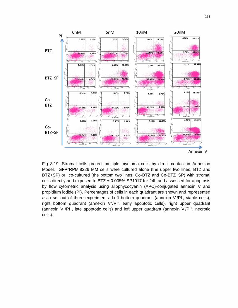
153
Fig 3.19. Stromal cells protect multiple myeloma cells by direct contact in Adhesion
Model. GFP+RPMI8226 MM cells were cultured alone (the upper two lines, BTZ and
BTZ+SP) or co-cultured (the bottom two lines, Co-BTZ and Co-BTZ+SP) with stromal
cells directly and exposed to BTZ ± 0.005% SP1017 for 24h and assessed for apoptosis
by flow cytometric analysis using allophycocyanin (APC)-conjugated annexin V and
propidium iodide (PI). Percentages of cells in each quadrant are shown and represented
as a set out of three experiments. Left bottom quadrant (annexin V-/PI-, viable cells),
right bottom quadrant (annexin V+/PI-, early apoptotic cells), right upper quadrant
(annexin V+/PI+, late apoptotic cells) and left upper quadrant (annexin V-/PI+, necrotic
cells).

154
1 10 100 10000
40
80
120BTZ
BTZ+SP1017
BTZ (nM)
Su
rviv
al (%
of
the
co
ntr
ol)
Fig.3.20. Cytotoxicity of BTZ ± 0.005%SP1017 to OMA-AD stromal cells after 24h
treatment. Data presented as mean ± SD. N=3.

155
A
B
Fig 3.21. IL-6 level in the supernatant of stromal cells and stromal cells co-cultured with
MM cells in Adhersion Model after (A) 4h and (B) 24h adhesion, followed by another 24h
drug treatments. Data presented as mean ± SD. N=3 (*P <0.05, **P<0.01).

156
improve sensitivity of myeloma cells to the drug. DEX, a glucocorticoid analog, used in
certain hematological malignancies, especially in the treatment of MM, in which DEX is
given alone in a high dose [68] or together with other anti-MM drugs. Combination
therapies with DEX plus BTZ, thalidomide, or lenalidomide have been extensively used
and are among the most effective treatments for patients with relapsed or refractory MM
[69, 70]. The combination of DEX with BTZ along with other drugs such as thalidomide,
doxorubicin, cisplatin, cyclophosphamide, and etoposide has resulted in improvements
in both response rates and long-term outcomes [69, 71]. Hematologic toxic effects are
quite frequent when thalidomide, lenalidomide, or bortezomib is used together with
conventional chemotherapy but are less frequent when these drugs are used with DEX
alone[69]. It is widely accepted that the ability of glucocorticoids like DEX to achieve MM
cell apoptosis is mediated via initial binding to their cognate receptor, the glucocorticoid
receptor (GR). Ligand binding to the GR could result in inhibition of NFκB activity by
‘‘transactivation’’ via transcription of IκB and by ‘‘transrepression’’ via a reduction in
transcription of the NFκB genes, thus decreasing the NFκB-dependent anti-apoptotic
proteins [72,73]. Here we examined the effectiveness of the BTZ+DEX+SP1017 3-drug
combination in MM. DEX itself didn’t induce any cytotoxicity to both RPMI 8226 MM cell
line and ARH-77 MM cell line even at quite high concentration (Fig.3.22a, 3.22b).
However, the addition of DEX (1uM) potentiated the BTZ- or BTZ+SP1017- induced cell
death in both RPMI 8226 MM cell line and ARH-77 MM cell line (Fig.3.23a, 3.23b).
Meanwhile, SP1017 potentiated the BTZ-induced cell death in both absence and
presence of DEX. SP1017 was proved to potentiate the BTZ-induce of XIAP(one of
NFκB-dependent anti-apoptotic protein) deduction (Fig.3.17). Here we observed that
XIAP was further reduced by the addition of DEX (Fig.3.24).

157
A
0.1 1 10 1000
40
80
120
DEX (uM)
Su
rviv
al (%
of
the
co
ntr
ol)
B
0.1 1 10 1000
40
80
120
DEX (uM)
Su
rviv
al (%
of
the
co
ntr
ol)
Fig.3.22. Cell survival of (A) sensitive RPMI 8226 MM cell line and (B) ARH 77 cell line
treated with DEX with different doses for 24h. Data presented as mean ± SD. N=3.

158
A
5nM
10nM
0
40
80
120
BTZ
BTZ+SP1017
BTZ+DEX
BTZ+SP1017+DEX
DEX (1uM)
***
******
*
***
***
**
BTZ (nM)
Su
rviv
al (%
of
the
co
ntr
ol)
B
5nM
10nM
0
40
80
120BTZ
BTZ+SP1017
BTZ+DEX
BTZ+SP1017+DEX
DEX (1uM)
**
**
****
***
BTZ (nM)
Su
rviv
al (%
of
the
co
ntr
ol)
Fig 3.23. Cell survival of (A) sensitive RPMI 8226 MM cell line and (B) ARH 77 cell line
treated with 3-drug combination (BTZ+0.005%SP1017+1uMDEX) for 24h. Data
presented as mean ± SD. N=3 (***P<0.001,**P<0.01, and *P<0.05).

159
Fig.3.24. Expression of XIAP of sensitive RPMI 8226 MM cell line treated with 3-drug
combination for 24h. Data represented as a set out of three experiments.
160
3.4 Conclusions
In the previous published work, Pluronics at relative low concentration can
sensitize MDR cancer cells mainly by Pgp inhibition, selective ATP depletion, ROS
generation, inhibition of GSH/GST detoxification as well as triggering mitochondria
apoptotic signals, but not in sensitive cells [74, 75, 76, 77]. Here in our study we firstly
proved that at quite low concentration, the mixture of Pluronics SP1017 sensitize
sensitive MM cells to proteasome inhibitors discussed in Chapter II. Mechanistic studies
showed that the BTZ+SP1017 combination–induced apoptosis is associated with : 1)
increase of suppression of ChT-L proteolytic activity, concomitant with increased
accumulation of ubiquitinated proteins and proteotoxic stress; 2) accumulation of
Pluronic® L61 in ER and increase of ER stress response; 3) accumulation of Pluronic®
L61 in Golgi apparatus, increase of Golgi fragmentation and reduction of secretion of
paraprotein; 4) increase of GSH depletion, resulting in less ability to neutralize drug-
induced oxidative stress; 5) increase of activation of both mitochondria-dependent (quick
translocation of Pluronic® L61 into mitochondria, mitochondrial membrane potential loss,
cytochrome c release and activation of caspase 9/3 ) and independent (activation of
caspase 8/3) apoptotic pathways; 6) decrease of the expression of anti-apoptotic
proteins. Studies in the BM microenvironment models showed that the combination of
SP1017 and BTZ effectively overcame both adhesion-mediated drug resistance and
cytokines-mediated drug resistance, indicating the great potential to use the combination
for MM. One of the mechanisms is due to the significant reduction of secretion of IL-6,
the major growth factor by BMSCs treated by the combination rather than BTZ alone.
The findings provide the strong rationale for our further investigation of the treatment
efficiency of the combination therapy in human myeloma/SCID mice model and the
future clinical evaluation in patients with relapsed and refractory myeloma. In addition,

161
SP1017 in combination with BTZ and DEX leads to most pronounced anti-proliferative
effect, indicating the potential of SP1017 used in corticosteroid sparing regimens to
further improve the therapeutic efficacy as well as limiting significant side effects that
patient with multiple myeloma often experience.

162
3.5 References
1 Dou QP, Zonder JA. Overview of proteasome inhibitor-based anti-cancer
therapies: perspective on bortezomib and secondgeneration proteasome inhibitors versu
s future generation inhibitors of ubiquitin-proteasome system. Curr Cancer Drug Targets.
2014;14(6):517-536.
2 Adams J. The proteasome: a suitable antineoplastic target. Nat Rev Cancer. 2004
May;4(5):349-60.
3 Mitsiades N, Mitsiades CS, Poulaki V, Chauhan D, Fanourakis G, Gu X, Bailey C,
Joseph M, Libermann TA, Treon SP, Munshi NC, Richardson PG, Hideshima T,
Anderson KC. Molecular sequelae of proteasome inhibition in human multiple myeloma
cells. Proc Natl Acad Sci U S A. 2002 Oct 29;99(22):14374-14379
4Ciechanover A, Schwartz AL. The ubiquitin-proteasome pathway: the complexity and
myriad functions of proteins death.Proc Natl Acad Sci U S A. 1998 Mar 17;95(6):2727-
2730.
5 Herrmann J, Lerman LO, Lerman A. Ubiquitin and ubiquitin-like proteins in protein
regulation. Circ Res. 2007 May 11;100(9):1276-1291.
6 Chauhan D, Hideshima T, Anderson KC. Proteasome inhibition in multiple myeloma:
therapeutic implication. Annu Rev Pharmacol Toxicol. 2005;45:465-476.
7 Chen D, Frezza M, Schmitt S, Kanwar J, Dou QP.. Bortezomib as the First Proteasome
Inhibitor Anticancer Drug: Current Status and Future Perspectives. Curr Cancer Drug
Targets. 2011 March ; 11(3): 239–253.
8 Kloetzel PM, Ossendorp F. Proteasome and peptidase function in MHC-class-I-
mediated antigen presentation. Curr Opin Immunol. 2004 Feb;16(1):76-81.
9 Berkers CR, Verdoes M, Lichtman E, Fiebiger E, Kessler BM, Anderson KC, Ploegh
HL, Ovaa H, Galardy PJ. Activity probe for in vivo profiling of the specificity of

163
proteasome inhibitor bortezomib. Nat Methods. 2005 May;2(5):357-362.
10 Chauhan D, Catley L, Li G, Podar K, Hideshima T, Velankar M, Mitsiades C, Mitsiades
N, Yasui H, Letai A, Ovaa H, Berkers C, Nicholson B, Chao TH,Neuteboom
ST, Richardson P, Palladino MA, Anderson KC. A novel orally active proteasome
inhibitor induces apoptosis in multiple myeloma cells with mechanisms distinct from
Bortezomib. Cancer Cell. 2005 Nov;8(5):407-419.
11 Parlati F, Lee SJ, Aujay M, Suzuki E, Levitsky K, Lorens JB, Micklem DR, Ruurs
P, Sylvain C, Lu Y, Shenk KD, Bennett MK. Carfilzomib can induce tumor cell death
through selective inhibition of the chymotrypsin-like activity of the proteasome.
Blood. 2009 Oct 15;114(16):3439-47.
12 Kuhn DJ, Chen Q, Voorhees PM, Strader JS, Shenk KD, Sun CM, Demo SD, Bennett
MK, van Leeuwen FW, Chanan-Khan AA, Orlowski RZ. Potent activity of carfilzomib,
a novel, irreversible inhibitor of the ubiquitin-proteasome pathway, against preclinical
models of multiple myeloma. Blood. 2007 Nov 1;110(9):3281-3290.
13 Grant S. Enhancing proteotoxic stress as an anticancer strategy. Oncotarget. 2011
Apr;2(4):284-286.
14 Bush KT, Goldberg AL, Nigam SK. Proteasome inhibition leads to a heat-shock
response, induction of endoplasmic reticulum chaperones, andthermotolerance. J Biol
Chem. 1997 Apr 4;272(14):9086-9092.
15 Welch WJ. Mammalian stress response: cell physiology, structure/function of stress
proteins, and implications for medicine and disease. Physiol Rev. 1992 Oct;72(4):1063-
1081.
16 Hartl FU. Molecular chaperones in cellular protein folding. Nature. 1996 Jun
13;381(6583):571-579.
17Hartl FU, Hayer-Hartl M. Molecular chaperones in the cytosol: from nascent chain to
164
folded protein. Science. 2002 Mar 8;295(5561):1852-1858.
18 Awasthi N, Wagner BJ. Upregulation of heat shock protein expression by proteasome
inhibition: an antiapoptotic mechanism in thelens. Invest Ophthalmol Vis Sci. 2005
Jun;46(6):2082-2091.
19 Duus J, Bahar HI, Venkataraman G, Ozpuyan F, Izban KF, Al-Masri H, Maududi
T, Toor A, Alkan S. Analysis of expression of heat shock protein-90 (HSP90) and the
effects of HSP90 inhibitor (17-AAG) in multiple myeloma. Leuk Lymphoma. 2006
Jul;47(7):1369-1378.
20 Roué G, Pérez-Galán P, Mozos A, López-Guerra M, Xargay-Torrent S, Rosich
L, Saborit-Villarroya I, Normant E, Campo E, Colomer D. The Hsp90 inhibitor IPI-504
overcomes bortezomib resistance in mantle cell lymphoma in vitro and in vivo by down-
regulation of the prosurvival ER chaperone BiP/Grp78. Blood. 2011 Jan 27;117(4):1270-
1279.
21 Harding HP, Calfon M, Urano F, Novoa I, Ron D. Transcriptional and translational
control in the Mammalian unfolded protein response. Annu Rev Cell Dev
Biol. 2002;18:575-599.
22 Rao RV, Bredesen DE. Misfolded proteins, endoplasmic reticulum stress and
neurodegeneration. Curr Opin Cell Biol. 2004 Dec;16(6):653-662.
23 Lenna S, Trojanowska M. The role of endoplasmic reticulum stress and the unfolded
protein response in fibrosis. Curr Opin Rheumatol. 2012 Nov;24(6):663-668.
24 Harding HP, Zhang Y, Bertolotti A, Zeng H, Ron D. Perk is essential for translational
regulation and cell survival during the unfolded protein response. Mol Cell. 2000
May;5(5):897-904.
25 Nishitoh H. CHOP is a multifunctional transcription factor in the ER stress response. J
Biochem. 2012 Mar;151(3):217-219

165
26 Matsumoto M, Minami M, Takeda K, Sakao Y, Akira S. Ectopic expression
of CHOP (GADD153) induces apoptosis in M1 myeloblastic leukemia cells. FEBS
Lett. 1996 Oct 21;395(2-3):143-7.
27 Maytin EV, Ubeda M, Lin JC, Habener JF. Stress-inducible transcription factor CHOP/
gadd153 induces apoptosis in mammalian cells via p38 kinase-dependent and-
independent mechanisms. Exp Cell Res. 2001 Jul 15;267(2):193-204.
28 McCullough KD, Martindale JL, Klotz LO, Aw TY, Holbrook NJ. Gadd153 sensitizes
cells to endoplasmic reticulum stress by down-regulating Bcl2 and perturbing the cellular
redox state.Mol Cell Biol. 2001 Feb;21(4):1249-1259.
29 Han YH, Moon HJ, You BR, Yang YM, Kim SZ, Kim SH, Park WH. MG132,
a proteasome inhibitor, induced death of calf pulmonary artery endothelial cells via casp
ase-dependent apoptosis and GSH depletion. Anticancer Res. 2010 Mar;30(3):879-885.
30 Du ZX, Zhang HY, Meng X, Guan Y, Wang HQ. Role of oxidative stress and
intracellular glutathione in the sensitivity to apoptosis induced by proteasome inhibitor
in thyroid cancer cells. BMC Cancer. 2009 Feb 16;9:56.
31 Nerini-Molteni S, Ferrarini M, Cozza S, Caligaris-Cappio F, Sitia R. Redox
homeostasis modulates the sensitivity of myeloma cells to bortezomib. Br J Haematol.
2008 May;141(4):494-503.
32 Tait SW, Green DR. Mitochondria and cell death: outer membrane permeabilization
and beyond. Nat Rev Mol Cell Biol. 2010 Sep;11(9):621-32.
33 Ling YH, Liebes L, Zou Y, Perez-Soler R. Reactive oxygen species generation and
mitochondrial dysfunction in the apoptotic response to Bortezomib, anovel proteasome
inhibitor, in human H460 non-small cell lung cancer cells. J Biol Chem. 2003 Sep
5;278(36):33714-23.

166
34 Liu FT, Agrawal SG, Gribben JG, Ye H, Du MQ, Newland AC, Jia L.
Bortezomib blocks Bax degradation in malignant B cells during treatment with TRAIL.
Blood. 2008 Mar 1;111(5):2797-805
35 Wei MC, Zong WX, Cheng EH, Lindsten T, Panoutsakopoulou V, Ross AJ, Roth
KA, MacGregor GR, Thompson CB, Korsmeyer SJ. Proapoptotic BAX and BAK: a
requisite gateway to mitochondrial dysfunction and death. Science. 2001 Apr
27;292(5517):727-730.
36 De Raeve HR, Vanderkerken K. The role of the bone marrow microenvironment in
multiple myeloma. Histol Histopathol. 2005 Oct;20(4):1227-1250.
37 Manier S, Sacco A, Leleu X, Ghobrial IM, Roccaro AM. Bone marrow
microenvironment in multiple myeloma progression.
J Biomed Biotechnol. 2012;2012:157496.
38 Hazlehurst LA, Damiano JS, Buyuksal I, Pledger WJ, Dalton WS. Adhesion to
fibronectin via beta1 integrins regulates p27kip1 levels and contributes to cell adhesion
mediated drug resistance (CAM-DR). Oncogene. 2000 Sep 7;19(38):4319-4327.
39 Uchiyama H, Barut BA, Mohrbacher AF, Chauhan D, Anderson KC. Adhesion of
human myeloma-derived cell lines to bone marrow stromal cells stimulates interleukin-6
secretion. Blood. 1993 Dec 15;82(12):3712-3720.
40 Gupta D, Treon SP, Shima Y, Hideshima T, Podar K, Tai YT, Lin B, Lentzsch
S, Davies FE, Chauhan D, Schlossman RL, Richardson P, Ralph P, Wu L,Payvandi
F, Muller G, Stirling DI, Anderson KC. Adherence of multiple myeloma cells to bone
marrow stromal cells upregulates vascular endothelial growth factor
secretion: therapeutic applications.Leukemia. 2001 Dec;15(12):1950-61.
41 Hideshima T, Anderson KC. Molecular mechanisms of novel therapeutic approaches
for multiple myeloma. Nat Rev Cancer. 2002 Dec;2(12):927-937.

167
42 Hideshima T, Richardson P, Chauhan D, Palombella VJ, Elliott PJ, Adams
J, Anderson KC. The proteasome inhibitor PS-341 inhibits growth, induces apoptosis,
and overcomes drug resistance in humanmultiple myeloma cells. Cancer Res. 2001 Apr
1;61(7):3071-3076.
43 Batrakova EV, Li S, Alakhov VY, Miller DW, Kabanov AV. Optimal Structure
Requirements for Pluronic Block Copolymers in Modifying P-glycoprotein Drug Efflux
Transporter Activity in Bovine Brain Microvessel Endothelial Cells J Pharmacol Exp
Ther. 2003 Feb;304(2):845-854.
44 Wang M, Wey S, Zhang Y, Ye R, Lee AS. Role of the unfolded protein response
regulator GRP78/BiP in development, cancer, and neurological disorders. Antioxid
Redox Signal. 2009 Sep;11(9):2307-2316.
45 Amy S. Lee. The ER chaperone and signaling regulator GRP78/BiP as a monitor of
endoplasmic reticulum stress. Methods. 2005 Apr;35(4):373-381.
46 Batrakova E, Lee S, Li S, Venne A, Alakhov V, Kabanov A. Fundamental
Relationships Between the Composition of Pluronic Block Copolymers and Their
Hypersensitization Effect in MDR Cancer Cells. Pharm Res. 1999 Sep;16(9):1373-1379.
47 Batrakova EV, Li S, Alakhov VY, Miller DW, Kabanov AV.. Optimal Structure
Requirements for Pluronic Block Copolymers in Modifying P glycoprotein Drug Efflux
Transporter Activity in Bovine Brian Microvessel Endothelial Cells. J Pharmacol Exp
Ther. 2003 Feb;304(2):845-854.
48 Batrakova EV, Kabanov AV. Pluronic Block Copolymers:Evolution of Drug Delivery
Concept from Inert Nanocarriers to Biological Response Modifiers. J Control
Release. 2008 Sep 10;130(2):98-106.
49 Alakhova DY, Kabanov AV. Pluronics and MDR Reversal: An Update. Mol Pharm.
2014 Aug 4;11(8):2566-78.
168
50 Batrakova EV, Li S, Vinogradov SV, Alakhov VY, Miller DW, Kabanov AV. Mechanism
of Pluronic Effect on P-Glycoprotein Efflux System in Blood-Brain Barrier: Contributions
of Energy Depletion and Membrane Fluidization. J Pharmacol Exp Ther. 2001
Nov;299(2):483-493.
51 Batrakova EV, Li S, Li Y, Alakhov VY, Kabanov AV. Effect of pluronic P85 on ATPase
activity of drug efflux transporters. Pharm Res. 2004 Dec;21(12):2226-2233.
52 Hicks SW, Machamer CE.. Golgi structure in stress sensing and apoptosis. Biochimica
et Biophysica Acta 1744 (2005) 406 –414.
53 Farquhar MG, Palade GE. The Golgi apparatus: 100 years of progress and
controversy. Trends Cell Biol. 1998 Jan;8(1):2-10.
54 Petrosyan A, Cheng PW, Clemens DL, Casey CA. Downregulation of the small
GTPase SAR1A: a key event underlying alcohol-induced Golgi fragmentation in
hepatocytes. Sci Rep. 2015 Nov 26;5:17127.
55 Petrosyan A, Casey CA, Cheng PW. The role of Rab6a and phosphorylation of non-
muscle myosin IIA tailpiece in alcohol-induced Golgi disorganization. Sci Rep. 2016 Aug
18;6:31962.
56 Heher EC, Rennke HG, Laubach JP, and Richardson PG. Kidney Disease and
Multiple Myeloma Clin J Am Soc Nephrol. 2013 Nov 7; 8(11): 2007–2017.
57 Circu ML, Aw TY. Glutathione and modulation of cell apoptosis. Biochim Biophys Acta
2012 Oct;1823(10):1767-1777.
58 Fulda S, Debatin KM. Targeting apoptosis pathways in cancer therapy.
Curr Cancer Drug Targets. 2004 Nov;4(7):569-76.
59 Fulda S, Debatin KM. Extrinsic versus intrinsic apoptosis pathways in anticancer
chemotherapy. Oncogene. 2006 Aug 7;25(34):4798-4811.

169
60 Alakhova DY, Rapoport NY, Batrakova EV, Timoshin AA, Li S, Nicholls D, Alakhov
VY, Kabanov AV.Differential Metabolic Responses to Pluronic in MDR and non-MDR
Cells: A Novel Pathway for Chemosensitization of Drug Resistant CancersJ Control
Release. 2010 Feb 25;142(1):89-100.
61 Batrakova EV1, Li S, Elmquist WF, Miller DW, Alakhov VY, Kabanov AV. Mechanism
of sensitization of MDR cancer cells by Pluronic block copolymers: Selective energy dep
letion. Br J Cancer. 2001 Dec 14;85(12):1987-1997.
62 Peter ME, Krammer PH. The CD95(APO-1/Fas) DISC and beyond. Cell Death
Differ. 2003 Jan;10(1):26-35.
63 Gajate C, Mollinedo F. The antitumor ether lipid ET-18-OCH3 induces apoptosis
through translocation and capping of Fas/CD95 into membrane rafts in human leukemic
cells. Blood. 2001 Dec 15;98(13):3860-3863.
64 Gajate C, Mollinedo F. Edelfosine and perifosine induce selective apoptosis in multiple
myeloma by recruitment of death receptors and downstream signaling molecules into
lipid rafts. Blood. 2007 Jan 15;109(2):711-719.
65 Mollinedo F, de la Iglesia-Vicente J, Gajate C, Estella-Hermoso de Mendoza A, Villa-
Pulgarin JA, Campanero MA, Blanco-Prieto MJ. Lipid raft-targeted therapy in multiple
myeloma. Oncogene. 2010 Jul 1;29(26):3748-57.
66 Sahay G, Batrakova EV, Kabanov AV. Different internalization pathways of polymeric
micelles and unimers and their effects on vesicular transport. Bioconjug Chem. 2008
Oct;19(10):2023-2029.
67 Kapoor P, Ramakrishnan V, Rajkumar SV. Bortezomib combination therapy in multiple
myeloma. Semin Hematol. 2012 Jul;49(3):228-42.

170
68 Friedenberg WR, Kyle RA, Knospe WH, Bennett JM, Tsiatis AA, Oken MM. High-dose
dexamethasone for refractory or relapsing multiple myeloma. Am J Hematol. 1991
Mar;36(3):171-5.
69 Palumbo A, Anderson K. Multiple myeloma. N Engl J Med. 2011;364:1046–1060.
70 Nooka AK, Kastritis E, Dimopoulos MA, Lonial S. Treatment options for relapsed and
refractory multiple myeloma. Blood. 2015 May 14;125(20):3085-99.
71Kapoor P, Ramakrishnan V, Rajkumar SV. Bortezomib combination therapy in multiple
myeloma. Semin Hematol. 2012 Jul;49(3):228-42.
72 Sharma S, Lichtenstein A. Dexamethasone-induced apoptotic mechanisms
in myeloma cells investigated by analysis of mutant glucocorticoid receptors.Blood. 2008
Aug 15;112(4):1338-45.
73 Salem K, Brown CO, Schibler J, Goel A. Combination chemotherapy increases
cytotoxicity of multiple myeloma cells by modification of nuclear factor (NF)-κB activity.
Exp Hematol. Feb 2013; 41(2): 209–218.
74Batrakova EV, Li S, Alakhov VY, Elmquist WF, Miller DW, Kabanov AV.. Sensitization
of cells overexpressing multidrug-resistant proteins by pluronic p85. Pharm Res. 2003
Oct;20(10):1581-1590.
75 Batrakova EV, Li S, Elmquist WF, Miller DW, Alakhov VY, Kabanov AV. Mechanism of
sensitization of MDR cancer cells by Pluronic block copolymers: Selective energy
depletion. Br J Cancer. 2001 Dec 14;85(12):1987-1997.
76 Alakhova DY, Rapoport NY, Batrakova EV, Timoshin AA, Li S, Nicholls D, Alakhov
VY, Kabanov AV. Differential metabolic responses to pluronic in MDR and non-MDR
cells: A novel pathway for chemosensitization of drug resistant cancers . J Control
Release. 2010 Feb 25;142(1):89-100.

171
77 Minko T, Batrakova EV, Li S, Li Y, Pakunlu RI, Alakhov VY, Kabanov AV., Pluronic
block copolymers alter apoptotic signal transduction of doxorubicin in drug-resistant
cancer cells. J Control Release. 2005 Jul 20;105(3):269-278

172
CHAPTER IV: THE ANTI-TUMOR ACTIVITY OF THE BTZ+SP1017 COMBINATION
THERAPY IN HUMAN MM/SCID MODELS.
4.1 Introduction
To further evaluate potential of SP1017+BTZ combination for treating MM, an in
vivo murine model that recapitulates the characteristics of human disease condition of
MM is required. An animal model that accurately reflects human MM and takes into
account the protective nature of BMSCs would be powerful in confirming the efficacy of
therapeutic agents in vivo. There are currently three types of MM animal models: 1)
injection of pristane oil in BALB/c mice leads to intraperitoneal plasmacytomas but
without BM colonization and osteolysis; 2) injection of allogeneic malignant plasma cells
(5T2MM, 5T33) in the C57BL/KalwRij mouse induces BM proliferation and osteolytic
lesions; 3) injection of malignant plasma cell lines in immunodeficient mice SCID or
NOD/SCID [1]. The BALB /c oil-induced and transplantable plasmacytomas resemble
human MM in their ability to produce a monoclonal immunoglobulin [2]. The major
disadvantage of the first model is that plasma cells are restrictively localized in the
peritoneum and do not extend to the BM, which does not faithfully resemble human MM
[1]. The 5TMM model originates from spontaneously developed MM in aged
C57BL/KalwRij mice and has since been propagated by intravenous injection of BM cells
from MM bearing mice, into young naive syngeneic recipients. This model has been
extensively used for studying homing mechanisms of MM cells to BM, interaction of MM
cells with the BM environment, and evaluation of new therapies [3, 4]. However, the
5TMM models are limited by being solely murine-derived and representative of only one
type of myeloma. Xenograft models, in which human MM cell lines are injected into
immune compromised mice (for example, severe combined immunodeficient (SCID),

173
nude, NOD/SCID gamma (NSG) mice, etc.), can be used to establish systemic disease
(via intravenous injection of MM cells) or local disease (via subcutaneous or intratibial
injections) [5]. The models allow for the implantation of human MM cells, which are more
relevant for investigating therapeutic targets and preclinical therapeutic testing than
murine MM cells. Subcutaneous xenograft models not only fail to reflect the systemic
nature of diffuse lesions of MM but also place tumor cells in the cutaneous
microenvironment, which is radically different from the BM microenvironment, which
appears to play an important role in differentiation, migration, proliferation, survival, and
drug resistance of the malignant plasma cells [6], thus influencing the responsiveness to
therapy. Several NOD/SCID MM models using intravenous injection of human MM cell
lines, including U266, MM.1S, RPMI-8266 or primary human MM cells have been
described [7, 8, 9, 10]. MM cells are preferentially homing into BM via selective binding
of adhesion molecules on their surface to BMSCs and ECM proteins. For example, the
very-late antigen 4 on MM cells binds to fibronectin and lymphocyte function-associated
antigen-1 on MM cells binds to intercellular adhesion molecule-1 on BMSCs [11].
NOD/SCID IL-2Rγ(null) mice (NSG) mice, which lack the interleukin-2 (IL-2) gamma
chain, have recently been used to develop models of MM [12]. Compared to the SCID
and NOD/SCID strains which lack mature B and T lymphocytes, this strain lacks NK
cells, characterized by defects in macrophages, complement and dendritic cell function
and shows significant improvements in tumor cell engraftment associated with
abolishment of residual immune function and lack of thymic lymphoma development
accompanied by an extended lifespan [13, 14]. NSG mice were injected intravenously
via the tail vein with U266, XG-1, OPM-2 or patient-derived MM cells, and provided MM
models which feature consistent tumor burden and osteolytic bone disease including
paraplegia, paraprotein in the serum, osteolytic lesions and loss of trabecular bone
[15]. Though tumor growth was observed in the BM of xenograft models, some of

174
previous studies indicated that MM cells were found in various sites, including the
lungs, liver, kidneys, spleen [9, 10, 16]. Intra-tibial inoculation of tumor cells in NSG mice
has been developed as well, which exhibited typical disease symptoms exclusively
without organ involvement [17, 18].
In order to evaluate therapeutic benefit of SP1017+BTZ combination for treating
MM and provide more accurate preclinical evaluations of investigational therapies, we
successfully established the human MM/SCID mice model by injecting Luc+/GFP+ RPMI
8226 MM cells (2×10⁶/100ul PBS/Mouse) into NSG mice. In 4 weeks post tumor
implantation, mice were treated with saline, SP1017, BTZ or combination of SP1017 and
BTZ via tail vein injections twice weekly for 4 weeks. Total body bioluminescence
imaging (BLI), the serum levels of human immunoglobulin lambda paraprotein, changes
in the body weight and onset of hind limb paralysis at the late stages of the disease were
used to monitor the treatment efficacy.
4.2 Materials and methods
4.2.1 Materials
Bortezomib was purchased from Selleck Chemicals (Houston, TX, USA). SP1017
was kindly provided by Supratek Pharma. Inc (Montreal, Canada). F-Luc-GFP Lentivirus
was purchased from Capital Biosciences (Rockville, MD, USA). Human lambda EILSA
kit was purchased from Bethyl Laboratories Inc (Montgomery, TX, USA). Luciferase
assay system and cell culture lysis reagent were purchased from Promega (Madison,
WI, USA). Hank’s Balanced Salt Solution (HBSS), ACK lysing buffer were purchased
from Fisher Scientific (Pittsburgh, PA, USA). Hexadimethrine bromide (Polybrene) was
purchased from Sigma-Aldrich (St Louis, MO, USA). FITC anti-mouse CD 45 antibody

175
was purchased from Biolegend (San Diego, CA, USA). PE anti-mouse CD11b and
Purified Rat Anti-Mouse CD16/CD32 was purchased from BD Biosciences (San Jose,
CA, USA).
4.2.2 Labeling of RPMI 8226 MM cells with Luciferase (Luc) and GFP
To prepare stably transfected Luc+/GFP+ MM cells, RPMI 8226 MM cells were
infected with F-Luc-GFP Lentivirus. Briefly, 10^5 of cells were seeded in one well of a
24-well plate and mixed with 100ul of F-Luc-GFP Lentivirus (10^8 TU/ml) in 1ml of the
complete media in the presence of Polybrene (8 µg/mL). Cells was centrifuged at 300Xg
for 1h at room temperature and incubated at 37 ℃ in a humidified, 5% CO2 atmosphere
overnight. The cells were washed with PBS, fresh culture medium was added the next
morning and cells were incubated for the enlargement of the cell number. To enrich for
Luc positive cells, cells were sorted by Becton Dickinson FACSAria™ flow
cytometer with GFP expression.
4.2.3 The establishment of the human MM/SCID model
A three mice pilot study was conducted to establish the human MM/SCID
models. MM was generated by i.v. injection of Luc+ /GFP+ RPMI-8226 cells (2 ×10⁶ cells
suspended in 100 ul of PBS) via tail vein of NSG mice. The tumor progression was
evaluated by BLI, the serum levels of human immunoglobulin lambda paraprotein,
changes in the body weight and onset of hind limb paralysis. The BM of one the tumor-
beaing mouse and one control mouse was extracted by ice-cold HBSS. All cells were
collected and centrifuged down at 1500rpm for 10min. The supernatant was removed
and cells were incubated with 2ml of ACK lysis buffer in 37°C water bath for 2min to
remove the red cells. Another 25ml of HBSS was added to stop the lysis reaction. The
GFP positive tumor cells were analyzed using Becton Dickinson FACSCalibur™ flow
cytomerter and FACSDiva software (Version 8.0, Becton Dickinson, San Jose, CA).

176
4.2.4 The evaluation of the combination of SP1017+BTZ in human MM/SCID
model
Female six-week old NOD/SCIDγc null (NSG) mice were obtained from The
Jackson Laboratories and housed in AAALAC accredited facility. Food and reverse
osmosis water were available ad libitum throughout the study. Animals were quarantined
for 7 days prior to study initiation. All animal studies were conducted in accordance with
the protocol approved by the University of Nebraska Medical Center Institutional Animal
Care and Use Committee. MM was generated by i.v. injection of Luc+/GFP+ RPMI-8226
cells (2 ×10⁶ cells suspended in 100 ul of PBS) via tail vein of NSG mice. After
development of tumors (4 weeks after injection), animal were randomized (4 treatment
groups, n=10) and treated with saline, 0.1% SP1017 (100ul), 0.5mg/kg BTZ or
combination of 0.1% SP1017 (100ul) and 0.5mg/kg BTZ. Treatments were administrated
via tail vein injections twice weekly for 4 weeks (On day 1, 4, 8, 11, 15, 18, 22, 25).
Tumor progression was monitored by bioluminescence imaging (BLI) once weekly, the
procedure of which had been detailed in [19]. Blood samples were collected twice a
week prior to drug injection from each mouse from facial vein by using 4-5mm lancet for
the measurement of human Igλ in murine serum using human lambda ELISA Kit
according to the manufacturer’s instructions. Animal body weight was monitored every
second day starting from the day of malignant cell infusion and during tumor
development and drug treatment. General physical conditions, clinical signs and
ascending paralysis were assessed using a four-stage scale of 0-3, with “0” being
clinically normal, “1” being decreased tail tone or weak tail only, “2” being hind limb
weakness and “3” being hind limb paralysis. Upon initial stages of disease progression,
animals were monitored for clinical signs every second day. When initial clinical signs
are noticed (score 1-2), animals were monitored on daily basis. The paralyzed mice or

177
non-paralyzed mice with 20% of body weight loss or those becoming moribund were
euthanized via CO2 asphyxiation.
4.2.5 Preparation of Cy5-L61
Pluronic® L61 was labeled with Cyanine 5 fluorophore according to the method
described before20. Briefly, Pluronic® L61were activated with CDI, and then modified with
excess of ethylenediamine and purified by dialysis against 20% ethonal. Amino-modified
L61 was conjugated with Cyanine5 NHS ester (1:1) in dimethylformamide in the
presence of 1.2 eq of tertiary amine. Free Cyanine5 and Cyanine5-labeled L61 were
separated by Sephadex LH-20 (Sigma-Aldrich, St Louis, MO, USA) column using
methonal/ dichloromethane (1:1) as elution phase.
4.2.6 Biodistribution of Cy5-L61 in both tumor-bearing mice and control
mice
5 Female six-week old NOD/SCIDγc null (NSG) mice were inoculated with
Luc+/GFP+ RPMI-8226 cells as described above. In 6 weeks post MM cell inoculation,
saline (100ul as control) and Cy5-L61 (0.011% L61 equivalent) mixed with 0.089% F127
in saline (100ul), which compose 0.1% SP1017 were injected into both tumor-bearing
mice and control mice without tumor. After 24h, the images of the whole body, skeleton
as well as organs were taken using IVIS imaging system to track the distribution of Cy5-
L61. In addition, the BM of the tumor-bearing mice and control mice were extracted by
ice-cold HBSS. All cells were collected and centrifuged down at 1500rpm for 10min. The
supernatant was removed and cells were incubated with 2ml of ACK lysis buffer in 37°C
water bath for 2min to remove the red cells. Another 25ml of HBSS was added to stop
the lysis reaction. The Cy5-positive cells were analyzed using Becton Dickinson LSR
II flow cytometer and FACSDiva software (Version 8.0, Becton Dickinson, San Jose,

178
CA). An aliquot of bone morrow cells was lysed with cell culture lysis reagent and
supernatant was collected after removing cell debris by centrifugation (14000 rpm for 10
min). 20ul of supernatant were taken and plated in white polystyrene, flat opaque bottom
Corning® 96-well plate (Sigma-Aldrich, St Louis, MO, USA). Luciferase assay reagents
were prepared according to the manufacturer’s instructions. 100ul reagent was added to
each sample. Luminescence was determined as relative light units (RLU) using the
SpectraMax M5 microplate reader and the results were normalized by protein content
determined by BCA assay.
4.2.7 Cy5-L61 uptake in CD45+ hematopoietic cells and CD11b+ cells
(macrophages/monocytes) in BM in control mice.
6 male six-week old NOD/SCIDγc null (NSG) mice were injected with saline (100ul
as control) and Cy5-L61 (0.011% L61 equivalent) mixed with 0.089% F127 in saline
(100ul), which compose 0.1% SP1017. The BM of control mice were extracted by ice-
cold HBSS 24h post-injection. All cells were collected and centrifuged down at 1500rpm
for 10min. The supernatant was removed and cells were incubated with 2ml of ACK lysis
buffer in 37°C water bath for 2min to remove the red cells. Another 25ml of HBSS was
added to stop the lysis reaction. Cells were stained with FITC-CD45 and PE-CD11b
according to the manufacturer’s instructions. The cells were analyzed using Becton
Dickinson LSR II flow cytometer and FACSDiva software (Version 8.0, Becton
Dickinson, San Jose, CA).
4.2.8 Statistical analysis
For the antitumor study, the Kruska-Wallis test, a nonparametric test was used
for overall comparison of the group means for paraprotein and BLI under different types
of treatments as the data did not follow a normal distribution. To evaluate specific
differences between BTZ and BTZ+SP1017 groups, Tukey’s nonparametric multiple-

179
comparison method was used. Two-tailed P values less than 0.05 were considered
significant in both tests.
4.3 Results and discussion
The human MM/SCID mice model was successfully established by injecting
Luc+/GFP+ RPMI 8226 MM cells (2×10⁶/100ul PBS/Mouse) into mice via tail vein. The
tumor formation was validated by increased BLI signal observed in femur and other parts
of skeleton (some mice also had increased BLI signal in soft tissue), increased
expression of human lambda paraprotein, huge body weight loss and the progressing
hind limb paralysis in the late stage. Tumor engraftment was observed 4 weeks post-
inoculation by BLI, with concomitant elevation of serum human lambda levels. The
presence of tumor cells in the BM microenvironment was confirmed by whole-body and
skeleton BLI as well as the presence of GFP+ tumor cells in the extracted BM analyzed
by flow cytometry. From the BLI of skeleton, the tumor signal was observed in femur.
Among the BM extracted from the femur about 2.6% of cells are GFP+ tumor cells,
indicating the tumor cells homing to the BM (Fig.4.1). In addition, MM foci and osteolytic
lesions were observed to be present in multiple sites of the axial skeleton and confirmed
by histopathology and X-ray imaging, respectively (Data not shown). Thus, MM cells
were proven to effectively home to skeleton and develop MM lesions in skeletal sites in
this model. For the evaluation of treatment efficiency, 40 NSG mice were injected with
Luc+/GFP+ RPMI 8226 MM cells (2*10^6/100ul PBS/Mouse) via tail vein. In 4 weeks post
tumor implantation, tumor-bearing NSG mice were divided into 4 groups randomly (10
mice per group), treated with 1) 0.9% saline; 2) 0.1% SP1017 (100ul); 3) 0.5mg/kg BTZ;
4) 0.5mg/kg BTZ+0.1% SP1017 (100ul) for 4 weeks, twice weekly (on day 1, 4, 8, 11,
15, 18, 22, 25), which resemble the clinical BTZ regimen for MM. Treatment efficiency

180
A: Control mice without tumor
Left femur Right femur
B: Tumor-bearing mice
Left femur Right femur
Fig.4.1 Luc+/GFP+ RPMI 8226 MM cells in the femur of (A) one control mouse and (B)
one tumor-bearing NSG mouse.

181
was elevated by measuring serum paraprotein levels twice a week and by weekly BLI
imaging. After 4 weeks of treatments (day 24), the serum paraprotein level in the
untreated controls was significantly elevated (85-fold increase) while both BTZ and
BTZ+SP1017 combination treatments greatly suppressed the elevation of paraprotein
over the whole treatment regimen and delayed the disease progression (Fig.4.2A).
Notably, serum level of paraprotein was significantly lower in the group of mice treated
with BTZ+SP1017 combination compared to that of the BTZ alone after only 2 injections
(insert enlarged, Fig.4.2A) and the significant differences between treatments
continued to be observed even after the treatments were completed. These results were
validated by BLI data, which also showed significantly higher inhibition of disease
progression in mice treated with the combination therapy compared to BTZ alone
(Fig.4.2B). Fig.4.3 showed the BLI images of four treatment groups on Day 28 (the third
day after final injection), which visually indicates the better treatment efficiency of the
combination therapy. The two parameters, the level of serum paraprotein and BLI
signal, are well-correlated in indicating the tumor progression and the anti-tumor
efficiency. The serum level of paraprotein can be a more quantitative indicator of
disease progression while the BIL data can be a visualized reporter, which demonstrated
bone morrow tropism of the RPMI 8226 MM cells. In addition, the combination therapy
did not show systemic toxicity (no significant body weight change between groups,
Fig.4.4) or hematological toxicity (no significant differences were observed between the
treatment and control groups as determined by cell blood counting, Table.4.1) at the
dosage (0.5 mg/kg BTZ and 0.1% SP1017 (100ul)) we used.
In previous work, Pluronic® block copolymer P85 was found to be accumulated in
various tissues in the following order: liver>spleen>kidney>lung>brain [21]. However, no
previous studies indicated whether the Pluronic® block copolymer can accumulate in the

182
A B
Fig.4.2 In vivo antitumor efficacy of BTZ+SP1017 combination therapy in Luc+/GFP+
RPMI 8226 human MM xenograft-bearing NSG mice. (A) Relative paraprotein levels
(Pt/P0) and (B) changes in tumor volume as indicated by relative radiance units (Rt/R0)
measured by means of BLI over time following IV administration of 1) 0.9% saline; 2)
0.1% SP1017 (100ul); 3) 0.5mg/kg BTZ; 4) 0.5mg/kg BTZ+0.1% SP1017 (100ul) on Day
1, 4, 8, 11, 15, 18, 22 and 25. Paraprotein level and radiance are normalized to
respective values at the day before treatments were initiated (P0 and R0, respectively).
Insert in panel A shows close-up of results up to 21 days following treatment initiation.
Data presented as mean ± SEM.N=10 (*P<0.05, **P<0.01). Statistical significance
between BTZ+SP1017 and BTZ groups was determined using Tukey’s nonparametric
multiple-comparison method.
0
200
400
600
800
0 3 7 10 14 17 21 24 27 34 41
Pt/P
0
Days
SalineSP1017BTZBTZ+SP1017
* **
**
**
**
**
**
0
10
20
30
40
50
0 3 7 10 14 17 21
*
**
**
0
100
200
300
400
0 6 13 20 27 34 41
Bt/B
0
Days
SalineSP1017BTZBTZ+SP1017
*
*
**
*

183
Fig.4.3. BLI images on the third day after all the 8 injections (Day 28) of four groups of
NSG mice treated with of 1) 0.9% saline; 2) 0.1% SP1017 (100ul); 3) 0.5mg/kg BTZ; 4)
0.5mg/kg BTZ+0.1% SP1017 (100ul).
Dorsal
Saline
SP1017
BTZ
BTZ+ SP1017
Ventral

184
Fig.4.4. Body weight of tumor-bearing NSG mice which are received treatments of 1)
0.9% saline; 2) 0.1% SP1017 (100ul); 3) 0.5mg/kg BTZ; 4) 0.5mg/kg BTZ+0.1% SP1017
(100ul) on Day 1, 4, 8, 11, 15, 18, 22 and 25.
0
5
10
15
20
25
0 2 4 6 8 10 12 14 16 18 20 22 24 26 28
Gra
ms
Days after tumor implantation
SalineSP1017BTZBTZ+SP1017
Body weight

185
Table.4.1. Blood cell counting after 4 injections and 8 injections of 1) 0.9% saline; 2) 0.1% SP1017 (100ul); 3) 0.5mg/kg BTZ; 4) 0.5mg/kg BTZ+0.1% SP1017 (100ul).
The data represent mean ±SD (n=3)
*Significantly different compared to control group, p<0.05
** Significantly different compared to control group, p<0.01
After 4 injections After 8 injections
Control SP1017 BTZ BTZ+SP Control SP1017 BTZ BTZ+SP
WBC(10^3/ul) 6.0±0.9 10.1±0.9** 13.4±3.9* 9.5±1.9* 8.1±2.7 7.2±1.4 12.4±4.5 7.2±2.3
RBC (10^6/ul) 9.3±0.1 8.2±1.3 9.0±0.4 8.5±0.6 9.2±0.2 8.8±0.6 8.0±1.2 8.5±0.3*
PLT (10^3/ul) 38.0±5.7 102.7±24.9* 110.7±89.6 83.3±26.8 38.7±18.9 65.0±5.7 93.7±43.4 43.8±25.2
HGB(g/dL) 15.5±0.5 14.8±0.4 15.2±0.3 14.9±0.1 14.4±0.4 14.6±1.3 14.0±0.5 14.2±0.3
HCT (%) 43.3±0.7 38.5±5.4 42.8±1.9 40.5±2.2 41.7±0.4 40.3±2.6 37.9±5.5 40.3±1.2
MCV (fL) 46.7±0.9 47.7±0.9 47.0±0.0 47.7±0.9 45.3±0.9 45.7±1.2 47.3±0.5* 47.5±0.5*
MCHC (g/dL) 35.8±0.7 39.5±7.2 35.7±1.4 37.0±2.3 34.7±0.8 36.2±3.3 37.7±4.5 35.4±1.7
RDWc (%) 19.5±0.2 19.3±0.4 20.5±0.3* 20.7±0.6 19.0±0.0 18.7±0.2 20.6±0.4* 21.1±0.5*

186
A
Dorsal Ventral
B
Dorsal Ventral
Fig. 4.5 The fluorescence (Cy5) images of mice skeleton of both (A) tumor-bearing mice
and (B) control mice without tumor after 24h of injection of 0.011% Cy5-L61detected by
IVIS with Ex=640nm and Em=680nm.

187
A B
Fig. 4.6 The fluorescence (Cy5) images of mice organs (liver, heart, lung, kidney,
spleen) of both (A) tumor-bearing mice and (B) control mice without tumor after 24h of
injection of Cy5-L61 (0.011% L61 equivalent) detected by IVIS with Ex=640nm and
Em=680nm.
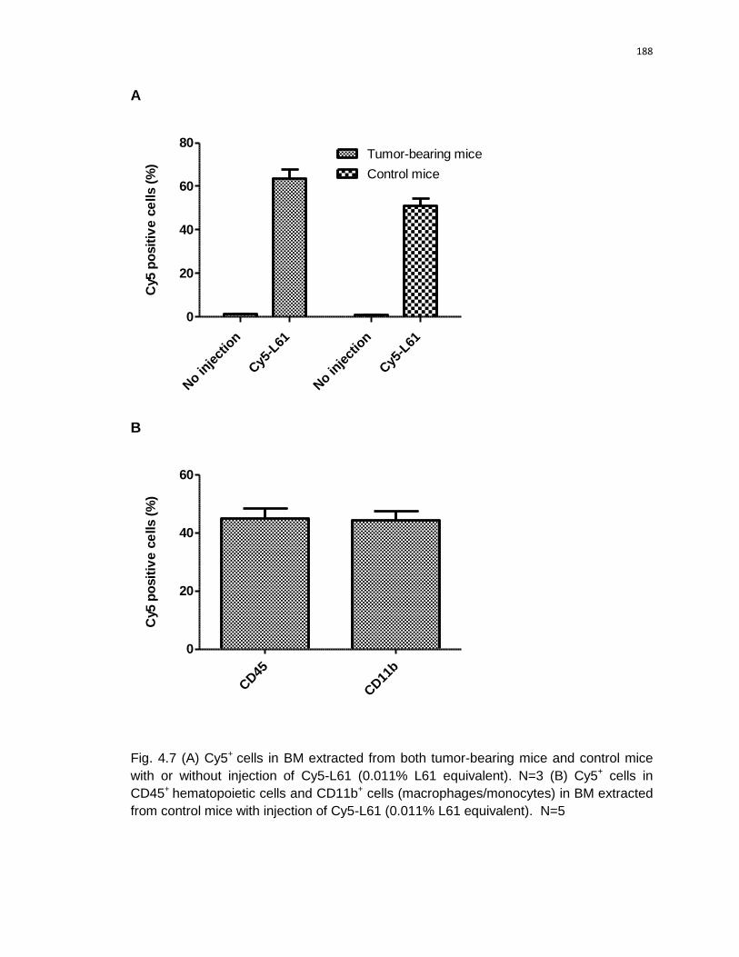
188
A
No in
ject
ion
Cy5
-L61
No in
ject
ion
Cy5
-L61
0
20
40
60
80Tumor-bearing mice
Control mice
Cy5
po
sit
ive
ce
lls (
%)
B
CD45
CD11
b
0
20
40
60
Cy5
po
sit
ive
ce
lls (
%)
Fig. 4.7 (A) Cy5+ cells in BM extracted from both tumor-bearing mice and control mice
with or without injection of Cy5-L61 (0.011% L61 equivalent). N=3 (B) Cy5+ cells in
CD45+ hematopoietic cells and CD11b+ cells (macrophages/monocytes) in BM extracted
from control mice with injection of Cy5-L61 (0.011% L61 equivalent). N=5

189
BM. Interestingly, the fluorescent signal of Cy5 was observed in the femur and spine in
both tumor-bearing mice and control mice 24h post-injection of Cy5-L61 (Fig. 4.5). We
also found the accumulation of Cy5-L61 in the organs, such as liver, kidney and lung in
both tumor-bearing mice and control mice (Fig 4.6). The BM from femur was extracted
and the Cy5+ cells were analyzed by flow cytometry. We found about 63% of Cy5+ cells
in tumor-bearing mice and 51% of Cy5+ cells in control mice (Fig. 4.7A). These data
indicate that Pluronic® L61 could accumulate in BM, thus playing an important role in
sensitizing MM cells in the BM to proteasome inhibitors. Moreover, 45.1% of CD45+ and
44.5% CD11b+ cells in BM were associated with Cy5-L61 (Fig. 4.7B), indicating the
uptake of the Pluronic® L61 by hematopoietic cells.
Dose-limiting peripheral neuropathy is the major toxicity issue with bortezomib,
which leads to dose reductions or discontinuation and may impact both long-term quality
of life and the ability to get subsequent therapies[22]. One way to reduce the systemic
toxicity could be using nanocarrier to encapsulate the hydrophobic drug BTZ or drug
combinations. Despite of the carrier function of Pluronic® micelles, the major limitations
of Pluronic® block copolymers include the low micellization and solubilization capacity to
hydrophobic drugs, as well as poor micellar stability upon dilution in the blood-steam due
to relatively high CMC values. In addition, whether and how much the Pluronic® unimers
can be truly transported to the tumor sites are big concerns. Thus, establishing a
micellar formation of BTZ or CFZ based on amphiphilic block copolymers which have
better micellar stability and can incorporate Pluronic® unimers to tumor sites as well as
releasing the Pluronic® unimers into cytosol for exerting chemosensitization effect could
be our future direction.
4.4 Conclusions

190
We demonstrated that combination of BTZ and SP1017 exerted enhanced
antitumor efficacy compared to BTZ, delaying the disease progression without causing
systemic toxicity or hematological toxicity. Moreover, we observed the accumulation of
the Cy5-L61 in the BM which was proved by both fluorescence imaging and flow
cytometry, indicating the Cy5-L61 could target and accumulate in the skeleton, thus
playing an important role in sensitizing MM cells in the BM to proteasome inhibitors.

191
4.5 References
1 Libouban H. The use of animal models in multiple myeloma. Morphologie. 2015
Jun;99(325):63-72.
2 Azar HA. Experimental plasmacytomas in relation to human multiple myeloma. Ann
1974 May-Jun;4(3):157-163.
3 Asosingh K, Radl J, Van Riet I, Van Camp B, Vanderkerken K. The 5TMM series:
a useful in vivo mouse model of human multiple myeloma.
Hematol J. 2000;1(5):351-356.
4 Vanderkerken K, Asosingh K, Croucher P, Van Camp B.. Multiple myeloma biology:
lessons from the 5TMM models. Immunol Rev. 2003 Aug;194:196-206.
5 Lwin ST, Edwards CM, Silbermann R. Preclinical animal models of multiple myeloma.
Bonekey Rep. 2016 Feb 3;5:772.
6 Manier S, Sacco A, Leleu X, Ghobrial IM, Roccaro AM. Bone
marrow microenvironment in multiple myeloma progression. J Biomed
Biotechnol. 2012;2012:157496.
7 Alsina M1, Boyce B, Devlin RD, Anderson JL, Craig F, Mundy GR, Roodman GD.
Development of an in vivo model of human multiple myeloma bone disease. Blood. 1996
Feb15;87(4):1495-1501.
8 Runnels JM1, Carlson AL, Pitsillides C, Thompson B, Wu J, Spencer JA, Kohler
JM, Azab A, Moreau AS, Rodig SJ, Kung AL, Anderson KC, Ghobrial IM, Lin CP.
Optical techniques for tracking multiple myeloma engraftment, growth, and response to
therapy. J Biomed Opt. 2011 Jan-Feb;16(1):011006.
9 Mitsiades CS, Mitsiades NS, Bronson RT, Chauhan D, Munshi N, Treon SP, Maxwell
CA, Pilarski L, Hideshima T, Hoffman RM, Anderson KC. Fluorescence imaging

192
of multiple myeloma cells in a clinically relevant SCID/NOD in vivo model: biologic and
clinical implications. Cancer Res. 2003 Oct 15;63(20):6689-6696.
10 Paton-Hough J, Chantry AD, Lawson MA. A review of current murine models of
multiple myeloma used to access the efficacy of therapeutic agents on tumour growth
and bone disease. Bone. 2015 Aug;77:57-68.
11 Hideshima T, Anderson KC. Molecular mechanisms of novel therapeutic approaches
for multiple myeloma. Nat Rev Cancer. 2002 Dec;2(12):927-937.
12 Miyakawa Y, Ohnishi Y, Tomisawa M, Monnai M, Kohmura K, Ueyama Y, Ito M, Ikeda
Y, Kizaki M, Nakamura M. Establishment of a new model of human multiple myeloma
using NOD/SCID/gammac(null) (NOG) mice. Biochem Biophys Res Commun. 2004 Jan
9;313(2):258-262.
13 Ito M, Hiramatsu H, Kobayashi K, Suzue K, Kawahata M, Hioki K, Ueyama Y,
Koyanagi Y, Sugamura K, Tsuji K, Heike T, Nakahata T. NOD/SCID/gamma (c) (null)
mouse: an excellent recipient mouse model for engraftment of human cells. Blood. 2002
Nov 1;100(9):3175-3182.
14 McDermott SP, Eppert K, Lechman ER, Doedens M, Dick JE. Comparison of human
cord blood engraftment between immunocompromised mouse strains. Blood. 2010 Jul
15;116(2):193-200.
15 Lawson MA, Paton-Hough JM, Evans HR, Walker RE, Harris W, Ratnabalan D,
Snowden JA, Chantry AD. NOD/SCID-GAMMA mice are an ideal strain to assess the
efficacy of therapeutic agents used in the treatment of myeloma bone disease. PLoS
One. 2015 Mar 13;10(3):e0119546.
16 Dewan MZ, Watanabe M, Terashima K, Aoki M, Sata T, Honda M, Ito M, Yamaoka
S, Watanabe T, Horie R, Yamamoto N. Prompt tumor formation and maintenance of

193
constitutive NF-kappaB activity of multiple myeloma cells in NOD/SCID/gammacnull
mice. Cancer Sci. 2004 Jul;95(7):564-8.
17 Schueler J, Wider D, Klingner K, Siegers GM, May AM, Wäsch R, Fiebig HH.
Engelhardt M. Intratibial injection of human multiple myeloma cells in NOD/SCID IL-2Rγ
(null) mice mimics human myeloma and serves as a valuable tool for the development of
anticancer strategies. PLoS One. 2013 Nov 6;8(11):e79939.
18 Fryer RA, Graham TJ, Smith EM, Walker-Samuel S, Morgan GJ, Robinson SP,
Davies FE. Characterization of a novel mouse model of multiple myeloma and its use in
preclinical therapeutic assessment. PLoS One. 2013;8(2):e57641.
19 Swapnil S. Desale, et al. Targeted delivery of platinum-taxane combination therapy in
ovarian cancer. J Control Release. 2015 Dec 28;220 (Pt B):651-659.
20 Batrakova EV, Li S, Alakhov VY, Miller DW, Kabanov AV. Optimal Structure
Requirements for Pluronic Block Copolymers in Modifying P-glycoprotein Drug Efflux
Transporter Activity in Bovine Brain Microvessel Endothelial Cells J Pharmacol Exp
Ther. 2003 Feb;304(2):845-854.
21 Batrakova EV, Li S, Li Y, Alakhov VY, Elmquist WF, Kabanov AV. Distribution kinetics
of a micelle-forming block copolymer Pluronic P85. J Control Release. 2004 Dec
10;100(3):389-397.
22 Saad A. Khan et al. Experimental Approaches in the Treatment of Multiple Myeloma.
Ther Adv Hematol. 2011 Aug; 2(4): 213–230.

194
CHAPTER V: SUMMARY
During the last decades, nanotechnology has provided new drug delivery
systems for anti-cancer drugs which can improve its water solubility, minimize the side
effects, increase the tumor-targeting distribution by passive or active targeting and
overcome MDR drug resistance. Pluronic® block copolymers are found to be an efficient
drug delivery system as well as chemosensitizers for MDR tumor cells. Here, in our
study, we investigated the sensitization effect of SP1017 (the mixture of Pluronic® L61
and F127 with ratio 1:8 w/w) on both sensitive and resistant MM cell lines to proteasome
inhibitors, explored the related molecular mechanisms and evaluated the treatment
efficiency of the combination of SP1017 and proteasome inhibitor BTZ in human
MM/SCID mice model.
Chapter II focuses on the sensitization effect of SP1017 on both sensitive and
resistant MM cells to both the reversible BTZ and CFZ rather than normal hematological
cell (PBMC) and non-hematological cells (MCF-7 human breast adenocarcinoma cells,
HepG2 human liver carcinoma cells and Hela human cervix adenocarcinoma cells).
0.005% SP1017 (below the CMC of Pluronic®
L61) potentiated 2-fold of both BTZ- and
irreversible CFZ- induced cell cytotoxicity of three different sensitive MM cell lines due to
cell apoptosis. SP1017 remained the comparable sensitization effect in BTZ-resistant
RPMI 8226 cells while gained significant higher sensitization factor in CFZ-resistant
RPMI 8226 cells due to the Pgp expression in CFZ-resistant MM cells rather than BTZ-
resistant RPMI 8226 MM cells. SP1017 did not sensitize BTZ-induced apoptosis of
normal hematological cell (PBMC), which indicates the potential of maintaining the anti-
MM efficacy of the combination therapy while reducing hematological toxicities.
Moreover, BTZ alone exhibiting less than encouraging results in non-hematologic tumor

195
cells compared with that to RPMI 8226 MM cells, indicating the specificity of the
combination therapy in dealing with hematologic malignancies.
Chapter III focuses on the molecular mechanisms of the combination of
SP1017+BTZ –induced MM cell apoptosis. Previous work indicated that Pluronics at
relative low concentration can only sensitize MDR cancer cells rather than sensitive
cells. Here in our study we firstly proved that at quite low concentration, SP1017
sensitized sensitive MM cells to proteasome inhibitors associated with : 1) increase of
suppression of ChT-L proteolytic activity, concomitant with increased accumulation of
ubiquitinated proteins and proteotoxic stress; 2) accumulation of Pluronic® L61 in ER and
increase of ER stress response; 3) accumulation of Pluronic® L61 in Golgi apparatus,
increase of Golgi fragmentation and reduction of secretion of paraprotein; 4) increase of
GSH depletion, resulting in less ability to neutralize drug-induced oxidative stress; 5)
increase of activation of both mitochondria-dependent (quick translocation of Pluronic®
L61 into mitochondria, mitochondrial membrane potential loss, cytochrome c release and
activation of caspase 9/3 ) and independent (activation of caspase 8/3) apoptotic
pathways; 6) decrease of the expression of anti-apoptotic proteins. Moreover, the
combination of SP1017 and BTZ effectively overcame both adhesion-mediated drug
resistance and cytokines-mediated drug resistance (significant reduction of IL-6
secretion by BMSCs), indicating the great potential to use the combination for MM. The
findings provide the strong rationale for the further investigation of the treatment
efficiency of the combination therapy in human MM/SCID mice model and the future
clinical evaluation in patients with relapsed and refractory myeloma. In addition, SP1017
in combination with BTZ and DEX leads to most pronounced anti-proliferative effect,
indicating the potential of SP1017 used in corticosteroid sparing regimens to further

196
improve the therapeutic efficacy as well as limiting significant side effects that patient
with multiple myeloma often experience.
Chapter IV focuses on the evaluation of the treatment efficacy of the combination
of SP1017+BTZ in human MM/SCID mice model. The combination of BTZ and SP1017
exerted enhanced antitumor efficacy compared to BTZ, delaying the disease
progression without causing systemic toxicity or hematological toxicity. Moreover, we
observed the accumulation of the Cy5-L61 in the BM which was proved by both
fluorescence imaging and flow cytometry, indicating the Cy5-L61 could target and
accumulate in the skeleton, thus playing an important role in sensitizing MM cells in the
BM to proteasome inhibitors.
Overall, the research supports the hypothesis SP1017 can sensitize multiple
myeloma in vitro and in vivo by targeting multiple molecular mechanisms. Therefore,
Pluronic® block copolymers are expected to be potential chemosensitizer used to
sensitize MM cells via not only overcoming drug efflux pumps, but also involving multiple
molecular mechanisms.
Our future study will focus on the evaluation of the sensitization effect of SP1017
on BTZ- and CFZ-resistant cells in vitro and in vivo and the exploration of the related
molecular mechanisms. In addition, establishing a micellar formation of BTZ or CFZ
based on amphiphilic block copolymers which have better micellar stability and can
incorporate Pluronic® unimers to tumor sites as well as releasing the Pluronic® unimers
into cytosol for exerting chemosensitization effect could be another future direction.
Copyright © 2022 FDOKUMEN