Multiple Myeloma associated Bone Disease - Preprints.org
24
1 Multiple Myeloma associated Bone Disease Stine Rasch 1,2 , Thomas Lund 1,3 , Jon Thor Asmussen 4 , Anne Lerberg Nielsen 5 , Rikke Faebo Larsen 1 , Mikkel Østerheden Andersen 6 and Niels Abildgaard 1,3 1 Department of Haematology, Odense University Hospital, Odense, Denmark 2 Department of Internal Medicine, Division of Haematology, Sydvestjysk Sygehus, Esbjerg, Denmark 3 Haematology Research Unit, Department of Clinical Research, University of Southern Denmark, Odense, Denmark 4 Department of Clinical Radiology, Odense University Hospital, Odense, Denmark 5 Department of Nuclear Medicine, Odense University Hospital, Odense, Denmark 6 Center for Spine Surgery & Research, Lillebaelt Hospital, Middelfart, Denmark Correspondence: [email protected]; Professor Niels Abildgaard, Haematology Research Unit, Department of Haematology, Odense University Hospital, Kloevervaenget 10, 12 th floor, DK-5000 Odense C, Denmark Abstract: The lytic bone disease is a hallmark of multiple myeloma, being present in about 80% of patients with newly diagnosed MM, and in more during the disease course. The myeloma associated bone disease (MBD) severely affects the morbidity and quality of life of the patients. MBD defines treatment demanding MM. In recent years, knowledge of the underlying pathophysiology has increased, and novel imaging technologies, medical and non-pharmaceutical treatments have improved. In this review, we highlight the major achievements in understanding, diagnosing and treating MBD. For diagnosing MBD, low-dose whole-body CT is now recommended over conventional skeletal survey, but also more advanced functional imaging modalities, such as diffusion-weighted MRI and PET/CT are increasingly important in the assessment and monitoring of MBD. Bisphosphonates have, for many years, played a key role in management of MBD, but denosumab is now an alternative to bisphosphonates, especially in patients with renal impairment. Radiotherapy is used for uncontrolled pain, for impeding fractures and in treatment of impeding or symptomatic spinal cord compression. Cement augmentation has been shown to reduce pain from vertebral compression fractures. Cautious exercise programs are safe and feasible and may have the potential to improve the status of patients with MM. Keywords: Multiple myeloma, myeloma bone disease, pathophysiology, osteolysis, imaging, zoledronic acid, denosumab, vertebral augmentation, rehabilitation, exercise. Word counts: abstract: 198 / manuscript: 4,768. Preprints (www.preprints.org) | NOT PEER-REVIEWED | Posted: 5 July 2020 doi:10.20944/preprints202007.0041.v1 © 2020 by the author(s). Distributed under a Creative Commons CC BY license. Peer-reviewed version available at Cancers 2020, 12, 2113; doi:10.3390/cancers12082113
-
Upload
khangminh22 -
Category
Documents
-
view
4 -
download
0
Transcript of Multiple Myeloma associated Bone Disease - Preprints.org

1
Multiple Myeloma associated Bone Disease
Stine Rasch1,2, Thomas Lund1,3, Jon Thor Asmussen4, Anne Lerberg Nielsen5, Rikke Faebo Larsen1,
Mikkel Østerheden Andersen6 and Niels Abildgaard1,3
1 Department of Haematology, Odense University Hospital, Odense, Denmark
2 Department of Internal Medicine, Division of Haematology, Sydvestjysk Sygehus, Esbjerg, Denmark
3 Haematology Research Unit, Department of Clinical Research, University of Southern Denmark, Odense,
Denmark
4 Department of Clinical Radiology, Odense University Hospital, Odense, Denmark 5 Department of Nuclear Medicine, Odense University Hospital, Odense, Denmark 6 Center for Spine Surgery & Research, Lillebaelt Hospital, Middelfart, Denmark
Correspondence: [email protected]; Professor Niels Abildgaard, Haematology Research Unit,
Department of Haematology, Odense University Hospital, Kloevervaenget 10, 12th floor, DK-5000 Odense C,
Denmark
Abstract: The lytic bone disease is a hallmark of multiple myeloma, being present in about
80% of patients with newly diagnosed MM, and in more during the disease course. The
myeloma associated bone disease (MBD) severely affects the morbidity and quality of life
of the patients. MBD defines treatment demanding MM. In recent years, knowledge of the
underlying pathophysiology has increased, and novel imaging technologies, medical and
non-pharmaceutical treatments have improved. In this review, we highlight the major
achievements in understanding, diagnosing and treating MBD. For diagnosing MBD,
low-dose whole-body CT is now recommended over conventional skeletal survey, but
also more advanced functional imaging modalities, such as diffusion-weighted MRI and
PET/CT are increasingly important in the assessment and monitoring of MBD.
Bisphosphonates have, for many years, played a key role in management of MBD, but
denosumab is now an alternative to bisphosphonates, especially in patients with renal
impairment. Radiotherapy is used for uncontrolled pain, for impeding fractures and in
treatment of impeding or symptomatic spinal cord compression. Cement augmentation
has been shown to reduce pain from vertebral compression fractures. Cautious exercise
programs are safe and feasible and may have the potential to improve the status of
patients with MM.
Keywords: Multiple myeloma, myeloma bone disease, pathophysiology, osteolysis,
imaging, zoledronic acid, denosumab, vertebral augmentation, rehabilitation, exercise.
Word counts: abstract: 198 / manuscript: 4,768.
Preprints (www.preprints.org) | NOT PEER-REVIEWED | Posted: 5 July 2020 doi:10.20944/preprints202007.0041.v1
© 2020 by the author(s). Distributed under a Creative Commons CC BY license.
Peer-reviewed version available at Cancers 2020, 12, 2113; doi:10.3390/cancers12082113

2
1. Introduction
Multiple myeloma is an incurable B-cell malignancy characterized by proliferation and
expansion of clonal plasma cells in the bone marrow[1]. The presence of osteolytic lesions is
a hallmark of multiple myeloma and occurs in up to 80% of patients at diagnosis[2]. The
axial skeleton, particularly the spine, and the proximal long bones, are most often affected,
but any bone can be involved[3]. Myeloma bone disease also includes hypercalcemia,
pathological fractures, bone pain and risk of spinal cord compression, all of which are
associated with reduced quality of life[4,5]. Furthermore, skeletal-related events may have
a negative impact on survival[6,7]. Despite the new, more targeted anti-myeloma
treatments, which have significantly improved the overall survival for patients with
multiple myeloma[8,9], MBD remains a major problem[10].
2. Pathophysiology
Bone remodelling is a continuous, lifelong process where old bone is resorped by
osteoclasts and replaced by new bone created by the osteoblasts. The process is well
balanced and mediated by crosstalks between osteoblasts, osteoclasts, osteocytes, immune
cells and bone matrix bound factors, and is partly mediated by certain cytokines and
hormones[11]. In patients with MBD, the harmonious coupling of osteoclast and osteoblast
activity is lost. Increased osteoclast activity and supressed osteoblast activity lead to
increased bone resorption that is not compensated for by bone formation[12].
A crucial regulatory system of bone remodelling is the receptor activator of nuclear
factor kappa B (RANK)/RANK ligand (RANKL) signalling pathway. Through the RANK
receptor on the precursor osteoclasts, RANKL stimulates recruitment, differentiation and
activity of the osteoclasts. The bone marrow stromal cells (BMSC) and osteoblasts secrete
osteoprotegerin (OPG), a decoy receptor for RANKL, which inactivates RANKL, thereby
reducing osteoclast activation[13,14]. Myeloma cells interact with the bone marrow
microenvironment, activating molecular cascades that lead to increased RANKL and
decreased OPG expression[15,16]. Consequently, RANKL/OPG ratio is increased as the key
element in the increased osteoclast hyper-activation.
Secondly, osteoblast inhibition, and thereby reduced bone formation, plays an
important role in the severity of MBD. Several factors are involved in downregulation of
osteoblastic activity by interfering with the Wingless (Wnt)/ (DKK1) signalling pathway,
which is a key pathway for osteoblast recruitment and activation[17]. Dickkopf-1 (DKK1),
expressed by the myeloma cells and BMSC, antagonizes the WNT-pathway, blocks the
differentiation of osteoblasts, and high DKK1 expression in the bone marrow is associated
with more severe MBD [18–20].
Besides the signalling abnormalities involved in the control of osteoclast and osteoblast
activity, it has been suggested that direct myeloma cell invasion into the bone remodelling
compartment is involved in the pathophysiology[21] The remodelling compartment is a
closed microenvironment, which is shielded against the bone marrow space by a thin
canopy. It has been shown that these canopies may be infiltrated and disrupted by
myeloma cells, thereby causing uncoupling of the normal remodelling process [21].
Figure 1 summarizes the key pathophysiological abnormalities in MBD.
Beside the abovementioned pathways , many other molecular pathways and signalling
molecules are hypothesized to be involved in the pathophysiology of MBD, and some data
even indicate that the involved mechanisms may differ between patients Please find more
Preprints (www.preprints.org) | NOT PEER-REVIEWED | Posted: 5 July 2020 doi:10.20944/preprints202007.0041.v1
Peer-reviewed version available at Cancers 2020, 12, 2113; doi:10.3390/cancers12082113

3
information in a thorough, recent review[22] Understanding these mechanisms is crucial to
improve the management of MBD.
3. Imaging
Imaging plays a crucial role when diagnosing multiple myeloma (MM). First of all,
identification of lytic lesions is one of the CRAB-criteria (Calcium, Renal, Anemia, Bone)
that define organ damage and the need for starting anti-myeloma therapy[23]. Imaging is
also essential to distinguish solitary plasmacytoma from multiple myeloma, and for
identifying extramedullary disease[24,25]. Finally, imaging is increasingly important in
post-treatment response evaluation[26].
3.1. Definition of myeloma associated bone disease
In 2014, the International Myeloma Working Group (IMWG) updated the criteria for
the diagnosis of multiple myeloma and stated, that one or more typical punched-out lytic
bone destructions (≥5 mm in size) on CT/low-dose CT or PET/CT would meet the
CRAB-criteria regardless of its visualization on skeletal radiography[27]. Increased focal
FDG uptake on PET-CT alone is not sufficient to define bone disease; evidence of lytic bone
destruction must be present on the CT-part. The presence of osteoporosis or vertebral
compression fractures in the absence of lytic lesions is not evidence of MBD. Additionally,
more than one focal lesion on magnetic resonance imaging (MRI), reflecting “tumoral”
changes in the bone marrow, fulfils the imaging criteria for treatment-demanding MM [27].
Both MRI and PET/CT are able to detect what is referred to as focal lesions, however only
lytic bone lesions detected by CT are truly evidence of MBD[28].
Preprints (www.preprints.org) | NOT PEER-REVIEWED | Posted: 5 July 2020 doi:10.20944/preprints202007.0041.v1
Peer-reviewed version available at Cancers 2020, 12, 2113; doi:10.3390/cancers12082113

4
3.2 From conventional skeletal survey to whole-body CT
Conventional skeletal survey (CSS) has been the standard imaging technique in the
radiological diagnosis of multiple myeloma for many years[29]. A definite advantage of
CSS has been its general availability and low cost. However, CSS has limitations, especially
in relation to sensitivity. An older study from 1967[30] showed that lytic bone disease only
becomes detectible by CSS when over 30% of the trabecular bone is lost.
Particularly in the spine and pelvis, whole-body low dose CT (WBLDCT) has been
shown to have superior sensitivity in detecting osteolytic lesions. For instance,
superimposed air in the bowel can challenge the interpretation of the pelvis (Figure 2). In a
study of 32 patients with MM, it was shown that osteolytic lesions in the pelvis or spine
were found in 50% of the patients examined with radiographs, and in 74% of patients
examined with WBLDCT[31]. A large, retrospective, international, multicentre study
performed a blinded comparison of CSS and WBLDCT in patients with newly diagnosed
MM[32]. In general, WBLDCT was superior to CSS in identifying lytic lesions. However,
the difference in the sensitivity depended on the location of the lytic lesions. WBLDCT was
superior in detecting lesions in the spine and pelvis, whereas no significant difference in
sensitivity was observed in long bones. In a large sub-cohort of patients with apparent
smouldering MM (SMM), lytic lesions were identified by WBLDCT, but not by CSS, in 22.2%
of the patients. These patients had a higher probability of progression to symptomatic
myeloma compared to those without bone destructions[32]. These and similar, small cohort
study observations caused a change in diagnostic practice in many MM centres. WBLDCT
was implemented as the standard for diagnostic screening for MBD. Also, in the updated
IMWG 2014 guideline, WBLDCT was recommended over CSS[27].
The appendicular bone marrow consists partly of adipose tissue, but in multiple
myeloma patients, the bone marrow is diffusely or focally infiltrated by neoplastic plasma
cells to varying degrees. Bone marrow changes are traditionally mostly investigated and
reported by magnetic resonance imaging techniques (see below), but nodular or diffuse
infiltration of long bones can also be detected by WBLDCT and has been reported to have
prognostic significance. Identified focal and diffuse pattern in the appendicular bone
marrow by WCLDCT is associated with a shorter PFS and OS[33].
Preprints (www.preprints.org) | NOT PEER-REVIEWED | Posted: 5 July 2020 doi:10.20944/preprints202007.0041.v1
Peer-reviewed version available at Cancers 2020, 12, 2113; doi:10.3390/cancers12082113

5
Today, WBLDCT is considered standard of care in diagnostic screening for MBD[28]. If
WBLDCT is not available, CSS can still be used[28].
3.3 MRI as a diagnostic and prognostic tool in patients with multiple myeloma
Magnetic resonance imaging (MRI) has the ability to detect early focal and diffuse
infiltration patterns of the bone marrow [34]. Studies have shown that MRI, either axial or
whole body, has a higher sensitivity in detecting bone marrow involvement in multiple
myeloma compared to CSS and WBLDCT [35–37]. Thus, a study of 611 patients concluded
that MRI was able to detect more focal lesions than CSS, and the presence of more than
seven focal lesions on MRI was an independent adverse feature for survival [36]. However,
it should be noticed that a focal lesion in the bone marrow on MRI is not evidence of an
established lytic destruction; it reflects a dense cellular infiltration that may or may not
have a connected lytic lesion, or may (or may not) precede development of a lytic lesion.
Lytic destruction is identified by loss of bone on CT or radiographs. Thus, it is important to
realize that MRI and CT offer complementary information in many patients [38].
In line with this, MRI may identify focal lesions in patients with presumed SMM and
normal WBLDCT. Two independent studies found that the finding of more than one focal
lesion on axial or whole-body MRI was associated with a 70-80% risk of progression to
symptomatic disease within 2 years [39,40]. Based on this observation, the IMWG included
the criteria “more than one focal lesion on MRI” in the updated 2014 criteria for treatment
demanding disease[27]. Therefore, whole-body MRI should be the next diagnostic
procedure in a patient with normal findings on WBLDCT and no other CRAB-criteria. This
patient would traditionally have been diagnosed as a SMM patient, however, whole body
MRI may up-classify the patient to have treatment-demanding disease. However, it should
be realized that “more than one focal lesion” on MRI is not an unequivocal finding; MRI
findings are not specific, and there will be a role for interpretation. Dubious findings may
require confirmation by biopsy, or a wait-and-watch strategy with repeated MRI after 3-6
months. Progression of focal lesions or appearance of new focal lesions identify a subgroup
of patients with true active disease, whereas unchanged findings indicate low risk and
SMM phenotype[41]. In contrary to focal lesions, diffuse infiltration of the bone marrow on
MRI is not considered a myeloma-defining event, but should lead to follow-up imaging in
3-6 month.[27].
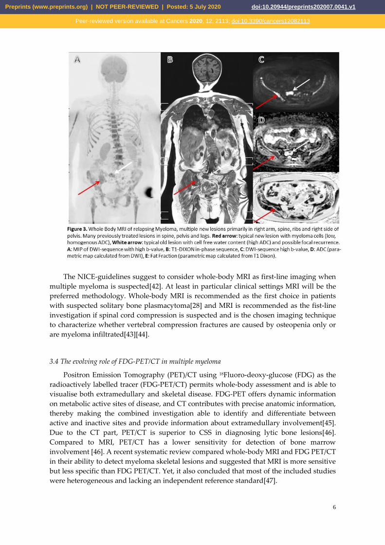
Figure 3 illustrates typical findings on whole-body MRI (WBMRI). WBMRI is
recommended over combined spinal and pelvic MRI as lesions in rib cage, shoulder girdles
and long bones could otherwise be missed.
Preprints (www.preprints.org) | NOT PEER-REVIEWED | Posted: 5 July 2020 doi:10.20944/preprints202007.0041.v1
Peer-reviewed version available at Cancers 2020, 12, 2113; doi:10.3390/cancers12082113
6
The NICE-guidelines suggest to consider whole-body MRI as first-line imaging when
multiple myeloma is suspected[42]. At least in particular clinical settings MRI will be the
preferred methodology. Whole-body MRI is recommended as the first choice in patients
with suspected solitary bone plasmacytoma[28] and MRI is recommended as the fist-line
investigation if spinal cord compression is suspected and is the chosen imaging technique
to characterize whether vertebral compression fractures are caused by osteopenia only or
are myeloma infiltrated[43][44].
3.4 The evolving role of FDG-PET/CT in multiple myeloma
Positron Emission Tomography (PET)/CT using 18Fluoro-deoxy-glucose (FDG) as the
radioactively labelled tracer (FDG-PET/CT) permits whole-body assessment and is able to
visualise both extramedullary and skeletal disease. FDG-PET offers dynamic information
on metabolic active sites of disease, and CT contributes with precise anatomic information,
thereby making the combined investigation able to identify and differentiate between
active and inactive sites and provide information about extramedullary involvement[45].
Due to the CT part, PET/CT is superior to CSS in diagnosing lytic bone lesions[46].
Compared to MRI, PET/CT has a lower sensitivity for detection of bone marrow
involvement [46]. A recent systematic review compared whole-body MRI and FDG PET/CT
in their ability to detect myeloma skeletal lesions and suggested that MRI is more sensitive
but less specific than FDG PET/CT. Yet, it also concluded that most of the included studies
were heterogeneous and lacking an independent reference standard[47].
Preprints (www.preprints.org) | NOT PEER-REVIEWED | Posted: 5 July 2020 doi:10.20944/preprints202007.0041.v1
Peer-reviewed version available at Cancers 2020, 12, 2113; doi:10.3390/cancers12082113

7
However, several studies have shown that PET-positive lesions offer prognostic
information, both at diagnosis, during and after treatment. The number of lesions, the
intensity of tracer uptake, and the presence of extramedullary disease have been shown to
be associated with inferior survival[48–51]. In the response criteria of minimal residual
disease negativity, FDG PET/CT is included and requires disappearance of abnormal tracer
uptake found on baseline scan or decrease to less than mediastinal blood pool or
surrounding normal tissue[26].
The IMWG recommends that PET/CT can be used in place of WBCT, but also in place
of WBMRI if imaging with MRI is not possible[28].
Sodium 18F-Fluoride (NaF) is a bone-seeking agent introduced in 1962 [52]. The uptake
of 18F-fluoride reflects blood flow and osteoblastic activity and thereby bone
remodeling[53,54]. NaF-PET is used in the assessment of malignant and benign skeletal
disease and has been suggested as a potentially valuable tool in the assessment of MM as
well [53,55]. Hypothetically, post-treatment NaF-PET could identify bone healing activity
in lytic lesions.[25]. However, so far, studies have not been able to demonstrate that
NaF-PET provides additional clinical information when assessing MBD or evaluating
treatment response compared to FDG-PET [56–58]. Figure 4 shows typical findings on
FDG-PET/CT and NaF-PET/CT in the same patient and illustrates how the findings
differentiate.
Other PET tracers, such as choline-based tracers, have been proposed for PET/CT
imaging in patients with MM. 11C‐Choline and 18F‐Fluorocholine PET/CT were initially
developed for prostate cancer imaging[59]. Choline is actively incorporated into the new
cell membranes [60]. Results from two smaller studies suggest that Choline PET/CT detects
up to 75% more focal lesions than FDG PET/CT in patients with MM suspected of
progression or relapse[61,62]. Thus, potentially there is a value in using other tracers than
FDG in MM; however, this needs to be explored further and validated in clinical trials.
3.5 Follow-up, response assessment and relapse
At the moment, there are no clear recommendations regarding routine follow-up, but
in general, CSS should not be used for disease monitoring[42]. It is recommended to repeat
relevant imaging of the same modality, PET/CT or WBMRI, as part of response evaluation
in patients where active disease sites or extramedullary disease were identified prior to
start of therapy. In patients with known extramedullary manifestations, imaging must be
repeated for response assessment. Oppositely, for now, there is no consensus that whole
body imaging should be performed as part of response evaluation in all patients. However,
Preprints (www.preprints.org) | NOT PEER-REVIEWED | Posted: 5 July 2020 doi:10.20944/preprints202007.0041.v1
Peer-reviewed version available at Cancers 2020, 12, 2113; doi:10.3390/cancers12082113

8
in patients with achieved complete remission after high-dose chemotherapy and
autologous stem-cell transplantation (ASCT), PET-positivity may persist and predict early
relapse and inferior outcome [63]. Moreover, IMWG has included FDG-PET/CT into
response assessment when evaluating MRD status[26] and recommends PET/CT
assessment at baseline and for response assessment in all patients included in clinical
trials[28]. If PET/CT is not available, diffusion-weighted WBMRI can be used and has
shown some promising ability to assess response to therapy[64].
For response assessment with FDG PET/CT as well as with WBMRI it applies that there
is a continued need for standardisation of the techniques, clear definition of response
criteria and prospective evaluation hereof.
4. Medical treatment
4.1 Bisphosphonates
Since Berenson s pamidronate trial in 1996, bisphosphonates have played a key role
and been standard of care in management of MBD[65]. Bisphosphonates are
pyrophosphate analogues that bind to bone and are ingested by the osteoclasts, leading to
inhibition of osteoclastic activity. There are different types of bisphosphonates:
Pamidronate, alendronate, ibandronate and zoledronate are all examples of
nitrogen-containing bisphosphonates and inhibit the mevalonate pathway.
Non-nitrogen-containing bisphosphonate, like clodronate, results in accumulation of
hydrolytical stable analogues of adenosine triphosphate. Zoledronate, pamidronate and
clodronate have been most intensively studied in MM. Both types of bisphosphonates
cause inhibition and apoptosis of osteoclasts. Furthermore, data indicate that
bisphosphonates, in addition to their bone-protective effects, may have antitumor activity
due to an uncoupling of the hypothesized vicious circle between bone resorption and
tumour growth in MM[66].
Few prospective, randomised trials comparing the different bisphosphonates head to
head have been conducted. The Rosen study from 2003[67] compared zoledronic acid to
pamidronate, in patients with either MM or breast cancer. Zoledronic acid was superior to
pamidronate in reducing the risk of skeletal related events (SRE), but the subgroup analysis
only found a significant difference in the breast cancer population. No data on overall
survival (OS) were provided. The UK MRC Myeloma XI study from 2011 compared
zoledronic acid with clodronate [68,69]. Zoledronic acid was found to be superior to
clodronate both in regard to SRE and overall survival. The lower risk of SRE was also
observed in patients without bone lesions at baseline [69].
A meta-analysis by the Cochrane database from 2017, including 24 randomised
controlled trials with a total of 7,293 patients, investigated the beneficial and adverse effects
associated with the use of different types of bisphosphonates in patients with MM [70].
They concluded that bisphosphonates reduce overall fractures and pain, and that
zoledronic acid improves overall survival compared to no bisphosphonate treatment. The
meta-analysis showed no significant difference between the different types of
bisphosphonate [70]. In contrast, a retrospective cohort study, of over 1,000 patients who
had been treated with either zoledronic acid or pamidronate, reported that zoledronic acid
compared to pamidronate reduced the risk of SRE by 25% and was associated with an
increased overall survival [71]. The current recommendation by IMWG is to initiate
Preprints (www.preprints.org) | NOT PEER-REVIEWED | Posted: 5 July 2020 doi:10.20944/preprints202007.0041.v1
Peer-reviewed version available at Cancers 2020, 12, 2113; doi:10.3390/cancers12082113

9
treatment with either zoledronic acid or pamidronate in all patients with symptomatic MM,
regardless of detectible osteolytic lesions on baseline imaging. [72].
In patients with smouldering myeloma, it has not been shown that bisphosphonates
prolong the time to progression to symptomatic disease [73,74] and it is therefore not
recommended.
The optimal duration of bisphosphonate treatment is still controversial. In most
randomized, controlled trials, bisphosphonates were administered up to 2 years. In the
Myeloma IX trial however, bisphosphonates were given until progression. A subanalysis
conducted in patients receiving treatment from year 2 and onward demonstrated persistent
superiority of the more potent zoledronic acid, both in regard to SRE and OS [75].
Interestingly, the cumulative incidence of renal complication and osteonecrosis of the jaw
(ONJ) seemed to reach a plateau between year 2 to 3 [76]. Another group investigated if 4
years treatment with zoledronic acid was superior to treatment for only 2 years. Prolonged
treatment reduced SRE but no difference in OS was observed.[77]. Some experts argue for
less bisphosphonate treatment in cases where the myeloma is well treated [78]. Indeed,
data from the Myeloma IX trial showed that a reduction in SRE was not observed in
patients achieving at least CR after ASCT, and that no survival benefit was seen in patients
achieving VGPR or better after ASCT [79].
All the referred studies used the standard dosing of pamidronate and zoledronic acid
every 3-4 weeks. However, an open-lable study by Himelstein et al. comprised 1,154
patients with bone metastases, including 278 patients with MM, and compared zoledronic
acid administrated every 4 weeks to every 12 weeks for up to 2 years [80]. No differences in
SRE or side effects were observed. Unfortunately, the study had a relatively high drop-out
rate of 31%, and because only about 25% of the included patients had MM, it is difficult to
draw firm conclusions about the possible adjustment of zoledronic acid scheduling in MM.
Other groups have proposed that the interval between zoledronic acid infusions could be
guided by the levels of the bone resorption marker Ntx-1 (N-terminal telopeptide of type 1
collagen) in the urine. [81]. Though this strategy is appealing and could reduce the risk of
developing ONJ, the evidence for doing this is still insufficient.
IMWG recommends that in patients who do not achieve very good partial response or
better, zoledronic acid should be administrated monthly until disease progression [72].
Otherwise, it is suggested that bisphosphonates should be administered for up to 2 years
and should be reinitiated at relapse, if discontinued earlier[72]. Rationally, and because
bisphosphonate treatment is prophylactic, re-initiation of zoledronic acid should be at
biochemical relapse and not postponed until clinical relapse. This is supported by a
Spanish study that randomised patients to zoledronic acid versus no bisphosphonate at
first sign of biochemical relapse. Although no effect was demonstrated on time to need of
treatment or survival, the patients who were re-initiated early with zoledronic acid had less
SREs at the time of treatment demanding relapse[82].
As mentioned, a serious but rare adverse event of bisphosphonate use is ONJ. Recent,
randomised, controlled trials showed an incidence of 3-4 % in myeloma patients receiving
zoledronic acid [68,83]. The median time from start of treatment to ONJ was found to be
13.6 months[83]. Invasive dental procedures, dental prostheses and intravenous
bisphosphonate administration and long-term treatment as well as the myeloma itself are
all risk factors associated with ONJ[84]. A case-control study showed that patients, who
were assessed by their dentist and had all necessary dental procedures done before
initiating treatment with zoledronic acid, had a three-fold decrease in the risk of
Preprints (www.preprints.org) | NOT PEER-REVIEWED | Posted: 5 July 2020 doi:10.20944/preprints202007.0041.v1
Peer-reviewed version available at Cancers 2020, 12, 2113; doi:10.3390/cancers12082113

10
developing ONJ [85]. If invasive dental procedures are required during bisphosphonate
treatment, a “drug holiday” before and after invasive dental procedures is generally
recommended[72], despite the fact that bisphosphonates remain in the skeleton for many
years[86,87]. A retrospective study indicated that prophylactic antibiotics during invasive
dental procedures may reduce the risk of developing ONJ [88]. Bisphosphonate-induced
nephrotoxicity is another major concern when treating patients with MM. Zoledronic acid
should be dose reduced already with a mild to moderate renal impairment (CrCl 30-60
mL/min) and is not recommended in patients with severe renal impairment (<30 mL/min).
A recent publication, including patients from 5 European countries, showed that 51 % of all
patients had renal insufficiency at the start of first line treatment, and 3 % had severe renal
impairment [89]. The study also found that a quarter of the patients with sufficient renal
function never started bisphosphonate treatment.
4.2 Denosumab
For patients with renal impairment and normal renal function, denosumab could be a
viable alternative to bisphosphonates. Denosumab is a human monoclonal antibody that
binds to and inhibits RANKL signalling and thereby blocks osteoclast activiation[90]. It is
not excreted through the kidneys, but degraded by endocytosis. Results from a large,
randomised, controlled, phase 3 study, including 1,718 patients with MM, showed that
denosumab was non-inferior to zoledronic acid in the time to first SRE and OS. Analysis of
the exploratory progression-free survival (PFS) endpoint favoured denosumab, and this
somewhat puzzling observation has been further analysed[91]. The PFS benefit was
restricted to the patients planned for (undergoing?) ASCT. One hypothesis could be that
RANKL signalling is involved in re-activation of “dormant” myeloma cells [92]. The
observation that the PFS improvement was restricted to younger ASCT-eligible patients
indicates that it could be the myelo- and stroma-ablative high-dose Melphalan in
combination with denosumab that is beneficial.
The incidence of ONJ was the same for denosumab and zoledronic acid, but a higher
incidence of hypocalcemia was observed among patients treated with denosumab.
Denosumab was given every 4 weeks, like zoledronic acid. The study only included
patients without renal impairment, and all patients had osteolysis at diagnosis. Sparse data
exist on the safety of denosumab in patients with MM with renal insufficiency. In patients
with bone metastases and severe renal insufficiency, denosumab can be given, but causes
an increased risk of electrolyte deficiencies [93]. Unlike bisphosphonates, denosumab is not
incorporated in the bone matrix and its effect declines rapidly after cessation [94]. This
could be a benefit in regard to “drug holidays” prior to invasive dental procedures. Indeed,
murine data indicate that ONJ may heal better after cessation of denosumab compared to
zoledronic acid [95]. The downside of this rapid cessation of effect is that a rebound effect
has been observed in patients with osteoporosis, where increased bone loss has been
observed when treatment is discontinued, presumably because of compensatory
upregulated RANKL signalling during denosumab treatment [96]. At the moment, there
are no data on how to stop treatment with denosumab in patients with MM, but it has been
suggested to switch treatment to bisphosphonate or to end the treatment with a single
dosing of zoledronic acid [97].
5. Non-pharmaceutical treatment
Preprints (www.preprints.org) | NOT PEER-REVIEWED | Posted: 5 July 2020 doi:10.20944/preprints202007.0041.v1
Peer-reviewed version available at Cancers 2020, 12, 2113; doi:10.3390/cancers12082113

11
5.1 Radiotherapy
Historically, radiotherapy has played an important part in managing MBD. One of the
most common indications for radiotherapy is pain reduction. A retrospective study found
that up to 84% of patients with myeloma obtained pain relief after radiotherapy[98]. Other
indications are prophylactic treatment of impending pathological fractures, spinal cord
compression and management of local neurological symptoms[99]. For patients with
myeloma with spinal cord compression, radiotherapy alone offers excellent response rates
(97%), local control (93% at 1 year, 82% at 2 years) and functional outcomes (64% of
non-ambulatory regained the ability to walk)[100].
Some concerns regarding depletion of the bone marrow reserve after concurrent
chemotherapy and radiotherapy exist. A smaller study, including 39 patients with
myeloma receiving radiotherapy alone or radiotherapy with concurrent novel
agents-based chemotherapy, concluded that concurrent treatment with radiotherapy and
systemic treatment was safe regarding hematologic toxicity and was well tolerated in the
majority of patients (87,5%)[101].
5. 2 Vertebral augmentation
Vertebroplasty and kyphoplasty are both minimal invasive fluoroscopic guided
percutaneous surgical procedures used to reduce pain caused by vertebral compression
fractures in patients with myeloma. Cement augmentation of the spine is possible at all
spinal levels. In the cervical region, the vertebral bodies can be accessed through an
anterior approach. Thoracic and lumbar vertebrae are reached through a transpedicular
approach with a Jamshidi needle. Bone cement (polymethylmethacrylate) is injected into
the vertebral body under imaging guidance. Kyphoplasty differs from vertebroplasty as
the height of the fractured vertebra is restored with an inflatable balloon catheter prior to
injection of bone cement. The void created by the balloon catheter while restoring the
vertebral height allows for more controlled delivery of cement, reducing the risk of bone
cement leakage.
Both procedures can be performed under local anaesthesia in an outpatient setting.
However, kyphoplasty is often performed under general anaesthesia as some patients
experience pain while the vertebral height is restored. For patient safety reasons, the
procedure is performed under local anaesthesia, allowing the patient to communicate
radiating pain, which could indicate that the needles are out of target, thereby minimizing
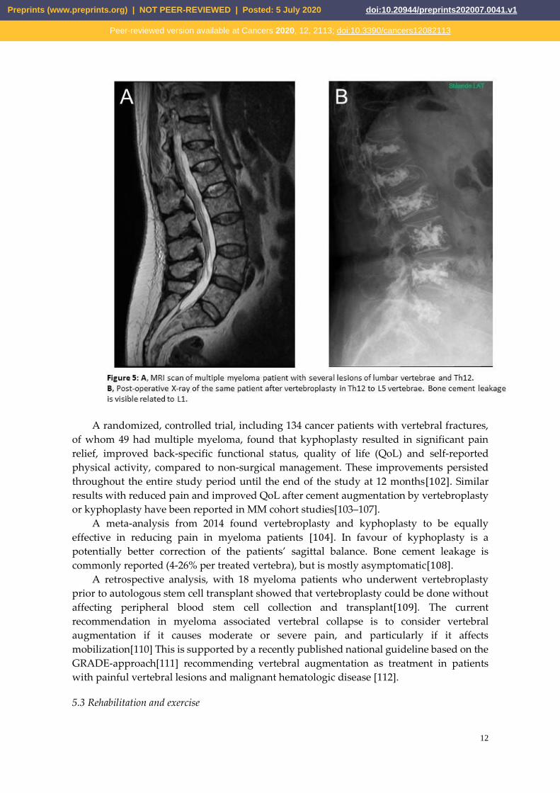
the risk of neurological injury. Figure 4 illustrates typical lumbar spine MRI findings prior
to vertebroplasty, and the final radiological appearance after the procedure.
Preprints (www.preprints.org) | NOT PEER-REVIEWED | Posted: 5 July 2020 doi:10.20944/preprints202007.0041.v1
Peer-reviewed version available at Cancers 2020, 12, 2113; doi:10.3390/cancers12082113
12
A randomized, controlled trial, including 134 cancer patients with vertebral fractures,
of whom 49 had multiple myeloma, found that kyphoplasty resulted in significant pain
relief, improved back-specific functional status, quality of life (QoL) and self-reported
physical activity, compared to non-surgical management. These improvements persisted
throughout the entire study period until the end of the study at 12 months[102]. Similar
results with reduced pain and improved QoL after cement augmentation by vertebroplasty
or kyphoplasty have been reported in MM cohort studies[103–107].
A meta-analysis from 2014 found vertebroplasty and kyphoplasty to be equally
effective in reducing pain in myeloma patients [104]. In favour of kyphoplasty is a
potentially better correction of the patients’ sagittal balance. Bone cement leakage is
commonly reported (4-26% per treated vertebra), but is mostly asymptomatic[108].
A retrospective analysis, with 18 myeloma patients who underwent vertebroplasty
prior to autologous stem cell transplant showed that vertebroplasty could be done without
affecting peripheral blood stem cell collection and transplant[109]. The current
recommendation in myeloma associated vertebral collapse is to consider vertebral
augmentation if it causes moderate or severe pain, and particularly if it affects
mobilization[110] This is supported by a recently published national guideline based on the
GRADE-approach[111] recommending vertebral augmentation as treatment in patients
with painful vertebral lesions and malignant hematologic disease [112].
5.3 Rehabilitation and exercise
Preprints (www.preprints.org) | NOT PEER-REVIEWED | Posted: 5 July 2020 doi:10.20944/preprints202007.0041.v1
Peer-reviewed version available at Cancers 2020, 12, 2113; doi:10.3390/cancers12082113
13
Exercise has been demonstrated to have a significant beneficial effect on QoL and
physical function in patients with cancer[113,114], but only few studies have been
conducted on patients with MM[115,116]. This is probably explained by the MBD and
suspected increased risk of pathological fractures. However, two literature reviews found
that exercise appeared safe and acceptable for patients with MM, but also concluded that
data are limited and that no conclusion regarding the effectiveness of exercise could be
drawn[115,116]. All studies in patients with MM have been conducted in patients before,
during or after ASCT.
Baseline data from a randomized controlled trial indicate that patients with newly
diagnosed multiple myeloma generally had lower physical function compared to the
normal population, and this goes particularly for patients with bone disease and
fractures[117]. A feasibility study, evaluated 30 patients with newly diagnosed myeloma
who were randomized 1:1 to usual care or usual care and individualized, supervised
exercise combined with home-based exercise for 10 weeks. Sixty-seven percent of the
patients had bone involvement. The study showed that even in older patients and in
patients with MBD, individualized physical exercise is feasible and safe around the time of
diagnosis[118]. The following expanded effect trial included 100 patients with newly
diagnosed MM in a randomized setting did not show effect on physical function, physical
activity, QoL, or pain [119]. However, the results of physical function indicated a trend for
less loss of muscle strengths in the intervention group, but there is a need to pay attention
to pain, since this might be worsened by the intervention [119].
6. Conclusion
Despite improved anti-myeloma treatments, MBD remains a significant problem. The
understanding of the pathophysiology has improved and may lead the way for
development of new bone directed treatments. Until then, anti-resorptive treatment with
bisphosphonates or denosumab is standard of care. Modern imaging with CT, PET/CT and
MRI play an essential role in diagnosing and monitoring MBD and help to guide
supplementary treatment with irradiation and vertebral augmentation. Exercise in patients
with MM is safe and feasible when relevant restrictions are taken into account; however, so
far no studies have demonstrated definite benefit of training.
Author Contributions: SR, TL and NA made the first draft. Special contributions; ALN,
PET imaging, JTA, radiology and MR imaging, MØ A, vertebral augmentation, RFL,
rehabilitation and exercise. All authors have read and agreed to the published version of
the manuscript.
Funding: This research received no external funding.
Acknowledgments: Vicky Svane Kristensen is highly acknowledged for critical English
reading.
Conflicts of Interest: The authors declare no conflicts of interest.
Preprints (www.preprints.org) | NOT PEER-REVIEWED | Posted: 5 July 2020 doi:10.20944/preprints202007.0041.v1
Peer-reviewed version available at Cancers 2020, 12, 2113; doi:10.3390/cancers12082113
14
References
1. Raab, M.S.; Podar, K.; Breitkreutz, I.; Richardson, P.G.; Anderson, K.C. Multiple myeloma. The
Lancet 2009, 374, 324–339, doi:10.1016/S0140-6736(09)60221-X.
2. Kyle, R.A.; Gertz, M.A.; Witzig, T.E.; Lust, J.A.; Lacy, M.Q.; Dispenzieri, A.; Fonseca, R.;
Rajkumar, S.V.; Offord, J.R.; Larson, D.R.; et al. Review of 1027 patients with newly diagnosed
multiple myeloma. Mayo Clin. Proc. 2003, 78, 21–33, doi:10.4065/78.1.21.
3. Shortt, C.P.; Carty, F.; Murray, J.G. The Role of Whole-Body Imaging in the Diagnosis, Staging,
and Follow-Up of Multiple Myeloma. Semin Musculoskelet Radiol 2010, 14, 37–46,
doi:10.1055/s-0030-1248705.
4. Cocks, K.; Cohen, D.; Wisløff, F.; Sezer, O.; Lee, S.; Hippe, E.; Gimsing, P.; Turesson, I.; Hajek, R.;
Smith, A.; et al. An international field study of the reliability and validity of a disease-specific
questionnaire module (the QLQ-MY20) in assessing the quality of life of patients with multiple
myeloma. European Journal of Cancer 2007, 43, 1670–1678, doi:10.1016/j.ejca.2007.04.022.
5. Jordan, K.; Proskorovsky, I.; Lewis, P.; Ishak, J.; Payne, K.; Lordan, N.; Kyriakou, C.; Williams,
C.D.; Peters, S.; Davies, F.E. Effect of general symptom level, specific adverse events, treatment
patterns, and patient characteristics on health-related quality of life in patients with multiple
myeloma: results of a European, multicenter cohort study. Support Care Cancer 2014, 22, 417–426,
doi:10.1007/s00520-013-1991-4.
6. Terpos, E.; Berenson, J.; Cook, R.J.; Lipton, A.; Coleman, R.E. Prognostic variables for survival
and skeletal complications in patients with multiple myeloma osteolytic bone disease. Leukemia
2010, 24, 1043–1049, doi:10.1038/leu.2010.62.
7. Augustson, B.M.; Begum, G.; Dunn, J.A.; Barth, N.J.; Davies, F.; Morgan, G.; Behrens, J.; Smith,
A.; Child, J.A.; Drayson, M.T. Early mortality after diagnosis of multiple myeloma: analysis of
patients entered onto the United kingdom Medical Research Council trials between 1980 and
2002--Medical Research Council Adult Leukaemia Working Party. J. Clin. Oncol. 2005, 23, 9219–
9226, doi:10.1200/JCO.2005.03.2086.
8. Naymagon, L.; Abdul-Hay, M. Novel agents in the treatment of multiple myeloma: a review
about the future. J Hematol Oncol 2016, 9, 52, doi:10.1186/s13045-016-0282-1.
9. Landgren, O.; Iskander, K. Modern multiple myeloma therapy: deep, sustained treatment
response and good clinical outcomes. Journal of Internal Medicine 2017, 281, 365–382,
doi:10.1111/joim.12590.
10. Vallet, S.; Filzmoser, J.-M.; Pecherstorfer, M.; Podar, K. Myeloma Bone Disease: Update on
Pathogenesis and Novel Treatment Strategies. Pharmaceutics 2018, 10,
doi:10.3390/pharmaceutics10040202.
11. Xiao, W.; Wang, Y.; Pacios, S.; Li, S.; Graves, D.T. Cellular and Molecular Aspects of Bone
Remodeling. Front Oral Biol 2016, 18, 9–16, doi:10.1159/000351895.
12. Giuliani, N.; Rizzoli, V.; Roodman, G.D. Multiple myeloma bone disease: Pathophysiology of
osteoblast inhibition. Blood 2006, 108, 3992–3996, doi:10.1182/blood-2006-05-026112.
13. Boyle, W.J.; Simonet, W.S.; Lacey, D.L. Osteoclast differentiation and activation. Nature 2003,
423, 337–342, doi:10.1038/nature01658.
14. Han, Y.; You, X.; Xing, W.; Zhang, Z.; Zou, W. Paracrine and endocrine actions of bone-the
Preprints (www.preprints.org) | NOT PEER-REVIEWED | Posted: 5 July 2020 doi:10.20944/preprints202007.0041.v1
Peer-reviewed version available at Cancers 2020, 12, 2113; doi:10.3390/cancers12082113

15
functions of secretory proteins from osteoblasts, osteocytes, and osteoclasts. Bone Res 2018, 6, 16,
doi:10.1038/s41413-018-0019-6.
15. Giuliani, N.; Bataille, R.; Mancini, C.; Lazzaretti, M.; Barillé, S. Myeloma cells induce imbalance
in the osteoprotegerin/osteoprotegerin ligand system in the human bone marrow environment.
Blood 2001, 98, 3527–3533, doi:10.1182/blood.v98.13.3527.
16. Terpos, E.; Szydlo, R.; Apperley, J.F.; Hatjiharissi, E.; Politou, M.; Meletis, J.; Viniou, N.;
Yataganas, X.; Goldman, J.M.; Rahemtulla, A. Soluble receptor activator of nuclear factor kappaB
ligand-osteoprotegerin ratio predicts survival in multiple myeloma: proposal for a novel prognostic
index. Blood 2003, 102, 1064–1069, doi:10.1182/blood-2003-02-0380.
17. Kim, J.H.; Liu, X.; Wang, J.; Chen, X.; Zhang, H.; Kim, S.H.; Cui, J.; Li, R.; Zhang, W.; Kong, Y.; et
al. Wnt signaling in bone formation and its therapeutic potential for bone diseases. Ther Adv
Musculoskelet Dis 2013, 5, 13–31, doi:10.1177/1759720X12466608.
18. Qiang, Y.-W.; Chen, Y.; Stephens, O.; Brown, N.; Chen, B.; Epstein, J.; Barlogie, B.; Shaughnessy,
J.D. Myeloma-derived Dickkopf-1 disrupts Wnt-regulated osteoprotegerin and RANKL production
by osteoblasts: a potential mechanism underlying osteolytic bone lesions in multiple myeloma.
Blood 2008, 112, 196–207, doi:10.1182/blood-2008-01-132134.
19. Kristensen, I.B.; Christensen, J.H.; Lyng, M.B.; Møller, M.B.; Pedersen, L.M.; Rasmussen, L.M.;
Ditzel, H.J.; Abildgaard, N. Expression of osteoblast and osteoclast regulatory genes in the bone
marrow microenvironment in multiple myeloma: only up-regulation of Wnt inhibitors SFRP3 and
DKK1 is associated with lytic bone disease. Medline Abbreviated Title: Leuk Lymphoma 2014, 55,
911–919, doi:10.3109/10428194.2013.820288.
20. Palma, B.D.; Guasco, D.; Pedrazzoni, M.; Bolzoni, M.; Accardi, F.; Costa, F.; Sammarelli, G.;
Craviotto, L.; Filippo, M.D.; Ruffini, L.; et al. Osteolytic lesions, cytogenetic features and bone
marrow levels of cytokines and chemokines in multiple myeloma patients: Role of chemokine (C-C
motif) ligand 20. Leukemia 2016, 30, 409–416, doi:10.1038/leu.2015.259.
21. Andersen, T.L.; Søe, K.; Sondergaard, T.E.; Plesner, T.; Delaisse, J.-M. Myeloma cell-induced
disruption of bone remodelling compartments leads to osteolytic lesions and generation of
osteoclast-myeloma hybrid cells. Br. J. Haematol. 2010, 148, 551–561,
doi:10.1111/j.1365-2141.2009.07980.x.
22. Børset, M.; Sundan, A.; Waage, A.; Standal, T. Why do myeloma patients have bone disease?
A historical perspective. Blood Rev. 2020, 41, 100646, doi:10.1016/j.blre.2019.100646.
23. Criteria for the classification of monoclonal gammopathies, multiple myeloma and related
disorders: a report of the International Myeloma Working Group. British Journal of Haematology
2003, 121, 749–757, doi:10.1046/j.1365-2141.2003.04355.x.
24. Caers, J.; Paiva, B.; Zamagni, E.; Leleu, X.; Bladé, J.; Kristinsson, S.Y.; Touzeau, C.; Abildgaard,
N.; Terpos, E.; Heusschen, R.; et al. Diagnosis, treatment, and response assessment in solitary
plasmacytoma: updated recommendations from a European Expert Panel. J Hematol Oncol 2018,
11, doi:10.1186/s13045-017-0549-1.
25. Sevcikova, S.; Minarik, J.; Stork, M.; Jelinek, T.; Pour, L.; Hajek, R. Extramedullary disease in
multiple myeloma – controversies and future directions. Blood Reviews 2019, 36, 32–39,
doi:10.1016/j.blre.2019.04.002.
Preprints (www.preprints.org) | NOT PEER-REVIEWED | Posted: 5 July 2020 doi:10.20944/preprints202007.0041.v1
Peer-reviewed version available at Cancers 2020, 12, 2113; doi:10.3390/cancers12082113

16
26. Kumar, S.; Paiva, B.; Anderson, K.C.; Durie, B.; Landgren, O.; Moreau, P.; Munshi, N.; Lonial, S.;
Bladé, J.; Mateos, M.-V.; et al. International Myeloma Working Group consensus criteria for
response and minimal residual disease assessment in multiple myeloma. Lancet Oncol. 2016, 17,
e328–e346, doi:10.1016/S1470-2045(16)30206-6.
27. Rajkumar, S.V.; Dimopoulos, M.A.; Palumbo, A.; Blade, J.; Merlini, G.; Mateos, M.-V.; Kumar,
S.; Hillengass, J.; Kastritis, E.; Richardson, P.; et al. International Myeloma Working Group updated
criteria for the diagnosis of multiple myeloma. Lancet Oncol. 2014, 15, e538-548,
doi:10.1016/S1470-2045(14)70442-5.
28. Hillengass, J.; Usmani, S.; Rajkumar, S.V.; Durie, B.G.M.; Mateos, M.-V.; Lonial, S.; Joao, C.;
Anderson, K.C.; García-Sanz, R.; Riva, E.; et al. International myeloma working group consensus
recommendations on imaging in monoclonal plasma cell disorders. Lancet Oncol. 2019, 20, e302–
e312, doi:10.1016/S1470-2045(19)30309-2.
29. Dimopoulos, M.; Terpos, E.; Comenzo, R.L.; Tosi, P.; Beksac, M.; Sezer, O.; Siegel, D.; Lokhorst,
H.; Kumar, S.; Rajkumar, S.V.; et al. International myeloma working group consensus statement
and guidelines regarding the current role of imaging techniques in the diagnosis and monitoring of
multiple Myeloma. Leukemia 2009, 23, 1545–1556, doi:10.1038/leu.2009.89.
30. Edelstyn, G.A.; Gillespie, P.J.; Grebbell, F.S. The radiological demonstration of osseous
metastases. Experimental observations. Clin Radiol 1967, 18, 158–162,
doi:10.1016/s0009-9260(67)80010-2.
31. Hinge, M.; Andersen, K.T.; Lund, T.; Jørgensen, H.B.; Holdgaard, P.C.; Ormstrup, T.E.;
Ø stergaard, L.L.; Plesner, T. Baseline bone involvement in multiple myeloma - a prospective
comparison of conventional X-ray, low-dose computed tomography, and 18flourodeoxyglucose
positron emission tomography in previously untreated patients. Haematologica 2016, 101, e415–
e418, doi:10.3324/haematol.2016.146092.
32. Hillengass, J.; Moulopoulos, L.A.; Delorme, S.; Koutoulidis, V.; Mosebach, J.; Hielscher, T.;
Drake, M.; Rajkumar, S.V.; Oestergaard, B.; Abildgaard, N.; et al. Whole-body computed
tomography versus conventional skeletal survey in patients with multiple myeloma: a study of the
International Myeloma Working Group. Blood Cancer J 2017, 7, e599, doi:10.1038/bcj.2017.78.
33. Matsue, K.; Kobayashi, H.; Matsue, Y.; Abe, Y.; Narita, K.; Kitadate, A.; Takeuchi, M. Prognostic
significance of bone marrow abnormalities in the appendicular skeleton of patients with multiple
myeloma. Blood Adv 2018, 2, 1032–1039, doi:10.1182/bloodadvances.2017014720.
34. Moulopoulos, L.A.; Dimopoulos, M.A. Magnetic Resonance Imaging of the Bone Marrow in
Hematologic Malignancies. Blood 1997, 90, 2127–2147, doi:10.1182/blood.V90.6.2127.
35. Wolf, M.B.; Murray, F.; Kilk, K.; Hillengass, J.; Delorme, S.; Heiss, C.; Neben, K.; Goldschmidt,
H.; Kauczor, H.-U.; Weber, M.-A. Sensitivity of whole-body CT and MRI versus projection
radiography in the detection of osteolyses in patients with monoclonal plasma cell disease. Eur J
Radiol 2014, 83, 1222–1230, doi:10.1016/j.ejrad.2014.02.008.
36. Walker, R.; Barlogie, B.; Haessler, J.; Tricot, G.; Anaissie, E.; Shaughnessy, J.D.; Epstein, J.; van
Hemert, R.; Erdem, E.; Hoering, A.; et al. Magnetic Resonance Imaging in Multiple Myeloma:
Diagnostic and Clinical Implications. JCO 2007, 25, 1121–1128, doi:10.1200/JCO.2006.08.5803.
37. Baur-Melnyk, A.; Buhmann, S.; Becker, C.; Schoenberg, S.O.; Lang, N.; Bartl, R.; Reiser, M.F.
Preprints (www.preprints.org) | NOT PEER-REVIEWED | Posted: 5 July 2020 doi:10.20944/preprints202007.0041.v1
Peer-reviewed version available at Cancers 2020, 12, 2113; doi:10.3390/cancers12082113

17
Whole-Body MRI Versus Whole-Body MDCT for Staging of Multiple Myeloma. American Journal of
Roentgenology 2008, 190, 1097–1104, doi:10.2214/AJR.07.2635.
38. Lecouvet, F.E.; Vande Berg, B.C.; Malghem, J.; Maldague, B.E. Magnetic resonance and
computed tomography imaging in multiple myeloma. Semin Musculoskelet Radiol 2001, 5, 43–55,
doi:10.1055/s-2001-12920.
39. Kastritis, E.; Moulopoulos, L.A.; Terpos, E.; Koutoulidis, V.; Dimopoulos, M.A. The prognostic
importance of the presence of more than one focal lesion in spine MRI of patients with
asymptomatic (smoldering) multiple myeloma. Leukemia 2014, 28, 2402–2403,
doi:10.1038/leu.2014.230.
40. Hillengass, J.; Fechtner, K.; Weber, M.-A.; Bäuerle, T.; Ayyaz, S.; Heiss, C.; Hielscher, T.;
Moehler, T.M.; Egerer, G.; Neben, K.; et al. Prognostic significance of focal lesions in whole-body
magnetic resonance imaging in patients with asymptomatic multiple myeloma. J. Clin. Oncol. 2010,
28, 1606–1610, doi:10.1200/JCO.2009.25.5356.
41. Merz, M.; Hielscher, T.; Wagner, B.; Sauer, S.; Shah, S.; Raab, M.S.; Jauch, A.; Neben, K.; Hose,
D.; Egerer, G.; et al. Predictive value of longitudinal whole-body magnetic resonance imaging in
patients with smoldering multiple myeloma. Leukemia 2014, 28, 1902–1908,
doi:10.1038/leu.2014.75.
42. Pratt, G.; Morris, T.C. Review of the NICE guidelines for multiple myeloma. International
Journal of Laboratory Hematology 2017, 39, 3–13, doi:10.1111/ijlh.12581.
43. 1 Guidance | Metastatic spinal cord compression in adults: risk assessment, diagnosis and
management | Guidance | NICE Available online:
https://www.nice.org.uk/guidance/CG75/chapter/1-Guidance#imaging (accessed on Mar 4, 2020).
44. Mauch, J.T.; Carr, C.M.; Cloft, H.; Diehn, F.E. Review of the Imaging Features of Benign
Osteoporotic and Malignant Vertebral Compression Fractures. American Journal of Neuroradiology
2018, doi:10.3174/ajnr.A5528.
45. Role of 18 F-FDG PET/CT in the diagnosis and management of multiple myeloma and other
plasma cell disorders: a consensus statement by the International Myeloma Working Group-
ClinicalKey Available online:
https://www-clinicalkey-com.proxy2-bib.sdu.dk/#!/content/journal/1-s2.0-S1470204517301894
(accessed on Mar 5, 2020).
46. Zamagni, E.; Nanni, C.; Patriarca, F.; Englaro, E.; Castellucci, P.; Geatti, O.; Tosi, P.; Tacchetti, P.;
Cangini, D.; Perrone, G.; et al. A prospective comparison of 18F-fluorodeoxyglucose positron
emission tomography-computed tomography, magnetic resonance imaging and whole-body
planar radiographs in the assessment of bone disease in newly diagnosed multiple myeloma.
Haematologica 2007, 92, 50–55, doi:10.3324/haematol.10554.
47. Gariani, J.; Westerland, O.; Natas, S.; Verma, H.; Cook, G.; Goh, V. Comparison of whole body
magnetic resonance imaging (WBMRI) to whole body computed tomography (WBCT) or
18F-fluorodeoxyglucose positron emission tomography/CT (18F-FDG PET/CT) in patients with
myeloma: Systematic review of diagnostic performance. Critical Reviews in Oncology/Hematology
2018, 124, 66–72, doi:10.1016/j.critrevonc.2018.02.012.
48. Bartel, T.B.; Haessler, J.; Brown, T.L.Y.; Shaughnessy, J.D.; van Rhee, F.; Anaissie, E.; Alpe, T.;
Preprints (www.preprints.org) | NOT PEER-REVIEWED | Posted: 5 July 2020 doi:10.20944/preprints202007.0041.v1
Peer-reviewed version available at Cancers 2020, 12, 2113; doi:10.3390/cancers12082113

18
Angtuaco, E.; Walker, R.; Epstein, J.; et al. F18-fluorodeoxyglucose positron emission tomography
in the context of other imaging techniques and prognostic factors in multiple myeloma. Blood
2009, 114, 2068–2076, doi:10.1182/blood-2009-03-213280.
49. Zamagni, E.; Patriarca, F.; Nanni, C.; Zannetti, B.; Englaro, E.; Pezzi, A.; Tacchetti, P.; Buttignol,
S.; Perrone, G.; Brioli, A.; et al. Prognostic relevance of 18-F FDG PET/CT in newly diagnosed
multiple myeloma patients treated with up-front autologous transplantation. Blood 2011, 118,
5989–5995, doi:10.1182/blood-2011-06-361386.
50. Usmani, S.Z.; Mitchell, A.; Waheed, S.; Crowley, J.; Hoering, A.; Petty, N.; Brown, T.; Bartel, T.;
Anaissie, E.; van Rhee, F.; et al. Prognostic implications of serial 18-fluoro-deoxyglucose emission
tomography in multiple myeloma treated with total therapy 3. Blood 2013, 121, 1819–1823,
doi:10.1182/blood-2012-08-451690.
51. Moon, S.H.; Choi, W.H.; Yoo, I.R.; Lee, S.J.; Paeng, J.C.; Jeong, S.Y.; Lee, S.-W.; Kim, K.; Choi, J.Y.
Prognostic Value of Baseline 18F-Fluorodeoxyglucose PET/CT in Patients with Multiple Myeloma: A
Multicenter Cohort Study. Korean J Radiol 2018, 19, 481–488, doi:10.3348/kjr.2018.19.3.481.
52. Blau, M.; Nagler, W.; Bender, M.A. Fluorine-18: a new isotope for bone scanning. J. Nucl. Med.
1962, 3, 332–334.
53. Grant, F.D.; Fahey, F.H.; Packard, A.B.; Davis, R.T.; Alavi, A.; Treves, S.T. Skeletal PET with
18F-Fluoride: Applying New Technology to an Old Tracer. J Nucl Med 2008, 49, 68–78,
doi:10.2967/jnumed.106.037200.
54. Even-Sapir, E.; Mishani, E.; Flusser, G.; Metser, U. 18F-Fluoride Positron Emission Tomography
and Positron Emission Tomography/Computed Tomography. Seminars in Nuclear Medicine 2007,
37, 462–469, doi:10.1053/j.semnuclmed.2007.07.002.
55. Nishiyama, Y.; Tateishi, U.; Shizukuishi, K.; Shishikura, A.; Yamazaki, E.; Shibata, H.; Yoneyama,
T.; Ishigatsubo, Y.; Inoue, T. Role of 18F-fluoride PET/CT in the assessment of multiple myeloma:
initial experience. Ann Nucl Med 2013, 27, 78–83, doi:10.1007/s12149-012-0647-7.
56. Sachpekidis, C.; Goldschmidt, H.; Hose, D.; Pan, L.; Cheng, C.; Kopka, K.; Haberkorn, U.;
Dimitrakopoulou-Strauss, A. PET/CT studies of multiple myeloma using (18) F-FDG and (18) F-NaF:
comparison of distribution patterns and tracers’ pharmacokinetics. Eur. J. Nucl. Med. Mol. Imaging
2014, 41, 1343–1353, doi:10.1007/s00259-014-2721-y.
57. Ak, İ.; Onner, H.; Akay, O.M. Is there any complimentary role of F-18 NaF PET/CT in detecting
of osseous involvement of multiple myeloma? A comparative study for F-18 FDG PET/CT and F-18
FDG NaF PET/CT. Ann. Hematol. 2015, 94, 1567–1575, doi:10.1007/s00277-015-2410-3.
58. Sachpekidis, C.; Hillengass, J.; Goldschmidt, H.; Wagner, B.; Haberkorn, U.; Kopka, K.;
Dimitrakopoulou-Strauss, A. Treatment response evaluation with 18F-FDG PET/CT and 18F-NaF
PET/CT in multiple myeloma patients undergoing high-dose chemotherapy and autologous stem
cell transplantation. Eur J Nucl Med Mol Imaging 2017, 44, 50–62,
doi:10.1007/s00259-016-3502-6.
59. DeGrado, T.R.; Coleman, R.E.; Wang, S.; Baldwin, S.W.; Orr, M.D.; Robertson, C.N.; Polascik,
T.J.; Price, D.T. Synthesis and evaluation of 18F-labeled choline as an oncologic tracer for positron
emission tomography: initial findings in prostate cancer. Cancer Res. 2001, 61, 110–117.
60. Yoshimoto, M.; Waki, A.; Yonekura, Y.; Sadato, N.; Murata, T.; Omata, N.; Takahashi, N.;
Preprints (www.preprints.org) | NOT PEER-REVIEWED | Posted: 5 July 2020 doi:10.20944/preprints202007.0041.v1
Peer-reviewed version available at Cancers 2020, 12, 2113; doi:10.3390/cancers12082113

19
Welch, M.J.; Fujibayashi, Y. Characterization of acetate metabolism in tumor cells in relation to cell
proliferation: acetate metabolism in tumor cells. Nucl. Med. Biol. 2001, 28, 117–122,
doi:10.1016/s0969-8051(00)00195-5.
61. Nanni, C.; Zamagni, E.; Cavo, M.; Rubello, D.; Tacchetti, P.; Pettinato, C.; Farsad, M.;
Castellucci, P.; Ambrosini, V.; Montini, G.C.; et al. 11C-choline vs. 18F-FDG PET/CT in assessing
bone involvement in patients with multiple myeloma. World J Surg Oncol 2007, 5, 68,
doi:10.1186/1477-7819-5-68.
62. Cassou-Mounat, T.; Balogova, S.; Nataf, V.; Calzada, M.; Huchet, V.; Kerrou, K.; Devaux, J.-Y.;
Mohty, M.; Talbot, J.-N.; Garderet, L. 18F-fluorocholine versus 18F-fluorodeoxyglucose for PET/CT
imaging in patients with suspected relapsing or progressive multiple myeloma: a pilot study. Eur. J.
Nucl. Med. Mol. Imaging 2016, 43, 1995–2004, doi:10.1007/s00259-016-3392-7.
63. Moreau, P.; Attal, M.; Caillot, D.; Macro, M.; Karlin, L.; Garderet, L.; Facon, T.; Benboubker, L.;
Escoffre-Barbe, M.; Stoppa, A.-M.; et al. Prospective Evaluation of Magnetic Resonance Imaging
and [18F]Fluorodeoxyglucose Positron Emission Tomography-Computed Tomography at Diagnosis
and Before Maintenance Therapy in Symptomatic Patients With Multiple Myeloma Included in the
IFM/DFCI 2009 Trial: Results of the IMAJEM Study. J. Clin. Oncol. 2017, 35, 2911–2918,
doi:10.1200/JCO.2017.72.2975.
64. Pawlyn, C.; Fowkes, L.; Otero, S.; Jones, J.R.; Boyd, K.D.; Davies, F.E.; Morgan, G.J.; Collins, D.J.;
Sharma, B.; Riddell, A.; et al. Whole-body diffusion-weighted MRI: a new gold standard for
assessing disease burden in patients with multiple myeloma? Leukemia 2016, 30, 1446–1448,
doi:10.1038/leu.2015.338.
65. Berenson, J.R.; Lichtenstein, A.; Porter, L.; Dimopoulos, M.A.; Bordoni, R.; George, S.; Lipton,
A.; Keller, A.; Ballester, O.; Kovacs, M.J.; et al. Efficacy of pamidronate in reducing skeletal events
in patients with advanced multiple myeloma. Myeloma Aredia Study Group. N. Engl. J. Med. 1996,
334, 488–493, doi:10.1056/NEJM199602223340802.
66. Clézardin, P. Mechanisms of action of bisphosphonates in oncology: a scientific concept
evolving from antiresorptive to anticancer activities. Bonekey Rep 2013, 2,
doi:10.1038/bonekey.2013.1.
67. Rosen, L.S.; Gordon, D.; Kaminski, M.; Howell, A.; Belch, A.; Mackey, J.; Apffelstaedt, J.;
Hussein, M.A.; Coleman, R.E.; Reitsma, D.J.; et al. Long-term efficacy and safety of zoledronic acid
compared with pamidronate disodium in the treatment of skeletal complications in patients with
advanced multiple myeloma or breast carcinoma. Cancer 2003, 98, 1735–1744,
doi:10.1002/cncr.11701.
68. Morgan, G.J.; Davies, F.E.; Gregory, W.M.; Cocks, K.; Bell, S.E.; Szubert, A.J.; Navarro-Coy, N.;
Drayson, M.T.; Owen, R.G.; Feyler, S.; et al. First-line treatment with zoledronic acid as compared
with clodronic acid in multiple myeloma (MRC Myeloma IX): a randomised controlled trial. Lancet
2010, 376, 1989–1999, doi:10.1016/S0140-6736(10)62051-X.
69. Morgan, G.J.; Child, J.A.; Gregory, W.M.; Szubert, A.J.; Cocks, K.; Bell, S.E.; Navarro-Coy, N.;
Drayson, M.T.; Owen, R.G.; Feyler, S.; et al. Effects of zoledronic acid versus clodronic acid on
skeletal morbidity in patients with newly diagnosed multiple myeloma (MRC Myeloma IX):
secondary outcomes from a randomised controlled trial. Lancet Oncol 2011, 12, 743–752,
Preprints (www.preprints.org) | NOT PEER-REVIEWED | Posted: 5 July 2020 doi:10.20944/preprints202007.0041.v1
Peer-reviewed version available at Cancers 2020, 12, 2113; doi:10.3390/cancers12082113

20
doi:10.1016/S1470-2045(11)70157-7.
70. Mhaskar, R.; Kumar, A.; Miladinovic, B.; Djulbegovic, B. Bisphosphonates in multiple myeloma:
an updated network meta-analysis. Cochrane Database Syst Rev 2017, 12, CD003188,
doi:10.1002/14651858.CD003188.pub4.
71. Sanfilippo, K.M.; Gage, B.; Luo, S.; Weilbaecher, K.; Tomasson, M.; Vij, R.; Colditz, G.; Carson, K.
Comparative effectiveness on survival of zoledronic acid versus pamidronate in multiple myeloma.
Leuk Lymphoma 2015, 56, 615–621, doi:10.3109/10428194.2014.924117.
72. Terpos, E.; Morgan, G.; Dimopoulos, M.A.; Drake, M.T.; Lentzsch, S.; Raje, N.; Sezer, O.;
García-Sanz, R.; Shimizu, K.; Turesson, I.; et al. International Myeloma Working Group
recommendations for the treatment of multiple myeloma-related bone disease. J. Clin. Oncol.
2013, 31, 2347–2357, doi:10.1200/JCO.2012.47.7901.
73. D’Arena, G.; Gobbi, P.G.; Broglia, C.; Sacchi, S.; Quarta, G.; Baldini, L.; Iannitto, E.; Falcone, A.;
Guariglia, R.; Pietrantuono, G.; et al. Pamidronate versus observation in asymptomatic myeloma:
final results with long-term follow-up of a randomized study. Leuk. Lymphoma 2011, 52, 771–775,
doi:10.3109/10428194.2011.553000.
74. Musto, P.; Petrucci, M.T.; Bringhen, S.; Guglielmelli, T.; Caravita, T.; Bongarzoni, V.; Andriani,
A.; D’Arena, G.; Balleari, E.; Pietrantuono, G.; et al. A multicenter, randomized clinical trial
comparing zoledronic acid versus observation in patients with asymptomatic myeloma. Cancer
2008, 113, 1588–1595, doi:10.1002/cncr.23783.
75. Morgan, G.J.; Davies, F.E.; Gregory, W.M.; Szubert, A.J.; Bell, S.E.; Drayson, M.T.; Owen, R.G.;
Ashcroft, A.J.; Jackson, G.H.; Child, J.A. Effects of induction and maintenance plus long-term
bisphosphonates on bone disease in patients with multiple myeloma: the Medical Research
Council Myeloma IX Trial. Blood 2012, 119, 5374–5383, doi:10.1182/blood-2011-11-392522.
76. Jackson, G.H.; Morgan, G.J.; Davies, F.E.; Wu, P.; Gregory, W.M.; Bell, S.E.; Szubert, A.J.; Coy,
N.N.; Drayson, M.T.; Owen, R.G.; et al. Osteonecrosis of the jaw and renal safety in patients with
newly diagnosed multiple myeloma: Medical Research Council Myeloma IX Study results. British
Journal of Haematology 2014, 166, 109–117, doi:10.1111/bjh.12861.
77. Aviles, A.; Nambo, M.-J.; Huerta-Guzman, J.; Cleto, S.; Neri, N. Prolonged Use of Zoledronic
Acid (4 Years) Did Not Improve Outcome in Multiple Myeloma Patients. Clin Lymphoma Myeloma
Leuk 2017, 17, 207–210, doi:10.1016/j.clml.2017.02.007.
78. Durie, B.G.M. Use of Bisphosphonates in Multiple Myeloma: IMWG Response to Mayo Clinic
Consensus Statement. Mayo Clinic Proceedings 2007, 82, 516–517, doi:10.4065/82.4.516.
79. Larocca, A.; Child, J.A.; Cook, G.; Jackson, G.H.; Russell, N.; Szubert, A.; Gregory, W.M.; Brioli,
A.; Owen, R.G.; Drayson, M.T.; et al. The impact of response on bone-directed therapy in patients
with multiple myeloma. Blood 2013, 122, 2974–2977, doi:10.1182/blood-2013-04-498139.
80. Himelstein, A.L.; Foster, J.C.; Khatcheressian, J.L.; Roberts, J.D.; Seisler, D.K.; Novotny, P.J.; Qin,
R.; Go, R.S.; Grubbs, S.S.; O’Connor, T.; et al. Effect of Longer-Interval vs Standard Dosing of
Zoledronic Acid on Skeletal Events in Patients With Bone Metastases: A Randomized Clinical Trial.
JAMA 2017, 317, 48–58, doi:10.1001/jama.2016.19425.
81. Raje, N.; Vescio, R.; Montgomery, C.W.; Badros, A.; Munshi, N.; Orlowski, R.; Hadala, J.T.;
Warsi, G.; Argonza-Aviles, E.; Ericson, S.G.; et al. Bone Marker-Directed Dosing of Zoledronic Acid
Preprints (www.preprints.org) | NOT PEER-REVIEWED | Posted: 5 July 2020 doi:10.20944/preprints202007.0041.v1
Peer-reviewed version available at Cancers 2020, 12, 2113; doi:10.3390/cancers12082113

21
for the Prevention of Skeletal Complications in Patients with Multiple Myeloma: Results of the
Z-MARK Study. Clin Cancer Res 2016, 22, 1378–1384, doi:10.1158/1078-0432.CCR-15-1864.
82. García-Sanz, R.; Oriol, A.; Moreno, M.J.; de la Rubia, J.; Payer, A.R.; Hernández, M.T.; Palomera,
L.; Teruel, A.I.; Blanchard, M.J.; Gironella, M.; et al. Zoledronic acid as compared with observation
in multiple myeloma patients at biochemical relapse: results of the randomized AZABACHE Spanish
trial. Haematologica 2015, 100, 1207–1213, doi:10.3324/haematol.2015.128439.
83. Raje, N.; Terpos, E.; Willenbacher, W.; Shimizu, K.; Garcia-Sanz, R.; Durie, B.; Legiec, W.; Krejci,
M.; Laribi, K.; Zhu, L.; et al. Denosumab versus zoledronic acid in bone disease treatment of newly
diagnosed multiple myeloma: an international, double-blind, double-dummy, randomised,
controlled, phase 3 study. Lancet Oncol 2018, 19, 370–381, doi:10.1016/S1470-2045(18)30072-X.
84. Identification of Risk Factors for Bisphosphonate-Associated Atypical Femoral Fractures and
Osteonecrosis of the Jaw in a Pharmacovigilance Database - Rebecca S. Bejhed, Mohammad
Kharazmi, Pär Hallberg, 2016 Available online:
https://journals-sagepub-com.proxy1-bib.sdu.dk/doi/10.1177/1060028016649368 (accessed on
Mar 23, 2020).
85. Dimopoulos, M.A.; Kastritis, E.; Bamia, C.; Melakopoulos, I.; Gika, D.; Roussou, M.; Migkou, M.;
Eleftherakis-Papaiakovou, E.; Christoulas, D.; Terpos, E.; et al. Reduction of osteonecrosis of the
jaw (ONJ) after implementation of preventive measures in patients with multiple myeloma treated
with zoledronic acid. Annals of Oncology 2009, 20, 117–120, doi:10.1093/annonc/mdn554.
86. Khan, S.A.; Kanis, J.A.; Vasikaran, S.; Kline, W.F.; Matuszewski, B.K.; McCloskey, E.V.; Beneton,
M.N.; Gertz, B.J.; Sciberras, D.G.; Holland, S.D.; et al. Elimination and biochemical responses to
intravenous alendronate in postmenopausal osteoporosis. J. Bone Miner. Res. 1997, 12, 1700–
1707, doi:10.1359/jbmr.1997.12.10.1700.
87. Lin, J.H. Bisphosphonates: A review of their pharmacokinetic properties. Bone 1996, 18, 75–85,
doi:10.1016/8756-3282(95)00445-9.
88. Montefusco, V.; Gay, F.; Spina, F.; Miceli, R.; Maniezzo, M.; Ambrosini, M.T.; Farina, L.; Piva, S.;
Palumbo, A.; Boccadoro, M.; et al. Antibiotic prophylaxis before dental procedures may reduce the
incidence of osteonecrosis of the jaw in patients with multiple myeloma treated with
bisphosphonates. Leukemia & Lymphoma 2008, 49, 2156–2162,
doi:10.1080/10428190802483778.
89. Mateos, M.-V.; Fink, L.; Koneswaran, N.; Intorcia, M.; Giannopoulou, C.; Niepel, D.; Cavo, M.
Bone complications in patients with multiple myeloma in five European countries: a retrospective
patient chart review. BMC Cancer 2020, 20, 170, doi:10.1186/s12885-020-6596-y.
90. Baron, R.; Ferrari, S.; Russell, R.G.G. Denosumab and bisphosphonates: Different mechanisms
of action and effects. Bone 2011, 48, 677–692, doi:10.1016/j.bone.2010.11.020.
91. Terpos, E.; García-Sanz, R.; Shimizu, K.; Willenbacher, W.; Glennane, A.; Dai, T.; Pasteiner, W.;
Raje, N.S. Progression-Free Survival Analysis of Denosumab Vs Zoledronic Acid in Intent to
Transplant Multiple Myeloma Patients Based on Treatment Regimen and Baseline Characteristics.
Blood 2019, 134, 606–606, doi:10.1182/blood-2019-127290.
92. Lawson, M.A.; McDonald, M.M.; Kovacic, N.; Hua Khoo, W.; Terry, R.L.; Down, J.; Kaplan, W.;
Paton-Hough, J.; Fellows, C.; Pettitt, J.A.; et al. Osteoclasts control reactivation of dormant
Preprints (www.preprints.org) | NOT PEER-REVIEWED | Posted: 5 July 2020 doi:10.20944/preprints202007.0041.v1
Peer-reviewed version available at Cancers 2020, 12, 2113; doi:10.3390/cancers12082113

22
myeloma cells by remodelling the endosteal niche. Nat Commun 2015, 6, 8983,
doi:10.1038/ncomms9983.
93. Watkins, K.R.; Rogers, J.E.; Atkinson, B. Tolerability of denosumab in metastatic solid tumor
patients with renal insufficiency. Support Care Cancer 2015, 23, 1657–1662,
doi:10.1007/s00520-014-2521-8.
94. Bone, H.G.; Bolognese, M.A.; Yuen, C.K.; Kendler, D.L.; Miller, P.D.; Yang, Y.-C.; Grazette, L.;
San Martin, J.; Gallagher, J.C. Effects of Denosumab Treatment and Discontinuation on Bone
Mineral Density and Bone Turnover Markers in Postmenopausal Women with Low Bone Mass. J
Clin Endocrinol Metab 2011, 96, 972–980, doi:10.1210/jc.2010-1502.
95. Molon, R.S. de; Shimamoto, H.; Bezouglaia, O.; Pirih, F.Q.; Dry, S.M.; Kostenuik, P.; Boyce,
R.W.; Dwyer, D.; Aghaloo, T.L.; Tetradis, S. OPG-Fc but Not Zoledronic Acid Discontinuation
Reverses Osteonecrosis of the Jaws (ONJ) in Mice. Journal of Bone and Mineral Research 2015, 30,
1627–1640, doi:10.1002/jbmr.2490.
96. Lamy, O.; Stoll, D.; Aubry-Rozier, B.; Rodriguez, E.G. Stopping Denosumab. Curr Osteoporos
Rep 2019, 17, 8–15, doi:10.1007/s11914-019-00502-4.
97. Tsourdi, E.; Langdahl, B.; Cohen-Solal, M.; Aubry-Rozier, B.; Eriksen, E.F.; Guañabens, N.;
Obermayer-Pietsch, B.; Ralston, S.H.; Eastell, R.; Zillikens, M.C. Discontinuation of Denosumab
therapy for osteoporosis: A systematic review and position statement by ECTS. Bone 2017, 105,
11–17, doi:10.1016/j.bone.2017.08.003.
98. eTalamo, G.; eDimaio, C.; Abbi, K.K.; Pandey, M.K.; eMalysz, J.; Creer, M.H.; eZhu, J.; Mir, M.A.;
Varlotto, J.M. CURRENT ROLE OF RADIATION THERAPY FOR MULTIPLE MYELOMA. Frontiers in
Oncology 2015, 5, doi:10.3389/fonc.2015.00040.
99. Terpos, E.; Kleber, M.; Engelhardt, M.; Zweegman, S.; Gay, F.; Kastritis, E.; van de Donk,
N.W.C.J.; Bruno, B.; Sezer, O.; Broijl, A.; et al. European Myeloma Network Guidelines for the
Management of Multiple Myeloma-related Complications. Haematologica 2015, 100, 1254–1266,
doi:10.3324/haematol.2014.117176.
100. Rades, D.; Conde-Moreno, A.J.; Cacicedo, J.; Segedin, B.; Rudat, V.; Schild, S.E. Excellent
outcomes after radiotherapy alone for malignant spinal cord compression from myeloma. Radiol
Oncol 2016, 50, 337–340, doi:10.1515/raon-2016-0029.
101. Shin, S.M.; Chouake, R.J.; Sanfilippo, N.J.; Rapp, T.B.; Cook, P.; Formenti, S.C.; Mazumder,
A.; Silverman, J.S. Feasibility and Efficacy of Local Radiotherapy With Concurrent Novel Agents in
Patients With Multiple Myeloma. Clinical Lymphoma Myeloma and Leukemia 2014, 14, 480–484,
doi:10.1016/j.clml.2014.07.010.
102. Berenson, J.; Pflugmacher, R.; Jarzem, P.; Zonder, J.; Schechtman, K.; Tillman, J.B.; Bastian,
L.; Ashraf, T.; Vrionis, F. Balloon kyphoplasty versus non-surgical fracture management for
treatment of painful vertebral body compression fractures in patients with cancer: a multicentre,
randomised controlled trial. The Lancet Oncology 2011, 12, 225–235,
doi:10.1016/S1470-2045(11)70008-0.
103. Julka, A.; Tolhurst, S.; Srinivasan, R.; Graziano, G. Functional Outcomes and Height
Restoration for Patients With Multiple Myeloma-related Osteolytic Vertebral Compression
Fractures Treated With Kyphoplasty. Journal of Spinal Disorders and Techniques 2014, 27, 342–346,
Preprints (www.preprints.org) | NOT PEER-REVIEWED | Posted: 5 July 2020 doi:10.20944/preprints202007.0041.v1
Peer-reviewed version available at Cancers 2020, 12, 2113; doi:10.3390/cancers12082113

23
doi:10.1097/BSD.0b013e318260a076.
104. Khan, O.A.; Brinjikji, W.; Kallmes, D.F. Vertebral Augmentation in Patients with Multiple
Myeloma: A Pooled Analysis of Published Case Series. American Journal of Neuroradiology 2014,
35, 207–210, doi:10.3174/ajnr.A3622.
105. Mendoza, T.R.; Koyyalagunta, D.; Burton, A.W.; Thomas, S.K.; Phan, M.-H.V.; Giralt, S.A.;
Shah, J.J.; Cleeland, C.S. Changes in pain and other symptoms in patients with painful multiple
myeloma-related vertebral fracture treated with kyphoplasty or vertebroplasty. J Pain 2012, 13,
564–570, doi:10.1016/j.jpain.2012.03.003.
106. Malhotra, K.; Butler, J.S.; Yu, H.M.; Selvadurai, S.; D’Sa, S.; Rabin, N.; Kyriakou, C.; Yong, K.;
Molloy, S. Spinal disease in myeloma: cohort analysis at a specialist spinal surgery centre
indicates benefit of early surgical augmentation or bracing. BMC Cancer 2016, 16, 444,
doi:10.1186/s12885-016-2495-7.
107. Simony, A.; Hansen, E.J.; Gaurilcikas, M.; Abildgaard, N.; Andersen, M.Ø . Pain reduction
after percutaneous vertebroplasty for myeloma-associated vertebral fractures. Dan Med J 2014,
61, A4945.
108. Health Quality Ontario Vertebral Augmentation Involving Vertebroplasty or Kyphoplasty
for Cancer-Related Vertebral Compression Fractures: A Systematic Review. Ont Health Technol
Assess Ser 2016, 16, 1–202.
109. Tosi, P.; Sintini, M.; Molinari, A.L.; Imola, M.; Ciotta, G.; Tomassetti, S.; Mianulli, A.M.;
Ratta, M.; Mangianti, S.; Merli, A.; et al. Early application of percutaneous vertebroplasty reduces
pain without affecting peripheral blood stem cell (PBSC) collection and transplant in newly
diagnosed multiple myeloma (MM) patients. Eur J Cancer Care (Engl) 2014, 23, 773–778,
doi:10.1111/ecc.12158.
110. Kyriakou, C.; Molloy, S.; Vrionis, F.; Alberico, R.; Bastian, L.; Zonder, J.A.; Giralt, S.; Raje, N.;
Kyle, R.A.; Roodman, D.G.D.; et al. The role of cement augmentation with percutaneous
vertebroplasty and balloon kyphoplasty for the treatment of vertebral compression fractures in
multiple myeloma: a consensus statement from the International Myeloma Working Group
(IMWG). Blood Cancer J 2019, 9, 27, doi:10.1038/s41408-019-0187-7.
111. Guyatt, G.H.; Oxman, A.D.; Vist, G.E.; Kunz, R.; Falck-Ytter, Y.; Alonso-Coello, P.;
Schünemann, H.J.; GRADE Working Group GRADE: an emerging consensus on rating quality of
evidence and strength of recommendations. BMJ 2008, 336, 924–926,
doi:10.1136/bmj.39489.470347.AD.
112. Rousing, R.; Kirkegaard, A.O.; Nielsen, M.; Holtved, E.; Sørensen, L.H.; Lund, T.; Olesen, V.;
Andersen, M.Ø . Percutaneous vertebroplasty as treatment of malignant vertebral lesions: a
systematic review and GRADE evaluation resulting in a Danish national clinical guideline. Eur Spine
J 2020, doi:10.1007/s00586-020-06392-w.
113. Sweegers, M.G.; Altenburg, T.M.; Chinapaw, M.J.; Kalter, J.; Verdonck-de Leeuw, I.M.;
Courneya, K.S.; Newton, R.U.; Aaronson, N.K.; Jacobsen, P.B.; Brug, J.; et al. Which exercise
prescriptions improve quality of life and physical function in patients with cancer during and
following treatment? A systematic review and meta-analysis of randomised controlled trials. Br J
Sports Med 2018, 52, 505–513, doi:10.1136/bjsports-2017-097891.
Preprints (www.preprints.org) | NOT PEER-REVIEWED | Posted: 5 July 2020 doi:10.20944/preprints202007.0041.v1
Peer-reviewed version available at Cancers 2020, 12, 2113; doi:10.3390/cancers12082113

24
114. Campbell, K.L.; Winters-Stone, K.M.; Wiskemann, J.; May, A.M.; Schwartz, A.L.; Courneya,
K.S.; Zucker, D.S.; Matthews, C.E.; Ligibel, J.A.; Gerber, L.H.; et al. Exercise Guidelines for Cancer
Survivors: Consensus Statement from International Multidisciplinary Roundtable. Medicine &
Science in Sports & Exercise 2019, 51, 2375–2390, doi:10.1249/MSS.0000000000002116.
115. Smith, L.; McCourt, O.; Henrich, M.; Paton, B.; Yong, K.; Wardle, J.; Fisher, A. Multiple
myeloma and physical activity: a scoping review. BMJ Open 2015, 5, e009576,
doi:10.1136/bmjopen-2015-009576.
116. Gan, J.H.; Sim, C.Y.L.; Santorelli, L.A. The effectiveness of exercise programmes in
patients with multiple myeloma: A literature review. Crit Rev Oncol Hematol 2016, 98, 275–289,
doi:10.1016/j.critrevonc.2015.11.005.
117. Larsen, R.F.; Jarden, M.; Minet, L.R.; Frølund, U.C.; Möller, S.; Abildgaard, N. Physical
function in patients newly diagnosed with multiple myeloma; a Danish cohort study. BMC Cancer
2020, 20, doi:10.1186/s12885-020-6637-6.
118. Larsen, R.F.; Jarden, M.; Minet, L.R.; Frølund, U.C.; Abildgaard, N. Supervised and
home-based physical exercise in patients newly diagnosed with multiple myeloma—a randomized
controlled feasibility study. Pilot Feasibility Stud 2019, 5, doi:10.1186/s40814-019-0518-2.
119. Larsen, R.F.; Jaden, M.; Minet, L.R.; Frølund, U.C.; Möller, S.; Abildgaard, N. Exercise in
newly diagnosed patients with multiple myeloma – A Randomized, controlled trial of effects on
physical function, physical activity. European Haematology Association Annual Meeting, EHA
Congress, June 20202, abstract #1760.
Preprints (www.preprints.org) | NOT PEER-REVIEWED | Posted: 5 July 2020 doi:10.20944/preprints202007.0041.v1
Peer-reviewed version available at Cancers 2020, 12, 2113; doi:10.3390/cancers12082113