Incidence of Venous Thromboembolism in Multiple Myeloma ...
10
Citation: Gidaro, A.; Manetti, R.; Delitala, A.P.; Soloski, M.J.; Lambertenghi Deliliers, G.; Castro, D.; Soldini, D.; Castelli, R. Incidence of Venous Thromboembolism in Multiple Myeloma Patients across Different Regimens: Role of Procoagulant Microparticles and Cytokine Release. J. Clin. Med. 2022, 11, 2720. https://doi.org/ 10.3390/jcm11102720 Academic Editor: Thomas R. Chauncey Received: 3 March 2022 Accepted: 9 May 2022 Published: 11 May 2022 Publisher’s Note: MDPI stays neutral with regard to jurisdictional claims in published maps and institutional affil- iations. Copyright: © 2022 by the authors. Licensee MDPI, Basel, Switzerland. This article is an open access article distributed under the terms and conditions of the Creative Commons Attribution (CC BY) license (https:// creativecommons.org/licenses/by/ 4.0/). Journal of Clinical Medicine Article Incidence of Venous Thromboembolism in Multiple Myeloma Patients across Different Regimens: Role of Procoagulant Microparticles and Cytokine Release Antonio Gidaro 1, * , Roberto Manetti 2 , Alessandro Palmerio Delitala 2 , Mark Jon Soloski 3 , Giorgio Lambertenghi Deliliers 4 , Dante Castro 2 , Davide Soldini 5 and Roberto Castelli 2, * 1 Department of Biomedical and Clinical Sciences Luigi Sacco, Luigi Sacco Hospital, University of Milan, Via G.B. Grassi N ◦ 74, 20157 Milan, Italy 2 Department of Medical, Surgical and Experimental Sciences, University of Sassari, Piazza Università N ◦ 21, 07100 Sassari, Italy; [email protected] (R.M.); [email protected] (A.P.D.); [email protected] (D.C.) 3 Division of Rheumatology, Johns Hopkins University School of Medicine, Baltimore, MD 21224, USA; [email protected] 4 Fondazione Mattarelli, Largo della Crocetta, 2, 20122 Milan, Italy; [email protected] 5 Department of Internal Medicine, ASST Papa Giovanni XXIII, Piazza OMS, 1, 24127 Bergamo, Italy; [email protected] * Correspondence: [email protected] (A.G.); [email protected] (R.C.); Tel.: +39-023-9042391 (A.G.); +39-079-228446 (R.C.) Abstract: Introduction: Multiple myeloma (MM) is characterized by a high prevalence of thrombotic complications. Microvesicles (MVs) are small membrane vesicles released from activated cells, and they may potentially contribute to thrombosis. Methods: We have evaluated the plasma levels of MVs and cytokines (IL-10, IL-17, and TGF-β in MM and Watch and Wait Smoldering MM (WWSMM) from patients and related them to thrombotic complications. The secondary aim was to assess the impact of ongoing therapy on MV and on cytokine levels. Result: 92 MM and 31 WWSMM were enrolled, and 14 (12%) experienced a thrombotic episode. Using univariate analysis, TGF-β and MV were significantly higher in patients with thrombotic events (p = 0.012; p = 0.008, respectively). Utilizing a Cox proportional hazard model, we confirmed this difference (TGF-β p = 0.003; Odds ratio 0.001, 95% CI 0–0.003 and MV p = 0.001; Odds ratio 0.003, 95% CI 0.001–0.005). Active treatment management displayed higher levels of MV (p < 0.001) and lower levels of glomerular filtration-rate (p < 0.001), IL-17 (p < 0.001) as compared to the WWSMM group. The TGF-β values of immunomodulatory derivatives patients were lower in the WWSMM (p < 0.001) and Dexamethasone/Bortezomib subgroup (p < 0.001). Conclusion: The increased levels of MVs in active regimens add insight into the mechanisms of hypercoagulation in MM. In addition, a role for cytokine-related thrombosis is also suggested. Keywords: multiple myeloma; thrombosis; microvesicles; TGF-β; IL-17; immunomodulatory derivatives (IMiDs); dexamethasone/bortezomib; watch and wait strategy 1. Introduction Cancer patients have a 4.3-fold higher incidence of thrombotic diseases due to multiple risk factors. Among hematologic malignancies, multiple myeloma (MM) is characterized by a ~10% higher risk of developing venous thromboembolism (VTE) [1]. Increased blood viscosity due to high levels of immunoglobulin, the procoagulant activity of the monoclonal protein, and inflammatory cytokines are the main factors involved in MM-related VTE [2]. Over the last decade, advances in MM therapy have led to an increase in survival, even in relapsed/refractory MM and elderly patients [3–5]. After the introduction of immunomodulatory derivatives (IMiDs) as a therapeutic tool in the management of MM, VTE has emerged as one of the leading complications, particularly in newly diagnosed MM patients [6]. The incidence of VTE varies across J. Clin. Med. 2022, 11, 2720. https://doi.org/10.3390/jcm11102720 https://www.mdpi.com/journal/jcm
-
Upload
khangminh22 -
Category
Documents
-
view
2 -
download
0
Transcript of Incidence of Venous Thromboembolism in Multiple Myeloma ...

Citation: Gidaro, A.; Manetti, R.;
Delitala, A.P.; Soloski, M.J.;
Lambertenghi Deliliers, G.; Castro,
D.; Soldini, D.; Castelli, R. Incidence
of Venous Thromboembolism in
Multiple Myeloma Patients across
Different Regimens: Role of
Procoagulant Microparticles and
Cytokine Release. J. Clin. Med. 2022,
11, 2720. https://doi.org/
10.3390/jcm11102720
Academic Editor: Thomas
R. Chauncey
Received: 3 March 2022
Accepted: 9 May 2022
Published: 11 May 2022
Publisher’s Note: MDPI stays neutral
with regard to jurisdictional claims in
published maps and institutional affil-
iations.
Copyright: © 2022 by the authors.
Licensee MDPI, Basel, Switzerland.
This article is an open access article
distributed under the terms and
conditions of the Creative Commons
Attribution (CC BY) license (https://
creativecommons.org/licenses/by/
4.0/).
Journal of
Clinical Medicine
Article
Incidence of Venous Thromboembolism in Multiple MyelomaPatients across Different Regimens: Role of ProcoagulantMicroparticles and Cytokine ReleaseAntonio Gidaro 1,* , Roberto Manetti 2 , Alessandro Palmerio Delitala 2, Mark Jon Soloski 3,Giorgio Lambertenghi Deliliers 4, Dante Castro 2, Davide Soldini 5 and Roberto Castelli 2,*
1 Department of Biomedical and Clinical Sciences Luigi Sacco, Luigi Sacco Hospital, University of Milan,Via G.B. Grassi N◦ 74, 20157 Milan, Italy
2 Department of Medical, Surgical and Experimental Sciences, University of Sassari, Piazza Università N◦ 21,07100 Sassari, Italy; [email protected] (R.M.); [email protected] (A.P.D.); [email protected] (D.C.)
3 Division of Rheumatology, Johns Hopkins University School of Medicine, Baltimore, MD 21224, USA;[email protected]
4 Fondazione Mattarelli, Largo della Crocetta, 2, 20122 Milan, Italy; [email protected] Department of Internal Medicine, ASST Papa Giovanni XXIII, Piazza OMS, 1, 24127 Bergamo, Italy;
[email protected]* Correspondence: [email protected] (A.G.); [email protected] (R.C.); Tel.: +39-023-9042391 (A.G.);
+39-079-228446 (R.C.)
Abstract: Introduction: Multiple myeloma (MM) is characterized by a high prevalence of thromboticcomplications. Microvesicles (MVs) are small membrane vesicles released from activated cells, andthey may potentially contribute to thrombosis. Methods: We have evaluated the plasma levels of MVsand cytokines (IL-10, IL-17, and TGF-β in MM and Watch and Wait Smoldering MM (WWSMM) frompatients and related them to thrombotic complications. The secondary aim was to assess the impactof ongoing therapy on MV and on cytokine levels. Result: 92 MM and 31 WWSMM were enrolled,and 14 (12%) experienced a thrombotic episode. Using univariate analysis, TGF-β and MV weresignificantly higher in patients with thrombotic events (p = 0.012; p = 0.008, respectively). Utilizing aCox proportional hazard model, we confirmed this difference (TGF-β p = 0.003; Odds ratio 0.001, 95%CI 0–0.003 and MV p = 0.001; Odds ratio 0.003, 95% CI 0.001–0.005). Active treatment managementdisplayed higher levels of MV (p < 0.001) and lower levels of glomerular filtration-rate (p < 0.001), IL-17(p < 0.001) as compared to the WWSMM group. The TGF-β values of immunomodulatory derivativespatients were lower in the WWSMM (p < 0.001) and Dexamethasone/Bortezomib subgroup (p < 0.001).Conclusion: The increased levels of MVs in active regimens add insight into the mechanisms ofhypercoagulation in MM. In addition, a role for cytokine-related thrombosis is also suggested.
Keywords: multiple myeloma; thrombosis; microvesicles; TGF-β; IL-17; immunomodulatoryderivatives (IMiDs); dexamethasone/bortezomib; watch and wait strategy
1. Introduction
Cancer patients have a 4.3-fold higher incidence of thrombotic diseases due to multiplerisk factors. Among hematologic malignancies, multiple myeloma (MM) is characterizedby a ~10% higher risk of developing venous thromboembolism (VTE) [1]. Increased bloodviscosity due to high levels of immunoglobulin, the procoagulant activity of the monoclonalprotein, and inflammatory cytokines are the main factors involved in MM-related VTE [2].Over the last decade, advances in MM therapy have led to an increase in survival, even inrelapsed/refractory MM and elderly patients [3–5].
After the introduction of immunomodulatory derivatives (IMiDs) as a therapeutictool in the management of MM, VTE has emerged as one of the leading complications,particularly in newly diagnosed MM patients [6]. The incidence of VTE varies across
J. Clin. Med. 2022, 11, 2720. https://doi.org/10.3390/jcm11102720 https://www.mdpi.com/journal/jcm
J. Clin. Med. 2022, 11, 2720 2 of 10
different regimens in MM [6]. IMiDs-based treatments are associated with rates of VTEreaching values up to 14 to 26%, particularly when Dexamethasone or chemotherapy areadded [7,8]. Recent studies have shown that the microenvironment in MM plays a pivotalrole in disease progression and relapse [9]. For example, active crosstalk between MM cellsand bone marrow stromal mesenchymal cells has been recently shown [10]. Interestingly,microvesicles (MVs), small (0.1–1 µm) membrane vesicles released from activated cells, havebeen identified as a soluble factor participating in intercellular communications [11]. Thesurface of MVs can be highly procoagulant due to the presence of the procoagulant proteintissue factor (TF) and of negatively charged phospholipids, such as phosphatidylserine [11].TF is the most important procoagulant protein expressed by cancer cells and, togetherwith other procoagulant factors, contributes to the thrombotic phenotype of malignantdisease [11]. In patients with cancer, TF is also overexpressed by normal host blood cellstriggered by cancer-derived inflammatory stimulation [11].
Therefore, a subclinical activation of blood coagulation is typically present in MMpatients, as demonstrated by abnormalities of circulating thrombotic biomarkers [12]. In-flammatory cytokines promote the formation of MVs from various cell types, includingendothelial cells and monocytes [11]. Most prominent among those is the proinflammatorycytokine tumor necrosis factor-alpha (TNF-α), which is used in many studies as a modelagent to study MV formation. For example, experimental animal models have shown theprothrombotic activity of several cytokines, including interferon gamma (IFN)-γ, inter-leukin (IL)-6, IL-17A, transforming grow factor β (TGF-β chemokine (CC motif ligand 2)(CCL2), IL-9 and IL-1β. On the other hand, other cytokines such as IL-10, Tumor necrosisfactor α (TNF-α), and IL-8 promote thrombus resolution [13].
The first aim of this prospective study was to correlate the plasma levels of MVs withthe serum levels of immunoregulatory cytokines (TNF-α, IL-10, IL-17, and TGF-β) both inbasal conditions and in patients who presented thrombotic complications. A secondaryaim was to assess the impact of ongoing therapy on MV and immunoregulatory cytokines.
2. Materials and Methods
MM patients were serially enrolled from 30 March 2018 to 30 March 2021. The studywas conducted according to the guidelines of the Declaration of Helsinki and approved bythe Institutional Ethics Committee of Ospedale Maggiore Policlinico di Milano (N 206 date29 June 2013).
The inclusion criteria were: (1) International Staging System (ISS) grade I IgG MM(2) Watch and Wait (WW) Smoldering MM. The exclusion criteria were: (1) ongoingtherapy with at least one between: anti-inflammatory, anti-fibrinolytic, anti-coagulant,anti-platelet; (2) baseline risk of thrombosis: history of smoking (active or previous), historyof previous arterial or venous thrombosis, chronic liver disease, grade IV chronic kidneydisease (Estimated Glomerular Filtration Rate (eGFR) < 30 mL/min/1.73 m2), chronicinflammatory diseases, or other neoplasms.
To assess the impact of ongoing therapy on MV and immunoregulatory cytokines,the whole cohort was divided into three groups: (1) Dexamethasone and Bortezomib,(2) IMiDS-based treatment, and (3) Watch and Wait (WW) Smoldering MM. The IMiDSsubgroup (1) Lenalidomide alone and (2) Melphalan, Prednisone, and Thalidomide werecompared to confirm the homogeneity of the cluster.
2.1. Sample Collection and Storage
All blood samples were collected after 12 h of fasting and using sodium citrate 3.8% asan anti-coagulant. Antecubital venous blood samples were drawn from patients affected byMM at baseline established as the first visit for WW patients and after 1 month of therapyfor patients in active treatment. Tests were performed on the same day of sample collection.

J. Clin. Med. 2022, 11, 2720 3 of 10
2.2. Measurements
Microvesicles were isolated from peripheral blood. Briefly, supernatants from thecells were centrifuged at 800× g for 5 min and then centrifuged at 4500× g for 5 min todiscard large debris [14]. Microvesicles were isolated after centrifugation at 20,000× g for60 min at 4 ◦C, followed by washing and resuspension in PBS. Ultrastructural analysis ofthe isolated Microvesicles was conducted using flow cytometry, including the followingmarkers, Annexin V- PAC, anti-CD141 for platelets, anti-CD142 for tissue factors, anti-CD144 for endothelial cells, and anti-CD138 for plasma cells. TNF-α, IL-10, IL-17, and TGF-βwere measured using Sandwich ELISA immunoassays (Quantikine R&D System Inc. 614McKinley Place NE Minneapolis, MN, USA). The Glomerular Filtration Rate (eGFR) wasestimated using the CKD-EPI formula.
2.3. Statistical Analysis
The Kolmogorov–Smirnov test was conducted to evaluate the normality of the distri-bution of data. The qualitative data were expressed as both a number and a percentage.Chi-square or Fisher exact tests were used in the comparison of the groups. The quantitativedata were expressed as mean, standard deviation, median, and range. The Student t-testand Mann–Whitney test (for non-parametric data) were used for comparison between thegroups. A p-value less than 0.05 was considered statistically significant. The Cox propor-tional hazard model was used to evaluate the clinical parameters that statistically correlatedwith the insurgence of thrombotic complications at univariate analysis. Associations be-tween statistically significant covariates were investigated by Pearson correlation analyses.The statistical analysis of the data was conducted using Excel (Office program 2016) andSPSS (statistical package for social science-SPSS, Inc., Chicago, IL, USA, version 20).
3. Results
Ninety-two myeloma patients undergoing therapy and thirty-one WW SmolderingMM patients were enrolled, 49 were men (39.8%), and 74 were women (60.2%); the medianage was 74 years of age (range 70–82 years, Table 1). Twenty-nine patients were treatedwith Dexamethasone and Bortezomib. The IMiDS group constituted 63 patients; 32 weretreated with Lenalidomide, 31 with Melphalan, Prednisone, and Thalidomide. The medianduration of the follow-up was 12 months (10–16). During this time, 14 patients (12%)experienced a thrombotic episode and required hospitalization. The median time beforedeveloping a thrombotic event was 11.5 months (6.75–12). Venous thromboses included:four pulmonary embolisms, two splanchnic thromboses, and nine deep venous thromboses(VTE) of the lower limbs. Arterial thromboses included: three strokes/transient ischemicattacks, one myocardial infarction, and one retinal artery occlusion (six patients experiencedmore than one event).
When all of the patients were compared using univariate analysis, the levels of TGF-βand MV were significantly higher in patients with thrombotic events (p = 0.012; p = 0.008,respectively, Figure 1). Thirty-two patients (26%) had higher values of TGF-β than thenormal range (344–2382 pg/mL [15]), and five of them developed thrombotic events. Eighty-four (68.3%) had higher values of MV than the normal range (1000 MVs/mL utilizing theflow cytometry technique [14]), and twelve of them developed thrombotic events.
Utilizing a Cox proportional hazard model, we confirmed this difference (Table 2:TGF-β; p = 0.003; odds ratio 0.001, 95% CI 0–0.003, and MV (p = 0.001; Odds ratio 0.003,95% CI 0.001–0.005).

J. Clin. Med. 2022, 11, 2720 4 of 10
Table 1. Laboratory exams and inflammatory parameters in: A. whole cohort B. Dexamethasone andBortezomib C. IMiDs D. Watch and Wait Smoldering MM. Data are reported as “median (IQR)”.
A. WholePopulation
B. Dexamethasoneand Bortezomib C. IMiDs D. Watch and Waits
Smoldering MMp ValueB vs. C
p ValueB vs. D
p ValueC vs. D
Age [years] 74 (70–82) 68 (67–73.5) 80 (74–82) 72 (70–80) <0.001 0.025 0.002
Number of patients withthrombosis/number of
patients14/123 10/29 4/63 0/31 0.002 0.017 0.3
• Melphalan,Prednisone and
Thalidomide (MPT)4/32
• Lenalidomide 0/31
Median follow-up[months] 12 (10–16) 12 (8–13) 12 (8–13) 20 (12–32)
Microvescicles [N/mL] 1100(400–1200) 1200 (1100–1300) 1200
(1100–1230) 130 (120–190) 0.83 <0.001 <0.001
Hemoglobin [g/dL] 10.2 (9.6–11.3) 10.4 (9.4–11.4) 10.4 (9.6–11.4) 10 (9.7–10.7) 0.65 0.77 0.4
Glomerular FiltrationRate (eGFR)
[mL/min/1.73 m2]46 (40–48) 40 (30–46) 44 (40–46) 46 (46–48) 0.006 <0.001 <0.001
Number of patients withChronic kidney disease(CKD) Stage 3a: 45 to 59[mL/min/1.73 m2] eGFR
68 9 30 29 0.17 <0.001 <0.001
Number of patients withCKD Stage 3b: 30 to 44
[mL/min/ 1.73 m2] eGFR49 20 33 2 0.17 <0.001 <0.001
Platelets [×109/L]187,000
(142,000–210,000)
203,000(126,000–230,000)
178,000(142,000–203,000)
164,000(123,000–242,500) 0.27 0.9 0.9
Activated PartialThromboplastin
Time (aPTT)1.02 (0.96–1.08) 1 (0.96–1.06) 1.03 (0.97–1.06) 1.01 (0.97–1.08) 0.91 0.95 0.97
International NormalizedRatio (INR) 1 (0.95–1.05 1.01 (0.95–1.03) 0.99 (0.96–1.03) 1.01 (0.95–1.05) 0.97 0.91 0.93
TNF-α [pg/mL] 1 (1–2) 2 (1–2) 2 (1–2) 1 (1–2) 0.14 0.029 0.14
IL-17 [pg/mL] 20 (20–25) 20 (18–24) 20 (20–25) 25 (25–30) 0.93 <0.001 <0.001
TGF-β pg/mL 1100(300–2456) 2259 (1290–3150) 300 (300–600) 3300 (1200–4280) <0.001 0.11 <0.001
Monoclonal protein level[g/dL] 3 (2.6–3.5) 2.6 (2.4–3.5) 3 (3–3.5) 3 (3–3.5) 0.037 0.1 0.97
IL-10 [pg/mL] 12 (6–35) 6 (2–10) 25 (7–60) 12 (12–12) <0.001 <0.001 0.266

J. Clin. Med. 2022, 11, 2720 5 of 10
J. Clin. Med. 2022, 11, x FOR PEER REVIEW 5 of 11
When all of the patients were compared using univariate analysis, the levels of TGF-
β and MV were significantly higher in patients with thrombotic events (p = 0.012; p = 0.008,
respectively, Figure 1). Thirty-two patients (26%) had higher values of TGF-β than the
normal range (344–2382 pg/mL [15]), and five of them developed thrombotic events.
Eighty-four (68.3%) had higher values of MV than the normal range (1000 MVs/mL
utilizing the flow cytometry technique [14]), and twelve of them developed thrombotic
events.
Figure 1. Comparison of patients with thrombosis and without thrombosis: (A) Microvesicle levels
in patients with thrombosis and patients without thrombosis; (B) TGF-β levels in patients with
thrombosis and patients without thrombosis.
Utilizing a Cox proportional hazard model, we confirmed this difference (Table 2:
TGF-β; p = 0.003; odds ratio 0.001, 95% CI 0–0.003, and MV (p = 0.001; Odds ratio 0.003,
95% CI 0.001–0.005).
Table 2. MVs and serum levels of immunoregulatory cytokines (TNF-α, IL-10, IL-17, and TGF-β) in:
A. Whole cohort B. Patients with thrombosis C. Patients without thrombosis. Data are reported as
“median (IQR)”.
A. Whole
Population
B. Patients
with
Thrombosis
C. Patients
without
Thrombosis
p Value
Univariate
p Value
Multivariate
Cox Proportional
Hazard Model
Odds Ratio
Multivariate
Age [years] 74 (70–82) 71.5 (67.75–
77.25) 74 (70–82) 0.713
Number of patients 123 14 109
Microvescicles
[N/mL] 1100 (400–1200)
1100 (1087–
1200) 1100 (200–1200) 0.008 0.001
0.003 (0.001–
0.005)
TNF α [pg/mL] 1 (1–2) 1.5 (1–2) 1 (1–2) 0.228
IL-17 [pg/mL] 20 (20–25) 20 (14–21.25) 20 (20–25) 0.085
TGF-β ng/mL 1100 (300–2456) 1470 (1180–
3145) 700 (300–2360) 0.012 0.003 0.001 (0–0.003)
IL-10 [pg/mL] 12 (6–35) 7.5 (4.25–13.5) 12 (6.5–36) 0.181
Figure 1. Comparison of patients with thrombosis and without thrombosis: (A) Microvesicle levelsin patients with thrombosis and patients without thrombosis; (B) TGF-β levels in patients withthrombosis and patients without thrombosis.
Table 2. MVs and serum levels of immunoregulatory cytokines (TNF-α, IL-10, IL-17, and TGF-β) in:A. Whole cohort B. Patients with thrombosis C. Patients without thrombosis. Data are reported as“median (IQR)”.
A. WholePopulation
B. Patientswith
Thrombosis
C. Patientswithout
Thrombosis
p ValueUnivariate
p ValueMultivariate
Cox ProportionalHazard Model
Odds RatioMultivariate
Age [years] 74 (70–82) 71.5(67.75–77.25) 74 (70–82) 0.713
Number ofpatients 123 14 109
Microvescicles[N/mL] 1100 (400–1200) 1100
(1087–1200) 1100 (200–1200) 0.008 0.001 0.003(0.001–0.005)
TNF α [pg/mL] 1 (1–2) 1.5 (1–2) 1 (1–2) 0.228
IL-17 [pg/mL] 20 (20–25) 20 (14–21.25) 20 (20–25) 0.085
TGF-β ng/mL 1100 (300–2456) 1470(1180–3145) 700 (300–2360) 0.012 0.003 0.001
(0–0.003)
IL-10 [pg/mL] 12 (6–35) 7.5 (4.25–13.5) 12 (6.5–36) 0.181
Interestingly, when we compared the levels of MV and TGF-β, we found a significantindirect association between the two in the whole cohort (Figure 2A, r = −0.496, p < 0.001),which was confirmed in the subgroups of patients without thrombosis (Figure 2B, r = −0.57,p < 0.001). Regarding patients with thrombosis, there was a trend toward a direct associa-tion, but it did not reach statistical significance (Figure 2C, r = 0.102, p = 0.729).

J. Clin. Med. 2022, 11, 2720 6 of 10
J. Clin. Med. 2022, 11, x FOR PEER REVIEW 6 of 11
Interestingly, when we compared the levels of MV and TGF-β, we found a significant
indirect association between the two in the whole cohort (Figure 2A, r = −0.496, p < 0.001),
which was confirmed in the subgroups of patients without thrombosis (Figure 2B, r =
−0.57, p < 0.001). Regarding patients with thrombosis, there was a trend toward a direct
association, but it did not reach statistical significance (Figure 2C, r = 0.102, p = 0.729).
Figure 2. Correlation between MV and TGF-β (A) Indirect association between MV and TGF-β
whole population; (B) Indirect association between MV and TGF-β population without thrombosis).
(C) Direct associations between MV and TGF-β in the population with thrombosis.
Importantly, when compared to the WW Smoldering MM, the group of patients
undergoing active treatment had a significantly higher number of thrombotic events
(Fisher’s test 14/92 vs. 0/31; p = 0.02, Table 2). Therefore, in our study, all of the thrombotic
episodes were found to be on active treatment.
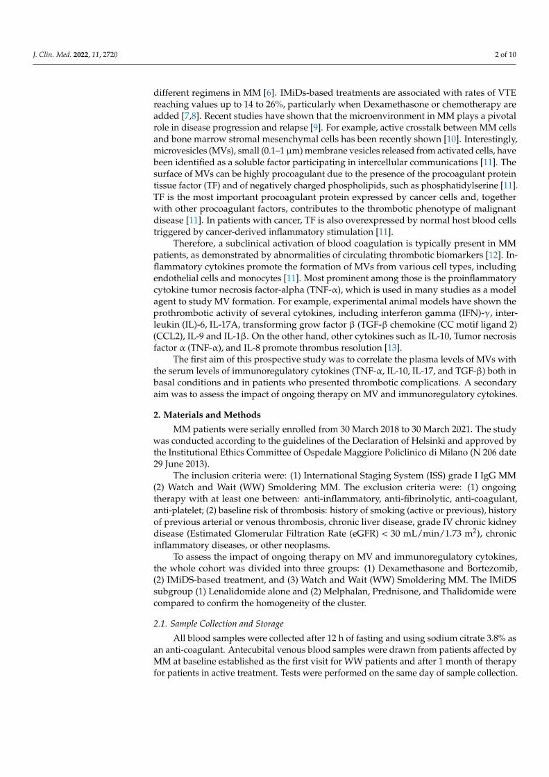
In addition, groups undergoing active treatment management (both the
Dexamethasone/Bortezomib and IMiDs group) displayed lower levels of IL-17 (p < 0.001
Figure 3A), glomerular filtration-rate (p < 0.001 Figure 3B), and higher levels of MV (p <
0.001, Figure 3C) compared to the WW Smoldering MM group (Table 1). The TGF-β values
of IMiDs patients were lower than the WW patients (p < 0.001), and the Dexamethasone
and Bortezomib subgroup (p < 0.001 Figure 3D).
Figure 2. Correlation between MV and TGF-β (A) Indirect association between MV and TGF-βwhole population; (B) Indirect association between MV and TGF-β population without thrombosis).(C) Direct associations between MV and TGF-β in the population with thrombosis.
Importantly, when compared to the WW Smoldering MM, the group of patients un-dergoing active treatment had a significantly higher number of thrombotic events (Fisher’stest 14/92 vs. 0/31; p = 0.02, Table 2). Therefore, in our study, all of the thrombotic episodeswere found to be on active treatment.
In addition, groups undergoing active treatment management (both the Dexametha-sone/Bortezomib and IMiDs group) displayed lower levels of IL-17 (p < 0.001 Figure 3A),glomerular filtration-rate (p < 0.001 Figure 3B), and higher levels of MV (p < 0.001, Figure 3C)compared to the WW Smoldering MM group (Table 1). The TGF-β values of IMiDs patientswere lower than the WW patients (p < 0.001), and the Dexamethasone and Bortezomibsubgroup (p < 0.001 Figure 3D).
J. Clin. Med. 2022, 11, x FOR PEER REVIEW 7 of 11
Figure 3. Comparison of patients based on different treatment: (A) IL-17 levels in Dexamethasone
and Bortezomib; IMiDs based treatment; Watch and Wait Smoldering MM patients; (B) Glomerular
Filtration Rate levels in Dexamethasone and Bortezomib; IMiDs based treatment; Watch and Wait
Smoldering MM patients (C) Microvesicles in Dexamethasone and Bortezomib; IMiDs based
treatment; Watch and Wait Smoldering MM patients (D) TGF-β in Dexamethasone and Bortezomib;
IMiDs based treatment; Watch and Wait Smoldering MM patients.
Intragroup IMiDs homogeneity was confirmed with no statistical difference in all the
variables tested other than the older age in the MPT group versus Lenalidomide,
respectively, 82 (77–82) vs. 74 (72–80) p = 0.005.
4. Discussion
To the best of our knowledge, this is the first report correlating circulating MVs and
cytokines to thrombotic complications in MM patients treated with a specific therapy.
Two studies proposed that hypercoagulability in MM arises from thrombin
generation due to clotting activation via TF and phospholipids activity [16,17]. Our result
is in agreement with Auwerda et al., who detailed, that in a MM cohort under
chemotherapy, elevated MV-TF activity in patients who developed VTE, in contrast to
patients who do not develop VTE [17]. This elevated MV-TF activity may be associated
with increased MVs, especially during anti-myeloma treatment. Importantly, our study
Figure 3. Cont.

J. Clin. Med. 2022, 11, 2720 7 of 10
J. Clin. Med. 2022, 11, x FOR PEER REVIEW 7 of 11
Figure 3. Comparison of patients based on different treatment: (A) IL-17 levels in Dexamethasone
and Bortezomib; IMiDs based treatment; Watch and Wait Smoldering MM patients; (B) Glomerular
Filtration Rate levels in Dexamethasone and Bortezomib; IMiDs based treatment; Watch and Wait
Smoldering MM patients (C) Microvesicles in Dexamethasone and Bortezomib; IMiDs based
treatment; Watch and Wait Smoldering MM patients (D) TGF-β in Dexamethasone and Bortezomib;
IMiDs based treatment; Watch and Wait Smoldering MM patients.
Intragroup IMiDs homogeneity was confirmed with no statistical difference in all the
variables tested other than the older age in the MPT group versus Lenalidomide,
respectively, 82 (77–82) vs. 74 (72–80) p = 0.005.
4. Discussion
To the best of our knowledge, this is the first report correlating circulating MVs and
cytokines to thrombotic complications in MM patients treated with a specific therapy.
Two studies proposed that hypercoagulability in MM arises from thrombin
generation due to clotting activation via TF and phospholipids activity [16,17]. Our result
is in agreement with Auwerda et al., who detailed, that in a MM cohort under
chemotherapy, elevated MV-TF activity in patients who developed VTE, in contrast to
patients who do not develop VTE [17]. This elevated MV-TF activity may be associated
with increased MVs, especially during anti-myeloma treatment. Importantly, our study
Figure 3. Comparison of patients based on different treatment: (A) IL-17 levels in Dexamethasoneand Bortezomib; IMiDs based treatment; Watch and Wait Smoldering MM patients; (B) Glomeru-lar Filtration Rate levels in Dexamethasone and Bortezomib; IMiDs based treatment; Watch andWait Smoldering MM patients (C) Microvesicles in Dexamethasone and Bortezomib; IMiDs basedtreatment; Watch and Wait Smoldering MM patients (D) TGF-β in Dexamethasone and Bortezomib;IMiDs based treatment; Watch and Wait Smoldering MM patients.
Intragroup IMiDs homogeneity was confirmed with no statistical difference in allthe variables tested other than the older age in the MPT group versus Lenalidomide,respectively, 82 (77–82) vs. 74 (72–80) p = 0.005.
4. Discussion
To the best of our knowledge, this is the first report correlating circulating MVs andcytokines to thrombotic complications in MM patients treated with a specific therapy.
Two studies proposed that hypercoagulability in MM arises from thrombin generationdue to clotting activation via TF and phospholipids activity [16,17]. Our result is inagreement with Auwerda et al., who detailed, that in a MM cohort under chemotherapy,elevated MV-TF activity in patients who developed VTE, in contrast to patients who donot develop VTE [17]. This elevated MV-TF activity may be associated with increasedMVs, especially during anti-myeloma treatment. Importantly, our study confirms thatDexamethasone and IMiDs carry an elevated risk of VTE by increasing the levels of MVsand the related TF activation of clotting. This result needs to be validated using other largerpatient cohorts.
It is known that there is a strong connection between inflammation, cancerogenesis,coagulation, and the immune system [18]. Therefore, serum levels of several key cytokineswere measured in our study.
Our finding of a correlation of TGF-β levels in the development of thrombosis confirmsprevious studies that show an increase in patients with VTE [13]. The inflammatory effectof TGF-β and its role in fibrogenesis underlies its contribution to endothelial dysfunctionand increased fibrosis [13].
TGF-β is part of a family of structurally related proteins that consists of activins/inhibins and bone morphogenic proteins [18]. Members of the TGF-β family controlnumerous cellular functions, including proliferation, apoptosis, differentiation, epithelial-mesenchymal transition, and migration. The first identified member, TGF-β, is implicatedin several human diseases, such as vascular diseases, autoimmune disorders, and carcino-genesis. The activation of the TGF-β receptor by its ligands induces the phosphorylationof serine/threonine residues and triggers the phosphorylation of intracellular effectors(SMADs). Upon activation, SMAD proteins translocate to the nucleus and induce transcrip-

J. Clin. Med. 2022, 11, 2720 8 of 10
tion of their target genes, regulating several cellular functions. TGF-β dysregulation hasbeen implicated in carcinogenesis and cancer progression. In the early stages of cancer, TGF-β exhibits tumor-suppressive effects by inhibiting cell cycle progression and promotingapoptosis. However, in the late stages, TGF-β exerts tumor-promoting effects, increasingtumor invasiveness and metastasis. Furthermore, the TGF-β signaling pathway communi-cates with other mediators in a synergistic or antagonistic manner and regulates cellularfunctions. Elevated TGF-β activity has been associated with poor clinical outcomes or theadvanced stages of cancer disease. In the setting of MM, TGF-β is linked with bone-relateddisease, and TGF-β acts as a potent immunosuppressive cytokine thought to exert effectson both cell differentiation and cell proliferation [15].
In our study, we observed a clear relationship between thrombotic events and activetreatment, increased Microvesicles, and TGF-β. A study by Yamaguchi et al. [18] showedthat the TGFβ1/SMAD/Plasminogen activator inhibitor-1 signaling pathway promotesthe release of Tissue Factor-Bearing Microvesicles.
In our analysis of disease status and treatment regimens in MM patients, the group ofMM patients in active treatment was found to have a higher number of thrombotic eventsand higher levels of MVs (p < 0.001) compared with the WW group. This confirms previouspapers that have reported a higher incidence of thrombotic events in actively treated MMpatients [7,8]. Chemotherapy, as well as other cellular stressors, such as heat shock, hypoxia,hypothermia, or oxidative stress, have also been reported to increase MV secretion [19].
In accordance with published studies [20–22], in the current study, patients in the WWmyeloma group had increased levels of IL-17 compared to the active treatment cohort;this could be related to the increased amounts of IL-6 in the bone marrow of myelomapatients with active disease that promotes the production of T helper 17 cells from CD4naive cells. In treated patients, there was a significant reduction in IL-17; the reductionin IL-17 probably reflects the disease response. Nevertheless, the relationship between Thelper 17 cells and T regulatory cells in MM requires further investigation [19].
The TGF-β values were lower in the IMiDs group compared to both the Dexametha-sone/Bortezomib and WW group. IMiDs agents, Thalidomide and Lenalidomide, areimmunomodulatory drugs. These treatments exhibit many important therapeutic proper-ties, such as anti-angiogenetic, antiproliferative, and pro-erythropoietic properties, whosemechanisms remain to be further clarified [23]. In addition, these drugs have shown effectson NK cytotoxic cells and cytokine production. In the literature, studies on the effect ofIMiDS on TGF-β are not in agreement. In an in vitro study, Galustian et al. found thatneither of these drugs (i.e., Lenalidomide and Pomalidomide) had an effect on secretedTGF-β [24]. In contrast, in agreement with our results, Hadjiaggelidou et al. demonstrated,in a small population of MM patients, a reduction in TGF-β levels in the Lenalidomideplus Dexamethasone treatment group versus those patients treated with Dexamethasoneplus Bortezomib [20]. Our results confirm and extend their studies.
Treatment with high doses of Dexamethasone and Bortezomib deserves special discus-sion; in this group, we observed high levels of TGF-β and MVs with an increased prevalenceof thrombosis. This observation, combined with the results from our IMDIS group, suggeststhe hypothesis of two different thrombotic pathways, one from MV-mediated TF thrombingeneration and one due to the TGF-β activity.
The addition of steroids, particularly at higher doses, is associated with a significantelevation in thrombosis risk. Rajkumar [25] reported a comparison between Lenalidomidewith high- or low-dose Dexamethasone. In this study, a high-dose was referred to as 40 mgof Dexamethasone on days 1–4, 9–12, and 17–20 of a 28-day cycle, versus a low dose, where40 mg of Dexamethasone was administered once weekly. The total dose of Dexametha-sone received in the ‘high-dose’ group was 480 mg/month, in line with the InternationalMyeloma Working Group’s (IMWG’s) later definition of ‘high-dose’ glucocorticoids [26].In the initial part of the study, VTE prophylaxis was recommended but not mandated. Ofthe first 266 enrolled patients, 18.2% developed VTE in the high-dose group and 3.7% in thelow-dose group, after which thromboprophylaxis became mandatory [25]. At 1 year from

J. Clin. Med. 2022, 11, 2720 9 of 10
study initiation, the VTE rate in the high-dose group was over double that of the low-dosegroup (26% vs. 12%), providing substantial supportive evidence for the thrombogenicpotential of high dose Dexamethasone [27].
Lastly, we note that the present study has several limitations, including the number ofpatients enrolled to investigate the risk of thrombosis and the low numbers of inflammatorycytokines investigated.
In addition, blood samples were drawn at baseline (at study entry for patients onWW Smoldering MM vs. 1 month of treatment for patients on active therapy), but thenno subsequent or serial blood analysis was conducted. However, most thromboembolismepisodes occurred many months after study enrollment, so we cannot rule out a change inthe variable tested during the follow-up period.
5. Conclusions
To conclude, this is the first prospective study correlating MVs levels with differentinflammatory cytokines in MM. Overall, we found that MVs increase in treated MMpatients across different regimens. The increased levels of MVs in these regimens addinsight into mechanisms of hypercoagulation in MM. In addition, the role of cytokine-related thrombosis is also suggested and remains to be further investigated.
Author Contributions: R.C. and A.G. defined the design of the study, studied and analyzed theresults and wrote the paper. A.G., R.M., A.P.D., D.S., D.C. and G.L.D. managed the data collectionprocess and analyzed data. A.P.D., M.J.S. and R.M. reviewed the manuscript. All authors have readand agreed to the published version of the manuscript.
Funding: The paper was funded by Progetto Fondazione di Sardegna year 2022–2023.
Institutional Review Board Statement: The study participants gave a written informed consent andlocal ethical committee approved the study (Ospedale Maggiore Policlinico di Milano; N 206 date 29June 2013).
Informed Consent Statement: Informed consent was obtained from all subjects involved in the study.
Data Availability Statement: The study data will be made available upon request to the correspond-ing author.
Conflicts of Interest: The authors declare no conflict of interest.
References1. Kristinsson, S.Y.; Fears, T.R.; Gridley, G.; Turesson, I.; Mellqvist, U.H.; Björkholm, M.; Landgren, O. Deep vein thrombosis after
monoclonal gammopathy of undetermined significance and multiple myeloma. Blood 2008, 112, 3582–3586. [CrossRef] [PubMed]2. Gogia, A.; Sikka, M.; Sharma, S.; Rusia, U. Hemostatic Abnormalities in Multiple Myeloma Patients. Asian Pac. J. Cancer Prev.
2018, 19, 127–130. [CrossRef] [PubMed]3. Castelli, R.; Gualtierotti, R.; Orofino, N.; Losurdo, A.; Gandolfi, S.; Cugno, M. Current and emerging treatment options for patients
with relapsed myeloma. Clin. Med. Insights Oncol. 2013, 7, 209–219. [CrossRef]4. Castelli, R.; Orofino, N.; Losurdo, A.; Gualtierotti, R.; Cugno, M. Choosing treatment options for patients with relapsed/refractory
multiple myeloma. Expert Rev. Anticancer Ther. 2014, 14, 199–215. [CrossRef] [PubMed]5. Castelli, R.; Pantaleo, G.; Gallipoli, P.; Gidaro, A.; Arquati, M.; Wu, M.A.; Lambertenghi Deliliers, G. Salvage therapy with
bortezomib and dexamethasone in elderly patients with relapsed/refractory multiple myeloma. Anticancer Drugs 2015, 26,1078–1082. [CrossRef]
6. Tiong, I.S.; Rodgers, S.E.; Lee, C.H.; McRae, S.J. Baseline and treatment-related changes in thrombin generation in patients withmultiple myeloma. Leuk. Lymphoma 2017, 58, 941–949. [CrossRef]
7. Zangari, M.; Berno, T.; Zhan, F.; Tricot, G.; Fink, L. Mechanisms of thrombosis in paraproteinemias: The effects of immunomodu-latory drugs. Semin. Thromb. Hemost. 2012, 38, 768–779. [CrossRef]
8. Cini, M.; Zamagni, E.; Valdré, L.; Palareti, G.; Patriarca, F.; Tacchetti, P.; Cavo, M. Thalidomide-dexamethasone as up-fronttherapy for patients with newly diagnosed multiple myeloma: Thrombophilic alterations, thrombotic complications, andthromboprophylaxis with low-dose warfarin. Eur. J. Haematol. 2010, 84, 484–492. [CrossRef]
9. Crowely, M.P.; Quinn, S.; Coleman, E.; Eustace, J.A.; Gilligan, O.M.; Shea, S.I. Differing coagulation profiles of patients withmonoclonal gammopathy of undetermined significance and multiple myeloma. J. Thromb. Thrombolysis 2015, 39, 245–249.[CrossRef]

J. Clin. Med. 2022, 11, 2720 10 of 10
10. Colombo, R.; Gallipoli, P.; Castelli, R. Thrombosis, and hemostatic abnormalities in hematological malignancies. Clin. LymphomaMyeloma Leuk. 2014, 14, 441–450. [CrossRef]
11. Zifkos, K.; Dubois, C.; Schäfer, K. Extracellular Vesicles and Thrombosis: Update on the Clinical and Experimental Evidence. Int.J. Mol. Sci. 2021, 22, 9317. [CrossRef]
12. Auwerda, J.J.; Sonneveld, P.; de Maat, M.P.; Leebeek, F.W. Prothrombotic coagulation abnormalities in patients with newlydiagnosed multiple myeloma. Haematologica 2007, 92, 279–280. [CrossRef] [PubMed]
13. Najem, M.Y.; Couturaud, F.; Lemarié, C.A. Cytokine and chemokine regulation of venous thromboembolism. J. Thromb. Haemost.2020, 18, 1009–1019. [CrossRef] [PubMed]
14. Shet, A.S.; Aras, O.; Gupta, K.; Hass, M.J.; Rausch, D.J.; Saba, N.; Koopmeiners, L.; Key, N.S.; Hebbel, R.P. Sickle blood containstissue factor-positive microparticles derived from endothelial cells and monocytes. Blood 2003, 102, 2678–2683. [CrossRef][PubMed]
15. Kubiczkova, L.; Sedlarikova, L.; Hajek, R.; Sevcikova, S. TGF-β—An excellent servant but a bad master. J. Transl. Med. 2012, 10,183. [CrossRef] [PubMed]
16. Nielsen, T.; Kristensen, S.R.; Gregersen, H.; Teodorescu, E.M.; Pedersen, S. Prothrombotic abnormalities in patients with multiplemyeloma and monoclonal gammopathy of undetermined significance. Thromb. Res. 2021, 202, 108–118. [CrossRef]
17. Auwerda, J.J.; Yuana, Y.; Osanto, S.; de Maat, M.P.; Sonneveld, P.; Bertina, R.M.; Leebeek, F.W. Microparticle-associated tissuefactor activity and venous thrombosis in multiple myeloma. Thromb. Haemost. 2011, 105, 14–20. [CrossRef]
18. Yamaguchi, R.; Sakamoto, A.; Yamaguchi, R.; Haraguchi, M.; Narahara, S.; Sugiuchi, H.; Yamaguchi, Y. Di-(2-Ethylhexyl)Phthalate Promotes Release of Tissue Factor-Bearing Microparticles From Macrophages via the TGFβ1/Smad/PAI-1 SignalingPathway. Am. J. Med. Sci. 2019, 357, 492–506. [CrossRef]
19. Kadota, T.; Fujita, Y.; Yoshioka, Y.; Araya, J.; Kuwano, K.; Ochiya, T. Emerging role of extracellular vesicles as a senescenceassoci-ated secretory phenotype: Insights into the pathophysiology of lung diseases. Mol. Asp. Med. 2018, 60, 92–103. [CrossRef]
20. Braga, W.M.; Atanackovic, D.; Colleoni, G.W. The Role of Regulatory T Cells and TH17 Cells in Multiple Myeloma. J. Immunol.Res. 2012, 2012, 293479. [CrossRef]
21. Bettelli, E.; Carrier, Y.; Gao, W.; Korn, T.; Strom, T.B.; Oukka, M.; Weiner, H.L.; Kuchroo, V.K. Reciprocal developmental pathwaysfor the generation of pathogenic effector TH17 and regulatory T cells. Nature 2006, 441, 235–238. [CrossRef] [PubMed]
22. Hadjiaggelidou, C.; Mandala, E.; Terpos, E.; Yiannaki, E.; Markala, D.; Triantafyllou, T.; Papatheodorou, A.; Gkastari, V.; Verrou,E.; Papanikolaou, A.; et al. Evaluation of regulatory T cells (Tregs) alterations in patients with multiple myeloma treated withbortezomib or lenalidomide plus dexamethasone: Correlations with treatment outcome. Ann. Hematol. 2019, 98, 1457–1466.[CrossRef] [PubMed]
23. Castelli, R.; Cannavò, A.; Conforti, F.; Grava, G.; Cortelezzi, A. Immunomodulatory drugs in multiple myeloma: From molecularmechanisms of action to clinical practice. Immunopharmacol. Immunotoxicol. 2012, 34, 740–753. [CrossRef] [PubMed]
24. Galustian, C.; Meyer, B.; Labarthe, M.C.; Dredge, K.; Klaschka, D.; Henry, J.; Todryk, S.; Chen, R.; Muller, G.; Stirling, D.; et al. Theanti-cancer agents lenalidomide and pomalidomide inhibit the proliferation and function of T regulatory cells. Cancer Immunol.Immunother. 2009, 58, 1033–1045. [CrossRef] [PubMed]
25. Rajkumar, S.V.; Blood, E.; Vesole, D.; Fonseca, R.; Greipp, P.R.; Eastern Cooperative Oncology Group. Phase III clinical trial ofthalidomide plus dexamethasone compared with dexamethasone alone in newly diagnosed multiple myeloma: A clinical trialcoordinated by the Eastern Cooperative Oncology Group. J. Clin. Oncol. 2006, 24, 431–436. [CrossRef] [PubMed]
26. Palumbo, A.; Rajkumar, S.V.; Dimopoulos, M.A.; Richardson, P.G.; San Miguel, J.; Barlogie, B.; Harousseau, J.; Zonder, J.A.;Cavo, M.; Zangari, M.; et al. Prevention of thalidomide- and lenalidomide-associated thrombosis in myeloma. Leukemia 2008, 22,414–423. [CrossRef]
27. Rajkumar, S.V.; Jacobus, S.; Callander, N.S.; Fonseca, R.; Vesole, D.H.; Williams, M.E.; Abonour, R.; Siegel, D.S.; Katz, M.;Greipp, P.R.; et al. Lenalidomide plus high-dose dexamethasone versus lenalidomide plus low-dose dexamethasone as initialtherapy for newly diagnosed multiple myeloma: An open-label randomised controlled trial. Lancet Oncol. 2010, 11, 29–37,Erratum in Lancet Oncol. 2010, 11, 14. [CrossRef]