Cas IIgly Induces Apoptosis in Glioma C6 Cells In Vitro and In Vivo through Caspase-Dependent and...
12
Cas IIgly Induces Apoptosis in Glioma C6 Cells In Vitro and In Vivo through Caspase-Dependent and Caspase-Independent Mechanisms 1 Cristina Trejo-Solı ´s *, Guadalupe Palencia *, Sergio Zu ´n ˜iga *, Andrea Rodrı ´guez-Ropon y , Laura Osorio-Rico *, Sanchez Torres Luvia y , Isabel Gracia-Mora z , Lucrecia Marquez-Rosado y , Aurora Sa ´ nchez *, Miguel E. Moreno-Garcı ´a y , Arturo Cruz § , Marı ´a Elena Bravo-Go ´mez z , Lena Ruiz-Ramı ´rez z , Sara Rodrı ´guez-Enriquez b and Julio Sotelo * *Departamento de Neuroinmunologı ´a, Instituto Nacional de Neurologı ´a y Neurocirugı ´a, SSA, Mexico, DF 14269, Mexico; y Departamento de Biologı ´a Celular, Centro de Investigaciones y Estudios Avanzados, Instituto Polite ´cnico Nacional, Mexico, DF, Mexico; z Departamento de Quı ´mica y Medicina Nuclear, Universidad Nacional Auto ´noma de Me ´xico, Mexico, DF, Mexico; § Departamento de Biologı ´a Celular, Instituto de Fisiologı ´a Celular, Universidad Nacional Auto ´noma de Me ´xico, Mexico, DF, Mexico; b Departamento de Bioquı ´mica, Instituto Nacional de Cardiologı ´a, SSA, Me ´xico, DF, Mexico Abstract In this work, we investigated the effects of Casiopeina II-gly (Cas IIgly)—a new copper compound exhibiting antineoplastic activity—on glioma C6 cells under both in vitro and in vivo conditions, as an approach to iden- tify potential therapeutic agents against malignant glioma. The exposure of C6 cells to Cas IIgly signifi- cantly inhibited cell proliferation, increased reactive oxygen species (ROS) formation, and induced apopto- sis in a dose-dependent manner. In cultured C6 cells, Cas IIgly caused mitochondrio-nuclear translocation of apoptosis induction factor (AIF) and endonuclease G at all concentrations tested; in contrast, fragmentation of nucleosomal DNA, cytochrome c release, and caspase-3 activation were observed at high concentrations. Ad- ministration of N-acetyl-L-cystein, an antioxidant, re- sulted in significant inhibition of AIF translocation, nucleosomal DNA fragmentation, and caspase-3 acti- vation induced by Cas IIgly. These results suggest that caspase-dependent and caspase-independent path- ways both participate in apoptotic events elicited by Cas IIgly. ROS formation induced by Cas IIgly might also be involved in the mitochondrio-nuclear trans- location of AIF and apoptosis. In addition, treatment of glioma C6 – positive rats with Cas IIgly reduced tumor volume and mitotic and cell proliferation indexes, and increased apoptotic index. Our findings support the use of Cas IIgly for the treatment of malignant gliomas. Neoplasia (2005) 7, 563–574 Keywords: Casiopeina II-gly, apoptosis, reactive oxygen species, apoptosis- inducing factor, glioma C6 cells. Introduction Apoptosis occurs spontaneously in malignant tumors, and this event is often potentiated by chemotherapeutic agents [1,2]. Morphologically, the process of apoptosis is characterized by cellular condensation, nuclear fragmentation, and engulfment of cell debris by phagocytes [3]. The induction of apoptosis may involve either extracellular triggering signals (such as a tumor necrosis factor) or endogenous signals [such as cytochrome c (cyt c)] [4], which are followed by the activa- tion of caspases [5] and endonucleases [6,7], thus causing disassembly of nuclear chromatin and degradation of oligo- nucleosomal DNA. Moreover, the translocation of apoptosis induction factor (AIF) leads to apoptosis through a caspase- independent pathway, producing the condensation of nuclear chromatin, as well as large-scale (f50 kb) DNA fragmenta- tion [8–10]. In addition, it has been suggested that Fe 3+ and Cu 2+ , two redox-active metal ions, are involved in reactive oxy- gen species (ROS) generation, and hence in apoptosis [11,12]. Oxidative stress is a putative mediator of apoptosis through many different mechanisms [13]. Among them, we can mention the following: 1) the intracellular increase of ROS [14] or the depletion of endogenous antioxidants [15]; 2) the action of some antioxidants—such as N-acetyl-L-cystein (NAC)—which act as intracellular ROS scavengers, thus inhibiting the Abbreviations: Cas IIgly, Casiopeina IIgly; PCD, programmed cell death; AIF, apoptosis- inducing factor; ROS, reactive oxygen species; NAC, N-acetyl-L-cystein; SOD, superoxide dismutase; GPx, glutathione peroxidase; MTT, 3[4,5-dimethylthiazol-2-yl]-2,5-diphenyl- tetrazolium bromide; FACS, fluorescence-activated cell sorter; NBT, 4-nitro blue tetrazolium chloride; BCIP, 5-bromide-4-chloride-3-indolyl-phosphate 4-toluidine salt; TUNEL, terminal deoxynucleotidyl transferase – mediated nick end labeling; R123, rhodamine 123; cyt c, cyto- chrome c; Endo G, endonuclease G; DCFH-DA, 2V,7V-dichlorofluorescein diacetate; DCF, 2V, 7V-dichlorofluorescein; TBARS, thiobarbituric acid reactive substances; PMSF, phenymethyl- sulfonyl fluoride; PCNA, proliferating cell nuclear antigen; PAGE, polyacrylamide gel electro- phoresis; ECL, enhanced chemiluminescence; SGOT, aspartate transaminase; SGPT, alanine transaminase; GGT, gamma glutamyl transferase Address all correspondence to: Dr. Cristina Trejo-Solı ´s, Departamento de Neuroinmunologı ´a, Instituto Nacional de Neurologı ´a y Neurocirugı ´a, Manuel Velasco Sua ´rez, Insurgentes Sur 3877, Mexico, DF 14269, Me ´xico. E-mail: [email protected] 1 This work was supported by CONACyT grants U41997-MA1 and C01-7677-Salud, 2002. Received 8 September 2004; Revised 18 January 2005; Accepted 18 January 2005. Copyright D 2005 Neoplasia Press, Inc. All rights reserved 1522-8002/05/$25.00 DOI 10.1593/neo.04607 Neoplasia . Vol. 7, No. 6, June 2005, pp. 563 – 574 563 www.neoplasia.com RESEARCH ARTICLE
Transcript of Cas IIgly Induces Apoptosis in Glioma C6 Cells In Vitro and In Vivo through Caspase-Dependent and...
Cas IIgly Induces Apoptosis in Glioma C6 Cells In Vitroand In Vivo through Caspase-Dependent andCaspase-Independent Mechanisms1
Cristina Trejo-Solıs*, Guadalupe Palencia*, Sergio Zuniga*, Andrea Rodrıguez-Ropon y, Laura Osorio-Rico*,Sanchez Torres Luvia y, Isabel Gracia-Mora z, Lucrecia Marquez-Rosadoy, Aurora Sanchez*,Miguel E. Moreno-Garcıa y, Arturo Cruz§, Marıa Elena Bravo-Gomez z, Lena Ruiz-Ramırez z,Sara Rodrıguez-Enriquez b and Julio Sotelo*
*Departamento de Neuroinmunologıa, Instituto Nacional de Neurologıa y Neurocirugıa, SSA, Mexico,DF 14269, Mexico; yDepartamento de Biologıa Celular, Centro de Investigaciones y Estudios Avanzados,Instituto Politecnico Nacional, Mexico, DF, Mexico; zDepartamento de Quımica y Medicina Nuclear,Universidad Nacional Autonoma de Mexico, Mexico, DF, Mexico; §Departamento de Biologıa Celular,Instituto de Fisiologıa Celular, Universidad Nacional Autonoma de Mexico, Mexico, DF, Mexico;bDepartamento de Bioquımica, Instituto Nacional de Cardiologıa, SSA, Mexico, DF, Mexico
Abstract
In this work, we investigated the effects of Casiopeina
II-gly (Cas IIgly)—a new copper compound exhibiting
antineoplastic activity—on glioma C6 cells under both
in vitro and in vivo conditions, as an approach to iden-
tify potential therapeutic agents against malignant
glioma. The exposure of C6 cells to Cas IIgly signifi-
cantly inhibited cell proliferation, increased reactive
oxygen species (ROS) formation, and induced apopto-
sis in a dose-dependent manner. In cultured C6 cells,
Cas IIgly caused mitochondrio-nuclear translocation of
apoptosis induction factor (AIF) and endonuclease G at
all concentrations tested; in contrast, fragmentation of
nucleosomal DNA, cytochrome c release, and caspase-3
activation were observed at high concentrations. Ad-
ministration of N-acetyl-L-cystein, an antioxidant, re-
sulted in significant inhibition of AIF translocation,
nucleosomal DNA fragmentation, and caspase-3 acti-
vation induced by Cas IIgly. These results suggest that
caspase-dependent and caspase-independent path-
ways both participate in apoptotic events elicited by
Cas IIgly. ROS formation induced by Cas IIgly might
also be involved in the mitochondrio-nuclear trans-
location of AIF and apoptosis. In addition, treatment of
glioma C6–positive rats with Cas IIgly reduced tumor
volume and mitotic and cell proliferation indexes, and
increased apoptotic index. Our findings support the
use of Cas IIgly for the treatment of malignant gliomas.
Neoplasia (2005) 7, 563–574
Keywords: Casiopeina II-gly, apoptosis, reactive oxygen species, apoptosis-inducing factor, glioma C6 cells.
Introduction
Apoptosis occurs spontaneously in malignant tumors, and
this event is often potentiated by chemotherapeutic
agents [1,2]. Morphologically, the process of apoptosis is
characterized by cellular condensation, nuclear fragmentation,
and engulfment of cell debris by phagocytes [3]. The induction
of apoptosis may involve either extracellular triggering signals
(such as a tumor necrosis factor) or endogenous signals [such
as cytochrome c (cyt c)] [4], which are followed by the activa-
tion of caspases [5] and endonucleases [6,7], thus causing
disassembly of nuclear chromatin and degradation of oligo-
nucleosomal DNA. Moreover, the translocation of apoptosis
induction factor (AIF) leads to apoptosis through a caspase-
independent pathway, producing the condensation of nuclear
chromatin, as well as large-scale (f50 kb) DNA fragmenta-
tion [8–10]. In addition, it has been suggested that Fe3+ and
Cu2+, two redox-active metal ions, are involved in reactive oxy-
gen species (ROS) generation, and hence in apoptosis [11,12].
Oxidative stress is a putative mediator of apoptosis through
many different mechanisms [13]. Among them, we can mention
the following: 1) the intracellular increase of ROS [14] or the
depletion of endogenous antioxidants [15]; 2) the action of
some antioxidants—such as N-acetyl-L-cystein (NAC)—which
act as intracellular ROS scavengers, thus inhibiting the
Abbreviations: Cas IIgly, Casiopeina IIgly; PCD, programmed cell death; AIF, apoptosis-
inducing factor; ROS, reactive oxygen species; NAC, N-acetyl-L-cystein; SOD, superoxide
dismutase; GPx, glutathione peroxidase; MTT, 3[4,5-dimethylthiazol-2-yl]-2,5-diphenyl-
tetrazolium bromide; FACS, fluorescence-activated cell sorter; NBT, 4-nitro blue tetrazolium
chloride; BCIP, 5-bromide-4-chloride-3-indolyl-phosphate 4-toluidine salt; TUNEL, terminal
deoxynucleotidyl transferase – mediated nick end labeling; R123, rhodamine 123; cyt c, cyto-
chrome c; Endo G, endonuclease G; DCFH-DA, 2V,7V-dichlorofluorescein diacetate; DCF, 2V,
7V-dichlorofluorescein; TBARS, thiobarbituric acid reactive substances; PMSF, phenymethyl-
sulfonyl fluoride; PCNA, proliferating cell nuclear antigen; PAGE, polyacrylamide gel electro-
phoresis; ECL, enhanced chemiluminescence; SGOT, aspartate transaminase; SGPT,
alanine transaminase; GGT, gamma glutamyl transferase
Address all correspondence to: Dr. Cristina Trejo-Solıs, Departamento de Neuroinmunologıa,
Instituto Nacional de Neurologıa y Neurocirugıa, Manuel Velasco Suarez, Insurgentes Sur
3877, Mexico, DF 14269, Mexico. E-mail: [email protected] work was supported by CONACyT grants U41997-MA1 and C01-7677-Salud, 2002.
Received 8 September 2004; Revised 18 January 2005; Accepted 18 January 2005.
Copyright D 2005 Neoplasia Press, Inc. All rights reserved 1522-8002/05/$25.00
DOI 10.1593/neo.04607
Neoplasia . Vol. 7, No. 6, June 2005, pp. 563 – 574 563
www.neoplasia.com
RESEARCH ARTICLE
activation of caspases [16]; 3) the overexpression of Mn
superoxide dismutase (SOD), which restores the mitochon-
drial transmembrane potential, thus inhibiting apoptosis
[16,17]; 4) the overexpression of Cu/Zn-SOD, which delays
apoptosis by scavenging O2.� [18,19]; and 5) the overex-
pression of the mitochondrial phospholipid hydroperoxide
glutathione peroxidase (GPx) that inhibits the generation of
ROS formation [20].
However, malignant gliomas are known to be resistant
to different forms of chemotherapy, and this is consistent
with evidence demonstrating that glioma cells are prone
to developing drug resistance during treatment. Therefore,
the search for novel and more effective chemotherapeutic
agents against glioma cells becomes more relevant than
ever. Moreover, the concept, that drugs based on endo-
genous (essential) metallic components may be more effec-
tive and less toxic than other currently used agents, prompted
several groups to develop copper-based drugs with poten-
tial use as therapeutic agents. In our case, we have recently
completed the synthesis of mixed copper-chelating com-
pounds with the generic name of casiopeinas, which pres-
ent the following basic formula: Cu(N – N)(A – A)]NO3
(A–AN–O, O–O) [21,22]. Furthermore, it has been demon-
strated that some copper-based generic compounds may
exhibit a higher antineoplastic potency than cisplatin for
human ovarian carcinoma (CH1), murine leukemia (L1210),
and various cervico-uterine carcinomas [23,24]. However,
casiopeinas still need to be characterized in their prop-
erties against tumors from the nervous system. Therefore,
the present study was undertaken to investigate the ef-
fects of Casiopeina II-gly (Cas IIgly) [Cu(4,7-dimethyl-1,
10-phenanthroline)(glycine)(H2O)]NO3 in rats with glioma
C6 and in cultured glioma C6 cells. Our data show that
Cas IIgly inhibited cell growth and induced programmed
cell death (PCD) in vitro and in vivo, thus involving an
active role of ROS formation in the positive actions of
casiopeina through both caspase-dependent and caspase-
independent mechanisms.
Materials and Methods
Reagents
Cas IIgly was synthesized as previously described [25]
and dissolved in sterile water. All other reagents were ob-
tained from known commercial sources.
In Vitro Experiments
Glioma cell culture Cultured glioma C6 cells from rats
(American Tissue Culture Collection, Rockville, MD) were
maintained at 37jC in 5% CO2 under sterile conditions in
Dulbecco’s modified Eagle’s medium (Sigma Chemical Co.,
St. Louis, MO), supplemented with 5% fetal bovine serum
(Sigma Chemical Co.), and treated during 24 hours with
increasing concentrations of Cas IIgly (1, 2.5, 5, and 10 mg/ml;
equivalent to 4.26 � 10�4, 1.0 � 10�3, 2.13 � 10�3, and 4.26 �10�3 mM, respectively), either in the presence or absence of
20 mM NAC (Sigma Chemical Co.).
Cell viability assay The effect of Cas IIgly on the survival
of glioma C6 cells was determined by the MTT (3[4,5-
dimethylthiazol-2-yl]-2,5-diphenyl-tetrazolium bromide) as-
say (Roche Diagnostics, Mannheim, Germany), which
measures mitochondrial activity. MTT is a yellow-colored
salt that is taken up and cleaved only by metabolically active
cells, reducing it to a colored, water-insoluble formazan
salt. The solubilized formazan products were quantified
in a 96-well–format spectrophotometer at 570 nm (Lab-
system Uniskan, Manchester, UK). Briefly, 5� 104 cells were
seeded in cell culture plates and preincubated overnight.
After exposure to increasing concentrations of Cas IIgly
for 24 hours at 37jC, 10 ml of MTT (5 mg/ml) was added,
and the cells were then incubated at 37jC for 4 hours.
The tetrazolium crystals were solubilized by the addition of
10% SDS in 0.01 N HCl. Absorbance was measured after
overnight incubation at 37jC. To observe cell morphology,
5� 104 cells were seeded on chamber slides and exposed
to Cas IIgly for 24 hours. Air-dried cells were fixed with
4% paraformaldehyde and stained with hematoxylin–eosin
(DakoCytomation, Carpinteria, CA), and cell morphology
was observed with a light microscope at �20.
Determination of apoptosis To assess apoptosis, frag-
mented DNA was isolated and analyzed by electrophoresis
on 2% (wt/vol) agarose gels and stained with ethidium
bromide as described in the Quantum PrepR AquaPure
Genomic DNA Kit (Bio-Rad, Hercules, CA). Induction of cell
death was also monitored as the appearance of the sub-G0
peak in cell cycle analysis [26]. Briefly, control and treated
cells (1� 106) were centrifuged and fixed overnight in 70%
ethanol at 4jC; then cells were washed three times with
PBS, incubated for 1 hour in the presence of 1 mg/ml RNase
A and 20 mg/ml propidium iodide at room temperature,
and analyzed for different cell cycle phases with a Becton
Dickinson (San Jose, CA) FACScan flow cytometer.
Apoptosis was assayed by a second method using the
in situ Cell Death Detection Kit, AP (Roche Diagnostics):
cells were seeded on 0.7-cm2 glass cover slides in eight-
well plates (Daigger, Vernon Hills, IL). After treatment with
Cas IIgly, cells were washed twice with PBS and fixed with
4% paraformaldehyde, washed with PBS, permeabilized
with 0.1% Triton X-100 in a 0.1% sodium citrate solution, and
incubated with the terminal deoxynucleotidyl transferase–
mediated nick end labeling (TUNEL) reaction mixture (con-
sisting of calf thymus deoxynucleotidyl transferase and
nucleotide mixture in a reaction buffer) for 60 minutes at
37jC. The slides were rinsed three times with PBS and
Converter-AP was added (antifluorescein antibody Fab frag-
ment from sheep, conjugated with alkaline phosphatase)
during 30 minutes at 37jC. The slides were rinsed again
three times with PBS, and the substrate solution was then
added [NBT (4-nitro blue tetrazolium chloride) and BCIP
(5-bromide-4-chloride-3-indolyl-phosphate 4-toluidine salt)].
Apoptotic cells were visualized with a light microscope.
Apoptosis was also analyzed by a third method using
a caspase-3 Colorimetric Assay (RyD System, Minneapolis,
MN): briefly, following the treatment with Cas IIgly for
564 Apoptotic Effect of Cas IIgly on Glioma C6 Cells Trejo-Solıs et al.
Neoplasia . Vol. 7, No. 6, 2005

24 hours, cells were washed in PBS, resuspended in lysis
buffer, and incubated on ice for 10 minutes. After centrifu-
gation at 10,000g during 1minute, caspase-3 activity was
measured in the supernatant at 37jC in 96 microplates;
the reaction contained the caspase-3 colorimetric substrate
(DEVD-pNA). The absorbance was measured at 405 nm
using a Labsystem Uniskan plate reader.
An apoptotic-related protein, AIF, was also analyzed
using Western blot analysis and immunocytochemistry
techniques. For immunocytochemistry, treated and control
cells were cultured on 0.7-cm2 glass cover slides in eight-
well plates (Daigger), fixed with 4% paraformaldehyde in
PBS for 10 minutes, washed three times with PBS, and
permeabilized with DAKO target retrieval solution (Dako-
Cytomation) for 30 minutes at 95jC, and then blocked with
3% hydrogen peroxide for 10 minutes at room temperature.
Cells were incubated with an AIF antibody (diluted 1:100)
in PBS for 30 minutes at room temperature, washed three
times, and then incubated with horseradish peroxidase–
conjugated antimouse IgG (DakoCytomation) at a 1:1000 di-
lution during 15 minutes. After three more washes, the slides
were processed by avidin–biotin–peroxidase and visualized
with a light microscope at �20.
Mitochondrial transmembrane potential assay Mitochon-
drial energization was evaluated through the analysis of
mitochondrial retention of the cationic fluorescent dye, rho-
damine 123 (R123) [27]. Briefly, 1� 105 cells were treated
with Cas IIgly for 24 hours, washed with PBS, and incubated
with 20 mg/ml R123 at 37jC for 20 minutes. After wash-
ing cells twice again with PBS, R123-related fluorescence
was analyzed by cytometry using a FACSCalibur cytom-
eter (Becton Dickinson) and analyzed with Cellquest 3.1f
analysis software.
Measurement of ROS formation 2V,7V-Dichlorofluorescein
diacetate (DCFH-DA) is a stable, nonfluorescent mole-
cule that is hydrolized by esterases to the nonfluorescent
DCFH. DCFH is then oxidized in the presence of ROS
(superoxide anion, hydrogen peroxide, and hydroxyl radi-
cal) to the highly fluorescent 2V,7V-dichlorofluorescein (DCF)
[28]. For ROS analysis, the DCFH-DA probe was used
as previously described [29]. Briefly, lysed cells were di-
luted 1:10 with 40 mM Tris (pH 7.4) and loaded with 5 mM
DCFH-DA (Molecular Probes, Eugene, OR) in methanol
for 15 minutes at 37jC. Afterward, the fluorescence was
recorded before and after 60 minutes of incubation. The
formation of the fluorescent oxidized derivative of DCFH,
named DCF, was monitored at an excitation wavelength
of 525 nm (slit, 5 nm). The bucket holder was thermostat-
ically maintained at 37jC. Autofluorescence of the cel-
lular lysate was always less than 6%. The fluorescent
signals of both methanol (as vehicle) and substrates were
recorded at the baseline, before the calculation of DCF
formation. DCF formation was quantified from a standard
curve (Sigma Aldrich, St. Louis, MO) in methanol. Analysis
was performed with a Perkin-Elmer (Boston, MA) LS50-B
luminescence spectrometer.
Assay of lipid peroxidation The amounts of aldehydic prod-
ucts generated by lipoperoxidation were determined in lysated
glioma C6 cells from control and Cas IIgly–treated groups
24 hours after incubation, using the thiobarbituric acid reac-
tion (Sigma Aldrich) [30], and modified as previously reported
[31]. Results were listed as nanomoles of thiobarbituric acid
reactive substances (TBARS) per milligram of protein.
Subcellular fractionation Control and treated glioma C6
cells were harvested, washed once with ice-cold PBS,
resuspended in 200 ml of ice-cold Cell Lysis-M Buffer (Sigma
Chemical Co.) plus protease inhibitors [10 mg/ml leupeptin,
1.0 mg/ml aprotinin, and 0.1 mM phenymethylsulfonyl fluo-
ride (PMSF)] (Sigma Chemical Co.), and then sonicated.
Insoluble debris was removed by centrifugation at 12,000g
for 15 minutes, and the supernatants were stored at �80jC.
The nuclear and cytoplasmic extractions were performed
with the NE-PER Nuclear and Cytoplasmic Extraction Re-
agents (Pierce Biotechnology, Rockford, IL), as previously
described [32]. Protein concentrations of each sample were
determined using the Bio-Rad protein assay system.
Western blot analysis The levels of proliferating cell nuclear
antigen (PCNA) protein expression were evaluated by Western
blot analysis. Samples containing equal amounts of protein
(30 mmg) were mixed with an equal volume of 2� sample
buffer (125 mM Tris–HCl, pH 6.8, 20% glycerol, 4% SDS3,
0.02% bromophenol blue, and 10% 2-mercaptoethanol) and
boiled during 5 minutes. The samples were cooled on ice for
5 minutes, centrifuged for a short time, and exposed to 10%
to 15% SDS polyacrylamide gel electrophoresis (PAGE).
Proteins were transferred to a nitrocellulose membrane for
2 hours at 70 V with 25 mM Tris–HCl, pH 8.0, 195 mM gly-
cine, and 10% methanol. The membrane was blocked with
5% light milk in 1� PBS for 1 hour, and the PCNA antibody
(Santa Cruz Biotechnology, Santa Cruz, Ca) was added to
the membrane for 24 hours at 4jC. After three consecutive
washes in PBS, the membrane was incubated with a sheep
antimouse IgG–horseradish peroxidase complex (Santa
Cruz Biotechnology) during 1 hour at room temperature,
followed by three washes with PBS. Then, chemilumines-
cence was visualized using the enhanced chemilumines-
cence (ECL) kit (Santa Cruz Biotechnology). The blot was
exposed to Kodak XAR-5-ray film (Sigma Chemical Co.)
for 10 minutes and then developed. A similar procedure
was used for AIF, cyt c, b-actin, lamin B (Santa Cruz Bio-
technology), and endonuclease G (Endo G) primary anti-
bodies (Chemicon International, Temecula, CA). The band
intensities were quantified in a Molecular Dynamics (Dur-
ham, NC) computing densitometer using ImageQuant soft-
ware version 3.2.2.
In Vivo Experiments
Tumor model and treatment Cultured C6 cells (1� 107)
were intraperitoneally inoculated into a 12-week–old Wistar:
Hsd rat. Fifteen days later, a multilobulated peritoneal tumor
developed. The tumor was mechanically dissociated at 4jC,
and a suspension of 1� 107 cells in 500 ml of saline solution
Apoptotic Effect of Cas IIgly on Glioma C6 Cells Trejo-Solıs et al. 565
Neoplasia . Vol. 7, No. 6, 2005
was subcutaneously inoculated into the back of 12-week–
old male Wistar:Hsd rats as previously described [33].
Subcutaneous tumors developed in 80% of the animals,
reaching a diameter of 1.5 cm, 14 to 17 days after cell im-
plantation [33]. These rats were randomly allocated to one of
three groups: group A (n = 30) was used as control and
injected subcutaneously with 0.5 ml of water every 24 hours
for 21 days, whereas groups B (n = 30) and C (n = 30) were
injected intraperitonerally every 24 hours for 21 days with
0.4 and 0.8 mg/kg Cas IIgly, respectively.
Antineoplastic evaluation and collateral effects of Cas IIgly
Twenty-one days after treatment, animals from all groups
were bled by intracardiac puncture to study chemical blood
parameters and then killed; the corresponding tumor was
removed from each animal and its volume was measured
by fluid displacement [34]. The tumors were dissected and
cut by the middle into eight parts; sections were stained by
the hematoxylin–eosin method for the morphologic study.
The mitotic index was determined by mean percentages of
mitoses in 10 different fields. For studies of cell proliferation,
sections were stained by immunohistochemistry with mono-
clonal antibodies against the PCNA (DAKO). The cell pro-
liferation index was obtained by the mean number of positive
cells in 10 different microscopic fields. All histologic evalua-
tions (�40) were made blindly to avoid bias.
The determination of apoptosis in 3 mm of glioma C6
sections was also analyzed using the in situ Cell Death
Detection Kit, AP (Roche Diagnostics; technique previously
described). The fraction of apoptotic cells was expressed as
an apoptotic rate, which represented the number of apoptotic
cells among 1000 glioma C6 nucleated cells.
Statistical analysis All in vitro studies were made in tripli-
cate. Data from in vitro and in vivo experiments were ana-
lyzed with the Student’s t test.
Results
Cas IIgly Inhibits Cell Proliferation and Induces
Morphologic Changes
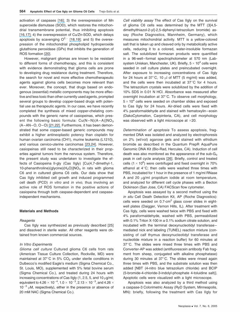
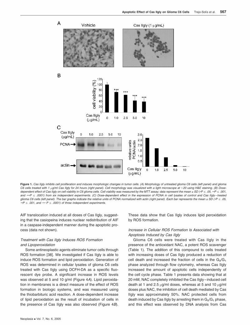
Treatment with Cas IIgly induced growth inhibition and
morphologic changes in a dose-dependent manner. Figure 1A
shows morphologic alterations in glioma C6 cells treated with
1 mg/ml Cas IIgly for 24 hours; such cells contracted, became
rounded, and detached from the culture dishes. In contrast,
nontreated glioma C6 cells remained morphologically un-
changed. Figure 1B depicts the dose-dependent effect of
Cas IIgly on glioma C6 cell viability; most cells died after
treatment with 5 and 10 mg/ml Cas IIgly for 24 hours. In addi-
tion, the Western blot analysis revealed that the levels of
PCNA were significantly decreased in cells treated with
higher concentrations of Cas IIgly (Figure 1C). These results
show that Cas IIgly has an antineoplastic effect on glioma C6
cells through the inhibition of cell proliferation, in a mecha-
nism apparently involving the induction of apoptosis.
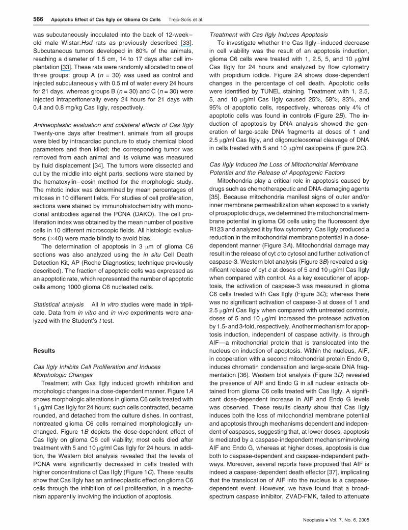
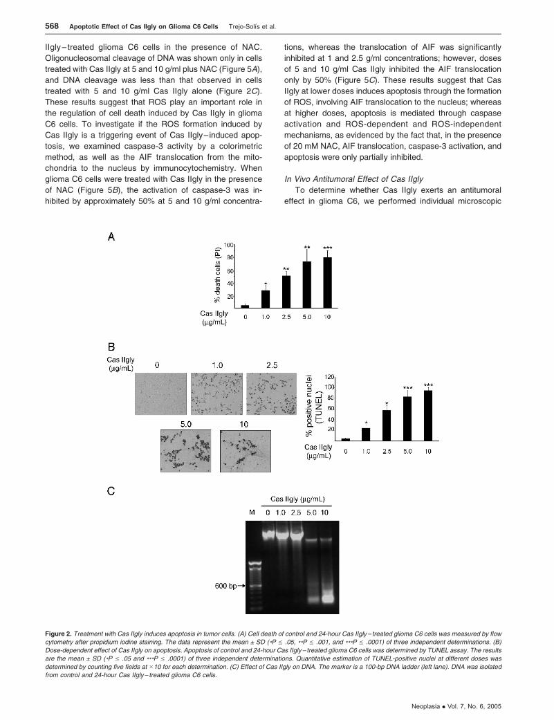
Treatment with Cas IIgly Induces Apoptosis
To investigate whether the Cas IIgly– induced decrease
in cell viability was the result of an apoptosis induction,
glioma C6 cells were treated with 1, 2.5, 5, and 10 mg/ml
Cas IIgly for 24 hours and analyzed by flow cytometry
with propidium iodide. Figure 2A shows dose-dependent
changes in the percentage of cell death. Apoptotic cells
were identified by TUNEL staining. Treatment with 1, 2.5,
5, and 10 mg/ml Cas IIgly caused 25%, 58%, 83%, and
95% of apoptotic cells, respectively, whereas only 4% of
apoptotic cells was found in controls (Figure 2B). The in-
duction of apoptosis by DNA analysis showed the gen-
eration of large-scale DNA fragments at doses of 1 and
2.5 mg/ml Cas IIgly, and oligonucleosomal cleavage of DNA
in cells treated with 5 and 10 mg/ml casiopeina (Figure 2C).
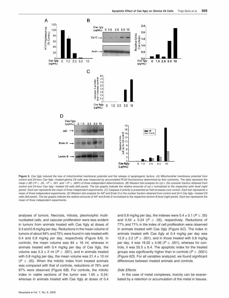
Cas IIgly Induced the Loss of Mitochondrial Membrane
Potential and the Release of Apoptogenic Factors
Mitochondria play a critical role in apoptosis caused by
drugs such as chemotherapeutic and DNA-damaging agents
[35]. Because mitochondria manifest signs of outer and/or
inner membrane permeabilization when exposed to a variety
of proapoptotic drugs, we determined the mitochondrial mem-
brane potential in glioma C6 cells using the fluorescent dye
R123 and analyzed it by flow cytometry. Cas IIgly produced a
reduction in the mitochondrial membrane potential in a dose-
dependent manner (Figure 3A). Mitochondrial damage may
result in the release of cyt c to cytosol and further activation of
caspase-3. Western blot analysis (Figure 3B) revealed a sig-
nificant release of cyt c at doses of 5 and 10 mg/ml Cas IIgly
when compared with control. As a key executioner of apop-
tosis, the activation of caspase-3 was measured in glioma
C6 cells treated with Cas IIgly (Figure 3C); whereas there
was no significant activation of caspase-3 at doses of 1 and
2.5 mg/ml Cas IIgly when compared with untreated controls,
doses of 5 and 10 mg/ml increased the protease activation
by 1.5- and 3-fold, respectively. Another mechanism for apop-
tosis induction, independent of caspase activity, is through
AIF—a mitochondrial protein that is translocated into the
nucleus on induction of apoptosis. Within the nucleus, AIF,
in cooperation with a second mitochondrial protein Endo G,
induces chromatin condensation and large-scale DNA frag-
mentation [36]. Western blot analysis (Figure 3D) revealed
the presence of AIF and Endo G in all nuclear extracts ob-
tained from glioma C6 cells treated with Cas IIgly. A signifi-
cant dose-dependent increase in AIF and Endo G levels
was observed. These results clearly show that Cas IIgly
induces both the loss of mitochondrial membrane potential
and apoptosis through mechanisms dependent and indepen-
dent of caspases, suggesting that, at lower doses, apoptosis
is mediated by a caspase-independent mechanisminvolving
AIF and Endo G, whereas at higher doses, apoptosis is due
both to caspase-dependent and caspase-independent path-
ways. Moreover, several reports have proposed that AIF is
indeed a caspase-dependent death effector [37], implicating
that the translocation of AIF into the nucleus is a caspase-
dependent event. However, we have found that a broad-
spectrum caspase inhibitor, ZVAD-FMK, failed to attenuate
566 Apoptotic Effect of Cas IIgly on Glioma C6 Cells Trejo-Solıs et al.
Neoplasia . Vol. 7, No. 6, 2005
AIF translocation induced at all doses of Cas IIgly, suggest-
ing that the casiopeina induces nuclear redistribution of AIF
in a caspase-independent manner during the apoptotic pro-
cess (data not shown).
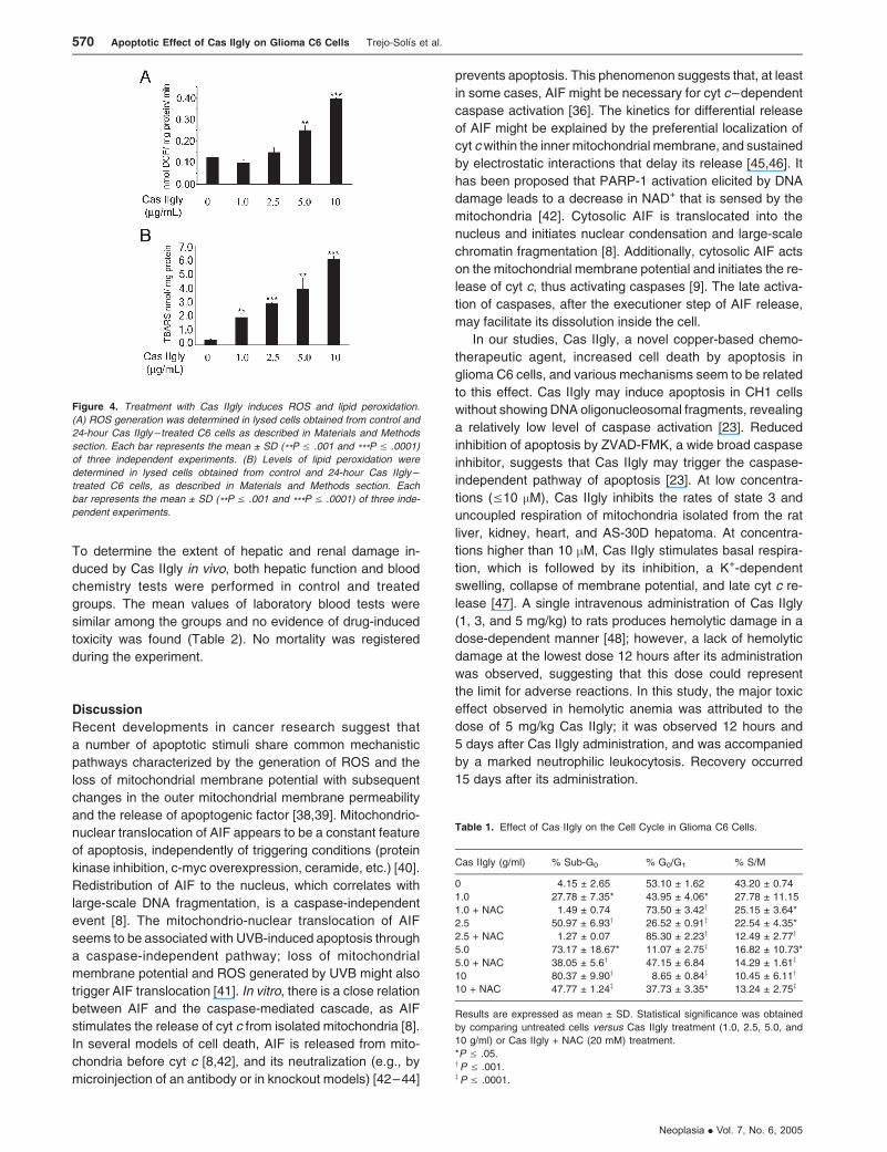
Treatment with Cas IIgly Induces ROS Formation
and Lipoperoxidation
Some antineoplastic agents eliminate tumor cells through
ROS formation [38]. We investigated if Cas IIgly is able to
induce ROS formation and lipid peroxidation. Generation of
ROS was determined in cellular lysates of glioma C6 cells
treated with Cas IIgly using DCFH-DA as a specific fluo-
rescent dye probe. A significant increase in ROS levels
was observed at 5 and 10 g/ml (Figure 4A). Lipid peroxida-
tion in membranes is a direct measure of the effect of ROS
formation in biologic systems, and was measured using
the thiobarbituric acid reaction. A dose-dependent increase
of lipid peroxidation as the result of incubation of cells in
the presence of Cas IIgly was also observed (Figure 4B).
These data show that Cas IIgly induces lipid peroxidation
by ROS formation.
Increase in Cellular ROS Formation Is Associated with
Apoptosis Induced by Cas IIgly
Glioma C6 cells were treated with Cas IIgly in the
presence of the antioxidant NAC, a potent ROS scavenger
(Table 1). The addition of this compound to cells treated
with increasing doses of Cas IIgly produced a reduction of
cell death and increased the fraction of cells in the G0/G1
phase analyzed through flow cytometry, whereas Cas IIgly
increased the amount of apoptotic cells independently of
the cell cycle phase. Table 1 presents data showing that at
20 mM, NAC completely inhibited the Cas IIgly– induced cell
death at 1 and 2.5 mg/ml doses, whereas at 5 and 10 mg/ml
doses plus NAC, the inhibition of cell death mediated by Cas
IIgly was approximately 50%. NAC protected cells from
death induced by Cas IIgly by arresting them in G0/G1 phase,
and this effect was observed by DNA analysis from Cas
Figure 1. Cas IIgly inhibits cell proliferation and induces morphologic changes in tumor cells. (A) Morphology of untreated glioma C6 cells (left panel) and glioma
C6 cells treated with 1 �g/ml Cas IIgly for 24 hours (right panel). Cell morphology was visualized with a light microscope at �20 using H&E staining. (B) Dose-
dependent effect of Cas IIgly on cell viability in C6 glioma cells. Cell viability was measured by the MTT assay; data represent the mean ± SD (*P V .05, **P V .001,
and ***P V .0001) from six independent experiments. (C) Dose-dependent effect in the expression of PCNA in cell lysates of control and Cas IIgly – treated
glioma C6 cells (left panel). The bar graphs indicate the relative units of PCNA normalized with actin (right panel). Each bar represents the mean ± SD (*P V .05,
**P V .001, and *** P V .0001) of three independent experiments.
Apoptotic Effect of Cas IIgly on Glioma C6 Cells Trejo-Solıs et al. 567
Neoplasia . Vol. 7, No. 6, 2005
IIgly– treated glioma C6 cells in the presence of NAC.
Oligonucleosomal cleavage of DNA was shown only in cells
treated with Cas IIgly at 5 and 10 g/ml plus NAC (Figure 5A),
and DNA cleavage was less than that observed in cells
treated with 5 and 10 g/ml Cas IIgly alone (Figure 2C).
These results suggest that ROS play an important role in
the regulation of cell death induced by Cas IIgly in glioma
C6 cells. To investigate if the ROS formation induced by
Cas IIgly is a triggering event of Cas IIgly– induced apop-
tosis, we examined caspase-3 activity by a colorimetric
method, as well as the AIF translocation from the mito-
chondria to the nucleus by immunocytochemistry. When
glioma C6 cells were treated with Cas IIgly in the presence
of NAC (Figure 5B), the activation of caspase-3 was in-
hibited by approximately 50% at 5 and 10 g/ml concentra-
tions, whereas the translocation of AIF was significantly
inhibited at 1 and 2.5 g/ml concentrations; however, doses
of 5 and 10 g/ml Cas IIgly inhibited the AIF translocation
only by 50% (Figure 5C). These results suggest that Cas
IIgly at lower doses induces apoptosis through the formation
of ROS, involving AIF translocation to the nucleus; whereas
at higher doses, apoptosis is mediated through caspase
activation and ROS-dependent and ROS-independent
mechanisms, as evidenced by the fact that, in the presence
of 20 mM NAC, AIF translocation, caspase-3 activation, and
apoptosis were only partially inhibited.
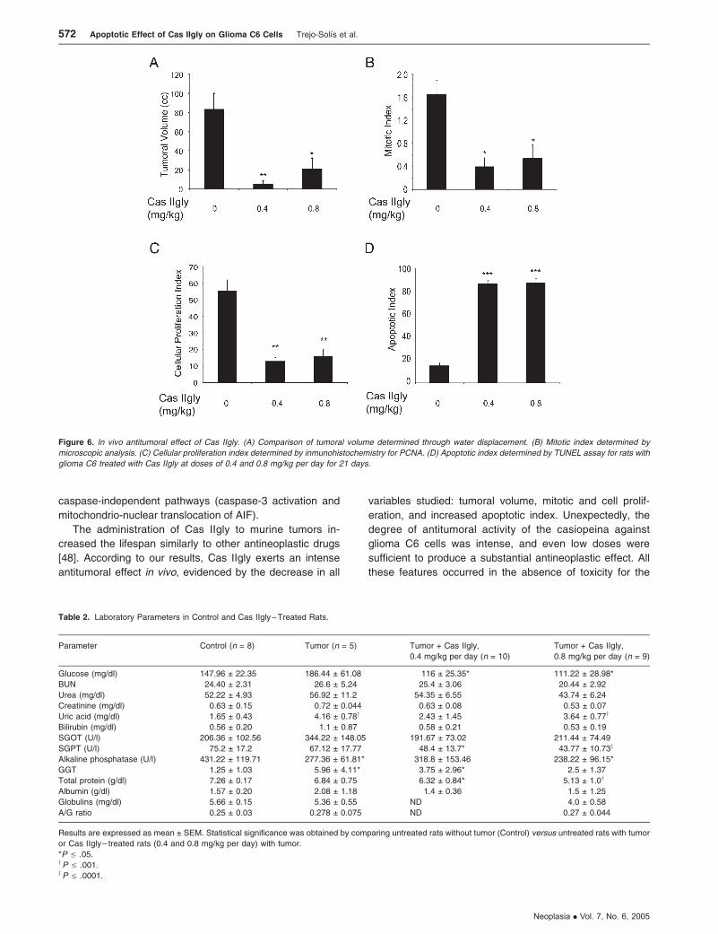
In Vivo Antitumoral Effect of Cas IIgly
To determine whether Cas IIgly exerts an antitumoral
effect in glioma C6, we performed individual microscopic
Figure 2. Treatment with Cas IIgly induces apoptosis in tumor cells. (A) Cell death of control and 24-hour Cas IIgly– treated glioma C6 cells was measured by flow
cytometry after propidium iodine staining. The data represent the mean ± SD (*P V .05, **P V .001, and ***P V .0001) of three independent determinations. (B)
Dose-dependent effect of Cas IIgly on apoptosis. Apoptosis of control and 24-hour Cas IIgly – treated glioma C6 cells was determined by TUNEL assay. The results
are the mean ± SD (*P V .05 and ***P V .0001) of three independent determinations. Quantitative estimation of TUNEL-positive nuclei at different doses was
determined by counting five fields at �10 for each determination. (C) Effect of Cas IIgly on DNA. The marker is a 100-bp DNA ladder (left lane). DNA was isolated
from control and 24-hour Cas IIgly– treated glioma C6 cells.
568 Apoptotic Effect of Cas IIgly on Glioma C6 Cells Trejo-Solıs et al.
Neoplasia . Vol. 7, No. 6, 2005
analyses of tumors. Necrosis, mitosis, pleomorphic multi-
nucleated cells, and vascular proliferation were less evident
in tumors from animals treated with Cas IIgly at doses of
0.4 and 0.8 mg/kg per day. Reductions in the mean volume of
tumors of about 94% and 75% were found in rats treated with
0.4 and 0.8 mg/kg per day, respectively (Figure 6A). In
controls, the mean volume was 84 ± 16 ml; whereas in
animals treated with 0.4 mg/kg per day of Cas IIgly, the
volume was 5.3 ± 2 ml (P V .001), and in animals treated
with 0.8 mg/kg per day, the mean volume was 21.4 ± 10 ml
(P V .05). When the mitotic index from treated animals
was compared with that of controls, reductions of 76% and
67% were observed (Figure 6B). For controls, the mitotic
index in viable sections of the tumor was 1.65 ± 0.24;
whereas in animals treated with Cas IIgly at doses of 0.4
and 0.8 mg/kg per day, the indexes were 0.4 ± 0.1 (P V .05)
and 0.54 ± 0.24 (P V .05), respectively. Reductions of
77% and 71% in the index of cell proliferation were observed
in animals treated with Cas IIgly (Figure 6C). The index in
animals treated with Cas IIgly at 0.4 mg/kg per day was
12.9 ± 2.2 (P V .001), and in those treated with 0.8 mg/kg
per day, it was 16.02 ± 4.06 (P V .001); whereas for con-
trols, it was 55.5 ± 6.4. The apoptotic index for the treated
groups was significantly higher than in controls (P V .0001)
(Figure 6D). For all variables analyzed, we found significant
differences between treated animals and controls.
Side Effects
In the case of metal complexes, toxicity can be exacer-
bated by a retention or accumulation of the metal in tissues.
Figure 3. Cas IIgly induced the loss of mitochondrial membrane potential and the release of apoptogenic factors. (A) Mitochondrial membrane potential from
control and 24-hour Cas IIgly – treated glioma C6 cells was measured by accumulated R123 fluorescence determined by flow cytometry. The data represent the
mean ± SD (*P V .05, **P V .001, and ***P V .0001) of three independent determinations. (B) Western blot analysis for cyt c, the cytosolic fraction obtained from
control and 24-hour Cas IIgly– treated C6 cells (left panel). The bar graphs indicate the relative amounts of cyt c normalized to the respective actin level (right
panel). Each bar represents the mean of three independent experiments. (C) Caspase-3 activity is presented as fold increases over control. Each bar represents a
mean of three independent experiments. (D) Western blot analysis for AIF and Endo G in the nuclear fraction obtained from control and 24 h Cas IIgly – treated C6
cells (left panel). The bar graphs indicate the relative amounts of AIF and Endo G normalized to the respective laminin B level (right panel). Each bar represents the
mean of three independent experiments.
Apoptotic Effect of Cas IIgly on Glioma C6 Cells Trejo-Solıs et al. 569
Neoplasia . Vol. 7, No. 6, 2005
To determine the extent of hepatic and renal damage in-
duced by Cas IIgly in vivo, both hepatic function and blood
chemistry tests were performed in control and treated
groups. The mean values of laboratory blood tests were
similar among the groups and no evidence of drug-induced
toxicity was found (Table 2). No mortality was registered
during the experiment.
Discussion
Recent developments in cancer research suggest that
a number of apoptotic stimuli share common mechanistic
pathways characterized by the generation of ROS and the
loss of mitochondrial membrane potential with subsequent
changes in the outer mitochondrial membrane permeability
and the release of apoptogenic factor [38,39]. Mitochondrio-
nuclear translocation of AIF appears to be a constant feature
of apoptosis, independently of triggering conditions (protein
kinase inhibition, c-myc overexpression, ceramide, etc.) [40].
Redistribution of AIF to the nucleus, which correlates with
large-scale DNA fragmentation, is a caspase-independent
event [8]. The mitochondrio-nuclear translocation of AIF
seems to be associated with UVB-induced apoptosis through
a caspase-independent pathway; loss of mitochondrial
membrane potential and ROS generated by UVB might also
trigger AIF translocation [41]. In vitro, there is a close relation
between AIF and the caspase-mediated cascade, as AIF
stimulates the release of cyt c from isolated mitochondria [8].
In several models of cell death, AIF is released from mito-
chondria before cyt c [8,42], and its neutralization (e.g., by
microinjection of an antibody or in knockout models) [42–44]
prevents apoptosis. This phenomenon suggests that, at least
in some cases, AIF might be necessary for cyt c–dependent
caspase activation [36]. The kinetics for differential release
of AIF might be explained by the preferential localization of
cyt cwithin the inner mitochondrial membrane, and sustained
by electrostatic interactions that delay its release [45,46]. It
has been proposed that PARP-1 activation elicited by DNA
damage leads to a decrease in NAD+ that is sensed by the
mitochondria [42]. Cytosolic AIF is translocated into the
nucleus and initiates nuclear condensation and large-scale
chromatin fragmentation [8]. Additionally, cytosolic AIF acts
on the mitochondrial membrane potential and initiates the re-
lease of cyt c, thus activating caspases [9]. The late activa-
tion of caspases, after the executioner step of AIF release,
may facilitate its dissolution inside the cell.
In our studies, Cas IIgly, a novel copper-based chemo-
therapeutic agent, increased cell death by apoptosis in
glioma C6 cells, and various mechanisms seem to be related
to this effect. Cas IIgly may induce apoptosis in CH1 cells
without showing DNA oligonucleosomal fragments, revealing
a relatively low level of caspase activation [23]. Reduced
inhibition of apoptosis by ZVAD-FMK, a wide broad caspase
inhibitor, suggests that Cas IIgly may trigger the caspase-
independent pathway of apoptosis [23]. At low concentra-
tions (V10 mM), Cas IIgly inhibits the rates of state 3 and
uncoupled respiration of mitochondria isolated from the rat
liver, kidney, heart, and AS-30D hepatoma. At concentra-
tions higher than 10 mM, Cas IIgly stimulates basal respira-
tion, which is followed by its inhibition, a K+-dependent
swelling, collapse of membrane potential, and late cyt c re-
lease [47]. A single intravenous administration of Cas IIgly
(1, 3, and 5 mg/kg) to rats produces hemolytic damage in a
dose-dependent manner [48]; however, a lack of hemolytic
damage at the lowest dose 12 hours after its administration
was observed, suggesting that this dose could represent
the limit for adverse reactions. In this study, the major toxic
effect observed in hemolytic anemia was attributed to the
dose of 5 mg/kg Cas IIgly; it was observed 12 hours and
5 days after Cas IIgly administration, and was accompanied
by a marked neutrophilic leukocytosis. Recovery occurred
15 days after its administration.
Figure 4. Treatment with Cas IIgly induces ROS and lipid peroxidation.
(A) ROS generation was determined in lysed cells obtained from control and
24-hour Cas IIgly – treated C6 cells as described in Materials and Methods
section. Each bar represents the mean ± SD (**P V .001 and ***P V .0001)
of three independent experiments. (B) Levels of lipid peroxidation were
determined in lysed cells obtained from control and 24-hour Cas IIgly–
treated C6 cells, as described in Materials and Methods section. Each
bar represents the mean ± SD (**P V .001 and ***P V .0001) of three inde-
pendent experiments.
Table 1. Effect of Cas IIgly on the Cell Cycle in Glioma C6 Cells.
Cas IIgly (g/ml) % Sub-G0 % G0/G1 % S/M
0 4.15 ± 2.65 53.10 ± 1.62 43.20 ± 0.74
1.0 27.78 ± 7.35* 43.95 ± 4.06* 27.78 ± 11.15
1.0 + NAC 1.49 ± 0.74 73.50 ± 3.42y 25.15 ± 3.64*
2.5 50.97 ± 6.93y 26.52 ± 0.91z 22.54 ± 4.35*
2.5 + NAC 1.27 ± 0.07 85.30 ± 2.23z 12.49 ± 2.77z
5.0 73.17 ± 18.67* 11.07 ± 2.75z 16.82 ± 10.73*
5.0 + NAC 38.05 ± 5.6y 47.15 ± 6.84 14.29 ± 1.61z
10 80.37 ± 9.90y 8.65 ± 0.84z 10.45 ± 6.11y
10 + NAC 47.77 ± 1.24z 37.73 ± 3.35* 13.24 ± 2.75z
Results are expressed as mean ± SD. Statistical significance was obtained
by comparing untreated cells versus Cas IIgly treatment (1.0, 2.5, 5.0, and
10 g/ml) or Cas IIgly + NAC (20 mM) treatment.
*P V .05.yP V .001.zP V .0001.
570 Apoptotic Effect of Cas IIgly on Glioma C6 Cells Trejo-Solıs et al.
Neoplasia . Vol. 7, No. 6, 2005

Our findings also indicate that Cas IIgly, at doses between
1 and 2.5 g/ml, induces ROS generation, loss of mitochon-
drial membrane potential, AIF and Endo G mitochondrio-
nuclear translocation and apoptosis without cytosolic
release of cyt c, caspase-3 activation, or DNA laddering.
An additional interesting finding is that NAC, a ROS scaven-
ger, blocked the mitochondrio-nuclear translocation of AIF
and apoptosis, suggesting that ROS generated by Cas IIgly
acts as a trigger factor for mitochondrio-nuclear translocation
of AIF only. In contrast, at higher doses of casiopeina (5 and
10 mg/ml), ROS production and loss of mitochondrial mem-
brane potential are larger, and apoptosis occurs with DNA
laddering associated with release of cyt c and caspase-3
activation, as well as mitochondrio-nuclear translocation of
AIF and Endo G. In the latter case, NAC blocks only 50%
of these processes. These results also suggest that ROS
generated by Cas IIgly is one of the triggering events for
apoptosis. The results mentioned above show that low doses
of Cas IIgly induce apoptosis in glioma C6 cells through a
caspase-independent pathway. Under these conditions, the
mitochondrio-nuclear translocation of AIF is associated with
this pathway, and ROS generated may be involved in trig-
gering its translocation, whereas at higher doses, Cas IIgly
induces apoptosis through both caspase-dependent and
Figure 5. Increase in cellular ROS is associated with apoptosis induced by Cas IIgly. (A) Effects of NAC on DNA of 24-hour Cas IIgly– treated cells in the presence
of 20 mM NAC. DNA extraction was performed as described in Materials and Methods section. The marker is a 1-kb extension ladder. (B) Immunocytochemistry of
AIF in control C6 cells, treated with Cas IIgly and Cas IIgly + 20 mM NAC for 24 hours (left panel). The figures shown are representative of at least three different
experiments for each experimental condition. Original magnification, �20. The bar graphs indicate the mean of three independent determinations (left panel).
Quantitative estimation of AIF-positive nuclei at different doses was determined by counting five fields of�20 for each determination. (C) Caspase-3 activity in control
C6 cells, treatedwith Cas IIgly andCas IIgly + 20mMNAC for 24 hours. Caspase-3 activity was determined as described inMaterials andMethods section. Caspase-3
activities were presented as fold increases over control. Each bar represents the mean ± SD (**P V .001 and ***P V .0001) of three independent experiments.
Apoptotic Effect of Cas IIgly on Glioma C6 Cells Trejo-Solıs et al. 571
Neoplasia . Vol. 7, No. 6, 2005
caspase-independent pathways (caspase-3 activation and
mitochondrio-nuclear translocation of AIF).
The administration of Cas IIgly to murine tumors in-
creased the lifespan similarly to other antineoplastic drugs
[48]. According to our results, Cas IIgly exerts an intense
antitumoral effect in vivo, evidenced by the decrease in all
variables studied: tumoral volume, mitotic and cell prolif-
eration, and increased apoptotic index. Unexpectedly, the
degree of antitumoral activity of the casiopeina against
glioma C6 cells was intense, and even low doses were
sufficient to produce a substantial antineoplastic effect. All
these features occurred in the absence of toxicity for the
Figure 6. In vivo antitumoral effect of Cas IIgly. (A) Comparison of tumoral volume determined through water displacement. (B) Mitotic index determined by
microscopic analysis. (C) Cellular proliferation index determined by inmunohistochemistry for PCNA. (D) Apoptotic index determined by TUNEL assay for rats with
glioma C6 treated with Cas IIgly at doses of 0.4 and 0.8 mg/kg per day for 21 days.
Table 2. Laboratory Parameters in Control and Cas IIgly – Treated Rats.
Parameter Control (n = 8) Tumor (n = 5) Tumor + Cas IIgly,
0.4 mg/kg per day (n = 10)
Tumor + Cas IIgly,
0.8 mg/kg per day (n = 9)
Glucose (mg/dl) 147.96 ± 22.35 186.44 ± 61.08 116 ± 25.35* 111.22 ± 28.98*
BUN 24.40 ± 2.31 26.6 ± 5.24 25.4 ± 3.06 20.44 ± 2.92
Urea (mg/dl) 52.22 ± 4.93 56.92 ± 11.2 54.35 ± 6.55 43.74 ± 6.24
Creatinine (mg/dl) 0.63 ± 0.15 0.72 ± 0.044 0.63 ± 0.08 0.53 ± 0.07
Uric acid (mg/dl) 1.65 ± 0.43 4.16 ± 0.78y 2.43 ± 1.45 3.64 ± 0.77y
Bilirubin (mg/dl) 0.56 ± 0.20 1.1 ± 0.87 0.58 ± 0.21 0.53 ± 0.19
SGOT (U/l) 206.36 ± 102.56 344.22 ± 148.05 191.67 ± 73.02 211.44 ± 74.49
SGPT (U/l) 75.2 ± 17.2 67.12 ± 17.77 48.4 ± 13.7* 43.77 ± 10.73z
Alkaline phosphatase (U/l) 431.22 ± 119.71 277.36 ± 61.81* 318.8 ± 153.46 238.22 ± 96.15*
GGT 1.25 ± 1.03 5.96 ± 4.11* 3.75 ± 2.96* 2.5 ± 1.37
Total protein (g/dl) 7.26 ± 0.17 6.84 ± 0.75 6.32 ± 0.84* 5.13 ± 1.0y
Albumin (g/dl) 1.57 ± 0.20 2.08 ± 1.18 1.4 ± 0.36 1.5 ± 1.25
Globulins (mg/dl) 5.66 ± 0.15 5.36 ± 0.55 ND 4.0 ± 0.58
A/G ratio 0.25 ± 0.03 0.278 ± 0.075 ND 0.27 ± 0.044
Results are expressed as mean ± SEM. Statistical significance was obtained by comparing untreated rats without tumor (Control) versus untreated rats with tumor
or Cas IIgly – treated rats (0.4 and 0.8 mg/kg per day) with tumor.
*P V .05.yP V .001.zP V .0001.
572 Apoptotic Effect of Cas IIgly on Glioma C6 Cells Trejo-Solıs et al.
Neoplasia . Vol. 7, No. 6, 2005
hapato-biliary or renal system. Hematic biometry performed
on Wistar:Hsd rats 21 days after treatment with Cas IIgly
(0.4 and 0.8 mg/kg per day, i.p.) showed a dose-dependent
effect. The higher dose produced only a moderate decrease
of hemoglobin (accompanied by light neutrophilic leukocytosis
but no hemolytic anemia), platelet count was normal, and
no histologic abnormalities were seen in the spleen (results
not shown).
Most currently used antineoplastic drugs have limited
efficacy and high toxicity, as they also affect normal cells.
Lipophilic cation drugs are concentrated by cells into mito-
chondria because of the large negative-inside electric mem-
brane potential [49,50]. The higher plasma and mitochondrial
membrane potentials of tumors cells [51,52] may enhance
the selective targeting of Cas IIgly into tumor cells and
mitochondria. AS-30D hepatoma mitochondria also ex-
hibited higher mitochondrial membrane potential values than
did mitochondria from the liver, the organ from which tumor
mitochondria were derived. Indeed, AS-30D and HeLa cells
in culture died within 48 hours of exposure to CAS IIgly [47].
The effect of Cas IIgly (1, 2.5, 5, and 10 mg/ml) after 24 hours
of incubation on the survival of normal fibroblasts was
determined by the MTT assay. At 1 and 2.5 mg/ml doses of
Cas IIgly, the cell viability of normal fibroblasts in culture was
100%; when the dose was increased to 5mg/ml, viability was
85%; and at 10 mg/ml, it was 50%, suggesting that metabolic
effects of Cas IIgly at 1, 2.5, or 5 mg/ml doses are fairly spe-
cific against glioma cells (results not shown).
Our results indicate that Cas IIgly is a promising chemo-
therapeutic option against glial malignant tumors.
Acknowledgements
We gratefully acknowledge the assistance of Martha Arroyo
and Carmen Escalante for performing the blood chemistry
tests. Isabel Perez Montfort corrected the English version of
the manuscript.
References[1] Kerr JF, Winterford CM, and Harmon BV (1994). Apoptosis. Its signifi-
cance in cancer and cancer therapy. Cancer 73, 2013 – 2026.
[2] Huschtscha LI, Bartier WA, Ross CE, and Tattersall MH (1996). Char-
acteristics of cancer cell death after exposure to cytotoxic drugs in vitro.
Br J Cancer 73, 54 – 60.
[3] Green DR and Reed JC (1998). Mitochondria and apoptosis. Science
28, 1309 – 1312.
[4] Bossy-Wetzel E, Newmeyer DD, and Green DR (1998). Mitochondria
cytochrome c release in apoptosis occurs upstream of DEVD-specific
caspase activation and independently of mitochondrial transmembrane
depolarization. EMBO J 17, 37 –49.
[5] Green D and Kroemer G (1998). The central executioners of apoptosis:
caspases or mitochondria? Trends Cell Biol 8, 267 – 271.
[6] Kroemer G, Zamzami N, and Susin SA (1997). Mitochondrial control of
apoptosis. Immunol Today 18, 44 –51.
[7] Sakahira H, Enari M, and Nagata S (1998). Cleavage of CAD inhibitor in
CAD activation and degradation during apoptosis. Nature 391, 96 –99.
[8] Susin SA, Lorenzo HK, Zamzami N, Marzo I, Snow BE, Brothers GM,
et al. (1999). Molecular characterization of mitochondrial apoptosis-
inducing factor. Nature 397, 441 –446.
[9] Susin SA, Daugas E, Ravagnan L, Samejima K, Zamzami N, Loeffler N,
et al. (2000). Two distinct pathways leading to nuclear apoptosis. J Exp
Med 192, 571 – 580.
[10] Daugas E, Nochy D, Ravagnan L, Loeffler N, Susin SA, Zamzami N,
et al. (2000). Apoptosis-inducing factor (AIF): a ubiquitous mitochon-
drial oxidoreductase involved in apoptosis. FEBS Lett 476, 118 –123.
[11] Winterbourn CC (1995). Toxicity of iron and hydrogen peroxide: the
Fenton reaction. Toxicol Lett 82/83, 969 – 974.
[12] Jimenez M and Velez PC (2004). Transition metal – induced apoptosis
in lymphocytes via hydroxyl radical generation, mitochondria dysfunc-
tion, and caspase-3 activation: an in vitro model for neurodegeneration.
Arch Med Res 35, 185 –193.
[13] Desagher S and Martinou JC (2000). Mitochondria as the central con-
trol point of apoptosis. Trends Cell Biol 10, 369 – 377.
[14] France-Lanord V, Brugg B, Michel PP, Agid Y, and Ruberg M (1997).
Mitochondrial free radical signal in ceramide-dependent apoptosis: a
putative mechanism for neuronal death in Parkinson’s disease. J Neu-
rochem 69, 1612 – 1621.
[15] Kane DJ, Sarafian TA, Anton R, Hahn H, Gralla EB, Valentine JS, et al.
(1993). Bcl-2 inhibition of neural death decreased generation of reactive
oxygen species. Science 262, 1274 –1277.
[16] Greenlund LJ, Deckwerth TL, and Johnson EM Jr (1995). Superoxide
dismutase delays neuronal apoptosis: a role for reactive oxygen spe-
cies in programmed neuronal death. Neuron 14, 303 –315.
[17] Majima HJ, Oberley TD, Furukawa K, Matsson MP, Yen HC, Szweda LI,
et al. (1998). Prevention of mitochondrial injury by manganese super-
oxide dismutase reveals a primary mechanism for alkaline-induced cell
death. J Biol Chem 273, 8217 –8224.
[18] Kiningham KK, Oberley TD, Lin S, Mattingly CA, and St Clair DK (1999).
Overexpression of manganese superoxide dismutase protects against
mitochondrial-initiated poly(ADP-ribose) polymerase – mediated cell
death. FASEB J 13, 1601 – 1610.
[19] Fujimura M, Morita-Fujimura Y, Noshita N, Sugawara T, Kawase M, and
Chan PH (2000). The cytosolic antioxidant copper/zinc-superoxide dis-
mutase prevents the early release of mitochondrial cytochrome c in is-
chemic brain after transient focal cerebral ischemia in mice. J Neurosci
20, 2817 – 2824.
[20] Nomura K, Imai H, Koumura T, Arai M, and Nakagawa Y (1999). Mito-
chondrial phospholipid hydroperoxide glutathione peroxidase sup-
presses apoptosis mediated by a mitochondrial death pathway. J Biol
Chem 274, 29294 –29302.
[21] Ruiz-Ramırez L, De La Rosa ME, Gracia-Mora I, Mendoza A, Perez G,
Ferrer-Sueta G, et al. (1995). Casiopeinas, metal-based drugs a new
class of antineoplastic and genotoxic compounds. J Inorg Biochem
207, 2 –3.
[22] Ruiz-Ramırez L, Gracia-Mora I, De La Rosa ME, Sumano H, Gomez C,
Pimentel E, et al. (1993). Cytostatic, mutagenic, antineoplastic activities
and preliminary toxicity of copper (II) drugs: casiopeinas I, II, III. J Inorg
Biochem 406, 1– 2.
[23] De Vizcaya-Ruiz A, Rivero-Muller A, Ruiz-Ramirez L, Kass GE,
Kelland LR, Orr RM, et al. (2000). Induction of apoptosis by a novel
copper-based anticancer compound, casiopeina II, in L1210 murine
leukaemia and CH1 human ovarian carcinoma cells. Toxicol In Vitro
14, 1 –5.
[24] Gracia-Mora I, Ruiz-Ramırez L, Gomez-Ruiz C, Tinoco-Mendez M,
Marquez-Quinones A, Romero de Lira L, et al. (2001). Knight’s move in
the periodic table, from copper to platinum, novel antitumor mixed chelate
copper compounds, casiopeinas, evaluated by an in vitro human and
murine cancer cell line panel. Met-Based Drug 8, 19– 29.
[25] Ruız-Azuara L (1993). US Patent, Ap. 21 (1992), no. 5,107,005; US
Patent, Re 35,458, February 18 (1997); US, Patent November 19
(1996), no. 5,576,326, 407543 SECOFI.
[26] Darzynkiewicz Z, Bedner E, and Smolewski P (2001). Flow cytometry in
analysis of cell cycle and apoptosis. Semin Hematol 38, 179 –193.
[27] Chen XC, Zhu YG, Chen LM, Fang F, Zhou YC, and Zhao CH (1998).
Nitric oxide induced PC12 cells apoptosis and the protective effect of
gingenoside Rg1. Chin Pharmacol Bull 18, 516 –519.
[28] Scott JA, Homcy CJ, Khaw BA, and Rabito CA (1988). Quantitation of
intracellular oxidation in a renal epithelial cell line. Free Radic Biol Med
4, 79 –83.
[29] LeBel CA, Ischiropoulous H, and Bondy SC (1992). Evaluation of the
probe 2V,7V-dichlorofluorescein as an indicator of reactive species for-
mation and oxidative stress. Chem Res Toxicol 5, 227 – 231.
[30] Buege JA and Aust SD (1978). Microsomal lipid peroxidation. Methods
Enzymol 52, 302 –310.
[31] Hernandez-Munoz R, Glender W, Dıaz-Munoz M, Garcıa-Sainz JA,
and Chagoya de Sanchez V (1984). Effects of adenosine on liver cell
damage induced by carbon tetrachloride. Biochem Pharmacol 33,
2599 – 2604.
[32] Smirnova IV, Douglas C, Bittel Rudravajhala R, Huimin J, and Glen KA
Apoptotic Effect of Cas IIgly on Glioma C6 Cells Trejo-Solıs et al. 573
Neoplasia . Vol. 7, No. 6, 2005
(2000). Zinc and cadmium can promote rapid nuclear translocation of
metal response element-binding transcription factor-1. J Biol Chem
275, 9377 – 9384.
[33] Guevara P and Sotelo J (1999). C6 rat glioma grown into the peritoneal
cavity, a large source of tumoral cells for subcutaneous transplant of
glioma. J Neuro-Oncol 44, 91– 92.
[34] Tamayko M and Reynolds P (1989). Determination of subcutaneous tumor
size in athymic (nude) mice. Cancer Chemother Pharmacol 24, 148 –154.
[35] Van GM, Festjens N, Van LG, Saelens X, and Vandenabeele P (2003).
Mitochondrial intermembrane proteins in cell death. Biochem Biophys
Res Commun 304, 487 – 497.
[36] Cande C, Cecconi F, Dessen F, and Kroemer G (2002). Apoptosis-
inducing factor (AIF): key to the conserved caspase-independent path-
ways of cell death? J Cell Sci 115, 4727 – 4734.
[37] Damien A, Brigitte G, Mariusz K, Juanita CS, Francesco C, and Richard
JY (2003). Mitochondrial release of AIF and Endo G requires caspase
activation downstream of Bax/Bak –mediated permeabilization. EMBO
J 22, 4385 –4399.
[38] Siro S, Minoru T, Kazuo U, and Masaya I (1998). Requirement of
caspase-3 (-like) protease-mediated hydrogen peroxide production for
apoptosis induced by various anticancer drugs. J Biol Chem 273,
26900 – 26907.
[39] Decaudin D, Marzo I, Brenner C, and Kroemer G (1998). Mitochondria
in chemotherapy-induced apoptosis: a prospective novel target of can-
cer therapy. Int J Oncol 12, 141 – 152.
[40] Daugas E, Susin SA, Zamzami N, Ferri KF, Irinopoulou T, Larochette N,
Prevost MC, Leber B, Andrews D, Penninger J, et al. (2000). Mitochondrio-
nuclear translocation of AIF in apoptosis and necrosis. FASEB J 14,
729 – 739.
[41] Murahashi H, Azuma H, Zamzami N, Furuya KJ, Ikebuchi K, Yamaguchi M,
et al. (2003). Possible contribution of apoptosis-inducing factor (AIF) and
reactive oxygen species (ROS) to UVB-induced caspase-independent cell
death in the T cell line Jurkat. J Leukoc Biol 73, 399 – 406.
[42] Yu SW, Wang H, Poitras MF, Coombs C, Bowers WJ, Federoff HJ,
et al. (2002). Mediation of poly(ADP-ribose) polymerase-1 –dependent
cell death by apoptosis-inducing factor. Science 297, 259 – 263.
[43] Ferri KF, Jacotot E, Blanco J, Este JA, Zamzami N, Susin SA, et al.
(2000). Apoptosis control in syncytia induced by the HIV-envelope
glycoprotein complex. Role of mitochondria and caspases. J Exp Med
192, 1081 – 1092.
[44] Joza N, Susin SA, Daugas E, Stanford WL, Cho SK, Li CY, et al.
(2001). Essential role of the mitochondrial apoptosis inducing factor
in programmed cell death. Nature 410, 549 –554.
[45] Salamon Z and Tolli G (1997). Interaction of horse heart cytochrome c
with lipid bilayer membranes: effects on redox potentials. J Bioenerg
Biomembr 29, 211 – 221.
[46] Jutila A, Rytomaa M, and Kinnunen PK (1998). Detachment of cyto-
chrome c by cationic drugs from membranes containing acidic phos-
pholipids: comparison of lidocaine, propranolol, and gentamycin. Mol
Pharmacol 54, 722 – 732.
[47] Marin-Hernandez A, Gracia-Mora I, Ruiz-Ramirez L, and Moreno-
Sanchez R (2003). Toxic effects of copper-based antineoplastic drugs
(Casiopeinas) on mitochondrial functions. Biochem Pharmacol 65,
1979 – 1989.
[48] Gracia-Mora I, Bravo-Gomez ME, Ruiz-Ramırez L, Tinoco-Mendez M,
and Huerta L (2004). New antineoplastic in vitro and in vivo screening
of mixed chelate copper(II) coordination compounds (casiopeinas) in
several tumoral models. J Bioorg Med Chem (in press).
[49] Vizcaya-Ruız A, Rivero-Muller A, Ruız-Ramirez L, Howarth JA, and
Dobrota M (2003). Hematotoxicity response in rats by the Koper-based
anticancer agent: casiopeina II. Toxicology 194, 103 –113.
[50] Nadakavukaren KK, Nadakavukaren JJ, and Chen LB (1985). In-
creased rhodamine 123 uptake by carcinoma cells. Cancer Res 45,
6093 – 6099.
[51] Koya K, Li Y, Wang H, Ukai T, Tatsuta N, Kawakami M, et al. (1996).
MKT-077, a novel rhodacyanine dye in clinical trials, exhibits anticarci-
noma activity in preclinical studies based on selective mitochondrial
accumulation. Cancer Res 56, 538 – 543.
[52] Davis S, Weiss MJ, Wong JR, Lampidis TJ, and Chen LB (1985). Mito-
chondria and plasma membrane potentials cause unusual accumulation
and retention of rhodamine 123 by human breast adenocarcinoma –
derived MCF-7 cells. J Biol Chem 260, 13844 – 13850.
574 Apoptotic Effect of Cas IIgly on Glioma C6 Cells Trejo-Solıs et al.
Neoplasia . Vol. 7, No. 6, 2005