Caspase, cathepsin-, and PERK-dependent regulation of MDA7/IL24-induced cell killing in primary...
29
Caspase-, cathepsin-, and PERK-dependent regulation of MDA-7/ IL-24-induced cell killing in primary human glioma cells Adly Yacoub 1 , Margaret A. Park 1 , Pankaj Gupta 6 , Mohammed Rahmani 2 , Guo Zhang 1 , Hossein Hamed 1 , David Hanna 1 , Devanand Sarkar 4,6 , Irina V. Lebedeva 6 , Luni Emdad 4,5 , Moira Sauane 6 , Nicollaq Vozhilla 6 , Sarah Spiegel 1 , Costas Koumenis 8 , Martin Graf 3 , David T. Curiel 7 , Steven Grant 1,2 , Paul B. Fisher 4,5,6 , and Paul Dent 1 1 Department of Biochemistry, Virginia Commonwealth University, Richmond, Virginia 2 Department of Medicine, Virginia Commonwealth University, Richmond, Virginia 3 Department of Neurosurgery, Virginia Commonwealth University, Richmond, Virginia 4 Department of Pathology, Herbert Irving Comprehensive Cancer Center, Columbia University Medical Center, College of Physicians and Surgeons, New York, New York 5 Department of Neurosurgery, Herbert Irving Comprehensive Cancer Center, Columbia University Medical Center, College of Physicians and Surgeons, New York, New York 6 Department of Urology, Herbert Irving Comprehensive Cancer Center, Columbia University Medical Center, College of Physicians and Surgeons, New York, New York 7 Division of Human Gene Therapy, Departments of Medicine, Pathology and Surgery, and the Gene Therapy Center, University of Alabama at Birmingham, Birmingham, Alabama 8 Department of Radiation Oncology, University of Pennsylvania School of Medicine, Philadelphia, Pennsylvania Abstract Melanoma differentiation-associated gene-7/interleukin-24 (mda-7/IL-24) is a novel cytokine displaying selective apoptosis-inducing activity in transformed cells without harming normal cells. The present studies focused on defining the mechanism(s) by which a GST-MDA-7 fusion protein inhibits cell survival of primary human glioma cells in vitro. GST-MDA-7 killed glioma cells with diverse genetic characteristics that correlated with inactivation of ERK1/2 and activation of JNK1-3. Activation of JNK1-3 was dependent on protein kinase R–like endoplasmic reticulum kinase (PERK), and GST-MDA-7 lethality was suppressed in PERK−/− cells. JNK1-3 signaling activated BAX, whereas inhibition of JNK1-3, deletion of BAX, or expression of dominant- negative caspase-9 suppressed lethality. GST-MDA-7 also promoted a PERK-, JNK-, and cathepsin B–dependent cleavage of BID; loss of BID function promoted survival. GST-MDA-7 suppressed BAD and BIM phosphorylation and heat shock protein 70 (HSP70) expression. GST- MDA-7 caused PERK-dependent vacuolization of LC3-expressing endosomes whose formation was suppressed by incubation with 3-methylade-nine, expression of HSP70 or BiP/GRP78, or knockdown of ATG5 or Beclin-1 expression but not by inhibition of the JNK1-3 pathway. Knockdown of ATG5 or Beclin-1 expression or overexpression of HSP70 reduced GST-MDA-7 Copyright © 2008 American Association for Cancer Research. Requests for reprints: Paul Dent, Department of Biochemistry, Virginia Commonwealth University, 401 College Street, Massey Cancer Center, Box 980035, Richmond, VA 23298-0035. Phone: 804-628-0861; Fax: 804-827-1309. [email protected]. Note: P. Dent is The Universal, Inc. Professor in Signal Transduction Research. P.B. Fisher is The Michael and Stella Chernow Urological Cancer Research Scientist and a SWCRF Investigator. NIH Public Access Author Manuscript Mol Cancer Ther. Author manuscript; available in PMC 2011 October 30. Published in final edited form as: Mol Cancer Ther. 2008 February ; 7(2): 297–313. doi:10.1158/1535-7163.MCT-07-2166. NIH-PA Author Manuscript NIH-PA Author Manuscript NIH-PA Author Manuscript
-
Upload
independent -
Category
Documents
-
view
0 -
download
0
Transcript of Caspase, cathepsin-, and PERK-dependent regulation of MDA7/IL24-induced cell killing in primary...
Caspase-, cathepsin-, and PERK-dependent regulation of MDA-7/IL-24-induced cell killing in primary human glioma cells
Adly Yacoub1, Margaret A. Park1, Pankaj Gupta6, Mohammed Rahmani2, Guo Zhang1,Hossein Hamed1, David Hanna1, Devanand Sarkar4,6, Irina V. Lebedeva6, Luni Emdad4,5,Moira Sauane6, Nicollaq Vozhilla6, Sarah Spiegel1, Costas Koumenis8, Martin Graf3, DavidT. Curiel7, Steven Grant1,2, Paul B. Fisher4,5,6, and Paul Dent11Department of Biochemistry, Virginia Commonwealth University, Richmond, Virginia2Department of Medicine, Virginia Commonwealth University, Richmond, Virginia3Department of Neurosurgery, Virginia Commonwealth University, Richmond, Virginia4Department of Pathology, Herbert Irving Comprehensive Cancer Center, Columbia UniversityMedical Center, College of Physicians and Surgeons, New York, New York5Department of Neurosurgery, Herbert Irving Comprehensive Cancer Center, Columbia UniversityMedical Center, College of Physicians and Surgeons, New York, New York6Department of Urology, Herbert Irving Comprehensive Cancer Center, Columbia UniversityMedical Center, College of Physicians and Surgeons, New York, New York7Division of Human Gene Therapy, Departments of Medicine, Pathology and Surgery, and theGene Therapy Center, University of Alabama at Birmingham, Birmingham, Alabama8Department of Radiation Oncology, University of Pennsylvania School of Medicine, Philadelphia,Pennsylvania
AbstractMelanoma differentiation-associated gene-7/interleukin-24 (mda-7/IL-24) is a novel cytokinedisplaying selective apoptosis-inducing activity in transformed cells without harming normal cells.The present studies focused on defining the mechanism(s) by which a GST-MDA-7 fusion proteininhibits cell survival of primary human glioma cells in vitro. GST-MDA-7 killed glioma cells withdiverse genetic characteristics that correlated with inactivation of ERK1/2 and activation ofJNK1-3. Activation of JNK1-3 was dependent on protein kinase R–like endoplasmic reticulumkinase (PERK), and GST-MDA-7 lethality was suppressed in PERK−/− cells. JNK1-3 signalingactivated BAX, whereas inhibition of JNK1-3, deletion of BAX, or expression of dominant-negative caspase-9 suppressed lethality. GST-MDA-7 also promoted a PERK-, JNK-, andcathepsin B–dependent cleavage of BID; loss of BID function promoted survival. GST-MDA-7suppressed BAD and BIM phosphorylation and heat shock protein 70 (HSP70) expression. GST-MDA-7 caused PERK-dependent vacuolization of LC3-expressing endosomes whose formationwas suppressed by incubation with 3-methylade-nine, expression of HSP70 or BiP/GRP78, orknockdown of ATG5 or Beclin-1 expression but not by inhibition of the JNK1-3 pathway.Knockdown of ATG5 or Beclin-1 expression or overexpression of HSP70 reduced GST-MDA-7
Copyright © 2008 American Association for Cancer Research.Requests for reprints: Paul Dent, Department of Biochemistry, Virginia Commonwealth University, 401 College Street, MasseyCancer Center, Box 980035, Richmond, VA 23298-0035. Phone: 804-628-0861; Fax: 804-827-1309. [email protected]: P. Dent is The Universal, Inc. Professor in Signal Transduction Research. P.B. Fisher is The Michael and Stella ChernowUrological Cancer Research Scientist and a SWCRF Investigator.
NIH Public AccessAuthor ManuscriptMol Cancer Ther. Author manuscript; available in PMC 2011 October 30.
Published in final edited form as:Mol Cancer Ther. 2008 February ; 7(2): 297–313. doi:10.1158/1535-7163.MCT-07-2166.
NIH
-PA Author Manuscript
NIH
-PA Author Manuscript
NIH
-PA Author Manuscript
lethality. Our data show that GST-MDA-7 induces an endoplasmic reticulum stress response thatis causal in the activation of multiple proapoptotic pathways, which converge on themitochondrion and highlight the complexity of signaling pathways altered by mda-7/IL-24 inglioma cells that ultimately culminate in decreased tumor cell survival.
IntroductionIn the United States, glioblastoma multiforme (GBM) is diagnosed in ~20,000 patients perannum. High-grade tumors, such as anaplastic astrocytoma and GBM, account for themajority of astrocytic tumors (1). Even under ideal circumstances, in which essentially all ofthe tumor can be surgically removed and the patients are maximally treated with radiationand chemotherapy, the mean survival of this disease is only extended from 2 to 3 months to1 year (1). These statistics emphasize the need to develop therapies against this devastatingand invariably fatal disease.
The mda-7 gene [recently renamed interleukin (IL)-24] was isolated from human melanomacells induced to undergo terminal differentiation by treatment with fibro-blast IFN andmezerein (2). The protein expression of MDA-7/IL-24 is decreased in advanced melanomas,with nearly undetectable levels in metastatic disease (2–4). This novel cytokine is a memberof the IL-10 gene family (5–12). Enforced expression of MDA-7/IL-24, by use of arecombinant adenovirus Ad.mda-7, inhibits the growth and kills a broad spectrum of cancercells, without exerting deleterious effects in normal human epithelial or fibroblast cells (9–14). Considering its potent cancer-specific apoptosis-inducing ability and tumor growth-suppressing properties in human tumor xenograft animal models, mda-7/IL-24 wasevaluated in a phase I clinical trial in patients with advanced cancers (10, 11, 15). This studyindicated that Ad.mda-7 injected intratumorally was safe, and with repeated injection,significant clinical activity was evident.
The apoptotic pathways by which Ad.mda-7 causes cell death in tumor cells are not fullyunderstood; however, current evidence suggests an inherent complexity and an involvementof proteins important for the onset of growth inhibition and apoptosis, including BCL-xL,BCL-2, BAX, and APO2/TRAIL (9–14). In melanoma cell lines but not in normalmelanocytes, Ad.mda-7 infection induces a significant decrease in both BCL-2 and BCL-xLlevels, with only a modest up-regulation of BAX and BAK expression (16). These datasupport the hypothesis that Ad.mda-7 enhances the ratio of proapoptotic to antiapoptoticproteins in cancer cells, thereby facilitating induction of apoptosis (9–14, 16, 17). The abilityof Ad.mda-7 to induce apoptosis in DU145 prostate cancer cells, which does not produceBAX, indicates that MDA-7/IL-24 can also mediate apoptosis in tumor cells by a BAX-independent pathway (9–12). In prostate cancer cells, overexpression of either BCL-2 orBCL-xL protects cells from Ad.mda-7-induced toxicity in a cell type–dependent fashion(18). Thus, MDA-7/IL-24 lethality seems to occur by multiple distinct pathways in differentcell types. More recently, MDA-7/IL-24 toxicity has been linked to alterations inendoplasmic reticulum (ER) stress signaling (19). In these studies, MDA-7/IL-24 physicallyassociates with BiP/GRP78 and inactivates the protective actions of this ER chaperoneprotein. In addition to virus-administered mda-7/IL-24, delivery of this cytokine as abacterially expressed GST fusion protein, GST-MDA-7, retains cancer-specific killing,selective ER localization and induces similar signal transduction changes in cancer cells. Wehave noted that high concentrations of GST-MDA-7 or infection with Ad.mda-7 kill rodentand human glioma cells (20–23). However, the precise mechanisms by which Ad.mda-7 andGST-MDA-7 modulate cell survival in nonestablished human glioma cells are presentlyunknown.
Yacoub et al. Page 2
Mol Cancer Ther. Author manuscript; available in PMC 2011 October 30.
NIH
-PA Author Manuscript
NIH
-PA Author Manuscript
NIH
-PA Author Manuscript
The ability of MDA-7/IL-24 to modulate cell signaling processes in transformed cells hasbeen investigated by several groups (20, 22, 24–27). Our laboratories have shown thatAd.mda-7 kills melanoma cells and established glioma cells in part by promoting p38mitogen-activated protein kinase–dependent activation of the growth arrest and DNAdamage-inducible genes, including GADD153, GADD45, and GADD34 (20, 24). Othergroups have argued that inhibition of phosphatidylinositol-3-kinase signaling, but notERK1/2 signaling, modestly promotes Ad.mda-7 lethality in breast and lung cancer cells(26, 27). Prior work by our groups has shown, using bacterially synthesized GST-MDA-7protein, that in the 0.25 to 2.0 nmol/L concentration range GST-MDA-7 primarily causesgrowth arrest with little cell killing, whereas at ~20-fold greater concentrations this cytokinecauses profound growth arrest and tumor cell death (20, 23, 24, 28). The toxicity of lownanomolar GST-MDA-7 concentrations were elevated by multiple agents that generatereactive oxygen species, which correlates with prolonged activation of the JNK1/2 pathwayand associates with tumor cell killing (16, 24). Using primary human GBM isolates culturedin vitro as well as transformed fibroblasts lacking expression of specific proapoptoticproteins, we currently examined the effect of GST-MDA-7 on cell viability with a focus onelucidating the molecular mechanisms by which GST-MDA-7 enhances cell death.
Materials and MethodsMaterials
Transformed protein kinase R–like ER kinase (PERK) −/− cells were a kind gift from Dr. D.Ron (Skirball Institute, New York University School of Medicine). Transformed cathepsin B−/− fibroblasts and matched wild-type (WT) fibroblasts were kindly supplied by Drs. C.Peters and T. Reinheckel (Medizinische Universitaetsklinik Freiburg) and Dr. P. Saftig(Christian-Albrechts-Universitaet Kiel; ref. 29). Plasmids expressing dominant-negativePERK (dnPERK), BiP/GRP78, and LC3-GFP were kindly supplied by Dr. A. Diehl(University of Pennsylvania), Dr. A. Lee (University of California-Los Angeles), and Dr. S.Spiegel (Virginia Commonwealth University). Short hairpin RNA constructs targetingATG5 (pLVTHM/ATG5) were a generous gift from Dr. Yousefi (Department ofPharmacology, University of Bern); Beclin-1 (pSRP-Beclin-1) was kindly provided by Dr.Yuan (Department of Cell Biology, Harvard Medical School; refs. 30–33). Antibodyreagents, kinase inhibitors, caspase inhibitors, cell culture reagents, primary human GBMcells, and noncommercial recombinant adenoviruses have been described previously (18, 21,23).
MethodsGeneration of Ad.mda-7 and Synthesis of GST-MDA-7—Recombinant type 5adenovirus to express MDA-7 (Ad.mda-7), control (CMV vector), or control (β-galactosi-dase) were generated using recombination in HEK293 cells as described in refs. 19–21, 25–27, 31.
Cell Culture and In vitro Exposure of Cells to GST-MDA-7 and Drugs—Allestablished cell lines were cultured at 37°C [5% (v/v) CO2] in vitro using RPMIsupplemented with 5% (v/v) FCS and 10% (v/v) nonessential amino acids. Primary humanglioma cells were subcultured in 2% (v/v) FCS to prevent growth of contaminating rodentfibroblasts for 1 week before in vitro analyses, after which cells were cultured in 5% (v/v)FCS. For short-term cell killing assays and immunoblotting, cells were plated at a density of3 × 103 per cm2 and 36 h after plating were treated with MDA-7/IL-24 and/or various drugsas indicated. In vitro small-molecule inhibitor treatments were from a 100 mmol/L stocksolution of each drug and the maximal concentration of vehicle (DMSO) in medium was0.02% (v/v). For adenoviral infection, cells were infected 12 h after plating and the
Yacoub et al. Page 3
Mol Cancer Ther. Author manuscript; available in PMC 2011 October 30.
NIH
-PA Author Manuscript
NIH
-PA Author Manuscript
NIH
-PA Author Manuscript
expression of the recombinant viral transgene was allowed to occur for 24 h before anyadditional experimental procedure. Cells were not cultured in reduced serum medium duringany study.
Cell Treatments, SDS-PAGE, and Western Blot Analysis—Cells were treated withvarious GST-MDA-7 concentrations as indicated in the figure legends. For SDS-PAGE andimmunoblotting, cells were lysed either in a nondenaturing lysis buffer and prepared forimmunoprecipitation as described in refs. 24, 28 or in whole-cell lysis buffer [0.5 mol/LTris-HCl (pH 6.8), 2% SDS, 10% glycerol, 1% β-mercaptoethanol, 0.02% bromophenolblue], and the samples were boiled for 30 min. After immunoprecipitation, samples wereboiled in whole-cell lysis buffer. The boiled samples were loaded onto 10% to 14% SDS-PAGE and electrophoresis was run overnight. Proteins were electrophoretically transferredonto 0.22-μm nitrocellulose and immunoblotted with indicated primary antibodies againstthe different proteins.
Recombinant Adenoviral Vectors: Infection In vitro—We generated and purchasedpreviously noted recombinant adenoviruses to express constitutively activated anddominant-negative AKT and MEK1 proteins, dominant-negative caspase-9, heat shockprotein 70 (HSP70), XIAP, c-FLIP-s, CRM A, and BCL-xL (Vector Biolabs). Cells wereinfected with these adenoviruses at approximate multiplicities of infection of 50. Cells wereincubated for 24 h to ensure adequate expression of transduced gene products before drugexposures.
Detection of Cell Death by Trypan Blue, Hoechst, Terminal DeoxynucleotidylTransferase–Mediated dUTP Nick End Labeling, and Flow Cytometric Assays—Cells were harvested by trypsinization with trypsin/EDTA for ~10 min at 37°C. As someapoptotic cells detached from the culture substratum into the medium, these cells were alsocollected by centrifugation of the medium at 1,500 rpm for 5 min. The pooled cell pelletswere resuspended and mixed with trypan blue dye. Trypan blue stain, in which blue dyeincorporating cells were scored as being dead, was done by counting of cells using a lightmicroscope and a hemacytometer. Five hundred cells from randomly chosen fields werecounted and the number of dead cells was counted and expressed as a percentage of the totalnumber of cells counted. For confirmatory purposes, the extent of apoptosis was evaluatedby assessing Hoechst-stained and terminal deoxynucleotidyl transferase–mediated dUTPnick end labeling–stained cytospin slides under fluorescent light microscopy and scoring thenumber of cells exhibiting the “classic” morphologic features of apoptosis and necrosis. Foreach condition, 10 randomly selected fields per slide were evaluated, encompassing at least1,500 cells. Alternatively, the Annexin V/propidium iodide (PI) assay was carried todetermine cell viability out as per the manufacturer’s instructions (BD PharMingen) using aBecton-Dickinson FACScan flow cytometer.
Preparation of S-100 Fractions and Assessment of Cytochrome c Release—Cells were harvested after GST-MDA-7 treatment by centrifugation at 600 rpm for 10 minat 4°C and washed in PBS. Cells (~1 × 10⁶) were lysed by incubation for 3 min in 100 μLlysis buffer containing 75 mmol/L NaCl, 8 mmol/L Na2HPO4, 1 mmol/L NaH2PO4, 1mmol/L EDTA, and 350 μg/mL digitonin. The lysates were centrifuged at 12,000 rpm for 5min, and the supernatant was collected and added to an equal volume of 2× Laemmli buffer.The protein samples were quantified and separated by 15% SDS-PAGE.
Plasmid Transfection—Plasmid DNA (0.5 μg/total plasmid transfected) was diluted into50 μL RPMI that lacked supplementation with FBS or penicillin-streptomycin.LipofectAMINE 2000 reagent (1 μL; Invitrogen) was diluted into 50 μL growth medium
Yacoub et al. Page 4
Mol Cancer Ther. Author manuscript; available in PMC 2011 October 30.
NIH
-PA Author Manuscript
NIH
-PA Author Manuscript
NIH
-PA Author Manuscript
that lacked supplementation with FBS or penicillin-streptomycin. The two solutions werethen mixed together and incubated at room temperature for 30 min. The total mix was addedto each well (4-well glass slide or 12-well plate) containing 200 μL growth medium thatlacked supplementation with FBS or penicillin-streptomycin. The cells were incubated for 4h at 37°C, after which time the medium was replaced with RPMI containing 5% (v/v) FBSand 1× penicillin-streptomycin.
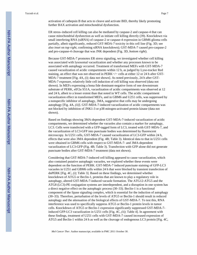
Microscopy for Acidic Endosomes and LC3-GFP Expression—Transfected cellswere pretreated with 3-methyl-adenine (3MA; 5 mmol/L; Sigma) 30 min before GST-MDA-7 exposure and then cultured for 12 to 48 h. Cells were then stained with LysotrackerRed Dye (Invitrogen) at the indicated time points for 20 min. Lysotracker Red Dye–stainedcells were visualized immediately after staining on a Zeiss Axiovert 200 microscope usingthe rhodamine filter. LC3-GFP-transfected cells were visualized at the indicated time pointson the Zeiss Axiovert 200 microscope using the FITC filter.
Data Analysis—Comparison of the effects of various treatments was done using one-wayANOVA and a two-tailed Student’s t test. Differences with P < 0.05 were consideredstatistically significant. Experiments shown are the means of multiple individual points frommultiple experiments (±SE).
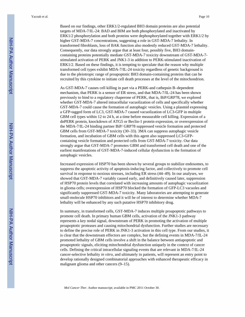
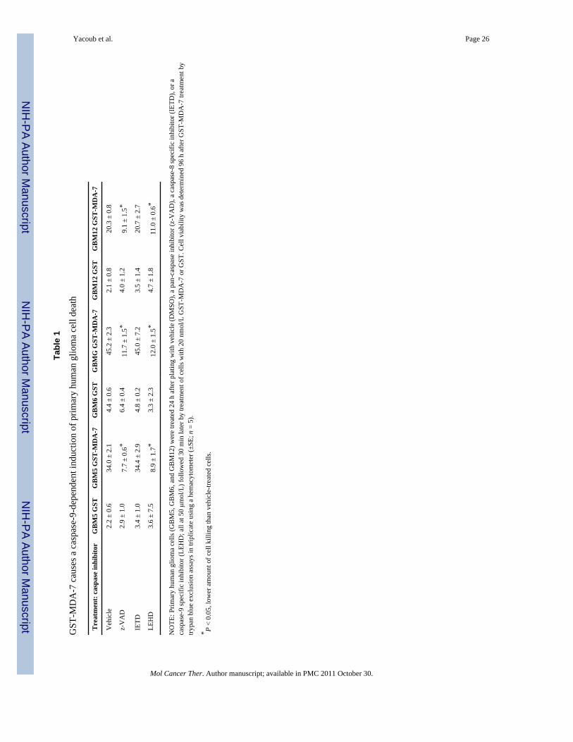
ResultsInitial experiments focused on defining potential mechanisms by which GST-MDA-7promotes cell killing in four independent primary human glioma cell populations: GBM5,GBM6, GBM12, and GBM14 (23). Treatment of GBM5, GBM6, GBM12, and GBM14cells with GST-MDA-7 was first noted to induce cell killing ~48 h after exposure andcaused significant cell killing within 72 h (Fig. 1A; Table 1; Supplementary Fig. S1).9 Cellkilling was blocked by a pan-caspase inhibitor and an inhibitor of caspase-9 but not by aninhibitor of caspase-8. Cell killing correlated with increased release of cytochrome c into thecytosol of GBM6 cells and with the cleavage of caspase-3 (Fig. 1A, top). In GBM6 cells,GST-MDA-7 treatment caused inactivation of ERK1/2 that correlated withdephosphorylation of BAD S112 and dephosphorylation and increased expression of BIM(Fig. 1B). In GBM6 cells, GST-MDA-7 treatment caused activation of JNK1-3 that wascausal in the activation of BAX and the cleavage of BID and caspase-3 (Fig. 1B). However,the decrease in BCL-xL protein expression caused by GST-MDA-7 treatment was noted tobe JNK independent. In all four glioma isolates, inhibition of caspase-8 function (IETD;expression of CRM A or c-FLIP-s) did not alter GST-MDA-7 lethality, whereas inhibitionof caspase-9 function (LEHD; expression of dominant-negative caspase-9 or XIAP or BCL-xL) significantly reduced GST-MDA-7 lethality as judged by cells becoming PI positive orretaining trypan blue dye (Fig. 1C; Supplementary Fig. S2; data not shown).9 GST-MDA-7lethality correlated with caspase-9-dependent cleavage of pro-caspase-3 (Fig. 1C, top inset).
To further investigate mechanisms of GST-MDA-7-induced mitochondrial dysfunction, weused SV40 large T antigen transformed mouse embryonic fibroblasts (MEF) devoid ofexpression of defined proapoptotic genes. Loss of BIM or BAK function had a modest butsignificant effect on GST-MDA-7 lethality, whereas loss of BAX and loss of BAX and BAKexpression profoundly reduced toxicity (Fig. 1D). In agreement with data in Fig. 1B, geneticmanipulation to delete expression of BID also significantly reduced GST-MDA-7 toxicity.Loss of BAX and BAK function or loss of BID function reduced the ability of GST-MDA-7to promote cytochrome c release into the cytosol (Fig. 1C, top immunoblotting). In
9Supplementary material for this article is available at Molecular Cancer Therapeutics Online (http://mct.aacrjournals.org/).
Yacoub et al. Page 5
Mol Cancer Ther. Author manuscript; available in PMC 2011 October 30.
NIH
-PA Author Manuscript
NIH
-PA Author Manuscript
NIH
-PA Author Manuscript

agreement with data showing that BAX activation and BID cleavage were JNK dependent,inhibition of JNK1-3 with JNK inhibitory peptide treatment suppressed the toxicity of GST-MDA-7 in GBM6 cells from 28.5 ± 1.3% above the GST control value with vehicletreatment to 7.6 ± 0.8% above the GST control value (±SE; n = 3). Collectively, thesefindings argue that GST-MDA-7 promoted activation of multiple proteins, which act toinduce mitochondrial dysfunction, and that activation of the intrinsic mitochondrial pathwayrepresents an important apoptotic mechanism for this cytokine in transformed cells.
In Fig. 1, we noted that BID function, but not caspase-8 function, correlated with GST-MDA-7-induced lethality. BID is a substrate for both caspase-8 and cathepsin proteases, andin glioma cells, cathepsin enzymes are overexpressed and play a key role in tumor invasionand angiogenesis (29, 34). GST-MDA-7 toxicity was nearly abolished by the loss ofcathepsin B expression comparing appropriate matched immortalized rodent fibroblast cells,of a different lineage to those used in Fig. 1, which correlated with a reduction in the GST-MDA-7-induced release of cytochrome c into the cytosol of these cells (Fig. 2A). Combinedinhibition of caspase-9 and cathepsin function was required to suppress GST-MDA-7lethality in transformed fibroblasts and in GBM6 and GBM12 cells (Table 2). Loss ofcathepsin B function suppressed the GST-MDA-7-induced degradation of BID andcaspase-3 in transformed fibroblasts, and inhibition of cathepsin B function suppressed theGST-MDA-7-induced degradation of BID in GBM6 cells [Fig. 2B, (i) and (ii)]. Thecleavage of p43 cathepsin B after GST-MDA-7 treatment was suppressed by inhibition ofJNK1-3 [Fig. 2B, (ii)]. Collectively, these findings suggest that GST-MDA-7 inducesmultiple parallel proapoptotic pathways in transformed cells that converge to causemitochondrial dysfunction: a JNK1-3-dependent activation of BAX; a JNK1-3-dependentactivation of cathepsin B, leading to a cathepsin B–dependent cleavage of BID; andincreased activity of BAD and BIM.
MDA-7/IL-24 has been suggested to promote ER stress signaling and cell death by bindingto and inactivating the PERK-binding protein BiP/GRP78 (19). Treatment of transformedfibroblasts that lacked expression of PERK (PERK−/−) with GST-MDA-7 causedsignificantly less cell killing than observed in their isogenic matched WT counterparts (Fig.3A). This correlated with reduced release of cytochrome c into the cytosol of GST-MDA-7treated PERK−/− cells; cytochrome c release into the cytosol was JNK dependent (Fig. 3B,top blotting). Of note, this observation was the opposite to treating these cells with anestablished inducer of ER stress, thapsigargin (Supplementary Fig. S3).9 Surprisingly, basedon known downstream targets of PERK signaling, expression of a dominant-negative eIF2αS51A protein only modestly modified the survival response of GBM6 cells treated withGST-MDA-7 (Supplementary Fig. S4).9 GST-MDA-7-induced BID cleavage, cathepsin Bcleavage, suppression of BCL-xL expression, inhibition of ERK1/2 phosphorylation, andincreased eIF2α S51 phosphorylation in transformed fibroblasts were PERK dependent (Fig.3C).
Considering published reports indicating that ER stress signaling is linked to the activationof the JNK pathway, and our present studies showing that GST-MDA-7-induced toxicity isJNK1-3 dependent and that inhibition of JNK signaling block cytochrome c release, wedetermined whether loss of PERK expression altered the activation of JNK1-3 followingGST-MDA-7 exposure (35–38). Treatment of WT-transformed fibroblasts with GST-MDA-7 promoted JNK1/2 activation, predominantly JNK1, which was causal in cell killing(Fig. 3C). In PERK−/− cells, JNK1/2 was very weakly activated by GST-MDA-7, whereas,of note, loss of cathepsin B function did not alter JNK1/2 activation. Hence, GST-MDA-7induces a PERK-dependent form of ER stress that promotes JNK pathway-dependentactivation of BAX and mitochondrial dysfunction as well as promoting a JNK-dependent
Yacoub et al. Page 6
Mol Cancer Ther. Author manuscript; available in PMC 2011 October 30.
NIH
-PA Author Manuscript
NIH
-PA Author Manuscript
NIH
-PA Author Manuscript
activation of cathepsin B that acts to cleave and activate BID, thereby likely promotingfurther BAX activation and mitochondrial dysfunction.
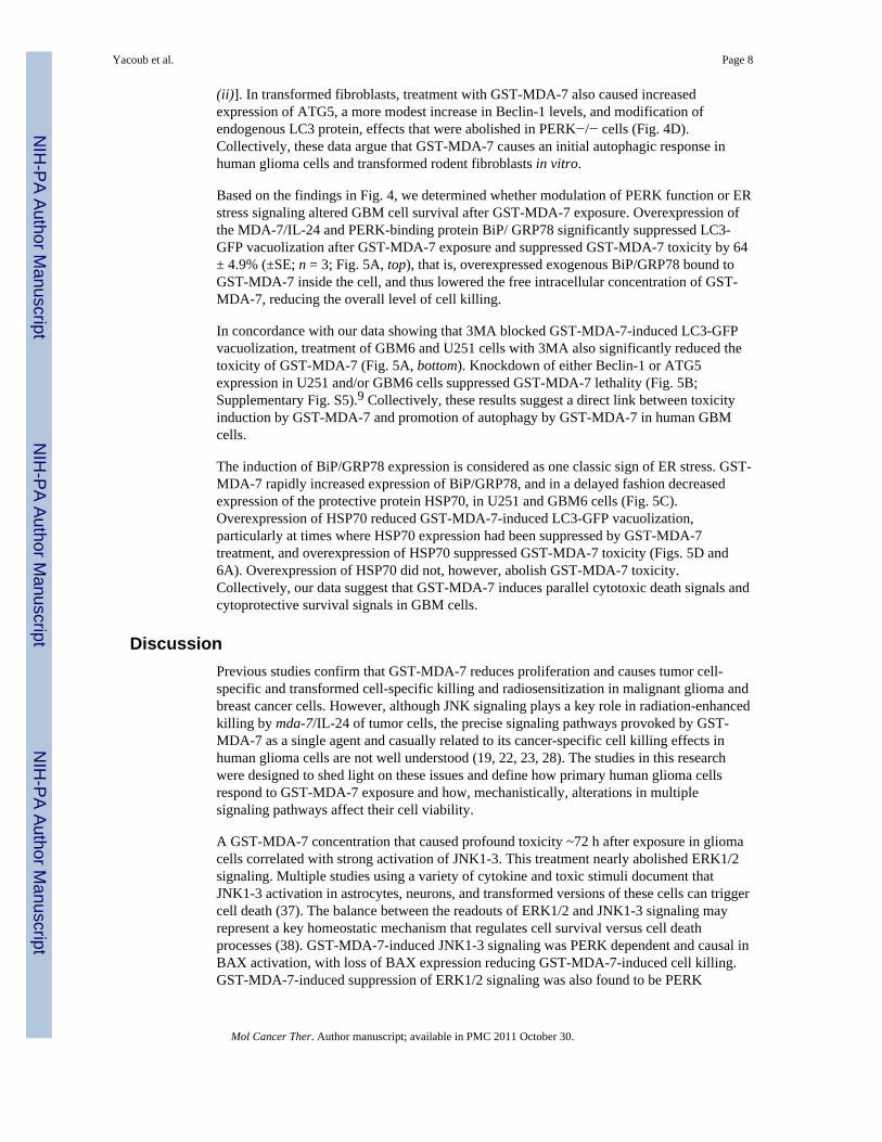
ER stress–induced cell killing can also be mediated by caspase-2 and caspase-4 that cancause mitochondrial dysfunction as well as initiate cell killing directly (39). Knockdown viasmall interfering RNA (siRNA) of caspase-2 or caspase-4 expression in GBM6 glioma cellspartially, albeit significantly, reduced GST-MDA-7 toxicity in this cell line (Fig. 3D; seealso inset on top right, confirming siRNA knockdown); GST-MDA-7 caused pro-caspase-2and pro-caspase-4 cleavage that was JNK dependent (Fig. 3D, bottom right).
Because GST-MDA-7 promotes ER stress signaling, we investigated whether cell killingwas associated with lysosomal vacuolization and whether any processes known to beassociated with autophagy occurred. Treatment of transformed MEFs with GST-MDA-7caused vacuolization of acidic compartments within 12 h, as judged by Lyso-tracker Redstaining, an effect that was not observed in PERK−/− cells at either 12 or 24 h after GST-MDA-7 treatment [Fig. 4A, (i); data not shown]. As noted previously, 24 h after GST-MDA-7 exposure, relatively little cell induction of cell killing was observed (data notshown). In MEFs expressing a bona fide dominant-negative form of one downstreamsubstrate of PERK, eIF2α S51A, vacuolization of acidic compartments was observed at 12and 24 h, albeit to a lesser extent than that noted in WT cells. The acidic compartmentvacuolization effect in transformed MEFs, and in GBM6 and U251 cells, was suppressed bya nonspecific inhibitor of autophagy, 3MA, suggestive that cells may be undergoingautophagy [Fig. 4A, (ii)]. GST-MDA-7-induced vacuolization of acidic compartments wasnot blocked by inhibition of JNK1-3 or p38 mitogen-activated protein kinase (data notshown).
Based on findings showing 3MA-dependent GST-MDA-7-induced vacuolization of acidiccompartments, we determined whether the vacuoles also contain a marker for autophagy,LC3. Cells were transfected with a GFP-tagged form of LC3, treated with GST-MDA-7, andthe vacuolization of LC3-GFP into punctuate bodies was determined by fluorescentmicroscopy. In U251 cells, GST-MDA-7 caused vacuolization of LC3-GFP within 24 h,effects that were also 3MA dependent (Fig. 4B; Table 3). Identical data to that in U251 cellswere obtained in GBM6 cells with respect to GST-MDA-7- and 3MA-dependentvacuolization of LC3-GFP (Fig. 4B; Table 3). Transfection with GFP alone did not generatepunctuate bodies after GST-MDA-7 treatment (data not shown).
Considering that GST-MDA-7-induced cell killing appeared to cause vacuolization, whichalso contained putative autophagic vacuoles, we explored whether these events weredependent on the function of PERK. GST-MDA-7 induced punctuate staining of GFP-LC3vacuoles in U251 and GBM6 cells within 24 h that were blocked by transient transfection ofdnPERK [Fig. 4C, (i); Table 3]. Based on these findings, we determined whetherknockdown of ATG5 or Beclin-1, proteins that are known to play a regulatory role inautophagy, altered GST-MDA-7-induced vacuole formation. The ATG12-ATG5 and theATG8 (LC3)-PE conjugation systems are interdependent, and a disruption in one system hasa direct negative effect on the autophagic process (30–33). Beclin-1 is a functionalcomponent of the lipase signaling complex, which is essential for the induction of autophagy(30–33). Therefore, perturbation of the levels of ATG5 or Beclin-1 should result in reducedautophagy and the attenuation of the biological effects of GST-MDA-7. To test this, RNAinterference was used to specifically suppress ATG5 or Beclin-1 protein levels in tumorcells. Knockdown of ATG5 or Beclin-1 expression significantly suppressed GST-MDA-7-induced GFP-LC3 vacuolization in U251 cells [Fig. 4C, (ii); Table 4]. In agreement withthese findings, treatment of U251 cells with GST-MDA-7 caused increased expression ofATG5 and Beclin-1 within 24 h as well as the cleavage of endogenous LC3 protein [Fig. 4C,
Yacoub et al. Page 7
Mol Cancer Ther. Author manuscript; available in PMC 2011 October 30.
NIH
-PA Author Manuscript
NIH
-PA Author Manuscript
NIH
-PA Author Manuscript
(ii)]. In transformed fibroblasts, treatment with GST-MDA-7 also caused increasedexpression of ATG5, a more modest increase in Beclin-1 levels, and modification ofendogenous LC3 protein, effects that were abolished in PERK−/− cells (Fig. 4D).Collectively, these data argue that GST-MDA-7 causes an initial autophagic response inhuman glioma cells and transformed rodent fibroblasts in vitro.
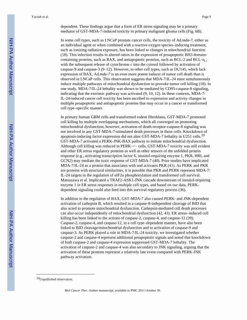
Based on the findings in Fig. 4, we determined whether modulation of PERK function or ERstress signaling altered GBM cell survival after GST-MDA-7 exposure. Overexpression ofthe MDA-7/IL-24 and PERK-binding protein BiP/ GRP78 significantly suppressed LC3-GFP vacuolization after GST-MDA-7 exposure and suppressed GST-MDA-7 toxicity by 64± 4.9% (±SE; n = 3; Fig. 5A, top), that is, overexpressed exogenous BiP/GRP78 bound toGST-MDA-7 inside the cell, and thus lowered the free intracellular concentration of GST-MDA-7, reducing the overall level of cell killing.
In concordance with our data showing that 3MA blocked GST-MDA-7-induced LC3-GFPvacuolization, treatment of GBM6 and U251 cells with 3MA also significantly reduced thetoxicity of GST-MDA-7 (Fig. 5A, bottom). Knockdown of either Beclin-1 or ATG5expression in U251 and/or GBM6 cells suppressed GST-MDA-7 lethality (Fig. 5B;Supplementary Fig. S5).9 Collectively, these results suggest a direct link between toxicityinduction by GST-MDA-7 and promotion of autophagy by GST-MDA-7 in human GBMcells.
The induction of BiP/GRP78 expression is considered as one classic sign of ER stress. GST-MDA-7 rapidly increased expression of BiP/GRP78, and in a delayed fashion decreasedexpression of the protective protein HSP70, in U251 and GBM6 cells (Fig. 5C).Overexpression of HSP70 reduced GST-MDA-7-induced LC3-GFP vacuolization,particularly at times where HSP70 expression had been suppressed by GST-MDA-7treatment, and overexpression of HSP70 suppressed GST-MDA-7 toxicity (Figs. 5D and6A). Overexpression of HSP70 did not, however, abolish GST-MDA-7 toxicity.Collectively, our data suggest that GST-MDA-7 induces parallel cytotoxic death signals andcytoprotective survival signals in GBM cells.
DiscussionPrevious studies confirm that GST-MDA-7 reduces proliferation and causes tumor cell-specific and transformed cell-specific killing and radiosensitization in malignant glioma andbreast cancer cells. However, although JNK signaling plays a key role in radiation-enhancedkilling by mda-7/IL-24 of tumor cells, the precise signaling pathways provoked by GST-MDA-7 as a single agent and casually related to its cancer-specific cell killing effects inhuman glioma cells are not well understood (19, 22, 23, 28). The studies in this researchwere designed to shed light on these issues and define how primary human glioma cellsrespond to GST-MDA-7 exposure and how, mechanistically, alterations in multiplesignaling pathways affect their cell viability.
A GST-MDA-7 concentration that caused profound toxicity ~72 h after exposure in gliomacells correlated with strong activation of JNK1-3. This treatment nearly abolished ERK1/2signaling. Multiple studies using a variety of cytokine and toxic stimuli document thatJNK1-3 activation in astrocytes, neurons, and transformed versions of these cells can triggercell death (37). The balance between the readouts of ERK1/2 and JNK1-3 signaling mayrepresent a key homeostatic mechanism that regulates cell survival versus cell deathprocesses (38). GST-MDA-7-induced JNK1-3 signaling was PERK dependent and causal inBAX activation, with loss of BAX expression reducing GST-MDA-7-induced cell killing.GST-MDA-7-induced suppression of ERK1/2 signaling was also found to be PERK
Yacoub et al. Page 8
Mol Cancer Ther. Author manuscript; available in PMC 2011 October 30.
NIH
-PA Author Manuscript
NIH
-PA Author Manuscript
NIH
-PA Author Manuscript
dependent. These findings argue that a form of ER stress signaling may be a primarymediator of GST-MDA-7-induced toxicity in primary malignant glioma cells (Fig. 6B).
In some cell types, such as LNCaP prostate cancer cells, the toxicity of Ad.mda-7, either asan individual agent or when combined with a reactive oxygen species–inducing treatment,such as ionizing radiation exposure, has been linked to changes in mitochondrial function(18). This infection results in altered ratios in the expression of proapoptotic BH3 domain-containing proteins, such as BAX, and antiapoptotic proteins, such as BCL-2 and BCL-xL,with the subsequent release of cytochrome c into the cytosol followed by activation ofcaspase-9 and caspase-3 (9–12). However, in other cell types, such as DU145, which lackexpression of BAX, Ad.mda-7 is an even more potent inducer of tumor cell death than isobserved in LNCaP cells. This observation suggests that MDA-7/IL-24 must simultaneouslyinduce multiple pathways of mitochondrial dysfunction to provoke tumor cell killing (18). Inone study, MDA-7/IL-24 lethality was shown to be mediated by CD95-caspase-8 signaling,indicating that the extrinsic pathway was activated (9, 10, 12). In these contexts, MDA-7/IL-24-induced cancer cell toxicity has been ascribed to expression and activity changes inmultiple proapoptotic and antiapoptotic proteins that may occur in a cancer or transformedcell type–specific manner.
In primary human GBM cells and transformed rodent fibroblasts, GST-MDA-7 promotedcell killing by multiple overlapping mechanisms, which all converged on promotingmitochondrial dysfunction; however, activation of death receptor-caspase-8 signaling wasnot involved in any GST-MDA-7-stimulated death processes in these cells. Knockdown ofapoptosis-inducing factor expression did not alter GST-MDA-7 lethality in U251 cells.10
GST-MDA-7 activated a PERK-JNK-BAX pathway to initiate mitochondrial dysfunction.Although cell killing was reduced in PERK−/− cells, GST-MDA-7 toxicity was still evidentand other ER stress regulatory proteins as well as other sensors of the unfolded proteinresponse (e.g., activating transcription factor 6, inositol-requiring enzyme 1, PKR, HRI, andGCN2) may mediate the toxic response of GST-MDA-7 (40). Prior studies have implicatedMDA-7/IL-24 as a protein that associates with and activates PKR (41). As PERK and PKRare proteins with structural similarities, it is possible that PKR and PERK represent MDA-7/IL-24 targets in the regulation of eIF2α phosphorylation and transformed cell survival.Matsuzawa et al. implicated a TRAF2-ASK1-JNK cascade downstream of inositol-requiringenzyme 1 in ER stress responses in multiple cell types, and based on our data, PERK-dependent signaling could also feed into this survival regulatory process (36).
In addition to the regulation of BAX, GST-MDA-7 also caused PERK- and JNK-dependentactivation of cathepsin B, which resulted in a caspase-8-independent cleavage of BID thatalso acted to promote mitochondrial dysfunction. Cathepsin-mediated cell death processescan also occur independently of mitochondrial dysfunction (42, 43). ER stress–induced cellkilling has been linked to the actions of caspase-2, caspase-4, and caspase-12 (39).Caspase-2, caspase-4, and caspase-12, in a cell type–dependent manner, have also beenlinked to BID cleavage/mitochondrial dysfunction and to activation of caspase-9 andcaspase-3. As PERK played a role in MDA-7/IL-24 toxicity, we investigated whethercaspase-2 and caspase-4 represent additional proapoptotic signals and noted that knockdownof both caspase-2 and caspase-4 expression suppressed GST-MDA-7 lethality. Theactivation of caspase-2 and caspase-4 was also secondary to JNK signaling, arguing that theactivation of these proteins represent a relatively late event compared with PERK-JNKpathway activation.
10Unpublished observation.
Yacoub et al. Page 9
Mol Cancer Ther. Author manuscript; available in PMC 2011 October 30.
NIH
-PA Author Manuscript
NIH
-PA Author Manuscript
NIH
-PA Author Manuscript
Based on our findings, other ERK1/2-regulated BH3 domain proteins are also potentialtargets of MDA-7/IL-24: BAD and BIM are both phosphorylated and inactivated byERK1/2 phosphorylation and both proteins were dephosphorylated together with ERK1/2 byhigher GST-MDA-7 concentrations, suggesting a role in GST-MDA-7 lethality. Intransformed fibroblasts, loss of BAK function also modestly reduced GST-MDA-7 lethality.Consequently, our data strongly argue that at least four, possibly five, BH3 domain-containing proteins potentially mediate GST-MDA-7 toxicity downstream of GST-MDA-7-stimulated activation of PERK and JNK1-3 in addition to PERK-stimulated inactivation ofERK1/2. Based on these findings, it is tempting to speculate that the reason why multipletransformed cell types exhibit MDA-7/IL-24 toxicity regardless of genetic background isdue to the pleiotropic range of proapoptotic BH3 domain-containing proteins that can berecruited by this cytokine to initiate cell death processes at the level of the mitochondrion.
As GST-MDA-7 causes cell killing in part via a PERK-and cathepsin B–dependentmechanism, that PERK is a sensor of ER stress, and that MDA-7/IL-24 has been shownpreviously to bind to a regulatory chaperone of PERK, that is, BiP/GRP78, we exploredwhether GST-MDA-7 altered intracellular vacuolization of cells and specifically whetherGST-MDA-7 could cause the formation of autophagic vesicles. Using a plasmid expressinga GFP-tagged form of LC3, GST-MDA-7 caused vacuolization of LC3-GFP in multipleGBM cell types within 12 to 24 h, at a time before measurable cell killing. Expression of adnPERK protein, knockdown of ATG5 or Beclin-1 protein expression, or overexpression ofthe MDA-7/IL-24 binding partner BiP/ GRP78 suppressed vesicle formation and protectedGBM cells from GST-MDA-7 toxicity (30–33). 3MA can suppress autophagic vesicleformation, and incubation of GBM cells with this agent also suppressed LC3-GFP-containing vesicle formation and protected cells from GST-MDA-7 toxicity. Our datastrongly argue that GST-MDA-7 promotes GBM and transformed cell death and one of theearliest manifestations of GST-MDA-7-induced cellular dysfunction is the formation ofautophagic vesicles.
Increased expression of HSP70 has been shown by several groups to stabilize endosomes, tosuppress the apoptotic activity of apoptosis-inducing factor, and collectively to promote cellsurvival in response to noxious stresses, including ER stress (44–49). In our analyses, weshowed that GST-MDA-7 variably caused early, and definitively caused later, suppressionof HSP70 protein levels that correlated with increasing amounts of autophagic vacuolizationin glioma cells; overexpression of HSP70 blocked the formation of GFP-LC3 vacuoles andsignificantly suppressed GST-MDA-7 toxicity. Many laboratories are attempting to generatesmall-molecule HSP70 inhibitors and it will be of interest to determine whether MDA-7lethality will be enhanced by any such putative HSP70 inhibitory drug.
In summary, in transformed cells, GST-MDA-7 induces multiple proapoptotic pathways topromote cell death. In primary human GBM cells, activation of the JNK1-3 pathwayrepresents a key nodal signal, downstream of PERK in promoting the activation of multipleproapoptotic proteases and causing mitochondrial dysfunction. Further studies are necessaryto define the precise role of PERK in JNK1-3 activation in this cell type. From our studies, itis clear that the downstream effectors are complex, but the defining events in MDA-7/IL-24promoted lethality of GBM cells involve a shift in the balance between antiapoptotic andproapoptotic signals, eliciting mitochondrial dysfunction uniquely in the context of cancercells. Defining the critical intracellular signaling events that are relevant in MDA-7/IL-24cancer-selective lethality in vitro, and ultimately in patients, will represent an entry point todevelop rationally designed combinatorial approaches with enhanced therapeutic efficacy inmalignant glioma and other cancers (9–15).
Yacoub et al. Page 10
Mol Cancer Ther. Author manuscript; available in PMC 2011 October 30.
NIH
-PA Author Manuscript
NIH
-PA Author Manuscript
NIH
-PA Author Manuscript
Supplementary MaterialRefer to Web version on PubMed Central for supplementary material.
AcknowledgmentsGrant support: Public Health Service grants P01-CA104177, R01-CA108325, and R01-DK52825, Jim Valvano“V” Foundation, and Department of Defense award DAMD17-03-1-0262 (P. Dent); Public Health Service grantsR01-CA63753 and R01-CA77141 and a Leukemia Society of America grant 6405-97 (S. Grant); Public HealthService grants P01-CA104177, R01-CA097318, R01-CA098172, and P01-NS031492, Samuel Waxman CancerResearch Foundation, and Michael and Stella Chernow Endowment (P.B. Fisher); and Public Health Service grantP01-CA104177 (D.T. Curiel).
References1. Robins HI, Chang S, Butowski N, Mehta M. Therapeutic advances for glioblastoma multiforme:
current status and future prospects. Curr Oncol Rep. 2007; 9:66–70. [PubMed: 17164050]2. Jiang H, Lin JJ, Su ZZ, Goldstein NI, Fisher PB. Subtraction hybridization identifies a novel
melanoma differentiation associated gene, mda-7, modulated during human melanomadifferentiation, growth and progression. Oncogene. 1995; 11:2477–86. [PubMed: 8545104]
3. Ekmekcioglu S, Ellerhorst J, Mhashilkar AM, et al. Down-regulated melanoma differentiationassociated gene (mda-7) expression in human melanomas. Int J Cancer. 2001; 94:54–9. [PubMed:11668478]
4. Ellerhorst JA, Prieto VG, Ekmekcioglu S. Loss of MDA-7 expression with progression ofmelanoma. J Clin Oncol. 2002; 20:1069–74. [PubMed: 11844832]
5. Huang EY, Madireddi MT, Gopalkrishnan RV, et al. Genomic structure, chromosomal localizationand expression profile of a novel melanoma differentiation associated (mda-7) gene with cancerspecific growth suppressing and apoptosis inducing properties. Oncogene. 2001; 20:7051–63.[PubMed: 11704829]
6. Parrish-Novak J, Xu W, Brender T, et al. Interleukins 19, 20, and 24 signal through two distinctreceptor complexes. Differences in receptorligand interactions mediate unique biological functions.J Biol Chem. 2002; 277:47517–23. [PubMed: 12351624]
7. Caudell EG, Mumm JB, Poindexter N, et al. The protein product of the tumor suppressor gene,melanoma differentiation-associated gene 7, exhibits immunostimulatory activity and is designatedIL-24. J Immunol. 2002; 168:6041–6. [PubMed: 12055212]
8. Pestka S, Krause CD, Sarkar D, Walter MR, Shi Y, Fisher PB. Interleukin-10 and related cytokinesand receptors. Annu Rev Immunol. 2004; 22:929–79. [PubMed: 15032600]
9. Gupta P, Su ZZ, Lebedeva IV, et al. mda-7/IL-24: multifunctional cancer-specific apoptosis-inducing cytokine. Pharmacol Ther. 2006; 111:596–628. [PubMed: 16464504]
10. Lebedeva IV, Sauane M, Gopalkrishnan RV, et al. mda-7/IL-24: exploiting cancer’s Achilles’ heel.Mol Ther. 2005; 11:4–18. [PubMed: 15585401]
11. Fisher PB, Gopalkrishnan RV, Chada S, et al. mda-7/IL-24, a novel cancer selective apoptosisinducing cytokine gene: from the laboratory into the clinic. Cancer Biol Ther. 2003; 2:S23–37.[PubMed: 14508078]
12. Fisher PB. Is mda-7/IL-24 a “magic bullet” for cancer? Cancer Res. 2005; 65:10128–38. [PubMed:16287994]
13. Su ZZ, Lebedeva IV, Gopalkrishnan RV, et al. A combinatorial approach for selectively inducingprogrammed cell death in human pancreatic cancer cells. Proc Natl Acad Sci U S A. 2001;98:10332–7. [PubMed: 11526239]
14. Su ZZ, Madireddi MT, Lin JJ, et al. The cancer growth suppressor gene mda-7 selectively inducesapoptosis in human breast cancer cells and inhibits tumor growth in nude mice. Proc Natl Acad SciU S A. 1998; 95:14400–5. [PubMed: 9826712]
15. Cunningham CC, Chada S, Merritt JA, et al. Clinical and local biological effects of an intratumoralinjection of mda-7 (IL24; INGN 241) in patients with advanced carcinoma: a phase I study. MolTher. 2005; 11:149–59. [PubMed: 15585416]
Yacoub et al. Page 11
Mol Cancer Ther. Author manuscript; available in PMC 2011 October 30.
NIH
-PA Author Manuscript
NIH
-PA Author Manuscript
NIH
-PA Author Manuscript
16. Lebedeva IV, Su ZZ, Chang Y, Kitada S, Reed JC, Fisher PB. The cancer growth suppressing genemda-7 induces apoptosis selectively in human melanoma cells. Oncogene. 2002; 21:708–18.[PubMed: 11850799]
17. Saeki T, Mhashilkar A, Swanson X, et al. Inhibition of human lung cancer growth followingadenovirus-mediated mda-7 gene expression in vivo. Oncogene. 2002; 21:4558–66. [PubMed:12085234]
18. Su ZZ, Lebedeva IV, Sarkar D, et al. Ionizing radiation enhances therapeutic activity of mda-7/IL-24: overcoming radiation- and mda-7/IL-24-resistance in prostate cancer cells over-expressingthe antiapoptotic proteins bcl-xL or bcl-2. Oncogene. 2006; 25:2339–48. [PubMed: 16331261]
19. Gupta P, Walter MR, Su ZZ, et al. BiP/GRP78 is an intracellular target for MDA-7/IL-24induction of cancer-specific apoptosis. Cancer Res. 2006; 66:8182–91. [PubMed: 16912197]
20. Su ZZ, Lebedeva IV, Sarkar D, et al. Melanoma differentiation associated gene-7, mda-7/IL-24,selectively induces growth suppression, apoptosis and radiosensitization in malignant gliomas in ap53-independent manner. Oncogene. 2003; 22:1164–80. [PubMed: 12606943]
21. Yacoub A, Mitchell C, Lister A, et al. Melanoma differentiation-associated 7 (interleukin 24)inhibits growth and enhances radiosensitivity of glioma cells in vitro and in vivo. Clin Cancer Res.2003; 9:3272–81. [PubMed: 12960112]
22. Yacoub A, Mitchell C, Lebedeva IV, et al. mda-7 (IL-24) inhibits growth and enhancesradiosensitivity of glioma cells in vitro via JNK signaling. Cancer Biol Ther. 2003; 2:347–53.[PubMed: 14508103]
23. Yacoub A, Mitchell C, Hong Y, et al. MDA-7 regulates cell growth and radiosensitivity in vitro ofprimary (non-established) human glioma cells. Cancer Biol Ther. 2004; 3:739–51. [PubMed:15197348]
24. Yacoub A, Mitchell C, Brannon J, et al. MDA-7 (interleukin-24) inhibits the proliferation of renalcarcinoma cells and interacts with free radicals to promote cell death and loss of reproductivecapacity. Mol Cancer Ther. 2003; 2:623–32. [PubMed: 12883035]
25. Sarkar D, Su ZZ, Lebedeva IV, et al. mda-7 (IL-24) mediates selective apoptosis in humanmelanoma cells by inducing the coordinated over-expression of the GADD family of genes bymeans of p38 MAPK. Proc Natl Acad Sci U S A. 2002; 99:10054–9. [PubMed: 12114539]
26. Mhashilkar AM, Stewart AL, Sieger K, et al. MDA-7 negatively regulates the β-catenin and PI3Ksignaling pathways in breast and lung tumor cells. Mol Ther. 2003; 8:207–19. [PubMed:12907143]
27. Chada S, Bocangel D, Ramesh R, et al. mda-7/IL24 kills pancreatic cancer cells by inhibition ofthe Wnt/PI3K signaling pathways: identification of IL-20 receptor-mediated bystander activityagainst pancreatic cancer. Mol Ther. 2005; 11:724 – 33. [PubMed: 15851011]
28. Sauane M, Gopalkrishnan RV, Choo HT, et al. Mechanistic aspects of mda-7/IL-24 cancer cellselectivity analysed via a bacterial fusion protein. Oncogene. 2004; 23:7679–90. [PubMed:15334067]
29. Guicciardi ME, Deussing J, Miyoshi H, et al. Cathepsin B contributes to TNF-α-mediatedhepatocyte apoptosis by promoting mitochondrial release of cytochrome c. J Clin Invest. 2000;106:1127–37. [PubMed: 11067865]
30. Yang YP, Liang ZQ, Gu ZL, Qin ZH. Molecular mechanism and regulation of autophagy. ActaPharmacol Sin. 2005; 26:1421–34. [PubMed: 16297339]
31. Levine B, Yuan J. Autophagy in cell death: an innocent convict? J Clin Invest. 2005; 115:2679 –88. [PubMed: 16200202]
32. Yousefi S, Perozzo R, Schmid I, et al. Calpain-mediated cleavage of Atg5 switches autophagy toapoptosis. Nat Cell Biol. 2006; 8:1124 – 32. [PubMed: 16998475]
33. Shibata M, Lu T, Furuya T, et al. Regulation of intracellular accumulation of mutant Huntingtin byBeclin 1. J Biol Chem. 2006; 281:14474–85. [PubMed: 16522639]
34. Wang M, Tang J, Liu S, Yoshida D, Teramoto A. Expression of cathepsin B and microvasculardensity increases with higher grade of astrocytomas. J Neurooncol. 2005; 71:3 –7. [PubMed:15719267]
Yacoub et al. Page 12
Mol Cancer Ther. Author manuscript; available in PMC 2011 October 30.
NIH
-PA Author Manuscript
NIH
-PA Author Manuscript
NIH
-PA Author Manuscript

35. Liang SH, Zhang W, McGrath BC, Zhang P, Cavener DR. PERK (eIF2α kinase) is required toactivate the stress-activated MAPKs and induce the expression of immediate-early genes upondisruption of ER calcium homoeostasis. Biochem J. 2006; 393:201–9. [PubMed: 16124869]
36. Matsuzawa A, Nishitoh H, Tobiume K, Takeda K, Ichijo H. Physiological roles of ASK1-mediatedsignal transduction in oxidative stress- and endoplasmic reticulum stress-induced apoptosis:advanced findings from ASK1 knockout mice. Antioxid Redox Signal. 2002; 4:415 – 25.[PubMed: 12215209]
37. Yoon S, Choi J, Yoon J, Huh JW, Kim D. Okadaic acid induces JNK activation, bim over-expression and mitochondrial dysfunction in cultured rat cortical neurons. Neurosci Lett. 2006;394:190–5. [PubMed: 16260088]
38. Xia Z, Dickens M, Raingeaud J, Davis RJ, Greenberg ME. Opposing effects of ERK and JNK-p38MAP kinases on apoptosis. Science. 1995; 270:1326 – 31. [PubMed: 7481820]
39. Golstein P, Kroemer G. Cell death by necrosis: towards a molecular definition. Trends BiochemSci. 2007; 32:37–43. [PubMed: 17141506]
40. Fels DR, Koumenis C. The PERK/eIF2α/ATF4 module of the UPR in hypoxia resistance andtumor growth. Cancer Biol Ther. 2006; 5:723 – 8. [PubMed: 16861899]
41. Pataer A, Vorburger SA, Chada S, et al. Melanoma differentiation-associated gene-7 proteinphysically associates with the double-stranded RNA-activated protein kinase PKR. Mol Ther.2005; 11:717–23. [PubMed: 15851010]
42. Yeung BH, Huang DC, Sinicrope FA. PS-341 (Bortezomib) induces lysosomal cathepsin B releaseand a caspase-2-dependent mitochondrial permeabilization and apoptosis in human pancreaticcancer cells. J Biol Chem. 2006; 281:11923–32. [PubMed: 16446371]
43. Hitomi J, Katayama T, Eguchi Y, et al. Involvement of caspase-4 in endoplasmic reticulum stress-induced apoptosis and Aβ-induced cell death. J Cell Biol. 2004; 165:347–56. [PubMed: 15123740]
44. Nylandsted J, Gyrd-Hansen M, Danielewicz A, et al. Heat shock protein 70 promotes cell survivalby inhibiting lysosomal membrane permeabilization. J Exp Med. 2004; 200:425–35. [PubMed:15314073]
45. Ravagnan L, Gurbuxani S, Susin SA, et al. Heat-shock protein 70 antagonizes apoptosis-inducingfactor. Nat Cell Biol. 2001; 3:839 – 43. [PubMed: 11533664]
46. Demidenko ZN, Vivo C, Halicka HD, et al. Pharmacological induction of Hsp70 protectsapoptosis-prone cells from doxorubicin: comparison with caspase-inhibitor- and cycle-arrest-mediated cytoprotection. Cell Death Differ. 2006; 13:1434–41. [PubMed: 16311509]
47. Mosser DD, Caron AW, Bourget L, et al. The chaperone function of hsp70 is required forprotection against stress-induced apoptosis. Mol Cell Biol. 2000; 20:7146–59. [PubMed:10982831]
48. Gurbuxani S, Schmitt E, Cande C, et al. Heat shock protein 70 binding inhibits the nuclear importof apoptosis-inducing factor. Oncogene. 2003; 22:6669 – 78. [PubMed: 14555980]
49. Mambula SS, Calderwood SK. Heat shock protein 70 is secreted from tumor cells by anonclassical pathway involving lysosomal endosomes. J Immunol. 2006; 177:7849–57. [PubMed:17114456]
Yacoub et al. Page 13
Mol Cancer Ther. Author manuscript; available in PMC 2011 October 30.
NIH
-PA Author Manuscript
NIH
-PA Author Manuscript
NIH
-PA Author Manuscript
Figure 1.GST-MDA-7 causes a caspase-9-dependent induction of primary human glioma cell death.A, primary human glioma cells (GBM6) were treated 24 h after plating with GST-MDA-7(30 nmol/L). At the indicated time points after GST-MDA-7 treatment, GBM6 cells wereisolated and subjected to SDS-PAGE, and immunoblotting was done to measure the releaseof cytochrome c into the cytosol, the expression of cleaved caspase-3 and β-actin, and cellkilling by terminal deoxynucleotidyl transferase–mediated dUTP nick end labeling assayand cell killing by Hoechst assay (n = 2). B, top, GBM6 cells were treated with 1 or 30nmol/L GST-MDA-7/GST 36 h after plating. Cells were isolated 48 h after exposure and100 μg protein was subjected to SDS-PAGE on 12% gels followed by immunoblotting todetermine the phosphorylation status of ERK1/2 and JNK1-3 and total expression of ERK2(n = 4). Bottom, GBM6 cells were cultured for 24 h and then treated with the JNK inhibitorypeptide based on sequence from JIP-1 (10 μmol/L; JNK-IP) 30 min before addition of GST
Yacoub et al. Page 14
Mol Cancer Ther. Author manuscript; available in PMC 2011 October 30.
NIH
-PA Author Manuscript
NIH
-PA Author Manuscript
NIH
-PA Author Manuscript
or GST-MDA-7 (30 nmol/L). Cells were isolated 24 h after GST-MDA-7 treatment and celllysates were split into two portions. In one portion, equal amounts of protein lysate weresubjected to immunoprecipitation using the anti-active BAX 6A7 antibody.Immunoprecipitates were subjected to SDS-PAGE and immunoblotted for BAX. In thesecond portion, equal amounts of protein lysate were subjected to SDS-PAGE andimmunoblotted for phosphorylated JNK1-3 (P-JNK1-3), BCL-xL, cleaved caspase-3, BAX,BAD, BAD S112, BIM, and ERK2. Data are from a representative experiment (n = 3). C,GBM6 cells were plated and 12 h after plating infected at a multiplicity of infection of 50 toexpress no gene (CMV), dominant-negative caspase-9 (dncasp9), or treatment with the JNKinhibitory peptide (10 μmol/L) as indicated. Twenty-four hours after infection, cells weretreated with 30 nmol/L GST or GST-MDA-7, and 72 h after GST-MDA-7 treatment, cellswere isolated and cell viability was determined by using Annexin V/PI staining assays intriplicate using a flow cytometer (±SE; n = 5; #, P < 0.05, lower amount of cell killing thanvehicle-treated cells). Top inset, at 48 h after 30 nmol/L GST-MDA-7 or GST treatment,GBM6 cells expressing dominant-negative caspase-9 or the caspase-8 inhibitor CRM Awere isolated and subjected to SDS-PAGE and immunoblotting was done to measure theexpression of ERK2 and the levels of cleaved caspase-3 (n = 2). D, SV40 large T proteintransformed MEFs, WT, lacking BIM (BIM−/−), lacking BAK (BAK−/−), lacking BAX(BAX−/−), lacking BAK and BAX (BAK−/−BAX−/−), and lacking BID (BID−/−) wereplated and 24 h after plating were treated with 100 nmol/L GST-MDA-7 or GST. Seventy-two hours after GST-MDA-7 treatment, cells were isolated and cell viability was determinedby trypan blue exclusion assays in triplicate using a hemacytometer. Data are presented asthe true percentage increase in cell death above GST-treated cells (±SE; n = 5; #, P < 0.05,lower amount of cell killing than WT cells). Inset, MEF cells were plated and 24 h afterplating treated with 100 nmol/L GST-MDA-7 (M7) or GST. Twenty-four and 48 h afterGST-MDA-7 treatment, cells were isolated, the cytosol was isolated and subjected to SDS-PAGE, and immunoblotting was done to measure the expression of cytochrome c andglyceraldehyde-3-phosphate dehydrogenase (GAPDH; n = 3).
Yacoub et al. Page 15
Mol Cancer Ther. Author manuscript; available in PMC 2011 October 30.
NIH
-PA Author Manuscript
NIH
-PA Author Manuscript
NIH
-PA Author Manuscript

Figure 2.Cathepsin B – dependent cleavage of BID plays an important role in GST-MDA-7 toxicityin transformed cells. A, mouse immortalized embryonic fibroblasts (WT; cathepsin B−/−)were cultured for 36 h and then treated with GST or GST-MDA-7 (0–100 nmol/L, asindicated). Cells were isolated for viability analyses 72 h after GST-MDA-7 treatment asjudged in triplicate by trypan blue dye exclusion assay (n = 3). Top flow cytometry, WT andcathepsin B−/− cells were cultured for 36 h and then treated with GST or GST-MDA-7 (150nmol/L). Cells were isolated for Annexin V/PI viability analyses 72 h after GST-MDA-7treatment as judged in triplicate (n = 3). Top immunoblotting, 48 hours after GST-MDA-7treatment, WT and cathepsin B−/− cells were isolated, the cytosol fraction was isolated andsubjected to SDS-PAGE, and immunoblotting was done to measure the levels of cytochromec and GAPDH (n = 3). B, top (i ), MEF cells were isolated for immunoblotting 48 h afterGST-MDA-7 treatment, as per A, and 100 μg protein at each time point was subjected toSDS-PAGE on 12% gels followed by immunoblotting to determine the total expression ofERK2, pro-caspase-3, and BID (representative blots; n = 3); Bottom (ii ), left, GBM6 cellswere cultured for 24 h after plating and then treated with the cathepsin B inhibitor (1 μmol/L; cath. B inhib.) 30 min before addition of GST or GST-MDA-7 (30 nmol/L). Cells were
Yacoub et al. Page 16
Mol Cancer Ther. Author manuscript; available in PMC 2011 October 30.
NIH
-PA Author Manuscript
NIH
-PA Author Manuscript
NIH
-PA Author Manuscript
isolated 48 h after GST-MDA-7 treatment and 100 μg protein at each time point wassubjected to SDS-PAGE on 12% gels followed by immunoblotting to determine the totalexpression of ERK2, pro-caspase-3, and BID (representative blots; n = 3). Right, GBM6cells were plated and cultured for 36 h and then treated with the JNK inhibitory peptide (10μmol/L) 30 min before addition of GST or GST-MDA-7 (30 nmol/L). Cells were isolated 24h after GST-MDA-7 treatment and equal amounts of protein lysate were subjected to SDS-PAGE and immunoblotted for expression of p43 cathepsin B and ERK2. Data are from arepresentative experiment (n = 3).
Yacoub et al. Page 17
Mol Cancer Ther. Author manuscript; available in PMC 2011 October 30.
NIH
-PA Author Manuscript
NIH
-PA Author Manuscript
NIH
-PA Author Manuscript
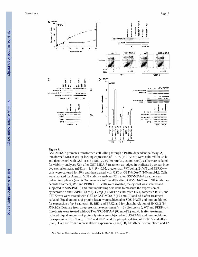
Figure 3.GST-MDA-7 promotes transformed cell killing through a PERK-dependent pathway. A,transformed MEFs: WT or lacking expression of PERK (PERK−/−) were cultured for 36 hand then treated with GST or GST-MDA-7 (0–60 nmol/L, as indicated). Cells were isolatedfor viability analyses 72 h after GST-MDA-7 treatment as judged in triplicate by trypan bluedye exclusion assay (±SE; n = 3; *, P < 0.05, greater than WT cells). B, WT and PERK−/−cells were cultured for 36 h and then treated with GST or GST-MDA-7 (100 nmol/L). Cellswere isolated for Annexin V/PI viability analyses 72 h after GST-MDA-7 treatment asjudged in triplicate (n = 3). Top immunoblotting, 48 h after GST-MDA-7 and JNK inhibitorypeptide treatment, WT and PERK B−/− cells were isolated, the cytosol was isolated andsubjected to SDS-PAGE, and immunoblotting was done to measure the expression ofcytochrome c and GAPDH (n = 3). C, top (i ), MEFs as indicated (WT, cathepsin B−/−, andPERK−/−) were treated with GST or GST-MDA-7 (60 nmol/L) and 48 h after treatmentisolated. Equal amounts of protein lysate were subjected to SDS-PAGE and immunoblottedfor expression of p43 cathepsin B, BID, and ERK2 and for phosphorylation of JNK1/2 (P-JNK1/2). Data are from a representative experiment (n = 3). Bottom (ii ), WT and PERK−/−fibroblasts were treated with GST or GST-MDA-7 (60 nmol/L) and 48 h after treatmentisolated. Equal amounts of protein lysate were subjected to SDS-PAGE and immunoblottedfor expression of BCL-xL, ERK2, and eIF2α and for phosphorylation of ERK1/2 and eIF2α(S51 ). Data are from a representative experiment (n = 2). D, GBM6 cells were plated and 12
Yacoub et al. Page 18
Mol Cancer Ther. Author manuscript; available in PMC 2011 October 30.
NIH
-PA Author Manuscript
NIH
-PA Author Manuscript
NIH
-PA Author Manuscript
h after plating portions of these cells were transfected with a scrambled siRNA (SCR), asiRNA to knockdown caspase-2 expression (si-2), or a siRNA to knockdown caspase-4expression (si-4). Other portions of cells were not transfected but treated with either vehicle(DMSO; VEH) or with the JNK inhibitory peptide (10 μmol/L). Forty-eight hours aftertransfection, cells were treated with GST or GST-MDA-7 at the indicated concentrations.Forty-eight hours (immunoblotting inset to the right) or, for viability analyses, 72 h afterGST-MDA-7 treatment, cells were isolated as judged in triplicate by trypan blue dyeexclusion assay (±SE; n = 2; #, P < 0.05, less than scrambled siRNA – transfected orvehicle-treated cells treated with the same concentration of GST-MDA-7).
Yacoub et al. Page 19
Mol Cancer Ther. Author manuscript; available in PMC 2011 October 30.
NIH
-PA Author Manuscript
NIH
-PA Author Manuscript
NIH
-PA Author Manuscript
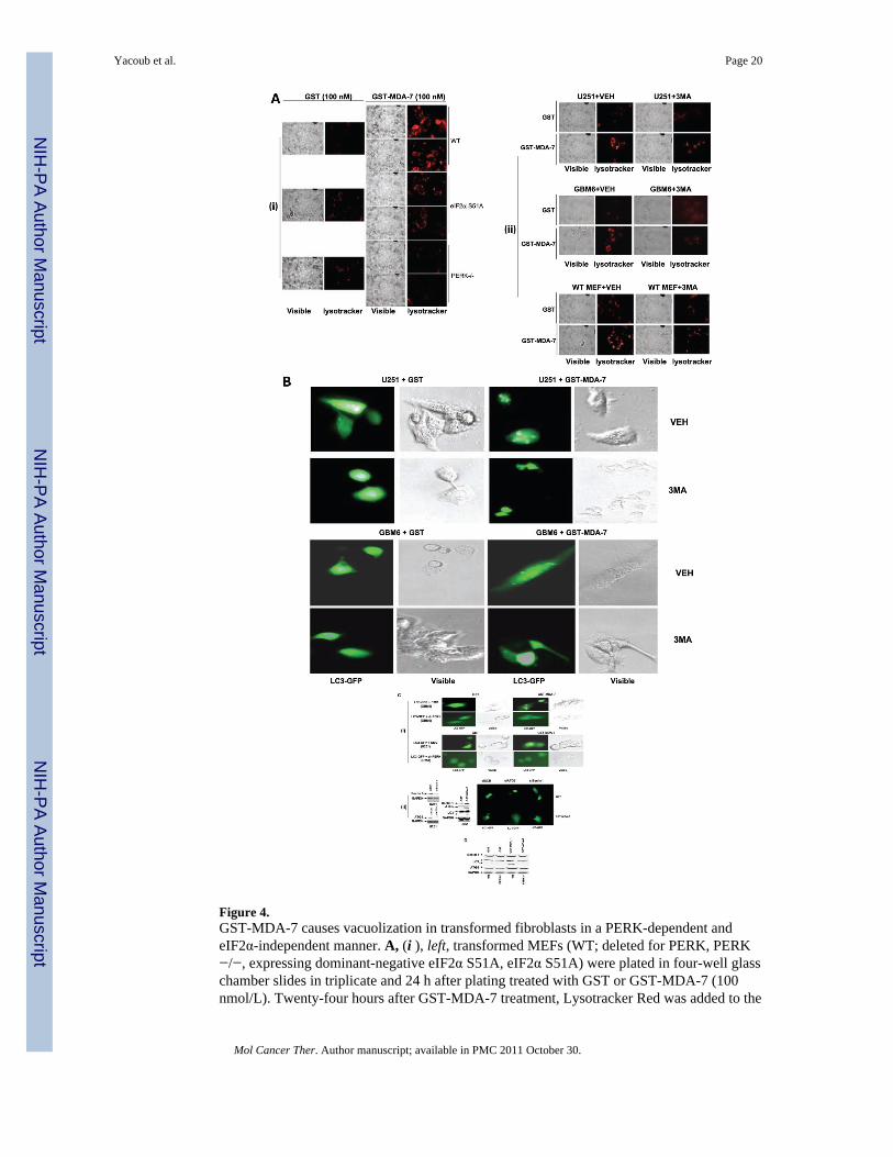
Figure 4.GST-MDA-7 causes vacuolization in transformed fibroblasts in a PERK-dependent andeIF2α-independent manner. A, (i ), left, transformed MEFs (WT; deleted for PERK, PERK−/−, expressing dominant-negative eIF2α S51A, eIF2α S51A) were plated in four-well glasschamber slides in triplicate and 24 h after plating treated with GST or GST-MDA-7 (100nmol/L). Twenty-four hours after GST-MDA-7 treatment, Lysotracker Red was added to the
Yacoub et al. Page 20
Mol Cancer Ther. Author manuscript; available in PMC 2011 October 30.
NIH
-PA Author Manuscript
NIH
-PA Author Manuscript
NIH
-PA Author Manuscript
culture medium and the cells were examined under visual light (visible) or under fluorescentlight (Lysotracker Red) at ×40 magnification. Representative images from the triplicateplating (n = 2). (ii ), right, WT MEF and U251 and GBM6 human glioma cells were treatedwith GST or GST-MDA-7 (100 nmol/L) and coincubated for the 24 h GST-MDA-7treatment with vehicle (PBS) or with 3MA (5 mmol/L). Twenty-four hours after GST-MDA-7 treatment, Lysotracker Red was added to the culture medium and the cells wereexamined under visual light (visible) or under fluorescent light (Lysotracker Red) at ×40magnification. Representative images from the triplicate plating (n = 2). B, U251 cells wereplated in four-well chamber glass slides in triplicate and 12 h after plating were transfectedwith a plasmid to express LC3-GFP. Twelve hours after transfection, cells were pretreatedwith vehicle (PBS) or with 3MA (5 mmol/L) and 30 min later were treated with GST orGST-MDA-7 (100 nmol/L). Twenty-four hours after GST-MDA-7 exposure, the U251 cellswere examined under visual light (visible) or under fluorescent light (LC3-GFP) at ×40magnification. Representative images from the triplicate plating (n = 2). C, (i ), GBM6 andU251 cells were plated in four-well chamber glass slides in triplicate and 12 h after platingtransfected with a plasmid to express LC3-GFP and in parallel cotransfected with either avector control plasmid (CMV) or with a plasmid to express dnPERK. Twelve hours aftertransfection, cells were treated with GST or GST-MDA-7 (100 nmol/L). Twenty-four hoursafter GST-MDA-7 exposure, the GBM6 and U251 cells were examined under visual light(visible) or under fluorescent light (LC3-GFP). Representative images from the triplicateplating (n = 2). (ii ), U251 cells were plated in four-well chamber glass slides in triplicateand 12 h after plating transfected with a plasmid to express LC3-GFP and in parallelcotransfected with either a vector control plasmid to express a nonspecific scrambled siRNA(siSCR) or plasmids to knockdown expression of Beclin-1 (siBeclin-1) or ATG5 (siATG5).Parallel studies also transfected cells with plasmids to express scrambled siRNA anduntagged GFP. Twelve hours after transfection, cells were treated with GST or GST-MDA-7(100 nmol/L). Twenty-four hours after GST-MDA-7 exposure, the U251 cells wereexamined under fluorescent light (LC3-GFP and GFP). Representative images from thetriplicate plating (n = 3). Immunoblotting, cells transfected with siRNA constructs tomodulate the expression of ATG5 and Beclin-1 were immunoblotted to determine theexpression of Beclin-1 and ATG5 48 h after transfection. Cells treated with GST-MDA-7and GST were immunoblotted 48 h after treatment to determine the expression of Beclin-1,ATG5, the cleavage status of LC3 and GAPDH (n = 2). D, transformed MEFs (WT; deletedfor PERK, PERK−/−) 24 h after plating were treated with GST or GST-MDA-7 (100 nmol/L). Twenty-four hours after GST-MDA-7 treatment, cells were isolated and subjected toSDS-PAGE to determine the expression of Beclin-1, ATG5, the cleavage status of LC3 andGAPDH (n = 2).
Yacoub et al. Page 21
Mol Cancer Ther. Author manuscript; available in PMC 2011 October 30.
NIH
-PA Author Manuscript
NIH
-PA Author Manuscript
NIH
-PA Author Manuscript
Yacoub et al. Page 22
Mol Cancer Ther. Author manuscript; available in PMC 2011 October 30.
NIH
-PA Author Manuscript
NIH
-PA Author Manuscript
NIH
-PA Author Manuscript
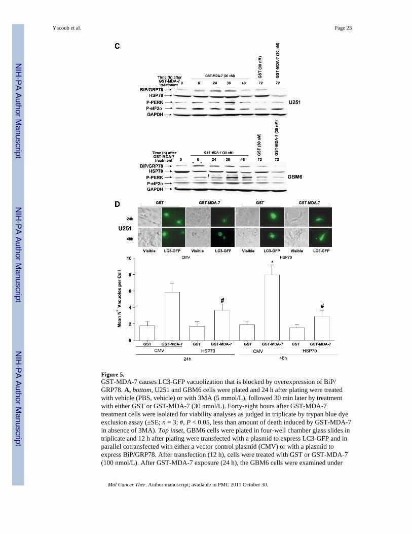
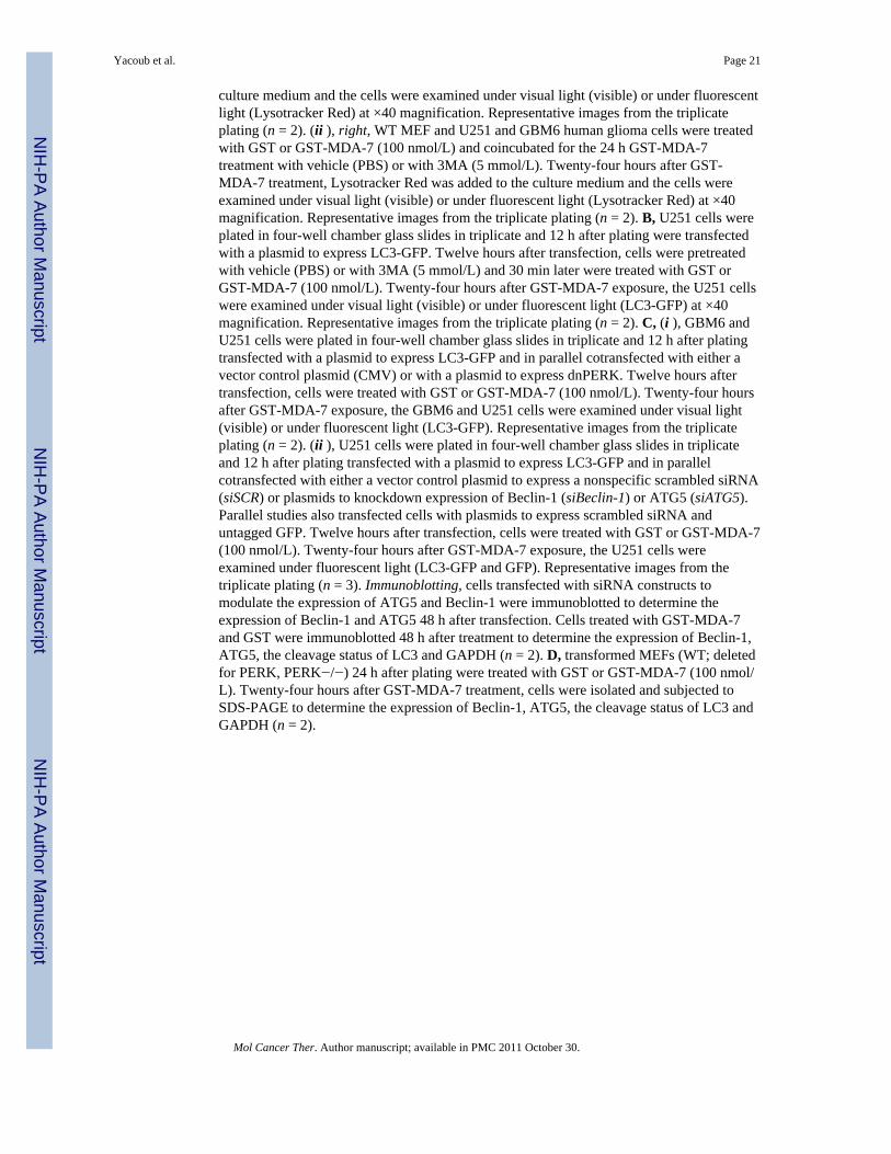
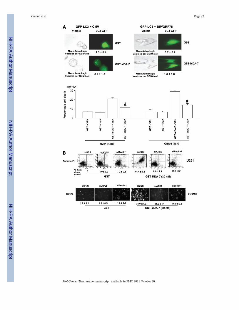
Figure 5.GST-MDA-7 causes LC3-GFP vacuolization that is blocked by overexpression of BiP/GRP78. A, bottom, U251 and GBM6 cells were plated and 24 h after plating were treatedwith vehicle (PBS, vehicle) or with 3MA (5 mmol/L), followed 30 min later by treatmentwith either GST or GST-MDA-7 (30 nmol/L). Forty-eight hours after GST-MDA-7treatment cells were isolated for viability analyses as judged in triplicate by trypan blue dyeexclusion assay (±SE; n = 3; #, P < 0.05, less than amount of death induced by GST-MDA-7in absence of 3MA). Top inset, GBM6 cells were plated in four-well chamber glass slides intriplicate and 12 h after plating were transfected with a plasmid to express LC3-GFP and inparallel cotransfected with either a vector control plasmid (CMV) or with a plasmid toexpress BiP/GRP78. After transfection (12 h), cells were treated with GST or GST-MDA-7(100 nmol/L). After GST-MDA-7 exposure (24 h), the GBM6 cells were examined under
Yacoub et al. Page 23
Mol Cancer Ther. Author manuscript; available in PMC 2011 October 30.
NIH
-PA Author Manuscript
NIH
-PA Author Manuscript
NIH
-PA Author Manuscript
visual light (visible) or under fluorescent light (LC3-GFP). The mean ±SE number of LC3-GFP vesicles per cell (n = 40) is indicated. Representative images from a triplicate plating (n= 2). B, GBM6 and U251 cells were plated in four-well chamber glass slides in triplicateand 12 h after plating transfected with either a vector control plasmid to express anonspecific scrambled siRNA or plasmids to knockdown expression of Beclin-1 or ofATG5. Twelve hours after transfection, cells were treated with GST or GST-MDA-7 (30nmol/L). Forty-eight hours after GST-MDA-7 exposure, the viability of the GBM6 andU251 cells was determined by terminal deoxynu-cleotidyl transferase – mediated dUTP nickend labeling assay on isolated cells (±SE; n = 3). Data shown are for GBM6 (terminal deox-ynucleotidyl transferase– mediated dUTP nick end labeling) and U251 cells was determinedby Annexin V/ PI flow cytometry (±SE; n = 2). C, U251 and GBM6 cells were treated withGST or with GST-MDA-7 (30 nmol/L). At the indicated time points, cells were isolated andlysates were subjected to immunoblotting to determine the expression of HSP70, BiP/GRP78, and GAPDH and the phosphorylation of PERK and eIF2α S51 (n = 2). D, U251cells were plated and 12 h after plating infected at a multiplicity of infection of 50 to expressno gene or HSP70. Twelve hours after infection, cells were transfected with a plasmid toexpress LC3-GFP. Twelve hours after transfection, cells were treated with GST or GST-MDA-7 (100 nmol/L). After GST-MDA-7 exposure (24 and 48 h), the U251 cells wereexamined under visual light (visible) or under fluorescent light (LC3-GFP). The mean ± SEnumber of LC3-GFP vesicles per cell (n = 40) is indicated (*, P < 0.05, greater thancorresponding 24-h value; #, P < 0.05, less than value in CMV-infected cells).Representative images from a triplicate plating (n = 2).
Yacoub et al. Page 24
Mol Cancer Ther. Author manuscript; available in PMC 2011 October 30.
NIH
-PA Author Manuscript
NIH
-PA Author Manuscript
NIH
-PA Author Manuscript

Figure 6.GST-MDA-7 causes cell killing by promoting PERK-dependent vacuolization and JNKpathway activation in transformed cells. A, U251 cells were plated and 12 h after platinginfected at a multiplicity of infection of 50 to express no gene or HSP70. Twenty-four hoursafter infection, cells were treated with GST or GST-MDA-7 (100 nmol/L). Forty-eight hoursafter GST-MDA-7 exposure, the viability of the U251 cells was determined by trypan blueexclusion assay and by Annexin V/PI staining (±SE; n = 2; #, P < 0.05, less than value inCMV-infected cells). B, treatment of transformed cells with GST-MDA-7 causes PERK-dependent activation of the JNK pathway and PERK-dependent inactivation of the ERK1/2pathway. JNK pathway signaling plays a key role in the activation of caspase-2 andcaspase-4 and cathepsin B; cathepsin B promotes the activation of the BH3 domain proteinBID. JNK pathway signaling also activates BAX. Inactivation of ERK1/2 promotesactivation of BAD and BIM. Thus, GST-MDA-7, via PERK signaling, promotes activationof at least four BH3 domain proteins, which cause mitochondrial dysfunction and promotecell killing.
Yacoub et al. Page 25
Mol Cancer Ther. Author manuscript; available in PMC 2011 October 30.
NIH
-PA Author Manuscript
NIH
-PA Author Manuscript
NIH
-PA Author Manuscript
NIH
-PA Author Manuscript
NIH
-PA Author Manuscript
NIH
-PA Author Manuscript
Yacoub et al. Page 26
Tabl
e 1
GST
-MD
A-7
cau
ses a
cas
pase
-9-d
epen
dent
indu
ctio
n of
prim
ary
hum
an g
liom
a ce
ll de
ath
Tre
atm
ent:
casp
ase
inhi
bito
rG
BM
5 G
STG
BM
5 G
ST-M
DA
-7G
BM
6 G
STG
BM
G G
ST-M
DA
-7G
BM
12 G
STG
BM
12 G
ST-M
DA
-7
Veh
icle
2.2
± 0.
634
.0 ±
2.1
4.4
± 0.
645
.2 ±
2.3
2.1
± 0.
820
.3 ±
0.8
z-V
AD
2.9
± 1.
07.
7 ±
0.6*
6.4
± 0.
411
.7 ±
1.5
*4.
0 ±
1.2
9.1
± 1.
5*
IETD
3.4
± 1.
034
.4 ±
2.9
4.8
± 0.
245
.0 ±
7.2
3.5
± 1.
420
.7 ±
2.7
LEH
D3.
6 ±
7.5
8.9
± 1.
7*3.
3 ±
2.3
12.0
± 1
.5*
4.7
± 1.
811
.0 ±
0.6
*
NO
TE: P
rimar
y hu
man
glio
ma
cells
(GB
M5,
GB
M6,
and
GB
M12
) wer
e tre
ated
24
h af
ter p
latin
g w
ith v
ehic
le (D
MSO
), a
pan-
casp
ase
inhi
bito
r (z-
VA
D),
a ca
spas
e-8
spec
ific
inhi
bito
r (IE
TD),
or a
casp
ase-
9 sp
ecifi
c in
hibi
tor (
LEH
D; a
ll at
50 μm
ol/L
) fol
low
ed 3
0 m
in la
ter b
y tre
atm
ent o
f cel
ls w
ith 2
0 nm
ol/L
GST
-MD
A-7
or G
ST. C
ell v
iabi
lity
was
det
erm
ined
96
h af
ter G
ST-M
DA
-7 tr
eatm
ent b
ytry
pan
blue
exc
lusi
on a
ssay
s in
tripl
icat
e us
ing
a he
mac
ytom
eter
(±SE
; n =
5).
* P <
0.05
, low
er a
mou
nt o
f cel
l kill
ing
than
veh
icle
-trea
ted
cells
.
Mol Cancer Ther. Author manuscript; available in PMC 2011 October 30.

NIH
-PA Author Manuscript
NIH
-PA Author Manuscript
NIH
-PA Author Manuscript
Yacoub et al. Page 27
Tabl
e 2
Cat
heps
in B
pla
ys a
n im
porta
nt ro
le in
GST
-MD
A-7
toxi
city
in tr
ansf
orm
ed c
ells
Cel
l typ
eT
reat
men
t
Veh
icle
z-V
AD
LE
HD
CM
V v
ehic
leD
omin
ant-n
egat
ive
casp
ase-
9 ve
hicl
eC
MV
cat
heps
in B
inhi
bito
rD
omin
ant-n
egat
ive
casp
ase-
9ca
thep
sin
B in
hibi
tor
WT
MEF
+ G
ST2.
1 ±
0.2
2.5
± 0.
22.
0 ±
0.3
WT
MEF
+ G
ST-M
DA
-717
.4 ±
0.3
6.3
± 0.
6*6.
9 ±
0.4*
Cat
heps
in B−
/− M
EF +
GST
1.6
± 0.
42.
9 ±
0.7
2.7
± 0.
5
Cat
heps
in B−
/− M
EF +
GST
-MD
A-7
6.8
± 0.
5*2.
7 ±
0.3*
3.1
± 0.
6*
GB
M6
+ G
ST3.
4 ±
0.3
5.0
± 0.
85.
7 ±
0.4
4.8
± 0.
8
GB
M6
+ G
ST-M
DA
-724
.2 ±
1.2
7.5
± 0.
2*9.
5 ±
1.1*
6.6
± 0.
2*
GB
M12
+ G
ST2.
0 ±
0.1
2.5
± 0.
23.
6 ±
0.5
5.1
± 0.
6
GB
M12
+ G
ST-M
DA
-716
.0 ±
0.6
5.3
± 0.
4*5.
4 ±
0.7*
4.5
± 0.
3*
NO
TE: M
EFs (
WT;
cat
heps
in B−
/−) w
ere
plat
ed a
nd 3
6 h
afte
r pla
ting
wer
e pr
etre
ated
with
veh
icle
(DM
SO),
pan-
casp
ase
inhi
bito
r (z-
VA
D, 5
0 μm
ol/L
), or
cas
pase
-9 in
hibi
tor (
LEH
D, 5
0 μm
ol/L
). Th
irty
min
utes
afte
r inh
ibito
r add
ition
, cel
ls w
ere
treat
ed w
ith G
ST o
r GST
-MD
A-7
(60
nmol
/L).
Inhi
bito
rs w
ere
resu
pple
men
ted
ever
y 24
h. C
ells
wer
e in
cuba
ted
for 7
2 h
afte
r whi
ch ti
me
cell
viab
ility
was
asse
ssed
in tr
iplic
ate
by tr
ypan
blu
e as
say
(±SE
; n =
3).
In p
aral
lel,
prim
ary
hum
an g
liom
a ce
lls (G
BM
6 an
d G
BM
12) w
ere
plat
ed a
nd 1
2 h
afte
r pla
ting
wer
e in
fect
ed w
ith a
deno
viru
ses a
t a fi
nal
mul
tiplic
ity o
f inf
ectio
n of
50
to e
xpre
ss a
s per
Fig
. 1: C
MV
or d
omin
ant-n
egat
ive
casp
ase-
9. C
ells
wer
e cu
lture
d fo
r 24
h af
ter i
nfec
tion
and
then
trea
ted
with
the
cath
epsi
n B
inhi
bito
r (1 μm
ol/L
, cat
heps
inB
inhi
bito
r) 3
0 m
in b
efor
e ad
ditio
n of
GST
or G
ST-M
DA
-7 (3
0 nm
ol/L
). C
ells
wer
e in
cuba
ted
for 7
2 h
afte
r whi
ch ti
me
cell
viab
ility
was
ass
esse
d in
trip
licat
e by
tryp
an b
lue
assa
y (±
SE; n
= 3
; #, P
< 0
.05,
low
er th
an C
MV
-infe
cted
veh
icle
-trea
ted
cells
with
GST
-MD
A-7
exp
osur
e; #
#, P
> 0
.05,
no
diff
eren
ce b
etw
een
GST
- and
GST
-MD
A-7
-trea
ted
cells
).
* P <
0.05
, low
er th
an v
ehic
le-tr
eate
d ce
lls w
ith G
ST-M
DA
-7 e
xpos
ure.
Mol Cancer Ther. Author manuscript; available in PMC 2011 October 30.
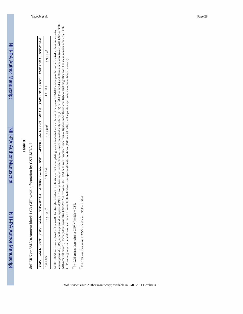
NIH
-PA Author Manuscript
NIH
-PA Author Manuscript
NIH
-PA Author Manuscript
Yacoub et al. Page 28
Tabl
e 3
dnPE
RK
or 3
MA
trea
tmen
t blo
ck L
C3-
GFP
ves
icle
form
atio
n by
GST
-MD
A-7
CM
V +
veh
icle
+ G
STC
MV
+ v
ehic
le +
GST
– M
DA
-7dn
PER
K +
veh
icle
+ G
STdn
PER
K +
veh
icle
+ G
ST +
MD
A-7
CM
V +
3M
A +
GST
CM
V +
3M
A +
GST
-MD
A-7
1.6
± 0.
55.
1 ±
0.8*
1.3
± 0.
41.
5 ±
0.3†
1.1
± 0.
41.
9 ±
0.4†
NO
TE: U
251
cells
wer
e pl
ated
in fo
ur-w
ell c
ham
ber g
lass
slid
es in
trip
licat
e an
d 12
h a
fter p
latin
g w
ere
trans
fect
ed w
ith a
pla
smid
to e
xpre
ss L
C3-
GFP
and
in p
aral
lel c
otra
nsfe
cted
with
eith
er a
vec
tor
cont
rol p
lasm
id (C
MV
) or w
ith a
pla
smid
to e
xpre
ss d
nPER
K. T
wel
ve h
ours
afte
r tra
nsfe
ctio
n, c
ells
wer
e pr
etre
ated
with
veh
icle
(PB
S) o
r 3M
A (5
mm
ol/L
) and
30
min
late
r wer
e tre
ated
with
GST
or G
ST-
MD
A-7
(100
nm
ol/L
). Tw
enty
-fou
r hou
rs a
fter G
ST-M
DA
-7 e
xpos
ure,
the
U25
1 ce
lls w
ere
exam
ined
und
er v
isua
l lig
ht o
r und
er fl
uore
scen
t lig
ht a
t ×40
mag
nific
atio
n. T
he m
ean
num
ber o
f int
ense
LC
3-G
FP st
aini
ng v
esic
les p
er c
ell w
as d
eter
min
ed fr
om m
ultip
le fi
elds
from
mul
tiple
trea
tmen
t con
ditio
ns (±
SE; n
= 4
0 ce
lls; n
= 3
sepa
rate
exp
erim
ents
; a re
pres
enta
tive
is sh
own)
.
* P <
0.05
gre
ater
than
val
ue in
CN
V +
Veh
icle
+ G
ST.
† P <
0.05
less
than
val
ue in
CN
V +
Veh
icle
+ G
ST –
ND
A-7
.
Mol Cancer Ther. Author manuscript; available in PMC 2011 October 30.
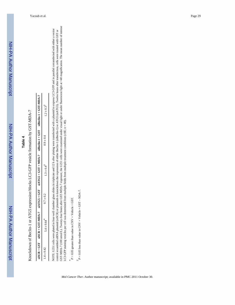
NIH
-PA Author Manuscript
NIH
-PA Author Manuscript
NIH
-PA Author Manuscript
Yacoub et al. Page 29
Tabl
e 4
Kno
ckdo
wn
of B
eclin
-1 o
r ATG
5 ex
pres
sion
blo
cks L
C3-
GFP
ves
icle
form
atio
n by
GST
-MD
A-7
siSC
R +
GST
siSC
R +
GST
-MD
A-7
siA
TG
5 +
GST
siA
TG
5 +
GST
+ M
DA
-7si
Bec
lin-1
+ G
STsi
Bec
lin-1
+ G
ST-M
DA
-7
1.4
± 0.
425.
4 ±
0.64
*0.
7 ±
0.2
1.3
± 0.
4†0.
9 ±
0.4
1.2
± 0.
3†
NO
TE: U
251
cells
wer
e pl
ated
in fo
ur-w
ell c
ham
ber g
lass
slid
es in
trip
licat
e an
d 12
h a
fter p
latin
g w
ere
trans
fect
ed w
ith a
pla
smid
to e
xpre
ss L
C3-
GFP
and
in p
aral
lel c
otra
nsfe
cted
with
eith
er a
vec
tor
cont
rol s
cram
bled
siR
NA
pla
smid
(siS
CR
) or p
lasm
ids t
o kn
ockd
own
the
expr
essi
on o
f eith
er B
eclin
-1 (s
iBec
lin-1
) or A
TG5
(siA
TG5)
. Tw
elve
hou
rs a
fter t
rans
fect
ion,
cel
ls w
ere
treat
ed w
ith G
ST o
rG
ST-M
DA
-7 (1
00 n
mol
/L).
Twen
ty-f
our h
ours
afte
r GST
-MD
A-7
exp
osur
e, th
e U
251
cells
wer
e ex
amin
ed u
nder
vis
ual l
ight
or u
nder
fluo
resc
ent l
ight
at ×
40 m
agni
ficat
ion.
The
mea
n nu
mbe
r of i
nten
seLC
3-G
FP st
aini
ng v
esic
les p
er c
ell w
as d
eter
min
ed fr
om m
ultip
le fi
elds
from
mul
tiple
trea
tmen
t con
ditio
ns (±
SE; n
= 4
0).
* P <
0.05
gre
ater
than
val
ue in
CN
V +
Veh
icle
+ G
ST.
† P <
0.05
less
than
val
ue in
CN
V +
Veh
icle
+ G
ST –
ND
A-7
.
Mol Cancer Ther. Author manuscript; available in PMC 2011 October 30.