Polyglutamic acid–PEG nanocapsules as long circulating carriers for the delivery of docetaxel

Docetaxel-induced apoptosis in melanoma cells isdependent on activation of caspase-2
Nizar M. Mhaidat, Yufang Wang, Kelly A. Kiejda,Xu Dong Zhang, and Peter Hersey
Immunology and Oncology Unit, Newcastle MisericordiaeHospital, Newcastle, New South Wales, Australia
AbstractTaxanes have a broad spectrum of activity against varioushuman cancers, including melanoma. In this study, wehave examined the molecular mechanism of docetaxel-induced apoptosis of human melanoma. We report thatdocetaxel induced varying degrees of apoptosis in a panelof melanoma cell lines but not in normal fibroblasts.Induction of apoptosis was caspase dependent andassociated with changes in mitochondrial membranepotential that could be inhibited by overexpression ofBcl-2. Docetaxel induced changes in Bax that correlatedwith sensitivity to docetaxel-induced apoptosis. Thesechanges in Bax were not inhibited by overexpression ofBcl-2. Kinetic studies of caspase-2 activation by Westernblotting and fluorogenic assays revealed that activationof caspase-2 seemed to be the initiating event. Inhibitionof caspase-2 with z-VDVAD-fmk or by small interferingRNA knockdown inhibited changes in Bax and mitochon-drial membrane potential and events downstream ofmitochondria. Activation of caspase-8 and Bid seemed tobe a late event, and docetaxel was able to induceapoptosis in cells deficient in caspase-8 and Bid. p53 didnot seem to be involved as a p53 null cell line wassensitive to docetaxel and an inhibitor of p53 did notinhibit apoptosis. Small interfering RNA knockdown ofPUMA and Noxa also did not inhibit apoptosis. Theseresults suggest that docetaxel induces apoptosis inmelanoma cells by pathways that are dependent onactivation of caspase-2, which initiates mitochondrialdependent apoptosis by direct or indirect activation ofBax. [Mol Cancer Ther 2007;6(2):752–61]
IntroductionThe treatment of patients with metastatic melanomacontinues to be a therapeutic challenge, and their prognosisis very poor. This is largely due to its unresponsiveness toconventional chemotherapeutic and biological reagents,which has been attributed to development of resistance toapoptosis (1–3). Understanding and overcoming resistancemechanism(s) of melanoma to apoptosis would thereforefacilitate identification of new therapeutic targets anddevelopment of new treatments (4–6).Taxanes, such as paclitaxel and docetaxel, represent an
important class of anticancer agents that have anticancereffects in vitro and in vivo against cancers of lung, ovaries,breast, leukemia, and malignant melanoma (7). They wereisolated from the bark of the American yew (Taxusbrevifolia) and later synthesized from the foliage and seedsof European yew (Taxus baccata). Initially, taxanes weredescribed as antimitotic agents that bind to h-tubulin,resulting in block of the cell cycle at the G2-M phase andapoptosis of cells (8, 9). When given as a single agent,taxanes have produced low response rates (3.3–17%) inpatients with melanoma. In some patients, taxanes wereassociated with prolonged duration of disease control.Higher response rates were seen (12–41%) when taxaneswere given in association with other anticancer agents,such as temozolomide, dacarbazine, cisplatin, carboplatin,or tamoxifen (10, 11).Chemotherapeutic agents in general are believed to
induce apoptosis through the intrinsic mitochondrialpathway via release of apoptogenic proteins such ascytochrome c and second mitochondrial-derived activatorof caspase/direct inhibitor of apoptosis–binding proteinwith low isoelectric point (Smac/DIABLO; refs. 12–16). Inone model of apoptotic regulation, apoptotic stimuli suchas damage to DNA or the cytoskeleton, or interaction ofcells with death-inducing ligands such as tumor necrosisfactor–related apoptosis–inducing ligand (TRAIL), induceactivation of proapoptotic BH3-only ‘‘sensor’’ proteins ofBcl-2 family, such as Noxa, PUMA, Bim, and Bid. Thesensor proteins are believed to bind to the antiapoptoticBcl-2 family members and promote binding of themultidomain proapoptotic proteins Bax and Bak to themitochondrial outer membrane, where they initiatechanges in mitochondrial membrane potential (17, 18).The biochemical pathways initiated by taxanes that
lead to cell death are poorly understood and seem tovary between different types of cancer. Lymphoma cellstreated with paclitaxel were reported to undergo deathreceptor–independent activation of caspase-8, which set upa mitochondrial amplification loop (8, 19). Studies onanother lymphoblastic cell line implicated death receptor–independent activation of caspase-10 as the key event inpaclitaxel-induced apoptosis (20).
Received 9/13/06; revised 11/20/06; accepted 12/20/06.
Grant support: New South Wales State Cancer Council and National Healthand Medical Research Council, Australia. X.D. Zhang is a Cancer InstituteNSW Fellow.
The costs of publication of this article were defrayed in part by thepayment of page charges. This article must therefore be hereby markedadvertisement in accordance with 18 U.S.C. Section 1734 solely toindicate this fact.
Requests for reprints: Peter Hersey or Xu Dong Zhang, Immunology andOncology Unit, Royal Newcastle Hospital, Room 443, David MaddisonClinical Sciences Building, Corner King and Watt Streets, Newcastle,NSW 2300, Australia. Phone: 61-2-49-236828; Fax: 61-2-49236184.E-mail: [email protected] or [email protected]
Copyright C 2007 American Association for Cancer Research.
doi:10.1158/1535-7163.MCT-06-0564
752
Mol Cancer Ther 2007;6(2). February 2007
on May 5, 2016. © 2007 American Association for Cancer Research. mct.aacrjournals.org Downloaded from

In the present study, we examined the apoptosis-inducing potential of docetaxel in cultured melanoma celllines and explored the biochemical pathways involved. Theresults indicate that apoptosis was caspase dependent andwas mediated through the mitochondria. Activation ofcaspase-2 seemed to be the initiating event, whereasactivation of caspase-8 was a delayed event resulting fromactivation of caspases.
Materials andMethodsCell LinesHuman melanoma cell lines Me4405, Me1007, IgR3, Mel-
FH, Mel-RM, Mel-CV, Mel-AT, SK-mel-28, SK-mel-110, andMM200 have been described previously (21). The cell lineswere cultured in DMEM containing 5% FCS (Common-wealth Serum Laboratories, Melbourne, Australia).
Antibodies and Other ReagentsDocetaxel (Taxotere) was kindly provided by Aventis
Pharma SA (Antony, France). Docetaxel was stored as a 100mmol/L solution in absolute ethanol at �80jC and dilutedwith the DMEM before use. Recombinant human TRAILwas supplied by Immunex (Seattle, WA). The rabbitpolyclonal antibodies against caspase-2, caspase-3, caspase-8, and caspase-9, and the mouse monoclonal antibodiesagainst cytochrome c and poly(ADP)ribose polymerase(PARP) were purchased from PharMingen (Bioclone, Mar-rickville, Australia). The rabbit polyclonal anti-Bax againstamino acids 1 to 20 was purchased from Upstate Biotech-nology (Lake Placid, NY). Propidium iodide was purchasedfrom Sigma-Aldrich (Castle Hill, NSW, Australia). The cell-permeable pan-caspase inhibitor Z-Val-Ala-Asp(OMe)-CH2F (z-VAD-fmk); the caspase-3– specific inhibitorZ-Asp(OMe)-Glu(OMe)-Val-Asp(OMe)-CH2F (z-DEVD-fmk); the caspase-9–specific inhibitor Z-Leu-Glu(Ome)-His-Asp(Ome)-CH2F (z-LEHD-fmk); the caspase-8–specificinhibitor Z-lle-Glu(Ome)-Thr-Asp(Ome)-CH2F (z-IETD-fmk); the caspase-2–specific inhibitor Z-Val-Ala-Asp(OMe)-Val-Ala-Asp(OMe)-CH2F (z-VDVAD-fmk); the mouseanti-Bak Ab-1 antibody; the caspase substrates z-VDVAD-AFC, z-DEVD-AFC, z-IETD-AFC, Ac-LEHD-AFC, to mea-sure caspase-2, caspase-3, caspase-8, and caspase-9 activities,respectively; and the p53 inhibitor 2-(2-imino-4,5,6,7-tetrahy-drobenzothiazol-3-yl)-1-(4-methylphenyl)ethanone (QB102)were purchased from Calbiochem (La Jolla, CA).
ApoptosisQuantitation of apoptotic cells by measurement of sub-G1
DNA content using the propidium iodide method wascarried out as described elsewhere (22). Quantitation ofapoptotic cells by Annexin V staining was carried out asdescribed previously and according to the manufacturer’sinstructions (23).In studies using QB102 (Calbiochem), a stock solution of
10 mmol/L was made by dissolving QB102 in DMSO, whichwas diluted with medium before use. Melanoma cells werepretreated with QB102 at 10 Amol/L for 3 h (24) beforeadding docetaxel for another 48 h. Cells were then collectedand the percentage of apoptotic cells was determined usingthe propidium iodide method in flow cytometry.
Flow CytometryImmunostaining on intact and permeabilized cells was
carried out as described previously (23). Analysis wascarried out using a Becton Dickinson (Mountain View, CA)FACScan flow cytometer. The percentage of antigen-positive cells was calculated as the difference in positivearea between the positive and negative control histograms.The positive area was that to the right of the intersection ofthe two curves (22).
Mitochondrial Membrane Potential (#ym)Tumor cells were seeded at 1 � 105 per well in 24-well
plates and allowed to reach exponential growth for 24 hbefore treatment. JC-1 staining was done according to themanufacturer’s instructions (Molecular Probes, Eugene,OR). Briefly, adherent cells and nonadherent cells werecollected and washed with PBS. Cells were then incubatedwith 10 Ag/mL of JC-1 in warm PBS at 37jC for 15 min.After washing with PBS, the cells were analyzed using aFACScan flow cytometer (Becton Dickinson, Sunnyvale,CA). Cells with polarized mitochondria presented in theupper-right quadrant of the dot plot due to the forma-tion of JC-1 aggregates, which emit orange fluorescence(590 nmol/L) when excited at 488 nmol/L. Cells with depo-larized mitochondria emit green fluorescence (530 nmol/L)and are visualized in the lower-right quadrant in the dotplot (25).
Western Blot and Protein Expression AnalysisThe protein content of cell extracts was determined by the
Bradford assay (Bio-Rad, Regents Park, NSW, Australia).A total of 20 to 30 Ag of protein were electrophoresed on10% to 15% SDS-PAGE gels and transferred to nitrocellu-lose membranes. Membranes were blocked, incubated withprimary antibodies at the appropriate concentration, andsubsequently incubated with horseradish peroxidase–conjugated goat anti-rabbit IgG or goat anti-mouse IgG(1:3,000 dilutions; Bio-Rad). Labeled bands were detectedby Immun-Star horseradish peroxidase chemiluminescentkit, and images were captured. The intensity of the bandswas quantitated with the Bio-Rad VersaDoc image system(Bio-Rad). The relative expression of certain protein wasdetermined by dividing the densitometric value of the testprotein by that of the control [glyceraldehyde-3-phosphatedehydrogenase (GAPDH) or actin].
CaspaseActivityAssayThe caspase substrates z-VDVAD-AFC, z-DEVD-AFC, z-
IETD-AFC, and Ac-LEHD-AFC were used to measurecaspase-2, caspase-3, caspase-8, and caspase-9 activities,respectively. Briefly, after incubation with docetaxel at20 nmol/L for different time intervals, both the floating andadherent cells were collected, washed twice in ice-cold PBS,and lysed in 100 AL of caspase lysis buffer [25 mmol/LHEPES (pH 7.5), 5 mmol/L EDTA, 5 mmol/L MgCl2,10 mmol/L DTT, 10 Ag/mL each of pepstatine andleupeptin, 0.5% Triton X-100, and 2 mmol/L phenyl-methylsulfonyl fluoride]. Cells were kept on ice for 15 to20 min with occasional mixing. The lysate was thencentrifuged at 15,000 � g for 20 min at 4jC. Proteinconcentration of the supernatant was determined using the
Molecular Cancer Therapeutics 753
Mol Cancer Ther 2007;6(2). February 2007
on May 5, 2016. © 2007 American Association for Cancer Research. mct.aacrjournals.org Downloaded from

Bradford method (bicinchoninic acid protein assayreagent). Subsequently, 40 Ag of sample were diluted intoa final volume of 200 AL with the assay buffer (50 mmol/LHEPES, 10% sucrose, 0.1% CHAPS, and 10 mmol/L DTT)containing the fluorogenic caspase substrate at a finalconcentration of 100 Amol/L and incubated for 1 to 2 h in a96-well plate at 30jC. The generation of free AFC wasdetermined by fluorescence measurement using FluostarOPTIMA (LABTECH, Offenburg, Germany) set at anexcitation wavelength of 400 nm and an emission wave-length of 505 nm.
PlasmidVector and TransfectionStable Mel-RM transfectants of Bcl-2 were established by
electroporation of the PEF-puro vector carrying humanBcl-2 provided by Dr. David Vaux (Walter and Eliza HallInstitute, Melbourne, Victoria, Australia) and describedelsewhere (26).
Preparation ofMitochondrial and Cytosolic FractionsMethods used for subcellular fraction were similar to the
methods described previously (22).
SiRNA-Mediated Silencing of Caspase-2, Noxa,and PUMACells were transfected in six-well plates with caspase-2–,
Noxa-, or PUMA-specific or nontarget control small inter-fering RNA (siRNA) constructs (Dharmacon, Lafayette, CO)
for 24 h according to the manufacturer’s protocol. Theconstruct used was the siRNA SMARTpool caspase-2,Noxa, or PUMA. Cells were transfected with 100 nmol/Lof the above siRNA using LipofectAMINE (Invitrogen,Carlsbad, CA) according to the manufacturer’s protocol.Following silencing, cells were treated for another 24 or48 h with or without docetaxel at 20 nmol/L, and apoptoticcells were quantified by the propidium iodide method inflow cytometry. Efficiency of RNA interference wasassessed by immunoblotting.
Statistical AnalysisData are expressed as mean F ISE. The statistical sig-
nificance of intergroup differences in normally distributedcontinuous variables was determined using Student’s t test.P values >0.05 were considered statistically significant.
ResultsDocetaxel-Induced Apoptosis ofMelanoma CellsWe examined the apoptosis-inducing potential of doce-
taxel in melanoma cells by treating two melanoma celllines, IgR3 and MM200, with a range of docetaxelconcentrations as follows: 0, 1, 2.5, 5, 10, 20, 40, and80 nmol/L for 24 h. As shown in Fig. 1A, docetaxel inducedapoptosis of the melanoma cells in a dose-dependentmanner, with the highest percentage of apoptotic cells seen
Figure 1. Docetaxel-induced apoptosis of melanoma cells. A, titration of docetaxel on induction of apoptosis. IgR3 and MM200 cells were treated withdocetaxel at indicated doses for 24 h before measurement of apoptosis by the propidium iodide method using flow cytometry. Points, mean of threeindividual experiments; bars, ISE. B, kinetics of induction of apoptosis by docetaxel. IgR3 and MM200 cells were treated with docetaxel at 20 nmol/L forthe indicated time periods before measurement of apoptosis by the propidium iodide method using flow cytometry. Points, mean of three individualexperiments; bars, ISE. C, representative dot plots showing fluorescence channel analyses of melanoma cells after dual staining with Annexin V andpropidium iodide. IgR3 and MM200 cells were treated with docetaxel at 20 nmol/L for the indicated time periods and then stained with FITC-conjugatedAnnexin V (green fluorescence, horizontal axis ) and propidium iodide (red fluorescence, vertical axis ) and analyzed using flow cytometry. Annexin Vbinding to phosphatidylserine in the absence of propidium iodide staining is indicative of early apoptosis. Representative of three individual experiments.D, docetaxel-induced apoptosis in a panel of melanoma cell lines but not in normal fibroblasts. Cells were treated with either TRAIL at 200 ng/mL ordocetaxel at 20 nmol/L for 24 and 48 h, respectively, and apoptosis was measured by the propidium iodide method using flow cytometry. Columns, meanof three individual experiments; bars, ISE.
Caspase-2 and Doxetaxel-Induced Apoptosis754
Mol Cancer Ther 2007;6(2). February 2007
on May 5, 2016. © 2007 American Association for Cancer Research. mct.aacrjournals.org Downloaded from

at 20 to 40 nmol/L. Figure 1B indicates that apoptosis ofIgR3 cells was detected after 3 h of treatment and peaked at36 to 48 h. This concentration seems clinically relevantbased on the reported plasma concentration of docetaxel inpatients treated with docetaxel (27).Dual staining with Annexin V and propidium iodide
shown in Fig. 1C indicated that reactivity with Annexin Vpreceded the staining with propidium iodide, consistentwith induction of apoptosis rather than necrosis. Forexample, only 4.2% of the sensitive IgR3 cells stained withpropidium iodide alone at 48 h. A summary of studies ofdocetaxel on a panel of melanoma cell lines is illustratedin Fig. 1D. TRAIL, known to induce apoptosis in twothirds of melanoma cells (23), was used as a positivecontrol (200 ng/mL for 16 h). Results showed that therewas very little concordance between the degree ofapoptosis induced by TRAIL and docetaxel in themelanoma cell lines.
Docetaxel Induces Caspase-Dependent Apoptosis inMelanoma CellsTo assess the role of caspases in docetaxel-induced
apoptosis, IgR3 and MM200 cells were pretreatedwith the pan-caspase inhibitor z-VAD-fmk (20 Amol/L),or specific inhibitors of caspase-2, z-VDVAD-fmk(50 Amol/L), caspase-3, z-DEVD-fmk (30 Amol/L), cas-pase-8, z-IETD-fmk (30 Amol/L), or caspase-9, z-LEHD-fmk (30 Amol/L). The inhibitors were added 1 h beforeadding TRAIL (200 ng/mL) or docetaxel (20 nmol/L) foranother 24 and 48 h, respectively. As shown in Table 1,the pan-caspase inhibitor z-VAD-fmk significantly re-duced docetaxel-induced apoptosis. Inhibition of doce-taxel-induced apoptosis by the specific inhibitors wasmost marked with that against caspase-2 and caspase-9in studies on the sensitive IgR3 melanoma cell line.
Inhibitor of caspase-8 had very little effect. All the inhi-bitors had a low level of activity against the low killingof the MM200 cell line.The involvement of caspases in docetaxel-induced
apoptosis was further examined by Western blot analysisbefore and after exposure to docetaxel. The kinetics ofcaspase activation in the two cell lines is shown in Fig. 2A.In IgR3 cells, cleavage of caspase-2 was evident by 12 h,whereas cleavage of caspase-9 and caspase-3 was evidentby 16 h. Cleavage of PARP was evident by 16 h. Activationof caspase-8 was only evident at 24 to 36 h. In MM200 cells,cleavage of caspase-3, caspase-9, and PARP was detectableonly at 24 to 36 h. Figure 2A also shows the effect oftreatment with docetaxel on X-linked inhibitor of apoptosisexpression, which was reported to be cleaved by activatedcaspase-3 (28). Reduction in density of the X-linkedinhibitor of apoptosis protein bands was evident in thesensitive cell line IgR3 by 16 to 36 h but not in the resistantMM200 cells.Previous studies suggested that caspase-2 can act up-
stream of mitochondria and induce cytochrome c and AIFrelease (29–31). To examine whether caspase-2 is requiredfor docetaxel-induced apoptosis, we assessed the kinetics ofactivation of caspase-2, caspase-8, and caspase-9 and theeffector caspase-3 using the caspase fluorogenic substrates z-VDVAD-AFC, z-DEVD-AFC, z-IETD-AFC, and z-LEHD-AFC to measure caspase-2, caspase-3, caspase-8, andcaspase-9 activities, respectively (Fig. 2B). Caspase-2appeared to be activated first and to a higher degreecompared with other caspases at 3 h after treatment ofIgR3 cells with docetaxel. Activated caspase-3 correlatedwell with activated caspase-9. Activation of caspase-8 wasdelayed and became active after caspase-3 activation,indicating that caspase-8 was unlikely to be an initiator ofdocetaxel-induced apoptosis.
Docetaxel Activates the Mitochondrial ApoptoticPathwayTo identify the apoptotic pathway being activated in
response to docetaxel, we measured the mitochondrialmembrane potential in melanoma cells treated withdocetaxel for different time periods. Changes in mito-chondrial membrane potential were studied using afluorescent cationic dye 5,5¶,6,6¶-tetrachloro-1,1¶,3,3¶-tetraethyl-benzamidazolocarbocyanin iodide, known asJC-1. In healthy cells, JC-1 exists as a monomer in thecytosol (FL1-positive; green) and also accumulates asaggregates in the mitochondria (FL2-positive; red). Inapoptotic cells, JC-1 exists exclusively in monomer formand produces a green cytosolic signal (32). As shown inFig. 2C, docetaxel induced mitochondrial depolarizationdetected at 12 h in both IgR3 and MM200 cells andpeaked at 48 h. Reduction in mitochondrial membranepotential was at low levels in MM200 cells comparedwith that in IgR3 cells.Docetaxel-induced mitochondrial membrane potential
changes were further examined by studying the releaseof cytochrome c and apoptosis-inducing factor (AIF) fromthe mitochondria to the cytosol in both sensitive and
Table 1. Effect of caspase inhibitors on docetaxel-inducedapoptosis of melanoma cells
Percentage inhibition*
Cell linec IgR3 MM200
Caspase inhibitor Docetaxel TRAIL Docetaxel TRAIL
No inhibitorb 35.5 F 3.2 11.3 F 1.9 10.3 F 1.8 67.8 F 4.61z-VAD-fmk 67 F 3.1 97 F 1.2 27 F 2.1 98.2 F 0.98z-VDVAD-fmk 56 F 2.4 68 F 4.21 23 F 1.7 43.1 F 3.1z-DEVD-fmk 37 F 2.89 98 F 1.1 25 F 2 72.1 F 3.98z-IETD-fmk 12.6 F 2.9 98 F 0.78 19 F 1.9 82 F 4.1z-LEHD-fmk 45 F 3.16 98 F 1 19.3 F 1.2 87 F 3.67
*Data are the mean F SE of three individual experiments.cIgR3 and MM200 cells were pretreated with the pan-caspase inhibitor z-VAD-fmk (20 Amol/L), the caspase-2 inhibitor z-VDVAD-fmk (50 Amol/L),the caspase-3 inhibitor z-DEVD-fmk (30 Amol/L), the caspase-8 inhibitor z-IETD-fmk (30 Amol/L), or the caspase-9 inhibitor z-LEHD-fmk (30 Amol/L),1 h before adding TRAIL (200 ng/mL) or docetaxel (20 nmol/L) for another24 and 48 h, respectively. Apoptosis was measured by the propidium iodidemethod using flow cytometry.bApoptosis level induced by docetaxel and TRAIL.
Molecular Cancer Therapeutics 755
Mol Cancer Ther 2007;6(2). February 2007
on May 5, 2016. © 2007 American Association for Cancer Research. mct.aacrjournals.org Downloaded from
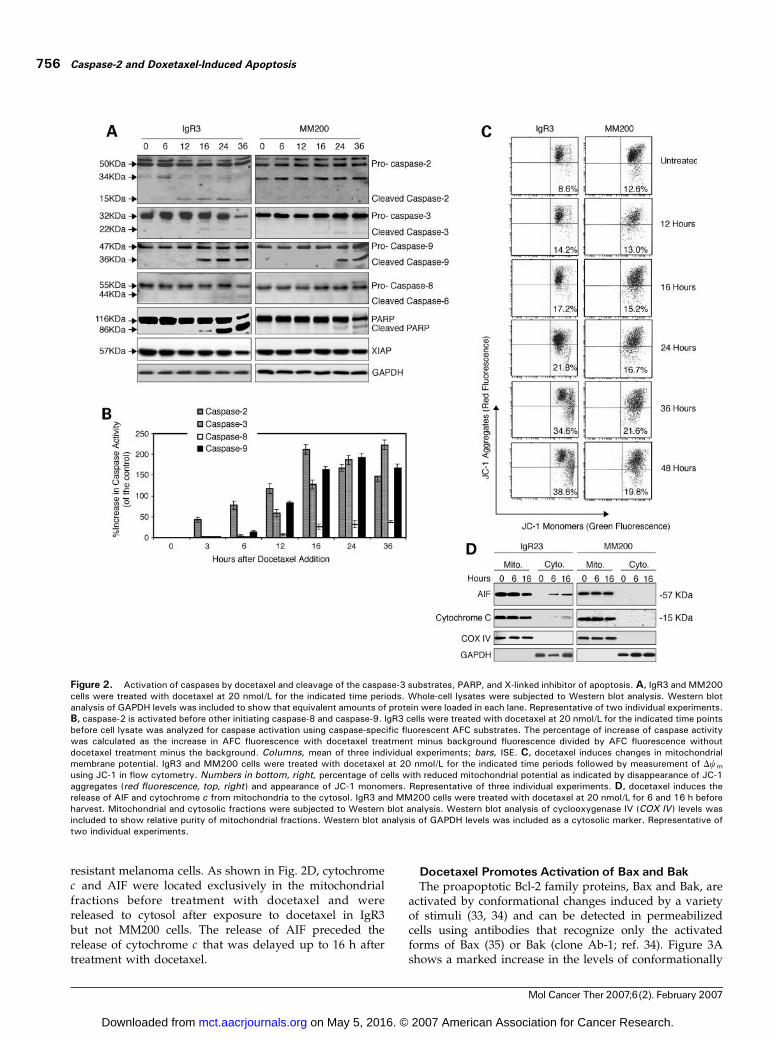
resistant melanoma cells. As shown in Fig. 2D, cytochromec and AIF were located exclusively in the mitochondrialfractions before treatment with docetaxel and werereleased to cytosol after exposure to docetaxel in IgR3but not MM200 cells. The release of AIF preceded therelease of cytochrome c that was delayed up to 16 h aftertreatment with docetaxel.
Docetaxel Promotes Activation of Bax and BakThe proapoptotic Bcl-2 family proteins, Bax and Bak, are
activated by conformational changes induced by a varietyof stimuli (33, 34) and can be detected in permeabilizedcells using antibodies that recognize only the activatedforms of Bax (35) or Bak (clone Ab-1; ref. 34). Figure 3Ashows a marked increase in the levels of conformationally
Figure 2. Activation of caspases by docetaxel and cleavage of the caspase-3 substrates, PARP, and X-linked inhibitor of apoptosis. A, IgR3 and MM200cells were treated with docetaxel at 20 nmol/L for the indicated time periods. Whole-cell lysates were subjected to Western blot analysis. Western blotanalysis of GAPDH levels was included to show that equivalent amounts of protein were loaded in each lane. Representative of two individual experiments.B, caspase-2 is activated before other initiating caspase-8 and caspase-9. IgR3 cells were treated with docetaxel at 20 nmol/L for the indicated time pointsbefore cell lysate was analyzed for caspase activation using caspase-specific fluorescent AFC substrates. The percentage of increase of caspase activitywas calculated as the increase in AFC fluorescence with docetaxel treatment minus background fluorescence divided by AFC fluorescence withoutdocetaxel treatment minus the background. Columns, mean of three individual experiments; bars, ISE. C, docetaxel induces changes in mitochondrialmembrane potential. IgR3 and MM200 cells were treated with docetaxel at 20 nmol/L for the indicated time periods followed by measurement of Dwmusing JC-1 in flow cytometry. Numbers in bottom, right, percentage of cells with reduced mitochondrial potential as indicated by disappearance of JC-1aggregates (red fluorescence, top, right ) and appearance of JC-1 monomers. Representative of three individual experiments. D, docetaxel induces therelease of AIF and cytochrome c from mitochondria to the cytosol. IgR3 and MM200 cells were treated with docetaxel at 20 nmol/L for 6 and 16 h beforeharvest. Mitochondrial and cytosolic fractions were subjected to Western blot analysis. Western blot analysis of cyclooxygenase IV (COX IV ) levels wasincluded to show relative purity of mitochondrial fractions. Western blot analysis of GAPDH levels was included as a cytosolic marker. Representative oftwo individual experiments.
Caspase-2 and Doxetaxel-Induced Apoptosis756
Mol Cancer Ther 2007;6(2). February 2007
on May 5, 2016. © 2007 American Association for Cancer Research. mct.aacrjournals.org Downloaded from

changed Bax and Bak in IgR3, which started at 6 h forBax and 12 h for Bak and peaked at 36 h of treatmentwith docetaxel. In contrast, activated Bax and Bakappeared later in MM200 cells at 24 h, and changes weremaximal at 36 to 48 h.We examined the expression of Bax in different subcel-
lular fractions of IgR3 and MM200 cells with or withoutexposure to docetaxel. As shown in Fig. 3B, the Bax proteinwas predominantly located in the cytosol before treatmentwith docetaxel. In contrast, after treatment with docetaxelfor 16 h, Bax was observed in the mitochondrial fractionswith corresponding decrease in the levels of expression inthe cytosol. This was most evident in the docetaxel-sensitive IgR3 cells.
Overexpression of Bcl-2 Inhibits Docetaxel-InducedApoptosis inMelanoma CellsTo investigate whether the observed mitochondrial
dysfunction is required for docetaxel-induced apoptosis,
we transfected cDNA encoding Bcl-2 into Mel-RM cells asshown in Fig. 3C. There was a marked increase in the levelsof Bcl-2 in the Bcl-2–transfected cells, but the levels in thecells transfected with the vector alone were similar to thosein the parental cells. Apoptosis of melanoma induced byTRAIL, which is known to induce apoptosis of melanomapredominantly through the mitochondrial apoptotic path-way, was almost completely inhibited in the Bcl-2 trans-fectants. Similarly, the levels of docetaxel-inducedapoptosis in Bcl-2–transfected cells were markedly de-creased compared with those in vector alone–transfectedcells. As shown in Fig. 3D, docetaxel-induced changes in themitochondrial membrane potential were reversed in Bcl-2–transfected cells but not in cells transfected with the vectoralone. Similarly, docetaxel-induced caspase-3 activationand Bak conformational changes were inhibited by Bcl-2overexpression. Docetaxel-induced Bax conformationalchanges were not affected in Bcl-2–overexpressing cells.
Figure 3. Docetaxel activates the mitochondrial apoptosis pathway. A, docetaxel induces conformational changes of Bax and Bak. IgR3 and MM200cells were treated with or without docetaxel at 20 nmol/L for the indicated time periods before being subjected to flow cytometry analysis using either aBax NH2-terminal epitope-specific antibody or anti-Bak Ab-1 that specifically recognize activated forms of Bax or Bak, respectively. Representative of threeindividual experiments. B, docetaxel induces Bax relocation from the cytosol to mitochondria. IgR3 and MM200 cells were treated with docetaxel for16 h before harvest. The mitochondrial and cytosolic fractions were subjected to Western blot analysis. Western blot analysis of cyclooxygenase IV andGAPDH levels was included as controls. Representative of two individual experiments. C, overexpression of Bcl-2 inhibits docetaxel-induced apoptosis.Left, Bcl-2 was overexpressed in Mel-RM cells transfected with the cDNA encoding Bcl-2, but not in the cells transfected with the vector alone. Whole-celllysates were subjected to Western blot analysis. Western blot analysis of GAPDH levels was included to show that equivalent amounts of protein wereloaded in each lane. Representative of two individual experiments. Right, Mel-RM cells transfected with c-DNA for Bcl-2 or vector alone were treated witheither TRAIL at 200 ng/mL or docetaxel at 20 nmol/L for 24 and 48 h, respectively, before measurement of apoptosis by the propidium iodide method usingflow cytometry. Columns, mean of three individual experiments; bars, ISE.D, overexpression of Bcl-2 inhibited docetaxel-induced Dwm changes, caspase-3 activation, and conformational changes of Bak but not Bax. Mel-RM cells transfected with cDNA for Bcl-2 or vector alone were treated with docetaxel at20 nmol/L for 24 h. The changes in Dwm were measured by JC-1 using flow cytometry. Activated forms of caspase-3, Bax, and Bak were measured in flowcytometry using a monoclonal antibody that recognizes the processed caspase-3, a Bax NH2-terminal epitope-specific antibody, or anti-Bak Ab-1,respectively. Columns, mean of three individual experiments; bars, ISE.
Molecular Cancer Therapeutics 757
Mol Cancer Ther 2007;6(2). February 2007
on May 5, 2016. © 2007 American Association for Cancer Research. mct.aacrjournals.org Downloaded from

Premitochondrial Caspase-2 Activation Is Requiredfor Docetaxel-Induced ApoptosisTo explore the role of caspase-2 in docetaxel-induced
apoptosis, z-VDVAD-fmk, a specific inhibitor of caspase-2, was used. As shown in Fig. 4A, inhibition of caspase-2significantly impaired mitochondrial membrane potentialchanges induced by docetaxel and decreased release ofAIF (by 49% of that induced by docetaxel alone) andcytochrome c (by 52%) from the mitochondria. Consistentwith these results, cleavage of pro-caspase-9, pro-caspase-3, and PARP was markedly reduced after pretreat-ment with the caspase-2 inhibitor (Fig. 4B). Caspase-2seemed to act upstream of activation of the multidomainproteins Bax and Bak. Caspase-2 inhibitor reduced acti-vation of both Bax and Bak and inhibits Bax transloca-tion to mitochondria (Fig. 4C). Furthermore, inhibition ofcaspase-2 using a siRNA specific for caspase-2 resultedin a marked inhibition of docetaxel-induced apoptosis(Fig. 4D).
Docetaxel-Induced Apoptosis Is Independent of p53Previous studies have shown that caspase-2 may be
activated by a complex of a p53-induced death domaincontaining protein and an adaptor protein RAIDD (36). Inview of this, we examined activation of p53 in IgR3 andMM200 cell lines by Western blots and used an inhibitor ofp53 to determine its possible role in docetaxel-inducedactivation. Our results showed that p53 levels wereincreased by 16 h in the resistant MM200 cells, but p53did not undergo significant change in the sensitive IgR3cells (Fig. 5A). ME4405 cells do not express p53 but weresensitive to docetaxel (data are not shown). Noxa andPUMA are known targets of p53. Noxa was up-regulated inboth cell lines from 6 to 24 h, and PUMA was up-regulatedin both cells by 16 h. Knockdown of these proteinsindividually by siRNA, however, did not decrease doce-taxel-induced apoptosis of the sensitive IgR3 line (Fig. 5B).Combining siRNA for both Noxa and PUMA also did notdecrease docetaxel-induced apoptosis (data not shown).
Figure 4. Caspase-2 is activated upstream of mitochondria, caspase-9, and caspase-3. A, caspase-2 is upstream of the mitochondrial membranechanges (left ). IgR3 and MM200 cells were treated with or without the caspase-2 inhibitor z-VDVAD-fmk for 1 h before the addition of docetaxel at20 nmol/L for another 48 h, followed by measurement of Dwm by JC-1 using flow cytometry. Columns, mean of three individual experiments; bars, ISE.Docetaxel-induced release of AIF and cytochrome c is suppressed by caspase-2 inhibition (right ). IgR3 cells with or without pretreatment with caspase-2inhibitor for 1 h were treated with docetaxel at 20 nmol/L for 16 h. Mitochondrial (M ) and cytosolic (C ) fractions were subjected to Western blot analysis.Western blot of cyclooxygenase IV levels was included to show relative purity of the mitochondrial fractions. Western blot analysis of GAPDH levels wasincluded as a cytosolic marker. Representative of two individual experiments. B, caspase-2 is upstream of caspase-9, caspase-3 activation, and PARPcleavage. IgR3 cells with or without pretreatment with the caspase-2 inhibitor z-VDVAD-fmk for 1 h were treated with docetaxel at 20 nmol/L for 16 h.Whole-cell lysates were subjected to Western blot analysis. Western blot analysis of GAPDH levels was included to show that equivalent amounts ofprotein were loaded in each lane. Representative of two individual experiments. C, caspase-2 is upstream of Bax and Bak activation. Inhibition of caspase-2suppresses conformational changes of Bax and Bak (left ). IgR3 cells with or without pretreatment with the caspase-2 inhibitor z-VDVAD-fmk at 50 Amol/Lfor 1 h were treated with docetaxel at 20 nmol/L for 36 h before flow cytometry analysis using a Bax NH2-terminal epitope-specific antibody or anti-BakAb-1. Representative of three individual experiments. Right, inhibition of caspase-2 suppresses Bax relocation from the cytosol to mitochondria. IgR3 cellswith or without pretreatment with the caspase-2 inhibitor z-VDVD-fmk for 1 h were treated with docetaxel at 20 nmol/L for 16 h before harvest. Themitochondrial and cytosolic fractions were subjected to Western blot analysis. Cyclooxygenase IV and GAPDH levels were included as controls asdescribed above. Representative of two individual experiments. D, down-regulation of caspase-2 expression in IgR3 cells inhibits docetaxel-inducedapoptosis. IgR3 cells were transfected with caspase-2–specific siRNA sequence (Dharmacon) for 24 h. The whole-cell lysates were subjected to Westernblot analyses (left ). Transfected cells were exposed to docetaxel for 48 h and assessed for apoptosis using the propidium iodide method (right ).Representative of two individual experiments.
Caspase-2 and Doxetaxel-Induced Apoptosis758
Mol Cancer Ther 2007;6(2). February 2007
on May 5, 2016. © 2007 American Association for Cancer Research. mct.aacrjournals.org Downloaded from

To further examine the role of p53, we used QB102, achemical inhibitor of p53, which is able to inhibit p53-dependent apoptosis by reversible inhibition of both p53transactivation activity and p53 downstream events (24). Inour studies, QB102 at 10 Amol/L was able to inhibitcisplatin-induced p53 up-regulation and its downstreamgene p21 in melanoma cells (data not shown). IgR3, Mel-AT, ME4405, and ME1007 were pretreated with QB102 at10 Amol/L for 3 h before adding docetaxel for another 48 h.As shown in Fig. 5C, p53 inhibition did not protectmelanoma cells against docetaxel-induced apoptosis.
DiscussionIn the present study, we show that docetaxel inducedvarying degrees of apoptotic cell death in a panel of
melanoma cell lines but did not induce apoptosis in normalfibroblasts. Apoptosis induced in melanoma cells includedthose that were resistant to TRAIL, a member of the tumornecrosis factor family that induces apoptosis in about twothirds of melanoma cell lines (23). In contrast to TRAIL-induced apoptosis (22), docetaxel-induced apoptosis ofmelanoma proceeded much more slowly (3–48 h) andseemed to involve activation of caspase-2 as the initiatingcaspase. This was shown by early activation of caspase-2compared with other caspases using Western blot andfluorogenic substrate assays for caspase-2, caspase-3,caspase-8, and caspase-9. Moreover, inhibition of caspase-2 by z-VDVAD-fmk or by siRNA knockdown significantlyprotected melanoma cells from docetaxel-induced apopto-sis. Apoptosis seemed to proceed via the mitochondrialpathway, as shown by changes in mitochondrial membrane
Figure 5. Docetaxel-induced apoptosis does not involve p53 activation. A, IgR3 and MM200 cells were treated with docetaxel at 20 nmol/L for theindicated time periods and whole-cell lysates were subjected to Western blot analysis. Docetaxel did not induce changes in p53 protein levels in thesensitive IgR3 cells, whereas p53 and p21 was up-regulated in the resistant MM200 cells. GAPDH blot was included to show that equivalent amounts ofprotein were loaded in each lane. Representative of two individual experiments. B, down-regulation of Noxa or PUMA expression in IgR3 cells does notaffect docetaxel-induced apoptosis. IgR3 cells were transfected with either Noxa- or PUMA-specific siRNA sequence (Dharmacon) for 24 h. Cells were thentreated with or without docetaxel for another 16 h and whole-cell lysates were subjected to Western blot analyses (left ), or were exposed to docetaxel for24 h and assessed for apoptosis using the propidium iodide method (right ). Representative of three individual experiments. Columns, mean of threeindividual experiments; bars, ISE. C, inhibition of p53 does not protect melanoma cells against docetaxel-induced apoptosis. IgR3, Me1007, and Mel-ATcells were treated with the p53 inhibitor QB102 at 10 Amol/L 3 h before adding docetaxel at 20 nmol/L for another 48 h. Apoptosis was measured by thepropidium iodide method using flow cytometry. Columns, means of three individual experiments; bars, ISE.
Molecular Cancer Therapeutics 759
Mol Cancer Ther 2007;6(2). February 2007
on May 5, 2016. © 2007 American Association for Cancer Research. mct.aacrjournals.org Downloaded from

potential associated with release of AIF and cytochrome c,activation of caspase-9 and caspase-3, and cleavage ofPARP. Moreover, docetaxel-induced apoptosis was inhi-bited by overexpression of Bcl-2 in melanoma cells.Analysis of changes in the multidomain proapoptotic
proteins Bax and Bak by flow cytometry revealed thattreatment with docetaxel resulted in conformationalchanges in both proteins that correlated with the onset ofdocetaxel-induced apoptosis. These changes were notinhibited by overexpression of Bcl-2, suggesting that theywere upstream of the mitochondria. Importantly, inhibitionof caspase-2 with z-VDVAD-fmk prevented docetaxel-induced changes in Bax, indicating that changes incaspase-2 were responsible for conformational changesin Bax.Several previous studies have also suggested that caspase-2
is an apical caspase for stress-induced apoptosis via acti-vation of Bax (29, 37). The mechanisms involved, however,were not clear. Caspase-2 has a caspase recruitment domainprodomain, which can associate with RAIDD, an adaptorprotein containing a caspase recruitment domain anddeath domain (38). RAIDD was proposed to interact withPIP1 and thereby to link tumor necrosis factor receptor 1activationwith caspase-2–mediated apoptosis (38, 39).Morerecently, caspase-2 was shown to be recruited to the DISCcomplex in T and B cells but did not initiate apoptosismediated through Fas (40). Paroni et al. (41) suggested thatcaspase-2 was able to trigger mitochondrial dysfunctionin response to genotoxic agents, in complex with the p53-induced death domain–containing protein and the adaptorprotein RAIDD (36).Several of these proposed mechanisms, such as involve-
ment of death receptors, seem unlikely to explain its role indocetaxel-induced killing in that melanoma cells deficientin caspase-8 (Me-1007) or Bid (IgR3) were sensitive to theagent. Mechanisms dependent on activation of p53 also didnot seem to be involved as p53 did not undergo significantincreases in response to docetaxel in the sensitive IgR3cells. ME4405 cells, which do not contain p53, weresensitive to docetaxel. Inhibition of p53 activation withQB102, a chemical that reversibly blocks p53-dependenttransactivation of p53-responsive genes and inhibits p53-mediated apoptosis (24), was unable to protect melanomacells against docetaxel-induced apoptosis. We detected up-regulation of the p53-dependent protein Noxa and PUMA,but siRNA knockdown studies of these proteins had noeffect on docetaxel-induced apoptosis. Studies on corticalneurons reported cytoprotective mechanisms in which Baxspecifically interacted with certain proteins that sup-pressed Bax translocation. It was proposed that caspase-2could inhibit this interaction and allow Bax to be relocatedto mitochondria (42, 43).In summary, docetaxel-induced apoptosis of melanoma
cells seems to depend largely on activation of caspase-2,which induces conformational changes in Bax and
subsequent events leading to apoptosis. To our knowl-edge, this is the first study to identify caspase-2 as animportant mediator of apoptosis induced by taxanes inmelanoma. Whether the same mechanism applies againstfresh isolates of melanoma is yet to be determined but ifsubstantiated in such isolates, the present studies mayprovide valuable insights into the basis for sensitivity orresistance of melanoma to docetaxel. As reported else-where, cisplatin induces more rapid apoptosis of melano-ma through the mitochondrial pathway (44). However,apoptosis induced by cisplatin or by vincristine does notinvolve caspase-2,1 suggesting that the taxanes induceapoptosis by different mechanism to these drugs. Theseand ongoing studies promise to provide valuable insightsthat may assist in more effective use of the taxanes intreatment of melanoma.
References
1. Soengas MS, Lowe SW. Apoptosis and melanoma chemoresistance.Oncogene 2003;22:3138–51.
2. Hersey P, Zhang X. How melanoma cells evade TRAIL-inducedapoptosis. Nat Rev Cancer 2001;1:142–50.
3. Hersey P, Zhang XD. Overcoming resistance of cancer cells toapoptosis. J Cell Physiol 2003;196:9–18.
4. Debatin KM, Poncet D, Kroemer G. Chemotherapy: targeting themitochondrial cell death pathway. Oncogene 2002;21:8786–803.
5. Lewanski CR, Gullick WJ. Radiotherapy and cellular signalling. LancetOncol 2001;2:366–70.
6. Wesselborg S, Engels IH, Rossmann E, Los M, Schulze-Osthoff K.Anticancer drugs induce caspase-8/FLICE activation and apoptosis inthe absence of CD965 receptor/ligand interaction. Blood 1999;93:3053–63.
7. Rowinsky EK, Donehower RC. The clinical pharmacology and use ofantimicrotubule agents in cancer chemotherapeutics. Pharmacol Ther1991;52:35–84.
8. Goncalves A, Braguer D, Carles G, Andre N, Prevot C, Briand C.Caspase-8 activation independent of CD95/CD95-L interaction duringpaclitaxel-induced apoptosis in human colon cancer cells (HT29-D4).Biochem Pharmacol 2000;60:1579–84.
9. Bhalla KN. Microtubule-targeted anticancer agents and apoptosis.Oncogene 2003;22:9075–85.
10. Gogas H, Bafaloukos D, Bedikian AY. The role of taxanes in thetreatment of metastatic melanoma. Melanoma Res 2004;14:415–20.
11. Rao RD, Holtan SG, Ingle JN, et al. Combination of paclitaxel andcarboplatin as second-line therapy for patients with metastatic melanoma.Cancer 2006;106:375–82.
12. Liu X, Kim CN, Yang J, Jemmerson R, Wang X. Induction of apoptoticprogram in cell-free extracts: requirement for dATP and cytochrome c . Cell1996;86:145–57.
13. Susin SA, Lorenzo HK, Zamzami N, et al. Mitochondrial release ofcaspase-2 and -9 during the apoptotic process. J Exp Med 1999;189:381–93.
14. Du C, Fang M, Li Y, Li L, Wang X. Smac, a mitochondrial protein thatpromotes cytochrome c -dependent caspase activation by eliminating IAPinhibition. Cell 2000;102:33–42.
15. Verhagen AM, Ekert PG, Pakusch M, et al. Identification of DIABLO, amammalian protein that promotes apoptosis by binding to and antagoniz-ing IAP proteins. Cell 2000;102:43–53.
16. Hegde R, Srinivasula SM, Zhang Z, et al. Identification of Omi/HtrA2 as a mitochondrial apoptotic serine protease that disrupts inhi-bitor of apoptosis protein-caspase interaction. J Biol Chem 2002;277:432–8.
17. Cheng EH, Wei MC, Weiler S. BCL-2, BCL-X(L) sequester BH3domain-only molecules preventing BAX- and BAK-mediated mitochondrialapoptosis. Mol Cell 2001;8:705–11.
18. Bouillet P, Strasser A. BH3-only proteins—evolutionarily conserved1 In preparation.
Caspase-2 and Doxetaxel-Induced Apoptosis760
Mol Cancer Ther 2007;6(2). February 2007
on May 5, 2016. © 2007 American Association for Cancer Research. mct.aacrjournals.org Downloaded from

proapoptotic Bcl-2 family members essential for initiating programmed celldeath. J Cell Sci 2002;115:1567–74.
19. von Haefen C, Wieder T, Essmann F, Schulze-Osthoff K, Dorken B,Daniel PT. Paclitaxel-induced apoptosis in BJAB cells proceeds via deathreceptor-independent, caspase-3/8-driven mitochondrial amplificationloop. Oncogene 2003;22:2236–47.
20. Park SJ, Wu CH, Gordon JD, Zhong X, Emami A, Safa AR. Taxol indu-ces caspase-10-dependent apoptosis. J Biol Chem 2004;279:51057–67.
21. Zhang XD, Borrow JM, Zhang XY, Nguyen T, Hersey P. Activation ofERK1/2 protects melanoma cells from TRAIL-induced apoptosis byinhibiting Smac/DIABLO release from mitochondria. Oncogene 2003;22:2869–81.
22. Zhang XD, Zhang XY, Gray CP, Nguyen T, Hersey P. Tumor necrosisfactor-related apoptosis-inducing ligand-induced apoptosis of humanmelanoma is regulated by Smac/DIABLO release from mitochondria.Cancer Res 2001;61:7339–48.
23. Zhang XD, Franco A, Myers K, et al. Relation of TNF-relatedapoptosis-inducing ligand (TRAIL) receptor and FLICE-inhibitory proteinexpression to TRAIL-induced apoptosis of melanoma. Cancer Res 1999;59:2747–53.
24. Komarov PG, Komarova EA, Kondratov RV, et al. A chemical inhibitorof p53 that protects mice from the side effects of cancer therapy. Science1999;285:1733–7.
25. Li R, Moudgil T, Ross HJ, Hu HM. Apoptosis of non-small-cell lungcancer cell lines after paclitaxel treatment involves the BH3-onlyproapoptotic protein Bim. Cell Death Differ 2005;12:292–303.
26. Zha J, Harada H, Yang E, Jockel J, Korsmeyer SJ. Serinephosphorylation of death agonist BAD in response to survival factorresults in binding to 14-3-3 not Bcl-X. Cell 1996;87:619–28.
27. Georgoulias V. Doectaxel (Taxotere) in the treatment of non-small celllung cancer. Curr Med Chem 2002;9:869–77.
28. Johnson DE, Gastman BR, Wieckowski E, et al. Inhibitor of apoptosisprotein hILP undergoes caspase-mediated cleavage during T lymphocyteapoptosis. Cancer Res 2000;60:1818–23.
29. Lassus P, Optiz-Araya X, Lazebnik Y. Requirement for caspase-2 instress-induced apoptosis before mitochondrial permeabilization. Science2002;297:1352–4.
30. Robertson JD, Enoksson M, Suomela M, Zhivotovsky B, Orrenius S.Caspase-2 acts upstream of mitochondria to promote cytochrome crelease during etoposide-induced apoptosis. J Biol Chem 2002;277:29803–9.
31. Guo Y, Srinivasula SM, Druilhe A, Fernandes-Alnemri T, Alnemri ES.
Caspase-2 induces apoptosis by releasing proapoptotic proteins frommitochondria. J Biol Chem 2002;277:13430–7.
32. Nihal M, Ahmad N, Mukhtar H, Wood GS. Anti-proliferative andproapoptotic effects of (�)-epigallocatechin-3-gallate on human melano-ma: possible implications for the chemoprevention of melanoma. Int JCancer 2005;114:513–21.
33. Hsu YT, Youle RT. Bax in murine thymus is soluble monomeric proteinthat displays differential detergent-induced conformations. J Biol Chem1998;273:10777–83.
34. Griffiths GJ, Dubrez L, Morgan CP, et al. Cell damage-inducedconformational changes of the pro-apoptotic protein Bak in vivo precedethe onset of apoptosis. J Cell Biol 1999;144:903–14.
35. Dewson G, Snowden RT, Almond JB, Dyer MJ, Cohen GM.Conformational changes and mitochondrial translocation of Bax accom-pany proteasome inhibitor-induced apoptosis of chronic lymphocyticleukemia cells. Oncogene 2003;22:2643–54.
36. Tinel A, Tschopp J. The PIDDosome, a protein complex implicated inactivation of caspase-2 in response to genotoxic stress. Science 2004;304:843–6.
37. Harvey NL, Butt AJ, Kumar S. Functional activation of Nedd2/ICH-1(caspase-2) is an early process in apoptosis. J Biol Chem 1997;272:13134–9.
38. Duan H, Dixit VM. RAIDD is a new ‘‘death’’ adaptor molecule. Nature1997;385:86–9.
39. Ahmad M, Seinivasula SM, Wang L, et al. CRADD, a novel humanapoptotic adaptor molecule for caspase-2, and FasL/tumor necrosis factorreceptor-interacting protein RIP. Cancer Res 1997;57:615–9.
40. Lavrik IN, Golks A, Baumann S, Krammer PH. Caspase-2 is activatedat the CD95 death-inducing signaling complex in the course of CD95-induced apoptosis. Blood 2006;108:559–65.
41. Paroni G, Henderson C, Schneider C, Brancolini C. Caspase-2 cantrigger cytochrome c release and apoptosis from the nucleus. J Biol Chem2002;277:15147–61.
42. Sawada M, Hayes P, Matsuyama S. Cytoprotective membrane-permeable peptides designed from Bax-binding domain of Ku70. Nat CellBiol 2003;5:352–7.
43. Guo B, Zhai D, Cabezas E, et al. Human peptide suppresses apoptosisby interfering with Bax activation. Nature 2003;423:456–61.
44. Zhang XD, Wu JJ, Gillespie SK, Borrow JM, Hersey P. Humanmelanoma cells selected for resistance to apoptosis by prolonged exposureto TRAIL are more vulnerable to necrotic cell death induced by Cisplatin.Clin Cancer Res 2006;12:1355–64.
Molecular Cancer Therapeutics 761
Mol Cancer Ther 2007;6(2). February 2007
on May 5, 2016. © 2007 American Association for Cancer Research. mct.aacrjournals.org Downloaded from

2007;6:752-761. Mol Cancer Ther Nizar M. Mhaidat, Yufang Wang, Kelly A. Kiejda, et al. dependent on activation of caspase-2Docetaxel-induced apoptosis in melanoma cells is
Updated version
http://mct.aacrjournals.org/content/6/2/752
Access the most recent version of this article at:
Cited articles
http://mct.aacrjournals.org/content/6/2/752.full.html#ref-list-1
This article cites 43 articles, 19 of which you can access for free at:
Citing articles
http://mct.aacrjournals.org/content/6/2/752.full.html#related-urls
This article has been cited by 8 HighWire-hosted articles. Access the articles at:
E-mail alerts related to this article or journal.Sign up to receive free email-alerts
Subscriptions
Reprints and
To order reprints of this article or to subscribe to the journal, contact the AACR Publications
Permissions
To request permission to re-use all or part of this article, contact the AACR Publications
on May 5, 2016. © 2007 American Association for Cancer Research. mct.aacrjournals.org Downloaded from
Copyright © 2022 FDOKUMEN