Chitosan/Pluronic F127 Thermosensitive Hydrogel as ... - MDPI
Upload
independentCategory
view
2download
0
Accepted Manuscript
Title: Effect of HIFU Treatment on Tumor Targeting Efficacyof Docetaxel-loaded Pluronic Nanoparticles
Author: Keun Sang Oh Hyounkoo Han Byeong Deok YoonMinae Lee Hyuncheol Kim Dong Wan Seo Jae Hong SuhKwangmeyung Kim Ick Chan Kwon Soon Hong Yuk
PII: S0927-7765(14)00233-1DOI: http://dx.doi.org/doi:10.1016/j.colsurfb.2014.05.007Reference: COLSUB 6412
To appear in: Colloids and Surfaces B: Biointerfaces
Received date: 13-3-2014Revised date: 2-5-2014Accepted date: 3-5-2014
Please cite this article as: K.S. Oh, H. Han, B.D. Yoon, M. Lee, H. Kim, D.W.Seo, J.H. Suh, K. Kim, I.C. Kwon, S.H. Yuk, Effect of HIFU Treatment on TumorTargeting Efficacy of Docetaxel-loaded Pluronic Nanoparticles, Colloids and SurfacesB: Biointerfaces (2014), http://dx.doi.org/10.1016/j.colsurfb.2014.05.007
This is a PDF file of an unedited manuscript that has been accepted for publication.As a service to our customers we are providing this early version of the manuscript.The manuscript will undergo copyediting, typesetting, and review of the resulting proofbefore it is published in its final form. Please note that during the production processerrors may be discovered which could affect the content, and all legal disclaimers thatapply to the journal pertain.
Page 1 of 33
Accep
ted
Man
uscr
ipt
1
Manuscript for Colloids and Surfaces B: Biointerfaces1
2
3
Effect of HIFU Treatment on Tumor Targeting Efficacy of Docetaxel-loaded Pluronic 4
Nanoparticles5
6
Keun Sang Oha, Hyounkoo Hanb,e, Byeong Deok Yoon a, Minae Lee a, Hyuncheol Kimb, Dong 7
Wan Seoc, Jae Hong Suhd, Kwangmeyung Kime, Ick Chan Kwone and Soon Hong Yuka,d*8
9
10aCollege of Pharmacy, Korea University, 2511 Sejongro, Sejong 339-700, Republic of Korea11
bChemical and Biomolecular Engineering, Sogang University, 35 Baekbeom Ro, Mapo-gu, 12
Seoul 121-742, Republic of Korea13
cDepartment of Internal Medicine, Asan Medical Center, University of Ulsan, College of 14
Medicine, 388-1 Pungnap-dong, Songpa-gu, Seoul 138-736, Republic of Korea15
dBiomedical Research Center, Korea University Guro Hospital, 148-1 Gurodong-ro, Guro-gu, 16
Seoul 152-703, Republic of Korea17
eCenter for Theragnosis, Korea Institute of Science and Technology, 39-1 Hawolgok-dong,18
Sungbuk-gu, Seoul 136-791, Republic of Korea19
20
21
* Corresponding authors: Soon Hong Yuk, Ph.D. (E-mail: [email protected])22
23
Page 2 of 33
Accep
ted
Man
uscr
ipt
2
Abstract23
Numerous studies have been performed to identify the microenvironment of solid 24
tumors, which is responsible for the insufficient delivery of anticancer drugs to tumor cells 25
due to the poorly organized vasculature and the increased interstitial fluid pressure. As a 26
result, the extravasation of convection-dependent agents including NPs is severely limited. 27
Therefore, we have demonstrated the feasibility of targeting an enhancement of docetaxel-28
loaded Pluronic nanoparticles (NPs) using high-intensity focused ultrasound (HIFU) as an 29
external stimulus-induced clinical system in tumor tissue. The efficient extravasation of NPs 30
into the interior cells in tumor tissue was induced by relatively low HIFU exposure without 31
apparent acute tissue damage. The enhanced targeting of NPs with near-infrared fluorescence 32
dye was observed in tumor-bearing mice with various HIFU exposures. As a result, the 33
greatest accumulation of NPs at the tumor tissue was observed at an HIFU exposure of 20 34
W/cm2. However, the tumor tissue above at 20 W/cm2 appeared to be destroyed and the35
tumor targetability of NPs was significantly decreased owing to thermal ablation with 36
necrosis, resulting in the destruction of the tumor tissue and the blood vessels. In particular, a37
cross-sectional view of the tumor tissue verified that the NPs migrated into the middle of the 38
tumor tissue upon HIFU exposure. The preliminary results here demonstrate that HIFU 39
exposure through non-thermal mechanisms can aid with the extravasation of NPs into the40
interior cells of tumors and increase the therapeutic effect in enhanced and targeted cancer 41
therapy.42
43
44
Keywords: Targeting enhancement; ultrasound-induced; extravasation; pluronic45
nanoparticles; effective cancer therapy46
Page 3 of 33
Accep
ted
Man
uscr
ipt
3
1. Introduction47
Numerous studies have been conducted in an effort to identify the microenvironment of 48
solid tumors which is responsible for the insufficient delivery of anticancer drugs to tumor 49
cells [1,2]. Due to the poorly organized vasculature and a lack of functional lymphatics, 50
increased interstitial fluid pressure is generated in tumor tissue compared to normal tissue. As 51
a result, the extravasation of convection-dependent agents, including NPs, is severely limited 52
[3,4]. In particular, the extravasation of drug-loaded NPs is further exacerbated by the 53
presence of extracellular matrix (ECM), which is denser and more highly cross-linked than 54
healthy tissue [5,6].55
Recently, high-intensity focused ultrasound (HIFU) has been used effectively to ablate 56
solid tumors, where relatively long, continuous exposures are employed [7]. HIFU has also 57
been used noninvasively to enhance the local delivery of various macromolecules into 58
different tissue types by decreasing the temporal average intensities with shorter pulses and 59
relatively short duty cycles [8-10]. Many studies have reported that the acoustic energy of 60
focused ultrasound can induce reversible blood-brain barrier (BBB) disruption without 61
apparent acute tissue damage at the target site [11-15], and the ability of ultrasound to 62
permeabilize blood-tissue barriers makes it a potential method for the targeted drug delivery 63
of drugs, antibodies or genes. There are many different reports about changes in the degree of64
vessel permeability according to ultrasound-induced membrane porosity; moreover, the 65
duration of BBB disruption can transiently last up to 4 hours after sonication [16,17].66
Due to the presence of hydrophilic PEO chains on the surface, PEGylated NPs were 67
shown to have an increased circulation time in the body. In addition, they are believed to 68
accumulate in tumors due to the enhanced permeability and retention effect (EPR effect) [18-69
19]. However, it was also reported that the targeting of this type of NPs could be hindered by 70
the microenvironment of the solid tumor, as described above. This can be overcome with71
Page 4 of 33
Accep
ted
Man
uscr
ipt
4
HIFU, which may increase the permeability of the vascular endothelial cells, allowing the 72
NPs to escape the vascular space into the interstitial space of the tumor and aiding with the 73
distribution of the chemotherapeutic agent into the tumor, as schematically described in Fig. 74
1 [10,20].75
Pluronics are the tri-block copolymers of poly (ethylene oxide)-poly (propylene oxide)-76
poly (ethylene oxide) (PEO-PPO-PEO) of different molecular weights. Through the self-77
assembly of Pluronics in aqueous media, Pluronics form a micelle which is composed of an 78
inner hydrophobic core formed by PPO chain blocks and an outer hydrophilic shell formed 79
by nontoxic and non-immunogenic PEO blocks [21,22]. Using a combination of two 80
Pluronics, specifically Pluronic L61 and Pluronic F127, to form a Pluronic micelle, a 81
doxorubicin (DOX) formulation (SP1049C) was designed, demonstrating that SP1049C is 82
highly effective against multidrug-resistant cells [23,24]. A new type of Pluronic-based 83
nanoparticles (NPs) was formed in the melt state of a Pluronic/polyethylene glycol (PEG) 84
mixture through a temperature-induced phase transition. The PEG core containing paclitaxel 85
(PTX) was stabilized by Pluronic F-68 to form core/shell NPs, which showed enhanced 86
antitumor efficacy compared with a Cremophor EL-based PTX formulation [25,26]. Through 87
conjugation with rabies virus glycoprotein, the brain-targeted delivery of a protein was also 88
successfully demonstrated with Pluronic-based NPs, showing that this method can be utilized 89
for the treatment of brain diseases such as tumors, Alzheimer’s disease and Parkinson’s 90
disease [27]. 91
In this study, a targeting enhancement through the increased extravasation of docetaxel 92
(DTX)-loaded Pluronic NPs was demonstrated with HIFU exposure. To verify the feasibility 93
of the DTX-loaded Pluronic NPs as a delivery system for chemotherapy using drug-loaded 94
NPs with HIFU exposure, we evaluated the drug release patterns and in vivo bio-distribution95

Page 5 of 33
Accep
ted
Man
uscr
ipt
5
and conducted a histological analysis (TUNEL assay) of the Pluronic NPs with and without 96
HIFU exposure.97
98
2. Materials and methods99
2.1. Materials100
Pluronic F-68 (Pluronics, poly (ethylene oxide)-poly (propylene oxide)-poly 101
(ethylene oxide) triblock copolymer (Mw = 8,350; (EO)79(PO)28(EO)79) was obtained from 102
BASF Corp., Korea, and was used as received. Docetaxel (DTX, anhydrous form) was 103
purchased from Parling Pharma Tech Co., Ltd. (Shanghai, China). Tween 80 and soybean oil 104
were purchased from Sigma (St. Louis, MO, USA). The monoreactive hydroxysuccinimide 105
ester of Cy5.5 (Cy5.5-NHS) was obtained from Amersham Bioscience (Piscataway, USA).106
107
2.2. Preparation of the Pluonic NPs108
The DTX-loaded Pluronic NPs were prepared by a temperature-induced phase 109
transition. 300 mg of soybean oil/Tween 80 mixtures (weight ratio: 0.1) and 20 mg of DTX 110
were mixed to form a homogeneous drug-loaded core phase, which was subsequently mixed 111
with 200 mg of Pluronic F-68. The loading amount of DTX in the NPs was 3.84 wt. %. As 112
the temperature was increased to 60 °C, the mixture melted into a liquid phase. Equilibrium 113
was maintained at 60 °C for 10 minutes with stirring, and the liquid mixture was then cooled114
to 0 °C for about 10 minutes until it turn into a paraffin-like solid phase to induce a phase 115
transition. To evaluate the physicochemical characteristics of the DTX-loaded Pluronic NPs, 116
we added a pre-determined amount of distilled-deionized water or phosphate buffered 117
solution (PBS, pH 7.4) to the dried sample.118
To visualize in vivo tumor targetability using a NIR fluorescence imaging system, the 119
DTX-loaded Pluronic NPs were labeled with NIR dye cyanine 5.5 (Cy5.5). For this purpose, 120
Page 6 of 33
Accep
ted
Man
uscr
ipt
6
1 mg of Cy5.5 was mixed with a drug-loaded phase and subsequently mixed with Pluronic F-121
68 before the temperature induced phase transition.122
123
2.3. Particle size and morphology of the NPs124
The average diameter and size distribution of NPs (1 mg/mL of the NPs dispersed in125
phosphate buffered saline (PBS, pH 7.4)) were measured via dynamic light scattering (Zeta 126
Sizer Nano Series) at 632.8 nm and 25 °C. Transmittance electron microscopy (TEM) 127
measurement images were also taken to observe the morphology of Pluronic NPs. For the 128
TEM measurement, the freeze-dried NPs were dispersed in distilled-deionized water to obtain 129
a solution of 0.1 wt. %. To prepare a given sample, each solution was dropped on a carbon-130
coated grid and then dried at 25 °C in a vacuum oven for 24 hours. Samples were examined 131
with a Hitachi 7600 microscope operated at 100k V.132
133
2.4. In vitro drug release characteristics of the DTX-loaded Pluronic NPs134
To measure the release pattern of DTX from the Pluronic NPs, 10 mg of the NPs was 135
dispersed in 10 mL of PBS and put into a dialysis bag (MWCO: 500,000, Spectrum®, Rancho 136
Dominquez, CA), which was immersed in 15 mL of PBS, containing 0.1 % (w/v) Tween 80.137
The experimental setup was placed in a shaking water bath maintained at 37 °C and shaken 138
horizontally at 100 rpm. At predetermined time intervals, 2 mL aliquots of the release 139
medium (PBS) were withdrawn, and the total release medium was replaced with 15 ml of 140
fresh PBS to maintain the sink conditions. The quantification of released DTX was 141
determined by reverse-phase high performance liquid chromatography (RP-HPLC) using a 142
Capcell-pack C18 column and an acetonitrile/water (55/45, v/v) mobile phase over 15 minutes143
at a flow rate of 1.0 mL/min. The eluent was monitored by UV absorption at 227 nm.144
145
Page 7 of 33
Accep
ted
Man
uscr
ipt
7
2.5. Cytotoxicity and cellular uptake behavior of the Pluronic NPs146
Murine squamous cell carcinoma (SCC-7) cells were cultured in RPMI 1640 (Gibco, 147
Grand Island, NY) containing 10 % (v/v) FBS (Gibco) and 1 % (w/v) penicillin-streptomycin 148
at 37 °C in a humidified 5 % CO2-95 % air atmosphere. The cytotoxicity of free DTX149
(commercial DTX formulation (Taxotere®)), empty NPs, and the DTX-loaded Pluronic NPs 150
was evaluated using a MTT assay. The cells were seeded at a density of 5×10³ cells/well in 151
96-well flat bottomed plates, and allowed to adhere overnight. The cells were washed twice 152
with PBS and incubated for 24 hours with various concentrations of free DTX, empty NPs, 153
and DTX-loaded Pluronic NPs. The cells were then washed twice with PBS to eliminate the 154
remaining drugs. 25 L of MTT solution (5 mg/mL in PBS) was added to each well and the 155
cells were incubated further for 2 hours at 37 °C. The cells were added and dissolved in 200 156
L of DMSO. Absorbance at 570 nm was measured with a microplate reader (VERSAmax™, 157
Molecular Devices Corp., Sunnyvale, CA). Cell viability was calculated as a percentage 158
compared to the culture medium only (negative control groups). Free DTX and the empty 159
NPs (NPs without DTX) were tested in parallel (equivalent DTX concentrations, and the160
empty NPs with the same NPs concentrations, respectively).161
To verify the cellular uptake of Cy5.5-/DTX-loaded Pluronic NPs, SCC-7 tumor162
cells (1×10⁴ cells) were seeded onto a dish with cover slip and allowed to attach for 1 day. 163
After cell attachment, the medium was replaced with 2 mL of serum with a culture medium 164
containing the Cy5.5-/DTX-loaded Pluronic NPs (5 mg of DTX) and then incubated for 1 165
hour. The cells were then washed twice with PBS (pH 7.4) and fixed with a 4 % 166
paraformaldehyde solution. For nuclear staining, the cells were incubated with DAPI (3 167
mmol) for 5 minutes at 25 °C, following several washes in PBS. The intracellular localization 168
of Cy5.5-/DTX-loaded Pluronic NPs was observed using an IX81-ZDC focus drift 169
Page 8 of 33
Accep
ted
Man
uscr
ipt
8
compensating microscope (Olympus, Tokyo, Japan), where the excitation and emission 170
wavelengths were 673 nm and 692 nm. IC50 was calculated from the cell viability data as the 171
drug concentration in which cell growth was inhibited by 50 %.172
173
2.6. HIFU exposures for enhanced and targeted deliveryof the DTX-loaded Pluronic NPs174
The HIFU system used in the study and the method by which the tumors were treated 175
were previously described [28]. A commercially clinical available HIFU system (FEP-BY02,176
Yuande Bio-Medical Engineering, Beijing, China) was used to generate pulsed HIFU. Briefly, 177
the therapeutic procedure was guided by real-time US. A DU3 US imaging device (Esaote, 178
Genova, Italy) was used. This imaging probe is located in the center of the high intensity 179
focused ultrasound transducer. Ultrasound energy was produced by a transducer with a 5 x 5180
cm diameter and a focal length of 10 cm operated at a frequency of 0.8 MHz. Coupling of the 181
HIFU device to the skin was accomplished by warmed (37 °C) and degassed water. The 182
HIFU treatment was performed under intraperitoneal anesthesia with a solution containing 8 183
mg/mL ketamine (Ketalar®, Panpharma, Fougères, France) and 0.8 mg/mL xylazine 184
(Rompun®, Bayer Pharma, Puteaux, France) at 0.015 mL/g of body weight. After complete185
anesthesia was induced, the mice were carefully positioned on the left leg (tumor-bearing 186
site) so that the skin overlaying the lesion to be treated could be easily put in contact with the 187
degassed water. A vertical scanning mode was chosen and a line scan was used in the therapy. 188
The distance between slices was 5 mm and the treatment power was increased stepwise after 189
starting, and the ablation was terminated after the increased grey scale covered the tumor 190
margin. Exposure power of 10, 20, 30, and 80 W/cm2 was used. A typical exposure for an 191
entire tumor was within approximately 10 to 20 seconds. Prior work with these exposure 192
variables showed temperature elevations in SCC-7 tumor cells on the order of 4 °C to 5 °C.193
194
Page 9 of 33
Accep
ted
Man
uscr
ipt
9
2.7. In vivo biodistribution and tumor-targeting capability of the Pluronic NPs195
For in vivo experiments, SCC-7 (squamous cell carcinoma) cells were induced in 196
male C3H/HeN mice (5.5 weeks old, ORIENT BIO Inc. Korea) by means of a subcutaneous 197
injection of 1.0 x 10⁶ cells suspended in a cell culture medium (RPMI 1640, 10 % fetal 198
bovine serum, 1 % antibiotic agent). When the tumor volume reached approximately 250 199
mm3 to 300 mm3, the mice received intravenous injection of the Cy5.5-/DTX-loaded Pluronic 200
NPs with 10 mg of DTX/kg with and without HIFU exposure. HIFU exposure was initiated 201
after intravenous injection of the NPs. To measure the tissue distribution, the time-dependent202
excretion profile, and the tumor-targeting capability, we positioned each animal on an 203
eXplore Optix system (Advanced Research Technologies Inc., Montreal, Canada). The mice 204
under an anesthetic state by inhalation of Gerolan sol. (enflurane as the active agent, Choong 205
Wae PHARMA Co., Korea) were automatically moved into the imaging chamber for 206
scanning. Laser power was optimized at 6 W, and the count time was set to 0.3 s per point. 207
Excitation and emission spots were raster-scanned in 1 mm steps over the selected region of 208
interest to generate emission wavelength scans. A 670 nm pulsed laser diode was used to 209
excite the Cy5.5 molecules. NIR fluorescence (NIRF) emission at 700 nm was collected and 210
detected with the aid of a fast photomultiplier tube (Hammamatsu, Japan) and a time-211
correlated single photon counting system (Becker and Hickl GmbH, Berlin, Germany). The 212
in vivo characteristics of Cy5.5-/DTX-loaded Pluronic NPs were confirmed by measuring the 213
NIRF intensity in the SCC-7 tumor-bearing mice (n=3 mice per group). Data were calculated 214
by using the region of interest (ROI) function of the Analysis Workstation software 215
(Advanced Research Technologies). To compare the tissue and tumor distributions of the 216
Cy5.5-/DTX-loaded Pluronic NPs, the mice were sacrificed 3 days post-injection. The major 217
organs including the liver, lung, spleen, kidney, and heart, as well as the tumor, were 218
dissected from the mice, and their fluorescence intensities were determined by using a 12-bit 219
Page 10 of 33
Accep
ted
Man
uscr
ipt
10
CCD camera (Image Station 4000 MM; Kodak, New Haven, CT) equipped with a special C-220
mount lens and a Cy5.5 bandpass emission filter (680-720 nm; Omega Optical). Identical 221
illumination settings (e.g., lamp voltage, filter, exposure time) were used in all animal 222
imaging studies. Quantification of in vivo tumor target specificity was verified as the total 223
photons per centimeter squared per steradian (p/s/cm2/sr) per each tumor (n=3 mice per224
group). All animal studies were carried out in accordance with the guidelines for animal 225
experiments at the Korea Institute of Science and Technology, Republic of Korea. To further 226
evaluate the distribution of the Cy5.5-/DTX-loaded Pluronic NPs in the tumor tissues, the 227
excised tumors were embedded in optimum cutting temperature tissue compound (OCT 228
compound, Sakura, Tokyo) for cryo-section. The 8 m thick frozen sections were not stained 229
with any dyes and were observed with fluorescence microscopy at 100 x magnification and 230
the number of apoptotic cells was counted in 4 random fields (100 x).231
232
2.8. Ex vivo histological analysis of the Pluronic NPs.233
After various HIFU exposures, tumors were removed at 24 hours after treatment and 234
fixed in 10 % buffered formalin (pH 7.4) and embedded in paraffin for hematoxylin-eosin 235
(H&E) and terminal deoxynucleotidyl transferased UTP nickend labeling (TUNEL). The 236
paraffin blocks were cut into 5 m tissue sections. Hematoxylin-eosin (H&E) staining was 237
used to observe the inflammatory reaction and morphology of tumor tissue. TUNEL staining 238
was performed using an ApopTag® Peroxidase In Situ Apoptosis Detection Kit 239
(Chemicon/Millipore, Billerica, MA) according to the manufacturer's protocol for observing 240
the apoptosis of tumor tissue by its effects despite short therapeutic time.241
242
2.9. Statistical analysis.243
Page 11 of 33
Accep
ted
Man
uscr
ipt
11
The data are expressed as the means ± SD of at least three experiments. The data for 244
in vivo characteristics were calculated using the region of interest (ROI) function of the 245
Analysis Workstation software (ART Advanced Research Technologies Inc., Montreal, 246
Canada). All data processing was performed using the ORIGIN® 8.0 statistical software 247
program (OriginLab Corp., Northampton, MA, USA). Statistical analysis was carried out 248
using one-way ANOVA and in all cases, a p value < 0.05 was considered significant, as249
noted in figures with asterisks.250
251
3. Results 252
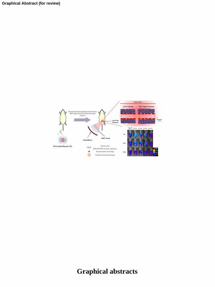
3.1. Preparation and characterization of the Pluronic NPs253
The DTX-loaded Pluronic NPs were prepared in a molten mixture of DTX dissolved 254
in soybean oil/Tween 80 mixtures and Pluronic F-68 through a temperature-induced phase 255
transition at 60 °C for 20 minutes. Pluronic F-68 showed a melting transition at around 55 °C256
and the mixtures composed of soybean oil/Tween 80 and Pluronic F-68 showed a melting 257
transition at around 48 °C [29]. Therefore, we anticipated that the mixture composed of DTX258
dissolved in soybean oil/Tween 80 mixtures and Pluronic F-68 would melt into a 259
homogeneous solution at about 60 °C. The formation of DTX-loaded Plunonic NPs was 260
observed when the temperature was decreased to 0 °C. The morphology and size distribution 261
of Pluronic NPs were examined by TEM and a particle size analyzer, respectively (Fig. 2A).262
The Pluronic NPs had a spherical form with approximately 178 ± 24.67 nm diameters and 263
showed a typical sustained release pattern observed in drug release from a homogeneous 264
polymer matrix (Fig. 2B).265
266
3.2. Physicochemical properties of the Pluronic NPs267
Page 12 of 33
Accep
ted
Man
uscr
ipt
12
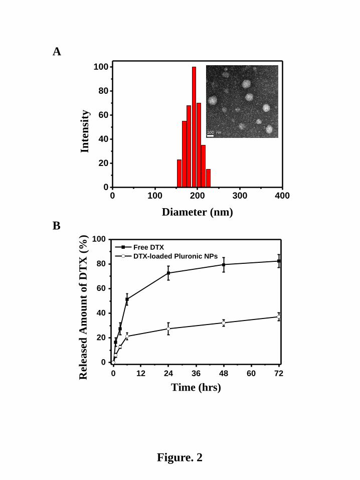
The cytotoxicity of Pluronic NPs was observed using a MTT assay. As a control, free 268
DTX was used under the same conditions. As expected, the empty Pluronic NPs exhibited 269
almost 100 % cell viability at a concentration of 500 g/mL. Even at a higher concentration 270
of 1,000 g/mL, the empty Pluronic NPs showed 90 % cell viability, indicating excellent 271
biocompatibility in the cell culture system. Also, free DTX and the DTX-loaded Pluronic272
NPs showed drug concentration-dependent cytotoxicity in the cell culture system. When the 273
DTX concentration was higher than 0.1 g/mL, the cytotoxicity of Pluronic NPs was lower 274
than that of free DTX. Less than 40 % of the SCC-7 tumor cells were viable after 2 days of 275
exposure to 1 g/mL of free DTX, whereas more than 75 % of the cells survived under the 276
same concentration of DTX in the Pluronic NPs (Fig. 3A). The low cytotoxicity of Pluronic277
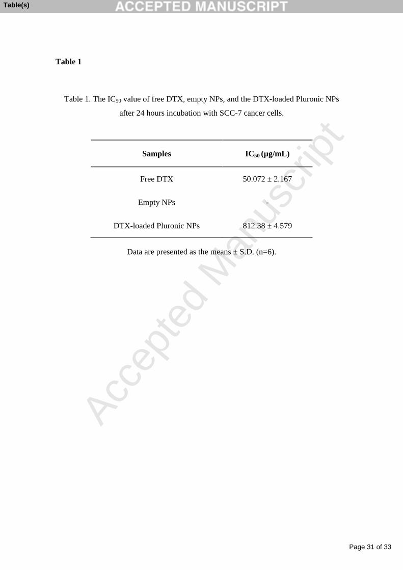
NPs might be due to the sustained release of PTX from the NPs. As shown in Table 1, the 278
IC50 values of free DTX and the DTX-loaded Pluronic NPs (50.072 ± 2.167, 812.38 ± 4.579 279
g/mL, respectively) also verified that the DTX-loaded Pluronic NPs was less cytotoxic than 280
free DTX.281
The Pluronic NPs can penetrate and be taken up by SCC-7 tumor cells through an 282
endocytosis process [30]. Therefore, we monitored the cellular uptake characteristics of 283
Pluronic NPs in a cell culture system after 60 minutes post-incubation. The Cy5.5-/DTX-284
loaded Pluronic NPs were incubated in SCC-7 tumor cells (Fig. 3B). When their sub-cellular 285
localization was examined under a fluorescent microscope, fluorescence from the Pluronic286
NPs was clearly observed in the cytoplasm but not in the nuclear compartment of SCC-7 287
tumor cells.288
289
3.3. Tumor targeting enhancement of the Pluronic NPs by HIFU exposure290
Page 13 of 33
Accep
ted
Man
uscr
ipt
13
Enhanced tumor targeting of the Pluronic NPs was monitored with and without the 291
HIFU exposure after intravenous injection of the Cy5.5-/DTX-loaded Pluronic NPs into 292
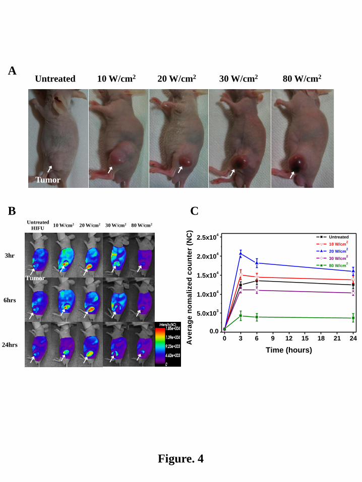
SCC-7 tumor-bearing mice using an NIRF optical imaging system. (Fig. 4) As a control, the293
NIRF signal of Pluronic NPs was measured at the tumor site without HIFU exposure. We 294
were able to distinguish tumors from the surrounding background tissue and the NIRF signal295
reached its maximum level 6 hours after injection. 296
Enhanced tumor targeting of the Pluronic NPs was observed with the HIFU297
exposure. After showing a maximum value at 20 W/cm2 (The following variables were 298
employed for exposure: transducer with a 5 x 5 cm, a focal length = 10 cm; pulse repetition 299
frequency = 0. 8 MHz; duty cycle = 10 % (100 ms ON and 900 ms OFF); slice distance= 5 300
mm), tumor targetability of the Pluronic NPs decreased.301
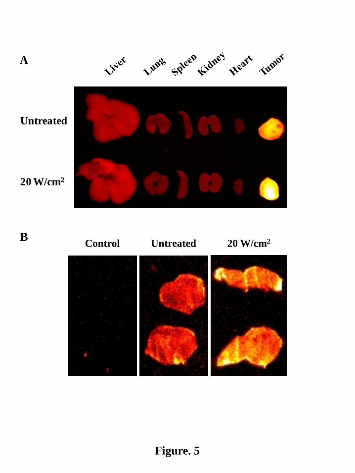
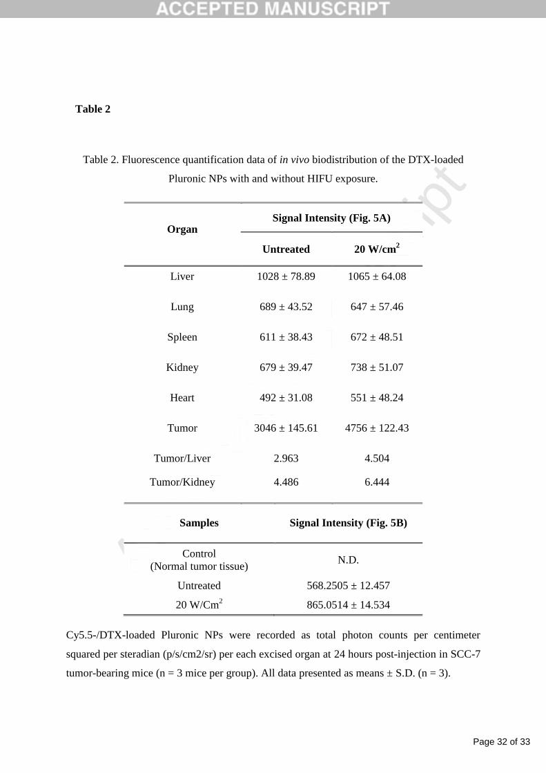
The enhancement of tumor targetability was further confirmed by ex vivo NIRF302
images of dissected major organs (liver, lung, kidneys, spleen, heart, and tumor), as shown in 303
Fig. 5a. With and without HIFU exposure, higher accumulations of the Pluronic NPs were304
observed at tumors compared with all organs. More significant enhancement of NIRF305
intensity in tumors, however, was observed with HIFU exposure (Fig. 5A and Table 2).306
Cross-sectional views of tumor tissue were examined to verify the extravasation of the 307
Pluronic NPs into the tumor tissue (Fig. 5B and Table 2). Without HIFU exposure, 308
background noise of NIRF (appeared as red color) was observed throughout the tumor tissue 309
and yellow color from the Cy5.5-/DTX-loaded Pluronic NPs was observed on the surface 310
area of the tumor tissue. However, with HIFU exposure, yellow color from the Cy5.5-/DTX-311
loaded Pluronic NPs became dominant in the middle of the tumor tissue.312
313
3.4. Ex vivo histological analysis of the Pluronic NPs314

Page 14 of 33
Accep
ted
Man
uscr
ipt
14
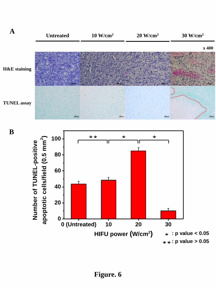
H&E staining of the excised tumors was performed after 24 hours post-treatment as 315
shown in Fig. 6a. The Pluronic NPs-treated tumors without HIFU exposure were used as a 316
control. Significant morphological change was not found below HIFU exposure of 20 W/cm2317
compared with the control. However, with HIFU exposure of 30 W/cm2, severe tissue318
damage and hemolysis were observed with inflammatory cells.319
TUNEL staining was also performed to determine whether HIFU exposure induced320
apoptosis of the cancer cells. After showing a maximum value at 20 W/cm2, the number of 321
apoptotic cells decreased as shown in Fig. 6B. This pattern was already observed in the 322
enhancement change of tumor targeting as a function of HIFU intensity.323
324
4. Discussion325
The objective of this study is to demonstrate the synergistic effect of HIFU exposure326
on targeting efficacy of DTX-loaded Pluronic NPs based on the EPR effect. If the anti-cancer 327
drug-loaded NPs circulate for sufficient time, they should accumulate preferentially in tumors328
based on the EPR effect. This is thought to be due to the leaky and disorganized 329
characteristics of the blood vessels around the tumor tissue, which allow the NPs of 330
appropriate size to escape from the blood vessel. With the application of HIFU at 20 W/cm2, 331
the ability of ultrasound to permeabilize blood-tissue barriers is expected and makes HIFU a 332
potential method for the targeted drug delivery of DTX. Therefore, substantially enhanced 333
antitumor efficacy is expected with reduced side effect. As presented in Figs. 3 and 4, the 334
DTX-loaded Pluronic NPs accumulated at tumor tissue and this new DTX formulation of the 335
Pluronic NPs may minimize the adverse effects of the current free DTX formulation 336
containing Tween 80 and ethanol [31]. Note also that a previous study by the authors showed 337
enhanced antitumor efficacy of the DTX-loaded Pluronic NPs, compared with commercial 338
DTX formulation (Taxotere®) [29].339
Page 15 of 33
Accep
ted
Man
uscr
ipt
15
In normal tissues, the hydrostatic pressure from vessel to tissue and the osmotic 340
pressure from tissue to vessels are balanced for the transfer of oxygen, nutrients and waste 341
into and out of the tissue. Low interstitial pressure is usually maintained in normal tissue by 342
the lymphatic system to eliminate excess fluid from the tissue. However, the balance between343
these forces is severely disrupted with the formation of tumors [32]. A dense and highly 344
cross-linked extracellular matrix is formed in tumor tissue and this leads to an increase in the 345
interstitial pressure with a reduction of hydraulic flow to the tumor periphery [33,34]. For 346
efficient treatment of cancer, drug-loaded NPs exit the tumor blood vessel and enter the 347
tumor interstitial space to access the site of action. However, due to the microenvironment of 348
tumors described above, the driving forces that would normally assist the extravasation of 349
NPs may be reduced.350
HIFU exposure has been used to noninvasively and nondestructively enhance the 351
delivery of a variety of systemically administered agents [35,36]. Similar enhancement on the352
extravasation of NPs is expected with HIFU exposure. As shown in Fig. 4, the enhanced 353
accumulation of Pluronic NPs on tumor tissue was observed with HIFU exposure. After 354
showing a maximum value at 20 W/cm2, the accumulation of Pluronic NPs at tumor tissue 355
decreased. HIFU is presently being used for thermal ablation and direct tumor destruction. 356
During thermal ablation, tumor cells are destroyed through necrosis [7]. Above HIFU 357
intensity of 30 W/cm2, tumor cells appeared to be destroyed through thermal ablation (Fig. 6).358
Because thermal ablation with necrosis resulted in destruction of tumor tissue, a decrease in 359
the tumor targetability of Pluronic NPs was observed. 360
The in vivo biodistribution study, which was performed with 20 W/cm2 HIFU 361
intensity, revealed that the greatest accumulation of Pluronic NPs at the tumor was observed 362
(see Figs. 4 and 5A). In particular, as shown in Fig. 5B, the cross-sectional view of tumor 363
tissue verified that the Pluronic NPs migrated into the middle of tumor tissue with HIFU 364
Page 16 of 33
Accep
ted
Man
uscr
ipt
16
exposure. Regarding a possible explanation of the manner by which the HIFU exposure 365
enhanced the therapeutic effect of injected agents, it has been suggested that HIFU exposure 366
enlarges the effective pore size in the tumor, leading to the better penetration and distribution 367
of injected agents [37, 38]. This is supported by the enhancement of tumor targetability of the 368
Pluronic NPs with HIFU exposure observed in the present study.369
HIFU exposures are clinically used in continuous mode to maximize the heat 370
generation for tissue ablation. However, for more delicate and non-thermal mechanisms, the 371
rate of energy disposition should be reduced. In the combination with Pluronic NPs, HIFU 372
exposure with 20 W/cm2 showed the highest number of apoptotic cells (see Fig. 6) indicating 373
that effective cancer treatment could be accomplished through a noninvasive approach with374
HIFU exposure.375
376
5. Conclusions377
Because of prolonged circulation of the Pluronic NPs in the blood, the DTX-loaded 378
Pluronic NPs are believed to be effective in targeted cancer therapy based on the EPR effect.379
However, with a more detailed understanding of the tumor microenvironment, elevated 380
interstitial tumor pressure and the dense tumor extracellular matrix have been recognized as 381
formidable barriers to the extravasation of NPs. Enhanced targeting at tumor and effective 382
extravasation into interior cells in the tumor tissue were demonstrated using the Pluronic NPs 383
with HIFU exposure. This approach can transiently increase the effective pore size of tumor 384
tissue with the enhanced permeability of Pluronic NPs through non-thermal mechanisms. 385
This was confirmed by observing the in vivo biodistribution of Pluronic NPs with HIFU 386
exposure. The preliminary results in this study demonstrate that HIFU exposure through non-387
thermal mechanisms can aid the extravasation of NPs into interior cells in tumors and 388
increase the therapeutic effect in targeted cancer therapy.389
Page 17 of 33
Accep
ted
Man
uscr
ipt
17
Acknowledgments
This work was supported by the National Research Foundation (NRF) of Korea grants
funded by the Korean government (MEST) (20110027932, 2012028831 and 2013063969).
Page 18 of 33
Accep
ted
Man
uscr
ipt
18
References
[1] Yang. C, Liu. Y, He. Y, Du. Y, Wang. W, Shi. X, Gao. F, The use of HA oligosaccharide-
loaded nanoparticles to breach the endogenous hyaluronan glycocalyx for breast cancer
therapy, Biomaterials 34 (2013) 6829-6838.
[2] Gao. W, Xiang. B, Meng. TT, Liu. F, Qi. XR, Chemotherapeutic drug delivery to cancer
cells using a combination of folate targeting and tumor microenvironment-sensitive
polypeptides, Biomaterials 34 (2013) 4137-4149.
[3] Stohrer. M, Boucher. Y, Stangassinger. M, Jain. RK, Oncotic pressure in solid tumors is
elevated, Cancer Res. 60 (2000) 4251-4255.
[4] Roose. T, Netti. PA, Munn. LL, Boucher. Y, Jain. RK, Solid stress generated by spheroid
growth estimated using a linear poroelasticity model small star, filled, Microvasc. Res. 66
(2003) 204-212.
[5] Netti. PA, Berk. DA, Swartz. MA, Grodzinsky. AJ, Jain. RK, Role of extracellular matrix
assembly in interstitial transport in solid tumors, Cancer Res. 60 (2000) 2497-2503.
[6] Nicholas. JW, Bae. YH, Odyssey of a cancer nanoparticle: From injection site to site of
action, Nano Today 7 (2012) 606-618.
[7] Kennedy. JE, High-intensity focused ultrasound in the treatment of solid tumours, Nat.
Rev. Cancer 5 (2005) 321-327.
[8] Dromi. S, Frenkel. V, Luk. A, Traughber. B, Angstadt. M, Bur. M, Poff. J, Xie. J, Libutti.
SK, Li. KC, Wood. BJ, Pulsed-high intensity focused ultrasound and low temperature-
sensitive liposomes for enhanced targeted drug delivery and antitumor effect, Clin. Cancer
Res. 13 (2007) 2722-2727.
[9] Miller. DL, Dou. C, Song. J, DNA transfer and cell killing in epidermoid cells by
diagnostic ultrasound activation of contrast agent gas bodies in vitro, Ultrasound Med. Biol.
29 (2003) 887-893.
Page 19 of 33
Accep
ted
Man
uscr
ipt
19
[10] Ziadloo. A, Xie. J, Frenkel. V, Pulsed focused ultrasound exposures enhance locally
administered gene therapy in a murine solid tumor model, J. Acoust. Soc. Am. 133 (2013)
1827-1834.
[11] Chen. CC, Sheeran. PS, Wu. SY, Olumolade. OO, Dayton. PA, Konofagou. EE,
Targeted drug delivery with focused ultrasound-induced blood-brain barrier opening using
acoustically-activated nanodroplets, J. Control. Release 172 (2013) 795-804.
[12] Alkins. RD, Brodersen. PM, Sodhi. RN, Hynynen. K, Enhancing drug delivery for boron
neutron capture therapy of brain tumors with focused ultrasound, Neuro. Oncol. 15 (2013)
1225-1235.
[13] Park. EJ, Zhang. YZ, Vykhodtseva. N, McDannold. N, Ultrasound-mediated blood-
brain/blood-tumor barrier disruption improves outcomes with trastuzumab in a breast cancer
brain metastasis model, J. Control. Release 163 (2012) 277-284.
[14] Liu. HL, Hua. MY, Yang. HW, Huang. CY, Chu. PC, Wu. JS, Tseng. IC, Wang. JJ, Yen
TC, Chen. PY, Wei. KC, Magnetic resonance monitoring of focused ultrasound/magnetic
nanoparticle targeting delivery of therapeutic agents to the brain, Proc. Natl. Acad. Sci. USA
107 (2010) 15205-15210.
[15] Sheikov. N, McDannold. N, Jolesz. F, Zhang. YZ, Tam. K, Hynynen. K, Brain arterioles
show more active vesicular transport of blood-borne tracer molecules than capillaries and
venules after focused ultrasound-evoked opening of the blood-brain barrier, Ultrasound Med.
Biol. 32 (2006) 1399-1409.
[16] Sheikov. N, McDannold. N, Sharma. S, Hynynen. K, Effect of focused ultrasound
applied with an ultrasound contrast agent on the tight junctional integrity of the brain
microvascular endothelium, Ultrasound Med. Biol. 34 (2008) 1093-1104.
Page 20 of 33
Accep
ted
Man
uscr
ipt
20
[17] Xie. F, Boska. MD, Lof. J, Uberti. MG, Tsutsui. JM, Porter. TR, Effects of transcranial
ultrasound and intravenous microbubbles on blood brain barrier permeability in a large
animal model, Ultrasound Med. Biol. 34 (2008) 2028-2034.
[18] Gulati. N, Rastogi. R, Dinda. AK, Saxena. R, Koul. V, Characterization and cell material
interactions of PEGylated PNIPAAM nanoparticles, Colloids Surf. B Biointerfaces 79
(2010) 164-173.
[19] Oh. KS, Lee. H, Kim. JY, Koo. EJ, Lee. EH, Park. JH, Kim, SY, Kim, K, Kwon, IC,
Yuk, SH, The multilayer nanoparticles formed by layer by layer approach for cancer-
targeting therapy, J. Control. Release 165 (2013) 9-15.
[20] Frenkel. V, Ultrasound mediated delivery of drugs and genes to solid tumors, Adv. Drug
Del. Rev. 60 (2008) 1193-1208.
[21] Kadam. Y, Yerramilli. U, Bahadur A, Bahadur P, Micelles from PEO-PPO-PEO block
copolymers as nanocontainers for solubilization of a poorly water soluble drug
hydrochlorothiazide, Colloids Surf. B Biointerfaces 83 (2011) 49-57.
[22] Alexandridis. P, Holzwarth. JF, Hatton. TA, Micellization of poly(ethylene oxide)-
poly(propylene oxide)-poly(ethylene oxide) triblock copolymers in aqueous solutions:
thermodynamics of copolymer association, Macromolecules 27 (1994) 2414-2425.
[23] Salama. HA, Mahmoud. AA, Kamel. AO, Abdel Hady. M, Awad. GA, Phospholipid
based colloidal poloxamer-nanocubic vesicles for brain targeting via the nasal route, Colloids
Surf. B Biointerfaces, 100 (2012) 146-154.
[24] Alakhov. VY, Moskaleva. EY, Batrakova. EV, Kabanov. AV, Hypersensitization of
multidrug resistant human ovarian carcinoma cells by Pluronic P85 block copolymer,
Bioconjugate Chem. 7 (1996) 209-216.
Page 21 of 33
Accep
ted
Man
uscr
ipt
21
[25] Oh. KS, Song. JY, Cho. SH, Lee. BS, Kim. SY, Kim. K, Jeon. H, Kwon. IC, Yuk. SH,
Paclitaxel-loaded Pluronic nanoparticles formed by a temperature-induced phase transition
for cancer therapy, J. Control. Release 148 (2010) 344-350.
[26] Yuk. SH, Oh. KS, Cho. SH, Kim. SY, Oh. S, Lee. JH, Kim. K, Kwon. IC, Enhancement
of the targeting capabilities of the paclitaxel-loaded pluronic nanoparticles with a glycol
chitosan/heparin composite, Mol. Pharm. 9 (2012) 230-236.
[27] Kim. JY, Choi. WI, Kim. YH, Tae. G, Brain-targeted delivery of protein using chitosan-
and RVG peptide-conjugated, pluronic-based nano-carrier, Biomaterials 34 (2013) 1170-
1178.
[28] Dittmar. KM, Xie. J, Hunter. F, Trimble. C, Bur. M, Frenkel. V, Li. KC, Pulsed high-
intensity focused ultrasound enhances systemic administration of naked DNA in squamous
cell carcinoma model: initial experience, Radiology 235 (2005) 541-546.
[29] Yuk. SH, Oh. KS, Park. J, Lee. EH, Kim. K, Kwon. IC, Docetaxel-loaded composite
nanoparticles formed by a temperature-induced phase transition for cancer therapy, J. Bioact.
Compat. Polym. 27 (2012) 441-452.
[30] Rapoport. N, Marin. A, Luo. Y, Prestwich. GD, Muniruzzaman. MD, Intracellular
uptake and trafficking of Pluronic micelles in drug-sensitive and MDR cells: effect on the
intracellular drug localization, J. Pharm. Sci. 91 (2002) 157-170.
[31] Baker. J, Ajani. J, Scotté. F, Winther. D, Martin. M, Aapro. MS, von Minckwitz G,
Docetaxel-related side effects and their management, Eur. J. Oncol. Nurs. 13 (2009) 49-59.
[32] Heldin. CH, Rubin. K, Pietras. K, Ostman. A, High interstitial fluid pressure-An obstacle
in cancer therapy, Nat. Rev. Cancer 4 (2004) 806-813.
[33] Boucher. Y, Jain. RK, Microvascular pressure is the principal driving force for
interstitial hypertension in solid tumors: implications for vascular collapse, Cancer Res. 52
(1992) 5110-5114.
Page 22 of 33
Accep
ted
Man
uscr
ipt
22
[34] Boucher. Y, Salehi. H, Witwer. B, Harsh GR. 4th, Jain. RK, Interstitial fluid pressure in
intracranial tumours in patients and in rodents, Br. J. Cancer 75 (1997) 829-836.
[35] Hancock. HA, Smith. LH, Cuesta. J, Durrani. AK, Angstadt. M, Palmeri. ML, Kimmel.
E, Frenkel. V, Investigations into pulsed high-intensity focused ultrasound-enhanced
delivery: Preliminary evidence for a novel mechanism, Ultrasound Med. Biol. 35 (2009)
1722-1736.
[36] Wang. CH, Kang. ST, Lee. YH, Luo. YL, Huang. YF, Yeh. CK, Aptamer-conjugated
and drug-loaded acoustic droplets for ultrasound theranosis, Biomaterials 33 (2012) 1939-
1947.
[37] O’Neill. BE, Vo. H, Angstadt. M, Li. KC, Quinn. T, Frenkel. V, Pulsed high intensity
focused ultrasouond mediated nanoparticle delivery: Mechanisms and efficacy in murine
muscle. Ultrasound Med. Biol. 35 (2009) 416-424.
[38] Wang. X, Chen. H, Zheng. Y, Ma. M, Chen. Y, Zhang. K, Zeng. D, Shi. J, Au-
nanoparticle coated mesoporous silica nanocapsule-based multifunctional platform for
ultrasound mediated imaging, cytoclasis and tumor ablation, Biomaterials 34 (2013) 2057-
2068.
Page 23 of 33
Accep
ted
Man
uscr
ipt
23
Figure Captions
Fig. 1. (A) Schematic description of enhanced tumor targeting with HIFU exposure and (B)
expected behavior of the Pluronic NPs in the vessel.
Fig. 2. (A) The size distribution and morphology of DTX-loaded Pluronic NPs and (B) in
vitro release behavior of DTX in the Pluronic NPs. Data are presented as the mean ± SD
(n=4).
Fig. 3. (A) The cellular uptake of Cy5.5-/DTX-loaded Pluronic NPs with SCC-7 tumor cells
and (B) in vitro cytotoxicity at different concentrations of free DTX, the empty NPs and the
DTX-loaded Pluronic NPs. Data are presented as the means ± SD (n=6).
Fig. 4. (A) Damage assessment of tumor in mice treated with different HIFU exposure
intensities, (B) in vivo whole body non-invasive biodistribution and (C) fluorescence
quantification analysis of localized the Cy5.5-/DTX-loaded NPs at tumor using in vivo NIRF
imaging system.
Fig. 5. (A) Representative ex vivo fluorescence images of dissected major organs (liver, lung,
spleen, kidney, heart and tumor) and (B) cross-sectional images of excised tumor tissues of
sacrificed SCC-7 tumor-bearing mice after 24 hours with and wiout HIFU exposure.
Fig. 6. (A) Representative histological images and (B) quantification data of apoptotic cells
of sliced tumor tissues with different HIFU exposure intensity, stained using hematoxylin and
eosin (H&E) and TUNEL assay. Tumors were excised at 24 hours after HIFU exposure. Data
are presented as the means ± SD (n=4).
Page 24 of 33
Accep
ted
Man
uscr
ipt
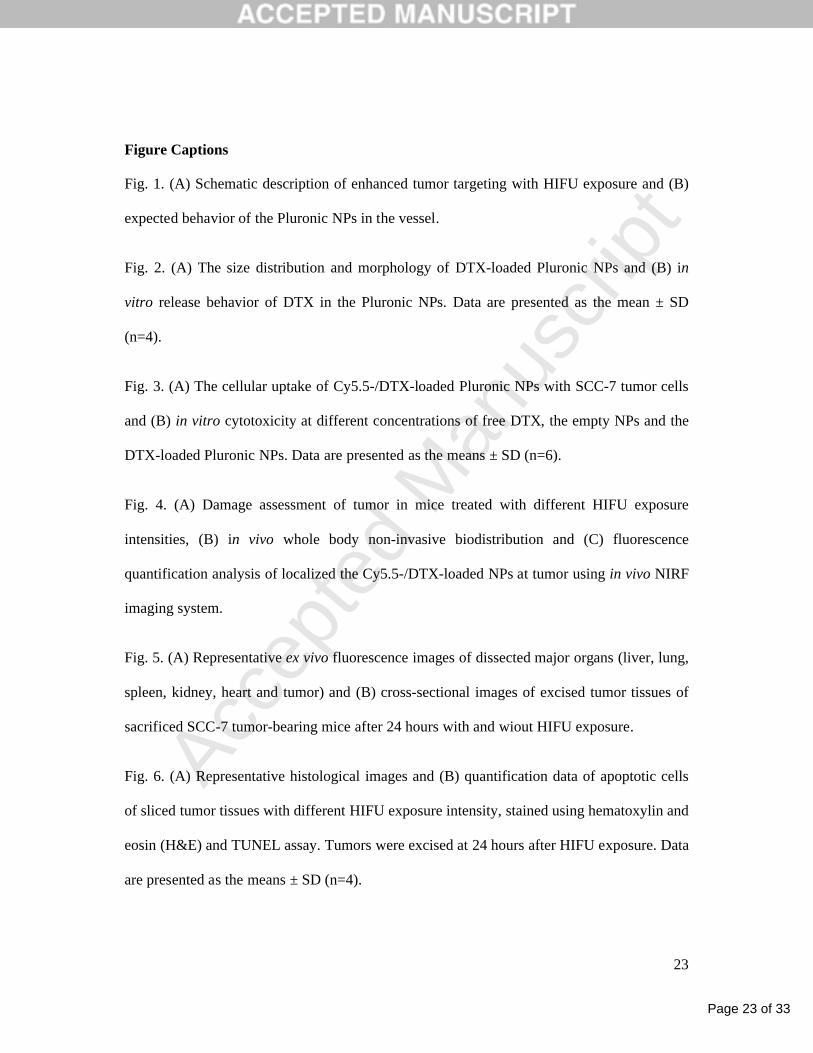
Graphical abstracts
DTX-loaded Pluronic NPs Pluronic F-68
(PEO-PPO-PEO tri-block copolymer)
Docetaxel (anti-cancer drug)
Soybean oil/Tween 80 mixtures
Ultrasound-induced targeting enhancement by
HIFU (high-intensity focused ultrasound)
exposure
Local site
(Tumor)
TransducerHIFU beam
Tumor TissueNormal Tissue
Free Drug Passive Targeting HIFU-triggered Targeting
Angiogenic
vessel
Untreated
HIFU10 W/cm2 20 W/cm2 30 W/cm2
3hr
6hrs
24hrs
80 W/cm2
Graphical Abstract (for review)

Page 25 of 33
Accep
ted
Man
uscr
ipt
Figure 1.
A
B
Figure(s)
Page 26 of 33
Accep
ted
Man
uscr
ipt
Figure. 2
A
0 100 200 300 4000
20
40
60
80
100
Inte
nsi
ty
Diameter (nm)
B
0 12 24 36 48 60 72
0
20
40
60
80
100
Rele
ase
d A
mo
un
t o
f D
TX
(%
)
Time (hrs)
Free DTX
DTX-loaded Pluronic NPs
Page 27 of 33
Accep
ted
Man
uscr
ipt
Figure. 3
A
B
0.1 1 10 100 500 10000
25
50
75
100
125
Ce
ll v
iab
ilit
y (
%)
Concentration (g/mL)
Empty NPs
0.1 1 10 100 500 10000
25
50
75
100
125
Ce
ll v
iab
ilit
y (
%)
Concentration (g/ml)
Free DTX
DTX-loaded Pluronic NPs
Page 28 of 33
Accep
ted
Man
uscr
ipt
Figure. 4
A
B C
0 3 6 9 12 15 18 21 240.0
5.0x10³
1.0x10⁴
1.5x10⁴
2.0x10⁴
2.5x10⁴
Untreated
10 W/cm2
20 W/cm2
30 W/cm2
80 W/cm2
Av
era
ge
no
ma
lize
d c
ou
nte
r (N
C)
Time (hours)
Untreated
HIFU10 W/cm2 20 W/cm2 30 W/cm2
3hr
6hrs
24hrs
80 W/cm2
Tumor
Untreated 10 W/cm2 20 W/cm2 30 W/cm2 80 W/cm2
Tumor
Page 29 of 33
Accep
ted
Man
uscr
ipt
B Control Untreated 20 W/cm2
Untreated
20 W/cm2
A
Figure. 5
Page 30 of 33
Accep
ted
Man
uscr
ipt
B
Figure. 6
A
0 (Untreated) 10 20 300
20
40
60
80
100
Nu
mb
er
of
TU
NE
L-p
os
itiv
e
ap
op
toti
c c
ell
s/f
ield
(0
.5 m
m2)
HIFU power (W/cm2) * : p value < 0.05
** : p value > 0.05
*** *
Untreated 10 W/cm2
x 400
20 W/cm2 30 W/cm2
H&E staining
TUNEL assay
Page 31 of 33
Accep
ted
Man
uscr
ipt
Table 1
Table 1. The IC50 value of free DTX, empty NPs, and the DTX-loaded Pluronic NPs
after 24 hours incubation with SCC-7 cancer cells.
Samples IC50 (µg/mL)
Free DTX 50.072 ± 2.167
Empty NPs -
DTX-loaded Pluronic NPs 812.38 ± 4.579
Data are presented as the means ± S.D. (n=6).
Table(s)
Page 32 of 33
Accep
ted
Man
uscr
ipt
Table 2
Table 2. Fluorescence quantification data of in vivo biodistribution of the DTX-loaded
Pluronic NPs with and without HIFU exposure.
Organ Signal Intensity (Fig. 5A)
Untreated 20 W/cm2
Liver 1028 ± 78.89 1065 ± 64.08
Lung 689 ± 43.52 647 ± 57.46
Spleen 611 ± 38.43 672 ± 48.51
Kidney 679 ± 39.47 738 ± 51.07
Heart 492 ± 31.08 551 ± 48.24
Tumor 3046 ± 145.61 4756 ± 122.43
Tumor/Liver
Tumor/Kidney
2.963
4.486
4.504
6.444
Samples Signal Intensity (Fig. 5B)
Control
(Normal tumor tissue) N.D.
Untreated 568.2505 ± 12.457
20 W/Cm2
865.0514 ± 14.534
Cy5.5-/DTX-loaded Pluronic NPs were recorded as total photon counts per centimeter
squared per steradian (p/s/cm2/sr) per each excised organ at 24 hours post-injection in SCC-7
tumor-bearing mice (n = 3 mice per group). All data presented as means ± S.D. (n = 3).
Page 33 of 33
Accep
ted
Man
uscr
ipt
Research Highlights
1. Targeting enhancement of docetaxel (DTX)-loaded Pluronic nanoparticles (NPs) was
demonstrated using high intensity focused ultrasound (HIFU) as an external stimuli-
responsive clinical system.
2. Efficient extravasation of the Pluronic NPs into the interior cells in the tumor tissue was
induced by the relatively low HIFU exposure where this can transiently increase the effective
pore size (endothelial cell tight junctions) of a tumor tissue with enhanced permeability of the
Pluronic NPs through non-thermal mechanisms.
3. To verify the targeting enhancement through the efficient extravasation, the biodistribution
of the Pluronic NPs was observed in in vivo animal model with various HIFU exposures.
*Highlights (for review)
Copyright © 2022 FDOKUMEN