Small Molecule Inhibitors Target the Tissue Transglutaminase and Fibronectin Interaction
Upload
independentCategory
view
1download
0
Small molecule inhibitors of Apaf-1-related caspase-3/-9 activation that control mitochondrial-dependentapoptosis
G Malet1, AG Martın2,3, M Orzaez3, MJ Vicent3, I Masip4,G Sanclimens4, A Ferrer-Montiel5, I Mingarro1, A Messeguer4,HO Fearnhead2 and E Perez-Paya*,3,6
1 Department of Biochemistry and Molecular Biology, Universitat de Valencia,E-46100 Burjassot, Valencia, Spain
2 Apoptosis Section, Regulation of Cell Growth Laboratory, NCI, NationalInstitutes of Health, Frederick, MD 21702, USA
3 Department of Medicinal Chemistry, Centro de Investigacion Prıncipe Felipe,E-46013 Valencia, Spain
4 Department of Biological Organic Chemistry, IIQAB (CSIC), E-08034Barcelona, Spain
5 Instituto de Biologıa Molecular y Celular, Universidad Miguel Hernandez,03202 Elx Alacant, Spain
6 Consejo Superior de Investigaciones Cientıficas (CSIC), E-46013 Valencia,Spain
* Corresponding author: E Perez-Paya, Centro de Investigacion Prıncipe Felipeand CSIC, Avda. Autopista del Saler, 16, E-46013 Valencia, Spain.Tel: þ 34-963289680; Fax: þ 34-963289701; E-mail: [email protected]
Received 27.5.05; revised 25.10.05; accepted 02.11.05; published online 09.12.05Edited by Y Tsujimoto
AbstractApoptosis is a biological process relevant to human diseasestates that is strongly regulated through protein–proteincomplex formation. These complexes represent interestingpoints of chemical intervention for the development ofmolecules that could modulate cellular apoptosis. Theapoptosome is a holoenzyme multiprotein complex formedby cytochrome c-activated Apaf-1 (apoptotic protease-activating factor), dATP and procaspase-9 that link mitochon-dria disfunction with activation of the effector caspases andin turn is of interest for the development of apoptoticmodulators. In the present study we describe the identifica-tion of compounds that inhibit the apoptosome-mediatedactivation of procaspase-9 from the screening of a diversity-oriented chemical library. The active compounds rescuedfrom the library were chemically optimised to obtainmolecules that bind to both recombinant and humanendogenous Apaf-1 in a cytochrome c-noncompetitivemechanism that inhibits the recruitment of procaspase-9 bythe apoptosome. These newly identified Apaf-1 ligandsdecrease the apoptotic phenotype in mitochondrial-mediatedmodels of cellular apoptosis.Cell Death and Differentiation (2006) 13, 1523–1532.doi:10.1038/sj.cdd.4401828; published online 9 December 2005
Keywords: apoptosis; apoptosome; Apaf-1; caspasa-3; caspa-
sa-9; combinatorial libraries; inhibitor; molecular recognition;
peptoid; protein–protein interactions; small molecule
Abbreviations: Apaf-1, apoptotic protease-activating factor;
DEVDase, hydrolysis of Ac-DEVD-afc; DTT, dithiothreitol;
FCS, fetal-calf serum; MEFs, mouse embryo fibroblasts;
MMP, mitochondrial membrane potential; MTT, 3-(4,5-di-
methylthiazol-2-yl)-2,5-diphenyltetrazolium bromide; Ni-NTA,
(Ni2þ -nitrilotriacetate)-agarose; peptoid f1a, 50-60 carboxy-
fluorescein-labelled peptoid 1a; PBS, phosphate-buffered
saline; PGA, poly-(L-glutamic acid); rApaf-1, recombinant
Apaf-1
Introduction
Protein–protein interactions play a critical role in almostall cellular and biological processes. Consequently, theinteractions between proteins represent points of chemicalintervention for therapeutic gain in the biological processesassociated with disease. Apoptosis is an interesting biologicalprocess because of its importance in a wide variety ofbiological systems, including normal cell turnover, the immunesystem and embryonic development and its associationwith different diseases. Inappropriate apoptosis is involvedin many human pathologies, including neurodegenerativediseases such as Alzheimer’s and Huntington’s, ischaemia,autoimmune disorders and several forms of cancer.1 Diverseapoptotic stimuli, including activation of cell surfacedeath receptors, anticancer agents, irradiation, lack ofsurvival factors, and ischaemia (reviewed in Strasser et al2)induce signalling cascades that all activate a family of cysteineaspartyl proteases called caspases. It is these proteasesthat execute the apoptotic process. Effector caspases (e.g.caspases-3 and -7) are responsible for the disassemblyof cellular components while initiator caspases (e.g. cas-pases-8, -9 and -10) are responsible for activation of theeffector caspases. While different apoptotic stimuli activatedifferent initiators, these initiators activate a common set ofeffector caspases. Because of the critical consequencesof apoptosis malfunctioning, the activation of caspases isscrupulously controlled. Some apoptotic signals activatethe mitochondria-mediated or intrinsic pathway that utilisescaspase-9 as its initiator. Caspase-9 activation is triggeredby the release to the cytosol of proapoptotic proteins fromthe mitochondrial inter-membrane space, in particular cyto-chrome c and Smac/Diablo.3,4 The formation of the macro-molecular complex named apoptosome is a key eventin this pathway. The apoptosome is a holoenzyme multi-protein complex formed by cytochrome c-activated Apaf-1(apoptotic protease-activating factor), dATP and pro-caspase-9.5–8 In this macromolecular complex, apoptosome-associated caspase-9 is activated and then, in turn,activate effector caspases. To identify molecules that couldameliorate disease-associated apoptosis, drug discovery
Cell Death and Differentiation (2006) 13, 1523–1532& 2006 Nature Publishing Group All rights reserved 1350-9047/06 $30.00
www.nature.com/cdd

efforts have initially targeted caspase activity9,10 rather thanactivation. Nevertheless, protein–protein interactions up-stream of caspase activation can be also relevant pointsof intervention for the development of modulators of apoptosispathways. In particular, recent data propose the formationof the apoptosome as an interesting target for the develop-ment of apoptotic modulators.11–14 In the absence ofdetailed structural information, the conventional methodsused for the identification of modulators of the apopto-some have been based in indirect measurements ofthe cytochrome c- and dATP-induced activation of caspase-3-like activity on defined cytosolic extracts.15,16 Usingthis methodology Lademann et al.15 have identifiedinhibitors of the apoptosome through the screeningof small molecules using cytosolic extracts of selectedcells while Nguyen et al.16 reported the identification ofactivators.
We are currently engaged in a discovery program employ-ing an in vitro reconstituted active apoptosome assembledfrom its recombinant constituent proteins. Here we describethe identification of compounds that inhibit the apoptosome-mediated activation of procaspase-9 from the screening ofa diversity-oriented chemical library of N-alkylglycines. Theactive compounds rescued from the library were chemicallyoptimised to obtain molecules that bind to both recombinantand human endogenous Apaf-1 and decrease the apoptoticphenotype in mitochondrial-mediated models of cellularapoptosis.
Results
Screening of a positional scanning library ofpeptoids as a source for apoptosome inhibitors
Oligomers of N-alkylglycines, also known as peptoids,constitute a family of non-natural molecules attractive for thedrug discovery process due to their broad variety of biologicalactivities and to the proteolytic stability that they exhibit.17,18
We have early designed a positional scanning diversity-oriented combinatorial library of trimers of N-alkylglycines19
that allowed the identification of lead compounds in differentbiological assays.20–22 We have now modified the diversity ofthe library to generate an optimised version of the originallibrary in terms of composition and concentration of thedifferent components.23 The library consists in 52 controlledmixtures and a total of 5120 compounds. The mixtures werescreened for their ability to prevent the apoptosome-depen-dent activation of procaspase-9. The apoptosome wasassembled in vitro by incubating rApaf-1, cytochrome c, dATPand [35S]Met procaspase-9 (see Materials and Methodssection and Supplementary information). Samples weresubjected to SDS-PAGE and caspase-9 processing subse-quently visualised by phospho-imaging. Defined mixturesfrom the library inhibited the apoptosome-dependent activa-tion and allowed the deconvolution process and identifi-cation of four discrete compounds (peptoids 1–4, Figure 1a)that inhibited the apoptosome-dependent activation of
Figure 1 Peptoids rescued from the library inhibit apoptosome dependent procaspase-9 processing in an in vitro assay. (a) Chemical structures of peptoids 1 to 4rescued from the screening of the library of N-alkylglycines. (b) Chemical structures of peptoids 1a and 1b. (c) Peptoid 1a (upper panel) and peptoid 1b (lower panel)were evaluated in a concentration-dependent manner for their capacity to inhibit apoptosome-dependent activation of procaspase-9
Small molecule inhibitors of Apaf-1G Malet et al
1524
Cell Death and Differentiation

procaspase-9 to different extent in a concentration-dependentmanner. A peptoid from our stock collection was used asnegative control. The most potent inhibitor was peptoid 1.However, probably due to the overall intrinsic hydrophobicityof all four compounds, problems of reproducibility wereencountered from batch-to-batch solubilised peptoids. Thenwe decided to generate more soluble analogues of peptoid 1.Previous experience in the design of bioactive peptides24,25
suggested that the presence of positively charged side chainsat both N- and C-terminus ends of peptides increased theoverall peptide solubility without modifying their conforma-tional or biological properties. Furthermore, in our laboratory,we have used an N-substituted alkylglycine hexamer com-posed by residues containing a simple benzene ring or atertiary amino moiety attached to a bioactive hexapeptidesequence to increase both aqueous solubility and cell-membrane permeability (unpublished results). With theseprecedents, peptoids 1a and 1b containing two additionalN-alkylamine residues at the N- or C-terminus ends,respectively, were synthesised (Figure 1b) and their in vitroactivity tested. These two peptoids inhibited the apoptosome-mediated procaspase-9 activation with an excellent batch-to-batch reproducibility and an IC50 close to 10 mM (Figure 1c).Preliminary structure–activity relationship analysis on thepeptoid 1 family suggested that the dichlorophenylethylaminomoiety was important for activity. Peptoid 1d, derived frompeptoid 1b with a deletion at the N-terminus of anN-dichlorophenylethylglycine, had a reduced inhibitory activitywhen compare to peptoid 1b (results not shown) in the in vitroapoptosome activity assay.
Peptoid 1a inhibited caspase-3 processing (Figure 2a)when the apoptosome activity was followed in the presence ofrApaf-1, cytochrome c, dATP, and both in vitro translated[35S]Met procaspase-9 and -3 (see Materials and Methodssection). Furthermore, we tested apoptosome-dependentcaspase-3 activation by adding rApaf-1 to an Apaf-1 free cellextract14 and measuring Ac-DEVD-afc hydrolysis (DEVDaseactivity; Figure 2b). This confirmed a dose-dependentinhibitory activity of the peptoids in this assay that correlatedwith their ability to inhibit apoptosome formation (Figure 1c). Itis worth mentioning that a recombinant caspase 3 activity invitro assay (Calbiochem) showed that the inhibitory propertiesof peptoid 1a were not due to a direct inhibition of caspase 3(data not shown).
Peptoid 1a directly binds to rApaf-1 and induces anonactive apoptosome complex
The apoptosome is assembled when seven Apaf-1:cyto-chrome c heterodimers oligomerise to form a ‘wheel’ structurethat has the ability to recruit procaspase-9.6,26–28 To initiallycharacterise the binding site of the peptoids to the apopto-some, we synthesised a fluorescent analogue of peptoid 1a(peptoid f1a). Peptoid f1a bound to rApaf-1, but not tocytochrome c and other control proteins (Figure 3A), withdissociation constant (Kd) 57712 nM as determined in afluorescence polarisation assay. Nevertheless, the bindingconstant value contrasted with the concentration of peptoid 1athat was needed to inhibit 50% of caspase activity in ourassays (IC50B10 mM, Figure 1c). A putative explanation toreconcile these data is that the reticulocyte lysate used to invitro translate procaspase-9 contained components thatdiminished the effective concentration of peptoid 1a.Next, we evaluated whether or not peptoid 1a inhibitscytochrome c binding to rApaf-1. In the fluorescencepolarisation assay, the dissociation constant for the interac-tion rApaf-1/peptoid f1a was not influenced by the presenceof up to five-fold molar excess of cytochrome c (not shown).Furthermore, we also performed the in vitro apoptosomereconstitution assay in the presence of increasing concentra-tions of cytochrome c. Regardless of the concentrationof cytochrome c used, peptoid 1a exerted its inhibitoryactivity on apoptosome-dependent procaspase-9 cleavage(Figure 3B). These results suggest that peptoid 1a directlybinds to rApaf-1 in a binding site different from that ofthe cytochrome c recruitment site and induces a conforma-tional change in Apaf-1 that precludes procaspase-9 proces-sing. In principle, the peptoid may have prevented anyof the steps leading to caspase-9 activation including Apaf-1oligomerisation and caspase recruitment.26 To test whichstep of caspase activation was blocked, rApaf-1 wasincubated in the presence and absence of peptoid 1a,cytochrome c and dATP and the ability to activate caspasesfrom cell lysates was evaluated before and after chromato-graphy on Superose-6 H-R analytical gel filtrationcolumn. Before gel filtration (Figure 3C; sample a is acontrol only containing FT extract), the sample containingrApaf-1, cytochrome c and dATP (sample b) was able toactivate caspases and this was inhibited by peptoid 1a
Figure 2 Peptoid 1a inhibits apoptosome mediated caspase-3 activation. (a)Left panel. In vitro translated [35S]Met procaspase-9 and procaspase-3 wereincubated in the presence of rApaf-1, cytochrome c and in the presence orabsence of peptoid 1a (20 mM). Right panel. In vitro translated procaspase-9 and[35S]Met procaspase-3 were incubated in the presence of rApaf-1, cytochrome cand in the presence or absence of peptoid 1a. (b) DEVDase activity on FTextracts was measured in the presence or absence of increasing concentrationsof both peptoid 1a (&) and peptoid 1b (E). rApaf-1 was incubated with peptoidsat 301C for 30 min and combined with a 5 ml aliquot of FT extract. DEVDaseactivity was measured and referred to a positive control (100% activity)
Small molecule inhibitors of Apaf-1G Malet et al
1525
Cell Death and Differentiation
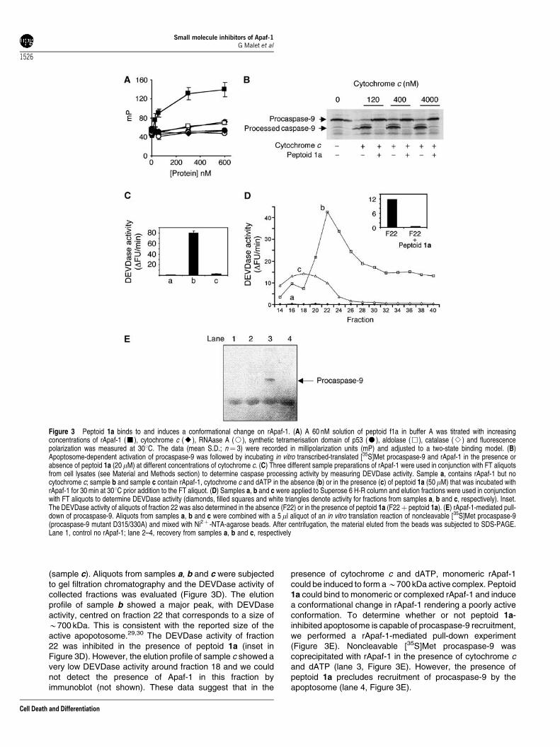
(sample c). Aliquots from samples a, b and c were subjectedto gel filtration chromatography and the DEVDase activity ofcollected fractions was evaluated (Figure 3D). The elutionprofile of sample b showed a major peak, with DEVDaseactivity, centred on fraction 22 that corresponds to a size ofB700 kDa. This is consistent with the reported size of theactive apopotosome.29,30 The DEVDase activity of fraction22 was inhibited in the presence of peptoid 1a (inset inFigure 3D). However, the elution profile of sample c showed avery low DEVDase activity around fraction 18 and we couldnot detect the presence of Apaf-1 in this fraction byimmunoblot (not shown). These data suggest that in the
presence of cytochrome c and dATP, monomeric rApaf-1could be induced to form aB700 kDa active complex. Peptoid1a could bind to monomeric or complexed rApaf-1 and inducea conformational change in rApaf-1 rendering a poorly activeconformation. To determine whether or not peptoid 1a-inhibited apoptosome is capable of procaspase-9 recruitment,we performed a rApaf-1-mediated pull-down experiment(Figure 3E). Noncleavable [35S]Met procaspase-9 wascoprecipitated with rApaf-1 in the presence of cytochrome cand dATP (lane 3, Figure 3E). However, the presence ofpeptoid 1a precludes recruitment of procaspase-9 by theapoptosome (lane 4, Figure 3E).
Figure 3 Peptoid 1a binds to and induces a conformational change on rApaf-1. (A) A 60 nM solution of peptoid f1a in buffer A was titrated with increasingconcentrations of rApaf-1 (’), cytochrome c (E), RNAase A (J), synthetic tetramerisation domain of p53 (K), aldolase (&), catalase (B) and fluorescencepolarization was measured at 301C. The data (mean S.D.; n¼ 3) were recorded in millipolarization units (mP) and adjusted to a two-state binding model. (B)Apoptosome-dependent activation of procaspase-9 was followed by incubating in vitro transcribed-translated [35S]Met procaspase-9 and rApaf-1 in the presence orabsence of peptoid 1a (20 mM) at different concentrations of cytochrome c. (C) Three different sample preparations of rApaf-1 were used in conjunction with FT aliquotsfrom cell lysates (see Material and Methods section) to determine caspase processing activity by measuring DEVDase activity. Sample a, contains rApaf-1 but nocytochrome c; sample b and sample c contain rApaf-1, cytochrome c and dATP in the absence (b) or in the presence (c) of peptoid 1a (50 mM) that was incubated withrApaf-1 for 30 min at 301C prior addition to the FT aliquot. (D) Samples a, b and c were applied to Superose 6 H-R column and elution fractions were used in conjunctionwith FT aliquots to determine DEVDase activity (diamonds, filled squares and white triangles denote activity for fractions from samples a, b and c, respectively). Inset.The DEVDase activity of aliquots of fraction 22 was also determined in the absence (F22) or in the presence of peptoid 1a (F22þ peptoid 1a). (E) rApaf-1-mediated pull-down of procaspase-9. Aliquots from samples a, b and c were combined with a 5 ml aliquot of an in vitro translation reaction of noncleavable [35S]Met procaspase-9(procaspase-9 mutant D315/330A) and mixed with Ni2þ -NTA-agarose beads. After centrifugation, the material eluted from the beads was subjected to SDS-PAGE.Lane 1, control no rApaf-1; lane 2–4, recovery from samples a, b and c, respectively
Small molecule inhibitors of Apaf-1G Malet et al
1526
Cell Death and Differentiation

Peptoid 1a inhibits human endogenous Apaf-1 anddecrease apoptotic phenotype in cellular modelsof drug induced apoptosis
Next, we tested whether peptoid 1a is able to inhibit cellularApaf-1 as it inhibits rApaf-1. We used a cell-free systemobtained from 293 cells.31 This extract was fractioned on aQ-sepharose column that, as mentioned before, results in afraction (named F1) enriched in endogenous Apaf-1 and aflow-through fraction (named FT) that contains cytochrome cand caspases, thus separating the components needed forcaspase-9/-3 activation. These two fractions could effectivelyreconstitute caspase activation. However, when the F1fraction was preincubated with peptoid 1a and then mixedwith FT, there was no detectable caspase activation(Figure 4). These results indicate that peptoid 1a inhibitsboth, endogenous cellular Apaf-1 and rApaf-1.
Initial examination of the ability to inhibit apoptosis in intactcells, suggested that peptoid 1a suffers of low capacity tocross cellular membranes. In order to overcome this difficulty,new series of peptoid 1 analogues were designed coveringthree different approaches that could facilitate cellular druginternalisation (Figure 5a). First, we synthesised a hybridpeptide/peptoid molecule where peptoid 1 was fused to thewell-characterised cell carrier peptide penetratin, derived fromthe sequence of the Drosophila transcription factor antenna-pedia.32,33 Secondly, we synthesised the analogue cyclo-peptoid 1, a conformationally constrained peptoid 1 mimetic.Third, a poly-(L-glutamic acid) (PGA) peptoid 1 conjugate,PGA-GG-peptoid 1, was also synthesised to explore theendocytic internalisation approach.34 Penetratin-GG-peptoid1 and cyclo-peptoid 1 were active as inhibitors in the cell freeassays (data not shown), showed lowered cell toxicity andallowed their use in cell based assay models. Cell-freeexperiments are no suitable as preliminary activity test forprodrugs, such as the derivative PGA-GG peptoid 1, becausepolymer conjugates adopt a unimolecular micelle 3D structurein solution hiding the active hydrophobic drug at the core.35 Toinvestigate whether these molecules would inhibit mitochon-drial mediated apoptosis, assays with: U937 human histiocytic
lymphoma cells challenged with doxorubicin; Saos-2 humanosteosarcoma cells with conditional expression of theproapoptotic protein Bax (Saos Bax tet-ON) and mouseembryo fibroblasts (MEFs) challenged with TNF-a wereperformed in culture. In the first cell-based model, doxorubicininduces apoptosis through DNA damage that is transducedto the mitochondria disturbing the mitochondrial membranepotential (MMP) and activating executioner caspases throughinvolvement of the apoptosome. After 12 h, doxorubicintreated cells demonstrated staining for annexin V-PE (thatspecifically binds to exposed phosphatidylserine) and lostof MMP (Figure 5b and c). When cells were treated withdoxorubicin in the presence of penetratin-GG-peptoid 1 or ofcyclo-peptoid 1, the extent of doxorubicin-induced apoptosiswas remarkably diminished as determined by the loweredpercentage of cells with apoptotic phenotype (Figure 5b and c).In the Saos-2 cell model, apoptosis is induced by theconditional expression of Bax through the tet-ON system(Clontech). Bax is a proapoptotic member of the Bcl-2 familythat induces apoptosis through liberation of cytochrome cfrom the mitochondria,36 thus activation of apoptosis solelyrelies in the mitochondrial pathway. In this model, PGA-GG-peptoid 1 showed a dose- and time-dependent prevention ofcell-viability lost induced by expression of Bax as measured in3-(4,5-dimethylthiazol-2-yl)-2,5-diphenyltetrazolium bromide(MTT) assays (data not shown). In addition and more relevantfor the present study, a time-dependent (up to 72 h) inhibitionof caspase-3 activity was observed (Figure 5d) consistent withPGA-GG-peptoid 1 inhibition of Apaf-1 dependent apoptosis.The time dependence of the process is due to thelysosomotropic intracellular drug delivery mechanism asso-ciated to PGA-GG-peptoid 1.34 In order to assess specificityof peptoid 1 for the mitochondrial pathway, apoptosis wasinduced by TNFa in MEFs. This is a type I system whereapoptosis is induced by ligation of death receptors of the TNFfamily without the involvement of cytochrome c liberation fromthe mitochondria. None of the modified forms of peptoid 1used had any detectable inhibitory effect on TNFa inducedapoptosis (data not shown), indicating that only the mitochon-drial pathway of caspase activation is inhibited by peptoid 1.
Discussion
Within the last decade enormous progress has been madein the understanding of the molecular basis of apoptosisparalleled with an increased interest in the potential clinicalapplications of molecules that could modulate the keyevents of the apoptotic machinery. Due to the unwantedactivation of the apoptosis program in several diseases, theinitial efforts were directed towards the inhibition of caspases.Caspase-3 has been one of the more intensely studiedenzymes in terms of potential target in neurodegenerativediseases and stroke.1,9,37 However, as it is true for othercellular pathways of relevance in therapy for a potentialtherapeutic intervention, more than one isolated protein hasto be targeted to increase the therapeutic value. Accordingly,the identification of new potential drugs for the treatment ofdiseases characterised by excessive apoptosis should beevaluated and developed. To pursue this goal, we used an
Figure 4 Peptoid 1a inhibits human endogenous Apaf-1 mediated activation ofcaspases. A measure of 3 ml aliquots of F1 extract were incubated at 301C by15 min in the presence or absence of peptoid 1a (50 mM) and then combined witha 5-ml aliquot of FT extract, 1 mM dATP and further incubated at 371C by 30 min.DEVDase activity was measured for each of the two samples (F1 andF1þ peptoid 1a) and for a negative control buffer A
Small molecule inhibitors of Apaf-1G Malet et al
1527
Cell Death and Differentiation

in vitro reconstituted apoptosome assay suitable to screenchemical libraries composed either of mixtures or of individualcompounds. A relevant application of this assay is theidentification of a novel class of trialkylglycine-based mole-cules that inhibit the apoptosome-dependent activationof caspase-9. Oligomers of N-substituted glycines providea class of small, non-natural molecules that are proteolyticallystable and have potent biological activities.38 The newlyidentified lead compound peptoid 1a binds reversibly to
Apaf-1 in a cytochrome c noncompetitive manner andprecludes the recruitment and activation of procaspase-9. Infact, here we show that rApaf-1 in the absence of dATP andcytochrome c behaves in a size exclusion column as a140 kDa protein that when incorporated to cellular extractscontaining caspases (FT extract) is able to activate theseenzymes (see Materials and Methods section and Supple-mentary information). Moreover, we obtain a functional(B700 kDa) apoptosome when rApaf-1 is subjected to size
Figure 5 Peptoid 1 analogues reduce doxorubicin-induced apoptosis in U937 cells and Bax-induced apoptosis in Saos-2 cells. (a) Structure of penetratin-GG-peptoid1, cyclo-peptoid 1 and PGA-GG-peptoid 1. The penetratin- and PGA-GG-peptoid 1 were synthesised (see Materials and Methods section) by covalently attaching thepenetratin sequence (one letter code for amino acids) and the poly(L-glutamic acid) (PGA) carrier, respectively, to the peptoid 1 structure flanked by a linker of two glycineresidues. (b) U937 cells were cultured in the absence (white bars) or in the presence of either doxorubicin 0.5 mM (black bars), or doxorubicin 0.5 mM plus penetratin-GG-peptoid 1, 5 mM (soft grey bars), or doxorubicin 0.5 mM plus cyclo-peptoid 1a, 5 mM (dark grey bars). Apoptosis was evaluated by flow cytometry as the percentage ofcells with exposed phosphatidylserine (annexinV-PE positive) and low DCm (DCm
low). (c) Flow cytometric analysis of PS exposure and DCm of cells treated with thesame procedures as in (b). From the left, control, doxorubicin, doxorubicin plus penetratin-GG-peptoid 1, and doxorubicin plus cyclo-peptoid 1a, respectively. Numbersrefer to the percentages of cells in the different regions. (d) Saos-2 cells were cultured in the absence (control) or in the presence of doxycycline 2mM (Doxy) ordoxycycline 2mM plus PGA-GG-peptoid 1 20 mM (Doxyþ PGA-GG-peptoid 1) at incubation times of 24 h (grey bars), 48 h (white bars) and 72 h (black bars). Caspase 3activity in Saos-2 cell extracts was measured by the fluorimetric DEVDase assay
Small molecule inhibitors of Apaf-1G Malet et al
1528
Cell Death and Differentiation

exclusion chromatography in the presence of the FT extractand the enzymatic activity is inhibited when such a fractioneluted from the column is treated with peptoid 1a (Figure 3D).However, when rApaf-1 in the presence of both the FT extractand peptoid 1a is subjected to size exclusion chromatography,the protein elutes as a major peak centred at elution volumesthat correspond to a larger complex that could be relatedwith the previously reported30 B1.4 kDa less active apopto-some complex (Figure 3D). It is tempting to speculate thatpeptoid 1a promotes the oligomerisation event by its bindingto rApaf-1 and induction of an inappropriate for correctoligomerisation conformation on the protein. If this is true, itis possible that Apaf-1 could have an allosteric site that couldbind to still undiscovered intrinsic inhibitors that couldparticipate in the physiological inactivation of the apoptosisprogram in certain cells.
Initial structure–activity relationship studies showed thata peptoid 1a analogue that was synthesised without one ofthe dichlorophenethyl moieties lost the ability to inhibit theapoptosome activity. Noticeably, Nguyen and Wells16 havereported recently the identification of small molecules bearinga dichlorobenzylamino moiety as direct activators of apoptosisand promoters of cytostatic and cytotoxic effects on a varietyof cancer cell lines. Compounds from an indolonone series inwhich the amino function was linked to a carbonyl groupdisplayed the most potent activity. Likewise, a halophenylmoiety, in this case bearing a trifluoromethyl group, is alsopresent in the diarylurea derivatives identified recently byLademann et al.15 as inhibitors of the formation of theapoptosome complex. Acylated amino residues locatedclose to the halophenyl group were also present in thesecompounds. Taken together with our results, it appears that ahaloaryl moiety linked directly or through a short alkyl spacerto an acylated amino group can exert an interaction with theapoptosome complex. However, those structural complemen-tary features that confer an activation effect or an inhibitoryactivity on the formation of the apoptosome complex and/orswitch on of the apoptosis machinery remain still to beelucidated.
The parent peptoid 1 exhibit low membrane permeability,thus exhibiting very modest efficacy arresting apoptosis incellular models. To overcome this handicap, we designedmore cell permeable molecules and demonstrated efficacy inthree independent cell models. Treatment of doxorubicin-challenged U937 cells with selected membrane-permeableanalogues of peptoid 1 decreases phenotypic markers ofapoptosis. When the Apaf-1 inhibitor was supplied throughlysosomotropic delivery, it protected Saos-2 cells fromapoptosis induced by overexpression of the proapoptoticprotein Bax, specific for the mitochondrial pathway. Moreover,these inhibitors could not prevent cell death activated by TNFain a mouse embryonic fibroblast model, indicating that onlyapoptosis induced through the mitochondrial pathway canbe blocked by peptoid 1. In summary, although there arequestions that still need to be addressed, a new structuralclass of apoptosome inhibitors has been designed based onoligomers of N-alkylglycines. The discovery of this new classof inhibitors provides a new medicinal chemistry tool in thesearch for lead compounds that could be developed for thetreatment of apoptosis-mediated diseases. Furthermore,
intracellular protein–protein interactions constitute majorcontrol points in many signalling pathways, yet havefrequently proven a difficult target for small moleculechemistry, often reflecting a protein interface that is extensive,shallow, and hydrophobic. Such endogenous control pointsare typically regulated by other protein domains or theirmodifications. Approaches such as the one presented herereinforce that synthetic molecule provide an alternativestrategy to probe protein–protein interactions and manipulatebiological pathways.
Materials and Methods
Chemistry
The library and individual N-alkylglycine trimers, pentamers, fluorescentand cyclic derivatives were synthesised following procedures previouslydescribed.19 For the case of peptoid 1b, introduction of the thirdN-alkylglycine residue was carried out by using the pre-formed N-(2,4-dichlorophenethyl)glycinamide. By this procedure, the formation of cyclicsideproducts promoted by the presence of a tertiary amino moiety in theN-alkylglycine present in the second position of the pentamer19 wascircumvented. The hybrid penetratin-GG-peptoid 1 molecule was preparedby Fmoc-based solid phase synthesis on a 433A Applied BiosystemsPeptide synthesiser where the preassembled peptoidyl 1-resin wasloaded. Peptoids purity was confirmed by HPLC and mass spectrometry.The HPLC analyses were carried out using a Kromasil 100 C8(15� 0.46 cm, 5 mm) column, with CH3CN/H2O mixtures containing0.1% TFA at 1 ml/min as mobile phase and monitoring at 220 nm andshowed purities over 80% for all the individual peptoids. High-resolutionmass spectra (HRMS) were carried out at the Mass Spectrometry Serviceof the University of Santiago de Compostela (Spain). To synthesise PGA-GG-peptoid 1 conjugate, poly(L-glutamic acid) sodium salt with amolecular weight (Mw) of approx. 20 000 Da (Sigma) was used as carrier.NH2-GG-peptoid 1 was conjugated to the PGA carrier by a carbodiimide-mediated coupling approach. To stop the reaction, the mixture was pouredinto chloroform. The resulting precipitate was collected and dried invacuum. The sodium salt of PGA-peptoid conjugate was obtained bydissolving the product in 1.0 M NaHCO3. The aqueous solution of PGA-peptoid conjugate was dialysed against distilled water (MW CO 12 000)and lyophilised to yield the desired conjugated (98% yield) with a 22%(w/w) peptoid loading as determined by UV.
Production and purification of recombinantApaf 1-XL in a baculovirus expression system
Recombinant Apaf-1-XL (rApaf-1) was obtained as described.3 Briefly,Apaf-1 cDNA (kindly provided by G Nunez, Michigan University) wassubcloned into pFastBac I vector with a C-terminal 9-His-tag. Theexpression plasmid was transformed into DH10Bac Escherichia coli cells.The recombinant bacmids were purified as recommended by themanufacturer (Invitrogen) and used to transfect SF9 insect cells. Thevirus stock was amplified and used to infect suspension cultures. Theinfected cells were harvested after 40 h, washed in cold phosphate-buffered saline (PBS), and resuspended in 5 volumes of buffer A(20 mM HEPES–KOH pH 8.0, 10 mM KCl, 1.5 mM MgCl2, 1 mM DTT(dithiothreitol)) supplemented with Complete Protease Inhibitor Cocktail(Roche). Cells were lysed with a dounce homogeniser and recombinantprotein was purified using a Ni-NTA (Ni2þ -nitrilotriacetate)-agarosecolumn. The eluted recombinant rApaf-1 protein was concentrated by
Small molecule inhibitors of Apaf-1G Malet et al
1529
Cell Death and Differentiation

filtration using Microcon YM10 membranes (Millipore), dialysed againstbuffer A and stored in multiple aliquots containing 20% glycerol at �801Cafter being snap frozen in liquid nitrogen. Before the apoptosome in vitroreconstitution assays, rApaf-1 was further analysed by size exclusionchromatography. UV absorption (at 280 nm) showed that protein eluted asa major single peak with an apparent molecular weight of approximately150 kDa, consistent with the size of monomeric Apaf-1.39,40 We thentested whether fractions from size exclusion chromatography activatedcaspases by mixing them with FT, a fraction from 293 cell extracts thatlacks Apaf-1 but contains caspase-9, -3 and cytochrome c.14,41 Caspaseactivation was triggered by a fraction corresponding to 150 kDa. Finally,the presence of Apaf-1 in this fraction was confirmed by immunoblot (seeSupplementary information).
In vitro apoptosome assay
Assays were performed incubating 200 ng of purified rApaf-1 with 30 nghorse heart cytochrome c (Sigma-Aldrich), 3 ml of in vitro translated[35S]Met procaspase-9 (obtained using the TNT kit from Promega),0.1 mM dATP, in a total volume of 20 ml of buffer A (20 mM HEPES–KOH,pH 7.5, 10 mM KCl, 1.5 mM MgCl2, 1 mM NaEDTA, 1 mM NaEGTA,1 mM DTT, 0.1 mM PMSF). Samples were incubated at 301C for 1 h andproteins resolved by SDS-PAGE. Gels were dried and caspase-9processing detected by phosphorimaging in a Fujifilm FLA-3000Phosphorimager. For the screening of the combinatorial library (seeSupplementary information) and for the final defined peptoids, eachpeptoid mixture (at a final concentration of 0.2 mg/ml) or defined peptoid(at concentrations detailed in the text and figures) were preincubated withrApaf-1 for 30 min at 301C in a total volume of 16ml before addingcytochrome c, and in vitro translated [35S]Met procaspase-9 andassessing caspase-9 processing as described above.
Cell culture and extract preparation
293 cells, a human embryonic kidney cell line, were cultured in Dulbecco’smodified Eagle’s medium, 10% fetal bovine serum, in 10% CO2. Cellswere seeded in 15 cm plates at 1–2� 106 cells per plate and harvestedwhen confluent (approximately 107 cells per plate). S-100 extracts from293 cells were prepared from 2� 108 cells and fractionated essentiallyas described.41 Briefly, fractionation of extracts was carried out by ion-exchange chromatography using a Mono Q column (AmershamPharmacia Biotech). An S-100 extract was separated into three fractions(F1, F2 and FT) and its composition was confirmed by immunoblot. F1(5mg of protein/ml) contained Apaf-1 and FT (30 mg of protein/ml)contained caspases-3 and -9, and cytochrome c. None of these fractionshad caspase activity but a combination of F1 and FT reconstitutedcaspase activation. The U937 human histiocytic lymphoma cell line wasobtained from the American Type Culture Collection (Rockville, MD, USA).The cells were grown in suspension in RPMI 1640 medium (Cellgro;Fischer Scientific) supplemented with 10% fetal-calf serum (FCS; OmegaScientific), penicillin (100 mg/ml), streptomycin (100mg/ml), and 2 mM L-glutamine (Gibco, UK). Cells were maintained at 371C in an atmosphere of5% carbon dioxide and 95% air and underwent passage twice weekly. TheSaos-2 cell line and MEFs were kindly provided by Karin Vousden (CancerResearch UK, Glasgow). The cells were grown in Dulbecco’s modifiedEagle’s medium (GIBCO) supplemented with 10% FCS (GIBCO). Cellswere maintained at 371C in an atmosphere of 5% carbon dioxide and95% air.
Saos-2 extracts were prepared from cells seeded in 3.5 cm plates at1� 105 cells per plate. After different treatments, cells were harvested and
the pellets resuspended in 30ml of extraction buffer (50 mM PIPES,50 mM KCl, 5 mM EGTA, 2 mM MgCl2, 2 mM DTT supplemented withprotease inhibitor cocktail from Sigma) and kept on ice 5 min. After threerounds of freeze and thaw, cell lysates were centrifuged at 14 000 r.p.m.5 min and supernatants were collected. Quantification of total proteinconcentration from these cell extracts was performed using thebicinchoninic acid method (Pierce).
Cell-free caspase activation assays (DEVDaseactivity)
rApaf-1 (5–10 ng) was added to FT fraction (5 ml) and incubated with 1 mMATP or dATP at 371C, for 30 min. Then, a 3 ml aliquot was mixed with200ml of caspases assay buffer (PBS, 10% glycerol, 0.1 mM EDTA, 2 mMDTT) containing 20mM Ac-DEVD-afc (Biomol). Caspase activity wascontinuously monitored following the release of fluorescent afc at301C using a Cytofluor 4000 fluorimeter (lexc¼ 400 nm; lem ¼ 508 nm).DEVDase activity was expressed as either as increase of relativefluorescence units per min (DFU/min) or as percentage of the initialfluorescence signal value obtained in the absence of inhibitor when theinhibitory activity of compounds was evaluated.
MTT cell viability assays
An MTT cell viability assay was used to measure cell recovery of Saos-2human osteosarcoma cells. Saos-2 cells were seeded in sterile 96-wellmicrotitre plates at a seeding density of 105 cells/ml and allow settling for24 h. Doxycycline (2 mg/ml in PBS) was then added and after 30 minpeptoid 1 analogues (0.2 mm filter sterilised) were also added to give afinal concentration of 5–20 mM drug-equiv. After 19 h, 43 and a 67 hincubation times, MTT (20 ml of a 5 mg/ml solution) was added to eachwell, and the cells were further incubated for 5 h. After removal of themedium, the precipitated formazan crystals were dissolved in optical gradeDMSO (100 ml), and the plates were read at 570 nm using a Wallac 1420Workstation.
Size exclusion chromatography
rApaf-1 (5mg) was incubated in the absence or in the presence ofcytochrome c (1 mM) at 301C for 15 min with ATP (1 mM) in buffer A,loaded onto a Superose-6 High Resolution (H-R) column (AmershamPharmacia Biotech) and eluted in 50 mM NaCl, HEPES-KOH 20 mM pH7.0, 0.1% Chaps, 5% sucrose, 5 mM DTT, flow rate 0.5 ml/min. Fractions(0.5 ml) were collected and to determine which fractions activatedcaspases, 4 ml of each fraction was mixed with FT (5 ml) and incubated at371C for 30 min, after that time caspase activity was assessed asdescribed above.
Pulldown assays
In total, 250 ng of rApaf-1 were incubated with peptoid 1a or buffer A for30 min at 301C in a total reaction volume of 20ml. Then 1 mM ATP, 1 mMcytochrome c and a 5 ml aliquot of an in vitro translated reaction ofnoncleavable [35S]Met procaspase-9 (procaspase-9 mutant D315/330A),26 were added and further incubated for 30 min at 371C. Thereaction was diluted 1 : 10 in cold buffer A and 20 ml of Ni2þ -NTA agarosebeads were used to pull down rApaf-1 complexes for 1 h at 41C. Afterthree washes with cold buffer A, samples were resolved by SDS-PAGEand caspase-9 detected by autoradiography.
Small molecule inhibitors of Apaf-1G Malet et al
1530
Cell Death and Differentiation

Fluorescence polarisation assay
Fluorescence polarisation measurements were made in a Victor2V 1420Multilabel HTS Counter. A 60 nM solution of 50-60 carboxyfluorescein-labelled peptoid 1a (named peptoid f1a; lexc ¼ 480 nm; lem¼ 535 nm) inbuffer A (total reaction volume was 200ml) was titrated with concentratedprotein solutions. Data were recorded by using a Wallac 1420 Workstationsoftware. The Kd values were calculated using a two state model.
Evaluation of cell toxicity and flow cytometryanalysis
U937 cells were treated in 24-well plates (1 ml/well) for the times indicated,with 0.5 mM doxorubicin. In apoptosis inhibition assays, cells were thentreated with 5 mM penetratin-GG-peptoid 1 or cyclo-peptoid 1a. Apoptosiswas analysed by flow cytometry by determining the changes in cellforward/side scatter and through the simultaneous determination ofphosphatidylserine (PS) exposure and mitochondrial membrane potential(DCm) loss with annexin V-PE (Caltag) and DiOC6(3) (Molecular Probes),respectively.42
Acknowledgements
This work was supported by grants from Spanish Ministry of Science andTechonology (SAF2001-2811, SAF2001-2286 and BIO2004-998), Funda-cion Areces and Fundacion Valenciana de Investigaciones Biomedicas.We thank Alicia Garcıa (Centro de Investigacion Prıncipe Felipe), andAdelina Calvino (IIQAB) for technical assistance; Dr. Gabriel Nunez andDr. Vishva Dixit for providing cDNAs of Apaf-1-XL and procaspase 9,respectively. We would like also thank to Dr. Patrice X. Petit for criticalreading of the manuscript and Dr. Isabel Marzo for help with theexperiments with U937 cells.
References
1. Reed JC (2001) Apoptosis-regulating proteins as targets for drug discovery.Trends Mol. Med. 7: 314–319
2. Strasser A, O’Connor L and Dixit VM (2000) Apoptosis signaling. Annu. Rev.Biochem. 69: 217–245
3. Zou H, Li Y, Liu X and Wang X (1999) An APAF-1.cytochrome c multimericcomplex is a functional apoptosome that activates procaspase-9. J. Biol.Chem. 274: 11549–11556
4. Chai J, Du C, Wu JW, Kyin S, Wang X and Shi Y (2000) Structural andbiochemical basis of apoptotic activation by Smac/DIABLO. Nature 406: 855–862
5. Li P, Nijhawan D, Budihardjo I, Srinivasula SM, Ahmad M, Alnemri ES andWang X (1997) Cytochrome c and dATP-dependent formation of Apaf-1/caspase-9 complex initiates an apoptotic protease cascade. Cell 91: 479–489
6. Acehan D, Jiang X, Morgan DG, Heuser JE, Wang X and Akey CW (2002)Three-dimensional structure of the apoptosome: implications for assembly,procaspase-9 binding, and activation. Mol. Cell 9: 423–432
7. Rodriguez J and Lazebnik Y (1999) Caspase-9 and APAF-1 form an activeholoenzyme. Genes Dev. 13: 3179–3184
8. Srinivasula SM, Ahmad M, Fernandes-Alnemri T and Alnemri ES (1998)Autoactivation of procaspase-9 by Apaf-1-mediated oligomerization. Mol. Cell1: 949–957
9. Scott CW, Sobotka-Briner C, Wilkins DE, Jacobs RT, Folmer JJ, Frazee WJ,Bhat RV, Ghanekar SV and Aharony D (2003) Novel small molecule inhibitorsof caspase-3 block cellular and biochemical features of apoptosis. J.Pharmacol. Exp. Ther. 304: 433–440
10. Garcia-Calvo M, Peterson EP, Leiting B, Ruel R, Nicholson DW and ThornberryNA (1998) Inhibition of human caspases by peptide-based and macromolecularinhibitors. J. Biol. Chem. 273: 32608–32613
11. Zhu S, Stavrovskaya IG, Drozda M, Kim BY, Ona V, Li M, Sarang S, Liu AS,Hartley DM, Wu du C, Gullans S, Ferrante RJ, Przedborski S, Kristal BS and
Friedlander RM (2002) Minocycline inhibits cytochrome c release and delaysprogression of amyotrophic lateral sclerosis in mice. Nature 417: 74–78
12. Wang X, Zhu S, Drozda M, Zhang W, Stavrovskaya IG, Cattaneo E, FerranteRJ, Kristal BS, Friedlander RM, Kim BY, Ona V, Li M, Sarang S, Liu AS,Hartley DM, Wu du C, Gullans S and Przedborski S (2003) Minocycline inhibitscaspase-independent and -dependent mitochondrial cell death pathways inmodels of Huntington’s disease. Proc. Natl. Acad. Sci. USA 100: 10483–10487
13. Mochizuki H, Hayakawa H, Migita M, Shibata M, Tanaka R, Suzuki A, Shimo-Nakanishi Y, Urabe T, Yamada M, Tamayose K, Shimada T, Miura M andMizuno Y (2001) An AAV-derived Apaf-1 dominant negative inhibitor preventsMPTP toxicity as antiapoptotic gene therapy for Parkinson’s disease. Proc.Natl. Acad. Sci. USA 98: 10918–10923
14. Martin AG and Fearnhead HO (2002) Apo cytochrome c blocks caspase-9activation and Bax-induced apoptosis. J. Biol. Chem. 277: 50834–50841
15. Lademann U, Cain K, Gyrd-Hansen M, Brown D, Peters D and Jaattela M(2003) Diarylurea compounds inhibit caspase activation by preventing theformation of the active 700-kilodalton apoptosome complex. Mol. Cell. Biol. 23:7829–7837
16. Nguyen JT, Wells JA, Lugovskoy AA, Degterev AI, Fahmy AF, Zhou P, GrossJD, Yuan J, Wagner G, Degterev A, Lugovskoy A, Cardone M, Mulley B andMitchison T (2003) Direct activation of the apoptosis machinery as amechanism to target cancer cells. Proc. Natl. Acad. Sci. USA 100: 7533–7538
17. Simon RJ, Kania RS, Zuckermann RN, Huebner VD, Jewell DA, Banville S, NgS, Wang L, Rosenberg S, Marlowe CK, Spellmeyer DC, Tan R, Frankel AD,Santi DV, Cohen FE and Bartlett PA (1992) Peptoids: a modular approach todrug discovery. Proc. Natl. Acad. Sci. USA 89: 9367–9371
18. Burkoth TS, Beausoleil E, Kaur S, Tang D, Cohen FE and Zuckermann RN(2002) Toward the synthesis of artificial proteins: the discovery of anamphiphilic helical peptoid assembly. Chem. Biol. 9: 647–654
19. Humet M, Carbonell T, Masip I, Sanchez-Baeza F, Mora P, Canton E,Gobernado M, Abad C, Perez-Paya E and Messeguer A (2003) A positionalscanning combinatorial library of peptoids as a source of biological activemolecules: identification of antimicrobials. J. Comb. Chem. 5: 597–605
20. Garcia-Martinez C, Humet M, Planells-Cases R, Gomis A, Caprini M, Viana F,De La Pena E, Sanchez-Baeza F, Carbonell T, De Felipe C, Perez-Paya E,Belmonte C, Messeguer A and Ferrer-Montiel A (2002) Attenuation of thermalnociception and hyperalgesia by VR1 blockers. Proc. Natl. Acad. Sci. USA 99:2374–2379
21. Planells-Cases R, Montoliu C, Humet M, Fernandez AM, Garcia-Martinez C,Valera E, Merino JM, Perez-Paya E, Messeguer A, Felipo V and Ferrer-MontielA (2002) A novel N-methyl-D-aspartate receptor open channel blocker with invivo neuroprotectant activity. J. Pharmacol. Exp. Ther. 302: 163–173
22. Montoliu C, Humet M, Canales JJ, Burda J, Planells-Cases R, Sanchez-BaezaF, Carbonell T, Perez-Paya E, Messeguer A, Ferrer-Montiel A and Felipo V(2002) Prevention of in vivo excitotoxicity by a family of trialkylglycines, a novelclass of neuroprotectants. J. Pharmacol. Exp. Ther. 301: 29–36
23. Masip I, Cortes N, Abad MJ, Guardiola M, Perez-Paya E, Ferragut J, Ferrer-Montiel A and Messeguer A (2005) Design and synthesis of an optimizedpositional scanning library of peptoids: identification of novel multidrugresistance reversal agents. Biorg. Med. Chem. 13: 1923–1929
24. Gonzalez-Navarro H, Mora P, Pastor M, Serrano L, Mingarro I and Perez-PayaE (2000) Identification of peptides that neutralize bacterial endotoxins usingbeta-hairpin conformationally restricted libraries. Mol. Divers 5: 117–126
25. Pastor MT, Lopez de la Paz M, Lacroix E, Serrano L and Perez-Paya E (2002)Combinatorial approaches: a new tool to search for highly structured beta-hairpin peptides. Proc. Natl. Acad. Sci. USA 99: 614–619
26. Bratton SB, Walker G, Roberts DL, Cain K and Cohen GM (2001) Caspase-3cleaves Apaf-1 into an approximately 30 kDa fragment that associates with aninappropriately oligomerized and biologically inactive approximately 1.4 MDaapoptosome complex. Cell Death Differ. 8: 425–433
27. Qin H, Srinivasula SM, Wu G, Fernandes-Alnemri T, Alnemri ES and Shi Y(1999) Structural basis of procaspase-9 recruitment by the apoptotic protease-activating factor 1. Nature 399: 549–557
28. Hill MM, Adrain C, Duriez PJ, Creagh EM and Martin SJ (2004) Analysis of thecomposition, assembly kinetics and activity of native Apaf-1 apoptosomes.EMBO J. 23: 2134–2145
29. Cain K, Brown DG, Langlais C and Cohen GM (1999) Caspase activationinvolves the formation of the aposome, a large (approximately 700 kDa)caspase-activating complex. J. Biol. Chem. 274: 22686–22692
Small molecule inhibitors of Apaf-1G Malet et al
1531
Cell Death and Differentiation

30. Cain K, Bratton SB, Langlais C, Walker G, Brown DG, Sun XM and Cohen GM(2000) Apaf-1 oligomerizes into biologically active approximately 700-kDa andinactive approximately 1.4-MDa apoptosome complexes. J. Biol. Chem. 275:6067–6070
31. Fearnhead HO (2001) Cell-free systems to study apoptosis. Methods Cell Biol.66: 167–185
32. Thoren PE, Persson D, Esbjorner EK, Goksor M, Lincoln P and Norden B(2004) Membrane binding and translocation of cell-penetrating peptides.Biochemistry 43: 3471–3489
33. Thoren PE, Persson D, Karlsson M and Norden B (2000) The antennapediapeptide penetratin translocates across lipid bilayers – the first directobservation. FEBS Lett. 482: 265–268
34. Duncan R (2003) The dawning era of polymer therapeutics. Nat. Rev. DrugDiscov. 2: 347–360
35. Mendichi R, Rizzo V, Gigli M and Schieroni AG (2002) Fractionation andcharacterization of a conjugate between a polymeric drug-carrier and theantitumor drug camptothecin. Bioconjug. Chem. 13: 1253–1258
36. Bouillet P and Strasser A (2002) BH3-only proteins – evolutionarily conservedproapoptotic Bcl-2 family members essential for initiating programmed celldeath. J. Cell Sci. 115: 1567–1574
37. Lee D, Long SA, Murray JH, Adams JL, Nuttall ME, Nadeau DP, Kikly K,Winkler JD, Sung CM, Ryan MD, Levy MA, Keller PM and DeWolf Jr WE (2001)Potent and selective nonpeptide inhibitors of caspases 3 and 7. J. Med. Chem.44: 2015–2026
38. Ostergaard S and Holm A (1997) Peptomers: a versatile approach for thepreparation of diverse combinatorial peptidomimetic bead libraries. Mol. Divers3: 17–27
39. Zou H, Henzel WJ, Liu X, Lutschg A and Wang X (1997) Apaf-1, a humanprotein homologous to C. elegans CED-4, participates in cytochrome c-dependent activation of caspase-3. Cell 90: 405–413
40. Zou H, Yang R, Hao J, Wang J, Sun C, Fesik SW, Wu JC, Tomaselli KJ andArmstrong RC (2003) Regulation of the Apaf-1/caspase-9 apoptosome bycaspase-3 and XIAP. J. Biol. Chem. 278: 8091–8098
41. Fearnhead HO, Rodriguez J, Govek EE, Guo W, Kobayashi R, Hannon Gand Lazebnik YA (1998) Oncogene-dependent apoptosis is mediated bycaspase-9. Proc. Natl. Acad. Sci. USA 95: 13664–13669
42. Marzo I, Perez-Galan P, Giraldo P, Lopez-Royuela N, Gomez-Benito M, LarradL, Lasierra P, Rubio-Felix D, Anel A and Naval J (2004) Farnesyltransferaseinhibitor BMS-214662 induces apoptosis in B-cell chronic lymphocytic leukemiacells. Leukemia 18: 1599–1604
Supplementary Information accompanies the paper on Cell Death and Differentiation website (http://www.nature.com/cdd)
Small molecule inhibitors of Apaf-1G Malet et al
1532
Cell Death and Differentiation
Copyright © 2022 FDOKUMEN