Microarray Analysis at Single-Molecule Resolution
18
Microarray analysis at single molecule resolution Leila Mureşan, Department of Knowledge-based Mathematical Systems, Johannes Kepler University, Linz, Austria Jarosław Jacak, Institute of Biophysics, Johannes Kepler University Erich Peter Klement, Department of Knowledge-based Mathematical Systems, Johannes Kepler University, Linz, Austria Jan Hesse, and Center for Biomedical Nanotechnology, Upper Austrian Research, Linz, Austria Gerhard J. Schütz Institute of Biophysics, Johannes Kepler University Abstract Bioanalytical chip-based assays have been enormously improved in sensitivity in the recent years; detection of trace amounts of substances down to the level of individual fluorescent molecules has become state of the art technology. The impact of such detection methods, however, has yet not fully been exploited, mainly due to a lack in appropriate mathematical tools for robust data analysis. One particular example relates to the analysis of microarray data. While classical microarray analysis works at resolutions of two to 20 micrometers and quantifies the abundance of target molecules by determining average pixel intensities, a novel high resolution approach [1] directly visualizes individual bound molecules as diffraction limited peaks. The now possible quantification via counting is less susceptible to labeling artifacts and background noise. We have developed an approach for the analysis of high-resolution microarray images. It consists first of a single molecule detection step, based on undecimated wavelet transforms, and second, of a spot identification step via spatial statistics approach (corresponding to the segmentation step in the classical microarray analysis). The detection method was tested on simulated images with a concentration range of 0.001 to 0.5 molecules per square micron and signal-to-noise ratio (SNR) between 0.9 and 31.6. For SNR above 15 the false negatives relative error was below 15%. Separation of foreground/background proved reliable, in case foreground density exceeds background by a factor of 2. The method has also been applied to real data from high-resolution microarray measurements. Keywords Single molecule imaging; microarrays COPYRIGHT ©2010 IEEE. This material is posted here with permission of the IEEE. Such permission of the IEEE does not in any way imply IEEE endorsement of any of UKPMC’s products or services. Internal or personal use of this material is permitted. However, permission to reprint/ republish this material for advertising or promotional purposes or for creating new collective works for resale or redistribution must be obtained from the IEEE by writing to [email protected]. By choosing to view this document, you agree to all provisions of the copyright laws protecting it. Europe PMC Funders Group Author Manuscript IEEE Trans Nanobioscience. Author manuscript; available in PMC 2010 July 30. Published in final edited form as: IEEE Trans Nanobioscience. 2010 March ; 9(1): 51–58. doi:10.1109/TNB.2010.2040627. Europe PMC Funders Author Manuscripts Europe PMC Funders Author Manuscripts
Transcript of Microarray Analysis at Single-Molecule Resolution
Microarray analysis at single molecule resolution
Leila Mure şan,Department of Knowledge-based Mathematical Systems, Johannes Kepler University, Linz,Austria
Jaros ław Jacak ,Institute of Biophysics, Johannes Kepler University
Erich Peter Klement ,Department of Knowledge-based Mathematical Systems, Johannes Kepler University, Linz,Austria
Jan Hesse , andCenter for Biomedical Nanotechnology, Upper Austrian Research, Linz, Austria
Gerhard J. SchützInstitute of Biophysics, Johannes Kepler University
AbstractBioanalytical chip-based assays have been enormously improved in sensitivity in the recent years;detection of trace amounts of substances down to the level of individual fluorescent molecules hasbecome state of the art technology. The impact of such detection methods, however, has yet notfully been exploited, mainly due to a lack in appropriate mathematical tools for robust dataanalysis. One particular example relates to the analysis of microarray data. While classicalmicroarray analysis works at resolutions of two to 20 micrometers and quantifies the abundance oftarget molecules by determining average pixel intensities, a novel high resolution approach [1]directly visualizes individual bound molecules as diffraction limited peaks. The now possiblequantification via counting is less susceptible to labeling artifacts and background noise.
We have developed an approach for the analysis of high-resolution microarray images. It consistsfirst of a single molecule detection step, based on undecimated wavelet transforms, and second, ofa spot identification step via spatial statistics approach (corresponding to the segmentation step inthe classical microarray analysis). The detection method was tested on simulated images with aconcentration range of 0.001 to 0.5 molecules per square micron and signal-to-noise ratio (SNR)between 0.9 and 31.6. For SNR above 15 the false negatives relative error was below 15%.Separation of foreground/background proved reliable, in case foreground density exceedsbackground by a factor of 2. The method has also been applied to real data from high-resolutionmicroarray measurements.
Keywords
Single molecule imaging; microarrays
COPYRIGHT ©2010 IEEE.
This material is posted here with permission of the IEEE. Such permission of the IEEE does not in any way imply IEEE endorsementof any of UKPMC’s products or services. Internal or personal use of this material is permitted. However, permission to reprint/republish this material for advertising or promotional purposes or for creating new collective works for resale or redistribution must beobtained from the IEEE by writing to [email protected] choosing to view this document, you agree to all provisions of the copyright laws protecting it.
Europe PMC Funders GroupAuthor ManuscriptIEEE Trans Nanobioscience. Author manuscript; available in PMC 2010 July 30.
Published in final edited form as:IEEE Trans Nanobioscience. 2010 March ; 9(1): 51–58. doi:10.1109/TNB.2010.2040627.
Europe P
MC
Funders A
uthor Manuscripts
Europe P
MC
Funders A
uthor Manuscripts

I. Introduction
Microarray technology is used in medical diagnostics and basic research for analyzing theglobal transcriptional state of biological samples. The massively parallel detection approachallows the determination of several thousand expression levels in a single experiment.Technologies for sample preparation like Fluorescence Activated Cell Sorting and LaserCapture Microdissection allow to isolate small subpopulations of cells and enableresearchers to investigate heterogeneities within their samples. For the global expressionanalysis of such small samples standard low-resolution methods require time consuming andpossibly distorting [2] pre-amplification steps. Recent developments in readout- [3] andplatform/array-technology [1], [4], dramatically extended the range of directly accessibleconcentrations by increasing detection efficiency and the resolution to the optical diffractionlimit.
In general, microarray technology is based on specific binding of fluorescent-tagged targetmolecules on different locations of the array and the subsequent determination of targetmolecule abundance by measuring fluorescence on the respective area. Classical methodsuse the pixel intensity values inside the pre-determined spot regions of the microarray scans,which is an indirect measure for the presence of hybridized molecules. The analysis isdivided in the following tasks: identification of spot locations in the pattern, segmentation ofsub-images containing single spots into signal and background pixels, construction ofsummaries of pixel intensity via appropriate statistics. Some further steps, typicallybackground subtraction and normalization, are intended to remove all non-biologicalvariation of the data. Several overviews of the classical microarray image analysis areavailable (e.g. [5]-[7]).
Conventional analysis is based on models of the microarray signal formation, like the onesproposed by [8]-[12]. These models include several aspects of the acquired data such asimage intensity, spot shapes, noise. However, the identification of true signal and the controlof the unspecific intensity variation is not a trivial task. Some of the main problems arerelated to the difficulty of signal detection, especially in the case of low signal-to-noise ratio(SNR) typical for small amounts of target molecules, correct background estimation andbackground subtraction, handling artifacts and accounting for the variability of the numberof fluorophores per molecule (for example by dye swap). In this work we show that ourapproach brings considerable improvement in solving the aforementioned problems.
Figure 1 gives an insight into the difference between our single molecule and the classicalmicroarray approach. It shows the images of four simulated spots at diffraction limitedresolution (200nm, upper row) and the same images downsampled to the scale of theclassical microarray techniques (4m lower row). Due to the low SNR, background noiseand the fluctuations of the single molecule signal intensities in the downsampled images theforeground/background contrast is low, making the segmentation impossible at lowconcentrations (leftmost images). However, the analysis is still possible at the originalresolution. The single bright peaks, representing the molecules, can be well detected andtheir concentration inside the spot of interest can be estimated. Note that the rectangular sub-image, obtained after the gridding step, usually contains beside the spot location(foreground) a background region where the peaks correspond to unspecifically boundmolecules or dirt, that should not influence the concentration estimation of the hybridizedmolecules.
The technology of high resolution microarrays offers direct access to the number ofhybridized molecules. In the classical case this information is hidden in the low-resolution
Mureşan et al. Page 2
IEEE Trans Nanobioscience. Author manuscript; available in PMC 2010 July 30.
Europe P
MC
Funders A
uthor Manuscripts
Europe P
MC
Funders A
uthor Manuscripts

pixel intensity value, that integrates background information, possible artifacts as well as theintensities of the hybridization signal inside the region corresponding to the respective pixel.
In this work we provide a method for the analysis of high resolution microarray images. Thedetection of single molecules is based on sparsity-adaptive wavelet thresholding, appliedafter a variance stabilization step. The estimation of the abundance of single molecules isperformed on the detection results, and separates specific hybridization from clutter(unspecifically bound signal, impurities, etc.). We show that the approach outperformsclassical counterparts for low concentration samples. The results are not hampered by theeffect of the unknown background and, normalization and dye swap post-processing stepsare not necessary.
The methods were validated by analyzing, on one hand, simulated data with known groundtruth and, on the other hand, high-resolution microarray images.
II. Methods
We describe here an original framework to measure hybridization on high-resolutionmicroarray data. The approximate positions of the microarray spots are found via classicalgridding methods (see for example [7], [13]) on a low resolution (software-binned) image ofthe microarray pattern. The gridding procedure returns a rectangular sub-imagecorresponding to the approximate area containing the spot of interest. Such a sub-image isshown in fig. 2(a). We propose an approach that finds the position and shape of the spot aswell as an estimate of the hybridization signal and of the background.
Our approach relies on two independent steps. First we present a wavelet based method todetect single molecules in each sub-image. Wavelet transform offers an attractive solutionfor the detection of small bright features, for e.g. in astronomical images [14] or in the caseof microscopy, for detection of sub-cellular structures [15]. The detection is based on theproperty of the wavelet transform to concentrate the information in a few waveletcoefficients, and subsequently thresholding the pixels corresponding to signal frombackground.
Second, we separate the detected molecules inside the spot of interest (the hybridizationsignal) from the unspecifically bound ones. We use two concentration estimation approachesbased on spatial statistics. The first algorithm matches the empirical moments with themoments of a mixture of two Poisson distributions representing counts of molecules outsideand inside the spot. The second algorithm separates spot-bound single molecules from dirtbased on nearest neighbour distances of all the detected peak locations, via an Expectation-Maximization (EM) approach. Since the surface was made anti-adsorptive for targetmolecules, we can assume that the concentration of peaks outside the spot is lower than theconcentration of the hybridized molecules inside the spot ([1]).
Each step of the high resolution image analysis is illustrated on a real spot in fig. 2.
A. Detection of single molecules
1) Isotropic undecimated wavelet transform (IUWT)— The wavelet transform isbased on dilations and translations of a “father” and “mother” wavelet: φjk(t) = 2−j/2φ(2−j t −k) and ψjk(t) = 2−j/2ψ(2−j t − k) for . The family {φjk, k = 0, 1, … , 2J − 1; ψjk, j ≥ j,k = 0, 1, … , 2j − 1} forms an orthonormal basis of L2([0, 1]). Any function f(t) ∈ L2([0, 1])can be arbitrarily well approximated by a wavelet series:
, where
Mureşan et al. Page 3
IEEE Trans Nanobioscience. Author manuscript; available in PMC 2010 July 30.
Europe P
MC
Funders A
uthor Manuscripts
Europe P
MC
Funders A
uthor Manuscripts

(1)
(2)
represent the approximations and detail coefficients, respectively.
The functions φ and ψ fulfill the dilation equations (see [16]):
(3)
with hk a discrete low-pass and gk a discrete band-pass filter (followed by downsampling),the approximation and detail coefficients can be computed as:
(4)
Note that the described wavelet transform is anisotropic 1D and not translation invariant.However, these two properties are essential to a good detection scheme. We proposetherefore to consider the isotropic undecimated wavelet transform (IUWT). The “à trous”scheme is thus used [14] and wavelet coefficients are now computed over the entire grid as:
(5)
(6)
The recursive computation of the dyadic wavelet transform becomes:
(7)
where ( , respectively) is obtained by inserting 2j − 1 zeros between each sample of hk(gk).
In order to preserve isotropy the filters h and g and the father and mother function φ and ψhave to be nearly isotropic. A popular choice is based on the B3 spline scaling function, hk =[1/16, 1/4, 3/8, 1/4, 1/16], and for the 2D case a separable filter h(k,l) = hkhl and g(k,l) = δk,l −h(k,l), where δk,l = 1 if (k, l) = (0, 0) and 0 otherwise.
The wavelet detail coefficients are given by: dj+1,(k,l) = aj,(k,l) − aj+1,(k,l) and thereconstruction is the sum of all details and the coarsest approximation:
(8)
When there is no confusion, a single index will be used to denote the 2D index (k, l). Thefirst index, j, denotes the scale. Further details on IUWT can be found in [17].
2) Thresholding based on False Discovery Rate (FDR)— The wavelet transformprovides a sparse representation of signals as the number of significant coefficients is small.
Mureşan et al. Page 4
IEEE Trans Nanobioscience. Author manuscript; available in PMC 2010 July 30.
Europe P
MC
Funders A
uthor Manuscripts
Europe P
MC
Funders A
uthor Manuscripts

The remaining coefficients of low amplitude can then be considered as noise and eliminatedvia thresholding. Hard thresholding of wavelet coefficients djk can be written as
(9)
Since signals produce significant wavelet coefficients, correlated across wavelet planes,while noise is supposed to be uncorrelated. A pixel i is considered signal if all itscorresponding wavelet coefficients dji , j = 1, … , J are exceeding the threshold T, e.g.
, where is the robust estimate of the noise variance at scale k and c is a constant.As estimate of the noise variance in [18], [19] is proposed:
and c is appropriately chosen, e.g c = 3 .
The difficulty of the detection task lies in the fact that it has to be robust for a whole rangeof single molecule concentrations. The unknown concentration of single molecules in theimage influences the sparsity of the signal, and implicitly the value of the parameter T thathas to be chosen in order to obtain a correct detection. Therefore the detection method has tobe driven by the (unknown) sparsity of the data. Some recent thresholding algorithms aresparsity adaptive as for instance Stein Unbiased Risk Estimator (SURE), FDR [20], [21] andempirical Bayes methods [22], [23].
The wavelet coefficient thresholding approach can be reformulated from a multiplehypothesis testing point of view. To each wavelet coefficient a (’no-signal’) hypothesis Hjk :djk = 0 is attached. The hypotheses are rejected if djk corresponds to signal (and thecorresponding wavelet coefficient kept in the expansion). Ideally, no coefficientscorresponding to noise should be rejected.
The False Discovery Rate is defined as the expectation of the proportion of erroneously keptcoefficients among all the coefficients kept in the representation. Applying the Benjamini-Hochberg method as described in [21] one maximizes the number of kept coefficientscontrolling meanwhile the FDR to a predefined level q. The algorithm consists of thefollowing steps:
1. For each d̂jk calculate the two-sided p-value:
2. Order ascendingly the computed pjk-s, p(1) ≤ p(2) ≤ … ≤ p(m)
3. Find the largest i such that p(i) ≤ (i/m)q and denote it i0. Compute i0 (j) = σΦ−1(1− p(i0)/2).
4. Threshold all coefficients at level i0 (j).
Again the significant pixels are those that have non-zero coefficients in all the J detail levels(except the finest, which usually is contaminated by noise). The binary image obtained from
the J detail levels, is an indicator image for the support of the detectedsingle molecules.
A denoised image is additionally obtained after applying the reconstruction step (equation 8)with thresholded detail coefficients. The wavelet detection algorithm has a certain“resolution”, two molecules that are spatially close together will be detected as one. In orderto correct for the estimation of the number of molecules, the binary image obtained after the
Mureşan et al. Page 5
IEEE Trans Nanobioscience. Author manuscript; available in PMC 2010 July 30.
Europe P
MC
Funders A
uthor Manuscripts
Europe P
MC
Funders A
uthor Manuscripts

detection step is combined with the denoised image such that all the local maxima of thedenoised image inside the support of the binary mask are considered distinct singlemolecules (see fig. 2, c).
3) Variance stabilization— Wavelet methods are typically designed for additive Gaussiannoise: X i = i + ∊i, where . However low intensities (small photon counts)collected by the sensor are not well modeled by Gaussian noise. A combination of Poisson(shot-noise) and Gaussian noise is more appropriate to describe photon count variations andread-out noise. The main difference is the heteroskedasticity of the new model (the varianceof the noise depends on the signal).
In order to take into account the characteristics of the noise, variance stabilizing transformsare applied prior to wavelet detection to the input image which transform the heteroskedasticnoise into Gaussian noise of variance approximately equal to one. In case of a Poisson noisemodel (suitable to describe the photon count model) the well known Anscombe transform
can be used: (it underestimates the intensity for values under 30).Modeling both the photon count noise as well as the read-out noise, one obtains the mixedPoisson-Gaussian image model [14], X i = α · Ni + ∊i, where α > 0 represents the gain of thedetector, Ni ~ Poi( i) and that can be stabilized via the generalized Anscombe
transform (GAT): . The parameters α, , σ are determinedfrom the image itself via robust fitting as described in [24].
B. Foreground/background separation and estimation of single molecule concentration
Not all the peaks detected in the subimage belong to the spot of interest (see fig 2, c). Thebackground might be heavily contaminated by unspecifically bound signal, impurities, etc.(clutter), which when unaccounted for could seriously distort the hybridization results.Therefore, peaks detected in the subimages have to be assigned either to foreground orbackground. In the concentration estimation step, we thus model both the spot and thebackground concentration.
In order to distinguish between peaks within the spot, representing true hybridization signaland those representing clutter a spatial mixture model is used. A similar approach used forsegmentation of classical microarrays, but based on Gaussian mixture models for pixelintensity values, was described in [25], where the mixtures had two (signal/background) orthree (signal/background/artifacts) components.
The peak locations obtained after the wavelet transform correspond either to peaks situatedin the spot or to peaks in the background. Assuming that in case of strong hybridizationthere are much more peaks inside the spot, we shall discriminate between foreground andbackground via the concentration of the peaks in the two regions. The model we adopt isthat of a mixture of two Poisson processes with piecewise constant intensities 1 and 2 (forforeground and background, respectively).
1) Estimation of concentrations based on method of moments (MOM)— A firstapproach for concentration estimation is to consider the count of the detected peaks insidenon-overlapping, systematic quadrats, covering the subimage: yi, where i is an index overthe lattice structure. The counts are modeled as a mixture of two Poisson distributions, withconstant concentrations 1, 2 (expressed in counts per quadrat):
, where η1 denotes the weight of the firstcomponent. This simple model doesn’t account for correlations between neighbouringquadrats.
Mureşan et al. Page 6
IEEE Trans Nanobioscience. Author manuscript; available in PMC 2010 July 30.
Europe P
MC
Funders A
uthor Manuscripts
Europe P
MC
Funders A
uthor Manuscripts

The three parameters 1, 2 and η1 are determined via the method of moments for Poissonmixtures distribution discussed by Everitt, as described in [26].
Although inefficient compared to other estimators, this simple method offers a closed formsolution in the case of mixture of two Poisson distributions, which is crucial for the speed ofthe analysis for such a large quantity of data.
Let Y i be the random variables representing the counts in the quadrat i and yi the measuredvalue of these variables. The first three factorial moments E(Hj(Y)| 1, 2, η1) of the random
variable Y , , j = 1, 2, 3 are matched with empirical moments obtained from
yi, .
Since E(Hj(Y)| k) = k the equation system for 1, 2 and η1 becomes:
with .
We tested several quadrat sizes, but the results obtained on real images were robust forquadrats of size 20 × 20 pixels and above. However further study is necessary to select theoptimal quadrat size.
2) Concentration estimation based on Expectation-Maximization approach—As a second approach we adopt the method of Byers and Raftery, used in minefielddetection [27]. The location of the detected peaks are treated as a mixture of two spatialPoisson processes with different concentrations for foreground and background.
In the case of a single spatial Poisson processes with constant concentration thedistribution Dk of the distance from a point of the Poisson process to it’s k-th nearestneighbor (kNN) can be written as
(10)
This leads to the density function:
Mureşan et al. Page 7
IEEE Trans Nanobioscience. Author manuscript; available in PMC 2010 July 30.
Europe P
MC
Funders A
uthor Manuscripts
Europe P
MC
Funders A
uthor Manuscripts

meaning that the follows a transformed Gamma distribution, . Here ismeasured in counts of single molecules per pixels squared.
The maximum likelihood estimate of the rate of the Poisson process is:
(11)
where di, i = 1, … , n are the realizations of the k-th nearest neighbour distances.
In the case of a mixture of two Poisson processes with two intensity rates 1 and 2, the
model for Dk can be written as:
As opposed to the method of moments’ η, p represents a proportion of the samples Dk. Thethree unknown parameters that describe the distribution DK: p, 1, 2, are estimated via theExpectation Maximization (EM) algorithm, together with the assignments to components(“missing data”) δi ∈ {0, 1}, where δi = 1 if the i-th point belongs to the first component(signal), and δi = 0 otherwise.
The expectation step is:
and the maximization:
As initial values for the three parameters one can use the results obtained through themethod of moments.
III. Results and discussion
The performance of the high resolution microarray image analysis was extensively tested onsimulations as well as real images.
Mureşan et al. Page 8
IEEE Trans Nanobioscience. Author manuscript; available in PMC 2010 July 30.
Europe P
MC
Funders A
uthor Manuscripts
Europe P
MC
Funders A
uthor Manuscripts

A. Evaluation of the detection method
The detection algorithm was tested on a set of simulation images with varied image qualityparameters (measured by the SNR), as well as several molecule concentrations. Each imageis of dimension 512 × 512 pixels and contains N = 10, 50, 100, 500 or 1000 randomlylocated simulated single molecules.
To a single molecule corresponds a diffraction limited spot, approximated by a two-dimensional Gaussian shape, with width s corresponding to the point spread function of theoptical system (1.1 pixels [3] in our simulations). Both the constant background intensityand the peak intensity S were chosen on a logarithmic scale between 10 and 100. Noise isgenerated for each pixel as described in II-A3: the photon count noise was modeled bydraws from Poisson distributions, and finally Gaussian noise was added to each pixel from
, where σ was chosen as 0, 5, 10, 15 and 20% of the maximum peak intensity. Forthis special case of Poisson-Gaussian model described, we used the following dimensionless
signal-to-noise (SNR) definition as in [28]: , where S represents themaximum intensity of the single molecule profile, B the (local) background of the image andσ the standard deviation of the read-out noise.
The SNR for our simulations was between 0.9 and 31.6. For each set of parameters 10images were generated and analyzed. The results are summarized in figure 3 (a and b). Thefraction of correctly detected peaks over the number of simulated peaks gives the ratio oftrue positives, while the number of detected peaks that do not correspond to any simulatedone represent the false positives of the method. For SNR above 10 (our fluorescence readeris at least 10, but more typically exceeds 15 [1]) the detection rate is higher than 80% truepositives for less than 500 simulated molecules per image, and decreases to ~ 60% for highconcentrations (N = 1000) due to overlapping peaks. Note that at this concentration regimeone can revert to classical ensemble analysis.
The ratio of false positives is below 5% for very low concentrations (N = 10) andsubstantially lower for N > 100. The detection performance was similar for simulation withthe same SNR using different weights of Poisson and Gaussian noise in the generation of thesimulated image.
B. Evaluation of concentration estimation
The concentration estimation algorithms were tested on data representing the position ofsingle molecules and clutter, respectively. We assumed that signal (molecules’ position) hasa higher concentration than clutter. For each data set, two spatial Poisson processes aresimulated: one of intensity 1 inside a disk of radius R (150 pixels in our case) and a secondone, of intensity 2, independent of the first one, in a rectangle excluding this area. Theparameter values were chosen such that (1, 2) ∈ [0.01, 0.05] × [0.005, 0.05], 2 < 1. Foreach ( 1, 2) parameter pair, ten data sets were generated. The results of the estimation ofthe signal concentration 1 are presented in figure 4. The results in the case of MOMestimation are biased to lower intensities 1 than the true value. When the twoconcentrations are close together one can see a stronger bias, due to the failure of separatingthe two components of the mixture. In the case of EM estimation there is almost no bias.The true value belongs to the estimated confidence interval. Although more computationallyexpensive, the increased accuracy of the EM method makes it preferable to the MOM. Themethod is insensitive to the shape that has to be detected, allowing for the analysis ofanomalous spots due to uneven drying, spotting errors etc. An example of unusual shape thatcould be analyzed is shown in fig. 5.
Mureşan et al. Page 9
IEEE Trans Nanobioscience. Author manuscript; available in PMC 2010 July 30.
Europe P
MC
Funders A
uthor Manuscripts
Europe P
MC
Funders A
uthor Manuscripts
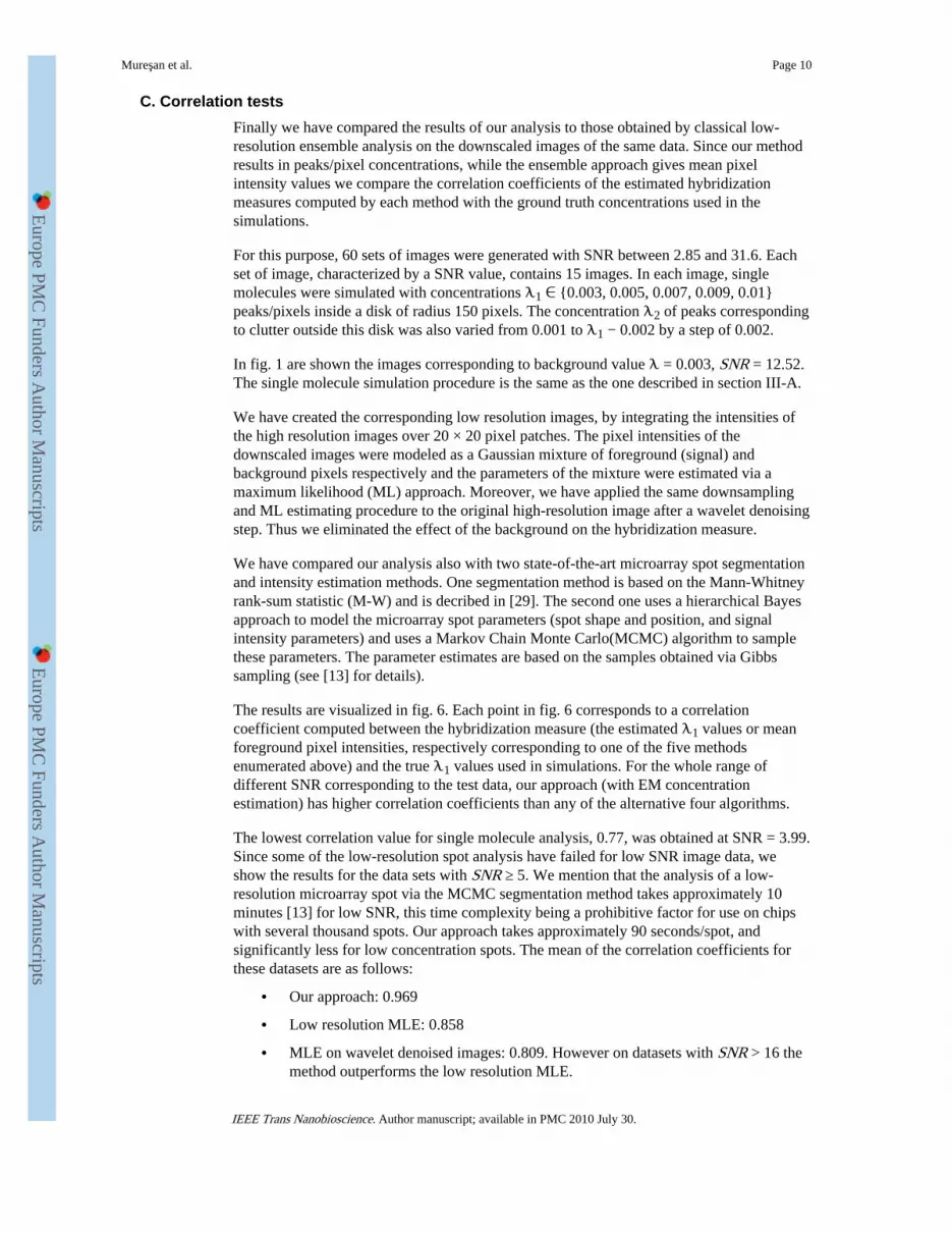
C. Correlation tests
Finally we have compared the results of our analysis to those obtained by classical low-resolution ensemble analysis on the downscaled images of the same data. Since our methodresults in peaks/pixel concentrations, while the ensemble approach gives mean pixelintensity values we compare the correlation coefficients of the estimated hybridizationmeasures computed by each method with the ground truth concentrations used in thesimulations.
For this purpose, 60 sets of images were generated with SNR between 2.85 and 31.6. Eachset of image, characterized by a SNR value, contains 15 images. In each image, singlemolecules were simulated with concentrations 1 ∈ {0.003, 0.005, 0.007, 0.009, 0.01}peaks/pixels inside a disk of radius 150 pixels. The concentration 2 of peaks correspondingto clutter outside this disk was also varied from 0.001 to 1 − 0.002 by a step of 0.002.
In fig. 1 are shown the images corresponding to background value = 0.003, SNR = 12.52.The single molecule simulation procedure is the same as the one described in section III-A.
We have created the corresponding low resolution images, by integrating the intensities ofthe high resolution images over 20 × 20 pixel patches. The pixel intensities of thedownscaled images were modeled as a Gaussian mixture of foreground (signal) andbackground pixels respectively and the parameters of the mixture were estimated via amaximum likelihood (ML) approach. Moreover, we have applied the same downsamplingand ML estimating procedure to the original high-resolution image after a wavelet denoisingstep. Thus we eliminated the effect of the background on the hybridization measure.
We have compared our analysis also with two state-of-the-art microarray spot segmentationand intensity estimation methods. One segmentation method is based on the Mann-Whitneyrank-sum statistic (M-W) and is decribed in [29]. The second one uses a hierarchical Bayesapproach to model the microarray spot parameters (spot shape and position, and signalintensity parameters) and uses a Markov Chain Monte Carlo(MCMC) algorithm to samplethese parameters. The parameter estimates are based on the samples obtained via Gibbssampling (see [13] for details).
The results are visualized in fig. 6. Each point in fig. 6 corresponds to a correlationcoefficient computed between the hybridization measure (the estimated 1 values or meanforeground pixel intensities, respectively corresponding to one of the five methodsenumerated above) and the true 1 values used in simulations. For the whole range ofdifferent SNR corresponding to the test data, our approach (with EM concentrationestimation) has higher correlation coefficients than any of the alternative four algorithms.
The lowest correlation value for single molecule analysis, 0.77, was obtained at SNR = 3.99.Since some of the low-resolution spot analysis have failed for low SNR image data, weshow the results for the data sets with SNR ≥ 5. We mention that the analysis of a low-resolution microarray spot via the MCMC segmentation method takes approximately 10minutes [13] for low SNR, this time complexity being a prohibitive factor for use on chipswith several thousand spots. Our approach takes approximately 90 seconds/spot, andsignificantly less for low concentration spots. The mean of the correlation coefficients forthese datasets are as follows:
• Our approach: 0.969
• Low resolution MLE: 0.858
• MLE on wavelet denoised images: 0.809. However on datasets with SNR > 16 themethod outperforms the low resolution MLE.
Mureşan et al. Page 10
IEEE Trans Nanobioscience. Author manuscript; available in PMC 2010 July 30.
Europe P
MC
Funders A
uthor Manuscripts
Europe P
MC
Funders A
uthor Manuscripts

• MCMC segmentation: 0.809
• M-W segmentation: 0.709.
IV. Conclusion
In this paper we have presented the analysis of microarray images at single moleculeresolution. The concentration of single molecules as a new measure of hybridization offersattractive advantages in the case of low abundance of target molecules, for backgroundsuppression, photon count fluctuation etc. We have shown that the single molecule detectionalgorithm performs well across a wide range of concentrations and image SNRs. Theseparation of the specifically bound molecules from clutter was also thoroughly tested.Furthermore we have shown that our approach provides good correlation results forconcentrations and SNR values where the low resolution methods fail. The algorithmspresented in the paper provide validated tools for other techniques based on the observationof individual fluorescent molecules.
Acknowledgments
This work was supported by the GEN-AU program of the Austrian Federal Ministry of Education, Science andCulture, by the Austrian Research Fund (FWF Project L422-N20), the European Commission (FP6 ProjectAutoscreen) and the state of Upper Austria.
References
[1]. Hesse J, Jacak J, Kasper M, Regl G, Eichberger T, Winklmayr M, Aberger F, Sonnleitner M,Schlapak R, Howorka S, Muresan L, Frischauf A, Schütz GJ. RNA expression profiling at thesingle molecule level. Genome Research. 2006; 16:1041–1045. [PubMed: 16809670]
[2]. Nygaard V, Holden M, Løland A, Langaas M, Myklebost O, Hovig E. Limitations of mRNAamplification from small-size cell samples. BMC Genomics. 2005; 6:147. [PubMed: 16253144]
[3]. Hesse J, Sonnleitner M, Sonnleitner A, Freudenthaler G, Jacak J, Höglinger O, Schindler H,Schütz G. Single-molecule reader for high-throughput bioanalysis. Anal. Chem. 2004; 76:5960–5964. [PubMed: 15456321]
[4]. Sonnleitner, M.; Freudenthaler, G.; Hesse, J.; Schüz, GJ. High-throughput scanning with single-molecule sensitivity; SPIE Proceedings; 2005; p. 202-210.
[5]. Bajcsy P. An overview of DNA microarray grid alignment and foreground separation approaches.EURASIP Journ. on Applied Signal Processing. 2006; 2006:1–13.
[6]. Yang YH, Buckley MJ, Speed T. Analysis of cDNA microarray images. Briefings inBioinformatics. 2001; 2:341–349. [PubMed: 11808746]
[7]. Yang YH, Buckley J, Dudoit S, Speed TP. Comparison of methods for image analysis on cDNAmicroarray data. Journal of Computational and Graphical Statistics. 2002; 11:108–136.
[8]. Balagurunathan Y, Wang N, Dougherty ER, Nguyen D, Chen Y, Bittner M, Tent J, Carroll R.Noise factor analysis for cdna microarrays. Journal of Biomedical Optics. 2004; 9(4):663–678.[PubMed: 15250753]
[9]. Angulo J. Polar modelling and segmentation of genomic microarray spots using mathematicalmorphology. Image Anal. Stereol. 2008; 27:107–124.
[10]. Chudin E, Kruglyak S, Baker S, Oeser S, Barker D, McDaniel T. A model of technical variationof microarray signals. Journal of Computational Biology. 2006; 13(4):996–1003. [PubMed:16761924]
[11]. Korn E, Habermann J, Upender M, Ried T, McShane L. Objective method of comparing DNAmicroarray image analysis systems. Bioimaging. 2004; 36(6):960–967.
[12]. Li Q, Fraley C, Bumgarner R, Yeung K, Raftery A. Donuts, scratches and blanks: robust model-based segmentation of microarray images. Bioinformatics. 2005; 21(12):2875–2882. [PubMed:15845656]
Mureşan et al. Page 11
IEEE Trans Nanobioscience. Author manuscript; available in PMC 2010 July 30.
Europe P
MC
Funders A
uthor Manuscripts
Europe P
MC
Funders A
uthor Manuscripts

[13]. Sarder P, Nehorai A, Davis PH, Stanley SLJ. Estimating gene signals from noisy microarrayimages. IEEE Transactions on Nanobioscience. 2008; 7(2)
[14]. Starck, J-L.; Murtagh, F.; Bijaoui, A. Image and Data Analysis: The Multiscale Approach.Cambridge University Press; 1998.
[15]. Olivo-Marin J-C. Extraction of spots in biological images using multiscale products. PatternRecognition. 2002; 35:1989–1996.
[16]. Mallat, S. A wavelet tour of signal processing. Academic Press; 1999.
[17]. Starck JL, Fadili J, Murtagh F. The undecimated wavelet decomposition and its reconstruction.IEEE Trans. on Image Processing. 2007; 16(2)
[18]. Donoho D, Johnstone I. Ideal spatial adaptation via wavelet shrinkage. Biometrika. 1994;81:425–455.
[19]. Donoho D. De-noising by soft-thresholidng. IEEE Transactions on Information Theory. 1995;41(3):613–627.
[20]. Benjamini Y, Hochberg Y. Controlling the false discovery rate: A practical and powerfulapproach to multiple testing. J. R. Statist. Soc. B. 1995; 57(1):289–300.
[21]. Abramovich F, Benjamini Y. Adaptive thresholding of wavelet coefficients. ComputationalStatistics and Data Analysis. 1996; 22:351–361.
[22]. Johnstone I, Silverman B. Needles and straw in haystacks: Empirical Bayes estimates of possiblysparse sequences. The Annals of Statistics. 2004; 32(4):1594–1649.
[23]. Johnstone I, Silverman B. Empirical Bayes selection of wavelet thresholds. The Annals ofStatistics. 2005; 33(4):1700–1752.
[24]. Boulanger, J.; Sibarita, J-B.; Kervrann, C.; Bouthemy, P. Non-parametric regression for patch-based fluorescence microscopy image sequence denoising; Proc. IEEE Int. Symp. on BiomedicalImaging: from nano to macro (ISBI); 2008;
[25]. Blekas K, Galatsanos NP, Likas A, Lagaris IE. Mixture model analysis of DNA microarrayimages. IEEE Transactions on Medical Imaging. 2005; 24:901–909. [PubMed: 16011320]
[26]. Frühwirth-Schnatter, S. Finite Mixture and Markov Switching Models. Springer; New York:2006.
[27]. Byers S, Raftery AE. Nearest-neighbor clutter removal for estimating features in spatial pointprocesses. Journal of the American Staistical Association. 1998; 93:577–584.
[28]. Murphy, DB. Fundamentals of light microscopy and electronic imaging. Willey-LISS; 2001.
[29]. Chen Y, Dougherty E, Bittner M. Ratio-based decisions and the quantitative analysis of cDNAmicroarray images. Journal of Biomedical Optics. 1997; 2(4):364–374. [PubMed: 23014960]
Mureşan et al. Page 12
IEEE Trans Nanobioscience. Author manuscript; available in PMC 2010 July 30.
Europe P
MC
Funders A
uthor Manuscripts
Europe P
MC
Funders A
uthor Manuscripts

Fig. 1.Simulation of microarrays spots. Upper row: spots at single molecule resolution (200nmpixel size) with different peak concentrations () inside each spot. From left to right: =0.005, 0.007, 0.009, 011 peaks per pixel. (Background concentration representing dirt,unspecific binding etc.: 0.003 peaks per pixel). Lower row: the same spots downsampled to4 m, the size used by existing commercial microarray systems.
Mureşan et al. Page 13
IEEE Trans Nanobioscience. Author manuscript; available in PMC 2010 July 30.
Europe P
MC
Funders A
uthor Manuscripts
Europe P
MC
Funders A
uthor Manuscripts

Fig. 2.Analysis of a spot in a high-resolution microarray image. (a) Original image, bright featurescorrespond to molecules bound to the chip. (b) Detection after undecimated waveletthresholding. (c) Selection of single molecule locations (local maxima on denoised imageinside the detection support in (b)), (d) Separation of hybridization signal from clutter.
Mureşan et al. Page 14
IEEE Trans Nanobioscience. Author manuscript; available in PMC 2010 July 30.
Europe P
MC
Funders A
uthor Manuscripts
Europe P
MC
Funders A
uthor Manuscripts

Fig. 3.Results of detection on simulations. Left: ratio of true positives, right: ratio of false positiveswith respect to the true number of simulated single molecules. The size of simulated imagesis 512 × 512 pixels. The typical SNR is higher than 10, indicated in the figures by a verticalline.
Mureşan et al. Page 15
IEEE Trans Nanobioscience. Author manuscript; available in PMC 2010 July 30.
Europe P
MC
Funders A
uthor Manuscripts
Europe P
MC
Funders A
uthor Manuscripts

Fig. 4.Background/foreground separation for three different concentrations via Method ofMoments (MOM) and Expectation Maximization (EM) methods (see II-B). The true values are represented as a stair-case function and for better visibility, the estimation resultswere slightly shifted on the abscissa.
Mureşan et al. Page 16
IEEE Trans Nanobioscience. Author manuscript; available in PMC 2010 July 30.
Europe P
MC
Funders A
uthor Manuscripts
Europe P
MC
Funders A
uthor Manuscripts

Fig. 5.Anomalous shape detection. A high concentration region in donut shape was simulated on abackground formed of low concentration clutter. The proposed approach is able to separatesignal from clutter.
Mureşan et al. Page 17
IEEE Trans Nanobioscience. Author manuscript; available in PMC 2010 July 30.
Europe P
MC
Funders A
uthor Manuscripts
Europe P
MC
Funders A
uthor Manuscripts

Fig. 6.Correlations between the estimated and the true signal concentrations. The single moleculeanalysis performs better than the analysis on the downscaled data (original and denoised viawavelet thresholding). The arrow indicates the correlation coefficient corresponding to theimages in figure 1.
Mureşan et al. Page 18
IEEE Trans Nanobioscience. Author manuscript; available in PMC 2010 July 30.
Europe P
MC
Funders A
uthor Manuscripts
Europe P
MC
Funders A
uthor Manuscripts