A Useful Approach to Identify Novel Small-Molecule Inhibitors of Wnt-Dependent Transcription
12
2010;70:5963-5973. Published OnlineFirst July 7, 2010. Cancer Res Kenneth Ewan, Bozena Pajak, Mark Stubbs, et al. of Wnt-Dependent Transcription A Useful Approach to Identify Novel Small-Molecule Inhibitors Updated Version 10.1158/0008-5472.CAN-10-1028 doi: Access the most recent version of this article at: Material Supplementary http://cancerres.aacrjournals.org/content/suppl/2010/07/02/0008-5472.CAN-10-1028.DC1.html Access the most recent supplemental material at: Cited Articles http://cancerres.aacrjournals.org/content/70/14/5963.full.html#ref-list-1 This article cites 40 articles, 17 of which you can access for free at: Citing Articles http://cancerres.aacrjournals.org/content/70/14/5963.full.html#related-urls This article has been cited by 4 HighWire-hosted articles. Access the articles at: E-mail alerts related to this article or journal. Sign up to receive free email-alerts Subscriptions Reprints and . [email protected] Department at To order reprints of this article or to subscribe to the journal, contact the AACR Publications Permissions . [email protected] Department at To request permission to re-use all or part of this article, contact the AACR Publications American Association for Cancer Research Copyright © 2010 on May 18, 2012 cancerres.aacrjournals.org Downloaded from Published OnlineFirst July 7, 2010; DOI:10.1158/0008-5472.CAN-10-1028
-
Upload
independent -
Category
Documents
-
view
0 -
download
0
Transcript of A Useful Approach to Identify Novel Small-Molecule Inhibitors of Wnt-Dependent Transcription
2010;70:5963-5973. Published OnlineFirst July 7, 2010.Cancer Res Kenneth Ewan, Bozena Pajak, Mark Stubbs, et al. of Wnt-Dependent TranscriptionA Useful Approach to Identify Novel Small-Molecule Inhibitors
Updated Version 10.1158/0008-5472.CAN-10-1028doi:
Access the most recent version of this article at:
MaterialSupplementary
http://cancerres.aacrjournals.org/content/suppl/2010/07/02/0008-5472.CAN-10-1028.DC1.htmlAccess the most recent supplemental material at:
Cited Articles http://cancerres.aacrjournals.org/content/70/14/5963.full.html#ref-list-1
This article cites 40 articles, 17 of which you can access for free at:
Citing Articles http://cancerres.aacrjournals.org/content/70/14/5963.full.html#related-urls
This article has been cited by 4 HighWire-hosted articles. Access the articles at:
E-mail alerts related to this article or journal.Sign up to receive free email-alerts
SubscriptionsReprints and
[email protected] atTo order reprints of this article or to subscribe to the journal, contact the AACR Publications
To request permission to re-use all or part of this article, contact the AACR Publications
American Association for Cancer Research Copyright © 2010 on May 18, 2012cancerres.aacrjournals.orgDownloaded from
Published OnlineFirst July 7, 2010; DOI:10.1158/0008-5472.CAN-10-1028
Published OnlineFirst July 7, 2010; DOI:10.1158/0008-5472.CAN-10-1028
Therapeutics, Targets, and Chemical Biology
CancerResearch
A Useful Approach to Identify Novel Small-MoleculeInhibitors of Wnt-Dependent Transcription
Kenneth Ewan1, Bożena Pająk1, Mark Stubbs2, Helen Todd3, Olivier Barbeau2, Camilo Quevedo2,Hannah Botfield1, Rodrigo Young4, Ruth Ruddle2, Lee Samuel1, Alysia Battersby1,Florence Raynaud2, Nicholas Allen1, Stephen Wilson4, Branko Latinkic1, Paul Workman2,Edward McDonald2, Julian Blagg2, Wynne Aherne2, and Trevor Dale1
Abstract
Authors' AUni ted KTherapeuticKingdom; 3
Scot landDevelopmeKingdom
Note: SupResearch O
CorresponSciences BPhone: 44cardiff.ac.u
doi: 10.115
©2010 Am
www.aacr
The Wnt signaling pathway is frequently deregulated in cancer due to mutations in genes encoding APC,β-catenin, and axin. To identify small-molecule inhibitors of Wnt signaling as potential therapeutics, a diversechemical library was screened using a transcription factor reporter cell line in which the activity of the path-way was induced at the level of Disheveled protein. A series of deconvolution studies was used to focus onthree compound series that selectively killed cancer cell lines with constitutive Wnt signaling. Activities of thecompounds included the ability to induce degradation of β-catenin that had been stabilized by a glycogensynthase kinase-3 (GSK-3) inhibitor. This screen illustrates a practical approach to identify small-moleculeinhibitors of Wnt signaling that can seed the development of agents suitable to treat patients with Wnt-dependent tumors. Cancer Res; 70(14); 5963–73. ©2010 AACR.
Introduction
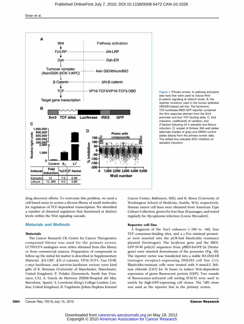
The Wnt signaling pathway is activated by Wnt ligands atmultiple stages of metazoan development and controls thedifferentiation and/or proliferation of stem cells in multipletissues. The “canonical” Wnt/β-catenin pathway is activatedfollowing Wnt ligand binding to a complex comprisingFrizzled (Fz) and LRP5/6 receptors and ultimately activatesβ-catenin/transcription factor–dependent transcription(Fig. 1A). Key steps in the pathway include the formation ofa ligand-activated receptor complex, the inhibition of intra-cellular β-catenin turnover, and the formation of a nuclearβ-catenin/transcription factor (TCF) transcription complex.Wnt/β-catenin signaling activates (and represses) transcrip-tion of genes whose promoters contain binding sites for TCF-transcription factors. Target genes include key developmentaland oncogenic targets (reviewed in ref. 1).The Wnt signaling pathway is considered a key therapeutic
target for cancer (1). Mutations to “core” Wnt signaling com-ponents, including β-catenin, adenomatous polyposis coli
ffiliations: 1School of Bioscience, Cardiff University, Cardiff,ingdom; 2Cancer Research UK Cent re for Cancers, The Institute of Cancer Research, Sutton, Surrey, UnitedVirology Department, Moredun Research Institute, Penicuik,, Un i ted K ingdom; and 4Depar tment o f Ce l l andntal Biology, University College London, London, United
plementary data for this article are available at Cancernline (http://cancerres.aacrjournals.org/).
ding Author: Trevor Dale, University of Cardiff, Biomedicaluilding, Museum Avenue, Cardiff CF10 3AX, United Kingdom.-29-2087-4652; Fax: 44-29-2087-4116; E-mail: DaleTC@k.
8/0008-5472.CAN-10-1028
erican Association for Cancer Research.
journals.org
American Asso Copyright © 2010 cancerres.aacrjoDownloaded from
(APC), and Axin, inappropriately activate the β-catenin/TCF branch of the Wnt signaling pathway in cancers arisingin a number of tissues, including colon and liver. Many othercancers show evidence of inappropriate Wnt pathway activa-tion, including raised levels of β-catenin (2). Unfortunately,the known intracellular components of the core Wnt path-way are not good targets for small-molecule inhibitors, asmany interactions comprise extended protein-protein inter-faces. Furthermore, drugs that target core components mayinduce side effects because the function of key components(e.g., β-catenin) is required for tissue homeostasis in multipleorgans, including the immune system and intestine (3).Small molecules that target “noncore” Wnt signaling
components have recently been identified (4). Of particularnote, ICG001 blocks the interaction between β-cateninand the noncore CREB binding protein (CBP) leading to areduction of adenoma formation in mouse models of coloncancer (5). The β-catenin–CBP interaction was shown topromote stem/progenitor marker expression, whereas the re-lated β-catenin–p300 interaction, which was not subject toblockade by ICG001, promoted expression of genes involvedin proliferative responses such as CMYC (6). The key pointfrom this example is that compounds that target noncorecomponents may be able to regulate distinct subsets of ther-apeutically relevant transcriptional targets. The recently dis-covered small molecules XAV939 and IWR2 inhibit Wntsignaling by blocking tankyrase activity, which is requiredfor degradation of the β-catenin scaffold protein Axin (3, 7).One of the main challenges in targeting noncore Wnt sig-
naling molecules is knowing which protein to target. A largenumber of potential target genes that regulate Wnt/β-cateninsignaling have been identified in whole-genome RNAi screens,mass spectrometry, and yeast two-hybrid studies (8–10), andit remains to be seen which protein should be the focus of
5963
ciation for Cancer Research on May 18, 2012urnals.org
Ewan et al.
5964
Published OnlineFirst July 7, 2010; DOI:10.1158/0008-5472.CAN-10-1028
drug discovery efforts. To overcome this problem, we used acell-based assay to screen a diverse library of small moleculesfor regulators of TCF-dependent transcription. We identifieda number of chemical regulators that functioned at distinctlevels within the Wnt signaling cascade.
Materials and Methods
MaterialsThe Cancer Research UK Centre for Cancer Therapeutics
compound library was used for the primary screen.CCT031374 analogues were either obtained from this libraryor from commercial sources. Preparation of compounds tofollow-up the initial hit matter is described in SupplementaryMaterial. ΔN-LRP, ΔN–β-catenin, VP16-TCF4, Tau-1N4R,c-myc-luciferase, and survivin-luciferase vectors were kindgifts of K. Brennan (University of Manchester, Manchester,United Kingdom), P. Polakis (Genentech, South San Fran-sisco, CA), A. García de Herreros (IMIM-Hospital del Mar,Barcelona, Spain), S. Lovestone (King's College London, Lon-don, United Kingdom), B. Vogelstein (Johns Hopkins Kimmel
Cancer Res; 70(14) July 15, 2010
American Asso Copyright © 2010 cancerres.aacrjoDownloaded from
Cancer Center, Baltimore, MD), and R. Moon (University ofWashington School of Medicine, Seattle, WA), respectively.Human tumor cell lines were obtained from American TypeCulture Collection, grown for less than 20 passages, and testedregularly for Mycoplasma infection (Lonza Mycoalert).
Reporter cell lineA fragment of the Xnr3 enhancer (−180 to −60), four
TCF consensus-binding sites, and a c-Fos minimal promot-er were inserted into the pUB-bsd blasticidin resistanceplasmid (Invitrogen). The luciferase gene and the IRES-GFP-SV40 poly(A) sequences from pIRES-hrGFP-2a (Strata-gene) were inserted downstream of the promoter (Fig. 1B).The reporter vector was transfected into a stable HA-Dvl2-ER(estrogen receptor)–expressing HEK293 cell line (11).Blasticidin-resistant cells were treated with 9 mmol/L lith-ium chloride (LiCl) for 16 hours to induce Wnt-dependentexpression of green fluorescent protein (GFP). Two roundsof fluorescence-activated cell sorting (FACS) were used toenrich for high-GFP-expressing cell clones. The 7df3 clonewas used as the reporter line in the primary screen.
Cancer Research
ciation for Cancer Research on May 18, 2012urnals.org
l
Figure 1. Primary screen. A, pathway activators(see text) that were used to induce Wnt/β-catenin signaling at distinct levels. B, thereporter construct used in the human epitheliaHEK293-based cell line. The bicistronicTCF-luciferase-IRES-GFP reporter containedthe Wnt response element from the Xnr3promoter and four TCF binding sites. C, foldinduction, coefficients of variation, andZ-factors following 24 h estradiol and lithiuminduction. D, subset of thirteen 384-well plates(alternate shades of gray) and DMSO controlplates (black) from the primary screen data.The dotted line indicates 50% inhibition ofestradiol induction.

Small-Molecule Inhibitors of Wnt Signaling
Published OnlineFirst July 7, 2010; DOI:10.1158/0008-5472.CAN-10-1028
Primary screenStructurally diverse, low molecular weight compounds
(63,040) were added individually to reporter cells at20 μmol/L 2 hours before addition of 10 μmol/L β-estradiolin 384-well plates. Luciferase assays were carried out 24 hoursafter β-estradiol addition using SteadyGlo reagent (Pro-mega). Activities of primary hits were reconfirmed in furtherluciferase assays and counterscreened for inhibition of lucife-rase enzyme (EasyLite kinase reagent, Perkin-Elmer) andnonspecific cell growth inhibition (Celltiter Blue, Promega).
Secondary assaysHit compounds (20 μmol/L) were assayed in transiently
transfectedHEK293 cells cotransfected with Topflash (Wnt re-porter, Upstate), TK-renilla (control), and cytomegalovirus-driven HA-Dvl2-ER expression plasmids. Twenty-four hoursafter transfection, cells were stimulated with 3 μmol/L β-estradiol or treated with the glycogen synthase kinase-3(GSK-3) inhibitor 6-bromoindirubin-3′-oxime (BIO; Calbio-chem). Luciferase activity was measured after 24 hours usingDualGlo reagent (Promega). For deconvolution assays,HEK293 cells were cotransfected with Topflash and CMV-lacZ reporter (control) plasmids together with constitutivelyactive core components of the pathway: ΔN-LRP, Axin-GID,ΔN–β-catenin, and VP16-TCF4. Compounds were added 24hours after transfection, and reporter activity was analyzeda further 24 hours later using BrightGlo and BetaGlo (Promega).Luciferase activity was normalized to β-galactosidase report-er activity. Experiments with c-myc and survivin-luciferasereporters were carried out in HEK293 cells using Axin-GIDas the inducer. SW480 cells were cotransfected with Topflashand CMV-lacZ reporter plasmids 24 hours before compoundaddition. Reporter assays were conducted after a further24 hours.
Growth inhibition (GI50)Cells were seeded to give a density of 25% confluence after
24 hours in either 96- or 384-well tissue culture plates. Com-pounds (0.05–100 μmol/L) were added at 24 hours; Cell TitreBlue or WST-1 (Roche) reagent was added after 72 hours(HT29, SW480 and HCT116) or 96 hours (SNU475 andCCD841Co).
β-Catenin stability experimentsMouse L-cells were cultured in DMEM/10% fetal bovine
serum and exposed to 7.5 μmol/L BIO, 9 mmol/L LiCl, 50%Wnt-3a conditioned medium (12), or 50 μmol/L MG-132(Merck Biosciences), and with compound for 6 hours beforemedium replacement as indicated. β-Catenin was assayed aspreviously described (13). The following antibodies wereused: β-catenin and GSK-3β (BD Transduction laboratories)and β-actin (Sigma). The β-catenin antibody from BD Trans-duction Laboratories was used for immunocytochemistry aspreviously described (14).For pulse-chase analyses, L-cells were treated throughout
with 7.5 μmol/L BIO. Cells were first starved in medium lack-ing methionine and cysteine for 1 hour then labeled for1 hour with 5 MBq per 25 cm2 flask of 35S-labeled methio-
www.aacrjournals.org
American Asso Copyright © 2010 cancerres.aacrjoDownloaded from
nine/cysteine. Cells were chased with unlabeled mediumfor the indicated periods. Cells were lysed, and immunopre-cipitation was carried out using 2 μg/sample monoclonalβ-catenin antibody and 50 μL of Protein G–coated beads(GE Healthcare). After three washes in lysis buffer, boundproteins were eluted in SDS sample buffer, separated byelectrophoresis, and analyzed as previously described (15).
Phosphorylated Tau experimentHEK293 cells were transfected with GSK-3β and/or
Tau-1N4R expression vectors (16). After 48 hours, cells wereexposed to 5 μmol/L BIO for 8 hours, after which they weretreated with 20 μmol/L CCT031374 and 5 μmol/L BIO for theindicated time periods. Immunoblotting for GSK-3β andphospho- and nonphospho-Tau antibodies (BT2, AT270,Autogen Bioclear) was carried out as previously described(16). Band peak areas were calculated using ImageJ (Scion).The phospho-Tau peak areas were normalized to the totalTau levels.
Glutathione S-transferase–E-cadherin pull down ofβ-cateninThe β-catenin binding region of E-cadherin (amino acids
730–882) was fused in framewith the glutathione S-transferase(GST) coding region of pGEX-5X-2 (GE Healthcare). GST–E-cadherin fusion protein was expressed and purified usingGlutathione Sepharose 4 Fast Flow beads (GE Healthcare).Ten-centimeterdishesofHEK293andSW480cellswere treatedwith CCT031374 and/or BIO and lysed to give a final volumeof 1 mL cell extract. Five micrograms of GST–E-cadherinwere combined with 500 μL of cell extract and precipitatedfollowing addition of glutathione beads. Bound β-cateninwas detected by immunoblotting.
Embryonic stem cell quantitative PCR for LEF1H9 human embryonic stem cells were cultured on feeders
using standard culture methods (17). Using a modification ofref. (17), neurogenic embryoid bodies were generated by cul-turing H9 colony fragments in suspension in neutralizing me-dium (ADF, Invitrogen 12634010) containing 10 μmol/LSB431542 (TGF-β Inhibitor, Tocris) and 10 μmol/L Y-27632(ROCK inhibitor, Calbiochem) for the first 48 hours. On day8, medium was supplemented with BIO (5 μmol/L) and/orCCT031374 (20 μmol/L) for 24 hours. RNA was extractedwith Rneasy (Qiagen). cDNA was synthesized with Super-script II Rnase H-RT (Invitrogen). Quantitative PCR reactionswere done in DyNAmoHS master mix (New England Biolabs)using the LEF1 primers 5′-accagattcttggcagaagg-3′ and5′-cagaccagcctggataaagc-3′.
Xenopus studiesXenopus laevis embryos were obtained, degelled, and cul-
tured using standard procedures (18), and staged accordingto Nieuwkoop and Faber (NF; ref. 19). Test compound orDMSO (control) was added at the 4- to 16-cell stage. Embryoswere scored at NF stages 35 to 38. For the animal cap assay,dissected Xenopus embryo animal caps were treated with300 mmol/L LiCl for 10 minutes followed by 2.5 hours
Cancer Res; 70(14) July 15, 2010 5965
ciation for Cancer Research on May 18, 2012urnals.org
Ewan et al.
5966
Published OnlineFirst July 7, 2010; DOI:10.1158/0008-5472.CAN-10-1028
exposure to CCT036477 at the indicated concentrations orwith DMSO control (indicated with dash). RNA extractionand reverse transcriptase-PCR conditions were carried outas described (20).
Zebrafish studiesZebrafish embryos were collected after natural spawning.
Embryos were treated from the 16-cell stage until midgastru-lation (8 hours postfertilization) with 20 μmol/L of compoundCCT036477 in 1% DMSO/fish water or with DMSO equivalent.Control and experimental embryos were imaged when at thepostgastrulation somitic stage (48 hours postfertilization).
Results
Optimization and outcome of high-throughput screenTo identify novel small-molecule regulators of the Wnt sig-
naling pathway, a HEK293-based reporter cell line (7df3) wasgenerated to allow the inducible induction of TCF-dependenttranscription. The rationale for this approach was twofold.First, inducible induction of the Wnt pathway was predictedto minimize positive and negative feedback from transcrip-tional targets, thereby simplifying the process of target decon-volution. Second, the cell line did not require Wnt signalingfor proliferation, thereby allowing the rapid distinction ofnonspecific growth inhibitors and compounds acting specifi-cally on the Wnt pathway.A bicistronic reporter coding for firefly luciferase and
GFP under the control of a promoter containing four ca-nonical TCF binding sites and the Wnt-responsive regionfrom the Xenopus Xnr3 promoter (ref. 21; Fig. 1B) was intro-duced into HEK293 cells that already contained an integra-ted Disheveled–estrogen receptor fusion (Dvl2-ER; ref. 11).Previous studies showed that estradiol induction of Dvl2 ac-tivity raised β-catenin levels within 30 minutes in these cells(11). Clones that showed tight regulation of TCF-dependenttranscription in response to estradiol and the GSK-3 inhib-itor LiCl (Fig. 1C) were identified by FACS sorting.Assays in a 384-well format showed reproducible 14-fold
and 10,389-fold induction with estradiol and LiCl, respec-tively, with low coefficients of variation (%CV) and high Z′factors (estradiol, 0.59; LiCl 0.89; Fig. 1C). Importantly, thecell line also showed robust behavior with multiple cellularpassages over more than 2 years. For the primary screen,63,040 chemically diverse compounds were assayed by se-quential addition of compound (20 μmol/L in 0.2% DMSO)and estradiol 24 hours before luciferase assay. Data from asubset of 384-well plates from the primary assay (Fig. 1D) il-lustrate the range of responses to compound addition. The1,902 compounds that showed >50% inhibition were re-assayed (n = 2) and counterscreened to exclude direct inhi-bitors of luciferase enzyme activity and to remove acutelycytotoxic compounds. At this stage, 306 molecules wereselected for subsequent analysis (Fig. 2A).
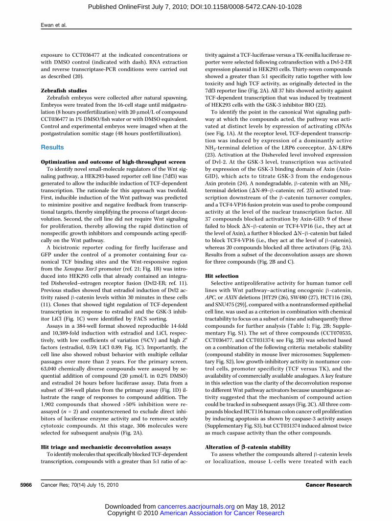
Hit triage and mechanistic deconvolution assaysTo identifymolecules that specifically blockedTCF-dependent
transcription, compounds with a greater than 5:1 ratio of ac-
Cancer Res; 70(14) July 15, 2010
American Asso Copyright © 2010 cancerres.aacrjoDownloaded from
tivity against a TCF-luciferase versus a TK-renilla luciferase re-porter were selected following cotransfection with a Dvl-2-ERexpression plasmid in HEK293 cells. Thirty-seven compoundsshowed a greater than 5:1 specificity ratio together with lowtoxicity and high TCF activity, as originally detected in the7df3 reporter line (Fig. 2A). All 37 hits showed activity againstTCF-dependent transcription that was induced by treatmentof HEK293 cells with the GSK-3 inhibitor BIO (22).To identify the point in the canonical Wnt signaling path-
way at which the compounds acted, the pathway was acti-vated at distinct levels by expression of activating cDNAs(see Fig. 1A). At the receptor level, TCF-dependent transcrip-tion was induced by expression of a dominantly activeNH2-terminal deletion of the LRP6 coreceptor, ΔN-LRP6(23). Activation at the Disheveled level involved expressionof Dvl-2. At the GSK-3 level, transcription was activatedby expression of the GSK-3 binding domain of Axin (Axin-GID), which acts to titrate GSK-3 from the endogenousAxin protein (24). A nondegradable, β-catenin with an NH2-terminal deletion (ΔN-89–β-catenin; ref. 25) activated tran-scription downstream of the β-catenin turnover complex,and a TCF4-VP16 fusion protein was used to probe compoundactivity at the level of the nuclear transcription factor. All37 compounds blocked activation by Axin-GID: 9 of thesefailed to block ΔN–β-catenin or TCF4-VP16 (i.e., they act atthe level of Axin), a further 8 blockedΔN–β-catenin but failedto block TCF4-VP16 (i.e., they act at the level of β-catenin),whereas 20 compounds blocked all three activators (Fig. 2A).Results from a subset of the deconvolution assays are shownfor three compounds (Fig. 2B and C).
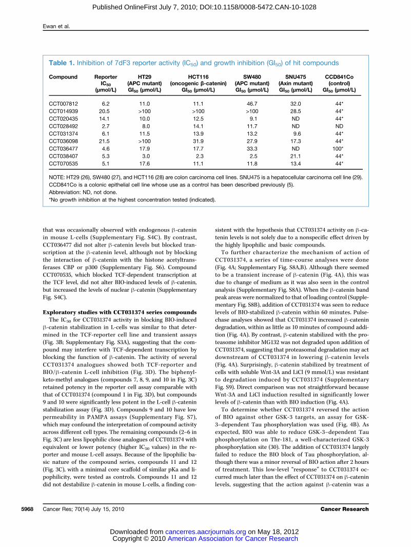
Hit selectionSelective antiproliferative activity for human tumor cell
lines with Wnt pathway–activating oncogenic β-catenin,APC, or AXIN deletions [HT29 (26), SW480 (27), HCT116 (28),and SNU475 (29)], compared with a nontransformed epithelialcell line, was used as a criterion in combination with chemicaltractability to focus on a subset of nine and subsequently threecompounds for further analysis (Table 1; Fig. 2B; Supple-mentary Fig. S1). The set of three compounds (CCT070535,CCT036477, and CCT031374; see Fig. 2B) was selected basedon a combination of the following criteria: metabolic stability(compound stability in mouse liver microsomes; Supplemen-tary Fig. S2), low growth-inhibitory activity in nontumor con-trol cells, promoter specificity (TCF versus TK), and theavailability of commercially available analogues. A key featurein this selection was the clarity of the deconvolution responseto different Wnt pathway activators because unambiguous ac-tivity suggested that the mechanism of compound actioncould be tracked in subsequent assays (Fig. 2C). All three com-pounds blockedHCT116 human colon cancer cell proliferationby inducing apoptosis as shown by caspase-3 activity assays(Supplementary Fig. S3), but CCT031374 induced almost twiceas much caspase activity than the other compounds.
Alteration of β-catenin stabilityTo assess whether the compounds altered β-catenin levels
or localization, mouse L-cells were treated with each
Cancer Research
ciation for Cancer Research on May 18, 2012urnals.org
Small-Molecule Inhibitors of Wnt Signaling
Published OnlineFirst July 7, 2010; DOI:10.1158/0008-5472.CAN-10-1028
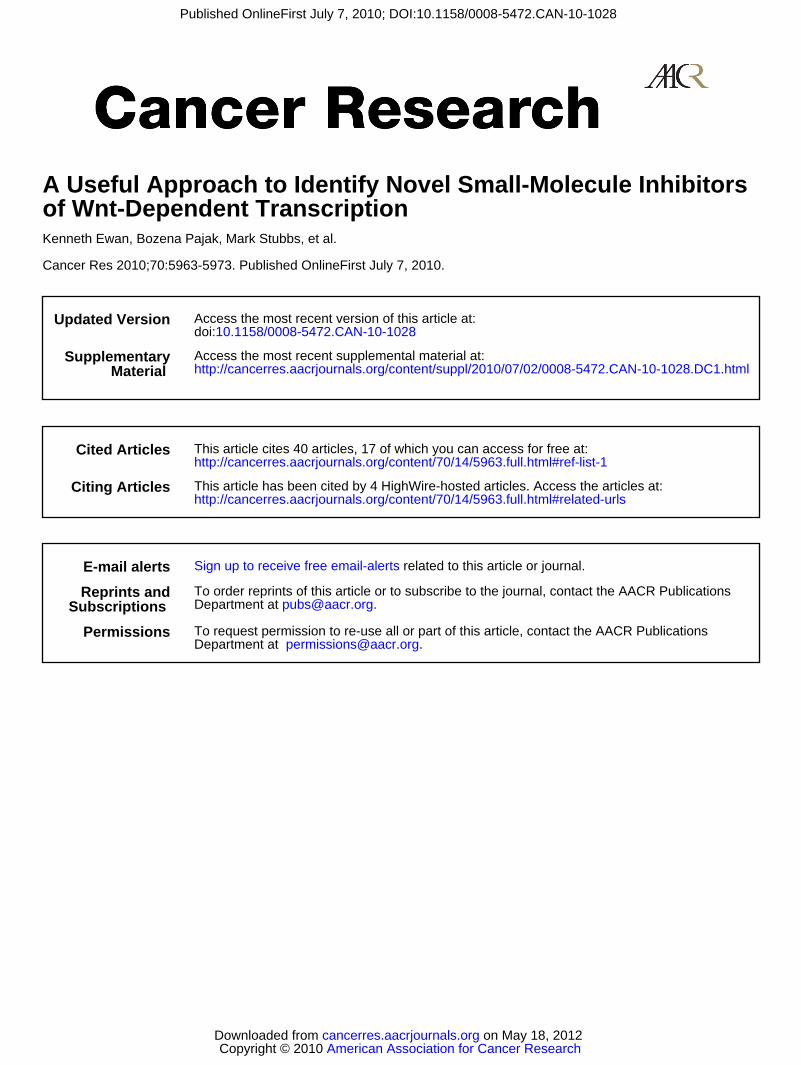
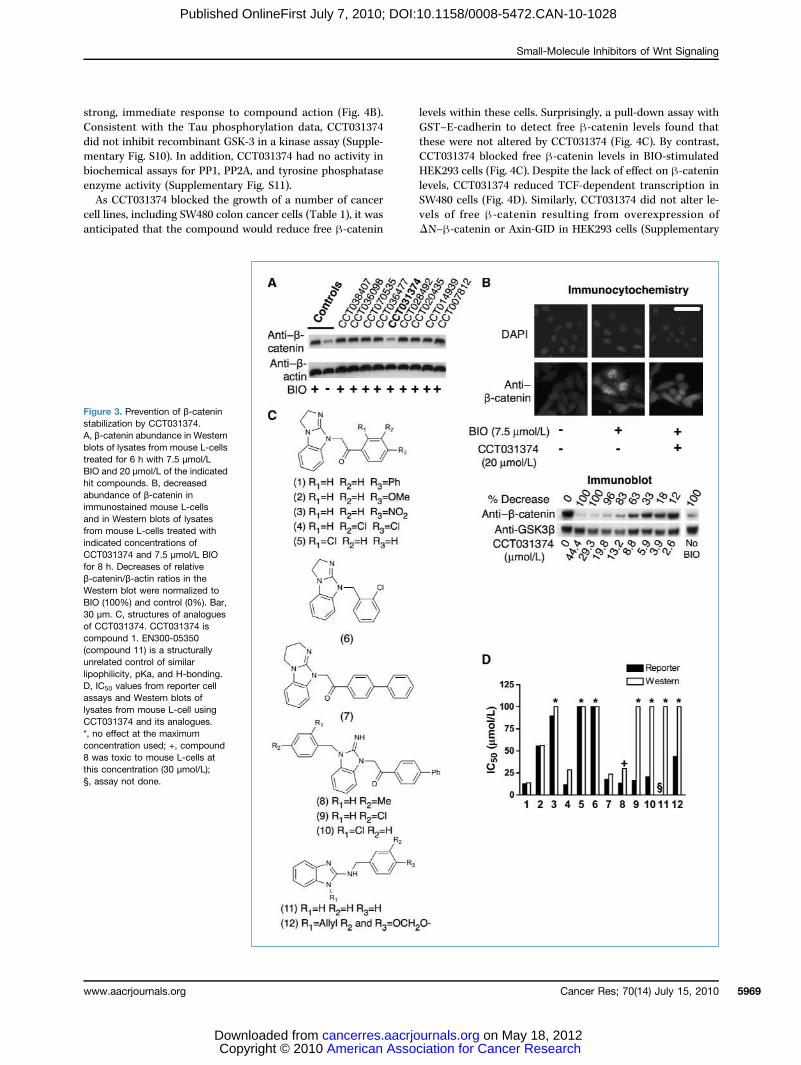
compound together with the GSK-3 inhibitor, BIO, to blockβ-catenin degradation. Following 8 hours of incubation,control BIO-treated cells showed a strong increase in totalβ-catenin levels that was associated with raised nuclearand cytosolic pools as determined by immunocytochemistry(Fig. 3A and B). Of the top nine compounds, only CCT031374prevented BIO-induced accumulation of β-catenin (Fig. 3A).
www.aacrjournals.org
American Asso Copyright © 2010 cancerres.aacrjoDownloaded from
The blockade of β-catenin accumulation by CCT031374 wasaccompanied by a reduction in both nuclear and cytosolicβ-catenin pools (Fig. 3B; Supplementary Figs. S4 and S5).In U2OS GFP–β-catenin human osteosarcoma cells (Bioimage,Thermo-Fisher), addition of CCT031374 induced formation ofGFP–β-catenin aggregates (Supplementary Fig. S4B) possiblydue to sequestration by subcellular organelles, a phenotype
Figure 2. Hit triage anddeconvolution. A, compoundattrition flow diagram leading toselected hits. B, structures of thethree hit compounds, CCT031374,CCT036477, and CCT070535.C, summary of the effects of30 μmol/L CCT031374,CCT036477, and CCT070535on TCF-dependent transcriptioninduced at different levels ofthe Wnt pathway. ControlGal4-luciferase reporter activitywas induced by VP16-Gal4.
Cancer Res; 70(14) July 15, 2010 5967
ciation for Cancer Research on May 18, 2012urnals.org
Ewan et al.
5968
Published OnlineFirst July 7, 2010; DOI:10.1158/0008-5472.CAN-10-1028
that was occasionally observed with endogenous β-cateninin mouse L-cells (Supplementary Fig. S4C). By contrast,CCT036477 did not alter β-catenin levels but blocked tran-scription at the β-catenin level, although not by blockingthe interaction of β-catenin with the histone acetyltrans-ferases CBP or p300 (Supplementary Fig. S6). CompoundCCT070535, which blocked TCF-dependent transcription atthe TCF level, did not alter BIO-induced levels of β-catenin,but increased the levels of nuclear β-catenin (SupplementaryFig. S4C).
Exploratory studies with CCT031374 series compoundsThe IC50 for CCT031374 activity in blocking BIO-induced
β-catenin stabilization in L-cells was similar to that deter-mined in the TCF-reporter cell line and transient assays(Fig. 3B; Supplementary Fig. S3A), suggesting that the com-pound may interfere with TCF-dependent transcription byblocking the function of β-catenin. The activity of severalCCT031374 analogues showed both TCF-reporter andBIO/β-catenin L-cell inhibition (Fig. 3D). The biphenyl-keto-methyl analogues (compounds 7, 8, 9, and 10 in Fig. 3C)retained potency in the reporter cell assay comparable withthat of CCT031374 (compound 1 in Fig. 3D), but compounds9 and 10 were significantly less potent in the L-cell β-cateninstabilization assay (Fig. 3D). Compounds 9 and 10 have lowpermeability in PAMPA assays (Supplementary Fig. S7),which may confound the interpretation of compound activityacross different cell types. The remaining compounds (2–6 inFig. 3C) are less lipophilic close analogues of CCT031374 withequivalent or lower potency (higher IC50 values) in the re-porter and mouse L-cell assays. Because of the lipophilic ba-sic nature of the compound series, compounds 11 and 12(Fig. 3C), with a minimal core scaffold of similar pKa and li-pophilicity, were tested as controls. Compounds 11 and 12did not destabilize β-catenin in mouse L-cells, a finding con-
Cancer Res; 70(14) July 15, 2010
American Asso Copyright © 2010 cancerres.aacrjoDownloaded from
sistent with the hypothesis that CCT031374 activity on β-ca-tenin levels is not solely due to a nonspecific effect driven bythe highly lipophilic and basic compounds.To further characterize the mechanism of action of
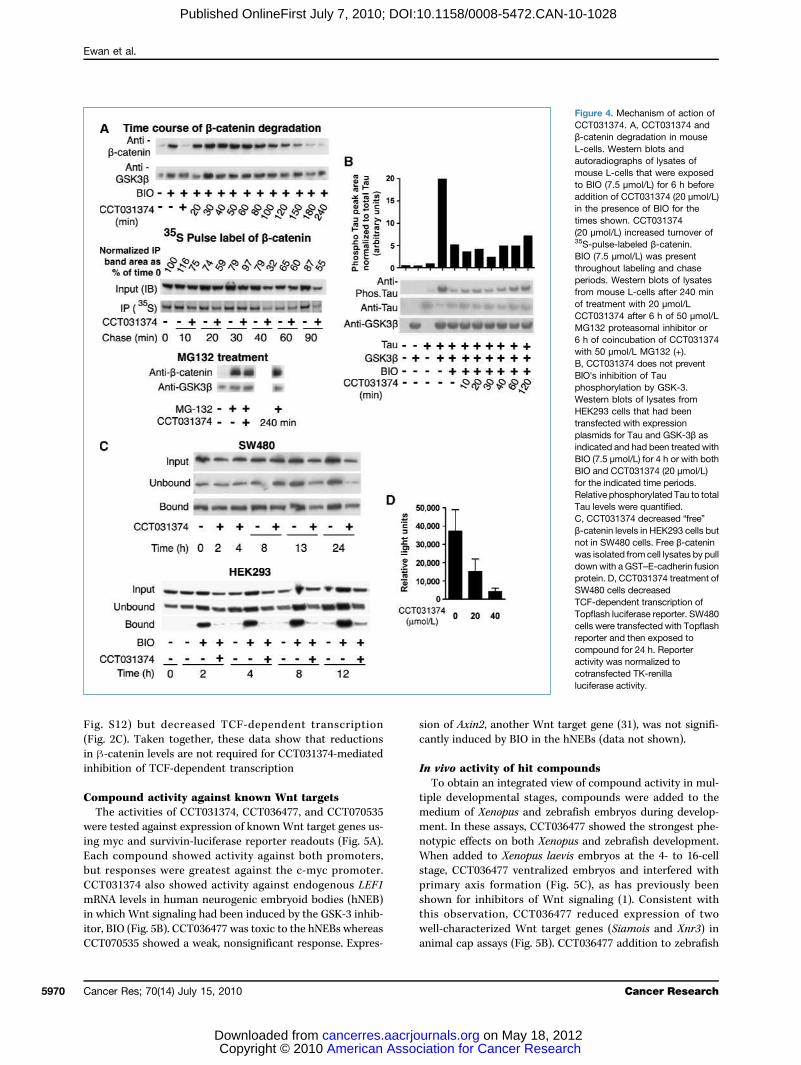
CCT031374, a series of time-course analyses were done(Fig. 4A; Supplementary Fig. S8A,B). Although there seemedto be a transient increase of β-catenin (Fig. 4A), this wasdue to change of medium as it was also seen in the controlanalysis (Supplementary Fig. S8A). When the β-catenin bandpeak areas were normalized to that of loading control (Supple-mentary Fig. S8B), addition of CCT031374 was seen to reducelevels of BIO-stabilized β-catenin within 60 minutes. Pulse-chase analyses showed that CCT031374 increased β-catenindegradation, within as little as 10 minutes of compound addi-tion (Fig. 4A). By contrast, β-catenin stabilized with the pro-teasome inhibitor MG132 was not degraded upon addition ofCCT031374, suggesting that proteasomal degradation may actdownstream of CCT031374 in lowering β-catenin levels(Fig. 4A). Surprisingly, β-catenin stabilized by treatment ofcells with soluble Wnt-3A and LiCl (9 mmol/L) was resistantto degradation induced by CCT031374 (SupplementaryFig. S9). Direct comparison was not straightforward becauseWnt-3A and LiCl induction resulted in significantly lowerlevels of β-catenin than with BIO induction (Fig. 4A).To determine whether CCT031374 reversed the action
of BIO against other GSK-3 targets, an assay for GSK-3–dependent Tau phosphorylation was used (Fig. 4B). Asexpected, BIO was able to reduce GSK-3–dependent Tauphosphorylation on Thr-181, a well-characterized GSK-3phosphorylation site (30). The addition of CCT031374 largelyfailed to reduce the BIO block of Tau phosphorylation, al-though there was a minor reversal of BIO action after 2 hoursof treatment. This low-level “response” to CCT031374 oc-curred much later than the effect of CCT031374 on β-cateninlevels, suggesting that the action against β-catenin was a
Table 1. Inhibition of 7dF3 reporter activity (IC50) and growth inhibition (GI50) of hit compounds
Compound
ReporterIC50(μmol/L)
HT29(APC mutant)GI50 (μmol/L)
HCT116(oncogenic β-catenin)
GI50 (μmol/L)
ciatiourna
SW480(APC mutant)GI50 (μmol/L)
n for Cancer R on May 1ls.org
SNU475(Axin mutant)GI50 (μmol/L)
esearch8, 2012
CCD841Co(control)
GI50 (μmol/L)
CCT007812
6.2 11.0 11.1 46.7 32.0 44* CCT014939 20.5 >100 >100 >100 28.5 44* CCT020435 14.1 10.0 12.5 9.1 ND 44* CCT028492 2.7 8.0 14.1 11.7 ND ND CCT031374 6.1 11.5 13.9 13.2 9.6 44* CCT036098 21.5 >100 31.9 27.9 17.3 44* CCT036477 4.6 17.9 17.7 33.3 ND 100* CCT038407 5.3 3.0 2.3 2.5 21.1 44* CCT070535 5.1 17.6 11.1 11.8 13.4 44*NOTE: HT29 (26), SW480 (27), and HCT116 (28) are colon carcinoma cell lines. SNU475 is a hepatocellular carcinoma cell line (29).CCD841Co is a colonic epithelial cell line whose use as a control has been described previously (5).Abbreviation: ND, not done.*No growth inhibition at the highest concentration tested (indicated).
Cancer Research
Small-Molecule Inhibitors of Wnt Signaling
Published OnlineFirst July 7, 2010; DOI:10.1158/0008-5472.CAN-10-1028
strong, immediate response to compound action (Fig. 4B).Consistent with the Tau phosphorylation data, CCT031374did not inhibit recombinant GSK-3 in a kinase assay (Supple-mentary Fig. S10). In addition, CCT031374 had no activity inbiochemical assays for PP1, PP2A, and tyrosine phosphataseenzyme activity (Supplementary Fig. S11).As CCT031374 blocked the growth of a number of cancer
cell lines, including SW480 colon cancer cells (Table 1), it wasanticipated that the compound would reduce free β-catenin
www.aacrjournals.org
American Asso Copyright © 2010 cancerres.aacrjoDownloaded from
levels within these cells. Surprisingly, a pull-down assay withGST–E-cadherin to detect free β-catenin levels found thatthese were not altered by CCT031374 (Fig. 4C). By contrast,CCT031374 blocked free β-catenin levels in BIO-stimulatedHEK293 cells (Fig. 4C). Despite the lack of effect on β-cateninlevels, CCT031374 reduced TCF-dependent transcription inSW480 cells (Fig. 4D). Similarly, CCT031374 did not alter le-vels of free β-catenin resulting from overexpression ofΔN–β-catenin or Axin-GID in HEK293 cells (Supplementary
Figure 3. Prevention of β-cateninstabilization by CCT031374.A, β-catenin abundance in Westernblots of lysates from mouse L-cellstreated for 6 h with 7.5 μmol/LBIO and 20 μmol/L of the indicatedhit compounds. B, decreasedabundance of β-catenin inimmunostained mouse L-cellsand in Western blots of lysatesfrom mouse L-cells treated withindicated concentrations ofCCT031374 and 7.5 μmol/L BIOfor 8 h. Decreases of relativeβ-catenin/β-actin ratios in theWestern blot were normalized toBIO (100%) and control (0%). Bar,30 μm. C, structures of analoguesof CCT031374. CCT031374 iscompound 1. EN300-05350(compound 11) is a structurallyunrelated control of similarlipophilicity, pKa, and H-bonding.D, IC50 values from reporter cellassays and Western blots oflysates from mouse L-cell usingCCT031374 and its analogues.*, no effect at the maximumconcentration used; +, compound8 was toxic to mouse L-cells atthis concentration (30 μmol/L);§, assay not done.
Cancer Res; 70(14) July 15, 2010 5969
ciation for Cancer Research on May 18, 2012urnals.org
Ewan et al.
5970
Published OnlineFirst July 7, 2010; DOI:10.1158/0008-5472.CAN-10-1028
Fig. S12) but decreased TCF-dependent transcription(Fig. 2C). Taken together, these data show that reductionsin β-catenin levels are not required for CCT031374-mediatedinhibition of TCF-dependent transcription
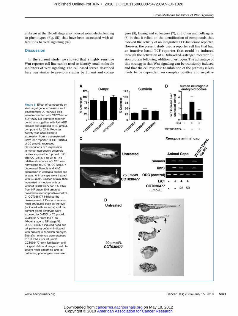
Compound activity against known Wnt targetsThe activities of CCT031374, CCT036477, and CCT070535
were tested against expression of knownWnt target genes us-ing myc and survivin-luciferase reporter readouts (Fig. 5A).Each compound showed activity against both promoters,but responses were greatest against the c-myc promoter.CCT031374 also showed activity against endogenous LEF1mRNA levels in human neurogenic embryoid bodies (hNEB)in which Wnt signaling had been induced by the GSK-3 inhib-itor, BIO (Fig. 5B). CCT036477 was toxic to the hNEBs whereasCCT070535 showed a weak, nonsignificant response. Expres-
Cancer Res; 70(14) July 15, 2010
American Asso Copyright © 2010 cancerres.aacrjoDownloaded from
sion of Axin2, another Wnt target gene (31), was not signifi-cantly induced by BIO in the hNEBs (data not shown).
In vivo activity of hit compoundsTo obtain an integrated view of compound activity in mul-
tiple developmental stages, compounds were added to themedium of Xenopus and zebrafish embryos during develop-ment. In these assays, CCT036477 showed the strongest phe-notypic effects on both Xenopus and zebrafish development.When added to Xenopus laevis embryos at the 4- to 16-cellstage, CCT036477 ventralized embryos and interfered withprimary axis formation (Fig. 5C), as has previously beenshown for inhibitors of Wnt signaling (1). Consistent withthis observation, CCT036477 reduced expression of twowell-characterized Wnt target genes (Siamois and Xnr3) inanimal cap assays (Fig. 5B). CCT036477 addition to zebrafish
ciation for Cancer Researc on May 18, 201urnals.org
Figure 4. Mechanism of action ofCCT031374. A, CCT031374 andβ-catenin degradation in mouseL-cells. Western blots andautoradiographs of lysates ofmouse L-cells that were exposedto BIO (7.5 μmol/L) for 6 h beforeaddition of CCT031374 (20 μmol/L)in the presence of BIO for thetimes shown. CCT031374(20 μmol/L) increased turnover of35S-pulse-labeled β-catenin.BIO (7.5 μmol/L) was presentthroughout labeling and chaseperiods. Western blots of lysatesfrom mouse L-cells after 240 minof treatment with 20 μmol/LCCT031374 after 6 h of 50 μmol/LMG132 proteasomal inhibitor or6 h of coincubation of CCT031374with 50 μmol/L MG132 (+).B, CCT031374 does not preventBIO's inhibition of Tauphosphorylation by GSK-3.Western blots of lysates fromHEK293 cells that had beentransfected with expressionplasmids for Tau and GSK-3β asindicated and had been treated withBIO (7.5 μmol/L) for 4 h or with bothBIO and CCT031374 (20 μmol/L)for the indicated time periods.Relative phosphorylated Tau to totalTau levels were quantified.C, CCT031374 decreased “free”β-catenin levels in HEK293 cells butnot in SW480 cells. Free β-cateninwas isolated from cell lysates by pulldown with a GST–E-cadherin fusionprotein. D, CCT031374 treatment ofSW480 cells decreasedTCF-dependent transcription ofTopflash luciferase reporter. SW480cells were transfected with Topflashreporter and then exposed tocompound for 24 h. Reporteractivity was normalized tocotransfected TK-renillaluciferase activity.
Cancer Research
h2
Small-Molecule Inhibitors of Wnt Signaling
Published OnlineFirst July 7, 2010; DOI:10.1158/0008-5472.CAN-10-1028
embryos at the 16-cell stage also induced axis defects, leadingto phenotypes (Fig. 5D) that have been associated with al-terations to Wnt signaling (32).
Discussion
In the current study, we showed that a highly sensitiveWnt reporter cell line can be used to identify small-moleculeinhibitors of Wnt signaling. The cell-based screen describedhere was similar to previous studies by Emami and collea-
www.aacrjournals.org
American Asso Copyright © 2010 cancerres.aacrjoDownloaded from
gues (5), Huang and colleagues (7), and Chen and colleagues(3) in that it relied on the identification of compounds thatblocked the activity of an integrated TCF-luciferase reporter.However, the present study used a reporter cell line that hadan inactive basal TCF-reporter that could be inducedthrough the activation of a Dishevelled–estrogen receptor fu-sion protein following addition of estrogen. The advantage ofthis strategy is that Wnt signaling can be transiently inducedand that the cell response to inhibition of the pathway is lesslikely to be dependent on complex positive and negative
Figure 5. Effect of compounds onWnt target gene expression anddevelopment. A, HEK293 cellswere transfected with CMYC-luc orSURVIVIN-luc promoter-reporterconstructs together with Axin-GIDinducer and exposed to 40 μmol/Lcompound for 24 h. Reporteractivity was normalized toexpression from a cotransfectedCMV-lacZ reporter. B, CCT031374,at 20 μmol/L, repressedBIO-induced LEF1 expressionin human neurogenic embryoidbodies exposed to 3 μmol/L BIOand CCT031374 for 24 h. Therelative abundance of LEF1 wasnormalized to ACTB. CCT036477decreased Siamois and Xnr3expression in Xenopus animal capassays. Animal caps were treatedwith 0.3 mol/L LiCl for 10 min, thenincubated in medium with orwithout CCT036477 for 3 h. RNAfrom NF stage 10.5 embryosprovided a second positive control.C, CCT036477 inhibited thedevelopment of Xenopus anteriorhead structures such as the eye(indicated with an arrow) and thecement gland. Embryos wereexposed to DMSO or 75 μmol/LCCT036477 from the 4- to16-cell stage to NF stage 38.D, CCT036477 induced head andtail patterning defects (indicatedwith arrows) in zebrafish embryos.Zebrafish embryos were exposedto 1% DMSO or 20 μmol/LCCT036477 from fertilization untilmidgastrulation. A range of mild tosevere head patterning and tailpatterning phenotypes were seen.
Cancer Res; 70(14) July 15, 2010 5971
ciation for Cancer Research on May 18, 2012urnals.org
Ewan et al.
5972
Published OnlineFirst July 7, 2010; DOI:10.1158/0008-5472.CAN-10-1028
feedback pathways commonly found in cancer cell lines (33–35). Furthermore, cell lines whose growth is dependent onthe Wnt pathway have been shown to undergo apoptosis(as seen for CCT031374, CCT036477, and CCT070535; Supple-mentary Fig. S3) following Wnt pathway inhibition, and thiscan be difficult to distinguish from nonspecific cell killingduring high-throughput screening.Multiple compounds were identified in the primary screen
that showed specificity for the TCF-reporter when comparedwith control promoters. A hit triage/deconvolution cascadewas designed to identify the most promising candidatesamong these compounds for further study. These assays in-cluded growth-inhibitory activity against tumor cell lineswith activated Wnt signaling. However, a key criterion wasthe “clarity” of the deconvolution response such that repro-ducible, mechanistically consistent readouts were identified,which allowed the logical pursuit of the mechanism of actionof the compound to distinct points in the Wnt pathway.From a total of 63,040 compounds, 9 compounds that oper-ated at either the β-catenin or TCF levels of the pathwaywere selected for further investigation. Downstream assaysfocused on 2 compounds that operated at the β-catenin leveland 1 that operated at the TCF level.CCT031374 acted at the β-catenin level based on the ob-
servation that it blocked TCF-dependent transcription in-duced by a stabilized form of β-catenin (NH2-terminaldeletion of 89 amino acids; ref. 36), but not by a constitutivelyactive TCF-VP16 fusion protein. Consistent with this mecha-nism, CCT031374 increased the degradation of endogenouswild-type β-catenin in mouse L-cells that had been stabilizedby the GSK-3 inhibitor BIO. CCT031374 did not increaseβ-catenin NH2-terminal phosphorylation in the presence ofBIO-treated cells (data not shown), suggesting that a path-way independent of phospho–β-catenin binding to β-TRCPinduced increased degradation. A number of alternativeβ-catenin degradation routes have been described. These in-clude pathways involving APC and the Siah1 ubiquitin ligase(37, 38), the presenilin/PKA complex (39), and the proteasecalpain (40). CCT031374 failed to induce β-catenin degrada-tion products characteristic of calpain-induced proteolysis(ref. 40; data not shown), suggesting that distinct novel path-ways are involved in its mechanism of action.Surprisingly, the block of TCF-dependent transcription in
SW480 colon cancer cells by CCT031374 was not accompa-nied by a corresponding reduction in β-catenin protein levels(Fig. 4F). Moreover, levels of transfected stabilized β-catenin(ΔN-89–β-catenin) were not reduced in HEK293 cells treatedwith CCT031374 (Supplementary Fig. S12), although the com-pound blocked TCF-dependent transcription in these cells(Fig. 2B). Taken together, these data argue that the degrada-tion of β-catenin may not be necessary for the blocking effect
Cancer Res; 70(14) July 15, 2010
American Asso Copyright © 2010 cancerres.aacrjoDownloaded from
of CCT031374 on TCF-dependent transcription in all cellularcontexts, suggesting that CCT031374 operates through morethan one mechanism. A similar correlation between β-cateninstability and TCF-dependent transcription was observed instudies of the prolyl oligopeptidase Pin-1. Pin-1 activity corre-lated with both β-catenin stability and TCF-dependent tran-scription in HeLa cells by modulating its interaction with APC(41). By contrast, in PTEN-deficient LNCaP cells, Pin-1 alteredTCF-dependent transcription without altering β-catenin le-vels by blocking β-catenin interactions with the androgen re-ceptor (42). CCT031374 may similarly function throughmolecular pathways that can be coupled to protein degrada-tion in selected contexts.Of the three compound series studied, only CCT036477
showed clear activity in vivo where it blocked developmentof zebrafish and Xenopus embryos and expression of Wnt tar-get genes. CCT031374 and CCT070535 did not show clear re-sponses in these systems, and it is currently unclear whetherthe compounds reached the required concentration to blocksignaling in these in vivo models or whether their lack ofin vivo activity reflects a specificity for cells with oncogeni-cally activated Wnt signaling. Overall, these studies haveidentified novel small molecules that target distinct levelsof Wnt signal transduction pathway. Further studies withthese compounds (see Supplementary Fig. S13) could leadto the identification of molecular targets of therapeutic bene-fit in multiple tumor types with activated Wnt signaling, andfurther investigations will be published in due course.
Disclosure of Potential Conflicts of Interest
T. Dale: commercial research grant, Merck Serono. Authors affiliated to theCancer Research UK Centre for Cancer Therapeutics are employees of theInstitute of Cancer Research, which has a commercial interest in inhibitorsof the Wnt pathway and has a funded research collaboration with MerckSerono, from which authors may benefit from a rewards to inventors scheme.The other authors disclosed no potential conflicts of interest.
Acknowledgments
We thank Teresa Brooks and Paul Rogers for help with reporter cell lineoptimization, Perihan Nalbant for help with the GFP-β-catenin assay, andBethan Lloyd-Lewis for assistance with the intensity quantitation ofimmunocytochemistry images.
Grant Support
Work at Cardiff University and The Cancer Research UK Centre for CancerTherapeutics was supported by CR-UK grant nos. C368/A5410 and C368/A7505,and C309/A2187, C309/A8274, and C309/A8365, respectively. National HealthService funding supported work at the National Institute for Health ResearchBiomedical Research Centre, The Institute of Cancer Research, and The RoyalMarsden NHS Foundation Trust. P. Workman is a CR-UK Life Fellow.
Received 03/25/2010; revised 05/17/2010; accepted 05/18/2010; publishedOnlineFirst 07/06/2010.
References
1. Clevers H. Wnt/β-catenin signaling in development and disease. Cell2006;127:469–80.2. Cowin P, Rowlands TM, Hatsell SJ. Cadherins and catenins in breast
cancer. Curr Opin Cell Biol 2005;17:499–508.3. Chen B, Dodge ME, Tang W, et al. Small molecule-mediated disrup-
tion of Wnt-dependent signaling in tissue regeneration and cancer.Nat Chem Biol 2009;5:100–7.
4. Ewan KB, Dale TC. The potential for targeting oncogenic WNT/β-catenin signaling in therapy. Curr Drug Targets 2008;9:532–47.
5. Emami KH, Nguyen C, Ma H, et al. A small molecule inhibitor of
Cancer Research
ciation for Cancer Research on May 18, 2012urnals.org
Small-Molecule Inhibitors of Wnt Signaling
Published OnlineFirst July 7, 2010; DOI:10.1158/0008-5472.CAN-10-1028
β-catenin/CREB-binding protein transcription [corrected]. Proc NatlAcad Sci U S A 2004;101:12682–7.
6. Miyabayashi T, Teo JL, Yamamoto M, McMillan M, Nguyen C,Kahn M. Wnt/β-catenin/CBP signaling maintains long-term murineembryonic stem cell pluripotency. Proc Natl Acad Sci U S A 2007;104:5668–73.
7. Huang SM, Mishina YM, Liu S, et al. Tankyrase inhibition stabilizesaxin and antagonizes Wnt signalling. Nature 2009;461:614–20.
8. DasGupta R, Kaykas A, Moon RT, Perrimon N. Functional genomicanalysis of the Wnt-wingless signaling pathway. Science 2005;308:826–33.
9. Tian Q. Proteomic exploration of the Wnt/β-catenin pathway. CurrOpin Mol Ther 2006;8:191–7.
10. Mosimann C, Hausmann G, Basler K. β-catenin hits chromatin: reg-ulation of Wnt target gene activation. Nat Rev Mol Cell Biol 2009;10:276–86.
11. Smalley MJ, Signoret N, Robertson D, et al. Dishevelled (Dvl-2) acti-vates canonical Wnt signalling in the absence of cytoplasmic puncta.J Cell Sci 2005;118:5279–89.
12. Willert K, Brown JD, Danenberg E, et al. Wnt proteins are lipid-modified and can act as stem cell growth factors. Nature 2003;423:448–52.
13. Kishida S, Yamamoto H, Kikuchi A. Wnt-3a and Dvl induce neuriteretraction by activating Rho-associated kinase. Mol Cell Biol 2004;24:4487–501.
14. Smalley MJ, Sara E, Paterson H, et al. Interaction of axin and Dvl-2proteins regulates Dvl-2-stimulated TCF-dependent transcription.EMBO J 1999;18:2823–35.
15. Salghetti SE, Kim SY, Tansey WP. Destruction of Myc by ubiquitin-mediated proteolysis: cancer-associated and transforming muta-tions stabilize Myc. EMBO J 1999;18:717–26.
16. Lovestone S, Reynolds CH, Latimer D, et al. Alzheimer's disease-likephosphorylation of the microtubule-associated protein tau by glyco-gen synthase kinase-3 in transfected mammalian cells. Curr Biol1994;4:1077–86.
17. Thomson JA, Itskovitz-Eldor J, Shapiro SS, et al. Embryonic stemcell lines derived from human blastocysts. Science 1998;282:1145–7.
18. Sive HL, Grainger RM, Harland RM. Early development of Xenopuslaevis: a laboratory manual. Cold Spring Harbor: Cold Spring HarborLaboratory Press; 2000.
19. Nieuwkoop PD, Faber J. Normal table of Xenopus laevis (Daudin): asystematical and chronological survey of the development from thefertilized egg till the end of metamorphosis. New York: Garland Pub-lishing; 1994.
20. Samuel LJ, Latinkic BV. Early activation of FGF and nodal pathwaysmediates cardiac specification independently of Wnt/β-catenin sig-naling. PLoS ONE 2009;4:e7650.
21. McKendry R, Hsu SC, Harland RM, Grosschedl R. LEF-1/TCF pro-teins mediate wnt-inducible transcription from the Xenopus nodal-related 3 promoter. Dev Biol 1997;192:420–31.
22. Polychronopoulos P, Magiatis P, Skaltsounis AL, et al. Structuralbasis for the synthesis of indirubins as potent and selective inhibitorsof glycogen synthase kinase-3 and cyclin-dependent kinases. J MedChem 2004;47:935–46.
23. Brennan K, Gonzalez-Sancho JM, Castelo-Soccio LA, Howe LR,Brown AM. Truncated mutants of the putative Wnt receptor LRP6/Arrow can stabilize β-catenin independently of Frizzled proteins.Oncogene 2004;23:4873–84.
24. Fraser E, Young N, Dajani R, et al. Identification of the Axin and Frat
www.aacrjournals.org
American Asso Copyright © 2010 cancerres.aacrjoDownloaded from
binding region of glycogen synthase kinase-3. J Biol Chem 2002;277:2176–85.
25. Munemitsu S, Albert I, Rubinfeld B, Polakis P. Deletion of an amino-terminal sequence β-catenin in vivo and promotes hyperphosporyla-tion of the adenomatous polyposis coli tumor suppressor protein.Mol Cell Biol 1996;16:4088–94.
26. Smith KJ, Johnson KA, Bryan TM, et al. The APC gene product innormal and tumor cells. Proc Natl Acad Sci U S A 1993;90:2846–50.
27. Nishisho I, Nakamura Y, Miyoshi Y, et al. Mutations of chromosome5q21 genes in FAP and colorectal cancer patients. Science 1991;253:665–9.
28. Morin PJ, Sparks AB, Korinek V, et al. Activation of β-catenin-Tcfsignaling in colon cancer by mutations in β-catenin or APC. Science1997;275:1787–90.
29. Satoh S, Daigo Y, Furukawa Y, et al. AXIN1 mutations in hepatocel-lular carcinomas, and growth suppression in cancer cells by virus-mediated transfer of AXIN1. Nature Genet 2000;24:245–50.
30. Goedert M, Jakes R, Crowther RA, et al. Epitope mapping of mono-clonal antibodies to the paired helical filaments of Alzheimer's dis-ease: identification of phosphorylation sites in tau protein. BiochemJ 1994;301:871–7.
31. Jho EH, Zhang T, Domon C, Joo CK, Freund JN, Costantini F. Wnt/β-catenin/Tcf signaling induces the transcription of Axin2, a negativeregulator of the signaling pathway. Mol Cell Biol 2002;22:1172–83.
32. Lekven AC, Thorpe CJ, Waxman JS, Moon RT. Zebrafish wnt8 en-codes two wnt8 proteins on a bicistronic transcript and is requiredfor mesoderm and neurectoderm patterning. Dev Cell 2001;1:103–14.
33. Gonzalez-Sancho JM, Aguilera O, Garcia JM, et al. The Wnt antago-nist DICKKOPF-1 gene is a downstream target of β-catenin/TCF andis downregulated in human colon cancer. Oncogene 2005;24:1098–103.
34. Li TW, Ting JH, Yokoyama NN, Bernstein A, van de Wetering M,Waterman ML. Wnt activation and alternative promoter repressionof LEF1 in colon cancer. Mol Cell Biol 2006;26:5284–99.
35. Caldwell GM, Jones CE, Soon Y, Warrack R, Morton DG, MatthewsGM. Reorganisation of Wnt-response pathways in colorectaltumorigenesis. Br J Cancer 2008;98:1437–42.
36. Munemitsu S, Albert I, Souza B, Rubinfeld B, Polakis P. Regulation ofintracellular β-catenin levels by the adenomatous polyposis coli(APC) tumor-suppressor protein. Proc Natl Acad Sci U S A 1995;92:3046–50.
37. Liu J, Stevens J, Rote CA, et al. Siah-1 mediates a novel β-catenindegradation pathway linking p53 to the adenomatous polyposis coliprotein. Mol Cell 2001;7:927–36.
38. Matsuzawa SI, Reed JC. Siah-1, SIP, Ebi collaborate in a novel path-way for β-catenin degradation linked to p53 responses. Mol Cell2001;7:915–26.
39. Hino S, Tanji C, NakayamaKI, Kikuchi A. Phosphorylation of β-cateninby cyclic AMP-dependent protein kinase stabilizes β-catenin throughinhibition of its ubiquitination. Mol Cell Biol 2005;25:9063–72.
40. Li G, Iyengar R. Calpain as an effector of the Gq signaling pathwayfor inhibition of Wnt/β-catenin-regulated cell proliferation. Proc NatlAcad Sci U S A 2002;99:13254–9.
41. Ryo A, Nakamura M, Wulf G, Liou YC, Lu KP. Pin1 regulates turnoverand subcellular localization of β-catenin by inhibiting its interactionwith APC. Nat Cell Biol 2001;3:793–801.
42. Chen SY, Wulf G, Zhou XZ, Rubin MA, Lu KP, Balk SP. Activation ofβ-catenin signaling in prostate cancer by peptidyl-prolyl isomerasePin1-mediated abrogation of the androgen receptor-β-catenin inter-action. Mol Cell Biol 2006;26:929–39.
Cancer Res; 70(14) July 15, 2010 5973
ciation for Cancer Research on May 18, 2012urnals.org