Cortactin scaffolds Arp2/3 and WAVE2 at the epithelia zonula adherens.
Characterization of two classes of small molecule inhibitors ofArp2/3 complex
B.J. Nolen1,*,#, N. Tomasevic2,*,§, A. Russell2, D.W. Pierce2,+, Z. Jia2, C.D. McCormick1, J.Hartman2, R. Sakowicz2,^, and T.D. Pollard1
1 Departments of Molecular Cellular and Developmental Biology, of Cell Biology and of MolecularBiophysics and Biochemistry, Yale University, New Haven, CT 06520 USA2 Cytokinetics, Inc., South San Francisco, CA 94080 USA
AbstractPolymerization of actin filaments directed by the Arp2/3 complex supports many types of cellularmovements1. However, questions remain regarding the relative contributions of Arp2/3 complexversus other mechanisms of actin filament nucleation to processes such as path finding byneuronal growth cones owing to the lack of simple methods to inhibit Arp2/3 complex reversiblyin living cells. Here we describe two classes of small molecules that bind to different sites onArp2/3 complex and inhibit its ability to nucleate actin filaments. CK-636 binds between Arp2 andArp3 where it appears to block movement of Arp2 and Arp3 into their active conformation.CK-548 inserts into the hydrophobic core of Arp3 and alters its conformation. Both classes ofcompounds inhibit formation of actin filament comet tails by Listeria and podosomes bymonocytes. Two inhibitors with different mechanisms of action provide a powerful approach forstudying Arp2/3 complex in living cells.
We used fluorescence assays to screen a library of >400,000 small molecules for inhibitorsof actin polymerization stimulated by human (Hs) Arp2/3 complex and either full-lengthHsWASp with acrylodan-actin or HsWASp residues 105–502 with pyrenyl-actin. Thesescreens each identified two inhibitors of human and bovine Arp2/3 complex, CK-0944636(abbreviated CK-636) and CK-0993548 (abbreviated CK-548) (Fig. 1a). These compoundsinhibited bovine (Bt) Arp2/3 complex with IC50 values of 32 μM for CK-636 and 11 μM forCK-548 (Fig. 1c). CK-636 inhibited actin polymerization stimulated by fission yeast Arp2/3complex (SpArp2/3 complex, IC50 = 24 μM), but 100 μM CK-548 did not (Fig. 1c, TableS1). Fluorescence microscopy of the products of these reactions stained with Alexa 488phalloidin showed branched actin filaments in controls (Fig. 1e, left panel). Samples with100 μM CK-636 contained fewer branched filaments (Fig. 1e, center panel), while sampleswith 100 μM CK-548 contained only unbranched filaments (Fig. 1e, right panel). We testeda number of compounds structurally related to CK-548 or CK-636 that had no effect on actin
Correspondence should be addressed to [email protected]. Reprints and permissions information is available atnpg.nature.com/reprintsandpermissions. Correspondence and requests for materials should be addressed to [email protected].*These authors contributed equally to this work#Current addresses: Department of Chemistry and the Institute of Molecular Biology, University of Oregon, Eugene, OR 97403§Kalobios Pharmaceuticals, Inc. South San Francisco, CA 94080;+Five Prime Therapeutics, San Francisco, CA USA 94158;^Gilead Sciences, Inc. Foster City, CA USA 94404.
AUTHOR CONTRIBUTIONSBJN, NT, AR, DWP, ZJ and JH designed and carried out experiments; CDM analyzed data; RS and TDP supervised research; BJN,NT, AR, JH and TDP wrote the paper.
AUTHOR INFORMATIONStructural data have been deposited in Protein Data Bank with accession codes 3DXK (CK-0944636) and 3DXM (CK-0993548).
NIH Public AccessAuthor ManuscriptNature. Author manuscript; available in PMC 2010 February 20.
Published in final edited form as:Nature. 2009 August 20; 460(7258): 1031–1034. doi:10.1038/nature08231.
NIH
-PA Author Manuscript
NIH
-PA Author Manuscript
NIH
-PA Author Manuscript
polymerization at concentrations up to 200 μM and are useful as controls for experimentswith cells (Fig. 2g). Table S2 lists one inactive compound from each class.
The following control experiments showed that both compounds interact with Arp2/3complex rather than actin or WASp. At 20 μM, neither compound inhibited polymerizationof actin alone or polymerization stimulated by formin Cdc12p (Fig. 1b), so neithercompound interacts directly with actin. Since actin can polymerize on its own, neithercompound eliminated actin polymerization in mixtures with Arp2/3 complex and WASp orN-WASP-VCA (Fig. 1c-d). Both compounds inhibited polymerization stimulated byHsArp2/3 complex and ActA, an Arp2/3 complex activating factor from Listeriamonocytogenes2, suggesting that they do not act on WASp (Supplementary Fig. 1). Afluorescence anisotropy assay showed that neither compound had a large effect on Arp2/3complex binding N-WASP-VCA (Supplementary Fig. 2). The affinity of rhodamine-N-WASP-VCA for BtArp2/3 complex was the same with 50 μM CK-636 (Kd = 470 ± 50 nM)as controls (Kd = 510 ± 30 nM), but 50 μM CK548 decreased the affinity approximatelytwo-fold (Kd = 1.0 ± 0.2 μM).
We used a Listeria motility assay to determine whether CK-636 and CK-548 can inhibitactin polymerization mediated by Arp2/3 complex in live cells. Both compounds reducedthe formation of actin filament comet tails by Listeria in infected SKOV3 cells (Fig. 2a–c).The concentration dependence of this inhibition gave IC50 values of 22 μM for CK-636 and31 μM for CK-548. We used the actin polymerization and Listeria comet tail assays tosearch for more potent inhibitors related to CK-636 and CK-548.
Compound CK-0944666 (abbreviated CK-666) has a fluorobenzene rather than thethiophene ring of CK-636 (Fig. 1a). CK-666 was a better inhibitor of actin polymerizationwith either BtArp2/3 complex (IC50 = 32 μM for CK-636 verses 17 μM for CK-666) orSpArp2/3 complex (IC50 = 24 μM for CK-636 versus 5 μM for CK-666) (Fig. 1d, TableS1). Both CK-636 and CK-666 had IC50 values of 4 μM for inhibiting HsArp2/3 complex.CK-666 reduced actin polymerization around intracellular Listeria to background levels(Fig. 2e and g) at lower concentrations than CK-636 (Fig. 2c). Actin filament halos andcomet tails reformed when the compounds were removed after one hour of treatment (Fig.2f–g). Inactive control compound CK-689 had no effect on comet tail formation (Fig. 2g).
CK-0157869 (abbreviated CK-869) has methoxy groups in the ortho and para positionsreplacing the ortho-hydroxy and meta-chlorine substituents of CK-548 (Fig. 1a). CK-869inhibited actin polymerization with BtArp2/3 complex similar to CK-548 (IC50 = 11 μM forboth compounds), but CK869 inhibited comet tail formation by Listeria more effectively(IC50 = 7 μM for CK-869 vs. 31 μM for CK-548). Like CK-548, CK-869 did not inhibiteither yeast Arp2/3 complex (Fig. 1d).
Both classes of compounds also inhibited the formation of podosomes by the THP-1monocyte cell line (Fig. 2h–k). Podosomes are adhesive structures that depend on Arp2/3complex, WASp and actin polymerization3. Podosomes also depend on microtubules4, butneither compound at 100 μM disrupted the microtubule network in THP-1 cells(Supplementary Fig. 3). Neither class of compound shows irreversible or off targetmorphological effects on cells, such as apoptosis, at concentrations up to 100 μM over 24hours. Our most potent compound, CK-666, had no effect on the mitotic index of SKOVcells at concentrations up to 80 μM, while inhibiting actin assembly around Listeriacompletely at 10 μM. Fifty micromolar CK-666 or CK-869 each altered the morphology andslightly slowed but did not stop the gliding motility of fish keratocytes (data not shown), asexpected, since ~10% of Arp2/3 complex (~500 nM) would still be active. Thus Listeria
Nolen et al. Page 2
Nature. Author manuscript; available in PMC 2010 February 20.
NIH
-PA Author Manuscript
NIH
-PA Author Manuscript
NIH
-PA Author Manuscript
comet tails and monocyte podosomes are more sensitive than lamellar motility to inhibitionof Arp2/3 complex.
We determined x-ray crystal structures of BtArp2/3 complex with either CK-636 or CK-548bound using molecular replacement with the apo-form BtArp2/3 complex as the searchmodel (1K8K.pdb)5. Structures of Arp2/3 complex co-crystallized with the compounds andwith compounds soaked into the crystals were nearly identical, but the resolution of soakedcrystals was higher (2.70 Å for CK-636 and 2.85 Å for CK-548, Table S3). As in previouscrystal structures of BtArp2/3 complex5–7, none of these structures had appreciable electrondensity for subdomains 1 and 2 of Arp2, which appears only after chemical crosslinking7.
After one round of rigid body refinement, strong Fo-Fc electron density was present at theinterface between Arp2 and Arp3 in maps calculated with data from crystals soaked in 1mM CK-636 (Fig. 3). This density improved upon further refinement (Supplementary Fig.4a) and CK-636 was modeled into the density when the Rf was 29.2 %. CK-636 binds in apocket between subdomain 4 of Arp2 and subdomain 1 of Arp3. The contact surface ismainly hydrophobic and buries 161 Å2 of the accessible surface of Arp2 and 79 Å2 of Arp3.CK-636 forms hydrogen bonds with two residues in the pocket. The side chain of Asp248forms a hydrogen bond with the nitrogen in the indole ring of the inhibitor and the backboneamide of Ala203 forms a hydrogen bond with the carbonyl oxygen on the linker between thetwo ring systems. Both trans- and cis- conformations of the amide functional group ofCK-636 fit into the electron density with the hydrogen bonds described above preserved inboth conformations. Because trans-amides are generally 1–5 kcal/mol more stable than cis-amides in solution 8, and the cis-conformation is rarely observed in small molecules, wemodeled CK-636 in the trans-conformation.
A difference electron density map shows that CK-636 causes minor conformational changesin the complex (Fig. 3b). Arp2 Arg250 from β strand 13 rotates out of the pocket and adoptsan extended conformation. Arp2 Ala203 from the loop (residues 199-208) connecting αE toαF moves toward the pocket, causing the side chain of Leu198 to adopt a new rotamer.These changes and small changes in the backbone of residues surrounding the pocket resultin an overall RMSD of 0.63 Å for an overlay of subdomains 3 and 4 (191 Cα atoms) ofArp2 with and without inhibitor bound.
We modeled CK-666 into the CK-636 binding pocket with the aromatic fluorine pointedtoward a concave surface in the back wall of the pocket formed by Tyr202 in Arp2 andresidues 118 to 120 in Arp3 (Supplementary Fig. 6). One hundred steps of conjugategradient minimization in CNS9 showed that CK-666 is stable in this conformation and doesnot clash with residues in the pocket. The fluorine atom provides additional van der Waalsinteractions with the back wall of the pocket, and the benzene ring completely fills thehydrophobic pocket created by Ile252, Tyr202 and the aliphatic portion of Thr119. Theseinteractions may allow CK-666 to bind more tightly than CK-636.
During formation of a branch Arp2 moves 31 Å relative to Arp3 from its position in inactiveArp2/3 complex10, so the location of the CK-636 binding pocket between Arp2 and Arp3suggests that CK-636 and CK-666 lock Arp2/3 complex in an inactive conformation.CK-636 at a concentration of 100 μM did not interfere with N-WASp-VCA binding toArp2/3 complex (Supplementary Figure 2), but it prevented WASp-VCA binding fromincreasing the fluorescence of Arp2/3 complex loaded with etheno-ATP (SupplementaryFigure 5). Thus CK-636 inhibits a conformational change caused by activator binding andsupports the hypothesis that CK-636 locks the complex in an inactive conformation.
The residues contributed by Arp2 and Arp3 to form the CK-636 binding pocket areconserved across a broad range of species (Table S4), so we expect CK-636 and CK-666
Nolen et al. Page 3
Nature. Author manuscript; available in PMC 2010 February 20.
NIH
-PA Author Manuscript
NIH
-PA Author Manuscript
NIH
-PA Author Manuscript
will be useful for inhibiting Arp2/3 complex from diverse species. These residues are alsoconserved in actin, but CK-636 does not inhibit actin polymerization at concentrations up to200 μM. Only half of the binding site is available on an actin monomer, and the two half-sites are not juxtaposed in actin filaments11.
CK-548 has a single stereo center and was used as a racemic mixture in our experiments.The 2S enantiomer binds in a hydrophobic cavity in the core of the Arp3 subunit (Fig. 4aand Supplementary Fig. 4b). The isolated 2R enantiomer, which does not fit in the bindingpocket, should provide a useful control for in vivo experiments. Binding of CK548 requiresa substantial conformational change: the loop (residues 76-85) connecting β6 (73-75) andαB (86-98) in subdomain 1 of Arp3 flips upward 7.2 Å, exposing the binding site in thehydrophobic core of Arp3 (Fig. 4b). CK-548 contacts 239 Å2 of the inner surface of thiscavity, which includes residues Cys12, Trp86, Met89, Met93, Leu112 and Ile78. Ahydrogen bond between the side chain amide of Asn118 and the carbonyl oxygen of CK548anchors the inhibitor to the loop between β7 (110-114) and αC (120-132), which forms thebottom lip of the cavity. The ability of CK-548 to bind crystallized Arp2/3 complex suggeststhat the β6/αB loop can open the hydrophobic binding pocket both in crystals and insolution, even in the absence of inhibitor. The average B-factor for all residues in the loop is63 Å2 in the closed conformation and 82 Å2 in the open conformation, suggesting that theloop is more flexible when open.
When modeled into the CK-548 binding site, the para-methoxy group of CK-869 projectstoward the upper lip of the pocket and is sandwiched by the side chains of Ile78, Glu84 andMet89 (Supplemetary Fig. 7). The ortho-methoxy group points toward a crease formed atthe ends of strands β1 and β7, which could be exploited to design higher affinity inhibitors.
Both CK-548 and CK-869 inhibited human and bovine Arp2/3 complexes, but neitherinhibited budding or fission yeast Arp2/3 complexes (Fig. 1c, d). The residues that contactCK548 are conserved in Arp2/3 complexes of most mammals, but tryptophan replacesMet93 at the back of the CK-548 binding pocket in many other species, including buddingand fission yeast (Table S5). The bent side chain of Met93 allows CK-548 to bind, buttryptophan at this position blocks binding to non-mammalian Arp3 and actin.
The mechanism of action of CK-548/869 is less apparent than that of CK-636/666. CK-548decreased the affinity of SpWsp1-Rho-VCA for Arp2/3 complex two-fold, but this shouldbe inconsequential under the conditions of our assay. The conformation induced byCK-548/869 must interfere with one or more of the reactions along the pathway to branchformation, such as binding of the pointed end of Arp3 to a mother filament10, nucleotidebinding to Arp3, or conformational changes that activate branch formation.
METHODS SUMMARYCompounds were purchased from Chemdiv, San Diego CA: CK-0944636 (Catalog number8012-5103), CK-0993548 (Catalog number K205-1650), CK-0944666 (Catalog number8012-5153) and CK-0157869 (Catalog number K205-0942). We purified native Arp2/3complex from human platelets12, bovine thymus6, Schizosaccharomyces pombe13 andSaccharomyces cerevisiae (Supplemental methods), actin from chicken skeletal muscle14and recombinant HsWASp, WASp105-502, WASp-VCA and Cdc4212, N-WASp-VCA428-505 (Supplemental methods), GST-ActA 36-170 (Supplemental methods) and S. pombeCdc12p(FH2)-His 973-139015 from E. coli. We used standard assays to measurepolymerization of pyrenyl-actin16 and to visualize actin filaments by fluorescencemicroscopy17. Binding of etheno-ATP to Arp2/3 complex was performed as describedpreviously with slight modifications18. We crystallized BtArp2/3 complex7 with either 0.5
Nolen et al. Page 4
Nature. Author manuscript; available in PMC 2010 February 20.
NIH
-PA Author Manuscript
NIH
-PA Author Manuscript
NIH
-PA Author Manuscript
mM CK-548 or 1 mM CK-636 in DMSO or soaked these compounds into crystals for 24hours before freezing in liquid nitrogen. Diffraction data were collected at beamline X29Aat Brookhaven National Laboratories. SKOV3 cells were infected with Listeriamonocytogenes and fixed with 2% formaldehyde, permeabilized with 0.1% Triton-X inPBS, stained with Listeria antibody (US Biologics, Cleveland, Ohio) and Alexa Fluor 568phalloidin (Molecular Probes, Eugene, OR), and imaged by fluorescence microscopy. Weused an Isodata threshold on background-subtracted images of Listeria to isolate individualbacterium and measure the ratio of colocalized actin to Listeria fluorescence. MonocyteTHP-1 cells were differentiated in 50 nM phorbol myristate acetate (Sigma-Aldrich-Fluka)to form podosomes before treatment with compounds. Black molly keratocytes19 wereobserved by time-lapse phase contrast microscopy.
METHODSProtein Purification
Human, bovine and S. pombe Arp2/3 complexes were purified as previouslydescribed11,12,20. S. cerevisiae Arp2/3 complex was purified from strain BN020 (MAT aor α, ura3-52, his3-200, leu2-3, lys2-801, trp1-1, Δarp2::TRP1, Δarp3::HIS, [pRS315-Arp3], [ pRS317-ARP2]). Cultures were grown overnight at 30° C to OD600 = 1 andpelleted by centrifuging at 5000g for 8 min. Cells were washed in lysis buffer (20 mM TrispH 8.0, 50 mM NaCl, 1 mM EDTA) and pelleted. The pellet was frozen in liquid nitrogenand stored at −80° C. Cells were thawed and 1 mL lysis buffer was added per g wet cellpellet. Protease inhibitor tablets (Sigma) were added (1 tablet per 100 mL cell suspension)and cells were lysed by five passes at 30,000 psi on a model 110EH Microfluidizer(Microfluidics). The lysate was spun at 30K g for 20 min and the supernatant was collectedand centrifuged for 1 hr 15 min at 125 Kg. The supernatant was filtered through cheeseclothand proteins were precipitated with 50% (NH4)2SO4. Pelleted proteins were resuspended in50 mL of PKME (25 mM PIPES pH 7.0, 50 mM KCl, 1mM EGTA, 3mM MgCl2, 1 mMEGTA, 1 mM DTT and 0.1 mM ATP) and dialyzed against the same buffer overnight. Thedialysate was purified on a GST-N-WASp-VCA affinity column and Arp2/3 complex-containing fractions were pooled and concentrated to 1 mL before loading onto aSuperdex200 gel filtration column (GE Healthcare) equilibrated in 20mM Tris pH 8.0 and100 mM NaCl. Peak fractions were pooled and concentrated to approximately 10– 20 μM,flash frozen in liquid nitrogen, and stored at −80°C. Bovine N-WASp-VCA (residues 428–505) was amplified from into pGEX-2T-NWASp-VCA (a gift of Henry Higgs) and clonedonto pGV67 using BamHI and EcoRI restriction sites. This vector was used to transform E.coli strain BL21(DE3). N-WASp-VCA was expressed and purified as previouslydescribed11. Chicken skeletal muscle was purified and labeled with pyreneiodoacetamide21. Recombinant full length human WASp was purified from 293 cells andrecombinant human WASp (residues 105–502) was purified from E. coli for the initialscreens with HsArp2/3 complex. Recombinant Listeria ActA residues 36–170 wereexpressed as an N-terminal GST fusion and purified from E. coli. S. pombe formin Cdc12FH2 domain (residues 973–1390) was purified from E. coli.
Actin Polymerization assayWe measured the nucleation activity of Arp2/3 complex from the time course of actinpolymerization. Polymerization reactions of 100 μL were assembled as previouslydescribed12 and fluorescence measurements were made at 8 s intervals in a 96 well plateusing a Gemini XPS spectrofluorimeter (Molecular Devices) with an excitation wavelengthof 365 nm and an emission wavelength of 407 nM. The rate of polymerization at each timepoint was determined by calculating the slope in 5 point intervals and multiplying by [totalpolymer]/Δ RFU, where [total polymer] is the total actin minus the critical concentration
Nolen et al. Page 5
Nature. Author manuscript; available in PMC 2010 February 20.
NIH
-PA Author Manuscript
NIH
-PA Author Manuscript
NIH
-PA Author Manuscript
(0.1 μM) and ΔRFU is RFUmax - RFUmin. We plotted the maximum elongation rate versesinhibitor concentration and fit the curve using the equation:
The products of polymerization reactions were diluted into Alexa 488-phalloidin andobserved by fluorescence microscopy16.
Fluorescence anisotropyThe endogenous cysteine (Cys431) near the end of the N-terminus in purified N-WASp-VCA was labeled with rhodamine maleimide12. Fixed concentrations of Rho-N-WASp-VCA were titrated with BtArp2/3 complex and the fluorescence anisotropy measured12.Binding constants were determined by fitting the anisotropy curves to the followingequation:
where rf is the signal of the free receptor (R), rb is the signal of the bound receptor, and [L]is the total concentration of the ligand (species titrated). rb and Kd were fit usingKaleidagraph (Synergy Software).
Etheno-ATP fluorescent assayBinding of etheno-ATP to Arp2/3 complex was performed as described previously withslight modifications17,18. Human Arp2/3 complex (0.7μM) was incubated with 2.5 μMetheno-ATP, 0.2 M acylamide (used as a etheno-ATP fluorescence quencher) in 2mM Trs-HCl pH 8.0, 50 mM KCl, 0.05 mM EGTA 0.8 mM MgCl2, 0 and 0.5 mM DTT. Ethano-ATP emission spectrums were acquired upon excitation at 340 nm using spectrofluorometerFluoroMax (Horiba JY). Inhibition of Arp2/3 complex by 100 μM CK-636 was performedin the presence or absence of 2.8 μM WASp-VCA.
Listeria comet, motility and podosome assaysSKOV3, THP-1 and Listeria monocytogenes (ATCC 984) cells were from ATCC. SKOV3cells were cultured in RPMI with 5% FBS without antibiotic at 37°C and 5% CO2. Listeriawere grown in shaking culture in brain heart infusion (Difco Inc, DF0037-15) at 37°C.SKOV3 cells were infected with Listeria monocytogenes, incubated for 90 min and thentreated for 60 minutes with either compounds dissolved in DMSO or a DMSO controlbefore fixing with 2% formaldehyde. Fixed cells were permeabilized with 0.1% Triton-X inPBS at RT for 15 min and reacted with a 1:200 dilution of Listeria antibody (US Biologics,Cleveland, Ohio) in PBS for 60 min at RT. Antibody was removed and cells were stainedwith a 1:100 dilution of Alexa Fluor 568 phalloidin and a 1:400 dilution Alexa Fluor 488goat anti-rabbit secondary antibody (both from Molecular Probes, Eugene, OR) for 60 minat RT. After washing, cells were imaged at 20x with an Axon Instruments automatedfluorescence microscope system (Molecular Devices). The THP-1 monocyte cell line wasgrown in RPMI with 10% FBS/2mM L-glutamine, differentiated in 50 nM phorbol myristateacetate (Sigma-Aldrich-Fluka) for 48 hrs at 37°C, trypsinized and plated at 5×10⁵ cells/ml inblack Nunc glass-bottom 96-well plates. After incubation at 37°C for 2 hr, cells were treatedwith compounds for 15 min before fixing and staining with Alexa Fluor 568 phalloidin and
Nolen et al. Page 6
Nature. Author manuscript; available in PMC 2010 February 20.
NIH
-PA Author Manuscript
NIH
-PA Author Manuscript
NIH
-PA Author Manuscript
DAPI (Sigma-Aldrich-Fluka) as above. Supplemental Figure 2 details the metrics used forpodosome quantification. Black molly keratocytes were isolated19, treated briefly withtrypsin and observed by time lapse phase contrast microscopy on plastic Petri dishes at roomtemperature.
X-ray CrystallographyCrystals of BtArp2/3 complex were grown at 4°C using the hanging drop vapor diffusionmethod as previously described7. Crystals were soaked for 24 hrs in 18 % polyethyleneglycol 8000, 50 mM HEPES pH 7.5, 100 mM potassium thiocyanate, 20 % glycerol andeither 0.5 mM CK548 or 1mM CK636 in DMSO before freezing in liquid nitrogen. Datawas collected at beamline X29A at Brookhaven National laboratories and processed withHKL200022. To generate initial models, protein atoms from 1K8K were refined against thedata using 30 cycles of minimization in which each subunit was allowed to move as aseparate rigid body. CK548 structure was refined in Refmac23 with topology files generatedin ccp4i sketcher. The CK636 structure was refined in CNS8 using topology files generatedwith the prodrg server24.
Supplementary MaterialRefer to Web version on PubMed Central for supplementary material.
AcknowledgmentsThis work was supported by Cytokinetics, Inc., NIH research grant GM-066311 to TDP, NSF Graduate ResearchFellowship to CDM and a Ruth Kirschstein postdoctoral fellowship GM074374-02 to BJN. The authors thank LisaBelmont, Zarak Khurshid, Obidimma Ezizika, Juliet Lee, Stephanie Leuenroth and Hongli Chen for help with theproject.
References1. Goley ED, Welch MD. The Arp2/3 complex: an actin nucleator comes of age. Nat Rev Mol Cell
Biol. 2006; 7:713–726. [PubMed: 16990851]
2. Welch MD, Rosenblatt J, Skoble J, Portnoy DA, Mitchison TJ. Interaction of human Arp2/3complex and the Listeria monocytogenes ActA protein in actin filament nucleation. Science. 1998;281:105–108. [PubMed: 9651243]
3. Linder S, et al. The polarization defect of Wiskott-Aldrich syndrome macrophages is linked todislocation of the Arp2/3 complex. J Immunol. 2000; 165:221–225. [PubMed: 10861055]
4. Gimona M, Buccione R, Courtneidge SA, Linder S. Curr Opin Cell Biol. 2008; 20:235–241.[PubMed: 18337078]
5. Robinson RC, et al. Crystal structure of Arp2/3 complex. Science. 2001; 294:1679–1684. [PubMed:11721045]
6. Nolen BJ, Littlefield RS, Pollard TD. Crystal structures of actin-related protein 2/3 complex withbound ATP or ADP. Proc Natl Acad Sci USA. 2004; 101:15627–15632. [PubMed: 15505213]
7. Nolen BJ, Pollard TD. Insights into the influence of nucleotides on actin family proteins from sevenstructures of Arp2/3 complex. Mol Cell. 2007; 26:449–457. [PubMed: 17499050]
8. Jabs A, Weiss MS, Hilgenfeld R. Non-proline cis peptide bonds in proteins. J Mol Biol. 1999;286:291–305. [PubMed: 9931267]
9. Brunger AT, et al. Crystallography & NMR system: A new software suite for macromolecularstructure determination. Acta Crystallogr D Biol Crystallogr. 1998; 54(Pt 5):905–921. [PubMed:9757107]
10. Rouiller I, et al. The structural basis of actin filament branching by the Arp2/3 complex. J CellBiol. 2008; 180:887–895. [PubMed: 18316411]
11. Holmes KC, Popp D, Gebhard W, Kabsch W. Atomic model of the actin filament. Nature. 1990;347:44–49. [PubMed: 2395461]
Nolen et al. Page 7
Nature. Author manuscript; available in PMC 2010 February 20.
NIH
-PA Author Manuscript
NIH
-PA Author Manuscript
NIH
-PA Author Manuscript
12. Tomasevic N, et al. Differential regulation of WASP and N-WASP by Cdc42, Rac1, Nck, andPI(4,5)P2. Biochemistry. 2007; 46:3494–3502. [PubMed: 17302440]
13. Nolen BJ, Pollard TD. Structure and biochemical properties of fission yeast Arp2/3 complexlacking the Arp2 subunit. J Biol Chem. 2008; 283:26490–26498. [PubMed: 18640983]
14. MacLean-Fletcher S, Pollard TD. Identification of a factor in conventional muscle actinpreparations which inhibits actin filament self-association. Biochem Biophys Res Commun. 1980;96:18–27. [PubMed: 6893667]
15. Kovar DR, Pollard TD. Insertional assembly of actin filament barbed ends in association withformins produces piconewton forces. Proc Natl Acad Sci USA. 2004; 101:14725–14730.[PubMed: 15377785]
16. Cooper JA, Walker SB, Pollard TD. Pyrene actin: documentation of the validity of a sensitiveassay for actin polymerization. J Muscle Res Cell Motil. 1983; 4:253–262. [PubMed: 6863518]
17. Blanchoin L, et al. Direct observation of dendritic actin filament networks nucleated by Arp2/3complex and WASP/Scar proteins. Nature. 2000; 404:1007–1011. [PubMed: 10801131]
18. Dayel MJ, Holleran EA, Mullins RD. Arp2/3 complex requires hydrolyzable ATP for nucleation ofnew actin filaments. Proc Natl Acad Sci USA. 2001; 98:14871–14876. [PubMed: 11752435]
19. Lee J, Ishihara A, Theriot JA, Jacobson K. Principles of locomotion for simple-shaped cells.Nature. 1993; 362:167–171. [PubMed: 8450887]
20. Higgs HN, Blanchoin L, Pollard TD. Influence of the C terminus of Wiskott-Aldrich syndromeprotein (WASp) and the Arp2/3 complex on actin polymerization. Biochemistry. 1999; 38:15212–22. [PubMed: 10563804]
21. Pollard TD. Polymerization of ADP-actin. J Cell Biol. 1984; 99:769–77. [PubMed: 6540783]
22. Otwinowski Z, Minor W. Processing of X-ray diffraction data dollected in oscillation mode.Methods in Enzymology. 1997; 276:307–326.
23. Dodson EJ, Winn M, Ralph A. Collaborative Computational Project, number 4: providingprograms for protein crystallography. Methods Enzymol. 1997; 277:620–33. [PubMed: 18488327]
24. Schuettelkopf AW, van Aalten DMF. PRODRG- a tool for high-throughput crystallography ofprotein-ligand complexes. Acta Crystallogr D Biol Crystallogr. 2004; D60 in press.
Nolen et al. Page 8
Nature. Author manuscript; available in PMC 2010 February 20.
NIH
-PA Author Manuscript
NIH
-PA Author Manuscript
NIH
-PA Author Manuscript
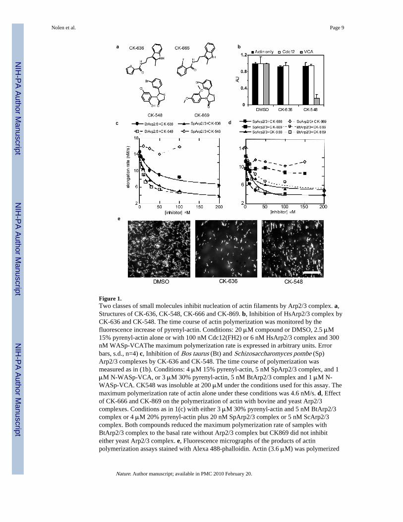
Figure 1.Two classes of small molecules inhibit nucleation of actin filaments by Arp2/3 complex. a,Structures of CK-636, CK-548, CK-666 and CK-869. b, Inhibition of HsArp2/3 complex byCK-636 and CK-548. The time course of actin polymerization was monitored by thefluorescence increase of pyrenyl-actin. Conditions: 20 μM compound or DMSO, 2.5 μM15% pyrenyl-actin alone or with 100 nM Cdc12(FH2) or 6 nM HsArp2/3 complex and 300nM WASp-VCAThe maximum polymerization rate is expressed in arbitrary units. Errorbars, s.d., n=4) c, Inhibition of Bos taurus (Bt) and Schizosaccharomyces pombe (Sp)Arp2/3 complexes by CK-636 and CK-548. The time course of polymerization wasmeasured as in (1b). Conditions: 4 μM 15% pyrenyl-actin, 5 nM SpArp2/3 complex, and 1μM N-WASp-VCA, or 3 μM 30% pyrenyl-actin, 5 nM BtArp2/3 complex and 1 μM N-WASp-VCA. CK548 was insoluble at 200 μM under the conditions used for this assay. Themaximum polymerization rate of actin alone under these conditions was 4.6 nM/s. d, Effectof CK-666 and CK-869 on the polymerization of actin with bovine and yeast Arp2/3complexes. Conditions as in 1(c) with either 3 μM 30% pyrenyl-actin and 5 nM BtArp2/3complex or 4 μM 20% pyrenyl-actin plus 20 nM SpArp2/3 complex or 5 nM ScArp2/3complex. Both compounds reduced the maximum polymerization rate of samples withBtArp2/3 complex to the basal rate without Arp2/3 complex but CK869 did not inhibiteither yeast Arp2/3 complex. e, Fluorescence micrographs of the products of actinpolymerization assays stained with Alexa 488-phalloidin. Actin (3.6 μM) was polymerized
Nolen et al. Page 9
Nature. Author manuscript; available in PMC 2010 February 20.
NIH
-PA Author Manuscript
NIH
-PA Author Manuscript
NIH
-PA Author Manuscript

with 6 nM HsArp2/3 complex, 100 nM WASp-105-502, 300 nM Cdc42 and 100 μMCK-636 Scale bar = 20 μm.
Nolen et al. Page 10
Nature. Author manuscript; available in PMC 2010 February 20.
NIH
-PA Author Manuscript
NIH
-PA Author Manuscript
NIH
-PA Author Manuscript
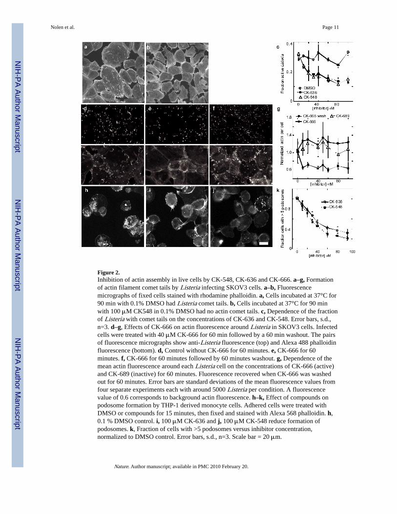
Figure 2.Inhibition of actin assembly in live cells by CK-548, CK-636 and CK-666. a–g, Formationof actin filament comet tails by Listeria infecting SKOV3 cells. a–b, Fluorescencemicrographs of fixed cells stained with rhodamine phalloidin. a, Cells incubated at 37°C for90 min with 0.1% DMSO had Listeria comet tails. b, Cells incubated at 37°C for 90 minwith 100 μM CK548 in 0.1% DMSO had no actin comet tails. c, Dependence of the fractionof Listeria with comet tails on the concentrations of CK-636 and CK-548. Error bars, s.d.,n=3. d–g, Effects of CK-666 on actin fluorescence around Listeria in SKOV3 cells. Infectedcells were treated with 40 μM CK-666 for 60 min followed by a 60 min washout. The pairsof fluorescence micrographs show anti-Listeria fluorescence (top) and Alexa 488 phalloidinfluorescence (bottom). d, Control without CK-666 for 60 minutes. e, CK-666 for 60minutes. f, CK-666 for 60 minutes followed by 60 minutes washout. g, Dependence of themean actin fluorescence around each Listeria cell on the concentrations of CK-666 (active)and CK-689 (inactive) for 60 minutes. Fluorescence recovered when CK-666 was washedout for 60 minutes. Error bars are standard deviations of the mean fluorescence values fromfour separate experiments each with around 5000 Listeria per condition. A fluorescencevalue of 0.6 corresponds to background actin fluorescence. h–k, Effect of compounds onpodosome formation by THP-1 derived monocyte cells. Adhered cells were treated withDMSO or compounds for 15 minutes, then fixed and stained with Alexa 568 phalloidin. h,0.1 % DMSO control. i, 100 μM CK-636 and j, 100 μM CK-548 reduce formation ofpodosomes. k, Fraction of cells with >5 podosomes versus inhibitor concentration,normalized to DMSO control. Error bars, s.d., n=3. Scale bar = 20 μm.
Nolen et al. Page 11
Nature. Author manuscript; available in PMC 2010 February 20.
NIH
-PA Author Manuscript
NIH
-PA Author Manuscript
NIH
-PA Author Manuscript
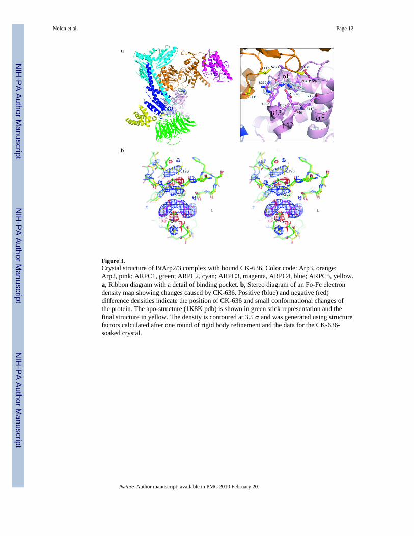
Figure 3.Crystal structure of BtArp2/3 complex with bound CK-636. Color code: Arp3, orange;Arp2, pink; ARPC1, green; ARPC2, cyan; ARPC3, magenta, ARPC4, blue; ARPC5, yellow.a, Ribbon diagram with a detail of binding pocket. b, Stereo diagram of an Fo-Fc electrondensity map showing changes caused by CK-636. Positive (blue) and negative (red)difference densities indicate the position of CK-636 and small conformational changes ofthe protein. The apo-structure (1K8K pdb) is shown in green stick representation and thefinal structure in yellow. The density is contoured at 3.5 σ and was generated using structurefactors calculated after one round of rigid body refinement and the data for the CK-636-soaked crystal.
Nolen et al. Page 12
Nature. Author manuscript; available in PMC 2010 February 20.
NIH
-PA Author Manuscript
NIH
-PA Author Manuscript
NIH
-PA Author Manuscript
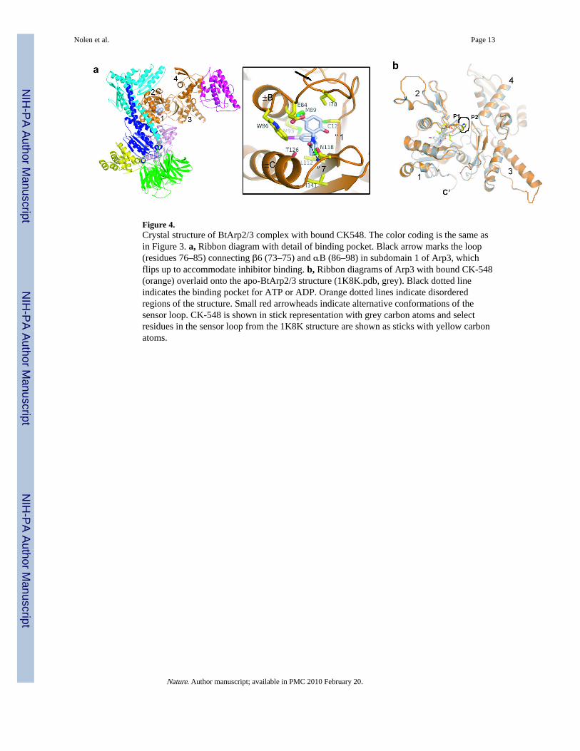
Figure 4.Crystal structure of BtArp2/3 complex with bound CK548. The color coding is the same asin Figure 3. a, Ribbon diagram with detail of binding pocket. Black arrow marks the loop(residues 76–85) connecting β6 (73–75) and αB (86–98) in subdomain 1 of Arp3, whichflips up to accommodate inhibitor binding. b, Ribbon diagrams of Arp3 with bound CK-548(orange) overlaid onto the apo-BtArp2/3 structure (1K8K.pdb, grey). Black dotted lineindicates the binding pocket for ATP or ADP. Orange dotted lines indicate disorderedregions of the structure. Small red arrowheads indicate alternative conformations of thesensor loop. CK-548 is shown in stick representation with grey carbon atoms and selectresidues in the sensor loop from the 1K8K structure are shown as sticks with yellow carbonatoms.
Nolen et al. Page 13
Nature. Author manuscript; available in PMC 2010 February 20.
NIH
-PA Author Manuscript
NIH
-PA Author Manuscript
NIH
-PA Author Manuscript
Copyright © 2022 FDOKUMEN