
PINCH1 regulates Akt1 activation and enhances radioresistance by inhibiting PP1α
Upload
khangminh22Category
view
0download
0
1
Inhibiting the Arp2/3 Complex Limits Infection of both Intracellular Mature
Vaccinia Virus and Primate Lentiviruses
Jun Komano*, Kosuke Miyauchi, Zene Matsuda, and Naoki Yamamoto
Laboratory of Virology and Pathogenesis, AIDS Research Center, National Institute of
Infectious Diseases, 4-7-1 Gakuen Musashi-Murayama, Tokyo 208-0011 Japan
*Corresponding author: Jun Komano
Laboratory of Virology and Pathogenesis, AIDS Research Center, National Institute of
Infectious Diseases, 4-7-1 Gakuen Musashi-Murayama, Tokyo 208-0011 Japan
Tel:+81-042-561-0771-335 Fax: +81-042-562-7875 E-mail:[email protected]
Running title: Arp2/3 complex supports viral entry
Abbreviations: Arp2/3, actin-related protein 2/3; CC, cytochalasin; GFP, green
fluorescent protein; HIV-1, human immunodeficiency virus type 1; HSV-1, herpes
simplex virus type 1; IMV, intracellular mature virus; MLV, murine leukemia virus; RLU,
relative light unit; SIV, simian immunodeficiency virus; VSV-G, vesicular stomatitis
virus G protein; VV, vaccinia virus; WASP, Wiscott-Aldrich syndrome protein;
Key words: HIV-1, SIV, IMV, inhibition of infection, actin

2
SUMMARY
Characterizing cellular factors involved in the life cycle of human
immunodeficiency virus type 1 (HIV-1) is an initial step toward controlling replication of
HIV-1. Actin polymerization mediated by the Arp2/3 complex has been found to play a
critical role in some pathogens’ intracellular motility. We have asked whether this
complex also contributes to the viral life cycles including that of HIV-1. We have used
both the acidic domains from actin-related protein (Arp) 2/3 complex-binding proteins
such as the Wiscott-Aldrich syndrome protein (N-WASP) or cortactin, and siRNA
directing toward Arp2 to inhibit viral infection. HIV-1, simian immunodeficiency virus
(SIV), and intracellular mature vaccinia virus (IMV) were sensitive to inhibition of the
Arp2/3 complex while MLV, HSV-1, and adenovirus were not. Interestingly,
pseudotyping HIV-1 with vesicular stomatitis virus G protein (VSV-G) overcame this
inhibition. Constitutive inhibition of the Arp2/3 complex in the T cell line H9 also
blocked replication of HIV-1. These data suggested the existence of an Arp2/3
complex-dependent event during the early phase of the life cycles of both primate
lentiviruses and IMV. Inhibiting the HIV-1’s ability to activate Arp2/3 complex could be a
potential chemotherapeutic intervention for acquired immunodeficiency syndrome
(AIDS).

3
INTRODUCTION
When human immunodeficiency virus type 1 (HIV-1) enters cells, the envelope
glycoprotein gp120 binds to CD4 and subsequently CXCR4 or CCR5 and initiates
membrane fusion at the cell surface. After the membrane fusion the reverse transcription
takes place while the viral core components migrate toward cell nucleus where the
proviral DNA integrates into the host cell chromosome. However, the protein-protein
interactions during these processes of disassembly/uncoating are the least understood
among the whole viral life cycle. Despite historical suggestions that actin plays a role in
the early phase of HIV-1 infection, its role remains largely unclear. Early studies used
chemical inhibitors of actin which were broadly active on cell physiology or
“non-specific” (Cudmore et al., 1997; Bukrinskaya et al., 1998; Iyengar et al., 1998). To
test for a specific role of actin in the early phase of HIV-1’s life cycle, we focused on
regulators of actin polymerization. It has now been shown that some bacteria and viruses
use cellular actin polymerization to propel themselves within cells (Gruenheid and Finlay,
2003). The key host proteins in these reactions are actin-related protein (Arp) 2/3
complex and its regulators. We hypothesized that Arp2/3 complex-dependent actin
nucleation might be required for efficient infection by primate lentiviruses including
HIV-1.
The Arp2/3 complex is a seven-subunit protein complex highly conserved among
eukaryotes that nucleates actin filaments from the sides of existing filaments (Higgs and
Pollard, 2001; Pantaloni et al., 2001). The Arp2/3 complex distributes throughout the cell
but is enriched especially at the cortical layer underneath the plasma membrane through

4
which viruses have to pass to infect cells (Flanagan et al., 2001). The Arp2/3 complex is
regulated by both Wiscott-Aldrich syndrome protein (WASP) family of proteins and
cortactin (Weaver et al., 2003). The carboxy terminal domain of WASP is called VCA
domain (verprolin homology, cofilin homology and acidic subdomains) and is also
named the WA domain. Intensive studies had revealed that VCA’s ability to bind
monomer actin through its V subdomain is critical for actin nucleation (Miki and
Takenawa, 1998). The CA subdomain confers to N-WASP its binding ability to the
Arp2/3 complex as evidenced by physicochemical assays (Machesky and Insall, 1998;
Marchand et al., 2001), X-ray crystallography and cross-linking experiments (Gournier et
al., 2001; Robinson et al., 2001; Zalevsky et al., 2001). Actin polymerization, nucleation
and branching are enhanced in the presence of VCA protein in vitro (Higgs et al., 1999;
Machesky et al., 1999; Rohatgi et al., 1999). Expression of the VCA protein sequesters
the Arp2/3 complex and displaces it from physiological regulation in vivo (Machesky and
Insall, 1998; Machesky et al., 1999; Rozelle et al., 2000; Castellano et al., 2001;
Harlander et al., 2003). By expressing in tissue culture cells, the VCA protein has been
used successfully as an inhibitor of Arp2/3 complex to study the role of Arp2/3 complex
in many biologic processes (Zhang et al., 1999; Krause et al., 2000; May et al., 2000;
Moreau et al., 2000; Rozelle et al., 2000; McGee et al., 2001; Zhang et al., 2002).
Another Arp2/3 complex regulator is cortactin, a filamentous actin-associated
protein originally identified as a substrate of Src (Weed and Parsons, 2001) that is also
implicated in the phagocytosis of several invasive bacteria (Dehio et al., 1995; Fawaz et
al., 1997; Cantarelli et al., 2000). Cortactin binds directly to the Arp2/3 complex through
5
its amino-terminal acidic domain, NTA, and activates it (Weed et al., 2000; Uruno et al.,
2001; Weaver et al., 2001). The NTA protein, like VCA, can serve as an inhibitor of
Arp2/3 complex.
We explored the possible involvement of Arp2/3 complex in the early phase of life
cycle of primate lentiviruses. In parallel, we tested different virus species including
adenovirus, herpes simplex virus type 1 (HSV-1), Moloney murine leukemia virus (MLV),
and intracellular mature vaccinia virus (IMV), all of which were reported to use the actin
cytoskeleton to infect cells, however the physical properties and mechanisms of their
entry vary (Rosenthal et al., 1985; Kizhatil and Albritton, 1997; Bukrinskaya et al., 1998;
Iyengar et al., 1998; Li et al., 1998). We also tested whether changing retroviral
envelopes, which forces viruses to enter through different routes, affected the efficiencies
of viral entry.
6
MATERIALS AND METHODS
Cells and Viruses
Human embryonic kidney (HEK) 293 cells and Chinese hamster ovary (CHO)-K1 cells
were maintained in Dulbecco's modified Eagle'smedium (DMEM, Sigma) supplemented
with 10% FBS (Hyclone), penicillin and streptomycin (Invitrogen). H9 cells were
maintained in RPMI1640 (Sigma) supplemented with 10% FBS, penicillin and
streptomycin. All the mammalian cell lines were incubated at 37℃ in the humidified 5%
CO2 atmosphere. Replication incompetent HIV-1 (HXB2 ∆vpr, ∆rev, ∆env, ∆nef) was
produced by transfecting the proviral DNA carrying renilla luciferase in place of nef open
reading frame into 293 T cells along with the expression plasmid for env, tat, rev and nef
(pIIIex). When pseudotyping HIV-1, rev expressing plasmid and either ecotropic MLV
envelope (Ragheb and Anderson, 1994) or vesicular stomatitis virus G (VSV-G)
expressing plasmid (Clontech) were co-transfected with the proviral DNA of HIV-1.
HXB2 was used for the replication competent HIV-1. The virus was prepared by
transfecting the proviral DNA into 293T cells and the supernatants were collected at 2
days posttransfection. SIV (∆nef) encoding firefly luciferase in place of nef open reading
frame was created based on the IL-2-carrying molecular clone of SIVmac239 (Gundlach
et al., 1997) (kindly provided by Dr. K. Mori). SIV was prepared by transfecting the
proviral DNA into COS7 cells and the supernatants were collected. MLV was produced
by transfecting pCMMP LacZIRESGFP, pCMMP eGFP (generous gift from Dr. J.
Young), pCMMP GFP-VCA, or pQcLIN (Clontech) into 293 cells along with gag/pol and
either ecotropic, amphotropic env or VSV-G expressing plasmids (Ragheb and Anderson,
7
1994). HSV-1 (KOS)Rid1/tk12 encoding beta-galactosidase under the regulation of the
immediate early promoter ICP4, vaccinia virus encoding T7 RNA polymerase (vTF7.3),
and adenovirus type 5 expressing T7 RNA polymerase were generous gifts from Drs.
Spear (Dean et al., 1994), Moss (Fuerst et al., 1986), and Ishii (Aoki et al., 1998),
respectively, and prepared according to the previous publications. Adenovirus, HSV-1
and IMV were titrated by the plaque assay using HEK293 or Hela cells; MLV by counting
reporter-positive HEK293 cells after two to three days post-infection, and HIV-1, VSV-G
HIV-1, and SIV by using the indicator cells that expressed beta-galactosidase or GFP
upon infection. The p24 concentration of eco HIV-1 was adjusted to that of HIV-1
(approximately 100 ng/mL). The neutralization assay using the antibody 2D5 that inhibits
infection of IMV but not that of EEV (Ichihashi and Oie, 1996) revealed that 99.4% of
our vaccinia virus preparation contained intracellular mature virus (IMV).
Plasmids
The VCA domain (amino acid 392-505) of N-WASP was amplified by PCR from
N-WASP cDNA kindly provided by Dr. Takenawa (Miki et al., 1996) with the following
primers: VCA sense 5’-CAATTGCCTTCTGATGGGGACCATCAGG-3’ and
5’-AAGCTTCAGTCTTCCCACTCATCATC-3’. The PCR product was subcloned into
pCR4 Blunt TOPO (Invitrogen), sequenced, digested with Mfe I and Eco RI, and cloned
into Eco RI site of pEGFP-C2 (Clontech), generating pGFP-VCA. Following two primers,
5’-AGATCTTAGTGGCTGATGGCCAAGAGTCCACACC-3’ and 5’-
CAATTGTCAGTCTTCCCACTCATCATC-3’, were used to amplify the CA domain

8
(amino acid 470-505) of N-WASP. The PCR fragment was cloned into Bgl II-Eco RI sites
of pEGFP-C2, giving rise to pGFP-CA. GFP-A expression plasmid was generated by
annealing following two oligonucleotides and cloned it into the Bgl II-Eco RI sites of
pEGFP-C2:
5’-GATCGATGAAGATGAAGATGAAGATGATGAAGAAGATTTTGAGGATGATG
ATGAGTGGGAAGACTGA-3’ and
5’-AATTTCAGTCTTCCCACTCATCATCATCCTCAAAATCTTCTTCATCATCTTCA
TCTTCATCTTCATC-3’. Following oligonucleotides were annealed and ligated into the
Bgl II site of pGFP-VCA giving rise to pGFP-VCA*:
5’-GATCTAGATAACTGATGCGGCCGCCG-3’ and
5’-GATCCGGCGGCCGCATCAGTTATCTA-3’. The NTA domain (amino acid 1-84) of
cortactin and Arp2 were amplified by PCR from 293T cDNA with following primers:
NTA sense 5’-GGATCCTCGAGATGTGGAAAGCTTCAGCAGGCCAC-3’, NTA
antisense 5’-CAATTGTCAATAGCCATGGGAAGCTTTTGGTCC-3’; Arp2 sense
5’-GGATCCTCGAGATGGACAGCCAGGGCAGGAAGG-3’, Arp2 antisense
5’-CAATTGTTATCGAACAGTCACACCAAGTTTC-3’. PCR fragments were cloned
into pCR4 Blunt TOPO, and the Bam HI-Mfe I fragments were cloned into Bgl II-Eco RI
sites of pEGFP-C2, generating pGFP-NTA and pGFP-Arp2. pCMMP GFP-VCA was
generated by digesting pCMMP GFP with Age I and Bam HI, ligated with Age I-Bam HI
fragment from pGFP-VCA. Transfection efficiencies were measured by phRL/CMV
(Promega) or pHIV-1 LTR-GFP-Luciferase. F10, the ecotropic MLV receptor from rat,
expression plasmid was created by digesting pcDNA F10-ecoR (Takase-Yoden and

9
Watanabe, 1999) with Nae I followed by the self-ligation. The human nectin 1 alpha
(HigR) expression plasmid was generated by inserting Eco RI fragment of pEF-BOS
human nectin 1 alpha 3xFLAG (Sakisaka et al., 2001) into Eco RI site of pcDNA3
(Invitrogen). The Xho I-Not I fragment of either pBCMGSneoCD4 or
pBCMGSneoCCR5 (a generous gift from Dr. Yamashita) was cloned into Xho I-Not I
sites of pcDNA3 (Invitrogen), generating pCD4 and pCCR5, respectively. The Sna
BI-Not I fragment from pCCR5 was cloned into pMACS4ires (Miltenyi Biotec),
generating pCCR5 IRES CD4. The CD4 lacking the cytoplasmic tail was amplified by
the following primers:
5’-GGATCCCGGGCCACCATGAACCGGGGAGTCCCTTTTAGGC-3’and 5’-
GAATTCGTGCCGGCACCTGACACAGAAGAAGATGCC-3’. The Xma I-Eco RI
fragment was cloned into the Age I-Eco RI sites of pEGFP-C2, generating pCD4∆cyt.
The T7 RNA polymerase (T7 RNAP) expression plasmid pCMMP T7RNAP IRES GFP
was created by inserting Xma I-Eco RI fragment from pVR1-T7 into Age I-Mfe I sites of
pCMMP IRES GFP (Aoki et al., 1998). The T7RNAP reporter plasmid pTM3Luci was
described previously (Aoki et al., 1998). The following pairs of oligonucleotides were
annealed and cloned into Apa I-Eco RI sites of pSilencer 1.0-U6 (Ambion) to generate
siRNA expressing vectors directing against GFP and Arp2: GFP sense
5’-GCTGACCCTGAAGTTCATCTTCAAGAGAGATGAACTTCAGGGTCAGCTTT
TTT-3’ and GFP antisense
5’-AATTAAAAAAGCTGACCCTGAAGTTCATCTCTCTTGAAGATGAACTTCAG
GGTCAGCGGCC-3’; Arp2 sense

10
5’-CAGCTTTACTTAGAACGAGTTCAAGAGACTCGTTCTAAGTAAAGCTGTTTT
TT-3’ and Arp2 antisense
5’-AATTAAAAAACAGCTTTACTTAGAACGAGTCTCTTGAACTCGTTCTAAGTA
AAGCTGGGCC-3’.
Transfection, magnetic selection, and infection
Plasmid DNAs were transfected into cells by using either lipofectamin/lipofectamin plus
reagent (Invitrogen) or X-tremeGENE siRNA transfection reagent (Roche) according to
the manufacturer’s protocol. The latter reagent, with which the transfection efficiencies
reached over 90% in 293 cells, was specifically used for preparing samples to carry out
Western blot analysis demonstrating the reduction of Arp2 levels. In brief, cells were fed
in 48-well plates 1 to 2 days before transfection. Cells were approximately in 50-60%
confluency at transfection. After transfection, cells were trypsinized and plated onto
96-well plates. Transfected cells were magnetically selected by using MACS system
directing toward CD4 (Miltenyi Biotec). Cells were infected with viruses of
approximately 0.1-0.5 multiplicity of infection by incubating in the virus-containing
culture medium at 37℃ for 1 to 4 hours.
Cell-to-cell fusion assay
The fusion assay was based upon the T7RNAP transcription-dependent reporter assay
originally described by Nussbaum et al. (Nussbaum et al., 1994), modified versions by
Sakamoto et al. (Sakamoto et al., 2003). In brief, the T7RNAP “donor” cells were

11
generated by transfecting 293 cells with both T7RNAP and HIV-1’s Env expression
vectors. The T7RNAP “acceptor” cells were generated by transfecting 293 cells with the
T7RNAP reporter (pT7-IRES-Luciferase) along with either CD4 or CD4∆cyt, GFP or
GFP-VCA, and the renilla luciferase expression vectors. At 48 hours post-transfection,
these cells were co-cultivated for 24 hours and lysed in the passive lysis buffer to carry
out the dual luciferase assay (Promega).
Detection of fluorescent signals
The transfected 293 cells were fixed by 4% formaldehyde in PBS at 48 hours
posttransfection and imaged by using confocal microscopy META 510 (Carl Zeiss). For
the images showing pseudopodial extensions, images of different focal planes were
projected to generate a single image. The transfected H9 cells were imaged similarly at 48
hours posttransfection without fixation. The light transmission image was merged with
the green fluorescent signal. Alternatively, the transfected cells were analyzed by the flow
cytometry (FACS calibur, Beckton Dickinson).
Western blot analysis
Cells were washed with PBS and lysed in a buffer containing 4% SDS, 100 mM Tris-HCl
(pH 6.8), 12% 2-ME, 20% glycerol, and bromophenol blue. Samples were boiled for 10
min. Protein lysates approximately equivalent to 5x10⁴ cells were separated in
SDS-PAGE (Perfect NT Gel, DRC, Tokyo), transferred to a polyvinylidene fluoride
(PVDF) membrane (Immobilon-PSQ, Millipore), and blocked with 5% dried non-fat milk

12
(Yuki-Jirushi, Tokyo) in PBS. For the primary antibody, we used anti-Arp2 antibody
H-84 (Santa Cruz), anti-actin antibody MAB1501 (Chemicon), or either a monoclonal
antibody or a polyclonal rabbit antiserum against GFP (Clontech). For the secondary
antibody, either a biotinylated anti-mouse antibody (Amersham-Pharmacia) or a
biotinylated anti-rabbit antibody was used. For the tertiary probe, a Horseradish
peroxidase (HRP)-conjugated streptoavidin (Amersham-Pharmacia) was used. Signals
were developed by incubating blots with the chemilumenescent HRP substrate
(Amersham-Pharmacia) and detected by using Lumi-Imager F1 (Boehringer Mannheim).
Reporter Assays
Cells were lysed in the Passive Lysis Buffer (Promega) and the dual luciferase assay was
performed to measure both firefly and renilla luciferase activities according to the
manufacturer’s protocol (Promega). The beta-galactosidase activity was measured by
using the LumiGal assy kit according to the manufacturer’s protocol (Clontech). The
chemiluminescence was detected by Lmax (Molecular Devices).
ELISA
The amount of p24 antigen of HIV-1 in the culture supernatants was quantified by using
Retro TEK p24 antigen ELISA kit according to the manufacturer’s protocol (Zepto
Metrix). The signals were measured by Vmax ELISA reader (Molecular Devices).

13
Statistical analysis
The significance of differences was tested by one-way analysis of variance (ANOVA) and
Student’s t-test. P values less than 0.05 were considered to be significant.

14
RESULTS
GFP-VCA inhibits infection of primate lentiviruses
We first tested whether the viral infection was affected by expressing a potential
inhibitor of the Arp2/3 complex GFP-VCA, the VCA domain of N-WASP fused to GFP
(Fig. 1A). The expression of GFP-VCA protein was verified by Western blot analysis (Fig
1A). The expression levels of GFP-VCA were similar to those of GFP as measured by the
flow cytometric analysis at 48 hours post-transfection (Fig. 1B). GFP-VCA preferentially
distributed to the cytoplasm and inhibited the pseudopodial extension as compared to
GFP alone in agreement with the previous report (Rozelle et al., 2000) (Fig. 1C). To
exclude the possibility that GFP fusion proteins negatively affects the cell surface
expression of membrane proteins, we confirmed that the distribution and the levels of
transiently and constitutively expressed CD4 and CXCR4 on the cell surface were similar
in both GFP- and GFP-VCA-expressing cells as assessed by both confocal microscopy
and the flow cytometric analysis (data not shown).
To evaluate the contribution of the Arp2/3 complex on the viral infection, we
employed a transient transfection/infection system in which target cells were transfected
with a mixture of plasmid DNA expressing (i) an Arp2/3 complex inhibitor GFP-VCA,
(ii) a viral receptor, and (iii) a reporter gene to normalize transfection efficiencies
(depicted in Fig. 1D). Due to the nature of cotransfection, the majority of cells uptook the
expression vector for GFP-VCA were transfected with the other plasmids because the
mixture of DNA contained 5-fold excess amount of the GFP-VCA expressing plasmid. At
48 hours posttransfection, a portion of transfected cells were collected to measure the

15
transfection efficiencies and others were challenged by a virus expressing a reporter gene
to monitor the efficiency of infection. Various times after infection, cells were lysed and
the virally encoded reporter gene activities were measured, which was divided by the
transfection efficiencies to normalize. Infection of viruses was restricted to the
transfected cells by using viral receptors: human CD4 for HIV-1, human CD4 and CCR5
for SIV, F10 for ecotropic MLV envelope-pseudotyped HIV-1 (eco HIV-1) and ecotropic
MLV (eco MLV), human nectin-1 alpha (HigR) for HSV-1. Viruses, receptors, and the
combination of reporter genes for each virus were summarized in the Table 1 (Table 1).
Primarily we targeted HEK293 cells because actin cytoskeletal system is shared among
eukaryotes. To monitor HSV-1 infection, CHO-K1 cells were used that lack entry
molecules for HSV-1. In order to monitor the efficiencies of single-round viral infection,
we used replication-incompetent HIV-1 and MLV. Alternatively, cells infected with
replication-competent viruses were lysed at time points before viruses entered the second
replication cycle, except SIV which required 48 hours to give sufficient signal to be
detected. Importantly, all the viruses tested in this study were reported to utilize actin
cytoskeleton to infect cells as examined by using chemical inhibitors against actin such as
cytochalasin (CC) (Rosenthal et al., 1985; Kizhatil and Albritton, 1997; Bukrinskaya et
al., 1998; Iyengar et al., 1998; Li et al., 1998). The infection efficiencies of viruses into
cells expressing GFP were set at 100% throughout the study unless stated and the
infection efficiencies into GFP-VCA-expressing cells relative to GFP-expressing cells
were calculated. For example, when eco HIV-1 was tested, the firefly luciferase activities
representing the transfection efficiencies for GFP- and GFP-VCA-transfected cells were

16
2,065 and 1,854 relative light unit (RLU), respectively, where the background signal was
3 RLU. The renilla luciferease activities reflecting the infection efficiencies into GFP-
and GFP-VCA-transfected cells were 4,018 and 1,254 RLU, respectively, where the
background was 15 RLU. In this case, the relative infection efficiency of eco HIV-1 into
GFP-VCA expressing cells was 34.4%. The relative infection efficiency was introduced
because we were able to integrate data from independent experiments. Results from the
indicated number of independent experiments were summarized in the Figure 1E (Fig.
1E). Expression of the GFP-VCA significantly reduced the relative infection efficiencies
of HIV-1, eco HIV-1, and SIV (30.3%, 35.8%, and 22.7%, respectively; P<0.01; Fig. 1E),
whereas did not those of eco MLV and HSV-1 (98.5% and 103.5%, respectively; Fig. 1E).
A ten-fold higher or lower titer of HSV-1 did not affect the results (data not shown).
Consistent with this, the average signal to noise ratio did not correlate with the efficiency
of inhibition (Fig. 1E and Table 1). The GFP-VCA did not inhibit eco MLV but did inhibit
the infection of eco HIV-1, suggesting that GFP-VCA’s ability to inhibit the infection of
eco HIV-1 was eco env-independent. It also suggested that the cell surface expression of
the F10 was not affected by GFP-VCA expression. In addition, our SIV clone did not
encode nef and HIV-1 vpr, demonstrating that both nef and vpr were not necessary for
primate lentiviruses (HIV-1 and SIV) to infect GFP-VCA expressing cells efficiently.
These results suggested that inhibiting the Arp2/3 complex by GFP-VCA negatively
affected the infection of primate lentiviruses.
HIV-1 pseudotyped with VSV-G overcomes the block of infection by GFP-VCA

17
To gain insight of the mechanism of GFP-VCA’s action to limit primate lentiviral
infection, we tested whether the GFP-VCA blocked infection of VSV-G-pseudotyped
HIV-1 (VSV-G HIV-1). HIV-1 enters cells by inducing the virus-cell membrane fusion at
the cell surface. When HIV-1 was pseudotyped with VSV-G, the route of viral entry
became endocytosis (Stein et al., 1987; Maddon et al., 1988). We transfected 293 cells
with GFP- or GFP-VCA-expressing plasmid along with one-tenth amount of CD4 and
F10 expressing plasmids. Then we enriched transfected cells by using magnetic beads
directing toward CD4 at 36-42 hours posttransfection. This allowed us to measure the
infection efficiencies into the transfected cells only (depicted in Fig. 2A). More than 90%
of the magnetically selected cells were green fluorescence-positive as examined by the
flow cytometric analysis (data not shown). At 48 hours posttransfection, cells were
infected either with HIV-1, eco HIV-1 or VSV-G HIV-1. For a comparison, amphotropic
MLV (ampho MLV) and VSV-G pseudotyped MLV (VSV-G MLV) were tested in parallel.
Infected cells were lysed at 2-3 days post-infection and the virally encoded reporter gene
activities were measured to estimate the relative infection efficiencies (depicted in Fig.
2A). For example, when eco HIV-1 was tested in parallel with VSV-G HIV-1, the renilla
luciferease activities representing the infection efficiencies of eco HIV-1 into
magnetically selected GFP- and GFP-VCA-expressing cells were 41,968 and 10,117
RLU, respectively, where the background was 30 RLU. In contrast, the renilla luciferase
activities of VSV-G HIV-1-infected cells were 6,346 RLU for GFP-expressing cells and
5,436 RLU for GFP-VCA-expressing cells where the background signal was 30 RLU. In
these cases, the relative infection efficiencies of eco and VSV-G HIV-1 into GFP-VCA

18
expressing cells were 24.1% and 85.6%, respectively. Results from a number of
independent experiments were summarized in the Figure 2B (Fig. 2B). The relative
infection efficiencies for HIV-1 and eco HIV-1 were significantly reduced when target
cells expressed GFP-VCA as compared to GFP alone (47.3%, 36.0%, P<0.01,
respectively; Fig. 2B). The magnitude of inhibition of HIV-1 infection in this assay was
smaller than that of the first experimental setup (Fig. 1D) presumably because the levels
of CD4 on the cell surface might have decreased after the magnetic selection (Fig. 1E).
On the other hand, the relative infection efficiency for eco HIV-1 (36.0%, P<0.01; Fig.
2B) was similar to the first experimental setup, indicating that the magnetic selection did
not detectably influence on the cellular susceptibility to viral infection. Interestingly,
VSV-G HIV-1 infected GFP-VCA-positive cells at almost equal efficiencies to
GFP-positive cells (101.7%, Fig. 2B). Similarly, both ampho MLV and VSV-G MLV
infected GFP-VCA-expressing cells as efficient as they did GFP-expressing cells (96.9%
for ampho MLV, 93.2% for VSV-G MLV; Fig. 2B). In this experimental setup, the signal
to noise ratio of HIV-1 and eco HIV-1 increased as compared to the first experimental
system, however, the results remained the same (Fig. 1E, 2B, and Table 2). These data
indicated that the GFP-VCA was unable to block HIV-1 infection when HIV-1 entered
cells through the VSV-G-mediated endocytosis. In other words, the reverse transcription,
nuclear import, and integration of HIV-1 genome into the host chromosome were able to
proceed in the presence of GFP-VCA.
The efficiency of the membrane fusion is not negatively affected by GFP-VCA
19
The membrane fusion is the critical event when enveloped viruses infect cells. We
next asked if the expression of GFP-VCA inhibited the membrane fusion through HIV-1’s
Env-receptor interaction. We carried out the cell-to-cell fusion assay in which
Env-positive cells expressing T7 RNA polymerase (T7RNAP) were fused to the
CD4-positive cells carrying the T7RNAP promoter-driven firefly luciferase reporter
plasmid (depicted in Fig. 2C). The efficiency of the cell-to-cell fusion was measured by
the firefly luciferase activity divided by the renilla luciferase activity representing the
transfection efficiency. The ratio of the firefly luciferase to renilla luciferase activities in
GFP-expressing cells was set at 100% and the relative efficiencies of cell-to-cell fusion of
GFP-VCA expressing cells were calculated. It revealed that the efficiencies of cell-to-cell
fusion initiated by the interaction between HIV-1’s Env and CD4 were not inhibited when
GFP-VCA was expressed in CD4-expressing cells as compared to GFP (114%, Fig. 2D).
To eliminate a possibility that the GFP-VCA inhibited the expression/distribution of
CD4 on the cell surface, we examined whether the CD4 lacking the cytoplasmic domain
(CD4∆cyt) was able to support the membrane fusion in the presence of GFP-VCA. Since
GFP-VCA distributed throughout the cytoplasm, it was unlikely that the motility of
CD4∆cyt on the cell surface was restricted by GFP-VCA. It was found that CD4∆cyt
induced the cell-to-cell fusion in the presence of GFP-VCA at almost equal efficiencies to
the full length CD4 (113%, Fig. 2D). In support of this, enveloped viruses (HSV-1, MLV,
and VSV-G HIV-1) were able to infect GFP-VCA-positive cells as efficient as
GFP-positive cells (Figs. 1E and 2B). In particular, when HSV-1 and ampho MLV enter
cells, like HIV-1, the membrane fusion takes place at the cell surface (Fuller and Spear,

20
1987; McClure et al., 1990; Wittels and Spear, 1991; Nussbaum et al., 1993). These data
indicated that the virus-cell membrane fusion was not inhibited by GFP-VCA. According
to the data presented hereby, it seemed likely that GFP-VCA did not negatively affect the
expression of receptors or viral attachment to receptors, or the virus-cell membrane
fusion. Since VSV-G HIV-1 infected cells in the presence of GFP-VCA, we assume that
GFP-VCA inhibits HIV-1’s life cycle after the membrane fusion before or at the reverse
transcription especially when HIV-1 enters cells through the membrane fusion at cell
surface where the viral core is placed in the cortical compartment.
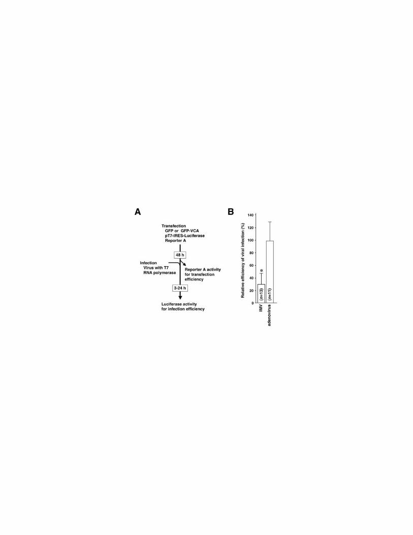
GFP-VCA inhibits infection of intracellular mature vaccinia virus (IMV) but not
adenovirus
To test whether the inhibition of viral entry by GFP-VCA was limited to the primate
lentiviruses, we examined both intracellular mature vaccinia virus (IMV) and adenovirus.
IMV enters cells via the membrane fusion at cell surface, which is accompanied by a
drastic actin cytoskeletal reorganization (reviewed by Smith et al., 2002). Adenovirus
infects cells through clathrin-dependent endocytosis (Wang et al., 1998). Adenovirus was
the only envelope-free virus studied in this paper. These viruses encoded T7 RNA
polymerase as a reporter (Table 3). To detect the infection signal from
transfected/infected cells, we introduced the T7 promoter-driven luciferase reporter into
293 cells (depicted in Fig. 3A). For example, when IMV was tested, the renilla luciferase
activities representing the transfection efficiencies for GFP- and GFP-VCA-transfected
cells were 485 and 556 RLU, respectively, where the background signal was 8 RLU. The

21
firefly luciferease activities reflecting the infection efficiencies of virus into GFP- and
GFP-VCA-transfected cells were 29,020 and 10,044 RLU, respectively, where the
background was 23 RLU. In this case, the relative infection efficiency of IMV into
GFP-VCA expressing cells was 30.3%. Results from a number of independent
experiments were summarized in the Figure 3B (Fig. 3B). The relative infection
efficiency of IMV was significantly reduced (30.6%, P<0.01; Fig. 3B) whereas
adenovirus infected GFP-VCA expressing cells as efficiently as GFP-expressing cells
(98.3%, Fig. 3B). IMV gave the highest signal to noise ratio throughout the study, yet its
infection was blocked efficiently by GFP-VCA (Table 3). Considering all the signal to
noise ratio data, it was suggested that the signal to noise ratio did not positively correlate
with the magnitude of inhibition of viral infection. Ten-fold higher or lower titer of either
IMV or adenovirus did not affect the results (data not shown). These data suggested that
inhibiting the Arp2/3 complex by GFP-VCA negatively affected infection of IMV as well
as primate lentiviruses. We focused on HIV-1 and IMV for the further studies.
GFP-VCA’s ability to nucleate actin filament is not necessary to inhibit viral entry
We attempted to locate the domain within VCA responsible for the inhibition of
HIV-1 and IMV infection. We generated a series of truncated GFP-VCA mutants (Fig.
4A). Their functions were also summarized according to the previous reports (Fig.
4A)(Takenawa and Miki, 2001; Weaver et al., 2003). Expression of each mutant was
verified in Western blot analysis (Fig. 4B). Introducing a stop codon into GFP-VCA after
the GFP open reading frame allowed VCA-encoding RNA to be expressed but not VCA

22
protein (GFP-VCA*). Expression of GFP-VCA* did not inhibit infection of both HIV-1
and IMV (82.9% and 112.6%, respectively; Fig. 4C), suggesting that the inhibition of
viral entry attributed to the VCA protein, not the VCA-encoding RNA (Fig. 4C). GFP-CA
retained the ability to limit both HIV-1 and IMV infection (46.7% and 46.4%, P<0.01 and
P<0.05, respectively, Fig. 4C). Expression of GFP-A inhibited HIV-1 infection less
efficiently than other derivatives (57.7%, P<0.05; Fig. 4C). However, GFP-A was unable
to limit IMV infection (110.8%, Fig. 4C) although synthetic peptides of A subdomain has
been shown to retain the binding affinity to the Arp2/3 complex as high as VCA in vitro
(Panchal et al., 2003). These data suggested that the V subdomain was not required for
the inhibition of HIV-1 and IMV infection. It seemed likely that C and A subdomains
functioned cooperatively in vivo to inhibit infection of both HIV-1 and IMV.
To further verify that the reduction of viral infection efficiencies was due to the
inhibition of Arp2/3 complex functions, we have tested whether another potential Arp2/3
complex inhibitor NTA, amino-terminal acidic domain of cortactin (amino acid 1-84),
was also able to limit both HIV-1 and IMV infection as did VCA. Cortactin is unrelated to
WASP family of proteins but is able to bind the Arp2/3 complex through the NTA domain.
Expression of GFP-NTA was verified by Western blot analysis (Fig. 4B). As expected,
GFP-NTA reduced the infection efficiencies of both HIV-1 and IMV (39% and 50%,
P<0.01 and P<0.05, respectively; Fig. 4C). These data confirmed that inhibiting
functions of the Arp2/3 complex was indeed responsible for limiting both HIV-1 and IMV
infection. As GFP-NTA lacked the ability to nucleate actin filaments similar to GFP-CA,
and due to the reported functions of CA subdomains (Fig. 4A), it was strongly suggested

23
that, not their abilities to enhance actin nucleation, but GFP fusion proteins’ abilities to
bind the Arp2/3 complex was primarily important to inhibit viral infection.
In addition, we tested whether downregulating expression of Arp2 by using siRNA
technique inhibited both HIV-1 and IMV infection. The GFP-VCA and -NTA were able to
inhibit functions of Arp2/3 complex by binding to it directly, whereas siRNA against
Arp2 downmodulated expression of Arp2, therefore decreased the number of Arp2/3
complex. Since siRNA against Arp2 was not able to inhibit the function of pre-existing
Arp2/3 complex directly, it was expected that siRNA against Arp2 should be able to limit
viral infection, if any, less efficiently than GFP-VCA. Transfecting the plasmid vector
expressing siRNA directing against Arp2 reduced the expression of endogenously
expressed Arp2 to 26.5% in 293 cells as demonstrated by Western blotting analysis (Fig.
4D). Viral infection efficiencies were measured using the experimental setups (Figs. 1D
and 3A) except the GFP or GFP-VCA expression plasmid was replaced with the siRNA
expression vectors against GFP or Arp2. When siRNA directing toward GFP was set as
100%, the relative efficiencies of both HIV-1 and eco HIV-1 infection in cells expressing
siRNA against Arp2 became 39.4% and 39.1%, respectively (both, P<0.01; Fig. 4E).
Adenovirus infection was slightly inhibited by the siRNA against Arp2 (69.4%, Fig. 4E).
However, the relative infection efficiencies of eco MLV, HSV-1, and IMV were not
significantly reduced by siRNA directed toward Arp2 (91.3%, 103.8%, and 104.4%,
respectively, Fig. 4E). Given that the other two Arp2/3 inhibitors were able to limit IMV
and HIV-1 infection, it seemed reasonable to speculate that the inhibition of HIV-1
infection by siRNA was due to the downmodulation of newly synthesized Arp2. However,

24
we were unable to limit the infection of IMV by siRNA against Arp2. We assumed that
this was either because siRNA was unable to downregulate expression of Arp2, therefore
Arp2/3 complex, at levels sufficient to block the entry of IMV or the number of the
pre-existing Arp2/3 complex might be sufficient to support IMV infection, or both.
HIV-1 replication is negatively affected when GFP-VCA is constitutively expressed in
H9 cells
Finally, we asked whether the replication of HIV-1 was inhibited in T cells, one of
the natural targets of HIV-1. To do this, we isolated H9 cell clones either expressing GFP
or GFP-VCA constitutively by infecting H9 cells with MLV vector followed by the
limiting dilution. Introducing expression plasmid for GFP-VCA did not alter the
morphology of H9 cells as compared to GFP-expressing cells or untransfected cells when
observed under the confocal microscopy at 48 hours post-transfection (Fig. 5A) as well as
stable cell clones (data not shown). H9 GFP-VCA clones proliferated at speed
indistinguishable from H9 GFP clones (data not shown). We verified the expression of
GFP and GFP-VCA in isolated clones by Western blot analysis (Fig. 5B). However, the
average expression levels of GFP-VCA appeared low as compared to those of GFP in H9
clones (Fig. 5B). We infected six H9-GFP and three H9-GFP-VCA clones with a
replication-competent HIV-1 (HXB2) and collected culture supernatants at different time
points to monitor the viral replication by measuring the amount of p24 viral antigen. The
replication kinetics of HIV-1 in GFP-expressing H9 clones showed a rapid propagation of
HIV-1 in culture. In contrast, the amount of p24 in the culture supernatants of H9

25
GFP-VCA clones did not accumulate, suggesting that the HIV-1 replication was
substantially suppressed in H9 GFP-VCA clones (Fig. 5C). The replication assay was
carried out twice consecutively and the similar data were obtained. On the average, the
peak of HIV-1 replication kinetics in GFP-VCA clones delayed a week as compared to
GFP clones. The maximum amount of p24 in the culture supernatants of H9 GFP-VCA
clones did not appear different from those of H9 GFP clones.
We next examined whether the viruses propagated in H9 GFP-VCA clones were
mutants capable of replicating in the presence of GFP-VCA. We isolated viral RNA from
the culture supernatants of a H9 GFP clone and three H9 GFP-VCA clones and sequenced
the gag/pro/pol region since the GFP-VCA’s ability to limit HIV-1 infection did not
depend on Env, Nef, Vpr, and Rev. However, we couldn’t find any nucleic acid alterations
when the viral sequences from an H9 GFP-VCA culture were compared to the one from
an H9 GFP culture. These data suggested that the levels of GFP-VCA in H9 clones might
be insufficient to confer the selective advantage for mutant viruses to take over the wild
type HIV-1. We found that the late phase of HIV-1 viral life cycle was slightly affected by
GFP-VCA. Transfecting proviral DNA into 293T cells along with the expression vector
for GFP-VCA yielded fewer amount of p24 antigen in the culture supernatant (72%)
compared to GFP alone. Taken together, the decreased replication kinetics of HIV-1 in H9
GFP-VCA was assumed to be mostly due to the inhibition of the early phase, partly the
late phase, of HIV-1’s life cycle. These data demonstrated that the constitutive inhibition
of the Arp2/3 complex by expressing GFP-VCA limited efficient replication of HIV-1 in
T cells as well as epithelial cell systems.

26
DISCUSSION
We have demonstrated that the Arp2/3 complex contributes to the efficient infection
of both primate lentiviruses (HIV-1 and SIV) and IMV but not MLV, HSV-1 and
adenovirus. Actin cytoskeleton has been shown to play a role in the infection of all the
viruses tested in this study according to previous studies using the chemical actin
inhibitor (Rosenthal et al., 1985; Kizhatil and Albritton, 1997; Bukrinskaya et al., 1998;
Iyengar et al., 1998; Li et al., 1998; Locker et al., 2000). In addition, the Arp2/3
complex-mediated actin nucleation is sensitive to CC (Welch et al., 1998). Therefore,
GFP-VCA’s ability to limit infection of primate lentiviruses and IMV might be a part of,
if not all, the mechanism by which CC reduced the efficiency of viral infection. In other
words, primate lentiviruses and IMV might utilize the Arp2/3 complex-dependent actin
polymerization system to support their early phase of viral life cycles. In contrast, MLV,
HSV-1, and adenovirus might utilize actin to enter cells in an Arp2/3
complex-independent manner. Perhaps, other functional aspects of actin are important for
their efficient infection such as the actin cable-dependent trafficking system. Our data
clearly demonstrated that the different viruses use actin system differently. It was reported
that the infection of HIV-1 was inhibited by CC at least two different levels: (i) by
limiting viral receptor/coreceptor clustering upon viral attachment (Iyengar et al., 1998);
and (ii) by disrupting establishment of active reverse transcription complex (Bukrinskaya
et al., 1998). Our findings suggest a possible involvement of the Arp2/3 complex in the
latter processe or a presence of another actin polymerization-dependent step between two
processes.
27
What is the molecular mechanism by which the Arp2/3 complex supports infection
of both primate lentiviruses and IMV? One of the possibilities is that these viruses may
activate the Arp2/3 complex and nucleate actin filaments to support their entry. It has
been reported that an acidic motif DDW or DEW can be found among cellular Arp2/3
complex-binding proteins including WASP, cortactin, and MyoD as well as Listeria
monocytogenes surface protein ActA (Weed and Parsons, 2001). We are unable to find
such a binding motif in proteins encoded by both HIV-1 and SIV. Therefore, these viruses
may not activate Arp2/3 complex due to a direct interaction between viral gene products
and Arp2/3 complex. There are two regulatory pathways known to activate the Arp2/3
complex. One is WASP/WAVE pathway and the other being cortactin pathway.
Expression of the dominant-negative derivatives of N-WASP, WAVEs, and cortactin were
unable to limit the infection of both HIV-1 and IMV (unpublished observation, consistent
with the Locker’s finding, Locker et al., 2000). The dominant-negative derivative of
cdc42, RhoGTPase family protein that locates upstream of WASP/WAVE pathway, was
also unable to reduce the relative infection efficiencies of both viruses (unpublished
observation). These data implied that the activation of Arp2/3 complex upon infection of
both lentiviruses and IMV might be mediated by novel virus-host interactions. Inhibiting
the Arp2/3 complex by GFP-VCA did not drastically reduce the efficiency of HIV-1
production as did that of HIV-1 infection, suggesting that the incoming HIV-1 might have
something unique which budding virus lacks. We speculate that viral gene products
cleaved by HIV-1’s protease may be responsible to induce activation of Arp2/3 complex
because viral protease become active when viral particles are released from cells such that

28
the cleaved proteins are present at high concentrations only in mature virus particles. VV
encodes envelope protein A36R that binds adaptor proteins Nck, Wip, Grb2 that recruits
and activates WASP-Arp2/3 complex system (Frischknecht et al., 1999; Rietdorf et al.,
2001; Scaplehorn et al., 2002). These molecular interactions are known to be important
for vaccinia virus to bud from infected cells as extracellular enveloped virus (EEV).
However, IMV does not have A36R on its envelope. Accordingly, IMV should initiate
activation of Arp2/3 complex via A36R-independent mechanisms.
Based upon our data as well as previous observations, we propose models in which
the Arp2/3 complex plays a role in the entry of both primate lentiviruses and IMV. HIV-1
enters cells via the membrane fusion on the cell surface (Stein et al., 1987; Maddon et al.,
1988). The viral core complex is released to the cytoplasm immediately after the
membrane fusion over the cortical layer. Our model is that the viral core components,
independent of Env, Nef, Vpr, and Rev, recruit adaptor proteins and activate the Arp2/3
complex to generate mechanical force by which HIV-1’s core complex passes through the
cortical layer and migrates toward the nucleus efficiently (Fig. 6A). McDonalds et al. had
shown that the microtubule system supports long distance movement of HIV-1’s core
complex (McDonald et al., 2002). It is possible that lentiviruses utilize the Arp2/3
complex-mediated active transport system to get access to a subcellular compartment
where they meet microtubule system. Consistent with our model, a short-distance rapid
movement of HIV-1’s core was observed in the time-lapse imaging, which suggested a
presence of an actin polymerization-dependent transport (McDonald et al., 2002). On the
other hand, IMV enters cells via the membrane fusion at the cell surface as suggested by

29
the intensive studies including electron microscopic studies (reviewed in Smith et al.,
2002). IMV infection induces the formation of actin-rich cell surface protrusions partly
due to GTPase Rac1 signaling (Locker et al., 2000) and is inhibited by treating cells with
CCD (Vanderplasschen et al., 1998; Locker et al., 2000). Taken together, the Arp2/3
complex-mediated actin polymerization may be required for not only the late phase but
also the early phase of IMV’s life cycle, specifically at the post-membrane fusion
processes (Fig. 6B). Also, it is necessary to reorganize the cortical actin network to
transport the virus-core toward the cytoplasmic subcompartment where the vaccinia virus
replicates. The Arp2/3 complex may play a role in the latter process (Fig. 6B). In any case,
IMV induces relatively global cytoskeletal reorganization in which a substantial number
of Arp2/3 complex should participate. This may account for the less efficient block of
IMV’s infection by both GFP-A and siRNA directing against Arp2 (Figs. 4C and E).
Our finding suggested a new therapeutic target to control HIV-1 replication in AIDS
patients. We should be able to limit HIV-1 replication by inhibiting the HIV-1’s ability to
activate Arp2/3 complex by a small chemical compound. It is underway to determine
which viral gene product is responsible to activate Arp2/3 complex.

30
ACHNOWLEDGEMENT
We thank Drs. Hironori Sato, Bill Sugden, and Tsutomu Murakami for critical
reading of the manuscript, Ms. Yuko Futahashi for the technical assistance. We also thank
Drs. T. Takenawa, A. Yamashita, J. Young, E. Freed, P. G. Spear, Y. Takai, S.
Takase-Yoden, W. Sugiura and K. Mori for generously sharing reagents and equipments.
This work was partly supported by both the Japan Health Science Foundation and the
grant from Japanese Ministry of Health, Labor, and Welfare.

31
FIGURE LEGENDS
Fig. 1 Inhibiting the Arp2/3 complex limited the entry of primate lentiviruses. (A) The
carboxy-terminus of N-WASP, VCA domain, was fused to GFP to give rise GFP-VCA.
Western blot analysis detected the 41 kD band, the predicted mol wt for GFP-VCA. (B)
The green fluorescence profiles of 293 cells transfected with either GFP or GFP-VCA
expression vector were similar to each other as measured by the flow cytometric analysis.
(C) GFP-VCA preferentially distributed evenly throughout the cytoplasm. The
pseudopodial extension was less active when GFP-VCA was expressed in 293 cells as
compared to GFP (magnification, x200; inset, x400). (D) The experimental procedure
was drawn schematically. (E) The relative infection efficiencies of HIV-1, HIV-1
pseudotyped with ecotropic MLV envelope (eco HIV-1), and SIV into cells expressing
GFP-VCA were significantly decreased whereas those of ecotropic MLV (eco MLV) and
HSV-1 were not (asterisk, P<0.01). The data represent the average and SD of indicated
number of independent experiments.
Fig. 2 GFP-VCA does not inhibit the infection efficiency of HIV-1 pseudotyped with
VSV-G or the membrane fusion induced by the HIV-1’s Env-CD4 interaction. (A) The
experimental procedure using the magnetic selection was drawn schematically. (B) The
relative infection efficiencies of HIV-1 and HIV-1 pseudotyped with ecotropic MLV
envelope (eco HIV-1) were significantly decreased (asterisk, P<0.01). In contrast, the
VSV-G-pseudotyped HIV-1 (VSV-G HIV-1) entered GFP-VCA expressing cells as
efficient as GFP-expressing cells similar to amphotropic MLV (amhpo MLV) and

32
VSV-G-pseudotyped MLV (VSV-G MLV). (C) The experimental procedure for the
cell-to-cell fusion assay was drawn schematically. (D) GFP-VCA did not negatively
affect the cell-to-cell fusion mediated by HIV-1’s Env and either the full length CD4 or
the CD4 without the cytoplasmic tail (CD4∆cyt) as compared to GFP. The data represent
the average and SD of indicated number of independent experiments.
Fig. 3 Limiting infection of IMV but not adenovirus by inhibiting the Arp2/3 complex.
(A) The experimental procedure was drawn schematically. (B) The significant decrease
of relative infection efficiency of IMV, but not adenovirus, was observed (asterisk,
P<0.01). The data represent the average and SD of indicated number of independent
experiments.
Fig. 4 Inhibitory effects of GFP-VCA derivatives, GFP-NTA, and siRNA against Arp2 on
the infection of both HIV-1 and IMV. (A) Schematic drawing of GFP-VCA derivatives
and GFP-NTA. (B) Western blot analysis detected 30, 41, 32, 30, 28, and 37 kD bands,
each predicted mol wt for GFP, GFP-VCA, -CA, -A, VCA*, and -NTA, respectively. (C)
The relative infection efficiencies of HIV-1 (filled) and IMV (open) were significantly
inhibited when cells expressed GFP-CA and GFP-NTA (asterisk, P<0.01; double
asterisks, P<0.05). Expression of GFP-A significantly reduced the relative infection
efficiency for HIV-1 but not for IMV. GFP-VCA* did not detectably inhibited infection
of both viruses. The data represent the average and SD of more than three independent
experiments. (D) The siRNA directed against Arp2 down-modulated expression of Arp2

33
by 73.5% on the average whereas the expression of siRNA against GFP did not as
demonstrated by Western blot analysis in which 293 cell lysates corresponding to the
5x10⁴ or 2x10⁵ were analyzed at 2 days post-transfection. (E) Introducing siRNA
against Arp2 significantly reduced the relative infection efficiencies of HIV-1 and HIV-1
pseudotyped with ecotropic MLV envelope (eco HIV-1; asterisk, P<0.01) but not
ecotropic MLV (eco MLV), HSV-1, adenovirus, and IMV. The data represent the average
and SD of indicated number of independent experiments.
Fig. 5 Slow replication kinetics of HIV-1 in H9 cells expressing GFP-VCA constitutively.
(A) The morphology of H9 cells was not drastically altered when GFP-VCA was
expressed as compared to GFP or untransfected cells. The green fluorescent image was
merged with the transmission image (magnification, x630). (B) H9 clones stably
expressed GFP or GFP-VCA as demonstrated by Western blot analysis (arrowheads). (C)
The amount of p24 antigen in the culture supernatants accumulated rapidly in H9-GFP
cell clones (red), whereas H9-GFP-VCA clones did not support the efficient HIV-1
replication (blue). Similar results were obtained by two independent experiments.
Fig. 6 Models in which the Arp2/3 complex supports infection of both primate
lentiviruses and IMV. (A) Primate lentiviruses enter cells via membrane fusion at cell
surface. After membrane fusion, viral gene products might initiate activation of Arp2/3
complex-dependent actin polymerization (red) behind the viral core to cross the cortical
layer (gray). (B) IMV also enters cells via membrane fusion at the cell surface. At or soon

34
after the attachment, the dynamic actin cytoskeletal reorganization takes place that
depends partly on the Arp2/3 complex-mediated actin polymerization (red), which may
facilitate viral entry. Alternatively, the Arp2/3 complex-mediated actin polymerization
(red) powers the viral core (green) to migrate towards the cytoplasmic compartment in
which vaccinia virus replicates.

35
REFERENCES
Aoki, Y., Aizaki, H., Shimoike, T., Tani, H., Ishii, K., Saito, I., Matsuura, Y., and
Miyamura, T. (1998). A human liver cell line exhibits efficient translation of HCV RNAs
produced by a recombinant adenovirus expressing T7 RNA polymerase. Virology 250,
140-150.
Bukrinskaya, A., Brichacek, B., Mann, A., and Stevenson, M. (1998). Establishment of a
functional human immunodeficiency virus type 1 (HIV-1) reverse transcription complex
involves the cytoskeleton. J Exp Med 188, 2113-2125.
Cantarelli, V.V., Takahashi, A., Akeda, Y., Nagayama, K., and Honda, T. (2000).
Interaction of enteropathogenic or enterohemorrhagic Escherichia coli with HeLa cells
results in translocation of cortactin to the bacterial adherence site. Infect Immun 68,
382-386.
Castellano, F., Le Clainche, C., Patin, D., Carlier, M.F., and Chavrier, P. (2001). A
WASp-VASP complex regulates actin polymerization at the plasma membrane. EMBO J
20, 5603-5614.
Cudmore, S., Reckmann, I., and Way, M. (1997). Viral manipulations of the actin
cytoskeleton. Trends Microbiol 5, 142-148.
Dean, H.J., Terhune, S.S., Shieh, M.T., Susmarski, N., and Spear, P.G. (1994). Single
amino acid substitutions in gD of herpes simplex virus 1 confer resistance to gD-mediated
interference and cause cell-type-dependent alterations in infectivity. Virology 199, 67-80.
Dehio, C., Prevost, M.C., and Sansonetti, P.J. (1995). Invasion of epithelial cells by
Shigella flexneri induces tyrosine phosphorylation of cortactin by a pp60c-src-mediated
signalling pathway. EMBO J 14, 2471-2482.
Fawaz, F.S., van Ooij, C., Homola, E., Mutka, S.C., and Engel, J.N. (1997). Infection
with Chlamydia trachomatis alters the tyrosine phosphorylation and/or localization of
several host cell proteins including cortactin. Infect Immun 65, 5301-5308.
Flanagan, L.A., Chou, J., Falet, H., Neujahr, R., Hartwig, J.H., and Stossel, T.P. (2001).
Filamin A, the Arp2/3 complex, and the morphology and function of cortical actin
filaments in human melanoma cells. J Cell Biol 155, 511-517.
Frischknecht, F., Moreau, V., Rottger, S., Gonfloni, S., Reckmann, I., Superti-Furga, G.,
and Way, M. (1999). Actin-based motility of vaccinia virus mimics receptor tyrosine

36
kinase signalling. Nature 401, 926-929.
Fuerst, T.R., Niles, E.G., Studier, F.W., and Moss, B. (1986). Eukaryotic
transient-expression system based on recombinant vaccinia virus that synthesizes
bacteriophage T7 RNA polymerase. Proc Natl Acad Sci U S A 83, 8122-8126.
Fuller, A.O., and Spear, P.G. (1987). Anti-glycoprotein D antibodies that permit
adsorption but block infection by herpes simplex virus 1 prevent virion-cell fusion at the
cell surface. Proc Natl Acad Sci U S A 84, 5454-5458.
Gournier, H., Goley, E.D., Niederstrasser, H., Trinh, T., and Welch, M.D. (2001).
Reconstitution of human Arp2/3 complex reveals critical roles of individual subunits in
complex structure and activity. Mol Cell 8, 1041-1052.
Gruenheid, S., and Finlay, B.B. (2003). Microbial pathogenesis and cytoskeletal function.
Nature 422, 775-781.
Gundlach, B.R., Linhart, H., Dittmer, U., Sopper, S., Reiprich, S., Fuchs, D., Fleckenstein,
B., Hunsmann, G., Stahl-Hennig, C., and Uberla, K. (1997). Construction, replication,
and immunogenic properties of a simian immunodeficiency virus expressing
interleukin-2. J Virol 71, 2225-2232.
Harlander, R.S., Way, M., Ren, Q., Howe, D., Grieshaber, S.S., and Heinzen, R.A. (2003).
Effects of ectopically expressed neuronal Wiskott-Aldrich syndrome protein domains on
Rickettsia rickettsii actin-based motility. Infect Immun 71, 1551-1556.
Higgs, H.N., Blanchoin, L., and Pollard, T.D. (1999). Influence of the C terminus of
Wiskott-Aldrich syndrome protein (WASp) and the Arp2/3 complex on actin
polymerization. Biochemistry 38, 15212-15222.
Higgs, H.N., and Pollard, T.D. (2001). Regulation of actin filament network formation
through ARP2/3 complex: activation by a diverse array of proteins. Annu Rev Biochem
70, 649-676.
Ichihashi, Y., and Oie, M. (1996). Neutralizing epitope on penetration protein of vaccinia
virus. Virology 220, 491-494.
Iyengar, S., Hildreth, J.E., and Schwartz, D.H. (1998). Actin-dependent receptor
colocalization required for human immunodeficiency virus entry into host cells. J Virol
72, 5251-5255.
Kizhatil, K., and Albritton, L.M. (1997). Requirements for different components of the
host cell cytoskeleton distinguish ecotropic murine leukemia virus entry via endocytosis
from entry via surface fusion. J Virol 71, 7145-7156.

37
Krause, M., Sechi, A.S., Konradt, M., Monner, D., Gertler, F.B., and Wehland, J. (2000).
Fyn-binding protein (Fyb)/SLP-76-associated protein (SLAP),
Ena/vasodilator-stimulated phosphoprotein (VASP) proteins and the Arp2/3 complex link
T cell receptor (TCR) signaling to the actin cytoskeleton. J Cell Biol 149, 181-194.
Li, E., Stupack, D., Bokoch, G.M., and Nemerow, G.R. (1998). Adenovirus endocytosis
requires actin cytoskeleton reorganization mediated by Rho family GTPases. J Virol 72,
8806-8812.
Locker, J.K., Kuehn, A., Schleich, S., Rutter, G., Hohenberg, H., Wepf, R., and Griffiths,
G. (2000). Entry of the two infectious forms of vaccinia virus at the plasma membane is
signaling-dependent for the IMV but not the EEV. Mol Biol Cell 11, 2497-2511.
Machesky, L.M., and Insall, R.H. (1998). Scar1 and the related Wiskott-Aldrich
syndrome protein, WASP, regulate the actin cytoskeleton through the Arp2/3 complex.
Curr Biol 8, 1347-1356.
Machesky, L.M., Mullins, R.D., Higgs, H.N., Kaiser, D.A., Blanchoin, L., May, R.C.,
Hall, M.E., and Pollard, T.D. (1999). Scar, a WASp-related protein, activates nucleation
of actin filaments by the Arp2/3 complex. Proc Natl Acad Sci U S A 96, 3739-3744.
Maddon, P.J., McDougal, J.S., Clapham, P.R., Dalgleish, A.G., Jamal, S., Weiss, R.A.,
and Axel, R. (1988). HIV infection does not require endocytosis of its receptor, CD4. Cell
54, 865-874.
Marchand, J.B., Kaiser, D.A., Pollard, T.D., and Higgs, H.N. (2001). Interaction of
WASP/Scar proteins with actin and vertebrate Arp2/3 complex. Nat Cell Biol 3, 76-82.
May, R.C., Caron, E., Hall, A., and Machesky, L.M. (2000). Involvement of the Arp2/3
complex in phagocytosis mediated by FcgammaR or CR3. Nat Cell Biol 2, 246-248.
McClure, M.O., Sommerfelt, M.A., Marsh, M., and Weiss, R.A. (1990). The pH
independence of mammalian retrovirus infection. J Gen Virol 71, 767-773.
McDonald, D., Vodicka, M.A., Lucero, G., Svitkina, T.M., Borisy, G.G., Emerman, M.,
and Hope, T.J. (2002). Visualization of the intracellular behavior of HIV in living cells. J
Cell Biol 159, 441-452.
McGee, K., Zettl, M., Way, M., and Fallman, M. (2001). A role for N-WASP in
invasin-promoted internalisation. FEBS Lett 509, 59-65.
Miki, H., Miura, K., and Takenawa, T. (1996). N-WASP, a novel actin-depolymerizing
protein, regulates the cortical cytoskeletal rearrangement in a PIP2-dependent manner
downstream of tyrosine kinases. EMBO J 15, 5326-5335.

38
Miki, H., and Takenawa, T. (1998). Direct binding of the verprolin-homology domain in
N-WASP to actin is essential for cytoskeletal reorganization. Biochem Biophys Res
Commun 243, 73-78.
Moreau, V., Frischknecht, F., Reckmann, I., Vincentelli, R., Rabut, G., Stewart, D., and
Way, M. (2000). A complex of N-WASP and WIP integrates signalling cascades that lead
to actin polymerization. Nat Cell Biol 2, 441-448.
Nussbaum, O., Broder, C.C., and Berger, E.A. (1994). Fusogenic mechanisms of
enveloped-virus glycoproteins analyzed by a novel recombinant vaccinia virus-based
assay quantitating cell fusion-dependent reporter gene activation. J Virol 68, 5411-5422.
Nussbaum, O., Roop, A., and Anderson, W.F. (1993). Sequences determining the pH
dependence of viral entry are distinct from the host range-determining region of the
murine ecotropic and amphotropic retrovirus envelope proteins. J Virol 67, 7402-7405.
Panchal, S.C., Kaiser, D.A., Torres, E., Pollard, T.D., and Rosen, M.K. (2003). A
conserved amphipathic helix in WASP/Scar proteins is essential for activation of Arp2/3
complex. Nat Struct Biol 10, 591-598.
Pantaloni, D., Le Clainche, C., and Carlier, M.F. (2001). Mechanism of actin-based
motility. Science 292, 1502-1506.
Ragheb, J.A., and Anderson, W.F. (1994). pH-independent murine leukemia virus
ecotropic envelope-mediated cell fusion: implications for the role of the R peptide and
p12E TM in viral entry. J Virol 68, 3220-3231.
Rietdorf, J., Ploubidou, A., Reckmann, I., Holmstrom, A., Frischknecht, F., Zettl, M.,
Zimmermann, T., and Way, M. (2001). Kinesin-dependent movement on microtubules
precedes actin-based motility of vaccinia virus. Nat Cell Biol 3, 992-1000.
Robinson, R.C., Turbedsky, K., Kaiser, D.A., Marchand, J.B., Higgs, H.N., Choe, S., and
Pollard, T.D. (2001). Crystal structure of Arp2/3 complex. Science 294, 1679-1684.
Rohatgi, R., Ma, L., Miki, H., Lopez, M., Kirchhausen, T., Takenawa, T., and Kirschner,
M.W. (1999). The interaction between N-WASP and the Arp2/3 complex links
Cdc42-dependent signals to actin assembly. Cell 97, 221-231.
Rosenthal, K.S., Perez, R., and Hodnichak, C. (1985). Inhibition of herpes simplex virus
type 1 penetration by cytochalasins B and D. J Gen Virol 66, 1601-1605.
Rozelle, A.L., Machesky, L.M., Yamamoto, M., Driessens, M.H., Insall, R.H., Roth, M.G.,
Luby-Phelps, K., Marriott, G., Hall, A., and Yin, H.L. (2000). Phosphatidylinositol
4,5-bisphosphate induces actin-based movement of raft-enriched vesicles through

39
WASP-Arp2/3. Curr Biol 10, 311-320.
Sakamoto, T., Ushijima, H., Okitsu, S., Suzuki, E., Sakai, K., Morikawa, S., and Muller,
W.E. (2003). Establishment of an HIV cell-cell fusion assay by using two genetically
modified HeLa cell lines and reporter gene. J Virol Methods 114, 159-166.
Sakisaka, T., Taniguchi, T., Nakanishi, H., Takahashi, K., Miyahara, M., Ikeda, W.,
Yokoyama, S., Peng, Y.F., Yamanishi, K., and Takai, Y. (2001). Requirement of
interaction of nectin-1alpha/HveC with afadin for efficient cell-cell spread of herpes
simplex virus type 1. J Virol 75, 4734-4743.
Scaplehorn, N., Holmstrom, A., Moreau, V., Frischknecht, F., Reckmann, I., and Way, M.
(2002). Grb2 and Nck act cooperatively to promote actin-based motility of vaccinia virus.
Curr Biol 12, 740-745.
Stein, B.S., Gowda, S.D., Lifson, J.D., Penhallow, R.C., Bensch, K.G., and Engleman,
E.G. (1987). pH-independent HIV entry into CD4-positive T cells via virus envelope
fusion to the plasma membrane. Cell 49, 659-668.
Takase-Yoden, S., and Watanabe, R. (1999). Contribution of virus-receptor interaction to
distinct viral proliferation of neuropathogenic and nonneuropathogenic murine leukemia
viruses in rat glial cells. J Virol 73, 4461-4464.
Takenawa, T., and Miki, H. (2001). WASP and WAVE family proteins: key molecules for
rapid rearrangement of cortical actin filaments and cell movement. J Cell Sci 114,
1801-1809.
Uruno, T., Liu, J., Zhang, P., Fan, Y., Egile, C., Li, R., Mueller, S.C., and Zhan, X. (2001).
Activation of Arp2/3 complex-mediated actin polymerization by cortactin. Nat Cell Biol
3, 259-266.
Vanderplasschen, A., Hollinshead, M., and Smith, G.L. (1998). Intracellular and
extracellular vaccinia virions enter cells by different mechanisms. J Gen Virol 79,
877-887.
Wang, K., Huang, S., Kapoor-Munshi, A., and Nemerow, G. (1998). Adenovirus
internalization and infection require dynamin. J Virol 72, 3455-3458.
Weaver, A.M., Karginov, A.V., Kinley, A.W., Weed, S.A., Li, Y., Parsons, J.T., and
Cooper, J.A. (2001). Cortactin promotes and stabilizes Arp2/3-induced actin filament
network formation. Curr Biol 11, 370-374.
Weaver, A.M., Young, M.E., Lee, W.L., and Cooper, J.A. (2003). Integration of signals to
the Arp2/3 complex. Curr Opin Cell Biol 15, 23-30.

40
Weed, S.A., Karginov, A.V., Schafer, D.A., Weaver, A.M., Kinley, A.W., Cooper, J.A.,
and Parsons, J.T. (2000). Cortactin localization to sites of actin assembly in lamellipodia
requires interactions with F-actin and the Arp2/3 complex. J Cell Biol 151, 29-40.
Weed, S.A., and Parsons, J.T. (2001). Cortactin: coupling membrane dynamics to cortical
actin assembly. Oncogene 20, 6418-6434.
Welch, M.D., Rosenblatt, J., Skoble, J., Portnoy, D.A., and Mitchison, T.J. (1998).
Interaction of human Arp2/3 complex and the Listeria monocytogenes ActA protein in
actin filament nucleation. Science 281, 105-108.
Wittels, M., and Spear, P.G. (1991). Penetration of cells by herpes simplex virus does not
require a low pH-dependent endocytic pathway. Virus Res 18, 271-290.
Zalevsky, J., Grigorova, I., and Mullins, R.D. (2001). Activation of the Arp2/3 complex
by the Listeria acta protein. Acta binds two actin monomers and three subunits of the
Arp2/3 complex. J Biol Chem 276, 3468-3475.
Zhang, J., Shehabeldin, A., da Cruz, L.A., Butler, J., Somani, A.K., McGavin, M.,
Kozieradzki, I., dos Santos, A.O., Nagy, A., Grinstein, S., Penninger, J.M., and
Siminovitch, K.A. (1999). Antigen receptor-induced activation and cytoskeletal
rearrangement are impaired in Wiskott-Aldrich syndrome protein-deficient lymphocytes.
J Exp Med 190, 1329-1342.
Zhang, J., Shi, F., Badour, K., Deng, Y., McGavin, M.K., and Siminovitch, K.A. (2002).
WASp verprolin homology, cofilin homology, and acidic region domain-mediated actin
polymerization is required for T cell development. Proc Natl Acad Sci U S A 99,
2240-2245.

a The signal to noise ratio was calculated by dividing the virally encoded reporter gene activities in the GFP-expressing cells by the background
signal. Data from all the trials shown in Figs. 1 and 4 were averaged.
Table 1. Viruses tested in the receptor-dependent infection system (see Fig. 1).
viurs virally encoded
reporter
reporter to normalize
infection efficiency
replication-
competency
infected cells harvested
(h postinfection)
receptor used in
this study S/N ratioa
HIV-1 renilla luciferase firefly luciferase incompetent 48-72 CD4 4.5
eco HIV-1 renilla luciferase firefly luciferase incompetent 48-72 F10 41.2
SIV firefly luciferase renilla luciferase competent 48 CD4 and CCR5 26.4
eco MLV beta-galactosidase firefly luciferase incompetent 48-72 F10 20.8
HSV-1 beta-galactosidase firefly luciferase competent 8 HigR 207.4

a The signal to noise ratio was calculated by dividing the virally encoded reporter gene activities in the GFP-expressing cells by the
background signal. Data from all the trials shown in Fig. 2 were averaged.
Table 2. Viruses tested in the magnetic selection system (see Fig. 2).
viurs virally encoded
reporter
reporter to normalize
infection efficiency replication-competency receptor used in this study S/N ratioa
HIV-1 renilla luciferase firefly luciferase incompetent CD4 7.2
eco HIV-1 renilla luciferase firefly luciferase incompetent F10 101.3
VSV-G HIV-1 renilla luciferase firefly luciferase incompetent 251.6
ampho MLV beta-galactosidase firefly luciferase incompetent 12.0
VSV-G MLV beta-galactosidase firefly luciferase incompetent 590.4

Table 3. Viruses tested in the T7 RNA polymerase system (see Fig. 3).
viurs reporter to normalize
infection efficiency replication-competency
infected cells harvested
(h postinfection) S/N ratioa
IMV renilla luciferase competent 3 1808.4
adenovirus renilla luciferase competent 24 901.3
a The signal to noise ratio was calculated by dividing the luciferase activities in the GFP-expressing cells by the background
signal. Data from all the trials shown in Figs. 3 and 4 were averaged.


Copyright © 2022 FDOKUMEN