Arp2/3-Mediated Actin-Based Motility: A Tail of Pathogen Abuse
14
Cell Host & Microbe Review Arp2/3-Mediated Actin-Based Motility: A Tail of Pathogen Abuse Matthew D. Welch 1, * and Michael Way 2, * 1 Department of Molecular and Cell Biology, University of California, Berkeley, Berkeley, CA 94720, USA 2 Cell Motility Laboratory, Cancer Research UK, London Research Institute, 44 Lincoln’s Inn Fields, London WC2A 3LY, UK *Correspondence: [email protected] (M.D.W.), [email protected] (M.W.) http://dx.doi.org/10.1016/j.chom.2013.08.011 Intracellular pathogens have developed elaborate mechanisms to exploit the different cellular systems of their unwilling hosts to facilitate their entry, replication, and survival. In particular, a diverse range of bacteria and viruses have evolved unique strategies to harness the power of Arp2/3-mediated actin polymerization to enhance their cell-to-cell spread. In this review, we discuss how studying these pathogens has revolutionized our molecular understanding of Arp2/3-dependent actin assembly and revealed key signaling pathways regulating actin assembly in cells. Future analyses of microbe-host interactions are likely to continue uncov- ering new mechanisms regulating actin assembly and dynamics, as well as unexpected cellular functions for actin. Further, studies with known and newly emerging pathogens will also undoubtedly continue to enhance our understanding of the role of the actin cytoskeleton during pathogenesis and potentially highlight future therapeutic approaches. Introduction The host cell actin cytoskeleton is a key target of microbial path- ogens. Bacterial pathogens frequently inhibit cellular processes by disabling the cytoskeleton using secreted toxins that target actin or its regulators (Aktories, 2011; Aktories et al., 2011). Alter- natively, many bacterial pathogens and most viruses use actin assembly to promote their invasion or uptake, enabling cellular colonization or replication (Carabeo, 2011; Taylor et al., 2011). Many pathogens have also evolved a capacity to hijack the force-generating capacity of actin polymerization to power intra- cellular or surface-associated motility. The frequent occurrence of pathogen exploitation of host cell actin has led to the proposal that perturbing actin may be a hallmark of infection or ‘‘pattern of pathogenesis’’ (Vance et al., 2009). Pathogens such as Listeria monocytogenes and Shigella flexneri use actin while they are within the cytosol to induce motility and promote collision of the pathogen with the plasma membrane, formation of a membranous protrusion containing the pathogen, and engulfment of the protrusion by adjoining cells, enabling cell-cell spread (Figure 1A). Pathogens such as vaccinia induce actin polymerization when the pathogen is bound to the outside of the cell in order to induce their motility and promote spread to adjacent cells (Figure 1B). Hijacking actin from inside or outside the cell represents topologically and biochemically distinct challenges that involve interfacing with components of signaling pathways that regulate actin assembly. Because pathogens exploit distinct layers of the actin regulatory machinery of their hosts, studies examining the mechanisms by which pathogens impact actin have illuminated key pathways of actin regulation. Mobilizing the host actin cytoskeleton requires that pathogens deploy proteins that interact with actin or mimic regulators that mediate or control its polymerization. Actin assembly is regu- lated by a variety of signaling molecules, including tyrosine kinases, adaptor proteins such as Nck and Grb2, and Rho family GTPases including Rho, Rac, and Cdc42. These factors act upstream of three major classes of proteins that nucleate actin filaments (F-actin) from actin monomers (G-actin) (Figure 2). One class, the formins, nucleate actin and processively asso- ciate with the fast-growing barbed end of the filament (Chesar- one et al., 2010). A second class, the tandem-monomer-binding family, nucleate actin but do not associate with growing filament ends (Qualmann and Kessels, 2009). Although both of these classes can be exploited by pathogens, in this review we will focus on the third class, the Arp2/3 complex, and its activators, the nucleation promoting factors (NPFs) (Rotty et al., 2013), as this is the primary class used by pathogens to promote actin- based motility during infection. The Arp2/3 complex is a weak actin nucleator, but when activated by NPFs it binds to an actin filament and robustly nucleates a new filament that emerges to form a Y-branch (Campellone and Welch, 2010; Rotty et al., 2013). Mammalian class I NPFs include the WASP/N-WASP, WAVE/Scar, WHAMM, WASH, and JMY proteins, each of which exhibits a specific intracellular localization and activates actin assembly during distinct cellular processes including lamellipo- dia protrusion (WAVE/Scar), endocytosis and endosome remod- eling (WASP/N-WASP, WASH), and anterograde transport (WHAMM) (Campellone and Welch, 2010; Rotty et al., 2013). Once nucleated, actin filament elongation provides the driving force for cellular movements. Many unrelated bacterial and viral pathogens mobilize the Arp2/3 complex to nucleate actin by mimicking or exploiting molecules ranging from tyrosine kinase substrates to NPFs, proving the flexibility of evolutionary strategies to hijack the cyto- skeleton (Haglund and Welch, 2011)(Figure 3). As it is unfortu- nately impossible to cover every pathogen that takes advantage of the Arp2/3 complex in the space available, we have focused our attention on Listeria monocytogenes, Shigella flexneri, vaccinia virus, and baculoviruses, each of which has developed the capacity to use Arp2/3-mediated actin polymerization to enhance their spread (Figure 3). We include early studies for historical perspective and contemporary studies for current 242 Cell Host & Microbe 14, September 11, 2013 ª2013 Elsevier Inc.
-
Upload
independent -
Category
Documents
-
view
0 -
download
0
Transcript of Arp2/3-Mediated Actin-Based Motility: A Tail of Pathogen Abuse

Cell Host & Microbe
Review
Arp2/3-Mediated Actin-Based Motility:A Tail of Pathogen Abuse
Matthew D. Welch1,* and Michael Way2,*1Department of Molecular and Cell Biology, University of California, Berkeley, Berkeley, CA 94720, USA2Cell Motility Laboratory, Cancer Research UK, London Research Institute, 44 Lincoln’s Inn Fields, London WC2A 3LY, UK*Correspondence: [email protected] (M.D.W.), [email protected] (M.W.)http://dx.doi.org/10.1016/j.chom.2013.08.011
Intracellular pathogens have developed elaborate mechanisms to exploit the different cellular systems oftheir unwilling hosts to facilitate their entry, replication, and survival. In particular, a diverse range of bacteriaand viruses have evolved unique strategies to harness the power of Arp2/3-mediated actin polymerization toenhance their cell-to-cell spread. In this review, we discuss how studying these pathogens has revolutionizedour molecular understanding of Arp2/3-dependent actin assembly and revealed key signaling pathwaysregulating actin assembly in cells. Future analyses of microbe-host interactions are likely to continue uncov-ering newmechanisms regulating actin assembly and dynamics, as well as unexpected cellular functions foractin. Further, studies with known and newly emerging pathogens will also undoubtedly continue to enhanceour understanding of the role of the actin cytoskeleton during pathogenesis and potentially highlight futuretherapeutic approaches.
IntroductionThe host cell actin cytoskeleton is a key target of microbial path-
ogens. Bacterial pathogens frequently inhibit cellular processes
by disabling the cytoskeleton using secreted toxins that target
actin or its regulators (Aktories, 2011; Aktories et al., 2011). Alter-
natively, many bacterial pathogens and most viruses use actin
assembly to promote their invasion or uptake, enabling cellular
colonization or replication (Carabeo, 2011; Taylor et al., 2011).
Many pathogens have also evolved a capacity to hijack the
force-generating capacity of actin polymerization to power intra-
cellular or surface-associated motility. The frequent occurrence
of pathogen exploitation of host cell actin has led to the proposal
that perturbing actin may be a hallmark of infection or ‘‘pattern of
pathogenesis’’ (Vance et al., 2009).
Pathogens such as Listeria monocytogenes and Shigella
flexneri use actin while they are within the cytosol to induce
motility and promote collision of the pathogen with the plasma
membrane, formation of a membranous protrusion containing
the pathogen, and engulfment of the protrusion by adjoining
cells, enabling cell-cell spread (Figure 1A). Pathogens such as
vaccinia induce actin polymerization when the pathogen is
bound to the outside of the cell in order to induce their motility
and promote spread to adjacent cells (Figure 1B). Hijacking actin
from inside or outside the cell represents topologically and
biochemically distinct challenges that involve interfacing with
components of signaling pathways that regulate actin assembly.
Because pathogens exploit distinct layers of the actin regulatory
machinery of their hosts, studies examining the mechanisms by
which pathogens impact actin have illuminated key pathways of
actin regulation.
Mobilizing the host actin cytoskeleton requires that pathogens
deploy proteins that interact with actin or mimic regulators that
mediate or control its polymerization. Actin assembly is regu-
lated by a variety of signaling molecules, including tyrosine
kinases, adaptor proteins such as Nck and Grb2, and Rho family
GTPases including Rho, Rac, and Cdc42. These factors act
242 Cell Host & Microbe 14, September 11, 2013 ª2013 Elsevier Inc
upstream of three major classes of proteins that nucleate actin
filaments (F-actin) from actin monomers (G-actin) (Figure 2).
One class, the formins, nucleate actin and processively asso-
ciate with the fast-growing barbed end of the filament (Chesar-
one et al., 2010). A second class, the tandem-monomer-binding
family, nucleate actin but do not associate with growing filament
ends (Qualmann and Kessels, 2009). Although both of these
classes can be exploited by pathogens, in this review we will
focus on the third class, the Arp2/3 complex, and its activators,
the nucleation promoting factors (NPFs) (Rotty et al., 2013), as
this is the primary class used by pathogens to promote actin-
based motility during infection. The Arp2/3 complex is a weak
actin nucleator, but when activated by NPFs it binds to an actin
filament and robustly nucleates a new filament that emerges to
form a Y-branch (Campellone and Welch, 2010; Rotty et al.,
2013). Mammalian class I NPFs include the WASP/N-WASP,
WAVE/Scar, WHAMM, WASH, and JMY proteins, each of which
exhibits a specific intracellular localization and activates actin
assembly during distinct cellular processes including lamellipo-
dia protrusion (WAVE/Scar), endocytosis and endosome remod-
eling (WASP/N-WASP, WASH), and anterograde transport
(WHAMM) (Campellone and Welch, 2010; Rotty et al., 2013).
Once nucleated, actin filament elongation provides the driving
force for cellular movements.
Many unrelated bacterial and viral pathogens mobilize the
Arp2/3 complex to nucleate actin by mimicking or exploiting
molecules ranging from tyrosine kinase substrates to NPFs,
proving the flexibility of evolutionary strategies to hijack the cyto-
skeleton (Haglund and Welch, 2011) (Figure 3). As it is unfortu-
nately impossible to cover every pathogen that takes advantage
of the Arp2/3 complex in the space available, we have focused
our attention on Listeria monocytogenes, Shigella flexneri,
vaccinia virus, and baculoviruses, each of which has developed
the capacity to use Arp2/3-mediated actin polymerization to
enhance their spread (Figure 3). We include early studies for
historical perspective and contemporary studies for current
.

invasion
actin
escape
protrusionvacuole
escape
vacuoleuptake
microtubule
replication
uptake
pedestal
A Intracellular bacterial pathogens B Vaccinia virus
or further spread
Figure 1. Schematic of Arp2/3-InducedActin Polymerization in Pathogen Spreadfrom Within or on the Surface of a Cell(A) Following invasion and escape from thevacuole, actin-based bacterial motility promotescollision with the plasma membrane, protrusionformation, and protrusion engulfment by an adja-cent cell.(B) After being transported to the cell periphery onmicrotubules, vaccinia fuses with the plasmamembrane and induces an outside-in signalingcascade to stimulate actin polymerization, whichpropels the virus onto neighboring cells. If theadjacent cell is already infected, the virus doesnot enter but again induces actin polymeriza-tion, which propels the virion across the cellsurface enhancing its chances of reaching anoninfected cell.
Cell Host & Microbe
Review
information and future directions. Developments in the field over
the past 25 years have highlighted that studying how pathogens
exploit actin has enhanced our understanding of pathogenesis
and revolutionized our knowledge of the host pathways that
regulate actin assembly in uninfected cells.
The Discovery of Bacterial Actin-Based MotilityIntracellular bacterial movement was first observed in the 1950s
by time-lapse microscopy of cells infected with Rickettsia rick-
ettsii (Schaechter et al., 1957). Motility resulted in the interaction
of bacteria with the host cell plasma membrane, the formation of
bacteria-containing protrusions, and the release of bacteria from
the cell. A similar phenomenon was subsequently described for
Shigella in the 1960s, suggesting a role for bacterial movement in
cell-to-cell spread (Ogawa et al., 1968). The direct association of
protrusions with cell-to-cell spread was confirmed using ultra-
structural analysis of epithelial cells infected with Listeria.
Bacteria-containing membrane protrusions were seen to extend
into invaginations in adjoining cells, where they became internal-
ized (Racz et al., 1970, 1972). Nevertheless, the molecular basis
of intracellular motility and its functional connection with spread
remained mysterious for nearly two decades.
actin alone
Arp2/3 complex + NPFs
formins
tandem monomer binders
pointed barbed
Figure 2. The ThreeDifferent Types of Actin NucleationMechanismsSpontaneous actin nucleation is the rate-limiting step in actin filamentassembly. There are three major classes of host actin-nucleating factors thataccelerate nucleation. The Arp2/3 complex is activated by NPFs, andnucleates a new filament from the side of an existing filament, linking the twofilaments into a Y-branch. Formins nucleate a filament and then remainprocessively associated with the fast-growing barbed end as it elongates.Tandem-monomer-binding proteins nucleate a filament and can remainattached to the slow-growing pointed end.
Ce
Insights into the mechanism of motility came in the late 1980s
fromseminal studies onListeria,Shigella, andRickettsia rickettsii,
which documented the association of cytoplasmic bacteria with
the host actin cytoskeleton (Bernardini et al., 1989; Heinzen et al.,
1993; Mounier et al., 1990; Tilney and Portnoy, 1989) (Figure 3).
The first comprehensive study focused on Listeria, which was
shown to associatewith actin in stages. The bacteriawere initially
surrounded by an actin cloud, and then trailed by an actin comet
tail (Tilney and Portnoy, 1989). Bacteria with actin tails often
extended into protrusions of the host cell plasma membrane,
with the bacterium at the tip. Some protrusions were internalized
by neighboring cells, resulting in a double-membrane intracel-
lular vacuole fromwhich the bacterium eventually escaped. Sub-
sequent studies confirmed this same pathway for Shigella and
Rickettsia conorii (Gouin et al., 1999). In support of a function
for actin in pathogenspread, treatment of infectedcellswith cyto-
chalasin D, an inhibitor of actin assembly, prevented protrusion
formation and spread of Listeria and Shigella (Bernardini et al.,
1989; Tilney and Portnoy, 1989), as well as release of Rickettsia
rickettsii from host cells (Heinzen et al., 1993). Together, these
data supported amodel inwhich the actin cytoskeletonpromotes
intracellular bacterial movement, protrusion formation, and
penetration into neighboring cells.
Time-lapse imaging confirmed this model and revealed for the
first time the kinetics of bacterial movement and spread. Listeria,
Shigella, and Rickettsia rickettsii were shown to move at rates
ranging from 2 to 60 mm/min, with variations between individual
bacteria in a single cell and between bacteria in different cell
types (Dabiri et al., 1990; Goldberg and Theriot, 1995; Sanger
et al., 1992). The relationship between movement and spread
was later directly observed for Listeria and Shigella. Moving bac-
teria collide with the plasma membrane and either ricochet back
into the cytosol or enter into protrusions, with relative fre-
quencies that depend on the strain and age of the cell monolayer
(Monack and Theriot, 2001; Robbins et al., 1999). Protrusions
that extend into the neighboring cell can be internalized, resolved
into a vacuole, and then disrupted as bacteria escape into the
cytosol. It is likely that this pathway of cell-to-cell spread is com-
mon to most intracellular bacterial pathogens that undergo
actin-based motility. An exception are Burkholderia species,
which have the intriguing ability to induce host cell-cell fusion
to enable direct access between cells, bypassing reliance on
actin-based protrusion formation and uptake (Stevens and
Galyov, 2004).
ll Host & Microbe 14, September 11, 2013 ª2013 Elsevier Inc. 243

Figure 3. Immunofluorescence Images ofActin Tails or EPEC and EHEC PedestalsPolymerized by the Indicated PathogenF-actin, red; pathogens, green. All scale bars =10 mm.
Cell Host & Microbe
Review
The organization and dynamics of actin enable the role of
actin polymerization in driving pathogen motility. Actin fila-
ments are orientated with their fast-growing barbed ends
facing the bacterium surface (Gouin et al., 1999; Tilney et al.,
1992a, 1992b), and actin assembly at the surface is coupled
to bacterial movement, resulting in the formation of the charac-
teristic comet tails (Sanger et al., 1992; Theriot et al., 1992).
Actin filaments in Listeria actin tails are organized into a den-
dritic network of Y-branches (Cameron et al., 2001), similar
to the organization of actin in cellular lamellipodia (Svitkina
and Borisy, 1999). Filaments in the comet tail remain fixed in
place and are depolymerized with a half-life of 30 s for Listeria
actin tails (Theriot et al., 1992) or 100 s for Rickettsia rickettsii
tails (Heinzen et al., 1993), remarkably similar to actin
dynamics in motile eukaryotic cells (Theriot and Mitchison,
1991; Theriot et al., 1992). Actin depolymerization is also
crucial for bacterial movement as it replenishes the G-actin
pool to fuel further actin assembly (Carlier et al., 1997; Rose-
nblatt et al., 1997). Based on the similarities in actin organiza-
tion and dynamics in bacterial comet tails and cellular
structures including lamellipodia, Listeria and Shigella motility
have been used as a model to study the molecular mecha-
nisms that control actin dynamics in cells. The use of such
model systems has also led to the identification of specific
244 Cell Host & Microbe 14, September 11, 2013 ª2013 Elsevier Inc.
bacterial proteins that regulate actin
dynamics in the context of pathogen
motility, providing insight into the under-
lying mechanisms of infectivity.
Bacterial Proteins Important forActin AssemblyThe identification of bacterial proteins
required for actin assembly was first
accomplished for Listeria and Shigella,
where genes encoding these proteins
were identified in screens for transposon
mutants deficient in cell-to-cell spread
and plaque formation. The discovery of
these proteins initiated a revolution in
our understanding of the molecular
mechanisms of actin assembly by patho-
gens and by the host cell in the absence
of infection.
IcsA of Shigella (also called VirG),
which is encoded by the icsA locus on
the virulence plasmid pWR100, was the
first protein identified (Bernardini et al.,
1989; Lett et al., 1989; Makino et al.,
1986). IcsA is a member of the autotrans-
porter (AT) family (or Type Va secretion
system) and features an N-terminal signal
sequence, central passenger domain,
and C-terminal translocation domain that mediates insertion
into the outer membrane of the Gram-negative bacterium
(Figure 4). Although passenger domain sequences are essential
for actin assembly (Suzuki et al., 1998), other than a series of
glycine-rich repeats, the passenger domain shows minimal
sequence similarity with other proteins known to be involved in
cellular actin dynamics. For this reason, although early studies
suggested that IcsA could be involved in pathogen motility and
bacterial actin assembly, themolecular mechanism of IcsA func-
tion remained unclear for years.
Listeria ActA was identified later than IcsA and is encoded by
actA on the bacterial chromosome (Kocks et al., 1992). ActA has
an N-terminal signal sequence, a surface exposed region, and a
single C-terminal transmembrane domain that inserts into the
cytoplasmic membrane of the Gram-positive bacterium. The
surface exposed region consists of sequence motifs including
acidic (A), central (C), and actin-binding (AB) motifs that are
crucial for actin assembly and exhibit some similarity to
sequences in Wiskott-Aldrich Syndrome family proteins (Gouin
et al., 2005) (Figure 4). Moreover, ActA contains 3–4 proline-
rich repeat motifs that are not essential for actin assembly but
are important for enhancing its efficiency (Lasa et al., 1995;
Niebuhr et al., 1997; Skoble et al., 2000; Smith et al., 1996). As
with IcsA, a lack of extensive sequence similarity with other

N-WASP WH1 G P W C AB W
Listeria ActA PSS W CA P P P
P W C ARickettsia RickA
PW C AWPbaculovirus p78/83
W C ABurkholderia BimA SS P
Pathogen Arp2/3 complex activating proteins (NPFs)
TM
AT
Listeria monocytogenes
Shigella flexneri
Vaccinia virus
Arp2/3
profilinactin
A W C P P P P
ActA VASP
plasma membrane
IcsA
A WC P G B
Toca-1
activated N-WASP
WH
1WIP
P
Y112
P
Y132
A36
Nck
P G B
WH
1
A
B
TM
Host Arp2/3 complex activating proteins (NPFs) used by pathogens
TM
W
A WC W
Gly-rich AT
Grb2
Figure 4. NPFs and Their Role in Pathogen Actin Assembly(A) Schematic representation of the domains andmotifs in pathogen NPFs andN-WASP, which is shown in its open conformation. SS = signal sequence;A = acidic; W = WASP homology 2 (WH2); C = central; p = proline rich; TM =transmembrane; AT = autotransporter; WH1 = WASP homology 1; B = basic;G = GTPase-binding.(B) Schematic representation of the components that Listeria, Shigella, andvaccinia use to stimulate Arp2/3-mediated actin polymerization. ActA binds
Ce
Cell Host & Microbe
Review
proteins slowed progress in identifying its molecular mechanism
of action.
Both IcsA and ActA are sufficient for actin assembly and
motility, as expression of these proteins on the surface of
bacteria that are unable to polymerize actin, for example E. coli
for IcsA (Goldberg and Theriot, 1995) or Listeria innocua or
Streptococcus pneumonia for ActA (Kocks et al., 1995; Smith
et al., 1995), enables motility. ActA is also sufficient to direct
motility in the absence of other bacterial proteins (Cameron
et al., 1999). Demonstrating this relied on techniques to reconsti-
tute motility in cell cytoplasmic extracts, which was an approach
used by cell biologists to identify key proteins in the actin
dynamics of cellular motility (Theriot et al., 1994). Subsequently,
within cytoplasmic extracts, ActA coated on the surface of
plastic beads was sufficient to support their motility (Cameron
et al., 1999). Thus, motility only requires display of ActA or IcsA
on the bacterial surface, and the bacteria are otherwise passive
participants, expending little of their own energy for movement
and spread.
Although IcsA and ActA were the first pathogen actin assem-
bly proteins to be identified, later studies revealed many such
proteins that are expressed by numerous bacterial pathogens,
including NPFs from Rickettsia and Burkholderia (Figure 4), and
other actin polymerizing proteins from Rickettsia, Chlamydia,
Mycobacterium, and Vibrio species. These proteins promote
actin assembly by mimicking or hijacking representatives of all
three major classes of actin assembly proteins in host cells
(Haglund and Welch, 2011) (Figure 2).
Listeria monocytogenes and the Rise of the Host Arp2/3ComplexBacteria expressing ActA or IcsA polymerize actin in cell extracts
(Goldberg and Theriot, 1995; Marchand et al., 1995). However,
ActA and IcsA themselves were unable to directly induce actin
filament assembly, suggesting additional host proteins are
required for actin nucleation (Loisel et al., 1999; Welch et al.,
1997). Identification of the host actin assembly factor was
made possible by the reconstitution ofmotility in cytoplasmic ex-
tracts, which could be fractionated and assayed for an activity
that promotes actin assembly on the Listeria surface (Theriot
et al., 1994; Welch et al., 1997). A host factor sufficient for actin
assembly was purified and identified as the Arp2/3 complex.
Arp2/3 had previously been identified in amoebae and was
proposed to function in actin nucleation (Machesky et al.,
1994), although no activity was detected in in vitro actin assem-
bly assays (Kelleher et al., 1995). The Arp2/3 complex has since
been shown to be necessary for Listeria actin assembly (Loisel
et al., 1999; May et al., 1999; Yarar et al., 1999). Notably,
although Arp2/3 can promote the assembly of actin by bacteria,
and activates the Arp2/3 complex while recruitment of VASP:profilin:actincomplexes by its proline-rich repeats enhances actin filament elongation andListeria actin-based motility. Shigella and vaccinia use different strategies torecruit host N-WASP to stimulate Arp2/3-induced actin polymerization. In thecase of Shigella, the glycine-rich repeats of IcsA interact directly with theN-WASP:WIP complex, which also requires Toca-1 for N-WASP activation andactin polymerization. In contrast, vaccinia recruits N-WASP via a signalingnetwork involving Nck and WIP, downstream of Src- and Abl-mediatedphosphorylation of tyrosine 112 of the integral viral membrane protein A36.Grb2 recruitment is not essential, but its interaction with the proline-richregions of WIP and N-WASP enhances actin tail formation.
ll Host & Microbe 14, September 11, 2013 ª2013 Elsevier Inc. 245

Cell Host & Microbe
Review
it is not sufficient to enablemotility, indicating that additional host
components are required for full reconstitution of motility (Welch
et al., 1997). Moreover, actin polymerization by Arp2/3 complex
at the Listeria surface requires ActA (Welch et al., 1997), consis-
tent with the essential nature of ActA in actin assembly during
infection.
The fact that actin assembly by Listeria requires both ActA and
Arp2/3 complex suggested that these factors act together to
nucleate actin assembly. Subsequent experiments using purified
ActA and Arp2/3 complex demonstrated that, although neither
factor alone was sufficient, together the proteins formed an effi-
cient nucleator (Welch et al., 1998). Based on the subunit
composition of Arp2/3 complex and the presence of actin related
proteins Arp2 and Arp3, it was proposed that ActA is an activator
or NPF for Arp2/3. Subsequent work showed that ActA was
indeed the first identified member of a broad class of NPF pro-
teins, which are characterized by the presence of actin-binding
WH2 domains (W), along with Arp2/3-binding C and A motifs
(collectively called WCA) (Campellone and Welch, 2010). The
WCA domain is the minimal region of NPF proteins that stimu-
lates Arp2/3-dependent actin nucleation. The NPF family also
includes other pathogen proteins such as baculovirus p78/83
(see later), Rickettsia spp. RickA (Gouin et al., 2004; Jeng
et al., 2004) and Burkholderia thailandensis BimA (Sitthidet
et al., 2010) (Figure 4). Thus, expressing proteins that mimic
NPFs is a conserved mechanism of pathogenesis, and studying
how pathogens deploy their NPFs will shed light on both patho-
genic strategies as well as the function and regulation of actin
assembly in uninfected cells.
It is noteworthy that Listeria actin-based motility appears to
occur largely independently of regulation by host signaling path-
ways (tyrosine kinases andGTPases) that control actin assembly
(Ebel et al., 1999; Marchand et al., 1995). However, the serine-
threonine kinase CK2 phosphorylates ActA, enhancing Arp2/3
binding and Listeria motility, similar to the function for CK2 in
phosphorylating host NPFs WASP and WAVE (Chong et al.,
2009). Notably, even without the activity of CK2, bacterially
expressed and purified ActA is active (Skoble et al., 2000; Welch
et al., 1998). Thus, Listeria has evolved the ability to bypass the
requirement for many host cell actin regulatory pathways, which
differs from the behavior of other pathogens, including Shigella
and vaccinia virus.
Shigella and the Discovery of Host NPFsThe discovery that ActA is an NPF for Arp2/3 complex suggested
that Shigella IcsA might possess a similar activity. However,
biochemical studies indicated that this is not the case (Egile
et al., 1999). The mechanism that IcsA employs to promote actin
nucleation emerged from a seminal study that implicated the
cellular NPF N-WASP in Shigella motility (Suzuki et al., 1998).
This study reported that N-WASP localizes at the site on Shigella
fromwhich the actin tail emerges (Figure 4). In contrast, N-WASP
is not recruited by Listeria. Interestingly, Shigella specifically
engages N-WASP but cannot recruit other NPFs, including the
closely related WASP that is expressed in hematopoietic cell
lineages (Suzuki et al., 2002). Consistent with this, Shigella
cannot undergo actin-based motility in macrophages, which
express WASP but not N-WASP (Suzuki et al., 2002). Moreover,
Shigella do not move in cells expressing dominant-negative
246 Cell Host & Microbe 14, September 11, 2013 ª2013 Elsevier Inc
variants of N-WASP or in N-WASP�/� cells (Lommel et al.,
2001; Snapper et al., 2001; Suzuki et al., 1998). Notably,
N-WASPwas implicated inShigellamotility before it was demon-
strated to be an NPF for Arp2/3, and thus studies with Shigella
were among the first to implicate N-WASP in actin nucleation.
The mechanism of N-WASP recruitment to Shigella involves
direct binding to the IcsA protein. In particular, the glycine-rich
repeats of IcsA, which are implicated in actin assembly, bind
N-WASP in vitro (Suzuki et al., 1998) (Figure 4). An influential
study showed that IcsA binding causes N-WASP to shift from
an inactive, autoinhibited conformation to an open state where
the WCA domain of N-WASP activates Arp2/3 complex (Egile
et al., 1999) (Figure 4). Consistent with this, Arp2/3 complex is
required for Shigella motility. This finding was remarkable
because it built on a contemporary report that the host signaling
molecules Cdc42 and PIP2 also bind to N-WASP and cause a
change from an inactive to an active NPF (Rohatgi et al., 1999).
Thus, it appears that Shigella IcsA evolved as a mimic of host
signaling pathways that recruit and activate N-WASP, in contrast
with Listeria ActA, which mimics activated N-WASP.
IcsA is not sufficient to activate N-WASP in host cells entirely
independently of host signaling proteins or pathways. The activ-
ities of Abl kinase (Burton et al., 2005) and Bruton’s tyrosine
kinase (Btk) (Dragoi et al., 2013) are important for N-WASP phos-
phorylation and Shigella motility. N-WASP activation and actin
assembly by Shigella also requires Toca-1 (Leung et al., 2008),
a cellular cofactor needed for N-WASP activation by Cdc42
and PIP2 (Ho et al., 2004) (Figure 4). However, N-WASP activa-
tion is independent of other cellular factors needed for
N-WASP activity, including Cdc42 or the WASP-interacting pro-
tein (WIP) (Moreau et al., 2000). Thus, IcsA bypasses the need for
Cdc42 and PIP2 in N-WASP activation, but not the requirement
for other N-WASP activating inputs.
Reconstitution of Actin-Based MotilityThe study of ActA and IcsA was instrumental in defining the
involvement of the NPF-Arp2/3 pathway in actin nucleation by
Listeria andShigella. However, it was clear that additional factors
beyond Arp2/3 and NPFs were required for motility, as purified
Arp2/3 only promoted actin assembly but not movement (Welch
et al., 1997). Just ten years after the initial discovery of bacterial
actin-based motility, a crowning achievement in the field was
the reconstitution of this process using purified proteins (Loisel
et al., 1999). Surprisingly, theminimal reconstitutionmix required
for bacterialmotility consistedof theNPF tethered to thebacterial
surface and four other components: actin, Arp2/3 complex,
capping protein, and ADF/cofilin. In the presence of these core
factors, slow motility occurred (0.5 mm/min). The role of actin
and Arp2/3 in motility was already discussed above. Two
mechanisms have been proposed for the function of capping
protein in the reconstitutedmotilitymix. The funneling hypothesis
proposes that capping older filaments and inhibiting their growth
increases the concentration of available actinmonomers, leading
to more rapid elongation of new uncapped filaments at the bac-
terial surface (Carlier et al., 1997). Themonomer-gating hypothe-
sis proposes that capping acts as a switch that gates actinmono-
mers to the Arp2/3 complex, enhancing the rate of actin
nucleation (Akin and Mullins, 2008). Resolving these hypotheses
will await further experimentation. The remaining essential factor,
.

Figure 5. Viral-Induced Actin-BasedMotilityThe panels taken fromMovies S1 and S2 show theactin-based motility of vaccinia virus (top, virus inred, actin in white) and baculovirus (bottom, virusin red, actin in green) at 8 and 1 hr postinfection,respectively. The scale bars represent 3 mm, andthe time is indicated in seconds.
Cell Host & Microbe
Review
ADF/cofilin, severs and depolymerizes actin filaments, enabling
monomer recycling for further polymerization (Carlier et al.,
1997; Rosenblatt et al., 1997). Thus, motility requires factors
that enhance both actin assembly and disassembly.
Although motility can be reconstituted with only four compo-
nents, further addition of the actin monomer-binding protein
profilin and the adaptor protein Ena/VASP (for Listeria only)
increases the speed of movement (to �3 mm/min). Profilin is
thought to promote filament elongation at barbed ends through
several mechanisms, including enhancing ADP/ATP exchange,
displacing monomers from sequestering proteins, and
enhancing the local concentration of actin monomers available
for assembly. Moreover, it is important for enhancing actin elon-
gation during Listeria motility (Grenklo et al., 2003). Ena/VASP
proteins,which specifically bind to theproline-rich repeat regions
of ActA (but not IcsA or N-WASP), enhance motility by recruiting
profilin (Auerbuchet al., 2003;Geeseet al., 2002) (Figure4) andby
enabling processive barbed end elongation while antagonizing
the activity of capping proteins (Breitsprecher et al., 2011;
Hansen and Mullins, 2010). These factors synergize with Arp2/3
to enable rapid filament elongation and actin-based motility.
The reconstitution of actin-based bacterial motility demon-
strates that the process is driven by a core set of proteins that
regulate actin assembly dynamics. It also provides insights into
the basic biochemical mechanisms that underlie host processes
like lamellipodia protrusionduring cellmigration,whichare driven
by the same set of factors (Campellone and Welch, 2010). The
reconstitution approach also highlights the fact that Listeria and
Shigella motility rely on a short list of components, whereas the
corresponding processes in host cells are more complex.
Because they represent a stripped-down system, these patho-
gens have served as very useful models for cell biologists and
biophysicists to study the basic mechanisms that control actin-
based movement. Future studies will continue to make use of
pathogens to study the biochemical basis of actin-based move-
ment and how the biochemical properties of the system enable
force generation to drive motility. In recent years, studies have
also highlighted the fact that actin assembly by pathogens can
play a role in processes outside of motility and spread.
Pathogen Actin Assembly and AutophagyIt was presumed for many years that the central function of
actin assembly by Listeria and Shigella was to enable cell-to-
Cell Host & Microbe 14, Se
cell spread. More recently, a new role
for actin polymerization has emerged.
Listeria actA mutants that fail to recruit
the host Arp2/3 complex or Ena/VASP
proteins are more readily modified by
ubiquitination, resulting in recruitment
of the autophagy machinery (Yoshikawa
et al., 2009). These results suggest that recruitment of the
actin assembly machinery inhibits bacterial destruction by
autophagy. Interestingly, actin recruitment appears to play
the opposite role during Shigella infection, as inhibition of actin
assembly by cytochalasin D or N-WASP depletion reduces
targeting of Shigella by the autophagy machinery (Mostowy
et al., 2010, 2011). Thus, the recruitment of actin nucleation
factors and actin itself modulates autophagy, but the precise
effect of actin differs between pathogens. This is an emerging
area of research with many questions still to be answered,
and future studies aimed at understanding how pathogens
exploit actin should focus on the potential role for actin in
autophagy. Such work also may reveal unexpected connec-
tions between autophagy and the cytoskeleton in uninfected
host cells.
Vaccinia: A Virus Stimulates Actin PolymerizationLike intracellular bacteria, most viruses manipulate or use the
actin cytoskeleton at some stage during their entry, replication,
and spread (Taylor et al., 2011). One of the most striking exam-
ples is vaccinia virus, a large double-stranded DNA virus that is
the most-studied member of the Orthopoxviridae. Vaccinia
was used as the vaccine in the WHO global vaccination program
against smallpox, a disease induced by its close relative, variola
virus. Vaccinia promotes its entry into cells by stimulating actin-
dependent macropinocytosis (Mercer and Helenius, 2008).
Once inside, the virus quickly establishes a complex replication
and assembly program in viral factories located near the
microtubule-organizing center of the cell (Roberts and Smith,
2008; Smith et al., 2002) (Figure 1B).
The first suggestion that vaccinia might use the actin cyto-
skeleton came from electron microscopy studies showing
virions on the tips of large microvilli projecting from infected cells
(Stokes, 1976). These viral-tipped projections, which appear late
in infection, contained actin, a-actinin, fimbrin, and filamin, but
not tropomyosin or myosin (Hiller et al., 1979, 1981; Krempien
et al., 1981). These initial studies were, however, largely
forgotten until Cudmore et al. (1995) reported that, like Listeria
and Shigella, vaccinia is moved by the power of actin polymeri-
zation on the tips of actin tails before extending out into adjacent
noninfected cells (Cudmore et al., 1995, 1996) (Figures 3 and 5
and Movie S1). As with Listeria and Shigella, actin tail assembly
involved the polarized nucleation of actin on the virus, and the
ptember 11, 2013 ª2013 Elsevier Inc. 247
Cell Host & Microbe
Review
filaments were orientated with their fast-growing ends toward
the virion (Cudmore et al., 1996).
From the start it was clear that not all viruses in an infected cell
induce actin tails (Cudmore et al., 1995). In an initial study using
wide-field fluorescent imaging, it was thought that cytoplasmic
intracellular enveloped virions (IEV) induced actin tails, as the
latter were absent when IEV assembly was inhibited (Cudmore
et al., 1995). The first suggestion that this might not be the
case came when it was found that cell-associated enveloped
virions (CEV) attached to the outside of the cell also induce actin
tails (Cudmore et al., 1996) (Figure 1B). Subsequent studies
demonstrated that actin tails are only induced by CEV when
the IEV fuse with the plasma membrane after being transported
from their perinuclear site of assembly on microtubules by
kinesin-1 (Dodding and Way, 2011).
A Viral Protein that Induces Actin AssemblyThe correlation between actin tail assembly and the presence of
IEV, which are required for CEV formation, suggested that an IEV
protein initiated actin tail formation. In 1996 only six viral proteins
(A33, A34, A36, A56, B5, and F13) were known to be associated
with the IEV membrane (Smith et al., 2002). Deletion of the genes
encoding A33, A34, A36, B5, or F13 resulted in a loss of actin tails
and a small plaque phenotype, which is indicative of defects in
cell-to-cell spread (Roper et al., 1998; Rottger et al., 1999;
Sanderson et al., 1998; Wolffe et al., 1997, 1998). However,
only A36, an integral membrane protein, was required for actin
tail formation and not IEV assembly (Rottger et al., 1999; Sander-
son et al., 1998; Wolffe et al., 1998). Nevertheless, the proposed
type II membrane topology of A36 (Parkinson and Smith, 1994)
meant that it was not exposed on the surface of IEV, raising
the question of how it could recruit the host proteins to stimulate
actin polymerization. This conundrum was resolved when it was
demonstrated that A36 has a type Ibmembrane topology with an
exposed cytoplasmic domain of �195 residues on the IEV
surface (Rottger et al., 1999; van Eijl et al., 2000) (Figure 4). More-
over, when IEV fuse with the plasma membrane A36 becomes
localized in the membrane beneath CEV (Smith et al., 2002;
van Eijl et al., 2000).
A36 was in the right place for the job, but in the absence of any
obvious domains or sequence homologies to any other protein,
it was not clear how it stimulated actin polymerization. Immuno-
fluorescence analysis of infected cells suggested that, like
Listeria and Shigella, vaccinia-induced actin polymerization
was likely to involve the Arp2/3 complex (Frischknecht et al.,
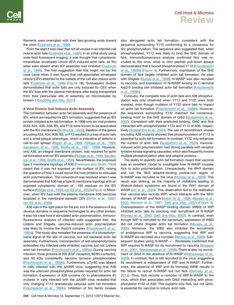
1999a). This study also revealed the presence of a phosphotyr-
osine signal at the site of vaccinia, but not bacterial actin tail
assembly. Furthermore, microinjection of anti-phosphotyrosine
antibodies into infected cells inhibited vaccinia but not Listeria
actin tail formation (Frischknecht et al., 1999a). During vaccinia
infection, three proteins at 200 (EGF-receptor), 80/85 (cortactin),
and 50 kDa consistently become tyrosine phosphorylated
(Frischknecht et al., 1999b). Furthermore, pTyr50 was absent
in cells infected with the DA36R virus, suggesting that A36
was the unknown phosphorylated protein required for actin tail
formation. Expression of A36 tyrosine (Y) to phenylalanine (F)
mutants in cells infected with the DA36R virus revealed that
only changing Y112 dramatically reduced actin tail formation
(Frischknecht et al., 1999b). Inhibition of Src family kinases
248 Cell Host & Microbe 14, September 11, 2013 ª2013 Elsevier Inc
also abrogated actin tail formation, consistent with the
sequence surrounding Y112 conforming to a consensus for
Src phosphorylation. The sequence also suggested that, when
phosphorylated, Y112 was likely to bind the SH2 domain of
Nck. Immunofluorescence analysis confirmed Nck was re-
cruited to the virus, while in vitro peptide pull-down assays
demonstrated that it bound phosphorylated Y112 (Frischknecht
et al., 1999b) (Figure 4). Furthermore, expression of the SH2
domain of Nck largely inhibited actin tail formation. As seen
with Shigella (Suzuki et al., 1998), N-WASP was also recruited
to vaccinia, and expression of N-WASP lacking its C-terminal
Arp2/3 binding site inhibited actin tail formation (Frischknecht
et al., 1999b).
Curiously, the complete loss of actin tails and A36 phosphor-
ylation was only observed when Y112 and Y132 were both
mutated, even though mutation of Y132 alone had no impact
on actin tail formation (Frischknecht et al., 1999b). However,
the sequences surrounding Y132 matched the consensus-
binding motif for the SH2 domain of Grb2 (Scaplehorn et al.,
2002). Consistent with their predicted binding, Grb2 and Nck
interacted with phosphorylated Y132 and Y112 of A36, respec-
tively (Scaplehorn et al., 2002). The use of recombinant viruses
encoding A36 mutants showed that phosphorylation of Y112 is
essential for actin tail formation. In contrast, Y132 only enhances
the number of actin tails (Scaplehorn et al., 2002). Vaccinia-
induced actin polymerization had strong parallels with receptor
tyrosine kinase signaling cascades, which also frequently involve
multiple phosphorylation sites and adaptor proteins.
The ability to quantify actin tail formation meant that vaccinia
was an excellent model to investigate how N-WASP couples
Nck to actin polymerization. Unexpectedly, the WH1 domain
and not the SH3 adaptor-binding proline-rich region of
N-WASP was recruited to the virus (Moreau et al., 2000). This
result was striking, as the majority of mutations leading to
Wiskott-Aldrich syndrome are found in the WH1 domain of
WASP (Jin et al., 2004). This observation led to the realization
that vaccinia also recruits WIP, which interacts with the WH1
domain of WASP and Nck (Anton et al., 1998; Moreau et al.,
2000; Ramesh et al., 1997; Zettl and Way, 2002) (Figure 4).
Overexpression of the WASP binding domain (WBD) of WIP
inhibited actin tails by blocking viral recruitment of N-WASP
(Moreau et al., 2000; Zettl and Way, 2002). In contrast, even
though WIP is recruited to the bacterium, expression of WBD
did not inhibit Shigella actin tail formation (Moreau et al.,
2000). Moreover, the WBD also inhibited the recruitment
of endogenous WIP to vaccinia, suggesting that WIP and
N-WASP are recruited as a complex (Moreau et al., 2000). Sub-
sequent studies using N-WASP�/� fibroblasts confirmed that
WIP requires N-WASP for its recruitment to vaccinia (Snapper
et al., 2001; Weisswange et al., 2009). There is also no recruit-
ment of Grb2 in the absence of N-WASP (Weisswange et al.,
2009). In contrast, Nck is still recruited to the virus, suggesting
its recruitment is independent of WIP and N-WASP. Further-
more, the absence of WIP and its homolog WIRE results in
the failure to recruit N-WASP but not Nck (Donnelly et al.,
2013). Thus, Nck recruits a complex of WIP:N-WASP to the
virus, which then associates with Grb2 interacting with phos-
phorylated Y132 of A36. This explains why Nck, but not Grb2,
is essential for vaccinia to induce actin tails.
.
Cell Host & Microbe
Review
Molecular Dissection of the Vaccinia Signaling NetworkUnraveling how a signaling cascade stimulates actin poly-
merization requires detailed knowledge of the interactions,
dynamics, and stoichiometry of the proteins in the network.
Unfortunately, many signaling networks controlling actin poly-
merization are not amenable to such quantitative analyses, as
their components and/or activation are often transient and
dispersed. In contrast, the signaling pathway used by vaccinia
to induce actin polymerization is localized and sustained. Taking
advantage of this, fluorescence recovery after photobleaching
(FRAP) was used to analyze the dynamics of GFP-tagged Nck,
Grb2, WIP, and N-WASP during vaccinia actin-based motility
(Weisswange et al., 2009). All four proteins undergo rapid ex-
change, with a half-time of recovery of 0.14, 0.8, 0.8, and 2.7 s
for Grb2, Nck, WIP, and N-WASP, respectively. The turnover
of Nck, WIP, and N-WASP increases significantly in the absence
of Grb2, demonstrating that Grb2 enhances vaccinia actin tail
formation by stabilizing the signaling complex. Surprisingly,
although Nck and WIP are responsible for N-WASP recruitment,
N-WASP exchanges �3.5 times slower than either of these pro-
teins. N-WASP turnover also did not occur in the absence of
Arp2/3 recruitment, suggesting that active actin polymerization
promotes exchange of the vaccinia signaling complex. Con-
sistent with this, the turnover rate of N-WASP depends on its
interaction with both Grb2 and the growing plus ends of actin
filaments. Loss of either of these interactions leads to a faster
rate of N-WASP exchange and virus movement. This suggests
that N-WASP not only activates the Arp2/3 complex, but also
modulates the rate of actin-based motility by regulating the
extent of actin polymerization, possibly by antagonizing filament
capping.
Nck and N-WASP play a key role in connecting phos-
photyrosine-based signaling to Arp2/3-mediated actin polymer-
ization during a wide variety of cellular processes, in addition to
driving actin-based motility of intracellular pathogens (Campel-
lone and Welch, 2010; Dodding and Way, 2009; Lommel et al.,
2001; Rotty et al., 2013; Snapper et al., 2001; Weisswange
et al., 2009). What is less clear is the precise role played by
WIP within Nck and N-WASP signaling networks. WIP inhibits
the ability of N-WASP to activate the Arp2/3 complex until
N-WASP receives the right signaling input (Ho et al., 2004;
Martinez-Quiles et al., 2001; Takano et al., 2008). Recent anal-
ysis using MEFs lacking WIP, which have also been treated
with RNAi against the WIP homolog WIRE, demonstrates that
an interaction of WIP with the second SH3 domain of Nck is
essential for vaccinia actin tail formation, as this interaction is
required to recruit the WIP:N-WASP complex (Donnelly et al.,
2013). Furthermore, the recruitment of N-WASP depends on
its interaction with WIP rather than Nck. Finally, the first and third
SH3 domains of Nck are not involved in recruiting the WIP:N-
WASP complex but are essential to stimulate actin assembly.
Vaccinia has thus provided essential insights into the connectiv-
ity within this important signaling network (Donnelly et al., 2013).
WBD overexpression studies and infection of WIP�/� cells sug-
gest that WIP is not required for Shigella actin tail formation
(Garber et al., 2012; Moreau et al., 2000). However, it remains
to be determined whether Shigella still can recruit N-WASP
and induce actin polymerization in the absence of both WIP
and WIRE.
Ce
Role of Src and Abl Kinases in Vaccinia ActinPolymerization and SpreadA36 only promotes actin tail formation beneath CEV, although it
is exposed on the surface of IEV. The reason is that Src activation
and phosphorylation of A36 only occurs after the virus fuses with
the plasma membrane (Newsome et al., 2004). This suggested
that one or more of the four integral viral membrane proteins
(A33, A34, A56, and B5) on the surface of CEV induces an
outside-in signal to activate Src. The molecular mechanism for
this still remains to be established, but the SCR4 domain of B5
is required to activate Src, phosphorylate A36, and induce actin
tail formation (Newsome et al., 2004). Phosphorylation of A36
also promotes the release of kinesin-1 after the virus fuses with
the plasma membrane.
Src is the prototypic member of a family of nonreceptor tyro-
sine kinases that play redundant roles in regulating a wide variety
of cellular processes. It was not surprising, then, that vaccinia
recruits multiple Src family kinases (Src, Fyn, and Yes) as well
as the related Abl family kinases (Abl and Arg) to promote actin
tail formation (Newsome et al., 2006; Reeves et al., 2005). While
additional tyrosine kinases may also phosphorylate A36, inhibi-
tion of both Src and Abl family kinases is sufficient to inhibit
vaccinia actin tail formation (Reeves et al., 2005). Consistent
with this, in vitro kinase assays demonstrate that Abl, Arg, Fyn,
Src, and Yes can phosphorylate Y112 of A36 (Newsome et al.,
2006). Interestingly, Abl and Arg but not Src family kinases are
also required to promote the release of CEV from infected cells
(Reeves et al., 2005). Moreover, treatment of infected mice
with Gleevec/STI-571/Imatinib, an Abl family kinase inhibitor
used to treat chronic myelogenous leukemia, reduces viral
spread and promotes survival from an otherwise lethal infection
(Reeves et al., 2005, 2011).
In addition to the roles for Src and Abl kinases described
above, recent work has begun to uncover additional potential
effects of these key kinases in actin-based motility and infection.
More than 30 years ago, Payne and Kristensson (1982) demon-
strated that inhibition of actin polymerization blocked release of
vaccinia from infected cells (Payne and Kristensson, 1982).
Recently, studies have provided some molecular insights into
this old observation (Horsington et al., 2013). The ability of Abl
to promote virus release is independent of its ability to phosphor-
ylate A36. Nevertheless, A36 phosphorylation and actin
polymerization are required to drive CEV out of plasma mem-
brane invaginations. In the absence of actin polymerization,
CEV remain trapped within these invaginations and are not
released unless the functionality of A34 or B5 is compromised.
Consistent with this, structured illumination microscopy reveals
that induction of actin polymerization polarizes A36 on the virus
(Horsington et al., 2013). Actin is not the only cellular factor
contributing to A36 polarization. Following their fusion with the
plasma membrane, but before actin tail formation, vaccinia re-
cruits clathrin in an AP-2-dependent fashion (Humphries et al.,
2012). The clathrin is, however, left behind when CEV stimulate
actin polymerization. Nevertheless, in the absence of clathrin
recruitment, it takes longer for the virus to induce actin polymer-
ization, and fewer actin tails are formed. Clathrin appears to have
an organizational role, promoting clustering of A36, which in
turn helps polarize and stabilize N-WASP, making initiation of
actin-based motility easier (Humphries et al., 2012).
ll Host & Microbe 14, September 11, 2013 ª2013 Elsevier Inc. 249

Cell Host & Microbe
Review
The activity of CK2 also enhances vaccinia actin tail formation
and cell-to-cell spread (Alvarez and Agaisse, 2012). Loss of CK2
does not affect CEV formation, but does reduce the ability to
recruit and activate Src. While the localization of CK2 during
infection and the molecular basis for these observations remain
to be established, it is curious that A36, which is heavily serine
phosphorylated, contains several predicted CK2 phosphoryla-
tion sites (Alvarez and Agaisse, 2012; Wolffe et al., 2001). It is
not just kinases that impact viral spread, however. The phos-
phoinositide 5-phosphatase SHIP2 acts as a negative regulator
of viral release, although it is not required for actin tail formation
(McNulty et al., 2011). The basis of this regulation remains to be
established. Interestingly, SHIP2 recruitment to actin tails is
dependent on its SH2 domain and N-WASP. Future analysis
will confirm whether the activity of SHIP2 is related to the recent
observations of Horsington et al. (2013).
Actin tail formation enhances the cell-to-cell spread of
vaccinia. It had been unclear, however, how the virus can spread
faster in a cell monolayer than its replication cycle would allow.
Live-cell imaging of viral spread during plaque formation has
now resolved this mystery (Doceul et al., 2010). If a virus (CEV
or EEV) lands on a recently infected neighboring cell that lacks
a virus factory, it forms a new actin tail without being internalized
(Figure 1B). Motility then drives the virus across the surface of the
infected cell and onto adjacent noninfected cells, where uptake
occurs. In this way, vaccinia ignores already-infected cells,
enhancing the rate of spread through the cell monolayer. This
‘‘super repulsion’’ is mediated by A33 and A36 on the surface
of recently infected cells. The SCR4 domain of B5 is also
required (Doceul et al., 2012). It remains to be established
whether ‘‘super repulsion’’ depends on Src- and Abl-mediated
phosphorylation of A36 and its downstream signaling network.
The striking similarities between these two actin-dependent
events, however, would suggest this is almost certainly the case.
A Common Mechanism to Promote the Spreadof Poxvirus InfectionActin-driven cell-to-cell spread of orthopoxviruses is likely to be
common, as A36 is highly conserved (http://www.poxvirus.org).
Consistent with this notion, variola and monkeypox viruses
induce Abl and Src family kinase-dependent actin tails, while
the A36 homolog of ectromelia virus, the causative agent of
mousepox, is required for viral spread and actin tail formation
(Lynn et al., 2012; Reeves et al., 2011). Actin tail formation is,
however, not restricted to orthopoxviruses, as Yaba-like disease
virus (YLDV; yatapoxvirus) and myxoma (leporipoxvirus) also
induce actin tails (Duteyrat et al., 2006; Law et al., 2004), even
though they lack an obvious A36 ortholog. Using a complemen-
tation approach, YL126 of YLDVwas found to promote Nck- and
N-WASP-dependent actin polymerization despite having less
than 15% sequence identity with vaccinia A36 (Dodding and
Way, 2009). Five phosphorylated tyrosines in YL126 can recruit
Nck to promote actin polymerization. However, YL126-mediated
actin tail formation, like that of A36, is also enhanced by the
recruitment of Grb2 by a single phosphorylated tyrosine. Highly
divergent YL126 orthologs in other vertebrate poxviruses, with
as little as 6% homology to each other, can also induce Nck-
and N-WASP-dependent actin polymerization. Actin-based
motility thus appears to be a commonmechanism used by verte-
250 Cell Host & Microbe 14, September 11, 2013 ª2013 Elsevier Inc
brate poxviruses (Chordopoxviridae) to enhance cell-to-cell
spread (Dodding and Way, 2009).
Baculoviruses Use the Arp2/3 Pathway for Motility andNuclear Actin AssemblyPoxviruses are not the only viruses to hijack the Arp2/3 complex
to promote actin polymerization. More than 20 years ago, it was
observed that the baculovirus Autographa californica multiple
nucleopolyhedrovirus (AcMNPV) induces thick actin cables in
the cytoplasm of its host (Charlton and Volkman, 1991). These
actin structures appear from 30min postinfection and are tipped
by a single nucleocapsid, suggesting the virus induces actin
polymerization (Charlton and Volkman, 1993). Intriguingly,
AcMNPV also stimulates the assembly of actin filaments in the
nucleus (Charlton and Volkman, 1991, 1993; Volkman et al.,
1992), a phenomenon that is essential for nucleocapsid assem-
bly and progeny virus production (Hess et al., 1989; Ohkawa and
Volkman, 1999; Volkman, 1988; Volkman et al., 1987, 1992).
Subsequent in vitro assays demonstrated that purified AcMNPV
nucleocapsids are capable of weakly stimulating actin polymer-
ization (Lanier and Volkman, 1998). Furthermore, two viral capsid
proteins, p39 and p78/83, were found to bind directly to actin.
Examining the sequence of p78/83 suggested that it might repre-
sent a viral WASP-like protein, as it contained a proline-rich
region, as well as a WCA motif that is indicative of G-actin and
Arp2/3 binding (Machesky et al., 2001) (Figure 4).
Fifteen years after the discovery of baculovirus interactions
with actin, p78/83 was shown to stimulate Arp2/3-dependent
actin polymerization in vitro. Recent live-cell imaging shows
that p78/83-mediated Arp2/3-induced actin polymerization pro-
pels AcMNPV nucleocapsids throughout the cytoplasm at rates
of 7–22 mm/min as early as 5 min postinfection (Ohkawa et al.,
2010). Actin-based motility promotes nuclear collisions, a pro-
cess that enables rapid nucleocapsid transit into the nucleus
to speed the onset of early gene expression. Interestingly,
once early gene expression is established, but before new viral
progeny are produced, remaining cytoplasmic nucleocapsids
accumulate in actin-rich protrusions at the cell surface, presum-
ably ready to bud and spread to neighboring cells. This behavior
may allow the virus to spread to neighboring cells before the
initially infected cell is removed by apoptosis and sloughing,
allowing the virus to establish an infection and enhancing its
rapid dissemination (Ohkawa et al., 2010).
Interestingly, p78/83 and Arp2/3-dependent actin polymeriza-
tion is also required later in infection for nuclear actin assembly
(Goley et al., 2006). The role of nuclear actin remains unclear,
however, and may include virus assembly, nuclear egress, and
nuclear envelope remodeling during viral envelopment. Actin
also plays a role in the nucleus of uninfected cells, yet the precise
form and function of nuclear actin remains unclear (Weston et al.,
2012). Baculoviruses represent an emerging system for studying
how pathogens hijack actin in the nucleus and for studying the
normal nuclear roles of actin.
What Can the Study of Pathogen Actin Assembly StillTell Us?Three decades after the discovery that Listeria and Shigella use
actin-basedmotility to promote their cell-to-cell spread, we have
achieved an understanding of how this process works at the
.

Cell Host & Microbe
Review
molecular level. Key proteins involved in Arp2/3-dependent
motility have been identified, and reconstitution of the process
has been achieved in vitro using purified proteins. These studies
have revolutionized our understanding of how pathogens exploit
actin and have revealed essential molecular pathways involved
in actin regulation in host cells.
Nevertheless, much still remains to be discovered.With regard
to Listeria and Shigella, we do not yet know at a biophysical level
how Arp2/3-dependent actin polymerization is coupled to force
generation to drive motility. Reconstitution of bacterial motility
represents an experimentally accessible system to address
this question, and what is learned will also apply to force-gener-
ating mechanisms involved in host cellular and intracellular
motility. We do not understand how host motility is coupled
with cell-to-cell spread, whatmembrane trafficking factorsmight
be important for this process, or how membrane-cytoskeleton
linkers like Ezrin and CD44 promote cell-to-cell spread (Pust
et al., 2005). Moreover, we do not know how the ability to recruit
Arp2/3 and actin plays other roles during infection, for example,
in avoidance of autophagy. The information gained from
studying Listeria and Shigella motility and cell-to-cell spread
will continue to be of major importance in uncovering basic cell
biological principles related to cytoskeletal function and
regulation. It is also important to note that other bacteria, for
example, Rickettsia and Burkholderia spp., are likely to exploit
distinct host actin polymerization pathways involving formins
or tandem-monomer-binding nucleators (Haglund and Welch,
2011). Thus, the study of how evolutionarily diverse bacterial
pathogens usurp actin will undoubtedly shed light on the
function and regulation of all three major host actin assembly
pathways.
Vaccinia virus has also been a powerful model, in particular for
understanding how a signaling network activated by Src and Abl
family kinases functions to stimulate actin polymerization.
However, as with bacterial pathogens, many outstanding ques-
tions remain. Vaccinia-induced actin polymerization is depen-
dent on the activation of Src and Abl family kinases (Frischknecht
et al., 1999b; Newsome et al., 2004, 2006; Reeves et al., 2005),
but the molecular basis of how CEV activate Src and Abl family
kinases remains to be determined. The temporal aspects of
vaccinia-induced kinase activation as the virus fuses with the
plasma membrane (or lands on another cell during ‘‘super repul-
sion’’) and its relationship to and/or role in the release of kinesin-
1 and recruitment of clathrin also need to be established. The
virus is amenable to live imaging, including FRAP and FRET-
based approaches, to address these important questions and
also provides a great system for the development of new sensors
tomonitor the activity and interactions of these proteins together
with components in the vaccinia-signaling cascade.
We also still lack a detailed understanding of how the initial
level of A36 tyrosine phosphorylation determines the final output
of the vaccinia-signaling network. Addressing this question is not
easy for most phosphorylation-based signaling networks. How-
ever, the ability to manipulate the number of phosphorylation-
competent A36 molecules beneath CEV (Humphries et al.,
2012), combined with quantitative live imaging (Weisswange
et al., 2009), provides a unique opportunity to determine how
actin-basedmotility and dynamics of a signaling network change
in response to the level of tyrosine phosphorylation. Within the
Ce
signaling network itself, we lack such basic information as the
stoichiometry of components and how many A36 molecules
are actually recruiting Nck. Does the slow exchange rate of
N-WASP compared to Nck and WIP mean there is an additional
binding partner in the system? We need to understand the exact
sequence of events in the Nck-mediated recruitment and activa-
tion of the WIP:N-WASP complex, ideally at the single-molecule
level. This will allow us to address such fundamental questions
as whether each N-WASP molecule activates single or multiple
Arp2/3 complexes before it dissociates from the virus. We
currently have no information on the number and organization
of actin filaments in the tail and why loss of clathrin recruitment
results in the slower disassembly of the tail.
Many of the imaging approaches and quantitative aspects that
are possible with vaccinia and other intracellular pathogens
would be difficult, if not impossible, in other systems. Further-
more, the quantitative data derived from pathogens can be
used to develop mathematical models that will provide addi-
tional molecular insights into the emergent properties and
cooperative nature of signaling networks and how they regulate
Arp2/3-dependent actin polymerization. The study of pathogens
also has the potential to reveal insights into new functions for
actin. One uncharted frontier in actin relates to its function in
the nucleus. Nuclear actin is thought to participate in RNA
biology, chromatin remodeling, nuclear shape, and cell differen-
tiation (Weston et al., 2012). However, the regulation, state,
dynamics, and roles of nuclear actin are poorly understood.
Because of their ability to polymerize and harness actin in the
nucleus, baculoviruses represent an outstanding model for
understanding the regulation and function of nuclear actin.
Finally, it should not be forgotten that pathogens cause a wide
range of diseases that can have serious social and economic
consequences. Understanding exactly how pathogens subvert
cell signaling and the host actin cytoskeleton thus offers the
potential to identify new therapeutic drug targets to combat
infection that in the case of bacteria are independent of any
acquired antibiotic resistance.
SUPPLEMENTAL INFORMATION
Supplemental Information includes two movies and can be found with thisarticle online at http://dx.doi.org/10.1016/j.chom.2013.08.011.
ACKNOWLEDGMENTS
Wewould like to thank David Barry (Way lab) for the vaccinia movie and AshleyHumphries (Way lab) for the vaccinia-infected cell image; Theresia Stradal(University of Munster, Germany) and Klemens Rottner (Bonn University,Germany) for images of Shigella-, EPEC-, and EHEC-infected cells; TaroOhkawa (University of California, Berkeley) for the image of a baculovirus-infected cell; and Rebecca Lamason (University of California, Berkeley) forthe image of a Rickettsia-infected cell. We also thank Klemens Rottner,Jasmine Abella (Way lab), and Taro Ohkawa (Welch lab) for comments onthe text. M.D.W. is supported by NIH grants R01 GM059609 and R01AI074760, and M.W. is supported by Cancer Research UK.
REFERENCES
Akin, O., and Mullins, R.D. (2008). Capping protein increases the rate of actin-based motility by promoting filament nucleation by the Arp2/3 complex. Cell133, 841–851.
Aktories, K. (2011). Bacterial protein toxins that modify host regulatoryGTPases. Nat. Rev. Microbiol. 9, 487–498.
ll Host & Microbe 14, September 11, 2013 ª2013 Elsevier Inc. 251

Cell Host & Microbe
Review
Aktories, K., Lang, A.E., Schwan, C., and Mannherz, H.G. (2011). Actin astarget for modification by bacterial protein toxins. FEBS J. 278, 4526–4543.
Alvarez, D.E., and Agaisse, H. (2012). Casein kinase 2 regulates vaccinia virusactin tail formation. Virology 423, 143–151.
Anton, I.M., Lu, W., Mayer, B.J., Ramesh, N., and Geha, R.S. (1998). TheWiskott-Aldrich syndrome protein-interacting protein (WIP) binds to theadaptor protein Nck. J. Biol. Chem. 273, 20992–20995.
Auerbuch, V., Loureiro, J.J., Gertler, F.B., Theriot, J.A., and Portnoy, D.A.(2003). Ena/VASP proteins contribute to Listeria monocytogenes pathogen-esis by controlling temporal and spatial persistence of bacterial actin-basedmotility. Mol. Microbiol. 49, 1361–1375.
Bernardini, M.L.,Mounier, J., d’Hauteville, H., Coquis-Rondon, M., and Sanso-netti, P.J. (1989). Identification of icsA, a plasmid locus of Shigella flexneri thatgoverns bacterial intra- and intercellular spread through interaction withF-actin. Proc. Natl. Acad. Sci. USA 86, 3867–3871.
Breitsprecher, D., Kiesewetter, A.K., Linkner, J., Vinzenz, M., Stradal, T.E.,Small, J.V., Curth, U., Dickinson, R.B., and Faix, J. (2011). Molecular mecha-nism of Ena/VASP-mediated actin-filament elongation. EMBO J. 30, 456–467.
Burton, E.A., Oliver, T.N., and Pendergast, A.M. (2005). Abl kinases regulateactin comet tail elongation via an N-WASP-dependent pathway. Mol. Cell.Biol. 25, 8834–8843.
Cameron, L.A., Footer, M.J., van Oudenaarden, A., and Theriot, J.A. (1999).Motility of ActA protein-coated microspheres driven by actin polymerization.Proc. Natl. Acad. Sci. USA 96, 4908–4913.
Cameron, L.A., Svitkina, T.M., Vignjevic, D., Theriot, J.A., and Borisy, G.G.(2001). Dendritic organization of actin comet tails. Curr. Biol. 11, 130–135.
Campellone, K.G., and Welch, M.D. (2010). A nucleator arms race: cellularcontrol of actin assembly. Nat. Rev. Mol. Cell Biol. 11, 237–251.
Carabeo, R. (2011). Bacterial subversion of host actin dynamics at the plasmamembrane. Cell. Microbiol. 13, 1460–1469.
Carlier, M.F., Laurent, V., Santolini, J., Melki, R., Didry, D., Xia, G.X., Hong, Y.,Chua, N.H., and Pantaloni, D. (1997). Actin depolymerizing factor (ADF/cofilin)enhances the rate of filament turnover: implication in actin-based motility.J. Cell Biol. 136, 1307–1322.
Charlton, C.A., and Volkman, L.E. (1991). Sequential rearrangement andnuclear polymerization of actin in baculovirus-infected Spodoptera frugiperdacells. J. Virol. 65, 1219–1227.
Charlton, C.A., and Volkman, L.E. (1993). Penetration of Autographa californ-ica nuclear polyhedrosis virus nucleocapsids into IPLB Sf 21 cells inducesactin cable formation. Virology 197, 245–254.
Chesarone, M.A., DuPage, A.G., and Goode, B.L. (2010). Unleashing forminsto remodel the actin and microtubule cytoskeletons. Nat. Rev. Mol. Cell Biol.11, 62–74.
Chong, R., Swiss, R., Briones, G., Stone, K.L., Gulcicek, E.E., and Agaisse, H.(2009). Regulatory mimicry in Listeria monocytogenes actin-based motility.Cell Host Microbe 6, 268–278.
Cudmore, S., Cossart, P., Griffiths, G., and Way, M. (1995). Actin-basedmotility of vaccinia virus. Nature 378, 636–638.
Cudmore, S., Reckmann, I., Griffiths, G., and Way, M. (1996). Vaccinia virus: amodel system for actin-membrane interactions. J. Cell Sci. 109, 1739–1747.
Dabiri, G.A., Sanger, J.M., Portnoy, D.A., and Southwick, F.S. (1990). Listeriamonocytogenes moves rapidly through the host-cell cytoplasm by inducingdirectional actin assembly. Proc. Natl. Acad. Sci. USA 87, 6068–6072.
Doceul, V., Hollinshead, M., van der Linden, L., and Smith, G.L. (2010). Repul-sion of superinfecting virions: a mechanism for rapid virus spread. Science327, 873–876.
Doceul, V., Hollinshead,M., Breiman, A., Laval, K., and Smith, G.L. (2012). Pro-tein B5 is required on extracellular enveloped vaccinia virus for repulsion ofsuperinfecting virions. J. Gen. Virol. 93, 1876–1886.
Dodding, M.P., and Way, M. (2009). Nck- and N-WASP-dependent actin-based motility is conserved in divergent vertebrate poxviruses. Cell HostMicrobe 6, 536–550.
252 Cell Host & Microbe 14, September 11, 2013 ª2013 Elsevier Inc
Dodding, M.P., and Way, M. (2011). Coupling viruses to dynein and kinesin-1.EMBO J. 30, 3527–3539.
Donnelly, S.K., Weisswange, I., Zettl, M., and Way, M. (2013). WIP provides anessential link between Nck and N-WASP during Arp2/3-dependent actin poly-merization. Curr. Biol. 23, 999–1006.
Dragoi, A.M., Talman, A.M., and Agaisse, H. (2013). Bruton’s tyrosine kinaseregulates Shigella flexneri dissemination in HT-29 intestinal cells. Infect.Immun. 81, 598–607.
Duteyrat, J.L., Gelfi, J., and Bertagnoli, S. (2006). Ultrastructural study ofmyxoma virus morphogenesis. Arch. Virol. 151, 2161–2180.
Ebel, F., Rohde, M., von Eichel-Streiber, C., Wehland, J., and Chakraborty, T.(1999). The actin-based motility of intracellular Listeria monocytogenes is notcontrolled by small GTP-binding proteins of the Rho- and Ras-subfamilies.FEMS Microbiol. Lett. 176, 117–124.
Egile, C., Loisel, T.P., Laurent, V., Li, R., Pantaloni, D., Sansonetti, P.J., andCarlier, M.F. (1999). Activation of the CDC42 effector N-WASP by the Shigellaflexneri IcsA protein promotes actin nucleation by Arp2/3 complex and bacte-rial actin-based motility. J. Cell Biol. 146, 1319–1332.
Frischknecht, F., Cudmore, S., Moreau, V., Reckmann, I., Rottger, S., andWay, M. (1999a). Tyrosine phosphorylation is required for actin-based motilityof vaccinia but not Listeria or Shigella. Curr. Biol. 9, 89–92.
Frischknecht, F., Moreau, V., Rottger, S., Gonfloni, S., Reckmann, I., Superti-Furga, G., and Way, M. (1999b). Actin-based motility of vaccinia virus mimicsreceptor tyrosine kinase signalling. Nature 401, 926–929.
Garber, J.J., Takeshima, F., Anton, I.M., Oyoshi, M.K., Lyubimova, A., Kapoor,A., Shibata, T., Chen, F., Alt, F.W., Geha, R.S., et al. (2012). EnteropathogenicEscherichia coli and vaccinia virus do not require the family of WASP-interacting proteins for pathogen-induced actin assembly. Infect. Immun. 80,4071–4077.
Geese, M., Loureiro, J.J., Bear, J.E., Wehland, J., Gertler, F.B., and Sechi, A.S.(2002). Contribution of Ena/VASP proteins to intracellular motility of listeriarequires phosphorylation and proline-rich core but not F-actin binding ormulti-merization. Mol. Biol. Cell 13, 2383–2396.
Goldberg, M.B., and Theriot, J.A. (1995). Shigella flexneri surface protein IcsAis sufficient to direct actin-basedmotility. Proc. Natl. Acad. Sci. USA 92, 6572–6576.
Goley, E.D., Ohkawa, T., Mancuso, J., Woodruff, J.B., D’Alessio, J.A., Cande,W.Z., Volkman, L.E., and Welch, M.D. (2006). Dynamic nuclear actin assemblyby Arp2/3 complex and a baculovirus WASP-like protein. Science 314,464–467.
Gouin, E., Gantelet, H., Egile, C., Lasa, I., Ohayon, H., Villiers, V., Gounon, P.,Sansonetti, P.J., and Cossart, P. (1999). A comparative study of the actin-based motilities of the pathogenic bacteria Listeria monocytogenes, Shigellaflexneri and Rickettsia conorii. J. Cell Sci. 112, 1697–1708.
Gouin, E., Egile, C., Dehoux, P., Villiers, V., Adams, J., Gertler, F., Li, R., andCossart, P. (2004). The RickA protein of Rickettsia conorii activates theArp2/3 complex. Nature 427, 457–461.
Gouin, E., Welch, M.D., and Cossart, P. (2005). Actin-basedmotility of intracel-lular pathogens. Curr. Opin. Microbiol. 8, 35–45.
Grenklo, S., Geese, M., Lindberg, U., Wehland, J., Karlsson, R., and Sechi,A.S. (2003). A crucial role for profilin-actin in the intracellular motility of Listeriamonocytogenes. EMBO Rep. 4, 523–529.
Haglund, C.M., and Welch, M.D. (2011). Pathogens and polymers: microbe-host interactions illuminate the cytoskeleton. J. Cell Biol. 195, 7–17.
Hansen, S.D., andMullins, R.D. (2010). VASP is a processive actin polymerasethat requires monomeric actin for barbed end association. J. Cell Biol. 191,571–584.
Heinzen, R.A., Hayes, S.F., Peacock, M.G., and Hackstadt, T. (1993). Direc-tional actin polymerization associated with spotted fever group Rickettsiainfection of Vero cells. Infect. Immun. 61, 1926–1935.
Hess, R.T., Goldsmith, P.A., and Volkman, L.E. (1989). Effect of cytochalasin Don cell morphology and AcMNPV replication in a Spodoptera frugiperda cellline. J. Invertebr. Pathol. 53, 169–182.
.

Cell Host & Microbe
Review
Hiller, G., Weber, K., Schneider, L., Parajsz, C., and Jungwirth, C. (1979). Inter-action of assembled progeny pox viruses with the cellular cytoskeleton.Virology 98, 142–153.
Hiller, G., Jungwirth, C., and Weber, K. (1981). Fluorescence microscopicalanalysis of the life cycle of vaccinia virus in chick embryo fibroblasts.Virus-cytoskeleton interactions. Exp. Cell Res. 132, 81–87.
Ho, H.Y., Rohatgi, R., Lebensohn, A.M., Le Ma, Li, J., Gygi, S.P., andKirschner, M.W. (2004). Toca-1 mediates Cdc42-dependent actin nucleationby activating the N-WASP-WIP complex. Cell 118, 203–216.
Horsington, J., Lynn, H., Turnbull, L., Cheng, D., Braet, F., Diefenbach, R.J.,Whitchurch, C.B., Karupiah, G., and Newsome, T.P. (2013). A36-dependentactin filament nucleation promotes release of vaccinia virus. PLoS Pathog.9, e1003239.
Humphries, A.C., Dodding,M.P., Barry, D.J., Collinson, L.M., Durkin, C.H., andWay, M. (2012). Clathrin potentiates vaccinia-induced actin polymerization tofacilitate viral spread. Cell Host Microbe 12, 346–359.
Jeng, R.L., Goley, E.D., D’Alessio, J.A., Chaga, O.Y., Svitkina, T.M., Borisy,G.G., Heinzen, R.A., and Welch, M.D. (2004). A Rickettsia WASP-like proteinactivates the Arp2/3 complex and mediates actin-based motility. Cell.Microbiol. 6, 761–769.
Jin, Y., Mazza, C., Christie, J.R., Giliani, S., Fiorini, M., Mella, P., Gandellini, F.,Stewart, D.M., Zhu, Q., Nelson, D.L., et al. (2004). Mutations of the Wiskott-Aldrich Syndrome Protein (WASP): hotspots, effect on transcription, and trans-lation and phenotype/genotype correlation. Blood 104, 4010–4019.
Kelleher, J.F., Atkinson, S.J., and Pollard, T.D. (1995). Sequences, structuralmodels, and cellular localization of the actin-related proteins Arp2 and Arp3from Acanthamoeba. J. Cell Biol. 131, 385–397.
Kocks, C., Gouin, E., Tabouret, M., Berche, P., Ohayon, H., and Cossart, P.(1992). L. monocytogenes-induced actin assembly requires the actA geneproduct, a surface protein. Cell 68, 521–531.
Kocks, C., Marchand, J.B., Gouin, E., d’Hauteville, H., Sansonetti, P.J., Carlier,M.F., and Cossart, P. (1995). The unrelated surface proteins ActA of Listeriamonocytogenes and IcsA of Shigella flexneri are sufficient to confer actin-based motility on Listeria innocua and Escherichia coli respectively. Mol.Microbiol. 18, 413–423.
Krempien, U., Schneider, L., Hiller, G., Weber, K., Katz, E., and Jungwirth, C.(1981). Conditions for pox virus-specific microvilli formation studied duringsynchronized virus assembly. Virology 113, 556–564.
Lanier, L.M., and Volkman, L.E. (1998). Actin binding and nucleation byAutographa california M nucleopolyhedrovirus. Virology 243, 167–177.
Lasa, I., David, V., Gouin, E., Marchand, J.B., and Cossart, P. (1995). Theamino-terminal part of ActA is critical for the actin-based motility of Listeriamonocytogenes; the central proline-rich region acts as a stimulator. Mol.Microbiol. 18, 425–436.
Law, M., Hollinshead, M., Lee, H.J., and Smith, G.L. (2004). Yaba-like diseasevirus protein Y144R, a member of the complement control protein family, ispresent on enveloped virions that are associated with virus-induced actin tails.J. Gen. Virol. 85, 1279–1290.
Lett, M.C., Sasakawa, C., Okada, N., Sakai, T., Makino, S., Yamada, M.,Komatsu, K., and Yoshikawa, M. (1989). virG, a plasmid-coded virulencegene of Shigella flexneri: identification of the virG protein and determinationof the complete coding sequence. J. Bacteriol. 171, 353–359.
Leung, Y., Ally, S., and Goldberg, M.B. (2008). Bacterial actin assemblyrequires toca-1 to relieve N-wasp autoinhibition. Cell Host Microbe 3, 39–47.
Loisel, T.P., Boujemaa, R., Pantaloni, D., and Carlier, M.F. (1999). Reconstitu-tion of actin-based motility of Listeria and Shigella using pure proteins. Nature401, 613–616.
Lommel, S., Benesch, S., Rottner, K., Franz, T., Wehland, J., and Kuhn, R.(2001). Actin pedestal formation by enteropathogenic Escherichia coli andintracellular motility of Shigella flexneri are abolished in N-WASP-defectivecells. EMBO Rep. 2, 850–857.
Lynn, H., Horsington, J., Ter, L.K., Han, S., Chew, Y.L., Diefenbach, R.J., Way,M., Chaudhri, G., Karupiah, G., and Newsome, T.P. (2012). Loss of cytoskel-etal transport during egress critically attenuates ectromelia virus infectionin vivo. J. Virol. 86, 7427–7443.
Ce
Machesky, L.M., Atkinson, S.J., Ampe, C., Vandekerckhove, J., and Pollard,T.D. (1994). Purification of a cortical complex containing two unconventionalactins from Acanthamoeba by affinity chromatography on profilin-agarose.J. Cell Biol. 127, 107–115.
Machesky, L.M., Insall, R.H., and Volkman, L.E. (2001). WASP homologysequences in baculoviruses. Trends Cell Biol. 11, 286–287.
Makino, S., Sasakawa, C., Kamata, K., Kurata, T., and Yoshikawa, M. (1986). Agenetic determinant required for continuous reinfection of adjacent cells onlarge plasmid in S. flexneri 2a. Cell 46, 551–555.
Marchand, J.B., Moreau, P., Paoletti, A., Cossart, P., Carlier, M.F., and Panta-loni, D. (1995). Actin-based movement of Listeria monocytogenes: actinassembly results from the local maintenance of uncapped filament barbedends at the bacterium surface. J. Cell Biol. 130, 331–343.
Martinez-Quiles, N., Rohatgi, R., Anton, I.M.,Medina, M., Saville, S.P., Miki, H.,Yamaguchi, H., Takenawa, T., Hartwig, J.H., Geha, R.S., and Ramesh, N.(2001). WIP regulates N-WASP-mediated actin polymerization and filopodiumformation. Nat. Cell Biol. 3, 484–491.
May, R.C., Hall, M.E., Higgs, H.N., Pollard, T.D., Chakraborty, T., Wehland, J.,Machesky, L.M., and Sechi, A.S. (1999). The Arp2/3 complex is essential forthe actin-based motility of Listeria monocytogenes. Curr. Biol. 9, 759–762.
McNulty, S., Powell, K., Erneux, C., and Kalman, D. (2011). The host phosphoi-nositide 5-phosphatase SHIP2 regulates dissemination of vaccinia virus.J. Virol. 85, 7402–7410.
Mercer, J., and Helenius, A. (2008). Vaccinia virus uses macropinocytosis andapoptotic mimicry to enter host cells. Science 320, 531–535.
Monack, D.M., and Theriot, J.A. (2001). Actin-based motility is sufficient forbacterial membrane protrusion formation and host cell uptake. Cell. Microbiol.3, 633–647.
Moreau, V., Frischknecht, F., Reckmann, I., Vincentelli, R., Rabut, G., Stewart,D., and Way, M. (2000). A complex of N-WASP and WIP integrates signallingcascades that lead to actin polymerization. Nat. Cell Biol. 2, 441–448.
Mostowy, S., Bonazzi, M., Hamon, M.A., Tham, T.N., Mallet, A., Lelek, M.,Gouin, E., Demangel, C., Brosch, R., Zimmer, C., et al. (2010). Entrapmentof intracytosolic bacteria by septin cage-like structures. Cell Host Microbe 8,433–444.
Mostowy, S., Sancho-Shimizu, V., Hamon, M.A., Simeone, R., Brosch, R., Jo-hansen, T., and Cossart, P. (2011). p62 and NDP52 proteins target intracyto-solic Shigella and Listeria to different autophagy pathways. J. Biol. Chem.286, 26987–26995.
Mounier, J., Ryter, A., Coquis-Rondon, M., and Sansonetti, P.J. (1990). Intra-cellular and cell-to-cell spread of Listeria monocytogenes involves interactionwith F-actin in the enterocytelike cell line Caco-2. Infect. Immun. 58, 1048–1058.
Newsome, T.P., Scaplehorn, N., and Way, M. (2004). SRC mediates a switchfrom microtubule- to actin-based motility of vaccinia virus. Science 306,124–129.
Newsome, T.P., Weisswange, I., Frischknecht, F., and Way, M. (2006). Ablcollaborates with Src family kinases to stimulate actin-based motility ofvaccinia virus. Cell. Microbiol. 8, 233–241.
Niebuhr, K., Ebel, F., Frank, R., Reinhard, M., Domann, E., Carl, U.D., Walter,U., Gertler, F.B., Wehland, J., and Chakraborty, T. (1997). A novel proline-richmotif present in ActA of Listeria monocytogenes and cytoskeletal proteins isthe ligand for the EVH1 domain, a protein module present in the Ena/VASPfamily. EMBO J. 16, 5433–5444.
Ogawa, H., Nakamura, A., and Nakaya, R. (1968). Cinemicrographic study oftissue cell cultures infected with Shigella flexneri. Jpn. J. Med. Sci. Biol. 21,259–273.
Ohkawa, T., and Volkman, L.E. (1999). Nuclear F-actin is required for AcMNPVnucleocapsid morphogenesis. Virology 264, 1–4.
Ohkawa, T., Volkman, L.E., and Welch, M.D. (2010). Actin-based motilitydrives baculovirus transit to the nucleus and cell surface. J. Cell Biol. 190,187–195.
ll Host & Microbe 14, September 11, 2013 ª2013 Elsevier Inc. 253

Cell Host & Microbe
Review
Parkinson, J.E., and Smith, G.L. (1994). Vaccinia virus gene A36R encodes aM(r) 43-50 K protein on the surface of extracellular enveloped virus. Virology204, 376–390.
Payne, L.G., and Kristensson, K. (1982). The effect of cytochalasin D andmonensin on enveloped vaccinia virus release. Arch. Virol. 74, 11–20.
Pust, S., Morrison, H., Wehland, J., Sechi, A.S., and Herrlich, P. (2005). Listeriamonocytogenes exploits ERM protein functions to efficiently spread from cellto cell. EMBO J. 24, 1287–1300.
Qualmann, B., and Kessels, M.M. (2009). New players in actin polymeriza-tion—WH2-domain-containing actin nucleators. Trends Cell Biol. 19, 276–285.
Racz, P., Tenner, K., and Szivessy, K. (1970). Electron microscopic studies inexperimental keratoconjunctivitis listeriosa. I. Penetration of Listeria monocy-togenes into corneal epithelial cells. Acta Microbiol. Acad. Sci. Hung. 17,221–236.
Racz, P., Tenner, K., and Mero, E. (1972). Experimental Listeria enteritis. I. Anelectron microscopic study of the epithelial phase in experimental listeriainfection. Lab. Invest. 26, 694–700.
Ramesh, N., Anton, I.M., Hartwig, J.H., and Geha, R.S. (1997). WIP, a proteinassociated with wiskott-aldrich syndrome protein, induces actin polymeriza-tion and redistribution in lymphoid cells. Proc. Natl. Acad. Sci. USA 94,14671–14676.
Reeves, P.M., Bommarius, B., Lebeis, S., McNulty, S., Christensen, J.,Swimm, A., Chahroudi, A., Chavan, R., Feinberg, M.B., Veach, D., et al.(2005). Disabling poxvirus pathogenesis by inhibition of Abl-family tyrosinekinases. Nat. Med. 11, 731–739.
Reeves, P.M., Smith, S.K., Olson, V.A., Thorne, S.H., Bornmann, W., Damon,I.K., and Kalman, D. (2011). Variola and monkeypox viruses utilize conservedmechanisms of virion motility and release that depend on abl and SRC familytyrosine kinases. J. Virol. 85, 21–31.
Robbins, J.R., Barth, A.I., Marquis, H., de Hostos, E.L., Nelson, W.J., andTheriot, J.A. (1999). Listeria monocytogenes exploits normal host cellprocesses to spread from cell to cell. J. Cell Biol. 146, 1333–1350.
Roberts, K.L., and Smith, G.L. (2008). Vaccinia virus morphogenesis anddissemination. Trends Microbiol. 16, 472–479.
Rohatgi, R., Ma, L., Miki, H., Lopez, M., Kirchhausen, T., Takenawa, T., andKirschner, M.W. (1999). The interaction between N-WASP and the Arp2/3complex links Cdc42-dependent signals to actin assembly. Cell 97, 221–231.
Roper, R.L., Wolffe, E.J., Weisberg, A., and Moss, B. (1998). The envelopeprotein encoded by the A33R gene is required for formation of actin-containingmicrovilli and efficient cell-to-cell spread of vaccinia virus. J. Virol. 72, 4192–4204.
Rosenblatt, J., Agnew, B.J., Abe, H., Bamburg, J.R., and Mitchison, T.J.(1997). Xenopus actin depolymerizing factor/cofilin (XAC) is responsible forthe turnover of actin filaments in Listeria monocytogenes tails. J. Cell Biol.136, 1323–1332.
Rottger, S., Frischknecht, F., Reckmann, I., Smith, G.L., and Way, M. (1999).Interactions between vaccinia virus IEV membrane proteins and their roles inIEV assembly and actin tail formation. J. Virol. 73, 2863–2875.
Rotty, J.D., Wu, C., and Bear, J.E. (2013). New insights into the regulation andcellular functions of the ARP2/3 complex. Nat. Rev. Mol. Cell Biol. 14, 7–12.
Sanderson, C.M., Frischknecht, F., Way, M., Hollinshead, M., and Smith, G.L.(1998). Roles of vaccinia virus EEV-specific proteins in intracellular actin tailformation and low pH-induced cell-cell fusion. J. Gen. Virol. 79, 1415–1425.
Sanger, J.M., Sanger, J.W., and Southwick, F.S. (1992). Host cell actin assem-bly is necessary and likely to provide the propulsive force for intracellularmovement of Listeria monocytogenes. Infect. Immun. 60, 3609–3619.
Scaplehorn, N., Holmstrom, A., Moreau, V., Frischknecht, F., Reckmann, I.,and Way, M. (2002). Grb2 and Nck act cooperatively to promote actin-basedmotility of vaccinia virus. Curr. Biol. 12, 740–745.
Schaechter, M., Bozeman, F.M., and Smadel, J.E. (1957). Study on the growthof Rickettsiae. II. Morphologic observations of living Rickettsiae in tissueculture cells. Virology 3, 160–172.
254 Cell Host & Microbe 14, September 11, 2013 ª2013 Elsevier Inc
Sitthidet, C., Stevens, J.M., Field, T.R., Layton, A.N., Korbsrisate, S., andStevens, M.P. (2010). Actin-based motility of Burkholderia thailandensis re-quires a central acidic domain of BimA that recruits and activates the cellularArp2/3 complex. J. Bacteriol. 192, 5249–5252.
Skoble, J., Portnoy, D.A., and Welch, M.D. (2000). Three regions within ActApromote Arp2/3 complex-mediated actin nucleation and Listeria monocyto-genes motility. J. Cell Biol. 150, 527–538.
Smith, G.A., Portnoy, D.A., and Theriot, J.A. (1995). Asymmetric distribution ofthe Listeria monocytogenes ActA protein is required and sufficient to directactin-based motility. Mol. Microbiol. 17, 945–951.
Smith, G.A., Theriot, J.A., and Portnoy, D.A. (1996). The tandem repeat domainin the Listeria monocytogenes ActA protein controls the rate of actin-basedmotility, the percentage of moving bacteria, and the localization of vasodi-lator-stimulated phosphoprotein and profilin. J. Cell Biol. 135, 647–660.
Smith, G.L., Vanderplasschen, A., and Law,M. (2002). The formation and func-tion of extracellular enveloped vaccinia virus. J. Gen. Virol. 83, 2915–2931.
Snapper, S.B., Takeshima, F., Anton, I., Liu, C.H., Thomas, S.M., Nguyen, D.,Dudley, D., Fraser, H., Purich, D., Lopez-Ilasaca, M., et al. (2001). N-WASPdeficiency reveals distinct pathways for cell surface projections and microbialactin-based motility. Nat. Cell Biol. 3, 897–904.
Stevens, M.P., and Galyov, E.E. (2004). Exploitation of host cells by Burkhol-deria pseudomallei. Int. J. Med. Microbiol. 293, 549–555.
Stokes, G.V. (1976). High-voltage electron microscope study of the release ofvaccinia virus from whole cells. J. Virol. 18, 636–643.
Suzuki, T., Miki, H., Takenawa, T., and Sasakawa, C. (1998). Neural Wiskott-Aldrich syndrome protein is implicated in the actin-based motility of Shigellaflexneri. EMBO J. 17, 2767–2776.
Suzuki, T., Mimuro, H., Suetsugu, S., Miki, H., Takenawa, T., and Sasakawa, C.(2002). Neural Wiskott-Aldrich syndrome protein (N-WASP) is the specificligand for Shigella VirG among the WASP family and determines the host celltype allowing actin-based spreading. Cell. Microbiol. 4, 223–233.
Svitkina, T.M., and Borisy, G.G. (1999). Arp2/3 complex and actin depolyme-rizing factor/cofilin in dendritic organization and treadmilling of actin filamentarray in lamellipodia. J. Cell Biol. 145, 1009–1026.
Takano, K., Toyooka, K., and Suetsugu, S. (2008). EFC/F-BAR proteins andthe N-WASP-WIP complex induce membrane curvature-dependent actinpolymerization. EMBO J. 27, 2817–2828.
Taylor, M.P., Koyuncu, O.O., and Enquist, L.W. (2011). Subversion of the actincytoskeleton during viral infection. Nat. Rev. Microbiol. 9, 427–439.
Theriot, J.A., and Mitchison, T.J. (1991). Actin microfilament dynamics inlocomoting cells. Nature 352, 126–131.
Theriot, J.A., Mitchison, T.J., Tilney, L.G., and Portnoy, D.A. (1992). The rate ofactin-based motility of intracellular Listeria monocytogenes equals the rate ofactin polymerization. Nature 357, 257–260.
Theriot, J.A., Rosenblatt, J., Portnoy, D.A., Goldschmidt-Clermont, P.J., andMitchison, T.J. (1994). Involvement of profilin in the actin-based motility ofL. monocytogenes in cells and in cell-free extracts. Cell 76, 505–517.
Tilney, L.G., and Portnoy, D.A. (1989). Actin filaments and the growth, move-ment, and spread of the intracellular bacterial parasite, Listeria monocyto-genes. J. Cell Biol. 109, 1597–1608.
Tilney, L.G., DeRosier, D.J., and Tilney, M.S. (1992a). How Listeria exploitshost cell actin to form its own cytoskeleton. I. Formation of a tail and howthat tail might be involved in movement. J. Cell Biol. 118, 71–81.
Tilney, L.G., DeRosier, D.J., Weber, A., and Tilney, M.S. (1992b). How Listeriaexploits host cell actin to form its own cytoskeleton. II. Nucleation, actinfilament polarity, filament assembly, and evidence for a pointed end capper.J. Cell Biol. 118, 83–93.
van Eijl, H., Hollinshead, M., and Smith, G.L. (2000). The vaccinia virus A36Rprotein is a type Ib membrane protein present on intracellular but not extracel-lular enveloped virus particles. Virology 271, 26–36.
Vance, R.E., Isberg, R.R., and Portnoy, D.A. (2009). Patterns of pathogenesis:discrimination of pathogenic and nonpathogenic microbes by the innateimmune system. Cell Host Microbe 6, 10–21.
.

Cell Host & Microbe
Review
Volkman, L.E. (1988). Autographa californica MNPV nucleocapsid assembly:inhibition by cytochalasin D. Virology 163, 547–553.
Volkman, L.E., Goldsmith, P.A., and Hess, R.T. (1987). Evidence for microfila-ment involvement in budded Autographa californica nuclear polyhedrosis virusproduction. Virology 156, 32–39.
Volkman, L.E., Talhouk, S.N., Oppenheimer, D.I., and Charlton, C.A. (1992).Nulcear F-actin: a functional component of baculovirus-infected lepidopterancells? J. Cell Sci. 103, 15–22.
Weisswange, I., Newsome, T.P., Schleich, S., and Way, M. (2009). The rate ofN-WASP exchange limits the extent of ARP2/3-complex-dependent actin-based motility. Nature 458, 87–91.
Welch, M.D., Iwamatsu, A., and Mitchison, T.J. (1997). Actin polymerization isinduced by Arp2/3 protein complex at the surface of Listeria monocytogenes.Nature 385, 265–269.
Welch, M.D., Rosenblatt, J., Skoble, J., Portnoy, D.A., and Mitchison, T.J.(1998). Interaction of human Arp2/3 complex and the Listeria monocytogenesActA protein in actin filament nucleation. Science 281, 105–108.
Weston, L., Coutts, A.S., and La Thangue, N.B. (2012). Actin nucleators in thenucleus: an emerging theme. J. Cell Sci. 125, 3519–3527.
Ce
Wolffe, E.J., Katz, E., Weisberg, A., and Moss, B. (1997). The A34R glycopro-tein gene is required for induction of specialized actin-containing microvilli andefficient cell-to-cell transmission of vaccinia virus. J. Virol. 71, 3904–3915.
Wolffe, E.J., Weisberg, A.S., and Moss, B. (1998). Role for the vaccinia virusA36R outer envelope protein in the formation of virus-tipped actin-containingmicrovilli and cell-to-cell virus spread. Virology 244, 20–26.
Wolffe, E.J., Weisberg, A.S., andMoss, B. (2001). The vaccinia virus A33R pro-tein provides a chaperone function for viral membrane localization and tyrosinephosphorylation of the A36R protein. J. Virol. 75, 303–310.
Yarar, D., To, W., Abo, A., and Welch, M.D. (1999). The Wiskott-Aldrichsyndrome protein directs actin-based motility by stimulating actin nucleationwith the Arp2/3 complex. Curr. Biol. 9, 555–558.
Yoshikawa, Y., Ogawa, M., Hain, T., Yoshida, M., Fukumatsu, M., Kim, M.,Mimuro, H., Nakagawa, I., Yanagawa, T., Ishii, T., et al. (2009). Listeriamonocytogenes ActA-mediated escape from autophagic recognition. Nat.Cell Biol. 11, 1233–1240.
Zettl, M., and Way, M. (2002). The WH1 and EVH1 domains of WASP andEna/VASP family members bind distinct sequence motifs. Curr. Biol. 12,1617–1622.
ll Host & Microbe 14, September 11, 2013 ª2013 Elsevier Inc. 255