
Effects of Arp2 and Arp3 nucleotide-binding pocket mutations on Arp2/3 complex function
14
THE JOURNAL OF CELL BIOLOGY © The Rockefeller University Press $8.00 The Journal of Cell Biology, Vol. 168, No. 2, January 17, 2005 315–328 http://www.jcb.org/cgi/doi/10.1083/jcb.200408177 JCB: ARTICLE JCB 315 Effects of Arp2 and Arp3 nucleotide-binding pocket mutations on Arp2/3 complex function Adam C. Martin, 1 Xiao-Ping Xu, 2 Isabelle Rouiller, 2 Marko Kaksonen, 1 Yidi Sun, 1 Lisa Belmont, 3 Niels Volkmann, 2 Dorit Hanein, 2 Matthew Welch, 1 and David G. Drubin 1 1 Barker Hall, Department of Molecular and Cell Biology, University of California, Berkeley, Berkeley, CA 94720 2 The Burnham Institute, La Jolla, CA 92037 3 Cytokinetics, Inc., South San Francisco, CA 94080 ontributions of actin-related proteins (Arp) 2 and 3 nucleotide state to Arp2/3 complex function were tested using nucleotide-binding pocket (NBP) mutants in Saccharomyces cerevisiae. ATP binding by Arp2 and Arp3 was required for full Arp2/3 complex nucleation activity in vitro. Analysis of actin dynamics and endocytosis in mutants demonstrated that nucleotide- bound Arp3 is particularly important for Arp2/3 complex function in vivo. Severity of endocytic defects did not cor- relate with effects on in vitro nucleation activity, suggesting that a critical Arp2/3 complex function during endocytosis C may be structural rather than catalytic. A separate class of Arp2 and Arp3 NBP mutants suppressed phenotypes of mutants defective for actin nucleation. An Arp2 suppressor mutant increased Arp2/3 nucleation activity. Electron microscopy of Arp2/3 complex containing this Arp2 suppressor identified a structural change that also occurs upon Arp2/3 activation by nucleation promoting factors. These data demonstrate the importance of Arp2 and Arp3 nucleotide binding for nucleating activity, and Arp3 nucleotide binding for maintenance of cortical actin cytoskeleton cytoarchitecture. Introduction Spatial and temporal regulation of actin polymerization are criti- cal for cellular processes such as cell migration and endocytosis (Welch and Mullins, 2002; Engqvist-Goldstein and Drubin, 2003). Because nucleation is rate limiting for actin polymeriza- tion, regulation of this step controls when and where actin is assembled in cells. An important nucleator of actin filaments is the Arp2/3 complex, a seven subunit complex that includes two actin-related proteins (Arps) 2 and 3 (Welch and Mullins, 2002). The Arp2/3 complex nucleates actin filaments de novo, and from the sides of preexisting filaments, generating a branched filament array (Blanchoin et al., 2000) resembling those observed at the leading edge of certain motile cells (Svitkina and Borisy, 1999). Nucleation promoting factors (NPFs), such as members of the Wiskott-Aldrich syndrome protein (WASP) family of proteins, activate Arp2/3 complex nucleation activity, allowing regulated actin assembly that can generate forces in cells (Welch and Mul- lins, 2002). Elucidating the mechanisms that regulate Arp2/3 complex function is essential for understanding the dynamics of actin-based force generation. Nucleotide binding plays a critical role in regulation of actin dynamics. ATP binding by actin promotes actin filament assembly by closing the nucleotide-binding pocket (NBP). ATP hydrolysis and P i release results in a transition from the “closed” to the “open” state, promoting actin filament disassembly (Bel- mont et al., 1999a; Sablin et al., 2002). Similarly, Arp2 and Arp3 bind ATP, and the Arp2/3 complex requires ATP for nu- cleation (Dayel et al., 2001; Le Clainche et al., 2001). Arp2 ATP hydrolysis was linked to both the dissociation of actin fila- ment branches (Le Clainche et al., 2003), and to nucleation (Dayel and Mullins, 2004). In the crystal structure of nucle- otide-free Arp2/3 complex, the Arp2 and Arp3 NBPs resemble that of actin in the “open” conformation (Robinson et al., 2001). Thus, ATP binding may close the NBPs of Arp2 and Arp3 such that they can serve as a nucleus for actin polymerization. In addition to regulating the association of the Arp2/3 complex with actin, nucleotide binding by Arp2/3 complex is linked to NPF binding. ATP-bound Arp2/3 exhibits higher affinity for NPFs than ADP-bound complex (Dayel et al., 2001). Furthermore, Arp2 ATP binding was reported to be enhanced by NPFs, and to be required for efficient Arp2/3 complex activation (Le Clainche et al., 2001). Similar to ATP, actin filaments also enhance Arp2/3 complex affinity for NPFs (Marchand et al., 2001). Because Arp2 and Arp3 are splayed apart in the crystal structure of the inactive Arp2/3 complex Correspondence to David G. Drubin: [email protected] Abbreviations used in this paper: Arp, actin-related protein; FRET, fluorescence resonance energy transfer; LY, Lucifer yellow; NBP, nucleotide-binding pocket; NPF, nucleation promoting factor; WASP, Wiskott-Aldrich syndrome protein. The online version of this article contains supplemental material. on March 8, 2016 jcb.rupress.org Downloaded from Published January 18, 2005 http://jcb.rupress.org/content/suppl/2005/01/19/jcb.200408177.DC1.html Supplemental Material can be found at:
-
Upload
independent -
Category
Documents
-
view
0 -
download
0
Transcript of Effects of Arp2 and Arp3 nucleotide-binding pocket mutations on Arp2/3 complex function

TH
EJ
OU
RN
AL
OF
CE
LL
BIO
LO
GY
©
The Rockefeller University Press $8.00The Journal of Cell Biology, Vol. 168, No. 2, January 17, 2005 315–328http://www.jcb.org/cgi/doi/10.1083/jcb.200408177
JCB: ARTICLE
JCB 315
Effects of Arp2 and Arp3 nucleotide-binding pocket mutations on Arp2/3 complex function
Adam C. Martin,
1
Xiao-Ping Xu,
2
Isabelle Rouiller,
2
Marko Kaksonen,
1
Yidi Sun,
1
Lisa Belmont,
3
Niels Volkmann,
2
Dorit Hanein,
2
Matthew Welch,
1
and David G. Drubin
1
1
Barker Hall, Department of Molecular and Cell Biology, University of California, Berkeley, Berkeley, CA 94720
2
The Burnham Institute, La Jolla, CA 92037
3
Cytokinetics, Inc., South San Francisco, CA 94080
ontributions of actin-related proteins (Arp) 2 and3 nucleotide state to Arp2/3 complex functionwere tested using nucleotide-binding pocket (NBP)
mutants in
Saccharomyces cerevisiae
. ATP binding byArp2 and Arp3 was required for full Arp2/3 complexnucleation activity in vitro. Analysis of actin dynamicsand endocytosis in mutants demonstrated that nucleotide-bound Arp3 is particularly important for Arp2/3 complexfunction in vivo. Severity of endocytic defects did not cor-relate with effects on in vitro nucleation activity, suggestingthat a critical Arp2/3 complex function during endocytosis
C
may be structural rather than catalytic. A separate classof Arp2 and Arp3 NBP mutants suppressed phenotypesof mutants defective for actin nucleation. An Arp2suppressor mutant increased Arp2/3 nucleation activity.Electron microscopy of Arp2/3 complex containing thisArp2 suppressor identified a structural change that alsooccurs upon Arp2/3 activation by nucleation promotingfactors. These data demonstrate the importance of Arp2and Arp3 nucleotide binding for nucleating activity, andArp3 nucleotide binding for maintenance of cortical actincytoskeleton cytoarchitecture.
Introduction
Spatial and temporal regulation of actin polymerization are criti-cal for cellular processes such as cell migration and endocytosis(Welch and Mullins, 2002; Engqvist-Goldstein and Drubin,2003). Because nucleation is rate limiting for actin polymeriza-tion, regulation of this step controls when and where actin isassembled in cells. An important nucleator of actin filaments isthe Arp2/3 complex, a seven subunit complex that includes twoactin-related proteins (Arps) 2 and 3 (Welch and Mullins, 2002).The Arp2/3 complex nucleates actin filaments de novo, and fromthe sides of preexisting filaments, generating a branched filamentarray (Blanchoin et al., 2000) resembling those observed at theleading edge of certain motile cells (Svitkina and Borisy, 1999).Nucleation promoting factors (NPFs), such as members of theWiskott-Aldrich syndrome protein (WASP) family of proteins,activate Arp2/3 complex nucleation activity, allowing regulatedactin assembly that can generate forces in cells (Welch and Mul-lins, 2002). Elucidating the mechanisms that regulate Arp2/3complex function is essential for understanding the dynamics ofactin-based force generation.
Nucleotide binding plays a critical role in regulation ofactin dynamics. ATP binding by actin promotes actin filamentassembly by closing the nucleotide-binding pocket (NBP). ATPhydrolysis and P
i
release results in a transition from the “closed”to the “open” state, promoting actin filament disassembly (Bel-mont et al., 1999a; Sablin et al., 2002). Similarly, Arp2 andArp3 bind ATP, and the Arp2/3 complex requires ATP for nu-cleation (Dayel et al., 2001; Le Clainche et al., 2001). Arp2ATP hydrolysis was linked to both the dissociation of actin fila-ment branches (Le Clainche et al., 2003), and to nucleation(Dayel and Mullins, 2004). In the crystal structure of nucle-otide-free Arp2/3 complex, the Arp2 and Arp3 NBPs resemblethat of actin in the “open” conformation (Robinson et al., 2001).Thus, ATP binding may close the NBPs of Arp2 and Arp3 suchthat they can serve as a nucleus for actin polymerization.
In addition to regulating the association of the Arp2/3complex with actin, nucleotide binding by Arp2/3 complex islinked to NPF binding. ATP-bound Arp2/3 exhibits higheraffinity for NPFs than ADP-bound complex (Dayel et al.,2001). Furthermore, Arp2 ATP binding was reported to beenhanced by NPFs, and to be required for efficient Arp2/3complex activation (Le Clainche et al., 2001). Similar to ATP,actin filaments also enhance Arp2/3 complex affinity for NPFs(Marchand et al., 2001). Because Arp2 and Arp3 are splayedapart in the crystal structure of the inactive Arp2/3 complex
Correspondence to David G. Drubin: [email protected] used in this paper: Arp, actin-related protein; FRET, fluorescenceresonance energy transfer; LY, Lucifer yellow; NBP, nucleotide-binding pocket;NPF, nucleation promoting factor; WASP, Wiskott-Aldrich syndrome protein.The online version of this article contains supplemental material.
on March 8, 2016
jcb.rupress.orgD
ownloaded from
Published January 18, 2005
http://jcb.rupress.org/content/suppl/2005/01/19/jcb.200408177.DC1.html Supplemental Material can be found at:

JCB • VOLUME 168 • NUMBER 2 • 2005316
(Robinson et al., 2001), it is possible that the cooperative bindingof ATP, NPFs, and actin filaments bring Arp2 and Arp3 togetherto form a template dimer for nucleation. Cryo-electron micros-copy of the Arp2/3 complex in actin filament branches revealedthat a large conformational change in the Arp2/3 complex repre-sented in the crystal model is required for Arp2/3 activation(Volkmann et al., 2001). These conformational changes arelikely to be influenced by the nucleotide state of Arp2 and Arp3.
Although ATP has been demonstrated to be important forArp2/3 nucleation in vitro, the contributions of ATP-boundArp2 or Arp3 to in vitro and in vivo function have not been de-termined individually. Here, we generated NPB mutations of
S.cerevisiae
Arp2 and Arp3 to explore the individual contribu-tions of nucleotide to the function of each protein.
Results
Identification of Arp2 and Arp3 mutants that affect nucleotide binding
To test the function of ATP binding by Arp2 and Arp3, we gen-erated 26 site-directed mutations that changed conserved resi-dues in the NBPs of these two proteins (Fig. 1; Table S1, avail-able at http://www.jcb.org/cgi/content/full/jcb.200408177/DC1).We attempted to make mutations that would affect nucleotide
affinity, hydrolysis, and exchange. Mutants were chosen basedon previous mutations of actin (Wertman et al., 1992; Chen etal., 1995; Belmont and Drubin, 1998; Belmont et al., 1999b)and on predictions based on the actin crystal structure (Kabschet al., 1990; Vorobiev et al., 2003). Residue numbers used torefer to mutants correspond to the amino acid position in theyeast actin sequence. Mutants were integrated into the endoge-nous
ARP2
and
ARP3
chromosomal loci and were assessed forgrowth on different media (Table S1). Immunoblotting re-vealed that mutants expressed wild-type levels of Arp2 andArp3 (unpublished data). Although viable
arp2
�
and
arp3
�
mutant strains have been observed (Winter et al., 1999), dele-tion of either gene resulted in lethality under our growth condi-tions. Surprisingly, all but one of the integrated Arp2 and Arp3mutants were viable, suggesting that they retain partial func-tion, and allowing further characterization of their effects.
Because ATP is required for Arp2/3 nucleation activity invitro (Dayel et al., 2001; Le Clainche et al., 2001), we predictedthat mutants affecting ATP binding would decrease Arp2/3function in vivo. Two mutants predicted to lower nucleotidebinding affinity,
arp3-D11A
and
arp3-G302Y
, showed growthdefects at high temperature and high osmolarity (Table S1). The
arp3-D11A
mutant was predicted to lower nucleotide affinity byremoving an aspartic acid that interacts with divalent cation,
Figure 1. Arp2 and Arp3 residues chosen for mutagenesis. Crystal structure of actin and NBPs of actin, Arp2, and Arp3 (Kabsch et al., 1990; Robinson et al.,2001). Subdomains of actin are indicated with Roman numerals. Because subdomains I and II of Arp2 are not present in the crystal structure, the backbone ofactin was substituted. Altered residues, ATP (blue), and Ca2� (orange) are identified by colors. Residue numbers correspond to the yeast actin peptide sequence. on M
arch 8, 2016jcb.rupress.org
Dow
nloaded from
Published January 18, 2005

GENETIC DISSECTION OF ATP BINDING BY ARP2/3 COMPLEX • MARTIN ET AL.
317
whereas the
arp3-G302Y
mutant was predicted to prevent nucle-otide binding by introducing a tyrosine into the adenine-bindingpocket. Interestingly, the analogous Arp2 mutations,
arp2-D11A
and
arp2-G302Y
, exhibited less severe growth phenotypes. Tofurther analyze the effects of these mutants, we crossed
arp3-G302Y
and
arp2-G302Y
to several
arp2
and
arp3
alleles, re-spectively. In addition, we crossed these mutants to
las17-11
,a temperature-sensitive allele of the gene encoding a yeastWASP-like NPF (Duncan et al., 2001).
arp2-G302Y
and
arp3-G302Y
showed synthetic lethality with each other and with
las17-11
(Table I). In addition,
arp2-G302Y
and
arp3-G302Y
exhibited synthetic lethality with
arp3-D11A
and
arp2-D11A
,respectively (Table I). Thus, the
arp2-D11A, arp3-D11A, arp2-G302Y,
and
arp3-G302Y
mutants impaired Arp2/3 function invivo, possibly indicating a defect in nucleotide binding.
Surprisingly, we identified two mutants,
arp2-D157E
and
arp2-Y306A,
that partially suppressed the
arp3-G302Y
phenotype, with
arp2-Y306A
being the stronger suppressor(Table I). Suppression was not allele specific because
arp2-Y306A
also partially suppressed
arp3-D11A
and
las17-11
(Ta-ble I). In addition,
arp2-Y306A
exhibited intragenic suppres-sion of
arp2-G13C
(Table I). The analogous Arp3 mutation,
arp3-F306A,
suppressed both
arp2-D11A
and
arp3-G15C
(Table I). Thus,
arp2-Y306A
and
arp3-F306A
represented aclass of NBP mutant that appeared to increase the activity ofthe Arp2/3 complex.
ATP binding to Arp2 and Arp3 mutants
To determine if the
D11A
,
G302Y
, and
Y(F)306A
mutations af-fected nucleotide binding, we purified the Arp2/3 complexfrom these mutants (Fig. 2 A). Similar amounts of purified pro-tein were obtained for each strain and most of these mutantcomplexes showed normal subunit compositions. However, wewere unable to determine the stoichiometry of Arc40 due to itsabnormal mobility on SDS-PAGE gels (Pan et al., 2004). Inter-estingly, Arp2/3 complex from
arp2-D11A
exhibited notice-
able Arp3 degradation, indicating that Arp2 can contribute tothe stability of Arp3 (Fig. 2 A).
The ability of mutant complexes to bind ATP was as-sessed using a photo-cross-linking assay previously used to ex-amine ATP binding to the Arp2/3 complex (Dayel et al., 2001;Le Clainche et al., 2001). For wild-type yeast Arp2/3 complex,
�
-[
32
P]ATP was cross-linked to Arp2 and Arp3, and addition ofthe Arp2/3-activating WCA domain of Las17 did not substan-tially affect this cross-linking, similar to what was observed for
Acanthamoeba
Arp2/3 complex (Fig. 2 B; Dayel et al., 2001).The
arp2-G302Y
and
arp3-G302Y
mutations dramaticallydecreased ATP cross-linking to Arp2 and Arp3, respectively,which is consistent with the prediction that this change steri-cally inhibits nucleotide binding (Fig. 2 B). These mutations didnot affect ATP cross-linking by the nonmutated Arp subunit,suggesting that ATP binding by Arp2 and Arp3 are indepen-dent, although we cannot rule out subtle effects on conforma-tion. Surprisingly, the
arp2-Y306A
and
arp3-F306A
mutationsalso decreased ATP cross-linking to Arp2 and Arp3, respec-tively, despite their ability to suppress other mutants. However,Arp2-Y306A ATP cross-linking was reproducibly increased to
�
70% of wild type by Las17 WCA (Fig. 2 B). The
arp2-D11A
and
arp3-D11A
mutations had a lesser effect on nucleotidecross-linking, suggesting that these mutants have milder effectson ATP binding (Fig. 2 B).
We confirmed the ATP binding defects of our mutants us-ing a fluorescent derivative of ATP, etheno-ATP, which in-creases in fluorescence upon binding to the Arp2/3 complex. Al-though this assay is more quantitative than the cross-linkingassay, it cannot be used to distinguish between binding to Arp2or Arp3 in the wild-type Arp2/3 complex. We found that wild-type yeast Arp2/3 complex binds to etheno-ATP with a
K
d
of2.3
�
M (Fig. 2 C). Arp2/3 complex from
arp3-G302Y
and
arp3-F306A
was saturated with etheno-ATP at
�
25% the fluo-rescence of wild-type Arp2/3 complex, further suggesting thatArp3-G302Y and Arp3-F306A did not bind ATP under these
Table I.
Genetic interactions of Arp2 and Arp3 mutants
arp2-G302Y[WT] x
Double mutant phenotype
arp3-G302Y[ts, ss, fs] x
Double mutant phenotype
arp3-D11A
SL
arp2-D11A
SL
arp3-T14A
SS (37
�
C)
arp2-T14A
SS (30
�
C)
arp3-Q137A
nd
arp2-Q137A
NE
arp3-Q137E
nd
arp2-Q137E
SS (25
�
C)
arp3-D157E
SS (NaCl)
arp2-D157E
a
Sup (fs)
arp3-V159N
NE
arp2-V159N
NE
arp3-G302Y
SL
arp2-G302Y
SL
arp3-F306A
NE
arp2-Y306A
a
Sup (ts,fs)
las17-11
SL
las17-11
SL
arp2-Y306A[WT] x
Double mutant phenotype
arp3-F306A[WT] x
Double mutant phenotype
arp3-D11A
Sup (ss, fs)
arp2-D11A
Sup (ss)
arp2-G13C
b
Sup (ts, fs)
arp3-G15C
b
Sup (ss, fs)
las17-11
Sup (fs)
las17-11 SS (30�C)
Genetic interactions were examined by dissecting at least seven tetrads for each cross and identifying double mutants using linked auxotrophic markers. The phenotypesof starting strains are shown in brackets. SL, synthetic lethal at 25�C; SS, synthetic sick; NE, no effect; Sup, suppression; ts, temperature sensitive; ss, salt sensitive; fs,formamide sensitive; nd, not determined. Growth under the condition shown in parentheses changes in the double mutant.aSuppression is of arp3-G302Y phenotype.bIntragenic interaction.
on March 8, 2016
jcb.rupress.orgD
ownloaded from
Published January 18, 2005

JCB • VOLUME 168 • NUMBER 2 • 2005318
conditions (Fig. 2 C). The residual fluorescence increase is likelydue to Arp2 etheno-ATP binding, with a calculated Kd of �20�M. This value agrees with previous results showing a low af-finity of Arp2 for ATP in the absence of an NPF (Le Clainche etal., 2001). arp3-D11A Arp2/3 complex showed a greater fluores-cence increase, but exhibited a 15-fold higher Kd, suggesting thatArp3-D11A bound etheno-ATP with lower affinity (Fig. 2 C). Incontrast, arp2 mutant complexes exhibited similar affinity towild-type Arp2/3 complex and only moderately decreased thefluorescence at saturation (Fig. 2 D). Thus, the fluorescence in-crease is mostly due to Arp3 etheno-ATP binding, with a calcu-lated Kd of �2 �M, similar to the previously determined value(Dayel et al., 2001). Overall, the cross-linking and etheno-ATPbinding experiments demonstrated that the D11A, G302Y, andY(F)306A mutants lower the affinity of Arp2 and Arp3 for ATP,with the G302Y mutant being most severely defective.
Arp3 nucleotide binding is required for normal actin organization and endocytosisThe Arp2/3 complex localizes to yeast cortical actin patches(Moreau et al., 1996; Winter et al., 1997), and Arp2/3 complex
mutants show defects in actin patch polarity (Moreau et al.,1996; Madania et al., 1999; Winter et al., 1999) and in endocy-tosis (Moreau et al., 1996, 1997). To test the importance ofArp2 and Arp3 nucleotide binding in vivo, we first examinedactin cytoskeleton organization in fixed cells using rhodaminephalloidin. At 25�C, actin patches in arp2-D11A, arp3-D11A,and arp3-G302Y mutants were more depolarized than in wild-type cells (Fig. 3 A, Table II). In addition, �10–20% of arp3-G302Y cells and a few percent of arp2-D11A and arp3-D11Acells exhibited cable-like actin aggregates, similar to those pre-viously observed in arp3-2 mutant cells (Winter et al., 1997).Actin patches became almost completely depolarized whenarp3-D11A and arp3-G302Y cells were shifted to the restric-tive temperature of 37�C for 1 h (Fig. 3 A). Consistent withprevious findings (Winter et al., 1999), actin cables werepresent in all mutants at all temperatures. In contrast to arp3-G302Y, actin patches in arp2-G302Y were of normal size andwere polarized at both 25�C and 37�C (Fig. 3 A). The Arp2/3complex localized to actin patches in both arp2 and arp3 mu-tants, although with slightly less intensity than in wild-typecells (Fig. 3 B). We were unable to examine arp3-G302Y local-ization because of synthetic sickness with HA-tagged ARC18.
Figure 2. ATP binding of Arp2 and Arp3 mutants. (A) Coomassie staining of purified mutant Arp2/3 complexes. Asterisk indicates Arp3 degradation.(B) ATP cross-linking to purified Arp2/3 complex mutants. Arp2/3 complex subunits were separated by SDS-PAGE and visualized by autoradiography(top) and Coomassie staining (bottom). Asterisks indicate Arp3 degradation. See Table II for quantification. (C and D) Etheno-ATP binding to purifiedArp2/3 complex mutants. Data points represent averages � SEM from two experiments. See Table II for Kd values.
on March 8, 2016
jcb.rupress.orgD
ownloaded from
Published January 18, 2005

GENETIC DISSECTION OF ATP BINDING BY ARP2/3 COMPLEX • MARTIN ET AL. 319
Interestingly, actin organization and Arp2/3 localization inarp2-Y306A and arp3-F306A mutants were indistinguishablefrom wild type (unpublished data), suggesting that these mu-tants did not decrease Arp2/3 activity.
Because the Arp2/3 complex and actin patches havebeen implicated in yeast endocytosis (Moreau et al., 1996,1997; Kaksonen et al., 2003), we tested endocytic function byvisualizing uptake of the fluid phase dye, Lucifer yellow (LY)at 25�C. The arp3-D11A and arp3-G302Y mutants showed adramatic reduction in LY staining in the vacuole (Fig. 3 C).
However, the arp2-Y306A and arp3-F306A mutants showedsimilar LY internalization to wild-type cells (Table II). Thearp2-Y306A mutant suppressed the LY internalization defectof arp3-G302Y (Fig. 3 C). Importantly, LY uptake was onlymoderately impaired in arp2-G302Y and arp2-D11A mutantcells, which is consistent with the more mild effect on growthand actin organization (Fig. 3 C). Therefore, Arp3 nucleotidebinding is more important compared with Arp2 nucleotidebinding for endocytosis, the major function of cortical actinpatches.
Figure 3. Actin cytoskeleton and endocytosis defects of Arp2 and Arp3 mutants. (A) Rhodamine phalloidin staining of yeast expressing indicated Arp2and Arp3 mutants. Cells were grown at 25�C, or shifted to 37�C for 1 h. Arrows indicate depolarized cells. Arrowheads indicate abnormal actin aggregates.(B) Arp2/3 complex localization in Arp2 and Arp3 mutants. Actin and Arp2/3 were visualized by indirect immunofluorescence after growth at 25�C.HA-tagged Arc18 was used to immunolocalize the Arp2/3 complex. Arrowheads indicate examples of colocalization. (C) Analysis of endocytosis byfluorescence microscopy using LY uptake. Cells were incubated with LY for 1 h at 25�C. Quantification is in Table II.
on March 8, 2016
jcb.rupress.orgD
ownloaded from
Published January 18, 2005

JCB • VOLUME 168 • NUMBER 2 • 2005320
Arp3 nucleotide binding is required for Abp1 and Sla1 internalizationWe next determined which steps in endocytic internalizationare affected in Arp3 mutants. An endocytic pathway has beenelucidated in which the behavior of yeast cortical patches iscorrelated with invariant changes in the protein composition ofthese patches over time (Kaksonen et al., 2003). Early compo-nents of this pathway, such as the endocytic adaptor Sla1, as-semble as patches at the plasma membrane and remain stablyassociated with the membrane until they are internalized after�20–30 s. Late patch components, such as actin, Arp2/3 com-plex, and the actin filament binding protein, Abp1, are re-cruited to Sla1 containing patches later, and movement of actinand Abp1 off the plasma membrane coincides with Sla1 inter-nalization. The Arp2/3 complex has previously been shown tobe important for actin patch motility (Winter et al., 1997), butits importance in the context of the recently defined endocyticpathway had not yet been investigated.
Abp1-GFP, a marker for filamentous actin, and Sla1-GFP were crossed to the D11A, G302Y, and Y(F)306A mu-tants to visualize the dynamics of these proteins in live mutantcells. Similar results were obtained using either Sac6-GFP orAbp1-GFP as a marker for actin (unpublished data), demon-strating that Abp1-GFP is a suitable marker for filamentousactin in these cells. The arp2-D11A, arp3-D11A, and arp3-G302Y mutants significantly decreased internalization of bothAbp1 and Sla1 patches at 25�C (Fig. 4, A and B; Table II; Vid-eos 1 and 2, available at http://www.jcb.org/cgi/content/full/jcb.200408177/DC1). This internalization defect resulted insignificantly longer lifetimes for Sla1 cortical patches in thesearp2 and arp3 mutants (Table II). In contrast, internalizationof Abp1 and Sla1 patches was only moderately reduced in thearp2-G302Y mutant (Fig. 4, A and B, Videos 1 and 2). Thus,Abp1 and Sla1 internalization defects correlated with endocy-tosis defects observed by LY uptake, which is consistent with
the conclusion that patch internalization represents endocyticinternalization (Kaksonen et al., 2003). The arp2-Y306A mu-tant, while exhibiting wild-type Abp1 and Sla1 patch inter-nalization, partially suppressed the internalization defects ofarp3-G302Y, which is consistent with its suppression of theLY internalization defect (Fig. 4 A, Table II).
The internalization defects of Arp3 mutants could haveresulted from loss of spatial or temporal regulation of actinassembly at endocytic sites. Interestingly, the rate of Abp1patch assembly was slightly decreased in both arp2-G302Yand arp3-G302Y (Fig. 4 C), resulting in longer Abp1 patchlifetimes in these mutants (Table II). In addition, the onset ofactin assembly was significantly delayed in both arp2-G302Y and arp3-G302Y relative to the initial appearance ofSla1 patches (Fig. 4 D). Thus, nucleotide-bound Arp2 andArp3 are required to initiate actin polymerization at en-docytic sites with the proper timing. However, the internal-ization defects observed in arp3-G302Y could not be explainedby actin assembly defects.
Arp2 and Arp3 mutants form defective actin comet tails in sla2� mutantsTo further investigate the basis for the Arp3 mutant internal-ization defect, we analyzed the effects of the arp2-G302Yand arp3-G302Y mutants on actin polymerization at endo-cytic sites by taking advantage of the sla2� mutant pheno-type. Deletion of the SLA2 gene blocks Sla1 internalizationand results in the formation of actin comet tails that associatewith Sla1-containing patches at the plasma membrane (Kak-sonen et al., 2003). Clusters of actin comet tails can be ob-served in sla2� cells expressing GFP-tagged actin, and thetreadmilling rate can be measured by the directional FRAP(Kaksonen et al., 2003).
Surprisingly, photo-bleaching of actin comet tails insla2� arp2-G302Y or sla2� arp3-G302Y mutants revealed that
Table II. Summary of Arp2 and Arp3 mutant biochemical and cellular phenotypes
Allele Growth ATP cross-linking Etheno-ATP Actin polarization LY uptakeAbp1 motility
(lifetime)Sla1 motility
(lifetime) Nucleation activity
% Kd (�M) % % % %
WT � 100 2.3 � 0.1 98 (90) 97 94.5 � 2.1(15 � 3 s)
85.4 � 5.1(33 � 10 s)
1.0
arp2-D11A ss 82.8 2.0 � 0.1 72 (59) 57 21.5 � 5.4 9.2 � 4.4 0.11arp2-G302Y � 16.9 1.8 � 0.1 98 (90) 91 75.9 � 8.2
(24 � 5 s)51.5 � 1.3
(69 � 24 s)0.28
arp2-Y306A � 23.1 2.4 � 0.2 nd 97 94.3 � 3.4 89.1 � 3.5(33 � 8 s)
1.12
arp3-D11A ts, fs, ss 68.1 34.5 � 2.2 43 (7) 14 14.5 � 9.4 3.3 � 2.9 0.28arp3-G302Y ts, fs, ss 17.1 22.0 � 4.4 18 (4) 15 6.2 � 4.9
(29 � 6 s)1.7 � 2.9
(148 � 52 s)0.24
arp3-F306A � 29.5 16.7 � 4.2 nd 88 94.3 � 3.0 78.2 � 1.7 0.96arp3-Y306A; arp3-G302Y
ss nd 1.5 � 106 nd 85 21.8 � 1.7(29 � 6 s)
11.0 � 9.3(98 � 27 s)
0.21
Characterization of Arp2 and Arp3 nucleotide-binding mutants. Growth: growth phenotypes on different media. ts, temperature sensitive (37°C); ss, salt sensitive; fs,formamide sensitive. ATP cross-linking: % of wild-type signal in the absence of WCA on Arp2 for arp2 mutants and Arp3 for arp3 mutants. Etheno-ATP Kd: averageKd � SEM calculated from data in Fig. 2 (C and D) as described in Materials and methods. Actin polarization: calculated as % of small-budded cells containing �5patches in the rear of the mother cell at 25°C and 37°C (parentheses). Actin was visualized with rhodamine phalloidin. LY (Lucifer yellow) uptake: measured visuallyas the % of the cells exhibiting bright vacuolar staining (Fig. 3 C, WT) versus faint or lack of staining (Fig. 3 C, arp3-G302Y). Abp1 motility and Sla1 motility: average% of motile patches � SD as determined from at least 60 patches for each strain. Parentheses: average lifespan � SD of patches in seconds as determined from atleast 20 patches for each strain. Nucleation activity: fraction of barbed ends generated relative to wild type. nd, not determined.
on March 8, 2016
jcb.rupress.orgD
ownloaded from
Published January 18, 2005

GENETIC DISSECTION OF ATP BINDING BY ARP2/3 COMPLEX • MARTIN ET AL. 321
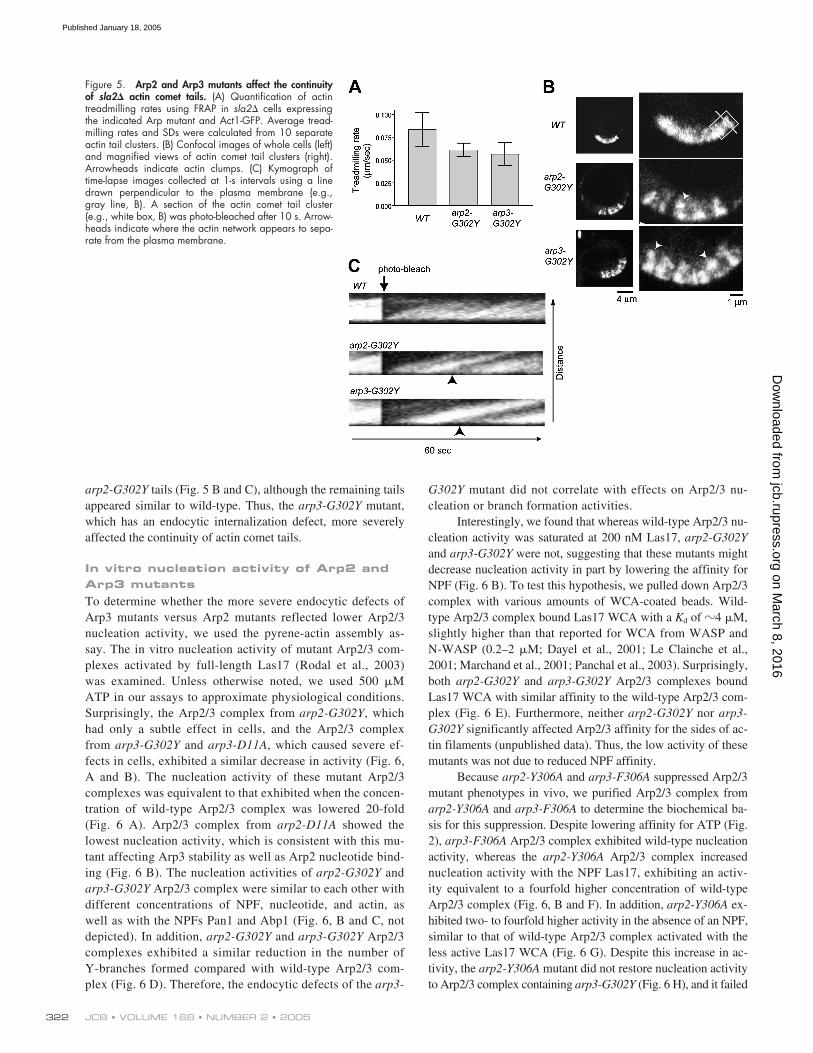
the rate of actin treadmilling was only slightly reduced com-pared with the rate in wild-type cells (Fig. 5 A). This wasconsistent with the relatively normal assembly of actin at en-docytic sites described above. Importantly, however, the con-tinuity of actin tails in sla2� arp3-G302Y cells was se-verely disrupted. Whereas actin tails in sla2� mutants appeared
smooth and continuous, all sla2� arp3-G302Y actin tails ex-hibited a broken structure (Fig. 5 B, Video 3, available at http://www.jcb.org/cgi/content/full/jcb.200408177/DC1). In thesemutants, clumps of actin appeared to separate from the plasmamembrane and to move into the cytoplasm at a continuous rate(Fig. 5 C). This phenotype was also observed for 40% of sla2�
Figure 4. Arp2 and Arp3 mutants affect actin patch protein internalization. (A) Tracking of individual cortical Abp1 patches. Abp1-GFP was visualizedevery 0.25 s and patch movement traces were obtained for the entire life of patches. Green and red dots indicate first and last positions, respectively.Dotted line indicates plasma membrane. (B) Internalization of Sla1 patches. Single frames (left) and kymographs (right) of time-lapse images collected at0.5-s intervals using a line drawn perpendicular to the plasma membrane (e.g., yellow). Dotted line: plasma membrane. (C) Fluorescence intensity of indi-vidual Abp1-GFP patches measured over time. (D) Single frames (left) and time series of single cortical patches (right) of cells expressing Abp1-GFP (red)and Sla1-CFP (green). Time-lapse between images was �4.55 s. Average times and SDs between Sla1 appearance and Abp1 recruitment were calculatedfrom at least 20 patches.
on March 8, 2016
jcb.rupress.orgD
ownloaded from
Published January 18, 2005
JCB • VOLUME 168 • NUMBER 2 • 2005322
arp2-G302Y tails (Fig. 5 B and C), although the remaining tailsappeared similar to wild-type. Thus, the arp3-G302Y mutant,which has an endocytic internalization defect, more severelyaffected the continuity of actin comet tails.
In vitro nucleation activity of Arp2 and Arp3 mutantsTo determine whether the more severe endocytic defects ofArp3 mutants versus Arp2 mutants reflected lower Arp2/3nucleation activity, we used the pyrene-actin assembly as-say. The in vitro nucleation activity of mutant Arp2/3 com-plexes activated by full-length Las17 (Rodal et al., 2003)was examined. Unless otherwise noted, we used 500 �MATP in our assays to approximate physiological conditions.Surprisingly, the Arp2/3 complex from arp2-G302Y, whichhad only a subtle effect in cells, and the Arp2/3 complexfrom arp3-G302Y and arp3-D11A, which caused severe ef-fects in cells, exhibited a similar decrease in activity (Fig. 6,A and B). The nucleation activity of these mutant Arp2/3complexes was equivalent to that exhibited when the concen-tration of wild-type Arp2/3 complex was lowered 20-fold(Fig. 6 A). Arp2/3 complex from arp2-D11A showed thelowest nucleation activity, which is consistent with this mu-tant affecting Arp3 stability as well as Arp2 nucleotide bind-ing (Fig. 6 B). The nucleation activities of arp2-G302Y andarp3-G302Y Arp2/3 complex were similar to each other withdifferent concentrations of NPF, nucleotide, and actin, aswell as with the NPFs Pan1 and Abp1 (Fig. 6, B and C, notdepicted). In addition, arp2-G302Y and arp3-G302Y Arp2/3complexes exhibited a similar reduction in the number ofY-branches formed compared with wild-type Arp2/3 com-plex (Fig. 6 D). Therefore, the endocytic defects of the arp3-
G302Y mutant did not correlate with effects on Arp2/3 nu-cleation or branch formation activities.
Interestingly, we found that whereas wild-type Arp2/3 nu-cleation activity was saturated at 200 nM Las17, arp2-G302Yand arp3-G302Y were not, suggesting that these mutants mightdecrease nucleation activity in part by lowering the affinity forNPF (Fig. 6 B). To test this hypothesis, we pulled down Arp2/3complex with various amounts of WCA-coated beads. Wild-type Arp2/3 complex bound Las17 WCA with a Kd of �4 �M,slightly higher than that reported for WCA from WASP andN-WASP (0.2–2 �M; Dayel et al., 2001; Le Clainche et al.,2001; Marchand et al., 2001; Panchal et al., 2003). Surprisingly,both arp2-G302Y and arp3-G302Y Arp2/3 complexes boundLas17 WCA with similar affinity to the wild-type Arp2/3 com-plex (Fig. 6 E). Furthermore, neither arp2-G302Y nor arp3-G302Y significantly affected Arp2/3 affinity for the sides of ac-tin filaments (unpublished data). Thus, the low activity of thesemutants was not due to reduced NPF affinity.
Because arp2-Y306A and arp3-F306A suppressed Arp2/3mutant phenotypes in vivo, we purified Arp2/3 complex fromarp2-Y306A and arp3-F306A to determine the biochemical ba-sis for this suppression. Despite lowering affinity for ATP (Fig.2), arp3-F306A Arp2/3 complex exhibited wild-type nucleationactivity, whereas the arp2-Y306A Arp2/3 complex increasednucleation activity with the NPF Las17, exhibiting an activ-ity equivalent to a fourfold higher concentration of wild-typeArp2/3 complex (Fig. 6, B and F). In addition, arp2-Y306A ex-hibited two- to fourfold higher activity in the absence of an NPF,similar to that of wild-type Arp2/3 complex activated with theless active Las17 WCA (Fig. 6 G). Despite this increase in ac-tivity, the arp2-Y306A mutant did not restore nucleation activityto Arp2/3 complex containing arp3-G302Y (Fig. 6 H), and it failed
Figure 5. Arp2 and Arp3 mutants affect the continuityof sla2� actin comet tails. (A) Quantification of actintreadmilling rates using FRAP in sla2� cells expressingthe indicated Arp mutant and Act1-GFP. Average tread-milling rates and SDs were calculated from 10 separateactin tail clusters. (B) Confocal images of whole cells (left)and magnified views of actin comet tail clusters (right).Arrowheads indicate actin clumps. (C) Kymograph oftime-lapse images collected at 1-s intervals using a linedrawn perpendicular to the plasma membrane (e.g.,gray line, B). A section of the actin comet tail cluster(e.g., white box, B) was photo-bleached after 10 s. Arrow-heads indicate where the actin network appears to sepa-rate from the plasma membrane.
on March 8, 2016
jcb.rupress.orgD
ownloaded from
Published January 18, 2005

GENETIC DISSECTION OF ATP BINDING BY ARP2/3 COMPLEX • MARTIN ET AL. 323
Figure 6. Effects of Arp2 and Arp3 mutants on Arp2/3 complex nucleation and branching. (A and F–H) Nucleation activity of Arp2 and Arp3 mutants.2 �M actin (5% pyrene labeled) was polymerized with the indicated concentration of Arp2/3 complex and, when indicated, 50 nM Las17 or 400 nMWCA. 500 �M ATP was present in nucleation reactions unless otherwise stated. (B and C) Barbed end concentrations at 50% polymerization generatedby Arp2/3 (20 nM) and different concentrations of Las17 (B) or with 50 nM Las17 and 7, 57, 107, or 507 �M ATP (C). (D) Branching activity of Arp2and Arp3 mutants. Actin filaments polymerized with 20 nM mutant Arp2/3 complex (2 nM wild-type Arp2/3) and 200 nM Las17 were visualized withrhodamine phalloidin. Average branching efficiency � SD was calculated as branch number per micrometer filament length, corrected for differences innucleation activity. (E) WCA binding to Arp2/3 complex mutants. 40 nM Arp2/3 complex was incubated with various concentrations of Las17 WCA-coated beads and the fraction of bound Arp2/3 was calculated by quantifying Arp3 left in the supernatant by immunoblotting. Data points represent av-erages � SEM from three experiments. Kd values of 4.5 � 0.9 �m, 3.9 � 1.0 �m, and 4.5 � 1.0 �m were calculated for wild-type, arp2-G302Y, andarp3-G302Y Arp2/3 complexes, respectively.
on March 8, 2016
jcb.rupress.orgD
ownloaded from
Published January 18, 2005

JCB • VOLUME 168 • NUMBER 2 • 2005324
to rescue Arp3 nucleotide binding of arp3-G302Y (Fig. 2 C),suggesting that in vivo suppression by the arp2-Y306A mutantdoes not simply restore nucleotide binding to Arp3.
The arp2-Y306A suppressor results in a conformational change that mimics Arp2/3 complex activationWe next used electron microscopy and image analysis to identifyconformational changes that occur upon activation of wild-typeArp2/3 complex. In addition, because the Arp2/3 complex con-taining the arp2-Y306A mutant exhibited constitutive activitysimilar to wild-type Arp2/3 complex activated by Las17 WCA(Fig. 6 G), we tested whether the arp2-Y306A mutation mimicsthe Arp2/3 complex activation step induced by NPF binding.Non-activated wild-type Arp2/3 complex, the arp2-Y306A Arp2/3mutant, and wild-type Arp2/3 complex bound to Las17 WCA,were imaged by electron microscopy in negative stain (Fig. 7).Single-particle analysis resulted in three reconstructions at �2.5nm resolution (Fig. 7 E), considerably higher than the 3.9 nmresolution previously reported for the yeast Arp2/3 complex(Volkmann et al., 2001). Fitting the atomic model of the nonacti-vated bovine Arp2/3 complex (Robinson et al., 2001) into the
three-dimensional maps revealed that the overall morphology ofall three reconstructions resembles that of the crystal structure(Fig. 7 A). However, differences in the density distribution be-tween the activated and the nonactivated wild-type Arp2/3 re-constructions are clearly visible in the Arp2 and Arc15 regions(Fig. 7, B and D). This observation shows for the first time adistinct structural change upon Arp2/3 complex activation byWCA. Interestingly, no significant differences were observedwhen the nonactivated arp2-Y306A mutant reconstruction wascompared with the activated wild-type Arp2/3 map. Thus, thestructural changes induced by NPF binding in the wild-typeArp2/3 complex are similar to those observed in the mutant com-plex in the absence of NPF. These structural similarities explainthe close resemblance of the nucleation activities of the mutantand the activated wild-type Arp2/3 complex (Fig. 6 G) and sug-gest that arp2-Y306A suppresses other mutants by causing theArp2/3 complex to resemble the active conformation.
DiscussionWe have generated mutants that affect either Arp2 or Arp3 nu-cleotide binding. Based on the effects of these mutants on en-
Figure 7. Single-particle reconstructions of Arp2/3complexes. (A) Docking of the nonactivated bovineArp2/3 crystal structure (ribbon diagram) into the recon-struction of nonactivated wild-type yeast Arp2/3 complex(chicken-wire representation). Color scheme follows C.The view in the second row is rotated by 90� horizon-tally from that in the first row; the view in the third row isrotated by 180� horizontally from that in the first row.(B) Three views of the reconstructions (solid blue surfacerepresentation) from nonactivated wild-type Arp2/3complex (WT), nonactivated arp2-Y306A mutant (arp2-Y306A), and activated wild-type Arp2/3 complex (WT-WCA). The views match those in A. Red arrows indicatethe region of Arp2 that shows differences between WTand WT-WCA/arp2-Y306A. Gray arrows indicate thedifferences in the Arc15 region. (C) Solvent exposed sur-face of the crystal structure; view matches third row in A.(D) Projection views of the three Arp2/3 reconstructions.Red arrows indicate the Arp2 region, highlighting differ-ences seen in B. (E) Fourier shell correlation graphs forthe three reconstructions calculated from two randomlyselected half sets of each respective data set. The 0.5 crite-rion indicates �2.5 nm resolution for all reconstructions.
on March 8, 2016
jcb.rupress.orgD
ownloaded from
Published January 18, 2005

GENETIC DISSECTION OF ATP BINDING BY ARP2/3 COMPLEX • MARTIN ET AL. 325
docytosis and in vitro nucleation activity, we present a modelfor the functions of nucleotide-bound Arp2 and Arp3 in Fig. 8.First, ATP-bound Arp2 and Arp3 are both required for full acti-vation of the Arp2/3 complex by NPFs in vitro (Fig. 8 A).Moreover, a structural change in Arp2 accompanies activationof the Arp2/3 complex by an NPF, as observed by electron mi-croscopy. The resulting nucleation activity is important for ef-ficiently initiating actin polymerization at endocytic sites (Fig.8 B, i). Once actin filaments are nucleated, actin assembly canproceed, albeit more slowly, with minimal Arp2/3 nucleationactivity. Finally, Arp2/3 complex, particularly nucleotide bind-ing by Arp3, plays a yet to be defined role, possibly anchoringactin filaments to each other and to the plasma membrane, toallow polymerization forces to be harnessed for endocytic in-ternalization (Fig. 8, A and B, ii). The evidence supporting thismodel is discussed below.
Importance of Arp2 and Arp3 nucleotide binding for actin filament nucleationOf the mutants we characterized, arp2-G302Y and arp3-G302Ymost dramatically inhibited ATP binding to Arp2 and Arp3, re-spectively. Previous studies reported that ATP binding is re-quired for Arp2/3 complex nucleation activity, but these stud-ies were unable to tease apart the individual contributions ofATP binding to Arp2 versus Arp3 (Dayel et al., 2001; LeClainche et al., 2001). Here, we demonstrated using the G302Ymutants that ATP-bound Arp2 and Arp3 both contribute toArp2/3 nucleation activity in vitro. Importantly, raising theATP concentration to physiological levels did not increase thein vitro nucleation activity of these mutant complexes, suggest-ing that their nucleation activities in vivo resemble those of nu-cleotide-free Arp2 and Arp3 complexes.
Previous studies suggested that nucleotide-bound Arp2and Arp3 are important for WCA binding (Dayel et al., 2001).Interestingly, arp2-G302Y and arp3-G302Y Arp2/3 complexesbound WCA with similar affinity to the wild-type complex,demonstrating that the decreased activity of these mutants isnot due to reduced NPF binding. A recent study probed confor-mational states of the human Arp2/3 complex using fluores-cence resonance energy transfer (FRET) between fluorophoresattached to different subunits of the complex (Goley et al.,2004). Using increase in FRET to monitor WCA binding, itwas demonstrated that the arp2-D11A and arp3-D11A mutantsreduce the ability of WCA to induce conformational changes inthe Arp2/3 complex (Goley et al., 2004). Previous studies usingmutations in WCA also suggested the existence of distinctWCA-binding and WCA-activation steps during Arp2/3 acti-vation (Marchand et al., 2001; Panchal et al., 2003). These re-sults support a model in which NPF-mediated Arp2/3 activa-tion requires ATP bound to Arp2 and Arp3.
Interestingly, the arp3-D11A mutant, which was ex-pected to bind ATP in our nucleation assays, had a similar nu-cleation defect to the arp3-G302Y mutant. This result sug-gested that the arp3-D11A mutation caused structural changesin the Arp2/3 complex even when nucleotide was bound toArp3. Consistent with this interpretation, both the arp3-D11Aand arp3-G302Y mutants prevented conformational changes in
human Arp2/3 complex that occur upon Arp3 nucleotide bind-ing as assessed by FRET (Goley et al., 2004). Thus, the arp3-D11A mutant appears to uncouple conformational changes inthe Arp2/3 complex from Arp3 nucleotide binding, mimickingthe nucleotide-free arp3-G302Y mutant. The similarity of thein vivo and in vitro phenotypes of the arp3-D11A and arp3-G302Y mutants suggests that defects in these mutants are spe-cifically related to Arp3 nucleotide binding. In addition, thedramatic endocytic phenotypes seen in arp3-D11A and arp3-G302Y demonstrate the importance of this conformationalchange to Arp2/3 function in vivo.
Mechanism of Arp2/3 complex activationUsing genetic crosses of our Arp2 and Arp3 mutants, we foundthat arp2-Y306A and arp3-F306A are suppressors of Arp2/3complex defects. Interestingly, Arp2/3 complex purified fromarp2-Y306A, but not from arp3-F306A, increased the nucle-ation activity of the Arp2/3 complex with and without Las17.This difference in activity was also seen in vivo, as arp2-Y306A, but not arp3-F306A, was able to suppress las17-11,identifying a possible functional difference between Arp2 andArp3. Because the NBP is critical for regulating actin structure,
Figure 8. Model for nucleotide’s role in Arp2/3 complex function.(A) Proposed relative contributions of Arp2 and Arp3 nucleotide binding toactin filament nucleation and a yet to be determined structural activity.(B) Proposed steps in an endocytic pathway (Merrifield et al., 2002; Kak-sonen et al., 2003) affected by Arp2 and Arp3 nucleotide-binding mutants.Both Arp2 and Arp3 mutants affect the initiation and the rate of actin as-sembly at endocytic sites (i), but Arp3 mutants more dramatically inhibitinternalization of endocytic proteins as a consequence of defects in actinnetwork structure and integrity (ii).
on March 8, 2016
jcb.rupress.orgD
ownloaded from
Published January 18, 2005

JCB • VOLUME 168 • NUMBER 2 • 2005326
and because changes in the Arp2 region were visible in elec-tron microscopy reconstructions of wild-type Arp2/3 complexupon activation, we speculated that the arp2-Y306A mutantcauses a conformational change that increases activities of theArp2/3 complex.
Because arp2-Y306A Arp2/3 complex exhibited similarnucleation activity to wild-type Arp2/3 complex activated byLas17 WCA, we examined the conformation of this mutant bycomparing its structure to both inactive and WCA-bound wild-type Arp2/3 complex. Indeed, in three-dimensional reconstruc-tions, arp2-Y306A Arp2/3 complex resembled wild-type Arp2/3complex activated by Las17 WCA, showing a similar differencein the region corresponding to Arp2. Previous evidence demon-strating that NPFs enhance Arp2 nucleotide binding support aconformational change in Arp2 upon NPF binding (Le Claincheet al., 2001). Because it has been proposed that the Arp2/3 com-plex must undergo a large conformational change to bring Arp2and Arp3 together to form a template dimer (Robinson et al.,2001; Volkmann et al., 2001), we suggest that a conformationalchange in Arp2 is a critical step in this process.
It was surprising that a mutant that lowers affinity for ATPresults in Arp2/3 activation. However, this lower affinity mightindicate greater conformational flexibility of the NBP, allowingArp2 to adopt an active conformation in the absence of an NPF.Interestingly, changing F306 in yeast actin to tyrosine increasedthe specificity of yeast actin for ATP versus GTP (Wen et al.,2002), possibly indicating a more compact conformation. Wealso found that in addition to the arp2-Y306A mutant, the arp2-D157E mutant also suppressed arp3 mutants. In actin, this mu-tation has been shown to increase the nucleotide exchange rate(Wolven et al., 2000), suggesting a more open conformation forthe NBP. We speculate that the conformational changes ob-served in the arp2-Y306A Arp2/3 complex result from greaterconformational flexibility of the Arp2 NBP.
Function of Arp2 and Arp3 nucleotide binding during endocytosis: general biological implicationsAnalysis of actin organization and endocytosis in arp2-G302Yand arp3-G302Y demonstrated an important functional dif-ference between nucleotide binding by Arp2 versus Arp3 invivo. The arp3-G302Y mutant caused severe endocytic defects,whereas arp2-G302Y showed only slight defects. However, thein vitro nucleation activities of these two mutants were indis-tinguishable. Importantly, the arp3-G302Y mutant assembledactin filaments at endocytic sites as well as arp2-G302Y, indi-cating that nucleation of actin filaments alone is not sufficientto drive endocytic internalization. We speculate that the struc-ture or integrity of the cortical actin network at endocytic sitesis disrupted in Arp3 mutants.
How might Arp3 contribute to actin cortical patch integ-rity and function? Because Arp3 nucleotide binding influencesthe conformation of the Arp2/3 complex (Goley et al., 2004),Arp3 mutants are likely to affect multiple interactions of theArp2/3 complex, possibly with one or several of the four knownyeast NPFs, Las17, Myo3/5, Pan1, and Abp1. Interestingly,Myo5 appears in patches immediately preceding internalization,
suggesting that its interaction with the Arp2/3 complex playsan important role in vesicle formation (Jonsdottir and Li,2004). Although wild-type, arp2-G302Y, and arp3-G302Y Arp2/3complexes bound Las17 WCA with similar affinity, conforma-tional changes upon actin filament binding and Arp2 ATPhydrolysis might alter interactions with NPFs subsequent tobranch formation (Marchand et al., 2001; Volkmann et al.,2001; Le Clainche et al., 2003; Dayel and Mullins, 2004). Con-nections between NPFs and the Arp2/3 complex in Y-branchesmight anchor branched actin filaments to the plasma membraneallowing forces to be applied for endocytic internalization. Inaddition, interactions with NPFs could be important for main-taining branched actin filament networks, such as those recentlyobserved in actin structures isolated from yeast (Young et al.,2004). The phenotype of arp3-G302Y sla2� actin tails, in whichclumps of actin appeared to separate from the plasma mem-brane, supports a defect in the integrity of plasma membrane at-tachment, actin network integrity, or both. Discontinuous actincomet tails have also been observed for NPF mutant “hopping”Listeria in vivo, and using a reconstituted motility system invitro, although the molecular basis for this phenomenon is notunderstood (Lasa et al., 1997; Bernheim-Groswasser et al.,2002). Further analysis of the molecular interactions of Arp2and Arp3 mutants is required to determine the mechanism bywhich Arp3 mutants affect the functionality of actin patches.
Because the arp2-G302Y and arp3-G302Y mutants af-fected in vitro nucleation activity indistinguishably, the simi-larities between these mutants provided insight into the func-tion of Arp2/3 complex nucleation. Both mutants exhibited aconsiderable delay in the onset of actin polymerization at en-docytic sites. This delay could have resulted from a lower nu-cleation rate or a delay in the recruitment of the Arp2/3 com-plex to endocytic sites. In addition, we cannot rule out thepossibility of Arp2/3-independent actin polymerization at thesesites. Interestingly, once initiated, the rate of actin assembly atendocytic sites in arp2-G302Y and arp3-G302Y mutants wasonly slightly lower in otherwise normal cells or in sla2� cells.This observation was similar to the moderate decrease in motil-ity rates for Listeria ActA mutants that significantly decreasedin vitro nucleation activity (Skoble et al., 2000). Therefore,Arp2/3 nucleation activity in these contexts appears more im-portant for initiating actin polymerization than for sustaining it.
In summary, we found that analysis of the yeast endocyticpathway provided a sensitive assay for distinguishing functionsof the Arp2/3 complex. The similarity of the effects of our NBPmutants of the yeast Arp2/3 complex to the analogous mutantsmade in the human Arp2 and Arp3 (Goley et al., 2004), and sim-ilarity in the recruitment of Arp2/3 complex to endocytic sites inyeast and mammalian cells (Kaksonen et al., 2003; Merrifield etal., 2004), suggest that our findings are directly applicable to thefunction of the Arp2/3 complex in multicellular eukaryotes.
Materials and methodsStrains, media, and genetic methodsYeast strains and plasmids used in this study are listed in Tables S2 and S3,available at http://www.jcb.org/cgi/content/full/jcb.200408177/DC1.
on March 8, 2016
jcb.rupress.orgD
ownloaded from
Published January 18, 2005

GENETIC DISSECTION OF ATP BINDING BY ARP2/3 COMPLEX • MARTIN ET AL. 327
Construction of mutant and tagged strainsAn ARP2::URA3 integration construct obtained from a previous study(Madania et al., 1999), pIMW18-U, and an ARP3::LEU2 integration con-struct, pDD1698, were mutagenized using QuikChange XL site-directedmutagenesis kit (Stratagene). Mutants were integrated into DDY2918 andDDY2919 as described previously (Wertman et al., 1992). Integration ofthe correct allele was confirmed by change in restriction site or sequenc-ing. ARC18 containing a COOH-terminal HA tag with a TEV protease sitewas generated using pBG265 (a gift from B. Goode, Brandeis University,Waltham, MA) and was integrated into the endogenous locus.
Protein purificationArp2/3 complex was purified using an HA-tagged Arc18 subunit andwas stored in HEKG5 (20 mM Hepes, pH 7.5, 1 mM EDTA, 50 mM KCl,5% glycerol) buffer. Full-length Las17 was purified as described previously(Rodal et al., 2003). GST-tagged Las17WCA was purified from bacteriausing pDD1122. Details of these purifications and actin purification proce-dures are provided in the supplemental materials.
ATP binding assaysATP cross-linking was performed using 5 �M ATP supplemented with 100�Ci �-[32P]ATP. Purified Arp2/3 complex (0.43 �M) with or without WCA(10 �M) in KMEI (50 mM KCl, 1 mM MgCl2, 1 mM EGTA, 10 mM imida-zole, pH 7.0) was equilibrated with ATP and irradiated at a 1-cm distancewith 254 nm UV light for 5 min using a hand-held Mineralight lamp. Sam-ples were subjected to SDS-PAGE and visualized/quantified using film ora Typhoon 9400 Phosphorimager (Molecular Dynamics). Etheno-ATP bind-ing was measured at 18�C using excitation and emission wavelengths of350 and 410 nm, respectively. Increasing amounts of etheno-ATP wereadded to 0.6 �M Arp2/3 complex in KMEI � 0.2 M acrylamide, and thebackground fluorescence of unbound etheno-ATP was subtracted, as de-scribed previously (Dayel et al., 2001). Kd values were calculated by fit-ting the data to a one-site binding equation using GraphPad Prism (Graph-Pad Software, Inc.).
MicroscopyPhalloidin staining, immunofluorescence, and LY uptake were per-formed as described previously (Belmont and Drubin, 1998). For livecell imaging, log phase cells grown at 25�C were adhered to the sur-face of a Con A–coated coverslip, inverted onto a glass slide, andsealed with vacuum grease (Dow Corning). All imaging was done at RTin synthetic media. Single fluorophore images (Abp1-GFP, Sla1-GFP,and rhodamine phalloidin) were obtained using a microscope (modelTE300; Nikon), 100�/NA 1.4 objective, and an Orca-100 Camera(Hamamatsu) controlled by Image Pro Plus software (Phase 3 Imaging).Dual fluorophore images (Abp1-GFP/Sla1-CFP) were obtained using amicroscope (model IX81; Olympus), 100�/NA 1.4 objective, and anOrca-ER Camera (Hamamatsu), controlled by Slidebook 4 software (In-telligent Imaging Innovations, Inc.). Dual fluorophore movies were ob-tained using a CFP-YFP filter set (JP4, Chroma) and motorized excitationand emission filter wheels (Sutter Instruments). Actin comet tails were ob-served using a confocal microscope (model 510; Carl Zeiss MicroImag-ing, Inc.) with a 63�/NA 1.4 objective. Photobleaching was per-formed by scanning a small region three times at full laser intensity at488 nm, followed by continued image acquisition at low laser intensity.Images used to track individual Abp1 patches were smoothed by 3 � 3pixel averaging and then thresholded with an adaptive thresholding al-gorithm, which takes into account the local intensity level. Patch posi-tions were determined by calculating the center of fluorescence intensityfor each patch. The patch positions of each frame were then connectedto trace patch trajectories. Patch intensity represents the sum of the in-tensity of each pixel making up an individual Abp1 patch. Custom-made software was used for all patch tracking calculations. Processingof individual images and movies was performed using Image J software(http://rsb.info.nih.gov/ij/).
AntibodiesArp2 and Arp3 were detected by immunoblotting using antibodies sc-11968 and sc-11973 (Santa Cruz Biotechnology, Inc.). Anti-HA 12CA5(Roche) was used for immunofluorescence with Arc18-HA.
Nucleation, branching, and WCA binding assaysActin assembly reactions were performed using 2 �M actin (5% pyrene la-beled) as described previously (Duncan et al., 2001). Barbed end concen-trations were calculated from the rate of actin assembly at 50% polymeriza-tion using a rate constant of 11.6 �M1 s1 (Higgs et al., 1999). Branched
filaments were observed by stabilizing actin filaments after polymerizationwas complete with equimolar rhodamine phalloidin (Molecular Probes) andwere processed for imaging as described previously (Amann and Pollard,2001). Branching was quantified by first calculating total filament length us-ing Fovea Pro 3.0 (Reindeer Graphics, Inc.) for Adobe Photoshop 7.0(Adobe Systems, Inc.), and then counting branches manually. More than8,000 or 13,000 �m of filaments were analyzed for wild-type or Arp2/3mutants, respectively. WCA binding was measured using glutathionebeads (Sigma-Aldrich) coated with GST-WCA. Binding was performed atRT for 30 min in G buffer (5 mM Tris, pH 7.5, 0.2 mM ATP, 0.2 mMCaCl2, 0.2 mM DTT) supplemented with 50 mM KCl, 2 mM MgCl2, and0.5 mM ATP. Amounts of bound Arp3 were quantified by immunoblottingusing a Typhoon 9400 Phosphorimager (Molecular Dynamics) and Kd valueswere calculated using GraphPad Prism (GraphPad Software, Inc.).
Electron microscopy50 nM Arp2/3 complex (wild-type or arp2-Y306A mutant) was diluted inF buffer (2 mM imidazole, pH 7.0, 50 mM KCl, 2 mM MgCl2, 1 mMEGTA, 0.2 mM DTT, 0.1 mM ATP, 0.02% azide), applied to a carboncoated EM grid and stained with 2% uranyl acetate. 500 nM Las17 WCAwas mixed with the wild-type Arp2/3 complex for generating the acti-vated Arp2/3 samples. Images were obtained under low-dose conditionsusing a Tecnai 12 microscope (FEI electron optics) equipped with a Lab6filament at 120 kV at a nominal magnification of 67,000� and at �1-�mdefocus. The micrographs were digitized on a SCAI scanner (Z/I Imaging)at 7-�m raster and compressed to a final pixel size of 0.6 nm.
Image processingInteractive selection from 16, 17, and 30 micrographs yielded 2,774,2,654, and 2,330 particles from nonactivated wild-type Arp2/3, nonacti-vated arp2-Y306A mutant, and wild-type yeast Arp2/3 with WCA, re-spectively. The particles were processed and analyzed using the EMAN(Ludtke et al., 1999) and CoAn (Volkmann and Hanein, 1999; Volkmannand Hanein, 2003) software packages. A correction for the contrast trans-fer function was applied for all images. Particles were first sorted into 89classes according to their mutual similarity without the use of an externalreference. Subsets of these views were used to generate initial referencesusing a Fourier-based common-line approach. The initial models were iter-atively refined against the data. Convergence was achieved after 7–10 it-erations. In addition to these reference-free models, a density map calcu-lated from the bovine Arp2/3 crystal structure was used for separaterefinements, which converged to the same reconstructions as the data-based models. To ensure the model independence of the differences be-tween activated and nonactivated complex, the final reconstruction of theactivated complex was used as a starting model with the nonactivateddata and vice versa. In both cases, the distinct features of the reconstruc-tions were recovered after a few rounds of refinement. A similar calcula-tion was performed with the nonactivated and the mutant complex, alsowith the result that the characteristic features were recovered after a few it-erations. The crystal structure of the nonactivated bovine Arp2/3 complexwas docked into each of the reconstructions using CoAn. The correlationbetween the docked structure and the reconstructions was between 84and 85% at 2.5-nm resolution.
Online supplemental materialSupplemental videos showing Abp1, Sla1 dynamics (Videos 1 and 2),and actin treadmilling in sla2� cells (Video 3); tables showing Arp2/3mutant growth phenotypes (Table S1), yeast strains used in this study(Table S2), and plasmids used in this study (Table S3); and supplementalmaterials and methods are available online at http://www.jcb.org/cgi/content/full/jcb.200408177/DC1.
We thank S. Almo and K. Shokat for advice on which mutations to make andfor helpful discussions about the effects of these mutations. We also thank B.Goode, B. Winsor, and members of the Drubin lab (M. Duncan, C. Toret,and J. Toshima) for providing reagents. Finally, we thank C. Le Clainche andE. Goley for critically reading the manuscript and members of the Drubin,Welch, Hanein, and Volkmann labs for helpful discussions.
This work was supported by National Institutes of Health (NIH) grantsGM50399 and GM42759 to D.G. Drubin and GM59609 to M. Welch.The electron microscopy and image analysis was supported by the NIH CellMigration Consortium (U54 GM646346) and NIH grant P01 GM66311for D. Hanein and N. Volkmann.
Submitted: 31August 2004Accepted: 1 December 2004
on March 8, 2016
jcb.rupress.orgD
ownloaded from
Published January 18, 2005

JCB • VOLUME 168 • NUMBER 2 • 2005328
ReferencesAmann, K.J., and T.D. Pollard. 2001. The Arp2/3 complex nucleates actin fila-
ment branches from the sides of pre-existing filaments. Nat. Cell Biol.3:306–310.
Belmont, L.D., and D.G. Drubin. 1998. The yeast V159N actin mutant revealsroles for actin dynamics in vivo. J. Cell Biol. 142:1289–1299.
Belmont, L.D., A. Orlova, D.G. Drubin, and E.H. Egelman. 1999a. A change inactin conformation associated with filament instability after Pi release.Proc. Natl. Acad. Sci. USA. 96:29–34.
Belmont, L.D., G.M. Patterson, and D.G. Drubin. 1999b. New actin mutants al-low further characterization of the nucleotide binding cleft and drugbinding sites. J. Cell Sci. 112(Pt 9):1325–1336.
Bernheim-Groswasser, A., S. Wiesner, R.M. Golsteyn, M.F. Carlier, and C.Sykes. 2002. The dynamics of actin-based motility depend on surfaceparameters. Nature. 417:308–311.
Blanchoin, L., K.J. Amann, H.N. Higgs, J.B. Marchand, D.A. Kaiser, andT.D. Pollard. 2000. Direct observation of dendritic actin filament net-works nucleated by Arp2/3 complex and WASP/Scar proteins. Nature.404:1007–1011.
Chen, X., J. Peng, M. Pedram, C.A. Swenson, and P.A. Rubenstein. 1995. Theeffect of the S14A mutation on the conformation and thermostability ofSaccharomyces cerevisiae G-actin and its interaction with adenine nucle-otides. J. Biol. Chem. 270:11415–11423.
Dayel, M.J., and R.D. Mullins. 2004. Activation of Arp2/3 complex: addition ofthe first subunit of the new filament by a WASP protein triggers rapidATP hydrolysis on Arp2. PLoS Biol. 2:E91.
Dayel, M.J., E.A. Holleran, and R.D. Mullins. 2001. Arp2/3 complex requireshydrolyzable ATP for nucleation of new actin filaments. Proc. Natl.Acad. Sci. USA. 98:14871–14876.
Duncan, M.C., M.J. Cope, B.L. Goode, B. Wendland, and D.G. Drubin. 2001.Yeast Eps15-like endocytic protein, Pan1p, activates the Arp2/3 com-plex. Nat. Cell Biol. 3:687–690.
Engqvist-Goldstein, A.E., and D.G. Drubin. 2003. Actin assembly and endocy-tosis: from yeast to mammals. Annu. Rev. Cell Dev. Biol. 19:287–332.
Goley, E.D., S.E. Rodenbusch, A.C. Martin, and M.D. Welch. 2004. Criticalconformational changes in the Arp2/3 complex are induced by nucle-otide and nucleation promoting factor. Mol. Cell. 16:269–279.
Higgs, H.N., L. Blanchoin, and T.D. Pollard. 1999. Influence of the C terminusof Wiskott-Aldrich syndrome protein (WASp) and the Arp2/3 complexon actin polymerization. Biochemistry. 38:15212–15222.
Jonsdottir, G.A., and R. Li. 2004. Dynamics of yeast Myosin I: evidence for apossible role in scission of endocytic vesicles. Curr. Biol. 14:1604–1609.
Kabsch, W., H.G. Mannherz, D. Suck, E.F. Pai, and K.C. Holmes. 1990. Atomicstructure of the actin:DNase I complex. Nature. 347:37–44.
Kaksonen, M., Y. Sun, and D.G. Drubin. 2003. A pathway for association ofreceptors, adaptors, and actin during endocytic internalization. Cell.115:475–487.
Lasa, I., E. Gouin, M. Goethals, K. Vancompernolle, V. David, J. Vandekerck-hove, and P. Cossart. 1997. Identification of two regions in the N-termi-nal domain of ActA involved in the actin comet tail formation by Listeriamonocytogenes. EMBO J. 16:1531–1540.
Le Clainche, C., D. Didry, M.F. Carlier, and D. Pantaloni. 2001. Activation ofArp2/3 complex by Wiskott-Aldrich syndrome protein is linked to en-hanced binding of ATP to Arp2. J. Biol. Chem. 276:46689–46692.
Le Clainche, C., D. Pantaloni, and M.F. Carlier. 2003. ATP hydrolysis on actin-related protein 2/3 complex causes debranching of dendritic actin arrays.Proc. Natl. Acad. Sci. USA. 100:6337–6342.
Ludtke, S.J., P.R. Baldwin, and W. Chiu. 1999. EMAN: semiautomated soft-ware for high-resolution single-particle reconstructions. J. Struct. Biol.128:82–97.
Madania, A., P. Dumoulin, S. Grava, H. Kitamoto, C. Scharer-Brodbeck, A.Soulard, V. Moreau, and B. Winsor. 1999. The Saccharomyces cerevi-siae homologue of human Wiskott-Aldrich syndrome protein Las17p in-teracts with the Arp2/3 complex. Mol. Biol. Cell. 10:3521–3538.
Marchand, J.B., D.A. Kaiser, T.D. Pollard, and H.N. Higgs. 2001. Interaction ofWASP/Scar proteins with actin and vertebrate Arp2/3 complex. Nat.Cell Biol. 3:76–82.
Merrifield, C.J., M.E. Feldman, L. Wan, and W. Almers. 2002. Imaging actinand dynamin recruitment during invagination of single clathrin-coatedpits. Nat. Cell Biol. 4:691–698.
Merrifield, C.J., B. Qualmann, M.M. Kessels, and W. Almers. 2004. NeuralWiskott-Aldrich syndrome protein (N-WASP) and the Arp2/3 complexare recruited to sites of clathrin-mediated endocytosis in cultured fibro-blasts. Eur. J. Cell Biol. 83:13–18.
Moreau, V., A. Madania, R.P. Martin, and B. Winsor. 1996. The Saccharomyces
cerevisiae actin-related protein Arp2 is involved in the actin cytoskeleton.J. Cell Biol. 134:117–132.
Moreau, V., J.M. Galan, G. Devilliers, R. Haguenauer-Tsapis, and B. Winsor.1997. The yeast actin-related protein Arp2p is required for the internal-ization step of endocytosis. Mol. Biol. Cell. 8:1361–1375.
Pan, F., C. Egile, T. Lipkin, and R. Li. 2004. ARPC1/Arc40 mediates the inter-action of the Arp2/3 complex with WASP family activators. J. Biol.Chem. 279:54629–54636.
Panchal, S.C., D.A. Kaiser, E. Torres, T.D. Pollard, and M.K. Rosen. 2003. Aconserved amphipathic helix in WASP/Scar proteins is essential for acti-vation of Arp2/3 complex. Nat. Struct. Biol. 10:591–598.
Robinson, R.C., K. Turbedsky, D.A. Kaiser, J.B. Marchand, H.N. Higgs, S.Choe, and T.D. Pollard. 2001. Crystal structure of Arp2/3 complex.Science. 294:1679–1684.
Rodal, A.A., A.L. Manning, B.L. Goode, and D.G. Drubin. 2003. Negative reg-ulation of yeast WASp by two SH3 domain-containing proteins. Curr.Biol. 13:1000–1008.
Sablin, E.P., J.F. Dawson, M.S. VanLoock, J.A. Spudich, E.H. Egelman, and R.J.Fletterick. 2002. How does ATP hydrolysis control actin’s associations?Proc. Natl. Acad. Sci. USA. 99:10945–10947.
Skoble, J., D.A. Portnoy, and M.D. Welch. 2000. Three regions within ActApromote Arp2/3 complex-mediated actin nucleation and Listeria mono-cytogenes motility. J. Cell Biol. 150:527–538.
Svitkina, T.M., and G.G. Borisy. 1999. Arp2/3 complex and actin depolymeriz-ing factor/cofilin in dendritic organization and treadmilling of actin fila-ment array in lamellipodia. J. Cell Biol. 145:1009–1026.
Volkmann, N., and D. Hanein. 1999. Quantitative fitting of atomic modelsinto observed densities derived by electron microscopy. J. Struct. Biol.125:176–184.
Volkmann, N., and D. Hanein. 2003. Docking of atomic models into reconstruc-tions from electron microscopy. Methods Enzymol. 374:204–225.
Volkmann, N., K.J. Amann, S. Stoilova-McPhie, C. Egile, D.C. Winter, L. Ha-zelwood, J.E. Heuser, R. Li, T.D. Pollard, and D. Hanein. 2001. Struc-ture of Arp2/3 complex in its activated state and in actin filament branchjunctions. Science. 293:2456–2459.
Vorobiev, S., B. Strokopytov, D.G. Drubin, C. Frieden, S. Ono, J. Condeelis,P.A. Rubenstein, and S.C. Almo. 2003. The structure of nonvertebrateactin: implications for the ATP hydrolytic mechanism. Proc. Natl. Acad.Sci. USA. 100:5760–5765.
Welch, M.D., and R.D. Mullins. 2002. Cellular control of actin nucleation.Annu. Rev. Cell Dev. Biol. 18:247–288.
Wen, K.K., X. Yao, and P.A. Rubenstein. 2002. GTP-yeast actin. J. Biol. Chem.277:41101–41109.
Wertman, K.F., D.G. Drubin, and D. Botstein. 1992. Systematic mutationalanalysis of the yeast ACT1 gene. Genetics. 132:337–350.
Winter, D., A.V. Podtelejnikov, M. Mann, and R. Li. 1997. The complex con-taining actin-related proteins Arp2 and Arp3 is required for the motilityand integrity of yeast actin patches. Curr. Biol. 7:519–529.
Winter, D.C., E.Y. Choe, and R. Li. 1999. Genetic dissection of the buddingyeast Arp2/3 complex: a comparison of the in vivo and structural roles ofindividual subunits. Proc. Natl. Acad. Sci. USA. 96:7288–7293.
Wolven, A.K., L.D. Belmont, N.M. Mahoney, S.C. Almo, and D.G. Drubin. 2000.In vivo importance of actin nucleotide exchange catalyzed by profilin.J. Cell Biol. 150:895–904.
Young, M.E., J.A. Cooper, and P.C. Bridgman. 2004. Yeast actin patches arenetworks of branched actin filaments. J. Cell Biol. 166:629–635.
on March 8, 2016
jcb.rupress.orgD
ownloaded from
Published January 18, 2005