
Untitled - Vyoma Online Classes
432
-
Upload
khangminh22 -
Category
Documents
-
view
1 -
download
0
Transcript of Untitled - Vyoma Online Classes

Demystified Series
Advanced Statistics DemystifiedAlgebra DemystifiedAnatomy DemystifiedAstronomy DemystifiedBiology DemystifiedBusiness Statistics DemystifiedCalculus DemystifiedChemistry DemystifiedCollege Algebra DemystifiedEarth Science DemystifiedEveryday Math DemystifiedGeometry DemystifiedPhysics DemystifiedPhysiology DemystifiedPre-Algebra DemystifiedProject Management DemystifiedStatistics DemystifiedTrigonometry Demystified
PHYSIOLOGY DEMYSTIFIED
The Hon. Dr. Dale Pierre Layman, Ph.D.,
Grand Ph.D. in Medicine (Belgium)
McGRAW-HILLNew York Chicago San Francisco Lisbon London
Madrid Mexico City Milan New Delhi San Juan
Seoul Singapore Sydney Toronto

Copyright © 2004 by The McGraw-Hill Companies, Inc. All rights reserved. Manufactured in the United States ofAmerica. Except as permitted under the United States Copyright Act of 1976, no part of this publication may bereproduced or distributed in any form or by any means, or stored in a database or retrieval system, without the priorwritten permission of the publisher.
0-07-147114-6
The material in this eBook also appears in the print version of this title: 0-07-143828-9.
All trademarks are trademarks of their respective owners. Rather than put a trademark symbol after every occurrenceof a trademarked name, we use names in an editorial fashion only, and to the benefit of the trademark owner, withno intention of infringement of the trademark. Where such designations appear in this book, they have been printedwith initial caps.
McGraw-Hill eBooks are available at special quantity discounts to use as premiums and sales promotions, or for usein corporate training programs. For more information, please contact George Hoare, Special Sales, [email protected] or (212) 904-4069.
TERMS OF USE
This is a copyrighted work and The McGraw-Hill Companies, Inc. (“McGraw-Hill”) and its licensors reserve allrights in and to the work. Use of this work is subject to these terms. Except as permitted under the Copyright Act of1976 and the right to store and retrieve one copy of the work, you may not decompile, disassemble, reverse engineer,reproduce, modify, create derivative works based upon, transmit, distribute, disseminate, sell, publish or sublicensethe work or any part of it without McGraw-Hill’s prior consent. You may use the work for your own noncommercialand personal use; any other use of the work is strictly prohibited. Your right to use the work may be terminated ifyou fail to comply with these terms.
THE WORK IS PROVIDED “AS IS.” McGRAW-HILL AND ITS LICENSORS MAKE NO GUARANTEES ORWARRANTIES AS TO THE ACCURACY, ADEQUACY OR COMPLETENESS OF OR RESULTS TO BEOBTAINED FROM USING THE WORK, INCLUDING ANY INFORMATION THAT CAN BE ACCESSEDTHROUGH THE WORK VIA HYPERLINK OR OTHERWISE, AND EXPRESSLY DISCLAIM ANY WARRANTY,EXPRESS OR IMPLIED, INCLUDING BUT NOT LIMITED TO IMPLIED WARRANTIES OFMERCHANTABILITY OR FITNESS FOR A PARTICULAR PURPOSE. McGraw-Hill and its licensors do notwarrant or guarantee that the functions contained in the work will meet your requirements or that its operation willbe uninterrupted or error free. Neither McGraw-Hill nor its licensors shall be liable to you or anyone else for anyinaccuracy, error or omission, regardless of cause, in the work or for any damages resulting therefrom. McGraw-Hillhas no responsibility for the content of any information accessed through the work. Under no circumstances shallMcGraw-Hill and/or its licensors be liable for any indirect, incidental, special, punitive, consequential or similardamages that result from the use of or inability to use the work, even if any of them has been advised of the possi-bility of such damages. This limitation of liability shall apply to any claim or cause whatsoever whether such claimor cause arises in contract, tort or otherwise.
DOI: 10.1036/0071438289

This book is fondly dedicated to my wonderful wife, Kathy. She is the‘‘Anchor,’’ the solid ‘‘Rock’’ upon which we can all lean in times of troubleor need. She is my best and most patient friend.I also wish to highlight Allison, one of my artistically talented daughters. It
is through her creative efforts that our host – Professor Joe, the TalkingSkeleton – has been brought to vibrant life.Finally, I wish to thank Janet M. Evans, President of the American
Biographical Institute, and Nicholas S. Law, Director General of theInternational Biographical Centre (Cambridge, England). In many of theirvolumes, they have described my ideas for ‘‘Intuitive Geometry and theA&P (Human Anatomy & Physiology) Text.’’And, now, through the help of Judy Bass and Scott Grillo at McGraw-Hill,
these ideas are being realized!

This page intentionally left blank
vii
CONTENTS
Preface xv
Acknowledgments xvii
PART 1 THE GOLDEN PATH OF BODYFUNCTION 1
CHAPTER 1 Physiology: Our Life Is A Path ThroughBodyspace 3All Life Starts with Biology! 4Two Paths Curve Apart in the Woods 8Contrasting Physiology with Plain Functions 13Letter Symbols for Anatomy, Physiology,
and Functions 13Some Characteristics of Living Body
Functions 17The Different Levels of Physiology 20Summary: A Three-Way System of
Classification for Key Body Facts 23Quiz 26Body-Level Grids for Chapter 1 28
For more information about this title, click here

CHAPTER 2 Control of the Internal Environment:A Story About Feedback and Homeostasis 30Enter Hippocrates: Human Health Seen as a
Balance 31Claude Bernard and His Dogs: A Preview of
Homeostasis 33Walter B. Cannon: The Wisdom of the Body
and Homeostasis 36Homeostasis and Modern Control System
Theory 37The Set-Point Theory 38Stressors or Stimuli: Disturbers of Body
Parameters 41Feedback Systems or Cycles 42Feedback Systems and Human Health 50Quiz 53Body-Level Grids for Chapter 2 55
Test: Part 1 59
PART 2 FUNCTIONS AT THE CHEMICAL ANDCELLULAR LEVELS 65
CHAPTER 3 Chemical Functions: ‘‘Hey! We’ve Got DirtMoving Around in Our Body Basement!’’ 67The Atom: An ‘‘Uncuttable’’ Particle with
Its Own Anatomy 68Chemical Bonds: Builders of Order and
Molecules 71Ions: First as Bonds, Then as ‘‘Goers-to’’ 74Energy and Metabolism 77‘‘Blood Hydrogen Homeostasis’’: Body
Acid-Base Balance 79
CONTENTSviii
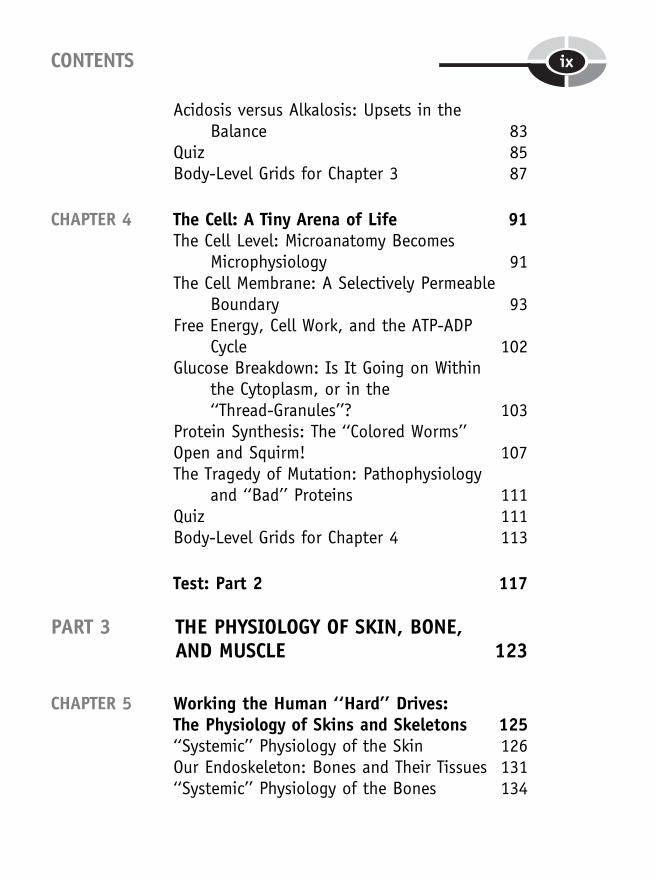
Acidosis versus Alkalosis: Upsets in theBalance 83
Quiz 85Body-Level Grids for Chapter 3 87
CHAPTER 4 The Cell: A Tiny Arena of Life 91The Cell Level: Microanatomy Becomes
Microphysiology 91The Cell Membrane: A Selectively Permeable
Boundary 93Free Energy, Cell Work, and the ATP-ADP
Cycle 102Glucose Breakdown: Is It Going on Within
the Cytoplasm, or in the‘‘Thread-Granules’’? 103
Protein Synthesis: The ‘‘Colored Worms’’Open and Squirm! 107The Tragedy of Mutation: Pathophysiology
and ‘‘Bad’’ Proteins 111Quiz 111Body-Level Grids for Chapter 4 113
Test: Part 2 117
PART 3 THE PHYSIOLOGY OF SKIN, BONE,AND MUSCLE 123
CHAPTER 5 Working the Human ‘‘Hard’’ Drives:The Physiology of Skins and Skeletons 125‘‘Systemic’’ Physiology of the Skin 126Our Endoskeleton: Bones and Their Tissues 131‘‘Systemic’’ Physiology of the Bones 134
CONTENTS ix

Quiz 143Body-Level Grids for Chapter 5 144
CHAPTER 6 ‘‘Gentlemen, Fire Up Your Engines!’’: ThePhysiology of Neurons and Muscle Fibers 148Lever Systems: Muscles, Bones, and Joints 149Motor Neurons and Muscle Fibers: The
Neuromuscular Connection 150Microanatomy of a Skeletal Muscle Fiber 153Muscle Fiber Contraction: The Sliding
Filament Theory 156Motor Units and the Concept of Threshold 163Muscle Endurance Versus Fatigue:
Biochemical Fiber Types 168Quiz 172Body-level Grids for Chapter 6 174
Test: Part 3 178
PART 4 THE PHYSIOLOGY OF NERVES ANDGLANDS 183
CHAPTER 7 ‘‘What Happens When We Step on aNail?’’: The Physiology of Nerves andReflexes 185Relationship of the Two Major Divisions of
the Nervous System 186The Synapse: Where Two Neurons ‘‘Clasp
Hands’’ 188Neuron Axons: Resting, Polarized, or
Active Depolarized? 190Neuron Axons: Naked, or Myelinated? 192
CONTENTSx
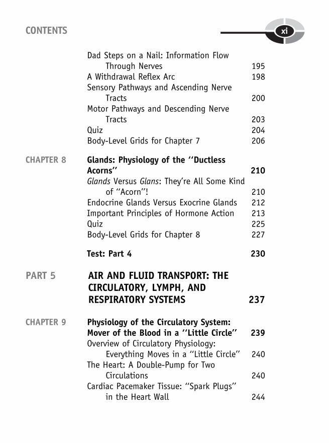
Dad Steps on a Nail: Information FlowThrough Nerves 195
A Withdrawal Reflex Arc 198Sensory Pathways and Ascending Nerve
Tracts 200Motor Pathways and Descending Nerve
Tracts 203Quiz 204Body-Level Grids for Chapter 7 206
CHAPTER 8 Glands: Physiology of the ‘‘DuctlessAcorns’’ 210Glands Versus Glans: They’re All Some Kind
of ‘‘Acorn’’! 210Endocrine Glands Versus Exocrine Glands 212Important Principles of Hormone Action 213Quiz 225Body-Level Grids for Chapter 8 227
Test: Part 4 230
PART 5 AIR AND FLUID TRANSPORT: THECIRCULATORY, LYMPH, ANDRESPIRATORY SYSTEMS 237
CHAPTER 9 Physiology of the Circulatory System:Mover of the Blood in a ‘‘Little Circle’’ 239Overview of Circulatory Physiology:
Everything Moves in a ‘‘Little Circle’’ 240The Heart: A Double-Pump for Two
Circulations 240Cardiac Pacemaker Tissue: ‘‘Spark Plugs’’
in the Heart Wall 244
CONTENTS xi
CONTENTSxii
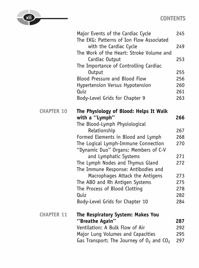
Major Events of the Cardiac Cycle 245The EKG: Patterns of Ion Flow Associated
with the Cardiac Cycle 249The Work of the Heart: Stroke Volume and
Cardiac Output 253The Importance of Controlling Cardiac
Output 255Blood Pressure and Blood Flow 256Hypertension Versus Hypotension 260Quiz 261Body-Level Grids for Chapter 9 263
CHAPTER 10 The Physiology of Blood: Helps It Walkwith a ‘‘Lymph’’ 266The Blood-Lymph Physiological
Relationship 267Formed Elements in Blood and Lymph 268The Logical Lymph-Immune Connection 270‘‘Dynamic Duo’’ Organs: Members of C-V
and Lymphatic Systems 271The Lymph Nodes and Thymus Gland 272The Immune Response: Antibodies and
Macrophages Attack the Antigens 273The ABO and Rh Antigen Systems 275The Process of Blood Clotting 278Quiz 282Body-Level Grids for Chapter 10 284
CHAPTER 11 The Respiratory System: Makes You‘‘Breathe Again’’ 287Ventilation: A Bulk Flow of Air 292Major Lung Volumes and Capacities 295Gas Transport: The Journey of O2 and CO2 297
Control of Respiration and BodyAcid-Base Balance 302
Quiz 307Body-Level Grids for Chapter 11 309
Test: Part 5 313
PART 6 PHYSIOLOGY FROM ‘‘THE LAND DOWNUNDER’’: THE DIGESTIVE ANDGENITOURINARY SYSTEMS 319
CHAPTER 12 The Digestive System: A Miraculous‘‘Grinder’’! 321The Dark and Mysterious Organ Systems
‘‘Below Our Belt’’ 322Modern Human Feces, or Ancient Roman
Fasces? Our ‘‘Dregs’’ Are All Packaged into‘‘Bundles’’ 322Enter the Accessory Digestive Organs 325Squirting Bile into the Digestive Tube: A
Partnership Between the Liver andGall Bladder 326
Too Much Acid? The Duodenum Just Keeps‘‘Secretin’’! 331
More Digestive Enzymes: The CCK ‘‘Bonus’’Effect 334
Villi and Microvilli in the Small IntestineWall 336
GI Tube Movements 337Vomiting and Diarrhea: Digestive
Movements That Can Upset Acid-BaseBalance 341
Quiz 342
CONTENTS xiii
Body-Level Grids for Chapter 12 344
CHAPTER 13 The Genitourinary System: It Allows Usto Release Fluids and ‘‘Beget’’ New Life! 347The Notion of Physiological Pathways
Within the Great Body Pyramid 348The Three Genitourinary Pathways 349Labor and Birth: A Beneficial Positive
Feedback Cycle 358Controlling the Output of Urine: The
Urinary Excretion Equation 361Body Water Balance and ADH 364Body Salt (Electrolyte) Balance 366Quiz 367Body-Level Grids for Chapter 13 369
Test: Part 6 373
Final Exam 378
Answers to Quiz, Test, andExam Questions 396
Index 403
CONTENTSxiv
PREFACE
‘‘Which comes first – the chicken, or the egg?’’ This book is about the‘‘chicken’’ – human physiology or living body functions. (The process of layingan egg is an aspect of chicken physiology, after all!) Its close companion,ANATOMY DEMYSTIFIED, is all about the ‘‘egg’’ – human anatomy orbody structure. (An unhatched egg is an example of anatomy.)
PHYSIOLOGY DEMYSTIFIED is for people who want to getacquainted with the fundamental concepts of human body function, withouthaving to take a formal course. But it can also serve as a supplemental text ina classroom, tutored, or home-schooling environment. In addition, it shouldbe useful for career changers who need to refresh their knowledge of thesubject. I recommend that you start at the beginning of this book andwork straight through.
This book seeks to provide you with an intuitive, highly visual grasp ofphysiology and its terminology. Starting with the Great Body Pyramid (aconcept borrowed from anatomy and the Ancient Egyptians),PHYSIOLOGY DEMYSTIFIED guides you along A Living Path ThroughBodyspace. Professor Joe, the Talking Skeleton, is our host. He is drawn as acartoon standing upright and pointing, whenever key facts about BiologicalOrder in the human body are being presented. But when he is fallen andfractured, our Good Professor is talking to you about facts of BiologicalDisorder within the human body. These key facts of Order-versus-Disorderwill be about Anatomy, Physiology, or just Plain Body Functions.
As you go from body system to body system, you will also learn where toput many facts of Biological Order/Disorder, briefly writing them within the‘‘grids or drawers’’ of the Great Body Pyramid. In this way, like putting yoursocks into a drawer, you will always know where to find the key body facts(‘‘socks’’) whenever you need them!
This introductory work also contains an abundance of practice quiz, test,and exam questions. They are all multiple-choice, and are similar to the sortsof questions used in standardized tests. There is a short quiz at the end ofevery chapter. The quizzes are ‘‘open-book.’’ You may (and should) refer to
xv
Copyright © 2004 by The McGraw-Hill Companies, Inc. Click here for terms of use.
the chapter texts when taking them. When you think you’re ready, take thequiz, write down your answers, and then give your list of answers to a friend.Have the friend tell you your score, but not which questions you got wrong.The answers are listed in the back of the book. Stick with the chapter untilyou get most of the answers correct.
This book is divided into six sections. At the end of each section is amultiple choice test. Take these tests when you’re done with the respectivesections and have taken all the chapter quizzes. The section tests are ‘‘closed-book,’’ but the questions are not as difficult as those in the quizzes. A satis-factory score is three-quarters of the answers correct. Again, answers are inthe back of the book.
There is a final exam at the end of this course. It contains questions drawnuniformly from all the chapters in the book. Take it when you have finishedall six section tests, and all of the chapter quizzes. A satisfactory score is atleast 75 percent correct answers.
With the section tests and the final exam, as with the quizzes, have a friendtell you your score without letting you know which questions you missed.That way, you will not subconsciously memorize the answers. You can checkto see where your knowledge is strong, and where it is not.
I recommend that you complete one chapter a week. An hour or two dailyought to be enough time for this. When you’re done with the course, you canuse this book, with its comprehensive index, as a permanent reference. Whatyou now hold in your hand, I think you will agree, is a most unusualapproach to the study of human physiology! We have, of course, our mostunique talking skeleton host, Professor Joe (and occasional glimpses of hissomewhat mischievous sidekick, Baby Heinie). More importantly, this bookrepresents the practical application of what I like to call Compu-think, or‘‘computer-like modes or ways of human thinking.’’ This is reflected in itsheavy emphasis upon binary (two-way) classifications, grid-associated rea-soning, and frequent occurrence of summary word equations.
Suggestions for future editions are welcome.Now, work hard! But, be sure to have fun! Best wishes for your success.
The Hon. Dr. Dale Pierre Layman, Ph.D., Grand Ph.D. in Medicine
PREFACExvi
ACKNOWLEDGMENTS
The most interesting and entertaining illustrations in this book are mainlydue to the talented efforts of one of my own daughters, Allison VictoriaLayman. It is through her gifted eyes that my visions for picturing keybody concepts have been successfully brought to life!
I extend thanks to Emma Previato of Boston University, who helped withthe technical editing of the manuscript of this book. Thanks also go toMaureen Allen and the staff at Keyword, who helped me winnow out variouserrors and inconsistencies within the original manuscript.
I particularly wish to thank Judy Bass, Senior Acquisitions Editor atMcGraw-Hill. Judy has been very enthusiastic and supportive of all mywriting efforts! This means a lot to a struggling author! She also deservescredit for her brilliant insight that we need two separate but closely linkedbooks – an ANATOMY DEMYSTIFIED, as well as a stand-alonePHYSIOLOGY DEMYSTIFIED – in this series.
Finally, Mr. Scott Grillo (publisher) has been a quiet, steadfast, andkindly presence behind all of my writing efforts. I am most pleased tohonor both Judy Bass and Scott Grillo within the distinguished pages ofthe Dedication Section in 2000 OUTSTANDING INTELLECTUALS OFTHE 21ST CENTURY (2nd Ed., 2003), just published by the InternationalBiographical Centre (Cambridge, England). The unusual and creative think-ing efforts being presented in these humble little volumes are closely beingwatched and reported on, within the Highest World Intellectual Circles!
xvii
Copyright © 2004 by The McGraw-Hill Companies, Inc. Click here for terms of use.

This page intentionally left blank
PART 1
The Golden Pathof Body Function
Copyright © 2004 by The McGraw-Hill Companies, Inc. Click here for terms of use.

This page intentionally left blank
3
CHAPTER1
Physiology: Our LifeIs A Path Through
Bodyspace
Copyright © 2004 by The McGraw-Hill Companies, Inc. Click here for terms of use.
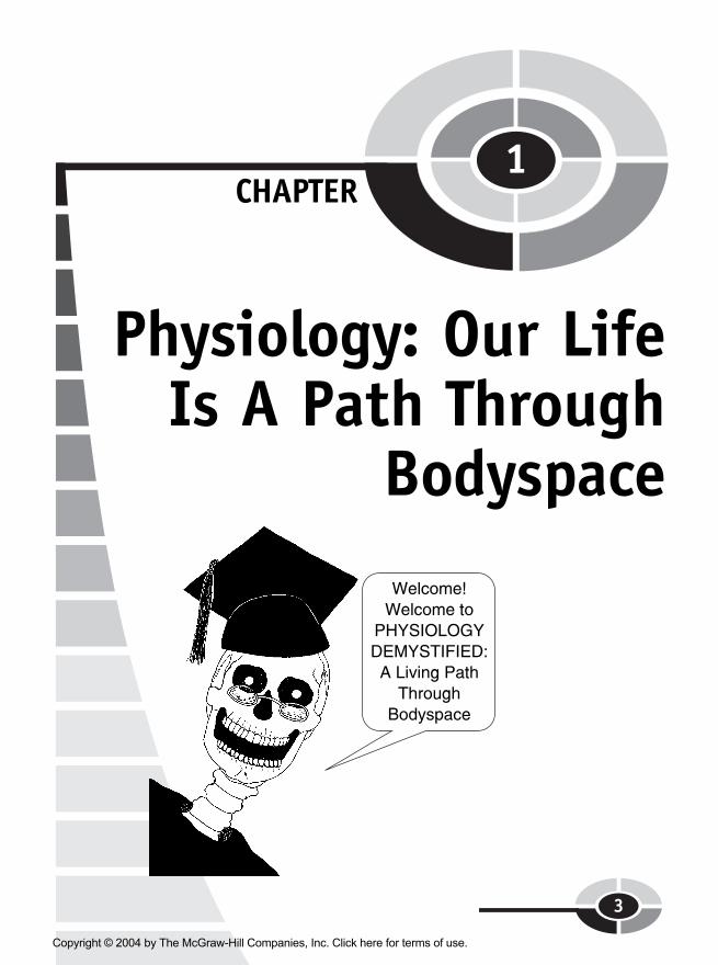
Hello, there! Who am I? Why, I am your host. They call me Professor Joe,the Talking Skeleton! I have been selected as your guide for this book,PHYSIOLOGY DEMYSTIFIED. I am here to give you a basic, ‘‘barebones’’ introduction to what happens in The Place Below Your Skin! Youand I are about to take a fascinating walk through the living body. We willdescribe this walk using a colorful phrase . . . A Living Path ThroughBodyspace.
All Life Starts with BiologyBefore we explore physiology (pronounced as fih-zee-AHL-uh-jee), we need toreview the broader subject of biology (buy-AHL-oh-jee). The word biology isreally a technical term that arises from two word parts of Ancient Greek – bi(‘‘life’’) plus -ology (‘‘study of ’’).
Thus biology literally means the ‘‘study of life.’’ (A related book,BIOLOGY DEMYSTIFIED, covers this broad subject of general biologyin considerable detail.) Since our subject of interest is human physiology,we will be focusing upon the part of general biology that includes it.
BIOLOGICAL ORDER ¼ ‘‘LIVING PATTERNS’’
Soon after you begin studying biology (or physiology), you become con-scious of its many distinct patterns. In general, a pattern is some particulararrangement of shapes, forms, colors, or designs. [Study suggestion: Closelyexamine some of the patterns you find outside and around your own home orapartment, such as its distinct grid or map of intersecting streets, variousbuildings, and sidewalks.]
For our treatment of physiology, however, we will be concentrating uponvarious patterns within the human organism (OR-gan-izm) – the entire livinghuman body. Speaking broadly, an organism is any living body with a highdegree of Biological Order. By Biological Order, we simply mean a recogniz-able pattern involving one or more organisms.
[Study suggestion: Look up into the sky and let your eyes trace the patternmade by a bird as it flies overhead. Does this pattern of flight represent a caseof Biological Order? How does this type of order differ from that found in thespecific arrangement of streets, buildings, and sidewalks around your home?]
Specifically, we want to study the patterns of Biological Order associatedwith the human body. Since the human body is a living organism, it shareswith other living creatures this fundamental relationship:
PART 1 The Golden Path of Body Function4
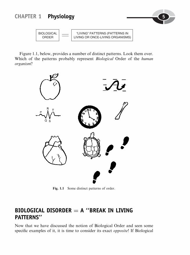
Figure 1.1, below, provides a number of distinct patterns. Look them over.Which of the patterns probably represent Biological Order of the humanorganism?
BIOLOGICAL DISORDER ¼ A ‘‘BREAK IN LIVINGPATTERNS’’
Now that we have discussed the notion of Biological Order and seen somespecific examples of it, it is time to consider its exact opposite! If Biological
CHAPTER 1 Physiology 5
Fig. 1.1 Some distinct patterns of order.

Order represents intact patterns, then Biological Disorder, of course, wouldrepresent the breaking or absence of such patterns:
Take a quick peek back at Figure 1.1 and its orderly patterns. Try topicture how breaking these patterns of order, thereby creating states of dis-order, would look. Now, follow this up with an examination of Figure 1.2.This time, ask yourself, ‘‘Which of the broken patterns probably representBiological Disorder of the human organism?’’
PART 1 The Golden Path of Body Function6
Fig. 1.2 Some broken patterns of order.
ICONS FOR BIOLOGICAL ORDER VERSUS DISORDER
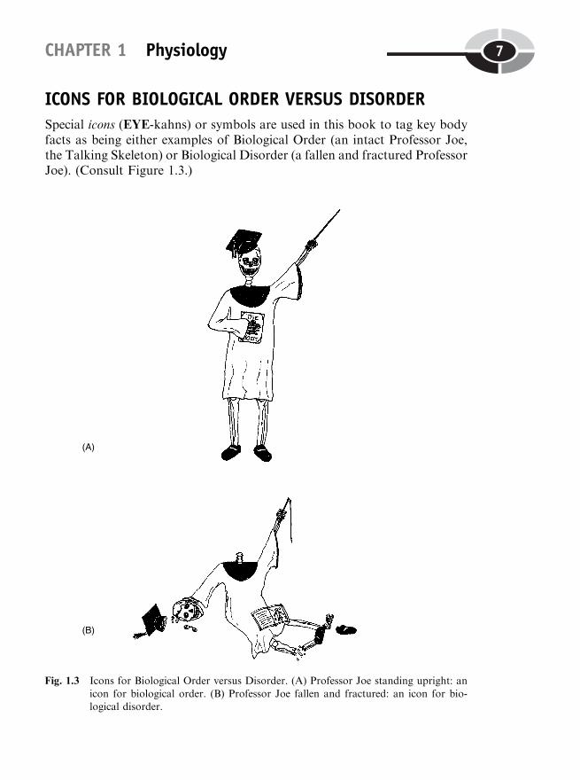
Special icons (EYE-kahns) or symbols are used in this book to tag key bodyfacts as being either examples of Biological Order (an intact Professor Joe,the Talking Skeleton) or Biological Disorder (a fallen and fractured ProfessorJoe). (Consult Figure 1.3.)
CHAPTER 1 Physiology 7
Fig. 1.3 Icons for Biological Order versus Disorder. (A) Professor Joe standing upright: an
icon for biological order. (B) Professor Joe fallen and fractured: an icon for bio-logical disorder.
(A)
(B)
Two Paths Curve Apart in the WoodsWe have said that we are on a nice walk, a nice walk through the Woods ofthe Human Body. But soon after we begin our exciting journey, we see twodifferent paths quickly splitting off, and curving away into the darkness. Oneof these paths we shall call anatomy (ah-NAT-oh-me), while the other will beknown as physiology. The splitting of these two paths of human body study –anatomy from physiology – formally began during the 1500s, and theirseparation has been getting wider ever since!
BODY STRUCTURE BECOMES ANATOMY
The word anatomy derives from Ancient Greek. It literally translates to mean‘‘the process of ’’ (-y) ‘‘cutting’’ (tom) ‘‘up or apart’’ (ana-). This exact trans-lation of anatomy clearly reflects its close connection to dissection (dih-SEK-shun), which has essentially the same meaning. In each case, the thing beingdissected is the human body and its structures. Anatomy, therefore, can bedefined as body structure and the study of body structures, primarily bymeans of dissection. (A close companion volume to this book, ANATOMYDEMYSTIFIED, looks at the various topics in human anatomy in muchgreater detail.)
Body structures would include such things as bones, muscles, the heart,and brain. Considered from a common grammar standpoint, each of thesebody structures can be considered a noun in a sentence. This is because anoun is a person, place, or thing. In the specific case of anatomy, then, thebody structures are important parts of a person (human organism).
Aristotle, the Father of Natural History
One very important name for you to know in the history of both anatomyand physiology is Aristotle (AIR-ist-ahtl). Living between 384 and 322 B.C. inAncient Greece, Aristotle is often considered the Father of Natural History –the collection and classification of plants and animals into certain groups.Aristotle is also often considered the world’s first great biologist. Aristotlegathered huge amounts of information about both the anatomy and thephysiology of numerous creatures in Nature.
PART 1 The Golden Path of Body Function8
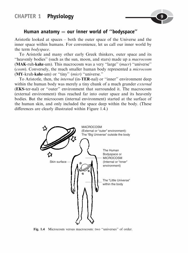
Human anatomy ¼ our inner world of ‘‘bodyspace’’
Aristotle looked at spaces – both the outer space of the Universe and theinner space within humans. For convenience, let us call our inner world bythe term bodyspace.
To Aristotle and many other early Greek thinkers, outer space and its‘‘heavenly bodies’’ (such as the sun, moon, and stars) made up a macrocosm(MAK-ruh-kahz-um). This macrocosm was a very ‘‘large’’ (macr) ‘‘universe’’(cosm). Conversely, the much smaller human body represented a microcosm(MY-kruh-kahz-um) or ‘‘tiny’’ (micr) ‘‘universe.’’
To Aristotle, then, the internal (in-TER-nal) or ‘‘inner’’ environment deepwithin the human body was merely a tiny chunk of a much grander external(EKS-ter-nal) or ‘‘outer’’ environment that surrounded it. The macrocosm(external environment) thus reached far into outer space and its heavenlybodies. But the microcosm (internal environment) started at the surface ofthe human skin, and only included the space deep within the body. (Thesedifferences are clearly illustrated within Figure 1.4.)
CHAPTER 1 Physiology 9
Fig. 1.4 Microcosm versus macrocosm: two ‘‘universes’’ of order.
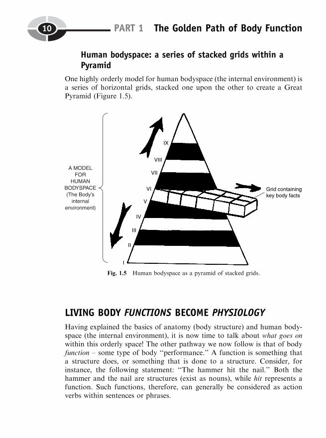
Human bodyspace: a series of stacked grids within aPyramid
One highly orderly model for human bodyspace (the internal environment) isa series of horizontal grids, stacked one upon the other to create a GreatPyramid (Figure 1.5).
LIVING BODY FUNCTIONS BECOME PHYSIOLOGY
Having explained the basics of anatomy (body structure) and human body-space (the internal environment), it is now time to talk about what goes onwithin this orderly space! The other pathway we now follow is that of bodyfunction – some type of body ‘‘performance.’’ A function is something thata structure does, or something that is done to a structure. Consider, forinstance, the following statement: ‘‘The hammer hit the nail.’’ Both thehammer and the nail are structures (exist as nouns), while hit represents afunction. Such functions, therefore, can generally be considered as actionverbs within sentences or phrases.
PART 1 The Golden Path of Body Function10
Fig. 1.5 Human bodyspace as a pyramid of stacked grids.
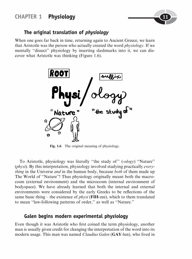
The original translation of physiology
When one goes far back in time, returning again to Ancient Greece, we learnthat Aristotle was the person who actually created the word physiology. If wementally ‘‘dissect’’ physiology by inserting slashmarks into it, we can dis-cover what Aristotle was thinking (Figure 1.6).
To Aristotle, physiology was literally ‘‘the study of ’’ (-ology) ‘‘Nature’’(physi). By this interpretation, physiology involved studying practically every-thing in the Universe and in the human body, because both of them made upThe World of ‘‘Nature’’! Thus physiology originally meant both the macro-cosm (external environment) and the microcosm (internal environment ofbodyspace). We have already learned that both the internal and externalenvironments were considered by the early Greeks to be reflections of thesame basic thing – the existence of physi (FIH-zee), which to them translatedto mean ‘‘law-following patterns of order,’’ as well as ‘‘Nature.’’
Galen begins modern experimental physiology
Even though it was Aristotle who first coined the term physiology, anotherman is usually given credit for changing the interpretation of the word into itsmodern usage. This man was named Claudius Galen (GAY-lun), who lived in
CHAPTER 1 Physiology 11
Fig. 1.6 The original meaning of physiology.
Greece and Rome during A.D. 130–200. Galen, a philosopher and physician,is often called the Father of Experimental Physiology. This is because Galenwas among the first to perform experiments – controlled trials – on livinganimals such as dogs, pigs, bears, and apes. The technical term for whathe did is vivisection (viv-uh-SEK-shun), the ‘‘process of cutting’’ (section)‘‘living’’ (vivi) things apart.
Galen’s thousands of vivisections of living animals (rather than merelydead humans or animals) brought their true body functions to light. Thus,the modern science of physiology was born! In its modern sense, physiology isthe study of living body functions, that is, it is the study of the nature of livingthings, only.
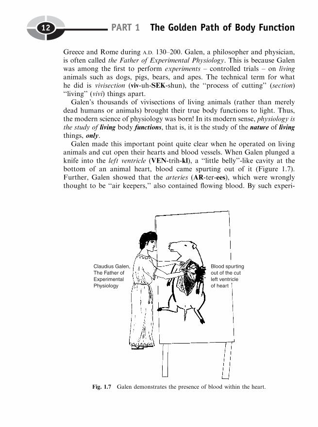
Galen made this important point quite clear when he operated on livinganimals and cut open their hearts and blood vessels. When Galen plunged aknife into the left ventricle (VEN-trih-kl), a ‘‘little belly’’-like cavity at thebottom of an animal heart, blood came spurting out of it (Figure 1.7).Further, Galen showed that the arteries (AR-ter-ees), which were wronglythought to be ‘‘air keepers,’’ also contained flowing blood. By such experi-
PART 1 The Golden Path of Body Function12
Fig. 1.7 Galen demonstrates the presence of blood within the heart.
ments on living animals, then, Galen showed that modern physiology isrestricted to the study of functions of the living body.
The living path: a curving path that passes through grids
We earlier modeled anatomy (body structure) as a series of stacked gridswithin a Body Pyramid. Physiology can thus be viewed as a curving‘‘Pathway of Life’’ that passes through these grids of Bodyspace.
Contrasting Physiology with Plain FunctionsIn addition to contrasting physiology from anatomy, it is also important tobe able to distinguish physiology from a number of plain functions that occurwithin the body. Plain functions are the actions that the various non-livingstructures in the body perform. Consider, for instance, the carbon (C) atom.The billions of carbon atoms in the human body tend to form chemical bondswith each other. The result is long strings of bonded carbon atoms. Sincecarbon atoms are, of course, non-living, their bonding together is an exampleof a plain body function, rather than physiology.
Letter Symbols for Anatomy,Physiology, and Functions
We have already classified key body facts as either being examples ofBiological Order (symbolized by an intact Professor Joe), or examples ofBiological Disorder (a fallen and fractured Professor Joe). Now, we will
CHAPTER 1 Physiology 13
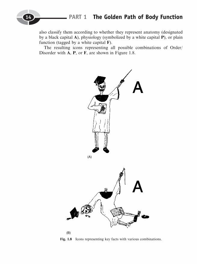
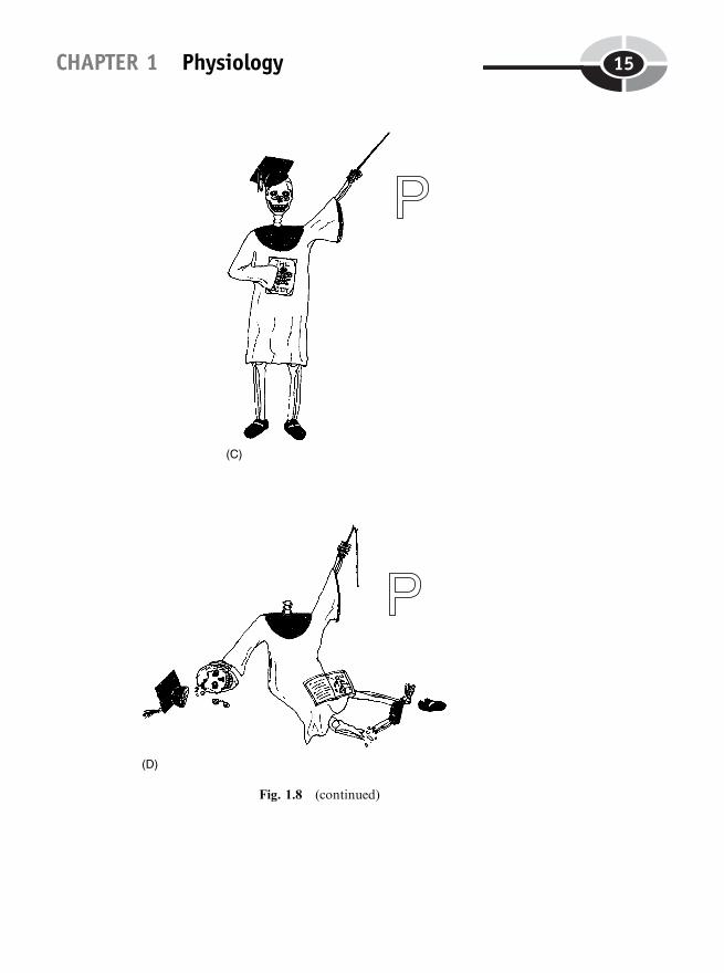
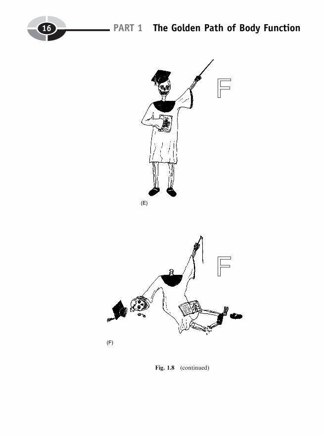
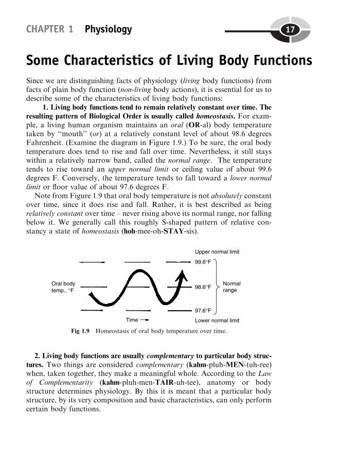
also classify them according to whether they represent anatomy (designatedby a black capital A), physiology (symbolized by a white capital P), or plainfunction (tagged by a white capital F).
The resulting icons representing all possible combinations of Order/Disorder with A, P, or F, are shown in Figure 1.8.
PART 1 The Golden Path of Body Function14
Fig. 1.8 Icons representing key facts with various combinations.
CHAPTER 1 Physiology 15
Fig. 1.8 (continued)
PART 1 The Golden Path of Body Function16
Fig. 1.8 (continued)
Some Characteristics of Living Body FunctionsSince we are distinguishing facts of physiology (living body functions) fromfacts of plain body function (non-living body actions), it is essential for us todescribe some of the characteristics of living body functions:
1. Living body functions tend to remain relatively constant over time. The
resulting pattern of Biological Order is usually called homeostasis. For exam-ple, a living human organism maintains an oral (OR-al) body temperaturetaken by ‘‘mouth’’ (or) at a relatively constant level of about 98.6 degreesFahrenheit. (Examine the diagram in Figure 1.9.) To be sure, the oral bodytemperature does tend to rise and fall over time. Nevertheless, it still stayswithin a relatively narrow band, called the normal range. The temperaturetends to rise toward an upper normal limit or ceiling value of about 99.6degrees F. Conversely, the temperature tends to fall toward a lower normallimit or floor value of about 97.6 degrees F.
Note from Figure 1.9 that oral body temperature is not absolutely constantover time, since it does rise and fall. Rather, it is best described as beingrelatively constant over time – never rising above its normal range, nor fallingbelow it. We generally call this roughly S-shaped pattern of relative con-stancy a state of homeostasis (hoh-mee-oh-STAY-sis).
2. Living body functions are usually complementary to particular body struc-
tures. Two things are considered complementary (kahm-pluh-MEN-tuh-ree)when, taken together, they make a meaningful whole. According to the Lawof Complementarity (kahm-pluh-men-TAIR-uh-tee), anatomy or bodystructure determines physiology. By this it is meant that a particular bodystructure, by its very composition and basic characteristics, can only performcertain body functions.
CHAPTER 1 Physiology 17
Fig 1.9 Homeostasis of oral body temperature over time.

Consider, for instance, the femur (FEE-mur), a long bone of the ‘‘thigh’’(femor), as well as the human eye. As is plain from an examination of Figure1.10, the femur has the basic structural characteristics of a long, white, rigidpillar. And the interior of the human eyeball consists of a complex series oflenses and fluid-filled compartments. This makes the eyeball somewhat like acomplicated telescope or camera in its structure.
The pillar-like structure of the femur, therefore, ‘‘determines’’ its majorbody function or physiology – supporting the body’s weight. And the cam-era-like anatomy of the eyeball ‘‘determines’’ its major function of focusinglight rays for the physiology of vision.
[Study suggestion: Pretend that you have plucked both of your eyeballsout of their sockets, and that you have amputated both of your lower legsjust above each femur. Now, jam your amputated femurs into your empty
PART 1 The Golden Path of Body Function18
Fig. 1.10 The Law of Complementary between body structure and function.
eye sockets, and strap both of your eyeballs to your leg stumps! Next, loweryour body onto the floor, and try to walk out of the room. Well, did youmake it? How does the Law of Complementarity make this situation soimpossible?]
3. Living body functions generally consume energy during metabolism, and
give off heat in the process. If a particular part of the body is alive, then it isalways engaging in metabolism (meh-TAB-ah-lizm) or a ‘‘state of change.’’Food that is eaten is soon changed by the chemical processes of metabolism.Energy is produced, which then performs body work. Such body work isusually some aspect of physiology, such as moving the parts of the bodyaround. And in this process of consuming energy and doing work, a con-siderable amount of heat is produced. (In other words, living things areusually a lot hotter than dead ones!)
4. Living body functions are usually sensitive to changes that occur within the
internal or external environment, and they respond to these changes. A stimulus(STIM-you-lus) is literally a ‘‘prod’’ or ‘‘goad.’’ (Picture a long stick thatpokes or prods the body.) In general, a stimulus is a detectable change in thebody’s internal or external environment. ‘‘What detects this change?’’ thecurious reader may well ask. The answer is: a sensory receptor (ree-SEP-ter). A sensory receptor is a group of specialized cells or modified nerveendings that ‘‘receive’’ (recept) the stimulus, and are excited or aroused byit. Consider, for example, a rise in oral body temperature towards its uppernormal limit of 99.6 degrees F. This rise is detected by groups of thermo-receptors (ther-moh-ree-SEP-ters) in the skin. The thermoreceptors are actu-ally naked nerve endings that are very sensitive to changes in body ‘‘heat’’(therm).
The thermoreceptors alter their physiology by firing off nerve impulses.These nerve impulses, in turn, activate certain body effectors (e-FEK-ters).An effector is part of the body that carries out a particular response, therebyhaving some ‘‘effect’’ upon the environment. The sweat glands in the skin, forinstance, are body effectors that respond to stimulation via nerve impulses byincreasing their rate of sweat secretion. As a result, the oral body temperaturesoon declines.
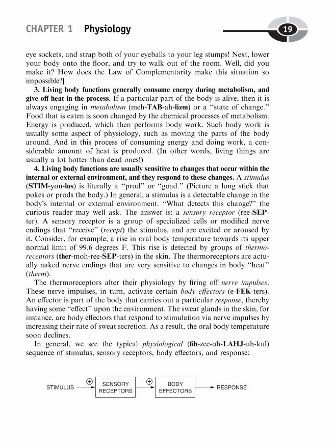
In general, we see the typical physiological (fih-zee-oh-LAHJ-uh-kul)sequence of stimulus, sensory receptors, body effectors, and response:
CHAPTER 1 Physiology 19
5. Whenever living body functions become highly irregular and disordered, a
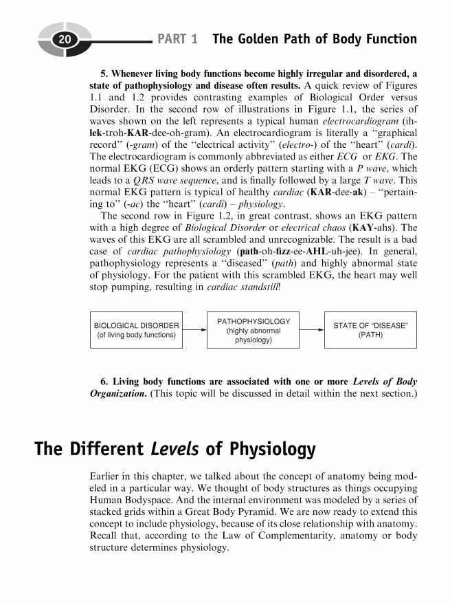
state of pathophysiology and disease often results. A quick review of Figures1.1 and 1.2 provides contrasting examples of Biological Order versusDisorder. In the second row of illustrations in Figure 1.1, the series ofwaves shown on the left represents a typical human electrocardiogram (ih-lek-troh-KAR-dee-oh-gram). An electrocardiogram is literally a ‘‘graphicalrecord’’ (-gram) of the ‘‘electrical activity’’ (electro-) of the ‘‘heart’’ (cardi).The electrocardiogram is commonly abbreviated as either ECG or EKG. Thenormal EKG (ECG) shows an orderly pattern starting with a P wave, whichleads to a QRS wave sequence, and is finally followed by a large T wave. Thisnormal EKG pattern is typical of healthy cardiac (KAR-dee-ak) – ‘‘pertain-ing to’’ (-ac) the ‘‘heart’’ (cardi) – physiology.
The second row in Figure 1.2, in great contrast, shows an EKG patternwith a high degree of Biological Disorder or electrical chaos (KAY-ahs). Thewaves of this EKG are all scrambled and unrecognizable. The result is a badcase of cardiac pathophysiology (path-oh-fizz-ee-AHL-uh-jee). In general,pathophysiology represents a ‘‘diseased’’ (path) and highly abnormal stateof physiology. For the patient with this scrambled EKG, the heart may wellstop pumping, resulting in cardiac standstill!
6. Living body functions are associated with one or more Levels of BodyOrganization. (This topic will be discussed in detail within the next section.)
The Different Levels of PhysiologyEarlier in this chapter, we talked about the concept of anatomy being mod-eled in a particular way. We thought of body structures as things occupyingHuman Bodyspace. And the internal environment was modeled by a series ofstacked grids within a Great Body Pyramid. We are now ready to extend thisconcept to include physiology, because of its close relationship with anatomy.Recall that, according to the Law of Complementarity, anatomy or bodystructure determines physiology.
PART 1 The Golden Path of Body Function20
THE GREAT PYRAMID OF STRUCTURE-FUNCTION ORDER:‘‘NORMAL’’ BODY PATTERNS
Every body structure in the anatomy of a living human organism essentiallydoes something! That is, every body structure performs a certain aspect ofeither plain function or physiology. Therefore, it is appropriate to talkabout Body Structure-Function Pyramids. Let us picture the GreatPyramid of Structure-Function Order. This Great Pyramid consists of‘‘Normal’’ Body Patterns, each of them occurring at a particular Level ofBody Organization.
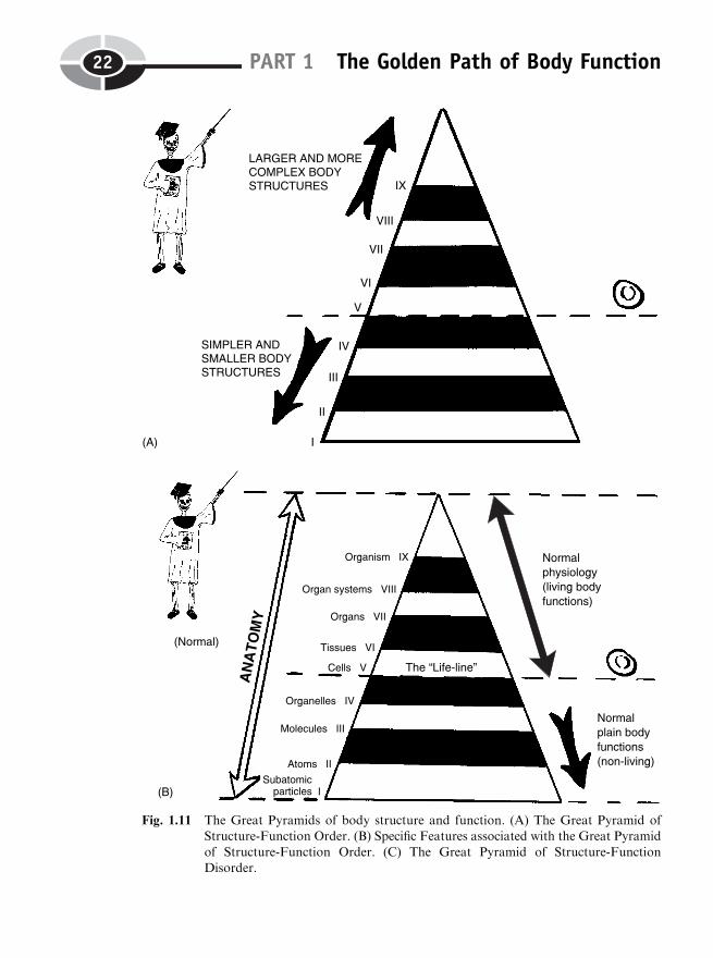
A Level of Body Organization is a particular degree of size and complexityof body structures. In general, we can picture the Great Pyramid ofStructure-Function Order as consisting of nine different levels of bodyorganization (Figure 1.11, A).
Near the base of the Pyramid, the body structures of anatomy are simplerand smaller. But as we climb toward the peak or apex of the Pyramid, thebody structures become progressively larger and more complex.
Figure 1.11 (B) gives specific names to each of the nine levels of bodyorganization in the Pyramid. Starting at the base, these are called thesubatomic (sub-ah-TAH-mik) particles, atoms, molecules, organelles (OR-gah-nels), cells, tissues, organs, organ systems, and the entire humanorganism.
THE GREAT PYRAMID OF STRUCTURE-FUNCTIONDISORDER: ‘‘ABNORMAL’’ BODY PATTERNS
‘‘What about body structures and functions that are in a state of BiologicalDisorder?’’ the inquiring reader needs to question. For a reply, one need onlylook at Figure 1.11 (C). Here we picture the Great Pyramid of Structure-Function Disorder. Shown alongside this Pyramid is our old friend,Professor Joe, in a fractured and fallen-down state. Once again, Levels I–IX (subatomic particles through the organism) are shown. But here the bodystructures and functions associated with these levels are in a state of disorder,such that they represent ‘‘abnormal’’ body patterns. Their anatomy can becalled pathological (path-oh-LAHJ-ih-kal) anatomy, while their physiologycan likewise be called pathophysiology. In short, both pathological anatomyand pathophysiology are similar in that they generally reflect ‘‘disease’’(path).
CHAPTER 1 Physiology 21
PART 1 The Golden Path of Body Function22
Fig. 1.11 The Great Pyramids of body structure and function. (A) The Great Pyramid ofStructure-Function Order. (B) Specific Features associated with the Great Pyramid
of Structure-Function Order. (C) The Great Pyramid of Structure-FunctionDisorder.
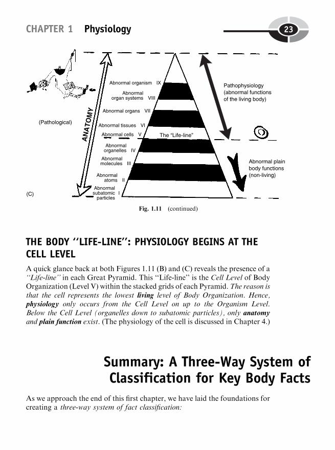
THE BODY ‘‘LIFE-LINE’’: PHYSIOLOGY BEGINS AT THECELL LEVEL
A quick glance back at both Figures 1.11 (B) and (C) reveals the presence of a‘‘Life-line’’ in each Great Pyramid. This ‘‘Life-line’’ is the Cell Level of BodyOrganization (Level V) within the stacked grids of each Pyramid.The reason isthat the cell represents the lowest living level of Body Organization. Hence,physiology only occurs from the Cell Level on up to the Organism Level.Below the Cell Level (organelles down to subatomic particles), only anatomyand plain function exist. (The physiology of the cell is discussed in Chapter 4.)
Summary: A Three-Way System ofClassification for Key Body Facts
As we approach the end of this first chapter, we have laid the foundations forcreating a three-way system of fact classification:
CHAPTER 1 Physiology 23
Fig. 1.11 (continued)
PART 1 The Golden Path of Body Function24
Organ 1
Organ 1
1. Classification according to Biological Order/Disorder. A key body factcan be labeled with Professor Joe standing and pointing if it represents a caseof Biological Order. If it is a case of Biological Disorder, however, then it islabeled with an icon of Professor Joe fallen and fractured.
2. Classification according to identity as either Anatomy, Physiology, or
Plain Body Function. If the key text fact being highlighted representsAnatomy, then a black capital A is placed under Professor Joe’s pointer(either intact or broken). If the fact is Physiology, then a white capital P isplaced under the intact/broken pointer. Finally, if the fact is just plainFunction, a white capital F is placed under the Professor’s pointer.
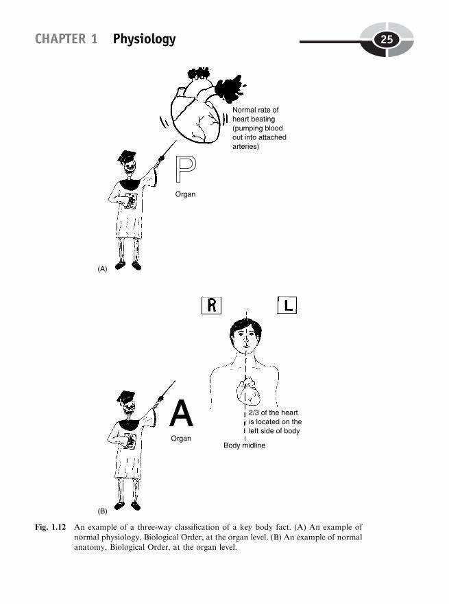
3. Classification according to Level of Body Organization. The third way ofclassifying a key fact is according to the level of body organization that itrepresents. Consider, for example, a key fact such as this one: ‘‘The normalresting heart usually beats at a rate of about 72 times per minute.’’
First, the standing Professor Joe is used, because the heart is in a state ofBiologicalOrder.Under the Professor’s pointer,we insert awhiteP, because anaspect of normal physiology of the heart (its rate of beating) is occurring. Andthirdly, we have the word organ, written under the P, because it is the physiol-ogy of the entire heart organ that is being described. (See Figure 1.12, A.)
Now consider this sentence: ‘‘About 2/3 of the heart is located on theleft side of the body midline, in most individuals.’’ The black capital A isused under standing Professor Joe for normal anatomy and a state ofBiological Order. The word organ is written under the A, because it isthe anatomy or structure of the entire heart that is being discussed. (SeeFigure 1.12, B.)
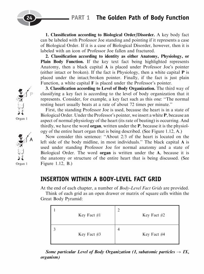
INSERTION WITHIN A BODY-LEVEL FACT GRID
At the end of each chapter, a number of Body-Level Fact Grids are provided.Think of each grid as an open drawer or matrix of square cells within the
Great Body Pyramid:
Some particular Level of Body Organization (I, subatomic particles! IX,organism)
1Key Fact #1
2Key Fact #2
3Key Fact #3
4Key Fact #4
CHAPTER 1 Physiology 25
Fig. 1.12 An example of a three-way classification of a key body fact. (A) An example of
normal physiology, Biological Order, at the organ level. (B) An example of normalanatomy, Biological Order, at the organ level.
Each of the four numbered cells or compartments within each grid pro-vides a specific place for you to briefly write-in and summarize the key factsyou have read, about a particular body structure and, quite often, its asso-ciated physiology. Since the grids essentially represent levels of body struc-ture (anatomy), the frequent placing of physiological facts into the gridssuggests that physiology is weaving A Golden Path – A Living PathwayThrough Bodyspace! (This colorful metaphor was presented at the beginningof this chapter.)
As you progress through PHYSIOLOGY DEMYSTIFIED, chapter bychapter, you can conveniently return to these Body-Level Grids and reviewand retrieve the information stored there. In this way, you will be learning tothink much like an Ancient Egyptian, who liked to put almost everythingsomewhere into a Pyramid!
QuizRefer to the text in this chapter if necessary. A good score is at least 8 correctanswers out of these 10 questions. The answers are listed in the back of thisbook.
1. Biology and physiology have in common:(a) Their concentration upon the structure of living plants and
animals(b) A primary focus upon living body functions(c) An avoidance of anything having to do with diseases(d) Study of dead people, plants, and animals
2. Biological Order can best be described as:(a) The study of the nature of living things(b) Essentially the same thing as randomness and chaos(c) The occurrence of various patterns within living organisms(d) Having no significant relationship to either anatomy or phys-
iology
3. The word anatomy is closest in its literal translation to the word:(a) Dissection(b) Physiology(c) Nature(d) Function
PART 1 The Golden Path of Body Function26
4. The Father of Natural History is:(a) Jules Verne(b) Baby Heinie(c) Hippocrates(d) Aristotle
5. The Ancient Greek idea of a Macrocosm was basically equivalentto:(a) The internal environment(b) Homeostasis(c) The normal range(d) The external environment
6. ‘‘The kite flew over the roof of the barn.’’ In this sentence, the wordflew represents:(a) Physiology(b) Anatomy(c) Plain function(d) A noun
7. Claudius Galen is usually considered the:(a) Inventor of the microscope(b) Creator of the word physiology(c) First person to systematically dissect human cadavers(d) The Father of Experimental Physiology
8. The sodium (Na) atoms in the human body fluids can form chemicalbonds with the chlorine (Cl) atoms. This bonding process is a caseof:(a) Plain body function(b) Biological Disorder(c) Physiology(d) Metabolism
9. Relative constancy of blood glucose (sugar) concentration over a24-hour period:(a) Upper normal limit(b) Homeostasis(c) Heterostasis(d) Sensory reception
CHAPTER 1 Physiology 27
10. A good example of the Law of Complementarity:(a) Lungfish crawling out onto the land to search for food(b) Human red blood cells exploding when placed into distilled
water(c) A gentlemen tipping his hat to a lady passing on the sidewalk(d) Teeth of sharks being put onto necklaces by South Seas natives
Body-Level Grids for Chapter 1Several key body facts were tagged with numbered icons in the page marginsof this chapter. Write a short summary of each of these key facts into anumbered cell or box within the appropriate Body-Level Grid that appearsbelow.
Anatomy and Biological Order Fact Grid for Chapter 1:
ORGANLevel
PART 1 The Golden Path of Body Function28
1
Physiology and Biological Order Fact Grid for Chapter 1:
ORGANLevel
CHAPTER 1 Physiology 29
1

30
CHAPTER2
Control of theInternal Environment:A Story AboutFeedback andHomeostasis
We have now begun our Magical Journey down a Living Path – HumanPhysiology, our Living Path through the World of ‘‘Bodyspace.’’
Chapter 1 taught us to look for normal patterns of Biological Order withinthe Great Pyramid of Structure-Function Order.
Copyright © 2004 by The McGraw-Hill Companies, Inc. Click here for terms of use.

Enter Hippocrates: Human HealthSeen as a Balance
Closely associated with patterns of order is the notion of balance. A balance,in general, is a rough equality between two or more different things.Consider, for example, the physiological concept of ‘‘eating a balanceddiet.’’ According to this view, to maintain their long-term health and well-being, people should not over-consume any one particular type of food intheir diet. Rather, they should eat all three major types of foodstuffs –proteins, lipids (LIP-ids), and carbohydrates (kar-boh-HIGH-draytes) – inroughly equal or ‘‘balanced’’ amounts.
This hearkens back to the time of the Ancient Greeks. In particular, it wasthe Greeks who advanced the concept of the Golden Mean, and of the classi-cal ideal of ‘‘Nothing to excess, but rather, moderation in all things.’’
Tracing back through the historical records, we repeatedly find the nameof one Ancient Greek, especially, who is famous for this philosophy. Hisname is Hippocrates (hih-PAHK-rah-tees). Hippocrates was a physician, tea-cher, and author who lived in Greece from about 460–370 B.C. Hippocrates iswidely considered the Father of Modern Medicine, because he broke withancient superstitions and saw human health and disease in more rational,cause-and-effect terms.
Before his birth, earlier Greek philosophers interpreted the Cosmos asbeing composed of four basic universal elements. These were air, fire, earth,and water. To Hippocrates (and later, Aristotle), this balance in the large,external macrocosm (universe) was also reflected as a balance within themuch smaller, internal microcosm (human body).
Specifically, Hippocrates believed in a balance of the four body humors(HYOO-mers) or ‘‘fluids.’’ This balance was formally called the Humoral(hyoo-MOR-al) Doctrine. The four body humors were the four primarytypes of fluid the Ancient Greeks thought made up the body. (ConsultFigure 2.1.)
The first body humor, blood, was hot and moist, like the moist universalelement, air. The second body humor, yellow bile from the liver, also seemedhot, but was more dry than blood (due to its lower water content). Thus, theGreeks equated yellow bile with the universal element, fire. The third bodyhumor, black bile, was mistakenly thought to come from the spleen. (This errormay have arisen from the frequent clinical observation of black, blood-filledvomit or stools in patients suffering internal bleeding.) Since black bile wasconsidered rather cold and dry, it was tied to the universal element, earth.
CHAPTER 2 The Internal Environment 31
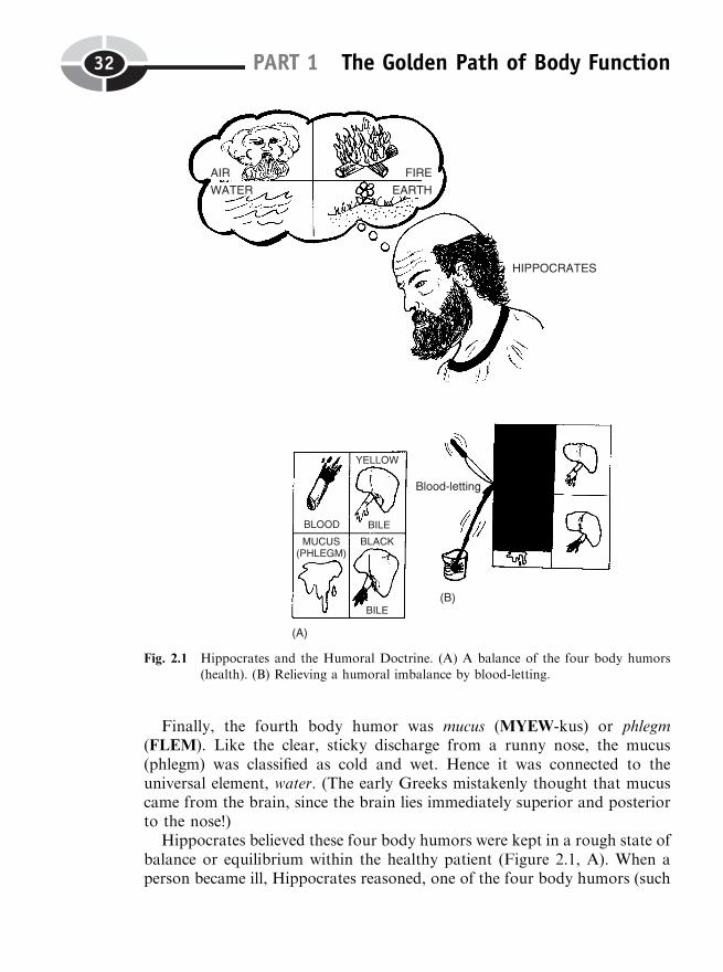
Finally, the fourth body humor was mucus (MYEW-kus) or phlegm(FLEM). Like the clear, sticky discharge from a runny nose, the mucus(phlegm) was classified as cold and wet. Hence it was connected to theuniversal element, water. (The early Greeks mistakenly thought that mucuscame from the brain, since the brain lies immediately superior and posteriorto the nose!)
Hippocrates believed these four body humors were kept in a rough state ofbalance or equilibrium within the healthy patient (Figure 2.1, A). When aperson became ill, Hippocrates reasoned, one of the four body humors (such
PART 1 The Golden Path of Body Function32
Fig. 2.1 Hippocrates and the Humoral Doctrine. (A) A balance of the four body humors(health). (B) Relieving a humoral imbalance by blood-letting.

as the blood) was present in a much greater amount than the other three.(Observe Figure 2.1, B.) A logical mode of treatment, therefore, was blood-letting. By cutting a vessel and letting out the excess blood, the Greek healersreasoned, the four body humors could be brought back into a rough equili-brium or balance, and a state of health be re-achieved.
Claude Bernard and His Dogs:A Preview of Homeostasis
The Golden Greek Ideal of the Humoral Doctrine (balance among the fourbody humors) did, when you really think about it, involve an equilibriumamong body fluids, rather than solid structures.
Almost 2,000 years later, on the Continent of Europe, a Frenchmannamed Claude Bernard (ber-NAR) made his own experimental follow-up toHippocrates and the Humoral Doctrine. Bernard was a vivisectionist, likeClaudius Galen (Chapter 1) long before him. As an experimental physiologist(fiz-ee-AHL-uh-jist), Bernard, of course, badly needed to observe the func-tioning of living organisms. But to the great horror of his rich wife, whoheard them howling, Bernard strapped dogs down onto lab tables in thebasement of his private lab, and operated upon them while they were stillalive! Even worse, Bernard did not give them any anesthesia (an-es-THEE-zha) to ‘‘remove’’ (an-) their pain ‘‘sensations’’ (esthes)! (Examine Figure 2.2.)
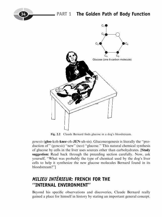
One of the main points of interest for Bernard was the physiology of thedigestive system. It was during surgery of the open abdominal (ahb-DAHM-ih-nal) cavity of one of his howling dogs that Bernard made an importantdiscovery. He found important information about the natural production ofglucose – a ‘‘sweet’’ (gluc) ‘‘carbohydrate’’ (-ose), that is, a sugar. Glucose isthe major sweet carbohydrate (sugar) used for fuel by the body’s cells.Bernard collected the blood draining from the dog’s liver through the hepatic(heh-PAT-ik) veins, which run below it. Now, Bernard had fed the dog onlymeat, which is mostly composed of protein. To his surprise, however, theblood draining from the dog’s liver was rich in glucose! Since the dog hadn’teaten any carbohydrates, Bernard correctly reasoned, the liver must havesynthesized (SIN-thuh-sized) the glucose itself, by putting the various partsof the molecule ‘‘together’’ (syn-).
This important observation provided the first strong evidence of naturalchemical synthesis – the making of new chemicals within the body. Thetechnical name for this particular type of chemical synthesis is gluconeo-
CHAPTER 2 The Internal Environment 33
Cell 1
Cell 2
genesis (gloo-koh-knee-oh-JEN-uh-sis). Gluconeogenesis is literally the ‘‘pro-duction of ’’ (genesis) ‘‘new’’ (neo) ‘‘glucose.’’ This natural chemical synthesisof glucose by cells in the liver uses sources other than carbohydrates. [Studysuggestion: Read back through the preceding section carefully. Now, askyourself, ‘‘What was probably the type of chemical used by the dog’s livercells to help it synthesize the new glucose molecules Bernard found in itsbloodstream?’’]
MILIEU INTERIEUR: FRENCH FOR THE‘‘INTERNAL ENVIRONMENT’’
Beyond his specific observations and discoveries, Claude Bernard reallygained a place for himself in history by stating an important general concept.
PART 1 The Golden Path of Body Function34
Fig. 2.2 Claude Bernard finds glucose in a dog’s bloodstream.

This concept is that of the milieu (MEEL-yoo) interieur (an-TARE-ee-er),which is French for ‘‘internal environment.’’
In this book, we have considered the internal environment as essentiallythe same thing as Human Bodyspace. We have defined it in a very broadsense, as including everything in the body that lies deep to the surface of theskin.
Claude Bernard, however, had a more narrow interpretation of the inter-nal environment (milieu interieur). For him, it primarily consisted of the innerbody fluids (including the blood), which provide a bathing medium for thecells. This bathing fluid includes important nutrients (NEW-tree-unts) or‘‘nourishing substances,’’ such as oxygen and glucose. The cells receivethese nutrients, and utilize them for their physiology and metabolism. Thewaste products of metabolism, such as carbon dioxide (die-OX-eyed) and acid,are released from the cells, and pass into the internal environment.
To properly understand Bernard’s concept of the internal environment, weneed to take a closer look at the fluid both inside and outside of our cells.This will require some new terms. The prefix extra- means ‘‘outside of ’’(something). And the word cellular (SELL-you-lar) refers to ‘‘little cells.’’We can put this information together and create a single new term, extra-cellular (eks-trah-SELL-you-lar). The extracellular fluid, therefore, is thefluid that literally lies ‘‘outside the little cells.’’ (See Figure 2.3.)
Since all of our cells lie deep within the body, the extracellular fluid sur-rounding the cells acts as Bernard’s ‘‘internal environment.’’ Even though itis located outside the cells, the extracellular fluid still lies internal or deep tothe skin surface. Hence, this fluid is both internal (to the skin surface) andexternal (to the individual body cells), at the same time! Summarizing theseideas, we can write the following word equation:
CHAPTER 2 The Internal Environment 35
Cell 3
Fig. 2.3 The ‘‘internal environment’’ as the extracellular fluid bathing our cells.
EXTRACELLULARFLUID
¼ Fluid lying outside andaround the body cells,but still deep to skin
surface
¼ CLAUDEBERNARD’S‘‘INTERNAL
ENVIRONMENT’’
‘‘What about the fluid lying inside our body cells?’’ you may well be askingat this point. Taking the same approach to word-building, we create a newterm, intracellular (in-trah-SELL-you-lar). The intracellular fluid, conse-quently, is the fluid present ‘‘within or inside’’ (intra-) our ‘‘little cells’’(cellul).
Because both nutrients and waste products frequently pass into and out ofthe cell, many of the components of the intracellular fluid and extracellularfluid mix with one another. ‘‘It is the fixity of the internal environment that isthe condition of free and independent life,’’ Claude Bernard maintained.Because the components of the extracellular fluid (internal environment) arerelatively stable, the physiology within our cells is likewise relatively stable. AsBernard discovered, for example, the concentration of glucose within thebloodstream (and other portions of the body’s extracellular fluid) remainsrelatively constant over time. Hence, the body cells are assured of a fairlysteady supply of glucose energy for their continued long-term survival.
Walter B. Cannon: The Wisdom of the Bodyand Homeostasis
Claude Bernard (1813–1878) did most of his work in the mid-1800s. Anotherimportant physiologist, Walter B. Cannon, was born in 1871. Thus, Cannonwas only seven years old when Claude Bernard died.
Around 1915 Cannon began work on the body’s response to severe stress.It was Cannon who showed how the hormone called adrenaline (ah-DREN-ah-lin) or epinephrine (ep-ih-NEF-rin) was released into the bloodstream dur-ing the so-called ‘‘fight-or-flight’’ response. By the ‘‘fight-or-flight’’ response,we mean the reaction of a human or animal to severe stress – and perhaps,life-threatening danger! So the frightened deer or rabbit being chased by apack of howling wolves must either run away (‘‘take flight’’) or just standthere and ‘‘put up a fight’’!
Certainly, Cannon was vividly impressed by the linkage of strong emo-tions, such as those of rage and fear in the ‘‘fight-or-flight’’ response, to theincreased release of adrenaline (epinephrine) into the human bloodstream.
PART 1 The Golden Path of Body Function36
Tissue 1
Molecule 1
Cell 1
Cell 2
And epinephrine, once released, was found to powerfully stimulate dramaticincreases in heart rate, blood pressure, and various other body functions.
THE FIRST DEFINITION OF HOMEOSTASIS IS BORN
In 1926, Walter Cannon generalized his observations in an important book,called The Wisdom of the Body. It was in this book and associated researchpapers that Cannon showed the great ‘‘wisdom’’ to create the term homeo-stasis. His observation that the strong emotions and the hormone epin-ephrine greatly disrupted normal, fairly constant and stable, patterns ofbody functioning, logically led to the concept of homeostasis withinCannon’s mind.
Walter Cannon defined homeostasis as ‘‘a condition which may vary, butwhich is relatively constant.’’
HOMEOSTASIS (WalterCannon’s original definition)
¼ A RELATIVELY CONSTANTCONDITION WITHIN THE BODY’S
INTERNAL ENVIRONMENT
Homeostasis and ModernControl System Theory
Since the time of Claude Bernard and Walter Cannon, the techniques andinstruments for measuring various aspects of the body have been greatlyrefined. Hence, we now speak of specific anatomical and physiological para-meters (par-AM-uh-ters) within the body’s internal environment. (A para-meter, in general, is an aspect of some thing – such as oral body temperature– that has been measured and expressed as a certain number of units.) Andthe internal environment concept can be greatly broadened to include notonly the extracellular fluid bathing our cells, but all of Human Bodyspacelying deep to the surface of the skin.
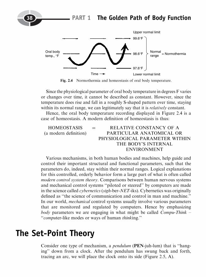
Recall (Chapter 1) that we record body parameters and see whether theircurrent values lie within their normal range – the distance between their lowernormal limit and their upper normal limit. Let us repeat and adapt fromFigure 1.9, which showed the example of oral body temperature recorded indegrees Fahrenheit (Figure 2.4). We can say that a mouth temperaturebetween 97.6 degrees F and 99.6 degrees F represents normothermia (nor-moh-THERM-ee-ah). This literally means ‘‘a condition of ’’ (-ia) ‘‘normal’’(normo-) body ‘‘heat’’ (therm) or temperature. Oral body temperature thuslies within its ‘‘normal’’ (normo-) range.
CHAPTER 2 The Internal Environment 37
Organ 1
Organism 1
PART 1 The Golden Path of Body Function38
Since the physiological parameter of oral body temperature in degrees F variesor changes over time, it cannot be described as constant. However, since thetemperature does rise and fall in a roughly S-shaped pattern over time, stayingwithin its normal range, we can legitimately say that it is relatively constant.
Hence, the oral body temperature recording displayed in Figure 2.4 is acase of homeostasis. A modern definition of homeostasis is thus:
HOMEOSTASIS(a modern definition)
¼ RELATIVE CONSTANCY OF APARTICULAR ANATOMICAL OR
PHYSIOLOGICAL PARAMETER WITHINTHE BODY’S INTERNAL
ENVIRONMENT
Various mechanisms, in both human bodies and machines, help guide andcontrol their important structural and functional parameters, such that theparameters do, indeed, stay within their normal ranges. Logical explanationsfor this controlled, orderly behavior form a large part of what is often calledmodern control system theory. Comparisons between human nervous systemsand mechanical control systems ‘‘piloted or steered’’ by computers are madein the science called cybernetics (sigh-ber-NET-iks). Cybernetics was originallydefined as ‘‘the science of communication and control in man and machine.’’In our world, mechanical control systems usually involve various parametersthat are monitored and regulated by computers. Hence by emphasizingbody parameters we are engaging in what might be called Compu-Think –‘‘computer-like modes or ways of human thinking.’’
The Set-Point TheoryConsider one type of mechanism, a pendulum (PEN-juh-lum) that is ‘‘hang-ing’’ down from a clock. After the pendulum has swung back and forth,tracing an arc, we will place the clock onto its side (Figure 2.5, A).
Fig. 2.4 Normothermia and homeostasis of oral body temperature.
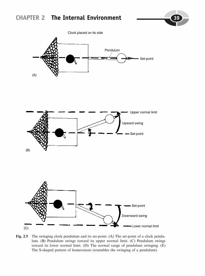
CHAPTER 2 The Internal Environment 39
Fig. 2.5 The swinging clock pendulum and its set-point. (A) The set-point of a clock pendu-lum. (B) Pendulum swings toward its upper normal limit. (C) Pendulum swings
toward its lower normal limit. (D) The normal range of pendulum swinging. (E)The S-shaped pattern of homeostasis (resembles the swinging of a pendulum).
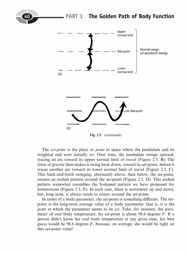
The set-point is the place or point in space where the pendulum and itsweighted end were initially set. Over time, the pendulum swings upward,tracing an arc toward its upper normal limit of travel (Figure 2.5, B). Theforce of gravity then makes it swing back down, toward its set-point, before ittraces another arc toward its lower normal limit of travel (Figure 2.5, C).This back-and-forth swinging, alternately above, then below, the set-point,creates an arched pattern around the set-point (Figure 2.5, D). This archedpattern somewhat resembles the S-shaped pattern we have proposed forhomeostasis (Figure 2.5, E). In each case, there is movement up and down,but, long term, it always tends to center around the set-point.
In terms of a body parameter, the set-point is something different. The set-point is the long-term average value of a body parameter; that is, it is thepoint at which the parameter seems to be set. Take, for instance, the para-meter of oral body temperature. Its set-point is about 98.6 degrees F. If aperson didn’t know her oral body temperature at any given time, her bestguess would be 98.6 degrees F, because, on average, she would be right onthis set-point value!
PART 1 The Golden Path of Body Function40
Fig. 2.5 (continued)

Stressors or Stimuli: Disturbers ofBody Parameters
A person with a probing mind might now question, ‘‘Well, if oral bodytemperature is actually set at a point or value of about 98.6 degrees F,then why doesn’t it just stay there? Why don’t we have an absolute constancyof oral body temperature over time, with no change in its value whatsoever?’’The answer is that numerous stressors (STRESS-ors) just won’t let the para-meter stay at its set-point value for very long!
A stressor is a change in the internal or external environment that disturbsa body parameter from its set-point level. For oral body temperature, thereare heat stressors that tend to push the value of oral body temperature aboveits set-point level. Drinking a cup of hot coffee, taking a warm bath, orvigorously exercising, for instance, could all act as heat stressors that pushthe body temperature up toward its upper normal limit of 99.6 degrees F.(Consult Figure 2.6, A.)
Conversely, drinking a glass of cold water, taking a cool shower, or restingon your back stripped down to your jockey shorts, could all act as coldstressors. These stressors would tend to push the value of oral body tempera-ture below its set-point level, and toward its lower normal limit of 97.6degrees F. (Examine Figure 2.6, B.)
Therefore, since stressors are present everywhere, all the time, absoluteconstancy of a particular body parameter is an unattainable goal. The bestyou can reasonably hope for is that a relative constancy of the body para-meter – homeostasis – can somehow be maintained.
Certain stressors are also stimuli (STIM-you-lie). A stimulus or ‘‘goad’’(mentioned in Chapter 1) is a detectable change in the body’s internal orexternal environment. A cold stressor, such as having a pail of icy waterdumped over your head, disturbs relative constancy of oral body tempera-ture, but it acts as a stimulus as well. How do we know this? – We can feel orsense it! [Study suggestion: Suppose there was a 500-pound block of radio-active material that someone hid, without your knowledge, under your deskat work or at school. Would you still come and sit there every day? Wouldthis radioactive material be a significant stressor? Would it also act as astimulus? – Why, or why not?]
CHAPTER 2 The Internal Environment 41

Feedback Systems or CyclesLet us return to a consideration of an ordinary clock pendulum. Interestinglyenough, temperature stressors can also ‘‘disturb’’ a clock pendulum! Whenthe room temperature gets hot, the rod in a clock pendulum expands, and itswings more slowly. Conversely, when the room gets cold, the rod in thependulum contracts, and it swings more rapidly. But no matter how fast or
PART 1 The Golden Path of Body Function42
Fig. 2.6 Contrasting effects of heat stressors versus cold stressors upon oral body tempera-
ture. (A) Heat stressors that raise body temperature above its set-point level.(B) Cold stressors that lower body temperature below its set-point level.
slowly the pendulum swings, it has a particular normal range of travel,swinging first to an upper normal limit at one end of its arc, and anotherat the opposite end of its arc. The regulation and control of the distance ofswinging of such a clock pendulum from its set-point position, therefore, is arelatively simple matter.
THE GENERAL OPERATION AND COMPONENTS OFFEEDBACK CYCLES
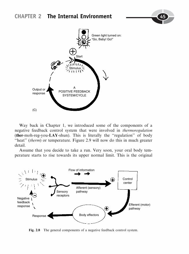
The regulation and control of various body parameters, such as oral bodytemperature in degrees Fahrenheit, however, is much more complex andinvolved! This regulation is accomplished by the action of what are formallycalled feedback systems or feedback cycles. A feedback system or cycle is asystem whose output or response curves or ‘‘feeds’’ back upon the start (thestimulus or stressor), thereby influencing whether the system repeats itself ornot. A general plan for a feedback system or cycle is traced in Figure 2.7, A.
There are two main types of feedback systems or cycles. The basic opera-tion of a negative feedback system is shown in Figure 2.7 (B), while that for apositive feedback system is depicted in Figure 2.7 (C).
A NEGATIVE FEEDBACK OR CONTROL SYSTEM
A negative feedback system is a system whose output or response curves or‘‘feeds’’ back upon the start in a negative manner, by removing or correctingthe conditions of the original stimulus. (This ‘‘negative’’ or ‘‘removing’’ effectupon the original stimulus is symbolized by the minus sign, and by a redtraffic light in Figure 2.7, B, which essentially tells the driver of a car, ‘‘Stop,Baby! Stop!’’)
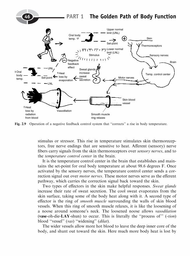
A group of sensory receptors originally sense or detect the stimulus (Figure2.8). The sensory receptors, after they are excited, send information over anafferent (A-fer-ent) or sensory pathway. An afferent or sensory pathway is apathway that ‘‘carries’’ (fer) information about the stimulus from the sensoryreceptors, and ‘‘toward’’ (af-) a control center. A control center is a collectionof anatomical and physiological components that establishes and maintainsthe set-point for a particular body parameter. A signal from the controlcenter is then sent over an efferent (EE-fer-ent) or motor pathway. An efferentor motor pathway is a pathway that ‘‘carries’’ (fer) motor or ‘‘movement’’-related information ‘‘away’’ (ef-) from the control center, and out towardbody effectors.
CHAPTER 2 The Internal Environment 43
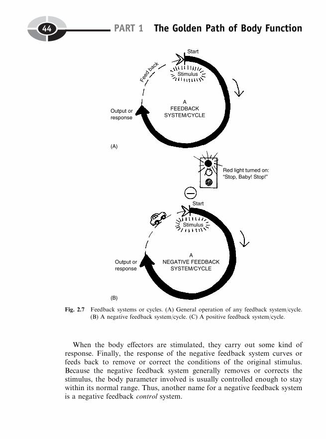
When the body effectors are stimulated, they carry out some kind ofresponse. Finally, the response of the negative feedback system curves orfeeds back to remove or correct the conditions of the original stimulus.Because the negative feedback system generally removes or corrects thestimulus, the body parameter involved is usually controlled enough to staywithin its normal range. Thus, another name for a negative feedback systemis a negative feedback control system.
PART 1 The Golden Path of Body Function44
Fig. 2.7 Feedback systems or cycles. (A) General operation of any feedback system/cycle.(B) A negative feedback system/cycle. (C) A positive feedback system/cycle.
Way back in Chapter 1, we introduced some of the components of anegative feedback control system that were involved in thermoregulation(ther-moh-reg-you-LAY-shun). This is literally the ‘‘regulation’’ of body‘‘heat’’ (therm) or temperature. Figure 2.9 will now do this in much greaterdetail.
Assume that you decide to take a run. Very soon, your oral body tem-perature starts to rise towards its upper normal limit. This is the original
CHAPTER 2 The Internal Environment 45
Fig. 2.8 The general components of a negative feedback control system.
stimulus or stressor. This rise in temperature stimulates skin thermorecep-tors, free nerve endings that are sensitive to heat. Afferent (sensory) nervefibers carry signals from the skin thermoreceptors over sensory nerves, and tothe temperature control center in the brain.
It is the temperature control center in the brain that establishes and main-tains the set-point for oral body temperature at about 98.6 degrees F. Onceactivated by the sensory nerves, the temperature control center sends a cor-rection signal out over motor nerves. These motor nerves serve as the efferentpathway, which carries the correction signal back toward the skin.
Two types of effectors in the skin make helpful responses. Sweat glandsincrease their rate of sweat secretion. The cool sweat evaporates from theskin surface, taking some of the body heat along with it. A second type ofeffector is the ring of smooth muscle surrounding the walls of skin bloodvessels. When this ring of smooth muscle relaxes, it is like the loosening ofa noose around someone’s neck. The loosened noose allows vasodilation(vase-oh-die-LAY-shun) to occur. This is literally the ‘‘process of ’’ (-tion)blood ‘‘vessel’’ (vas) ‘‘widening’’ (dilat).
The wider vessels allow more hot blood to leave the deep inner core of thebody, and shunt out toward the skin. Here much more body heat is lost by
PART 1 The Golden Path of Body Function46
Fig. 2.9 Operation of a negative feedback control system that ‘‘corrects’’ a rise in body temperature.
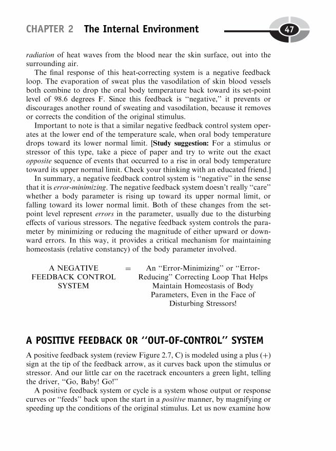
radiation of heat waves from the blood near the skin surface, out into thesurrounding air.
The final response of this heat-correcting system is a negative feedbackloop. The evaporation of sweat plus the vasodilation of skin blood vesselsboth combine to drop the oral body temperature back toward its set-pointlevel of 98.6 degrees F. Since this feedback is ‘‘negative,’’ it prevents ordiscourages another round of sweating and vasodilation, because it removesor corrects the condition of the original stimulus.
Important to note is that a similar negative feedback control system oper-ates at the lower end of the temperature scale, when oral body temperaturedrops toward its lower normal limit. [Study suggestion: For a stimulus orstressor of this type, take a piece of paper and try to write out the exactopposite sequence of events that occurred to a rise in oral body temperaturetoward its upper normal limit. Check your thinking with an educated friend.]
In summary, a negative feedback control system is ‘‘negative’’ in the sensethat it is error-minimizing. The negative feedback system doesn’t really ‘‘care’’whether a body parameter is rising up toward its upper normal limit, orfalling toward its lower normal limit. Both of these changes from the set-point level represent errors in the parameter, usually due to the disturbingeffects of various stressors. The negative feedback system controls the para-meter by minimizing or reducing the magnitude of either upward or down-ward errors. In this way, it provides a critical mechanism for maintaininghomeostasis (relative constancy) of the body parameter involved.
A NEGATIVEFEEDBACK CONTROL
SYSTEM
¼ An ‘‘Error-Minimizing’’ or ‘‘Error-Reducing’’ Correcting Loop That Helps
Maintain Homeostasis of BodyParameters, Even in the Face of
Disturbing Stressors!
A POSITIVE FEEDBACK OR ‘‘OUT-OF-CONTROL’’ SYSTEM
A positive feedback system (review Figure 2.7, C) is modeled using a plus (þ)sign at the tip of the feedback arrow, as it curves back upon the stimulus orstressor. And our little car on the racetrack encounters a green light, tellingthe driver, ‘‘Go, Baby! Go!’’
A positive feedback system or cycle is a system whose output or responsecurves or ‘‘feeds’’ back upon the start in a positive manner, by magnifying orspeeding up the conditions of the original stimulus. Let us now examine how
CHAPTER 2 The Internal Environment 47
a positive feedback system works. As for negative feedback, we will use asour example changes in oral body temperature from its set-point level.
Hyperthermia: body temperature rises ‘‘out of control’’at the high end
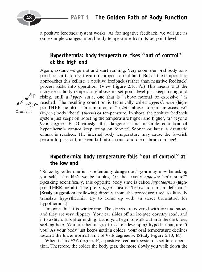
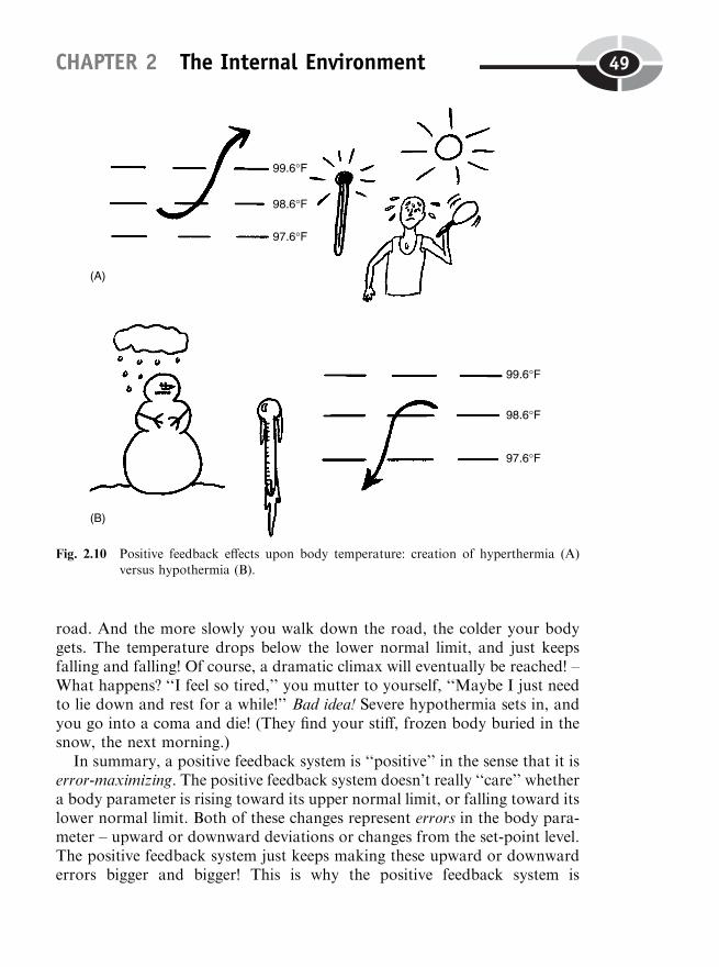
Again, assume we go out and start running. Very soon, our oral body tem-perature starts to rise toward its upper normal limit. But as the temperatureapproaches this ceiling, a positive feedback (rather than negative feedback)process kicks into operation. (View Figure 2.10, A.) This means that theincrease in body temperature above its set-point level just keeps rising andrising, until a hyper- state, one that is ‘‘above normal or excessive,’’ isreached. The resulting condition is technically called hyperthermia (high-per-THER-me-uh) – ‘‘a condition of ’’ (-ia) ‘‘above normal or excessive’’(hyper-) body ‘‘heat’’ (therm) or temperature. In short, the positive feedbacksystem just keeps on boosting the temperature higher and higher, far beyond99.6 degrees F. Obviously, this dangerous and unstable condition ofhyperthermia cannot keep going on forever! Sooner or later, a dramaticclimax is reached. The internal body temperature may cause the feverishperson to pass out, or even fall into a coma and die of brain damage!
Hypothermia: body temperature falls ‘‘out of control’’ atthe low end
‘‘Since hyperthermia is so potentially dangerous,’’ you may now be askingyourself, ‘‘shouldn’t we be hoping for the exactly opposite body state?’’Speaking scientifically, this opposite body state is called hypothermia (high-poh-THER-me-uh). The prefix hypo- means ‘‘below normal or deficient.’’[Study suggestion: Following directly from the procedure used to literallytranslate hyperthermia, try to come up with an exact translation forhypothermia.]
Imagine that it is wintertime. The streets are covered with ice and snow,and they are very slippery. Your car slides off an isolated country road, andinto a ditch. It is after midnight, and you begin to walk out into the darkness,seeking help. You are then at great risk for developing hypothermia, aren’tyou! As your body just keeps getting colder, your oral temperature declinestoward the lower normal limit of 97.6 degrees F. (Study Figure 2.10, B.)
When it hits 97.6 degrees F, a positive feedback system is set into opera-tion. Therefore, the colder the body gets, the more slowly you walk down the
PART 1 The Golden Path of Body Function48
Organism 1
road. And the more slowly you walk down the road, the colder your bodygets. The temperature drops below the lower normal limit, and just keepsfalling and falling! Of course, a dramatic climax will eventually be reached! –What happens? ‘‘I feel so tired,’’ you mutter to yourself, ‘‘Maybe I just needto lie down and rest for a while!’’ Bad idea! Severe hypothermia sets in, andyou go into a coma and die! (They find your stiff, frozen body buried in thesnow, the next morning.)
In summary, a positive feedback system is ‘‘positive’’ in the sense that it iserror-maximizing. The positive feedback system doesn’t really ‘‘care’’ whethera body parameter is rising toward its upper normal limit, or falling toward itslower normal limit. Both of these changes represent errors in the body para-meter – upward or downward deviations or changes from the set-point level.The positive feedback system just keeps making these upward or downwarderrors bigger and bigger! This is why the positive feedback system is
CHAPTER 2 The Internal Environment 49
Fig. 2.10 Positive feedback effects upon body temperature: creation of hyperthermia (A)versus hypothermia (B).

alternatively called a vicious circle or vicious cycle. Like a giant snowballrolling downhill, the bigger the snowball (error in a body parameter) gets,the faster it rolls, and the faster the snowball rolls, the bigger it gets! There isjust no stopping this giant snowball, is there? It will keep getting bigger andbigger (the parameter going ever farther into a hyper- or hypo- state) until adramatic climax is reached. (Maybe the giant snowball finally crashes into theside of a building!)
Positive feedback systems (vicious cycles or vicious circles) are definitelynot control systems! Rather, they are dramatically ‘‘out-of-control’’ systems!Hence, they tend to greatly disrupt homeostasis of body parameters, ratherthan maintain them.
A POSITIVEFEEDBACK SYSTEM
¼ An ‘‘Error-Maximizing’’ or ‘‘Error-Increasing’’Vicious Cycle That Usually Disrupts
Homeostasis of Body Parameters, PushingThem ‘‘Out of Control’’
Feedback Systems and Human HealthFrom the above discussion, we can conclude that the type of feedback systeminvolved – positive versus negative – can greatly influence human health andwell-being. Technically speaking, a state of clinical (KLIN-ih-kal) health isone where the body is basically healthy, so that there is no legitimate reasonto visit a ‘‘clinic’’ or physician’s office for treatment. A state of BiologicalOrder of the important body parameters is being maintained. This is gener-ally reflected in the occurrence of homeostasis (relative constancy) of theseparameters. And as we have seen from the time of the Ancient Greeks (suchas Hippocrates), human health is usually seen as a balance. Using moderncontrol system theory, we could also say that the negative feedback controlsystems are the ones that often maintain this balance or relative constancy ofthe various body parameters. This means that the body parameters staywithin their normo- (‘‘normal’’) range or normo zone over time.
For specific examples of clinical health and homeostasis, let us considerboth oral body temperature in degrees Fahrenheit, as well as blood glucoseconcentration, measured in milligrams of glucose per 100 ml (milliliters) ofblood.
Recall from earlier in this chapter that normothermia is a condition ofnormal body heat or temperature. This normothermia essentially involves a
PART 1 The Golden Path of Body Function50
Organism 2
state of the entire organism, since the temperature measured in the mouthgenerally represents the temperature in the entire human body. When thisnormothermia is maintained over time, then we can say that this high degreeof Biological Order is an instance of homeostasis or relative constancy, main-tained by negative feedback mechanisms. And the oral body temperature canbe legitimately described as clinically healthy.
Remember the work of Claude Bernard on the relative constancy of thebody’s ‘‘internal environment’’ (for him, the extracellular fluid bathing ourcells). This included a careful measurement of blood glucose concentration.This anatomical parameter is that of a particular type of molecule – glucose –and its concentration within the bloodstream. Just as we used normothermiato describe oral body temperature within its normal range, we can coin theterm normoglycemia (nor-moh-gleye-SEE-me-ah). This term indicates a‘‘blood condition of ’’ (-emia) ‘‘glucose’’ (glyc) within its ‘‘normal’’ (normo-)range. Under fasting conditions, the normal range for blood glucose concen-tration is from a lower normal limit of 70 mg/100ml blood, to an uppernormal limit of about 110 mg/100 ml blood. When the amount of upwardand downward change from the set-point value of 90 mg/100 ml bloodstays relatively ‘‘balanced’’ between these limits over time, then a state ofhomeostasis of blood glucose concentration is said to exist.
Both the normothermic (nor-moh-THERM-ik) and normoglycemic (nor-moh-gleye-SEE-mik) conditions represent states of clinical health of thesecritically important body parameters. Again and again in this book, we willsee that other body parameters lying in their normo- (‘‘normal’’) ranges like-wise represent states of homeostasis and clinical health.
POSITIVE FEEDBACK, MORBIDITY, AND MORTALITY
Positive feedback, as we have seen, maximizes or increases the amount ofupward or downward ‘‘error’’ from the set-point level of a particular bodyparameter. Positive feedback at the upper normal limit commonly results in ahyper- (‘‘above normal or excessive’’) state of the body parameter. In the caseof body temperature, this hyper- state would be called hyperthermia. (ReviewFigure 2.10, A, if desired.) And in the case of blood glucose concentration,this above-normal or excessive state is called hyperglycemia (high-per-gleye-SEE-me-ah).
At the opposite end of the normal range, positive feedback at the lowernormal limit creates a hypo- (‘‘below normal or deficient’’) state of the bodyparameter. For oral body temperature, of course, this hypo- state is called
CHAPTER 2 The Internal Environment 51
Tissue 1
Tissue 2
Tissue 1

hypothermia. (Review Figure 2.10, B.) And for deficient or below-normalblood glucose concentration, the scientific term is hypoglycemia (high-poh-gleye-SEE-me-ah). Either of these conditions results in a loss or disruption ofhomeostasis in the parameters involved.
Morbidity and mortality
Either a hyper- state or a hypo- state typically involves ‘‘diseased’’ (patho-logical) conditions of body structures and functions. This means pathologicalanatomy and pathophysiology. Such deficient or excessive pathological statesare lumped into a general category called morbidity (more-BID-ih-tee) –‘‘conditions of ’’ (-ity) ‘‘illness’’ (morbid). If these morbid (MOR-bid) or‘‘ill’’ states become very severe, they may result in mortality (more-TAL-ih-tee) – ‘‘a condition of ’’ (-ity) ‘‘death’’ (mortal).
POSITIVE FEEDBACK LEADING TO ‘‘HELPFUL’’ CLIMAXES
Our discussion of positive feedback systems (vicious circles or vicious cyclesof ever-increasing amounts of change) has generally been an unhappy one.From the important perspective of our clinical health, the dramatic climaxesand extreme hyper- or hypo- states reached by positive feedback mechanismshave been harmful. By disrupting the homeostasis of important body para-meters, the positive feedback cycles often result in morbidity (illness) ormortality (death).
Certainly, illness and death are dramatic climaxes, aren’t they? There are afew noteworthy examples, however, where positive feedback systems leadingto dramatic climaxes are most helpful (rather than harmful) to human survi-val. These include: sexual climax or orgasm, contractions of the womb leadingto childbirth, firing of nerve impulses, and clotting of the blood after vesselinjury. We will discuss these specific examples in more detail in later chapters.But for now, ask yourself this important question about blood clotting: ‘‘Ihave suffered cuts in my skin many times during my life. How come all ofthese cuts (say, from injuries suffered more than a month ago) aren’t stilloozing blood today? What type of feedback process has made all of thisblood-leaking shut off for good?’’
PART 1 The Golden Path of Body Function52

QuizRefer to the text in this chapter if necessary. A good score is at least 8 correctanswers out of these 10 questions. The answers are listed in the back of thisbook.
1. Widely acknowledged as the Father of Modern Medicine:(a) Socrates(b) Arnold Schwarzenegger(c) Hippocrates(d) Galen
2. The original rationale behind the medical practice of blood-lettingwas:(a) An excess of blood was upsetting the balance of body humors(b) The idea that blood was a natural body poison(c) Fear that excessive blood glucose would lead to diabetes(d) A common consensus that blood was an unimportant body fluid
3. Baby Heinie swallows a bucket of tomatoes and vinegar, which is highin acetic acid. He then suffers a bad case of acid indigestion. The aceticacid thus seems to be acting as a:(a) Control system(b) Stressor(c) Set-point(d) Normal range of a body parameter
4. The concept of the milieu interieur developed by Claude Bernardmainly refers to the:(a) Intracellular fluid and its dissolved particles(b) Four body humors of Ancient Greece(c) Secretions of the kidney into the urine(d) Extracellular fluid surrounding body cells
5. Walter Cannon’s original definition of homeostasis:(a) The ‘‘fight-or-flight’’ response to severe stress(b) An automatic secretion of adrenaline (epinephrine) into the
bloodstream whenever body parameters become disturbed(c) A relatively constant condition within the body’s internal envir-
onment
CHAPTER 2 The Internal Environment 53
(d) A state of absolute constancy of many of the body parametersover time
6. ‘‘Living organisms are machines’’ would best summarize:(a) The attitude of the early Chinese thinkers(b) The perspective of cybernetics and modern control system theory(c) A false rejection of the principles of the Humoral Doctrine(d) The ‘‘fight-or-flight’’ response
7. A relative constancy of a particular anatomical or physiological para-meter within the body’s internal environment:(a) Current definition of homeostasis(b) Outmoded superstitions about ‘‘ghosts’’ within the body(c) Description of the hyper- zone(d) A condition of illness
8. The long-term average value of a body parameter:(a) Normal range(b) Set-point(c) Upper normal limit(d) Chronic chaos
9. A negative feedback system generally operates to:(a) Restrain, inhibit, or minimize further change in a body parameter,
in some particular direction(b) Create ‘‘negative’’ conditions within the internal environment that
are harmful to survival(c) Stimulate, magnify, or accelerate the distance between the current
value of a body parameter and its set-point value(d) Halt any up-or-down movements whatsoever of a body parameter
from its set-point level
10. Most likely involved in establishing trends toward morbidity ormortality:(a) Normal body structures and functions(b) States of pathophysiology and pathological anatomy(c) Positive feedback mechanisms that promote blood clotting after
vessel injury(d) Negative feedback control systems operating at peak efficiency
PART 1 The Golden Path of Body Function54

Body-Level Grids for Chapter 2Several key body facts were tagged with numbered icons in the page marginsof this chapter. Write a brief summary of each of these key facts into anumbered cell or box within the appropriate Body-Level Grid that appearsbelow.
Anatomy and Biological Order Fact Grids for Chapter 2:
CELLLevel
TISSUELevel
CHAPTER 2 The Internal Environment 55
1 2
1
Anatomy and Biological Disorder Fact Grids for Chapter 2:
TISSUELevel
Physiology and Biological Order Fact Grids for Chapter 2:
MOLECULELevel
PART 1 The Golden Path of Body Function56
1 2
1
CELLLevel
TISSUELevel
ORGANISMLevel
CHAPTER 2 The Internal Environment 57
1 2
3
1
1 2
Physiology and Biological Disorder Fact Grids for Chapter 2:
ORGANLevel
ORGANISMLevel
PART 1 The Golden Path of Body Function58
1
1

59
Test: Part 1
DO NOT REFER TO THE TEXT WHEN TAKING THIS TEST. A goodscore is at least 18 (out of 25 questions) correct. Answers are in the back ofthe book. It’s best to have a friend check your score the first time, so youwon’t memorize the answers if you want to take the test again.
1. The word biology exactly translates to mean:(a) ‘‘Study of body functions’’(b) ‘‘Study of life’’(c) ‘‘Removal of living things’’(d) ‘‘A cutting apart’’(e) ‘‘Two kinds of living things’’
2. Biological Disorder equals:(a) A break in all natural patterns(b) The establishment of normal ranges(c) The application of parameters to living things only(d) A disruption in living patterns(e) Birds who fly in a straight path through the sky
Copyright © 2004 by The McGraw-Hill Companies, Inc. Click here for terms of use.
3. Body structures can be considered as within a sentence:(a) Verbs(b) Adverbs(c) Adjectives(d) Periods(e) Nouns
4. Aristotle created the word physiology to originally mean:(a) ‘‘The study of Nature’’(b) ‘‘Body structures and the study of body structures’’(c) ‘‘A state of relative constancy’’(d) ‘‘Body functions and the study of body functions’’(e) ‘‘The process of dissection’’
5. Blood pressure is measured and observed to remain between 90/60millimeters of mercury and 140/90 millimeters of mercury, over a24-hour period. We can logically conclude that:(a) No stressors were acting upon the blood pressure during this time(b) Homeostasis of blood pressure was being maintained(c) The blood pressure was absolutely constant over this period(d) Blood glucose concentration likewise remained within its normal
range(e) The blood was far too hot to permit accurate pressure readings
6. The sole of the foot is rather broad and flat, allowing it to bear weighteasily:(a) Law of Use and Disuse(b) Example of deadly force(c) Complementarity of body structure and function(d) An evolutionary error(e) Complained about by Aristotle
7. A stimulus is:(a) Always the same as a stressor(b) A detectable change in the body’s internal or external environ-
ment(c) A specific kind of sensory receptor(d) Usually the last event in a negative feedback cycle(e) Latin for ‘‘goat’’ with ‘‘horns’’
8. A level of body organization(a) Involves a particular size and complexity of body structures(b) Is defined in the book as the entire Pyramid of Life
PART 1 The Golden Path of Body Function60
(c) Starts with the cellular level(d) Bears no relationship to the Law of Complementarity(e) Just says something important about anatomy, not about
physiology
9. Hyperthermia generally reflects:(a) Precise regulation of body temperature(b) A dramatic drop in blood glucose concentration(c) A sudden fall in oral body temperature below its normal range(d) Proper maintenance of homeostasis(e) A loss of control of body temperature at its high end
10. A good example of a physiological parameter:(a) Femoral length in cm(b) Blood glucose concentration in mg glucose/100 ml blood(c) Density of nerve cells in the brain, in number of cells/cubic
centimeter of brain tissue(d) Weight of the liver in ounces(e) Rate of urine flow from the right kidney, in cc/minute
11. A plain functional parameter:(a) Heart rate in beats/minute(b) Car speed(c) Heat of exhaust emitted from a dumptruck tailpipe, in degrees
celsius(d) Lung capacity in liters(e) Bladder circumference in inches
12. The word anatomy exactly translates to mean:(a) ‘‘a process of cutting up or apart’’(b) ‘‘a penny for your thoughts’’(c) ‘‘inflammation of a sweetie’’(d) ‘‘production of new glucose’’(e) ‘‘presence of body structures (not functions)’’
13. The Ancient Greek idea of the Golden Mean:(a) A reflection of severe Biological Disorder(b) The product of unbalanced body humors(c) Was never supported in later eras(d) Is quite similar to the modern notion of the set-point(e) Has no valid application to current biological thinking
Test: Part 1 61
14. The Humoral Doctrine:(a) Assumed a balance among blood, yellow bile, black bile, and
phlegm(b) Believed that ‘‘The Devil is in the details!’’(c) ‘‘Eat, drink, and be merry; for tomorrow we may die’’(d) Had no connection to the universal element of water(e) Died out before the time of the American Revolution
15. The highest level of a normal body parameter:(a) Upper normal limit(b) Set-point value(c) Lower normal limit(d) Normal range(e) Subnormal range values
16. Claude Bernard used vivisection during his experiments because:(a) He was a notable animal rights activist(b) His rich wife refused to give him enough money to feed his lab
animals(c) He enjoyed torturing living dogs(d) Physiological mechanisms can only be observed in living organ-
isms(e) Not enough dog and pig cadavers were available
17. The production of new molecules of glucose from non-sugar sources:(a) Abdominocentesis(b) Gluconeogenesis(c) Glycogenolysis(d) Enzyme-splitting(e) Glucose uptake from the intestine
18. The milieu interieur:(a) An old German fairytale(b) A French phrase for ‘‘internal environment’’(c) First advanced by Walter B. Cannon(d) Intensely denounced by Hippocrates(e) The famous brainchild of Edward Jenner
19. Large amounts of adrenaline (epinephrine) released into the blood-stream:(a) Normal, resting body conditions(b) A slow response to sudden danger(c) The ‘‘fight-or-flight’’ response
PART 1 The Golden Path of Body Function62
(d) A quick return to hormonal equilibrium(e) Triggers a dramatic drop in body temperature
20. Baby Heinie pulls his Big Sister’s pigtails; Big Sister slaps Baby Heinie;Baby Heinie pulls Big Sister’s pigtails even harder; and Big Sister slapsBaby Heinie with even more force:(a) An imaginative story about negative feedback(b) A crucial ‘‘fight-or-flight’’ survival mechanism in operation(c) Normothermia of blood pressure is being maintained(d) Homeostasis of relevant body parameters(e) An entertaining real-life scenario of a positive feedback cycle
21. The falling of blood pressure into the range of hypotension (high-poh-TEN-shun):(a) The occurrence of set-point adjustment(b) The ‘‘survival of the fittest’’(c) A clinical example of the Humoral Doctrine(d) Probable starting of positive feedback near the lower normal limit(e) Negative feedback promoting blood pressure homeostasis
22. A clock pendulum swinging back and forth from its initial verticalposition:(a) Demonstration of the normal range concept for body parameters(b) A crude error in cybernetics(c) Rough model for the idea of morbidity(d) The removal of all feedback processes(e) A reflection of actual real-life vicious circles
23. The temperature control center:(a) Carries sensory information over an afferent pathway(b) Delivers a response from the body effectors in the skin(c) Monitors and adjusts the current body temperature to match the
set-point level(d) Removes all inhibitory influences upon blood glucose homeostasis(e) Is a hypothetical body structure that does not really exist
24. A distraught mother rushes into a hospital emergency room andscreams at the top of her lungs while the nurses are trying to applya Bandaid to a small scrape on her young daughter’s knee:(a) Good instance of a movement toward parameter equilibrium(b) An upsetting illustration of emotions rocketing into a hyper- state(c) Dumbing-down of medical therapy so that a lay person can
understand it
Test: Part 1 63
(d) Return of a previously morbid condition back into the normo-range
(e) Relative constancy around the ‘‘fight-or-flight’’ set-point
25. A single blood sample (taken at 8 A.M.) shows a glucose concentrationof 92 mg/100 ml blood. From this fact you can most appropriatelyconclude that:(a) Homeostasis of blood glucose concentration is being maintained(b) The tested patient is suffering from hypoglycemia(c) A diabetic coma is about to happen(d) Normoglycemia existed at 8 A.M.
(e) Clinical health of the patient is guaranteed
PART 1 The Golden Path of Body Function64
PART 2
Functions at theChemical andCellular Levels
Copyright © 2004 by The McGraw-Hill Companies, Inc. Click here for terms of use.

This page intentionally left blank
CHAPTER3
Chemical Functions:‘‘Hey! We’ve Got Dirt
Moving Around inOur Body Basement!’’
In Part 1 of PHYSIOLOGY DEMYSTIFIED, our aim was to present youwith the underlying history and basic concepts behind the modern study ofbody functions. Chapter 2, Control of the Internal Environment, brought tothe forefront the critical behind-the-scenes action of physiological controlsystems that help maintain homeostasis.
We will now, however, re-focus our vision upon the three bottom layers ofthe Great Body Pyramid. If you will remember, these were Levels I, II, andIII, which, taken all together, make up the Chemical Level of BodyOrganization:
67
Copyright © 2004 by The McGraw-Hill Companies, Inc. Click here for terms of use.
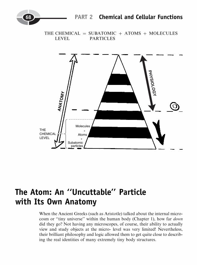
THE CHEMICALLEVEL
¼ SUBATOMICPARTICLES
þ ATOMS þ MOLECULES
The Atom: An ‘‘Uncuttable’’ Particlewith Its Own Anatomy
When the Ancient Greeks (such as Aristotle) talked about the internal micro-cosm or ‘‘tiny universe’’ within the human body (Chapter 1), how far downdid they go? Not having any microscopes, of course, their ability to actuallyview and study objects at the micro- level was very limited! Nevertheless,their brilliant philosophy and logic allowed them to get quite close to describ-ing the real identities of many extremely tiny body structures.
PART 2 Chemical and Cellular Functions68
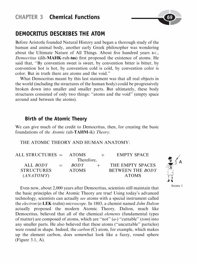
DEMOCRITUS DESCRIBES THE ATOM
Before Aristotle founded Natural History and began a thorough study of thehuman and animal body, another early Greek philosopher was wonderingabout the Ultimate Nature of All Things. About five hundred years B.C.,Democritus (dih-MAHK-ruh-tus) first proposed the existence of atoms. Hesaid that, ‘‘By convention sweet is sweet, by convention bitter is bitter, byconvention hot is hot, by convention cold is cold, by convention color iscolor. But in truth there are atoms and the void.’’
What Democritus meant by this last statement was that all real objects inthe world (including the structures of the human body) could be progressivelybroken down into smaller and smaller parts. But ultimately, these bodystructures consisted of only two things: ‘‘atoms and the void’’ (empty spacearound and between the atoms).
Birth of the Atomic Theory
We can give much of the credit to Democritus, then, for creating the basicfoundations of the Atomic (uh-TAHM-ik) Theory.
THE ATOMIC THEORY AND HUMAN ANATOMY:
ALL STRUCTURES ¼ ATOMS þ EMPTY SPACETherefore,
ALL BODYSTRUCTURES(ANATOMY)
¼ BODYATOMS
þ THE EMPTY SPACESBETWEEN THE BODY
ATOMS
Even now, about 2,000 years after Democritus, scientists still maintain thatthe basic principles of the Atomic Theory are true! Using today’s advancedtechnology, scientists can actually see atoms with a special instrument calledthe electron (e-LEK-trahn) microscope. In 1803, a chemist named John Daltonactually proposed the modern Atomic Theory. Dalton, much likeDemocritus, believed that all of the chemical elements (fundamental typesof matter) are composed of atoms, which are ‘‘not’’ (a-) ‘‘cuttable’’ (tom) intoany smaller parts. He also believed that these atoms (‘‘uncuttable’’ particles)were round in shape. Indeed, the carbon (C) atom, for example, which makesup the element carbon, does somewhat look like a fuzzy, round sphere(Figure 3.1, A).
CHAPTER 3 Chemical Functions 69
Atoms 1
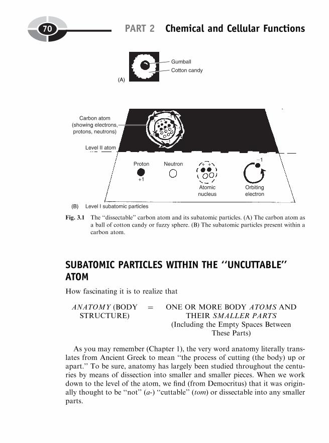
SUBATOMIC PARTICLES WITHIN THE ‘‘UNCUTTABLE’’ATOM
How fascinating it is to realize that
ANATOMY (BODYSTRUCTURE)
¼ ONE OR MORE BODY ATOMS ANDTHEIR SMALLER PARTS
(Including the Empty Spaces BetweenThese Parts)
As you may remember (Chapter 1), the very word anatomy literally trans-lates from Ancient Greek to mean ‘‘the process of cutting (the body) up orapart.’’ To be sure, anatomy has largely been studied throughout the centu-ries by means of dissection into smaller and smaller pieces. When we workdown to the level of the atom, we find (from Democritus) that it was origin-ally thought to be ‘‘not’’ (a-) ‘‘cuttable’’ (tom) or dissectable into any smallerparts.
PART 2 Chemical and Cellular Functions70
Fig. 3.1 The ‘‘dissectable’’ carbon atom and its subatomic particles. (A) The carbon atom as
a ball of cotton candy or fuzzy sphere. (B) The subatomic particles present within acarbon atom.

With the coming of the twentieth century, however, scientists were finallyable to split the supposedly uncuttable atom. Therefore the body atoms, likeall the larger structures within the human organism, are also ‘‘cuttable’’ or‘‘dissectable’’ into smaller pieces. These smaller pieces are technically calledthe subatomic particles. This is because they exist ‘‘under’’ (sub-) the higherlevel of the entire ‘‘atom.’’ The three most common types of subatomicparticles are the protons (PROH-tahns), neutrons (NEW-trahns), and elec-trons (e-LEK-trahns). Within the carbon atom, for instance, there are 6 pro-tons, 6 neutrons, and 6 electrons (Figure 3.1, B).
Each atom contains a nucleus (NEW-klee-us) – a rounded, centrallylocated ‘‘kernel’’ (nucle) or core. [Study suggestion: Think of the nucleus asa hard, round gumball.] The protons are subatomic particles with a positive(þ) charge in the nucleus, and they are often accompanied by neutronswhich, as their name suggests, are electrically ‘‘neutral’’ (having a chargeof 0). Circling at high speed around the nucleus is a cloud of electrons,which are tiny, negatively charged (�) particles also having some of thecharacteristics of waves. [Study suggestion: Consider the electron clouds asa sticky swirl of cotton candy surrounding the nucleus gumball.]
SUBATOMICPARTICLES
¼ (Protons þ Neutrons)THE NUCLEUS
þ (Orbiting Electrons)FUZZY CLOUD
AROUND NUCLEUS
Since the number of positively charged protons normally equals the num-ber of negatively charged electrons, the net electrical charge of most atoms iszero.
Chemical Bonds: Builders ofOrder and Molecules
We have shown how even the individual atom is dissectable into smallersubatomic particles, which exist at a lower level. But we can also workfrom the higher end, and link individual atoms together to create molecules.A molecule is a ‘‘little mass’’ (molecul) of matter that consists of two or moreatoms held together by chemical bonds.
A chemical bond is a sharing or transfer of outermost electrons betweenatoms. And as a result of this bonding (sharing or transfer of electrons), thecharacteristics of the formerly separate atoms are changed into the newcharacteristics of the molecule. In addition, the process of chemical bonding
CHAPTER 3 Chemical Functions 71
Subatomic
particles 1
Subatomic
particles 1

creates precise new patterns or arrangements of atoms in the body, therebybuilding Biological Order.
ORGANIC MOLECULES VERSUS INORGANIC MOLECULESAND THEIR BONDING
Molecules created by bonding are of two broad types – organic (or-GAN-ik)molecules versus inorganic (IN-or-gan-ik) molecules. The organic moleculesliterally make up most of the body ‘‘organs,’’ which are rich in bondedcarbon (C) atoms. The heart, lungs, brain, and other organs, then, all pri-marily consist of highly orderly arrangements of organic (carbon-containing)molecules.
The organic molecules have a chemical ‘‘backbone’’ that mainly includeseither C–C (carbon–carbon) or C–H (carbon–hydrogen) bonds. These types ofbonds are usually classified as nonpolar (nahn-POH-lar) covalent (koh-VALE-ent) bonds. A covalent bond always involves a sharing ‘‘together’’(co-) of outermost electrons between atoms. An important follow-up ques-tion to ask is, ‘‘Do the atoms share these outermost electrons equally, or dothey share them unequally?’’
If the electrons in the chemical bond are shared equally between atoms,then a nonpolar covalent bond results. Something that is nonpolar does‘‘not’’ (non-) have any poles or areas of opposite electrical charge. Whenelectrons, which have a negative (�) charge, are shared equally betweentwo atoms, their negative charges are also shared equally between theatoms. Thus, no electrical poles – areas of opposite positive (þ) or negative(�) charges – are created.
Consider the chemical bonding in our familiar glucose molecule (Figure3.2, A). The C–C bonds and C–O (oxygen) bonds forming its six-sided ringstructure are all considered nonpolar covalent. Thus, the electron cloudsaround each pair of shared electrons in the ring can be conveniently modeledas sticky cotton candy pulled halfway between the two atoms being bonded.
The bonding arrangement is markedly different within the water or H2Omolecule (Figure 3.2, B). Water is an inorganic molecule – one that does‘‘not’’ (in-) contain any ‘‘carbon’’ (organ) atoms. Recollect that the carbonatom tends to share its outermost electrons equally with other atoms. The O(oxygen) atom, however, is quite another story! Oxygen tends to be an elec-tron ‘‘hog,’’ pulling electrons away from other atoms, and into a closer orbitaround itself.
This is what happens when it bonds with hydrogen, creating water. Thetwo H–O (hydrogen–oxygen) bonds are classified as being polar (POH-lar)
PART 2 Chemical and Cellular Functions72
Subatomic
particles 2
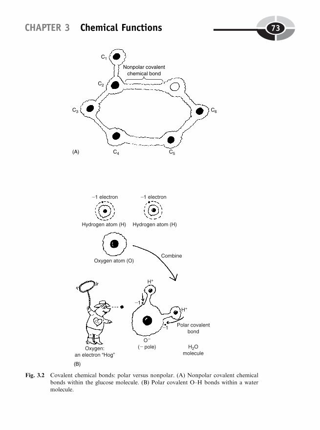
CHAPTER 3 Chemical Functions 73
Fig. 3.2 Covalent chemical bonds: polar versus nonpolar. (A) Nonpolar covalent chemical
bonds within the glucose molecule. (B) Polar covalent O–H bonds within a watermolecule.

covalent. A polar covalent chemical bond is a bond in which the outermostelectrons are shared unequally between the participating atoms, resulting in anegative pole and a positive pole. The negative pole is the end where theelectrons are pulled more closely, resulting in a net negative (�) charge.Conversely, the positive pole is the end where the electrons have been pulledfarther away, leaving it with a net positive (þ) charge. [Study suggestion:
Look back at the anatomy of the atomic nucleus (Figure 3.1). Why doespulling an outer electron away create a net positive charge?]
Covalent bond summary
We can state some simple equations summarizing covalent chemical bonds:
COVALENT BONDS ¼ THE SHARING OF ELECTRONSBETWEEN ATOMS
NONPOLARCOVALENT BONDS
¼ EQUAL SHARING OF ELECTRONS
versusPOLAR COVALENT
BONDS¼ UNEQUAL SHARING OF ELECTRONS
Ions: First as Bonds, Then as ‘‘Goers-to’’In certain types of bonds, things go far beyond an unequal sharing of elec-trons. Look at the case of ionic (eye-AH-nik) bonds. Ionic bonds are chemicalbonds that result from the complete transfer of one or more outermostelectrons between atoms, such that ions (EYE-ahns) are produced. An ionis an atom that has either a net excess or a deficiency of electrons, so that it iselectrically charged.
IONIC CHEMICALBONDS
¼ COMPLETE TRANSFER OF ELECTRONSBETWEEN ATOMS, CREATING IONS
A fine example is provided by sodium chloride (KLOR-eyed), symbolizedas NaCl. When the sodium (Na) atom gets near a chlorine (Cl) atom, it tendsto transfer one of its electrons to the chlorine. The net result is an ionic bondbetween Na and Cl. (Study Figure 3.3.)
The Na end has a net positive (þ) charge, while the Cl end has a netnegative (�) one. When enough of these ionic NaCl bonds are formed, anelegant crystal lattice structure of a salt cube results.
PART 2 Chemical and Cellular Functions74
Subatomic
particles 3
Subatomic
particles 4

CATIONS VERSUS ANIONS: TWO KINDS OF IONS
Let us now perform a little experiment. Grab a battery, which has a pair ofelectrodes (ih-LEK-trohds). (Examine Figure 3.4.) The negatively chargedelectrode is called the cathode (KATH-ohd). It is the source of a stream ofnegatively charged electrons. Place the cathode into a tank of water, which isa very good ‘‘dissolver’’ or solvent (SAHL-vent). At the other end of the largewater tank, insert an anode (AN-ohd). This is the positively charged elec-trode.
Now dump in a bunch of salt (NaCl) crystals. Salt is a type of electrolyte(ee-LEK-troh-light). An electrolyte is a substance that ‘‘breaks down’’ (lyt)into ions when placed into a water solvent, such that the resulting solutioncan conduct an ‘‘electrical current’’ (electro-).
Switch-on the electric current at the cathode end. The process calledelectrolysis (ih-lek-TRAHL-uh-sis) begins to occur. Electrolysis is the‘‘breakdown of ’’ (-lysis) some chemical substance under the influence of
CHAPTER 3 Chemical Functions 75
Fig. 3.3 Ionic bonds and the salt crystal.
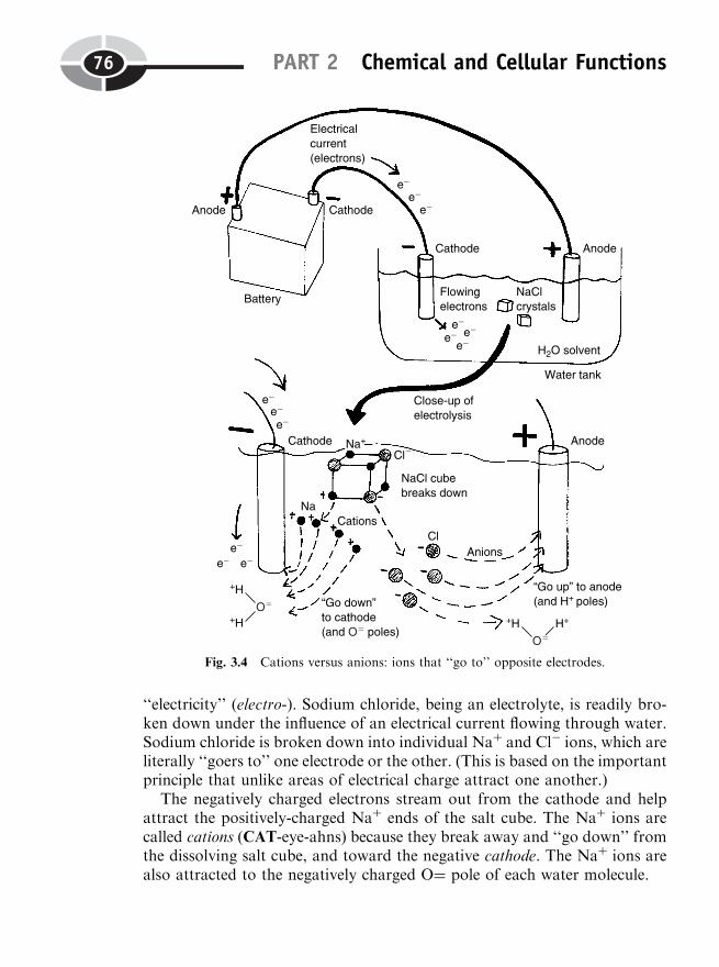
‘‘electricity’’ (electro-). Sodium chloride, being an electrolyte, is readily bro-ken down under the influence of an electrical current flowing through water.Sodium chloride is broken down into individual Naþ and Cl� ions, which areliterally ‘‘goers to’’ one electrode or the other. (This is based on the importantprinciple that unlike areas of electrical charge attract one another.)
The negatively charged electrons stream out from the cathode and helpattract the positively-charged Naþ ends of the salt cube. The Naþ ions arecalled cations (CAT-eye-ahns) because they break away and ‘‘go down’’ fromthe dissolving salt cube, and toward the negative cathode. The Naþ ions arealso attracted to the negatively charged O¼ pole of each water molecule.
PART 2 Chemical and Cellular Functions76
Fig. 3.4 Cations versus anions: ions that ‘‘go to’’ opposite electrodes.
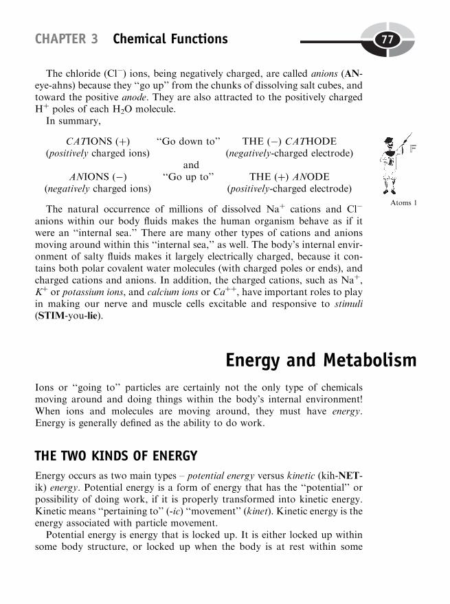
The chloride (Cl�) ions, being negatively charged, are called anions (AN-eye-ahns) because they ‘‘go up’’ from the chunks of dissolving salt cubes, andtoward the positive anode. They are also attracted to the positively chargedHþ poles of each H2O molecule.
In summary,
CATIONS (þ)(positively charged ions)
‘‘Go down to’’ THE (�) CATHODE(negatively-charged electrode)
andANIONS (�)
(negatively charged ions)‘‘Go up to’’ THE (þ) ANODE
(positively-charged electrode)
The natural occurrence of millions of dissolved Naþ cations and Cl�
anions within our body fluids makes the human organism behave as if itwere an ‘‘internal sea.’’ There are many other types of cations and anionsmoving around within this ‘‘internal sea,’’ as well. The body’s internal envir-onment of salty fluids makes it largely electrically charged, because it con-tains both polar covalent water molecules (with charged poles or ends), andcharged cations and anions. In addition, the charged cations, such as Naþ,Kþ or potassium ions, and calcium ions or Caþþ, have important roles to playin making our nerve and muscle cells excitable and responsive to stimuli(STIM-you-lie).
Energy and MetabolismIons or ‘‘going to’’ particles are certainly not the only type of chemicalsmoving around and doing things within the body’s internal environment!When ions and molecules are moving around, they must have energy.Energy is generally defined as the ability to do work.
THE TWO KINDS OF ENERGY
Energy occurs as two main types – potential energy versus kinetic (kih-NET-ik) energy. Potential energy is a form of energy that has the ‘‘potential’’ orpossibility of doing work, if it is properly transformed into kinetic energy.Kinetic means ‘‘pertaining to’’ (-ic) ‘‘movement’’ (kinet). Kinetic energy is theenergy associated with particle movement.
Potential energy is energy that is locked up. It is either locked up withinsome body structure, or locked up when the body is at rest within some
CHAPTER 3 Chemical Functions 77
Atoms 1
position or location. This potential energy is locked up, for example, withinthe chemical bonds of the glucose molecule. When the chemical bonds aresplit, however, some of the stored potential energy is released, and convertedinto kinetic energy. Kinetic energy is alternatively called free energy, becauseit is not stored or locked up. This free or kinetic energy then moves variousparticles around, and does body work.
ANABOLISM VERSUS CATABOLISM: THE ‘‘YIN–YANG’’ OFMETABOLISM
Chemical reactions are interactions between two or more chemicals. Forchemicals to interact, of course, they must move around and have kineticenergy. The most important chemical reactions occurring within the livinghuman organism are those of metabolism, a ‘‘state of change’’ (Chapter 1).Traditional Chinese medicine involved the Yin–Yang concept of two oppo-site but complementary energy forces.
Quite interestingly, metabolism, too, has its own Yin–Yang balance! Thetwo opposing faces of metabolism are anabolism (ah-NAB-oh-lizm) versuscatabolism (kah-TAB-oh-lizm). And in each case, their chemical reactions arespeeded up by enzymes (EN-zighms). Enzymes are protein catalysts (KAT-uh-lists) – substances that speed up chemical reactions without themselvesbeing changed in the process.
1. Synthesis reactions or anabolism (ah-NAB-oh-lizm) involve a ‘‘conditionof ’’ (-ism) ‘‘building up’’ (anabol) something from smaller pieces. (See Figure3.5, A.) During this process, free energy is usually added or consumed. Let usrefer back to our old friend, the individual glucose molecule. Suppose that weeat many glucose molecules, and they enter the liver from our bloodstream.Certain anabolic (an-ah-BAHL-ik) enzymes in the liver are activated. Theseanabolic enzymes then speed up the combining of many individual glucoseatoms together (like beads on a string). The result is the synthesis or anabo-lism of glycogen (GLEYE-koh-jen). The glycogen molecule thus stores a lotof potential energy within its chemical bonds. It is a well-known form ofstarch-like carbohydrate found within the liver cells and muscle fibers ofhumans and animals.
2. Breakdown reactions or catabolism (kah-TAB-oh-lizm) involve a ‘‘con-dition of ’’ (-ism) ‘‘casting (breaking) down’’ (catabol). (Examine Figure 3.5,B.) That is, a single large molecule, such as glycogen, is acted upon by variouscatabolic (KAH-tah-bahl-ik) enzymes. These enzymes break or cleave thechemical bonds between the component glucose molecules. The results reflectjust what the word glycogen literally means – ‘‘sweetness’’ (glyc) ‘‘producer’’
PART 2 Chemical and Cellular Functions78
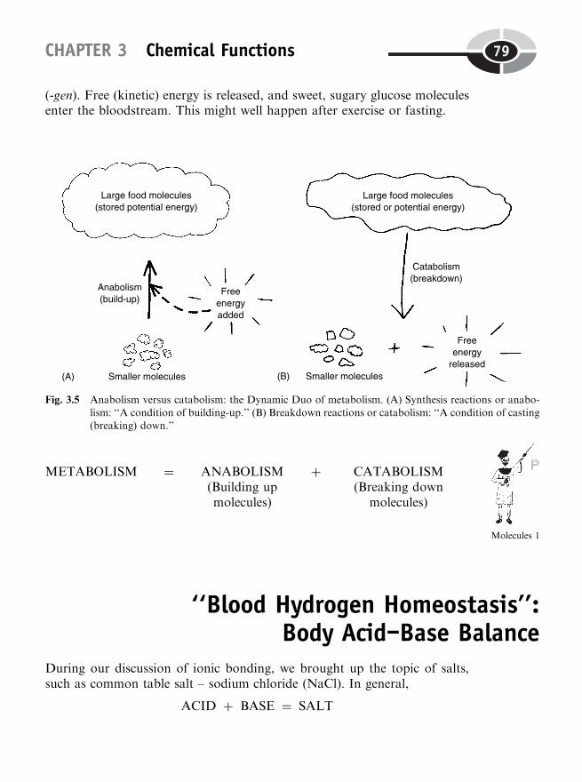
(-gen). Free (kinetic) energy is released, and sweet, sugary glucose moleculesenter the bloodstream. This might well happen after exercise or fasting.
METABOLISM ¼ ANABOLISM(Building upmolecules)
þ CATABOLISM(Breaking down
molecules)
‘‘Blood Hydrogen Homeostasis’’:Body Acid–Base Balance
During our discussion of ionic bonding, we brought up the topic of salts,such as common table salt – sodium chloride (NaCl). In general,
ACID þ BASE ¼ SALT
CHAPTER 3 Chemical Functions 79
Fig. 3.5 Anabolism versus catabolism: the Dynamic Duo of metabolism. (A) Synthesis reactions or anabo-lism: ‘‘A condition of building-up.’’ (B) Breakdown reactions or catabolism: ‘‘A condition of casting(breaking) down.’’
Molecules 1

To thoroughly understand the nature of the important salts or electrolytes(like NaCl) in our body fluids, therefore, we need to know a little bit aboutacids and bases.
Providing a specific example, we have:
HCl(Hydrochloric
acid)
þ NaHCO3
(Sodiumbicarbonate, a weak
base)
¼ NaCl(Commontable salt)
þ H2CO3
(Carbonicacid)
The above is often called the intestinal (in-TES-tih-nal) neutralization (new-tral-ih-ZAY-shun) equation. The reason is that this particular chemicalreaction helps neutralize excess stomach acid that often leaks into the smallintestine.
ACIDS AS HYDROGEN ION DONORS
An acid is a hydrogen ion or Hþ ion donor. An acid, such as hydrochloric(high-droh-KLOR-ik) acid, HCl, has a tendency to ‘‘donate’’ (give off ) itshydrogen (Hþ) ion. So,
HCl ¼ Hþ þ Cl�
Now, individual Hþ ions, themselves, are very dangerous to the body inhigh concentrations – Why? The reason is that the Hþ ions are highly reactiveand corrosive (koh-ROH-siv), tending to disintegrate or wear away anythingthey touch! A strong acid, such as HCl, is one that donates many Hþ ions, sothat it is very corrosive! This is just fine within the stomach, where HCldisintegrates and helps chemically digest eaten food. But within the smallintestine, a very high Hþ ion concentration may wear away the wall andcreate intestinal ulcers.
BASES OR ALKALI AS HYDROGEN ION ACCEPTORS
In direct contrast to the acids are the bases and alkali (AL-kah-lie). A base isan acceptor of Hþ ions (usually donated or given off by an acid). A weak base,such as sodium bicarbonate (buy-CAR-buh-nayt) or NaHCO3, is a base thataccepts just a few Hþ ions. Sodium bicarbonate (NaHCO3) is produced bymany cells in the liver and pancreas. It enters the small intestine from thesesources, where it reacts with the dangerous strong acid, HCl.
The Na atom from the NaHCO3 combines with the Cl from HCl, such thatNaCl and H2CO3 result. The H2CO3 is a chemical formula for carbonic (car-
PART 2 Chemical and Cellular Functions80
Molecules 1
Molecules 2
BAH-nik) acid. Carbonic acid is a weak acid, because it donates relatively fewHþ ions. Thus, the intestinal neutralization equation starts out with a strongacid (HCl) and eventually substitutes it with a weak acid (H2CO3). In thisway, the lining of the intestines is relatively safe, because the strong, corrosiveacid (HCl) has effectively been ‘‘neutralized’’ – rendered less dangerous orharmful by being replaced with a much weaker acid.
Alkali as very strong bases
We have said that sodium bicarbonate (NaHCO3) is a weak base. The alkali,in contrast, are very strong bases, because they can accept many Hþ ions.Some common alkali include lye and ammonia. Lye largely consists of thealkali (strong base) called sodium hydroxide (high-DRAHKS-eyed), NaOH.And ammonia, symbolized as NH3, is a strong base or alkali formed in thebody from the catabolism of many nitrogen-containing substances, such asproteins and amino (ah-MEE-noh) acids. Ammonia is very poisonous, and isquickly converted into urea (you-REE-ah) by the liver.
Besides being potent acceptors of Hþ ions from acids, both lye (NaOH)and ammonia (NH3) are extremely powerful detergents and dissolvers ofgreasy, lipid, or ‘‘fatty’’ material. [Study suggestion: Ask yourself the follow-ing question, ‘‘Well, if NaOH and NH3 are such good acceptors of Hþ ions,then why don’t we just drink a large quantity of lye and ammonia, so thatthey will very effectively neutralize any acid that happens to leak into thesmall intestine from the stomach? Wouldn’t they neutralize excess acid a lotmore effectively than sodium bicarbonate (NaHCO3), which is just a weakbase?’’]
THE CONCEPT OF pH
Besides potentially eating away at the insides of our stomach and intestines,the concentrations of acids and bases within our body fluids critically affectthe chemical environment both around and within our cells. Each of theenzymes (protein catalysts) that speed up metabolism, for instance, functionwithin a chemical environment filled with millions of hydrogen (Hþ) ions.Each enzyme has what is called an optimum pH, representing the Hþ ionconcentration where it functions most efficiently, as well as a permissiblerange of pH, which the enzyme must have if it is to function at all!
CHAPTER 3 Chemical Functions 81

Expressing Hþ ion concentration as pH
Since the concentration of Hþ ions is so critical for the functioning of ourbody’s metabolism, we should be able to measure and express it in appro-priate units. The concentration of hydrogen ions can be measured, for exam-ple, as the number of grams of Hþ ion present per liter of fluid (such as theblood). The chief problem with these units, however, is the extremely smallnumbers of hydrogen ions involved!
We frequently use a method called scientific notation for such amazinglysmall numbers. A neutral solution or fluid, such as pure water, is one that isneither acid nor base. Pure water (a neutral fluid) has a hydrogen ionconcentration of 1� 10�7 g of Hþ/liter. Expressed in decimals, we have0.0000001, or in fractions, only 1/10 millionth of a gram of Hþ ion/liter ofpure H2O!
This tiny Hþ ion concentration presents a huge mind-boggling problem forus in trying to understand and picture it. As a way of simplifying the situa-tion, we convert the hydrogen ion concentration into the pH unit. The pHdesignation is an abbreviation meaning ‘‘potential of Hydrogen.’’
A NEUTRAL FLUID(neither acid nor base):
Has pH¼ 7.00 or 1/10 millionth (10�7 of agram of Hþ ion) (note 7 decimal places to
the left of 1 in 0.0000001)
This is our starting point. We know that a pH of 7 is 1/10 millionth of agram of Hþ, or 0.0000001 (note 7 decimal places to the left of 1). If there ismore than 1/10 millionth of a gram of Hþ present, we have an acid. This isbecause an acid, or Hþ ion donor, certainly has a greater hydrogen ionconcentration than does pure water!
A Hþ ion concentration of 1 millionth of a gram, 1� 10�6 or 0.000001(note 6 decimal places to the left of 1), has a pH ¼ 6. This makes it an acid![Study suggestion: Would you rather inherit 1 millionth of a fortune, or 1/10millionth of it? Isn’t the 1 millionth more money (or Hþ ions)?]
AN ACID: Has pH ¼ 6.something or less, not 7!
A Hþ ion concentration of 1/100 millionth of a gram, 1� 10�8 or0.00000001 (note 8 decimal places to the left of 1), has a pH ¼ 8. Thismakes it a base! Certainly, a base would have a lower hydrogen ion concen-tration than even pure water! It pretty much has to be low, because a base isan acceptor of Hþ ions, isn’t it?
PART 2 Chemical and Cellular Functions82
A BASE OR ALKALI: Has pH ¼7. something, 8. something, or higher, notjust 7!
Yes, it seems pretty weird, but:
ACIDS: Have low pH, but a HIGH Hþ ion concentration (because theyhave plenty and donate Hþ ions)
BASES: Have HIGH pH, but a low Hþ ion concentration (because theyare ‘‘needy’’ and accept donated Hþ ions)
SAMPLE pH SCALE OF FAMILIAR FLUIDS
To help you visualize changes in pH and hydrogen ion concentration, consultthe sample pH scale of familiar fluids displayed in Figure 3.6. It may assistyou in noting that the word acid literally means ‘‘sour’’! So drinking sour
things with a lot of acid and hydrogen ions (such as lemon juice), make youpHeel (feel) low and sick and pucker your lips! But drinking some beers witha lot of base make you pHeel (feel) high and hops! (Bases in general tastebitter and feel soapy.)
ACID–BASE BALANCE
In general, we say that a person is in a state of acid-base balance or ‘‘bloodhydrogen homeostasis’’ if their blood pH is changing slightly around its set-point value of about 7.4. [Study suggestion: Does this make blood slightlyacid, or slightly basic?]
The blood pH can increase to an upper normal limit of about 7.5, anddecrease to a lower normal limit of about 7.3. We can then legitimately saythat a state of acid–base balance or blood hydrogen homeostasis is beingmaintained over time. (Check Figure 3.7.)
Acidosis Versus Alkalosis:Upsets in the Balance
Whenever blood hydrogen ion concentration rises beyond its upper normallimit of about 7.5, then a state of alkalosis (al-kah-LOH-sis) is said to exist.Alkalosis is an ‘‘abnormal condition of ’’ (-osis) too much base or alkali, or
CHAPTER 3 Chemical Functions 83
Subatomic
particles 5
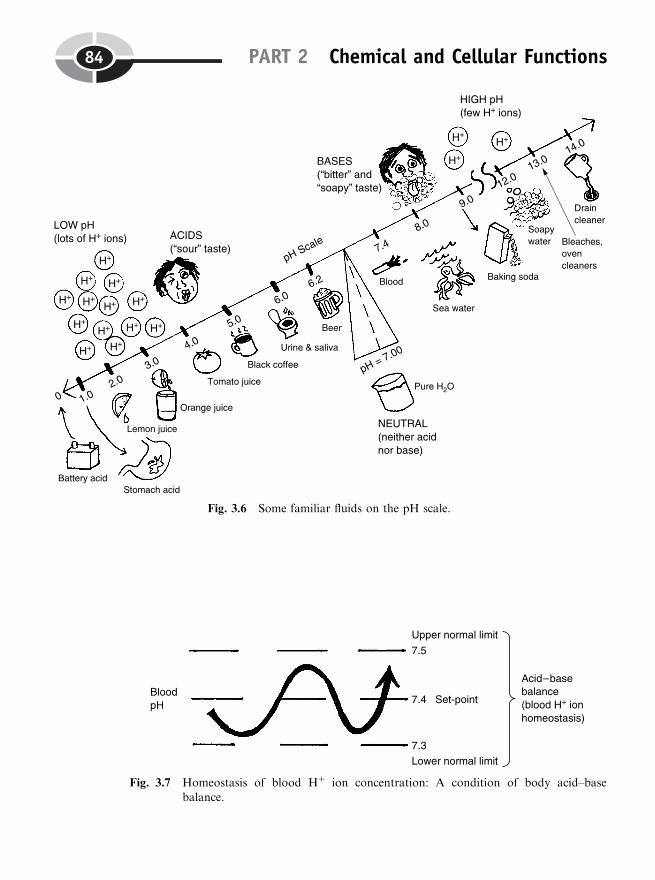
PART 2 Chemical and Cellular Functions84
Fig. 3.6 Some familiar fluids on the pH scale.
Fig. 3.7 Homeostasis of blood Hþ ion concentration: A condition of body acid–basebalance.
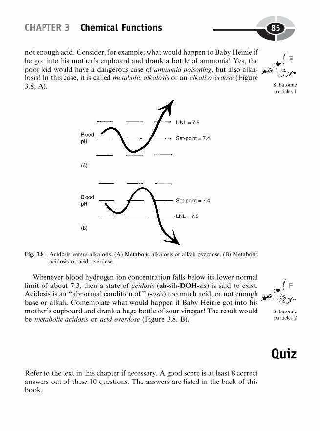
not enough acid. Consider, for example, what would happen to Baby Heinie ifhe got into his mother’s cupboard and drank a bottle of ammonia! Yes, thepoor kid would have a dangerous case of ammonia poisoning, but also alka-losis! In this case, it is called metabolic alkalosis or an alkali overdose (Figure3.8, A).
Whenever blood hydrogen ion concentration falls below its lower normallimit of about 7.3, then a state of acidosis (ah-sih-DOH-sis) is said to exist.Acidosis is an ‘‘abnormal condition of ’’ (-osis) too much acid, or not enoughbase or alkali. Contemplate what would happen if Baby Heinie got into hismother’s cupboard and drank a huge bottle of sour vinegar! The result wouldbe metabolic acidosis or acid overdose (Figure 3.8, B).
QuizRefer to the text in this chapter if necessary. A good score is at least 8 correctanswers out of these 10 questions. The answers are listed in the back of thisbook.
CHAPTER 3 Chemical Functions 85
Subatomic
particles 1
Fig. 3.8 Acidosis versus alkalosis. (A) Metabolic alkalosis or alkali overdose. (B) Metabolicacidosis or acid overdose.
Subatomic
particles 2
1. First proposed the existence of atoms:(a) Socrates(b) Sophie Tucker(c) Hippocrates(d) Democritus
2. The atom is not really ‘‘uncuttable’’ because:(a) Ions only occur in certain substances(b) Splitting atoms reveals their subatomic particles(c) No one has developed the necessary technology to try cutting
them(d) Atoms do not really exist!
3. Nonpolar covalent bonds:(a) Are mainly found in association with acids(b) Involve the net transfer of electrons between atoms(c) Create molecules with poles having opposite charge(d) Represent the equal sharing of outermost electrons between
atoms
4. NaCl is classified as an electrolyte, since:(a) Its crystals readily break down into ions that can help water con-
duct electricity(b) Its salty nature makes its taste ‘‘electrical’’(c) NaCl is decomposed into sodium anions and chloride cations(d) The crystal lattice has a C atom at each of its corners
5. Kinetic energy represents:(a) Stored energy that could potentially do work(b) Free energy associated with particle movement(c) Calories locked up into chemical bonds(d) Cold temperature conditions that can never be changed
6. The reaction, Glucose þ Fructose ¼ Sucrose, is an example of:(a) Catabolism(b) Electrolysis(c) Anode discharge(d) Anabolism
7. Acid þ Base ¼ :(a) Alkali(b) Complex carbohydrate(c) Cl�
(d) Salt
PART 2 Chemical and Cellular Functions86
8. Important Hþ ion acceptors:(a) Catabolic enzymes(b) Alkali(c) Acids(d) Most fruit juices
9. A fluid with a hydrogen ion concentration of 0.0000000001 g/liter hasa pH of:(a) 6(b) 8(c) 10(d) 12
10. Severe vomiting greatly reduces the amount of HCl left in the stomachand creates:(a) Intestinal ulcers(b) Metabolic acidosis(c) Respiratory alkalosis(d) Metabolic alkalosis
Body-Level Grids for Chapter 3Several key body facts were tagged with numbered icons in the page marginsof this chapter. Write a brief summary of each of these key facts into anumbered cell or box within the appropriate Body-Level Grid that appearsbelow.
Anatomy and Biological Order Fact Grids for Chapter 3:
SUBATOMICPARTICLELevel
CHAPTER 3 Chemical Functions 87
ATOMLevel
Physiology and Biological Order Fact Grids for Chapter 3:
MOLECULELevel
PART 2 Chemical and Cellular Functions88
1
1
1

Function and Biological Order Fact Grids for Chapter 3:
SUBATOMICPARTICLELevel
CHAPTER 3 Chemical Functions 89
1 2
3 4
5
ATOMLevel
MOLECULELevel
Function and Biological Disorder Fact Grids for Chapter 3:
SUBATOMICPARTICLELevel
PART 2 Chemical and Cellular Functions90
1
1 2
1 2

91
CHAPTER4
The Cell: A TinyArena of Life
We have now climbed up out of the Body Basement. You know – that wasthe Chemical Level (Chapter 3). Remember that we have just Plain BodyFunctions at the level of the subatomic particles, atoms, molecules. This is afact, of course, because it is only until we reach the Cell Level that Plain BodyFunctions finally become physiology.
The Cell Level: MicroanatomyBecomes Microphysiology
In our companion volume, ANATOMY DEMYSTIFIED, we read aboutmicroanatomy (MY-kroh-uh-NAT-uh-me), also called microscopic (my-kroh-SKAHP-ik) anatomy.
Copyright © 2004 by The McGraw-Hill Companies, Inc. Click here for terms of use.

MICROANATOMY(MICROSCOPICANATOMY)
¼ Study of Body Structures Too ‘‘Tiny’’To Be Seen With The Naked Eye
Technically speaking, microanatomy would include the subatomic parti-cles, atoms, molecules, organelles, and cells making up the structure of thehuman body. (Although the non-detailed characteristics of many tissues arevisible to the naked eye, the individual cells making up these tissues areusually studied with the microscope.)
The cell is the highest level of microanatomy, but at the same time it is thelowest level of microphysiology (my-kroh-fih-zee-AHL-uh-jee). We mean thatthe cell’s structures are so small that they must be viewed under a microscope,but the functions of the entire cell are now officially called physiology,because the cell is alive. The microphysiology of the cell is more oftendescribed by another phrase – cell physiology.
CELLPHYSIOLOGY
¼ MICROPHYSIOLOGYAT THE CELL
LEVEL
¼ FUNCTIONSCARRIED OUT BYTHE LIVING CELL
An interesting related question is, ‘‘What about the cell organelles?’’
PART 2 Chemical and Cellular Functions92
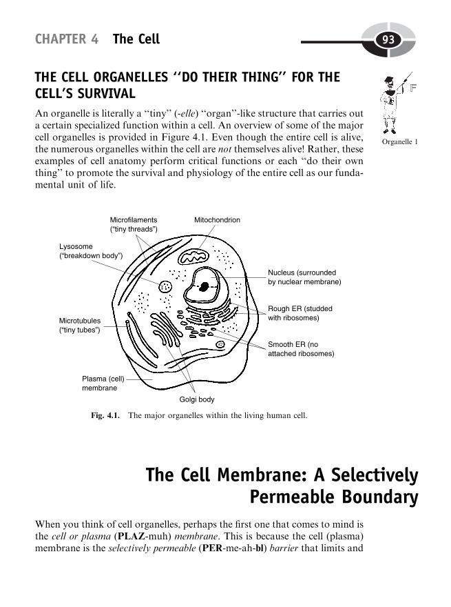
THE CELL ORGANELLES ‘‘DO THEIR THING’’ FOR THECELL’S SURVIVAL
An organelle is literally a ‘‘tiny’’ (-elle) ‘‘organ’’-like structure that carries outa certain specialized function within a cell. An overview of some of the majorcell organelles is provided in Figure 4.1. Even though the entire cell is alive,the numerous organelles within the cell are not themselves alive! Rather, theseexamples of cell anatomy perform critical functions or each ‘‘do their ownthing’’ to promote the survival and physiology of the entire cell as our funda-mental unit of life.
The Cell Membrane: A SelectivelyPermeable Boundary
When you think of cell organelles, perhaps the first one that comes to mind isthe cell or plasma (PLAZ-muh) membrane. This is because the cell (plasma)membrane is the selectively permeable (PER-me-ah-bl) barrier that limits and
CHAPTER 4 The Cell 93
Fig. 4.1. The major organelles within the living human cell.
Organelle 1
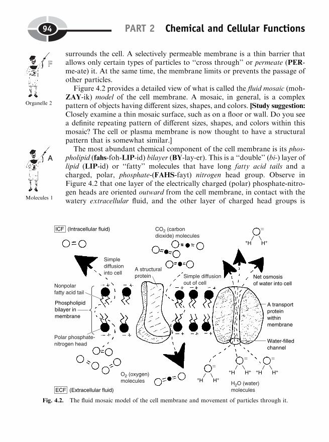
surrounds the cell. A selectively permeable membrane is a thin barrier thatallows only certain types of particles to ‘‘cross through’’ or permeate (PER-me-ate) it. At the same time, the membrane limits or prevents the passage ofother particles.
Figure 4.2 provides a detailed view of what is called the fluid mosaic (moh-ZAY-ik) model of the cell membrane. A mosaic, in general, is a complexpattern of objects having different sizes, shapes, and colors. [Study suggestion:Closely examine a thin mosaic surface, such as on a floor or wall. Do you seea definite repeating pattern of different sizes, shapes, and colors within thismosaic? The cell or plasma membrane is now thought to have a structuralpattern that is somewhat similar.]
The most abundant chemical component of the cell membrane is its phos-pholipid (fahs-foh-LIP-id) bilayer (BY-lay-er). This is a ‘‘double’’ (bi-) layer oflipid (LIP-id) or ‘‘fatty’’ molecules that have long fatty acid tails and acharged, polar, phosphate-(FAHS-fayt) nitrogen head group. Observe inFigure 4.2 that one layer of the electrically charged (polar) phosphate-nitro-gen heads are oriented outward from the cell membrane, in contact with thewatery extracellular fluid, and the other layer of charged head groups is
PART 2 Chemical and Cellular Functions94
Organelle 2
Fig. 4.2. The fluid mosaic model of the cell membrane and movement of particles through it.
Molecules 1
oriented inward from the membrane, in contact with the watery intracellularfluid.
SIMPLE DIFFUSION AND OSMOSIS: THE PRINCIPLE OF‘‘LIKE DISSOLVES LIKE’’
In determining which types of particles cross through (permeate) the cellmembrane, and how they pass through it, the principle of ‘‘like dissolveslike’’ is very essential. To be precise,
THE PRINCIPLE OF‘‘LIKE DISSOLVES LIKE’’:
Chemicals with similar or ‘‘like’’characteristics of bonding tend to mix ordissolve well together. So, charged particlestend to mix well with one another; anduncharged, nonpolar particles tend to mixwell with each other. Just don’t try to mixthe charged particles with the unchargedones!
Let us consider, for instance, the uncharged, nonpolar covalent, fatty acidtails of the phospholipid bilayer in the cell membrane. Being electricallyuncharged, this bilayer of fatty acid tails does not allow charged particles,such as polar covalent H2O molecules, or ions such as Naþ, to mix with it.The fatty acid tails do, however, let uncharged oxygen (O2) molecules mix,dissolve, and diffuse (dih-FUSE) or randomly ‘‘scatter’’ through them.Likewise, they allow uncharged carbon dioxide (die-AHKS-eyed) or CO2
molecules to mix and diffuse across the cell membrane.Simple diffusion is the random (chance) scattering of particles from a
region where their concentration is high, to a region where their concentra-tion is low. Oxygen (O2) molecules usually have a relatively high concentra-tion outside most of our cells, and a relatively low concentration within ourcells. [Study suggestion: Ask yourself, ‘‘Why does oxygen have such a lowconcentration within most of our body cells? What happens to the O2 mole-cules, when they diffuse in? Are they left alone, or are they used by cellmetabolism for something special?’’]
Therefore, there is usually a net (overall) simple diffusion of O2 moleculesacross the fatty acid bilayer, from the outside of the cell to the inside of thecell. Conversely, the net diffusion of CO2 molecules is in the opposite direc-tion (from the inside of the cell to the outside of the cell), because CO2 is athigher concentration within the cell. [Study suggestion: Think about thisquestion: ‘‘Where is the CO2 coming from, inside of the cell? What is
CHAPTER 4 The Cell 95
Molecules 1

going on to produce it?’’ We will come back to this problem later in thechapter.]
Glancing back at Figure 4.2, observe the presence of both structural pro-teins as well as transport proteins, occurring here and there within the cellmembrane. Note that the transport proteins have a water-filled channel thatextends all the way through them. These channels through transport proteinsare the way that the charged, polar H2O molecule can diffuse through themembrane. (They cannot mix with and diffuse through the nonpolar,uncharged fatty acid tails of the phospholipid bilayer, of course, becausethis would violate the ‘‘like dissolves like’’ principle.)
Osmosis (ahs-MOH-sis) is literally a ‘‘condition of ’’ (-osis) ‘‘thrusting’’(osm). Osmosis is defined as the simple diffusion of water (H2O) molecules,from a region of high water concentration to a region of low water concen-tration. In the cell membrane, such simple diffusion of water occurs throughthe channels in the transport proteins.
Water can, of course, pass through the channels in both directions. But ifthe water concentration is higher in the ECF (extracellular fluid), then therewill be a net (overall) osmosis of water into the cell. If the water concentrationis higher in the ICF (intracellular fluid), then there will be a net osmosis ofwater out of the cell. [Study suggestion: What do you predict will happen tothe size of the cell if there is a net osmosis of water into it? What if there is anet osmosis out of it?]
The idea of a ‘‘concentration gradient’’
You may have noted something in common about our discussions of bothsimple diffusion (random scattering of particles other than water) and osmo-sis (the diffusion of water molecules only). This is the fact that net diffusion orosmosis always occurs ‘‘downhill.’’ We mean that there is always a net oroverall movement of particles down their so-called concentration gradient(GRAY-dee-unt). The concentration gradient is literally a ‘‘grade’’ or ‘‘hill’’of concentration that exists between two different areas (as between the ECFand ICF).
NET DIFFUSION OROSMOSIS:
Always occurs ‘‘downhill’’ (from an area wherethe concentration of the moving particles ishigher, to an area where their concentration islower.)
[Study suggestion: Visualize a steep hill or gradient. A large bunch ofrandomly moving balls (chemical particles) are at the top of the hill, while
PART 2 Chemical and Cellular Functions96
Molecules 2
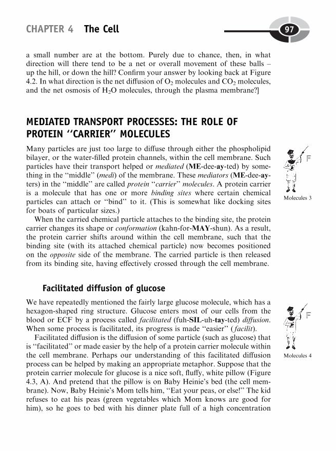
a small number are at the bottom. Purely due to chance, then, in whatdirection will there tend to be a net or overall movement of these balls –up the hill, or down the hill? Confirm your answer by looking back at Figure4.2. In what direction is the net diffusion of O2 molecules and CO2 molecules,and the net osmosis of H2O molecules, through the plasma membrane?]
MEDIATED TRANSPORT PROCESSES: THE ROLE OFPROTEIN ‘‘CARRIER’’ MOLECULES
Many particles are just too large to diffuse through either the phospholipidbilayer, or the water-filled protein channels, within the cell membrane. Suchparticles have their transport helped or mediated (ME-dee-ay-ted) by some-thing in the ‘‘middle’’ (medi) of the membrane. These mediators (ME-dee-ay-ters) in the ‘‘middle’’ are called protein ‘‘carrier’’ molecules. A protein carrieris a molecule that has one or more binding sites where certain chemicalparticles can attach or ‘‘bind’’ to it. (This is somewhat like docking sitesfor boats of particular sizes.)
When the carried chemical particle attaches to the binding site, the proteincarrier changes its shape or conformation (kahn-for-MAY-shun). As a result,the protein carrier shifts around within the cell membrane, such that thebinding site (with its attached chemical particle) now becomes positionedon the opposite side of the membrane. The carried particle is then releasedfrom its binding site, having effectively crossed through the cell membrane.
Facilitated diffusion of glucose
We have repeatedly mentioned the fairly large glucose molecule, which has ahexagon-shaped ring structure. Glucose enters most of our cells from theblood or ECF by a process called facilitated (fuh-SIL-uh-tay-ted) diffusion.When some process is facilitated, its progress is made ‘‘easier’’ ( facilit).
Facilitated diffusion is the diffusion of some particle (such as glucose) thatis ‘‘facilitated’’ or made easier by the help of a protein carrier molecule withinthe cell membrane. Perhaps our understanding of this facilitated diffusionprocess can be helped by making an appropriate metaphor. Suppose that theprotein carrier molecule for glucose is a nice soft, fluffy, white pillow (Figure4.3, A). And pretend that the pillow is on Baby Heinie’s bed (the cell mem-brane). Now, Baby Heinie’s Mom tells him, ‘‘Eat your peas, or else!’’ The kidrefuses to eat his peas (green vegetables which Mom knows are good forhim), so he goes to bed with his dinner plate full of a high concentration
CHAPTER 4 The Cell 97
Molecules 3
Molecules 4
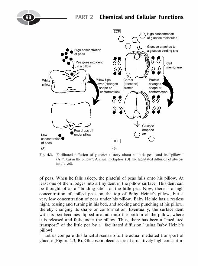
of peas. When he falls asleep, the plateful of peas falls onto his pillow. Atleast one of them lodges into a tiny dent in the pillow surface. This dent canbe thought of as a ‘‘binding site’’ for the little pea. Now, there is a highconcentration of spilled peas on the top of Baby Heinie’s pillow, but avery low concentration of peas under his pillow. Baby Heinie has a restlessnight, tossing and turning in his bed, and socking and punching at his pillow,thereby changing its shape or conformation. Eventually, the surface dentwith its pea becomes flipped around onto the bottom of the pillow, whereit is released and falls under the pillow. Thus, there has been a ‘‘mediatedtransport’’ of the little pea by a ‘‘facilitated diffusion’’ using Baby Heinie’spillow!
Let us compare this fanciful scenario to the actual mediated transport ofglucose (Figure 4.3, B). Glucose molecules are at a relatively high concentra-
PART 2 Chemical and Cellular Functions98
Fig. 4.3. Facilitated diffusion of glucose: a story about a ‘‘little pea’’ and its ‘‘pillow.’’
(A) ‘‘Peas in the pillow’’: A visual metaphor. (B) The facilitated diffusion of glucoseinto a cell.
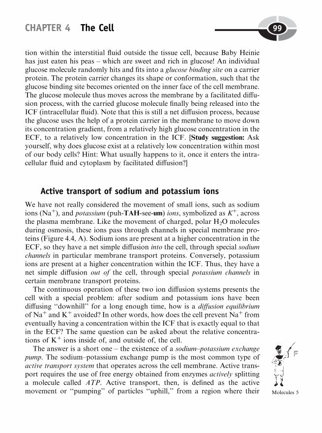
tion within the interstitial fluid outside the tissue cell, because Baby Heiniehas just eaten his peas – which are sweet and rich in glucose! An individualglucose molecule randomly hits and fits into a glucose binding site on a carrierprotein. The protein carrier changes its shape or conformation, such that theglucose binding site becomes oriented on the inner face of the cell membrane.The glucose molecule thus moves across the membrane by a facilitated diffu-sion process, with the carried glucose molecule finally being released into theICF (intracellular fluid). Note that this is still a net diffusion process, becausethe glucose uses the help of a protein carrier in the membrane to move downits concentration gradient, from a relatively high glucose concentration in theECF, to a relatively low concentration in the ICF. [Study suggestion: Askyourself, why does glucose exist at a relatively low concentration within mostof our body cells? Hint: What usually happens to it, once it enters the intra-cellular fluid and cytoplasm by facilitated diffusion?]
Active transport of sodium and potassium ions
We have not really considered the movement of small ions, such as sodiumions (Naþ), and potassium (puh-TAH-see-um) ions, symbolized as Kþ, acrossthe plasma membrane. Like the movement of charged, polar H2O moleculesduring osmosis, these ions pass through channels in special membrane pro-teins (Figure 4.4, A). Sodium ions are present at a higher concentration in theECF, so they have a net simple diffusion into the cell, through special sodiumchannels in particular membrane transport proteins. Conversely, potassiumions are present at a higher concentration within the ICF. Thus, they have anet simple diffusion out of the cell, through special potassium channels incertain membrane transport proteins.
The continuous operation of these two ion diffusion systems presents thecell with a special problem: after sodium and potassium ions have beendiffusing ‘‘downhill’’ for a long enough time, how is a diffusion equilibriumof Naþ and Kþ avoided? In other words, how does the cell prevent Naþ fromeventually having a concentration within the ICF that is exactly equal to thatin the ECF? The same question can be asked about the relative concentra-tions of Kþ ions inside of, and outside of, the cell.
The answer is a short one – the existence of a sodium–potassium exchangepump. The sodium–potassium exchange pump is the most common type ofactive transport system that operates across the cell membrane. Active trans-port requires the use of free energy obtained from enzymes actively splittinga molecule called ATP. Active transport, then, is defined as the activemovement or ‘‘pumping’’ of particles ‘‘uphill,’’ from a region where their
CHAPTER 4 The Cell 99
Molecules 5
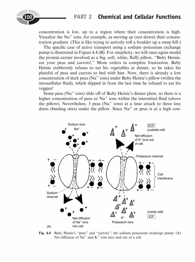
concentration is low, up to a region where their concentration is high.Visualize the Naþ ions, for example, as moving up (not down) their concen-tration gradient. (This is like trying to actively roll a boulder up a steep hill.)
The specific case of active transport using a sodium–potassium exchangepump is illustrated in Figure 4.4 (B). For simplicity, we will once again modelthe protein carrier involved as a big, soft, white, fluffy pillow. ‘‘Baby Heinie,eat your peas and carrots!,’’ Mom orders in complete frustration. BabyHeinie stubbornly refuses to eat his vegetables at dinner, so he takes hisplateful of peas and carrots to bed with him. Now, there is already a lowconcentration of dark peas (Naþ ions) under Baby Heinie’s pillow (within theintracellular fluid), which slipped in from the last time he refused to eat hisveggies!
Some peas (Naþ ions) slide off of Baby Heinie’s dinner plate, so there is ahigher concentration of peas or Naþ ions within the interstitial fluid (abovethe pillow). Nevertheless, 3 peas (Naþ ions) at a time attach to three tinydents (binding sites) under the pillow. Since Naþ or peas is at a high con-
PART 2 Chemical and Cellular Functions100
Fig. 4.4 Baby Heinie’s ‘‘peas’’ and ‘‘carrots’’: the sodium–potassium exchange pump. (A)Net diffusion of Naþ and Kþ ions into and out of a cell.
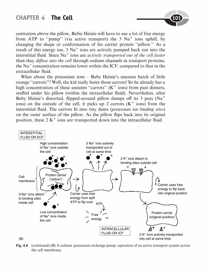
centration above the pillow, Babie Heinie will have to use a lot of free energyfrom ATP to ‘‘pump’’ (via active transport) the 3 Naþ ions uphill, bychanging the shape or conformation of his carrier protein ‘‘pillow.’’ As aresult of this energy use, 3 Naþ ions are actively pumped back out into theinterstitial fluid. Since Naþ ions are actively transported out of the cell fasterthan they diffuse into the cell through sodium channels in transport proteins,the Naþ concentration remains lower within the ICF, compared to that in theextracellular fluid.
What about the potassium ions – Baby Heinie’s uneaten batch of littleorange ‘‘carrots’’? Well, the kid really hates those carrots! So he already has ahigh concentration of these uneaten ‘‘carrots’’ (Kþ ions) from past dinners,stuffed under his pillow (within the intracellular fluid). Nevertheless, afterBaby Heinie’s distorted, flipped-around pillow dumps off its 3 peas (Naþ
ions) on the outside of the cell, it picks up 2 carrots (Kþ ions) from theinterstitial fluid. The carrots fit into tiny dents (potassium ion binding sites)on the outer surface of the pillow. As the pillow flips back into its originalposition, these 2 Kþ ions are transported down into the intracellular fluid.
CHAPTER 4 The Cell 101
Fig. 4.4 (continued) (B) A sodium–potassium exchange pump: operation of an active transport system acrossthe cell membrane.
As a result of this total active transport process, 3 Naþ ions are pumpedout of the cell for every 2 Kþ ions that are pumped into the cell. Therefore,this is a sodium-potassium 3-to-2 exchange pump.
SUMMARY OF PARTICLE MOVEMENT ACROSS THE CELLMEMBRANE
In summary,
THE THREEGENERAL TYPESOF PARTICLE
MOVEMENT ACROSSCELL MEMBRANE
¼ SIMPLEDIFFUSION
þ OSMOSIS þ MEDIATEDTRANSPORT
and
MEDIATEDTRANSPORT
(Use of protein carriers)
¼ FACILITATEDDIFFUSION
(No energy needed)
þ ACTIVETRANSPORT
(Requires use of energy)
Free Energy, Cell Work, and the ATP–ADP CycleIn addition to active transport across the cell membrane, there are manyenergy-requiring processes that occur within the cell. We have already men-tioned that a critically important molecule, ATP, was split by enzymes andprovided the free energy needed by the cell. ATP is an abbreviation foradenosine (ah-DEN-oh-seen) triphosphate (try-FAHS-fayt).
THE ATP–ADP CYCLE
The ATP (adenosine triphosphate) molecule is often symbolized as:A-P�P�P. The letter, A, of course, is an abbreviation for adenosine. Eachletter P denotes phosphate – a phosphorus-containing chemical group. WithinATP, there are ‘‘three’’ (tri-) phosphate groups attached to the adenosine. Thelast two chemical bonds in the ATP are special high-energy bonds. This isindicated by the squiggly line,�, before each of the last two phosphate groups(�P�P).
PART 2 Chemical and Cellular Functions102
Many cells contain a special type of enzyme called ATPase (ATP-ace),literally meaning ‘‘ATP splitter’’ (-ase). ATPase enzyme, therefore, acts tosplit the second high-energy phosphate bond within the ATP molecule. Whenthis bond is split, large amounts of previously stored potential energy isconverted into free (kinetic) energy and is used to do work, such as activetransport. After losing the end phosphate group, ATP or A-P�P�P,becomes ADP or adenosine diphosphate (die-FAHS-fate): A-P�P. ADP,therefore, is a reduced version of ATP that contains ‘‘two’’ (di-) phosphategroups rather than three.
When a person eats, say, a candybar or other foodstuff containing a highnumber of carbohydrate molecules (such as glucose), the individual carbohy-drate molecules are eventually broken down by catabolism. The potentialenergy stored in their chemical bonds is released. The free energy releasedfrom catabolism of food molecules is often used to re-attach the end phos-phate group back onto ADP, thereby re-creating more ATP. During exerciseor fasting, however, the available cell ATP molecules are broken back downinto ADP, such that more free energy is released to do the body’s work.
This back-and-forth process between ATP and ADP can technically becalled the ATP–ADP cycle. This energy storage–energy release cycle is acontinual process that goes around and around, for as long as the cell lives.
In summary, the ATP–ADP cycle:
ADP or A-P�P þ Excess free energy þ Phosphate (P) ¼ ATP or A-P�P�P(Energy storage)
andATP split by ATPase enzyme ¼ ADP þ Free energy to do cell work
Glucose Breakdown: Is It Going on Within theCytoplasm, or in the ‘‘Thread-Granules’’?
With all this talk of energy from ATP, and its use for numerous activitieswithin the cell, it is only natural for us to ask, ‘‘If free energy from ATP is soimportant for the cell, then quite a lot of it must be produced locally, rightthere within the cell, right?’’ Yes, this is quite correct!
CHAPTER 4 The Cell 103
Molecules 6
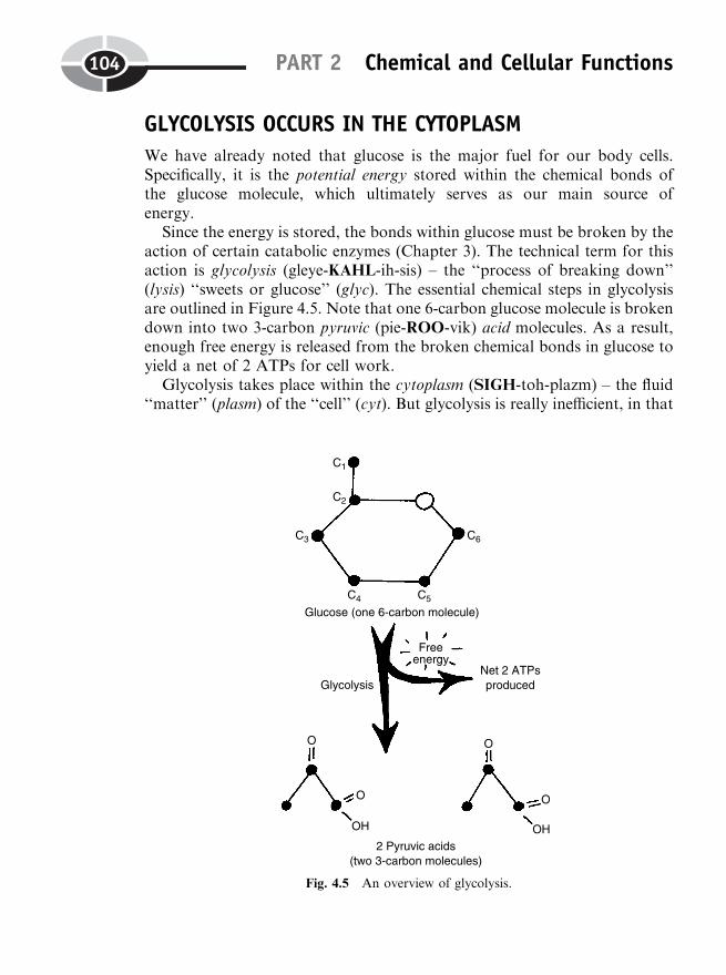
GLYCOLYSIS OCCURS IN THE CYTOPLASM
We have already noted that glucose is the major fuel for our body cells.Specifically, it is the potential energy stored within the chemical bonds ofthe glucose molecule, which ultimately serves as our main source ofenergy.
Since the energy is stored, the bonds within glucose must be broken by theaction of certain catabolic enzymes (Chapter 3). The technical term for thisaction is glycolysis (gleye-KAHL-ih-sis) – the ‘‘process of breaking down’’(lysis) ‘‘sweets or glucose’’ (glyc). The essential chemical steps in glycolysisare outlined in Figure 4.5. Note that one 6-carbon glucose molecule is brokendown into two 3-carbon pyruvic (pie-ROO-vik) acid molecules. As a result,enough free energy is released from the broken chemical bonds in glucose toyield a net of 2 ATPs for cell work.
Glycolysis takes place within the cytoplasm (SIGH-toh-plazm) – the fluid‘‘matter’’ (plasm) of the ‘‘cell’’ (cyt). But glycolysis is really inefficient, in that
PART 2 Chemical and Cellular Functions104
Fig. 4.5 An overview of glycolysis.

the breakdown of 1 glucose molecule results in a net of only 2 ATPs pro-duced. Now, doesn’t the cell deserve to get much more ATP energy ‘‘bang’’out of each of its glucose ‘‘bucks’’?
CELLULAR RESPIRATION OCCURS IN THEMITOCHONDRIA
Under normal conditions, most of the cells in the body operate within anaerobic (air-OH-bik) or ‘‘living’’ (bi) in ‘‘air’’ (aer) environment. As Figure4.6 reveals, the majority of our cells have a steady supply of oxygen (O2)being delivered to them from the bloodstream and the interstitial fluid. Thisoxygen, of course, ultimately comes from the air that we breathe. Hence, ourcells mostly operate under aerobic or oxidative (AHKS-ih-day-tiv) – ‘‘pertain-ing to’’ (-ive) ‘‘oxygen’’ (oxidat) – conditions.
When both oxygen and glucose are present within the cytoplasm, then the2 pyruvic acid molecules produced by glycolysis enter into cellular respiration(res-pir-AY-shun). Cellular respiration, then, is just the general phrase for allof the aerobic (oxygen-using) steps in glucose catabolism (breakdown) thatoccur after glycolysis.
Enter the mitochondria
Whereas glycolysis occurs in the cytoplasm, cellular respiration occurs inthe mitochondria (my-toh-KAHN-dree-ah). The mitochondria are literally‘‘thread’’ (mito) ‘‘granules’’ (chondr), because sometimes they are long andthin (like threads), while at other times they are round like little grains(granules). Each mitochondrion (my-toh-KAHN-dree-un) is surrounded byan outer membrane plus an inner membrane. The inner membrane is throwninto slender ‘‘crests’’ or ‘‘ridges’’ called cristae (KRIS-tee). And within themiddle of each mitochondrion is a fluid-filled ‘‘womb’’ or mitochondrialmatrix (MAY-tricks).
The mitochondrial matrix is where the 2 pyruvic acids diffuse, after theyare produced by glycolysis. Each pyruvic acid takes its turn entering theKrebs cycle. Named after the German biochemist, Hans Krebs, the Krebscycle is a repeating cycle of aerobic reactions that break down the pyruvicacids produced by glycolysis. The Krebs cycle rotates twice (once for each ofthe pyruvic acids fed into it). Along the way, a total of 2 ATPs, several CO2
molecules, and a number of hydrogen-carrier molecules result.
CHAPTER 4 The Cell 105
Organelles 1

PART 2 Chemical and Cellular Functions106
Fig. 4.6 An overview of cellular (aerobic) respiration.

The hydrogen-carrier molecules, once produced by the Krebs cycle, moveonto the nearby electron transport system. The electron transport system islocated along the cristae (inner crests or ridges) of the mitochondrion. Thelarge molecules of the electron transport system, as their name indicates,carry high-energy electrons from H atoms, down to progressively lowerand lower energy levels. As the electrons flow or are transported ‘‘downhill’’(energy-wise), they release considerable amounts of free or kinetic energyalong the way.
A net total of 34 more ATPs are eventually produced from this electrontransport process. Finally, the transported electrons, now depleted of most oftheir former energy, are transferred to an oxygen atom. The oxygen atomthen combines with two Hþ ions, thereby creating water (H2O).
The overall equation for cellular (aerobic) respiration, using glucose as thefuel molecule, is:
C6H12O6
Glucoseþ 6 O2
Oxygen! 6 CO2
Carbon dioxideþ 6 H2O
Waterþ 36 ATP
Free energy
SUMMARY OF AEROBIC GLUCOSE BREAKDOWN
Capsulizing this section, we have:
Glycolysis (occurs in cytoplasm) 2 ATPs/glucoseCellular respiration (Krebs cycle and 36 ATPs/glucoseelectron transport system in mitochondria)
Total ¼ 38 ATPs/glucose
Protein Synthesis: The ‘‘Colored Worms’’Open and Squirm!
So far, we have talked about the cell (plasma) membrane, the cytoplasm, andthe mitochondria. But there are several other key organelles deeply involvedin the process of protein synthesis, the making of new proteins for the cell.Proteins serve such critical functions as being enzymes (protein catalysts thatspeed up chemical reactions), structural proteins, transport proteins withchannels, and protein carrier molecules.
CHAPTER 4 The Cell 107
Molecules 7

The most important single place in the cell for protein synthesis is the cellnucleus. [Study suggestion: You have already heard about another kind of‘‘nucleus’’ in this book! – Do you remember it? At what level of BodyOrganization does it exist?]
Somewhat resembling a rounded ‘‘kernel’’ or ‘‘nut’’ (nucle), the cellnucleus has its own ‘‘skin’’ (like a peanut)! The ‘‘skin’’ in this case, however,is the nuclear (NEW-klee-ar) membrane surrounding the nucleus. Enclosedwithin the nuclear membrane are a number of important structures, suchas the chromosomes (KROH-moh-sohms) – ‘‘colored’’ (chrom) wormlike‘‘bodies’’ (-somes). (Consult Figure 4.7.) Each of these chromosomes, inturn, contains a tightly coiled DNA molecule. There are numerous genes(jeans) or sections strung along the DNA molecule.
DNA is an abbreviation for deoxyribonucleic (dee-ahk-see-RYE-boh-new-KLEE-ik) acid. This name reflects the fact that DNA is a nucleic (new-KLEE-ik) acid (NA), since it is an ‘‘acid’’ found within the cell ‘‘nucleus.’’ The D inthe abbreviation stands for deoxyribose (dee-ahk-see-RYE-bohs), a type ofsugar ‘‘lacking’’ (de-) an ‘‘oxygen’’ (oxy) atom.
But in looking at a deoxyribonucleic acid (DNA) molecule, one does notreally notice any sugars! Rather, one immediately notices the dramatic doublehelix (HEE-liks) or double ‘‘spiral’’ shape. The deoxyribose sugar moleculesoccur at intervals along each twisted chain of this double helix or twistedspiral.
Specifically, a deoxyribose sugar molecule forms part of the repeatingstructure of each gene within the DNA double helix. A single gene providesa chemical code for the synthesis of a particular protein. As Figure 4.7reveals, during protein synthesis, part of the DNA double helix unwindsand puffs out from the surface of the chromosome, almost as if it were acut-open, squirming earthworm on a fish hook!
DNA CODONS AS CHEMICAL CODE WORDS
A group of DNA codons (KOH-dahns) becomes exposed along the edges ofthis chromosome puff. A codon is a chemical ‘‘code word’’ consisting of threenitrogen-containing bases. Each codon is a code word for bringing in aparticular type of amino (ah-MEE-noh) acid. A protein is essentially a stringof connected amino acids. Therefore, a gene consists of a number of codons,all of them together providing a total code word to the cell for synthesizing acertain protein made of amino acids.
PART 2 Chemical and Cellular Functions108
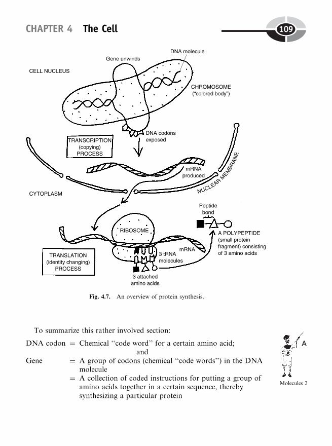
To summarize this rather involved section:
DNA codon ¼ Chemical ‘‘code word’’ for a certain amino acid;and
Gene ¼ A group of codons (chemical ‘‘code words’’) in the DNAmolecule
¼ A collection of coded instructions for putting a group ofamino acids together in a certain sequence, therebysynthesizing a particular protein
CHAPTER 4 The Cell 109
Molecules 2
Fig. 4.7. An overview of protein synthesis.
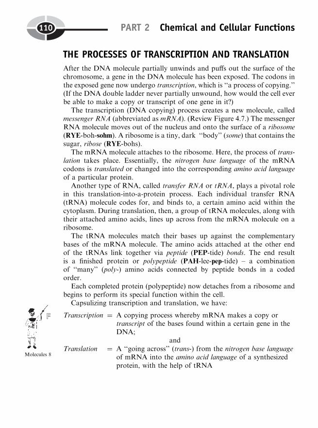
THE PROCESSES OF TRANSCRIPTION AND TRANSLATION
After the DNA molecule partially unwinds and puffs out the surface of thechromosome, a gene in the DNA molecule has been exposed. The codons inthe exposed gene now undergo transcription, which is ‘‘a process of copying.’’(If the DNA double ladder never partially unwound, how would the cell everbe able to make a copy or transcript of one gene in it?)
The transcription (DNA copying) process creates a new molecule, calledmessenger RNA (abbreviated as mRNA). (Review Figure 4.7.) The messengerRNA molecule moves out of the nucleus and onto the surface of a ribosome(RYE-boh-sohm). A ribosome is a tiny, dark ‘‘body’’ (some) that contains thesugar, ribose (RYE-bohs).
The mRNA molecule attaches to the ribosome. Here, the process of trans-lation takes place. Essentially, the nitrogen base language of the mRNAcodons is translated or changed into the corresponding amino acid languageof a particular protein.
Another type of RNA, called transfer RNA or tRNA, plays a pivotal rolein this translation-into-a-protein process. Each individual transfer RNA(tRNA) molecule codes for, and binds to, a certain amino acid within thecytoplasm. During translation, then, a group of tRNA molecules, along withtheir attached amino acids, lines up across from the mRNA molecule on aribosome.
The tRNA molecules match their bases up against the complementarybases of the mRNA molecule. The amino acids attached at the other endof the tRNAs link together via peptide (PEP-tide) bonds. The end resultis a finished protein or polypeptide (PAH-lee-pep-tide) – a combinationof ‘‘many’’ (poly-) amino acids connected by peptide bonds in a codedorder.
Each completed protein (polypeptide) now detaches from a ribosome andbegins to perform its special function within the cell.
Capsulizing transcription and translation, we have:
Transcription ¼ A copying process whereby mRNA makes a copy ortranscript of the bases found within a certain gene in theDNA;
andTranslation ¼ A ‘‘going across’’ (trans-) from the nitrogen base language
of mRNA into the amino acid language of a synthesizedprotein, with the help of tRNA
PART 2 Chemical and Cellular Functions110
Molecules 8

The Tragedy of Mutation: Pathophysiologyand ‘‘Bad’’ Proteins
Several times we have noted that pathological anatomy, usually associatedwith a certain degree of Biological Disorder within body structures, fre-quently results in pathophysiology and morbidity (disease) or mortality(death). In certain cases, this is also true for mutations (mew-TAY-shuns) –abnormal ‘‘changes’’ in the structure of body proteins.
Consider, for example, the frequently fatal disorder called sickle cell dis-ease. In persons with this disease, many of the normally rounded red bloodcells assume the strange, ‘‘sickled’’ shape of a half moon or crescent. Thisdeforming of the RBC (red blood cell) shape, surprisingly enough, is due toa mutation or error in just a single amino acid within the long hemoglobin(HE-moh-gloh-bin) molecule.
Hemoglobin is the main oxygen-carrying protein in our red blood cells(RBCs). Due to an error in the gene coding for hemoglobin synthesis, theresulting abnormal hemoglobin molecules in a person with sickle cell diseasecontain just one wrong amino acid! Nevertheless, this seemingly minor occur-rence of a Structural Disorder at the Molecular Level (hemoglobin) createsan abnormal hemoglobin that can produce deadly consequences for thepatient!
The sickled (crescent-shaped) RBCs are very fragile, and their plasmamembranes extremely sticky. Therefore, there is great risk of suffering a sicklecell crisis, wherein many sickled RBCs suddenly clump together or aggluti-nate (ah-GLUE-tih-nayt) in large numbers. Such large masses of agglutinated(ah-GLUE-tih-nay-ted) RBCs may completely occlude (ah-KLOOD) or ‘‘stopup’’ the openings of major blood vessels. As a result, considerable numbers ofbody cells and tissues may die, due to lack of adequate blood flow and itsvital oxygen supply.
QuizRefer to the text in this chapter if necessary. A good score is at least 8 correctanswers out of these 10 questions. The answers are listed in the back of thisbook.
1. The cell organelles, like the entire cell, are:(a) Living structures with their own physiology
CHAPTER 4 The Cell 111
Molecules 1

(b) Tiny organ-like structures having highly specific functions withinthe cell
(c) Each wrapped in a single plasma membrane(d) Usually functioning poorly (if at all!)
2. By describing something as ‘‘selectively permeable,’’ it means that:(a) The object is a totally ineffective barrier(b) Neither liquid nor solid can penetrate it(c) Some substances pass through it, while the movement of others is
prevented(d) Energy is always required to cross through it
3. The Principle of ‘‘like dissolves like’’ implies that:(a) Water and oil don’t mix(b) The phospholipid bilayer is easily penetrated by Naþ ions(c) Water and oil sometimes can mix well together(d) Charged, polar particles react well with those that are un-
charged
4. A condition of thrusting of H2O molecules:(a) Osmosis(b) Active transport(c) Facilitated diffusion(d) Mediated transport
5. Glucose enters most body cells by what specific process?(a) Active transport(b) Mitosis(c) Occurrence of a diffusion equilibrium(d) Facilitated diffusion
6. A sodium–potassium exchange pump is a good example of:(a) Cytokinesis(b) Diffusion down a concentration gradient(c) Facilitated diffusion through pores(d) Energy-primed movement up a concentration gradient
7. The ADP molecule becomes much more suited for supplying cellenergy when:(a) Mutations render it into a less harmful conformation(b) A terminal phosphate group breaks off the end of it(c) Nitrogen atoms are fused to it in great numbers(d) A terminal phosphate group is added
PART 2 Chemical and Cellular Functions112
8. Literally means the ‘‘process of breaking down sweets or glucose’’:(a) Glycolysis(b) Glycogen(c) Krebs cycle(d) Amino acid transference
9. Cellular respiration occurs within the of the mitochondrion:(a) Outer membrane(b) Ribosomes(c) Cristae and matrix(d) Matrix only
10. DNA codons function primarily as:(a) Alternative functions for membrane lipids(b) Protein carriers(c) Code words for amino acids(d) Translation of the language of peptide bonds into the language of
ionic bonds
Body-Level Grids for Chapter 4Several key body facts were tagged with numbered icons in the page marginsof this chapter. Write a brief summary of each of these facts into a numberedcell or box within the appropriate Body-Level Grid that appears below.
Anatomy and Biological Order Fact Grids for Chapter 4:
MOLECULELevel
CHAPTER 4 The Cell 113
ORGANELLELevel
Function and Biological Order Fact Grids for Chapter 4:
MOLECULELevel
PART 2 Chemical and Cellular Functions114
1 2
1
1 2
ORGANELLELevel
CHAPTER 4 The Cell 115
3 4
5 6
7 8
1 2
Anatomy and Biological Disorder Fact Grids for Chapter 4:
MOLECULELevel
PART 2 Chemical and Cellular Functions116
1

117
Test: Part 2
DO NOT REFER TO THE TEXT WHEN TAKING THIS TEST. A goodscore is at least 18 (out of 25 questions) correct. Answers are in the back ofthe book. It’s best to have a friend check your score the first time, so youwon’t memorize the answers if you want to take the test again.
1. The Chemical Level of Body Organization consists of:(a) Subatomic particles, organelles, and molecules(b) Cells, atoms, and molecules(c) Subatomic particles, atoms, and molecules(d) Atoms, tissues, and elements(e) Practically everything within the Great Body Pyramid!
2. Anatomy joins with the Atomic Theory in explaining that:(a) All body structures consist of atoms and the empty spaces
between these atoms(b) Several levels of human anatomy are nothing but atoms!(c) Atoms, like anatomy, have a corresponding physiology(d) Anatomy, like atoms, consists of structures that are ‘‘uncuttable’’(e) Neither area of human study involves any practical use of dissec-
tion
Copyright © 2004 by The McGraw-Hill Companies, Inc. Click here for terms of use.
3. Nonpolar covalent bonding differs from polar covalent bonding inits:(a) Transfer of innermost electrons between atoms(b) Tendency to break down into ions(c) Ability to share electron clouds equally between atoms(d) Frictional resistance to magnetic forces(e) Trend toward tearing apart into teeny little pieces
4. Very common cation within the body’s extracellular fluid:(a) Cl�
(b) U-235(c) Naþ
(d) Kþ
(e) HPO�4
5. A log sitting on the ground has much more of this energy, comparedto a log burning in a fire:(a) Anabolic(b) Metabolic(c) Kinetic(d) Human(e) Potential
6. Synthesis reactions are alternatively known as:(a) Protein catalysts(b) Anabolism(c) Destructogenesis(d) Catabolism(e) Exchange reactions
7. The so-called intestinal neutralization equation helps to ‘‘neutralize’’:(a) NaHCO3
(b) NaCl(c) H2O(d) H2CO3
(e) HCl
8. Serve as important Hþ ion acceptors:(a) Sponges(b) Acids(c) Bases or alkali(d) Electrolytes(e) Most cations
PART 2 Chemical and Cellular Functions118

9. The pH of a neutral solution is approximately:(a) 3(b) 5(c) 7(d) 7.4(e) 14
10. A bottle containing household ammonia has:(a) An extremely high pH(b) A low pH but a high Hþ ion concentration(c) Most of the same acid–base characteristics as does pure water(d) Both a high pH and a high Hþ concentration(e) Both a low pH and a low Hþ ion concentration
11. When Baby Heinie falls into a vat of lemon juice (containing lots ofascorbic acid) and gulps down large quantities of it, he is in mostdanger of suffering from:(a) A gall bladder attack!(b) Chronic acne(c) Metabolic acidosis(d) Respiratory alkalosis(e) Metabolic alkalosis
12. The cell is considered the lowest level where microphysiology exists,because:(a) The cell organelles are considered the ‘‘life-line’’(b) Only groups of related cells are actually functioning(c) Cell structure determines cell function(d) True physiology starts with the basic unit of life(e) Anatomy starts at the cell level
13. The portion of the cell or plasma membrane most resembling a soapbubble:(a) Polar phosphate-nitrogen head groups of the phospholipids(b) Water-filled channels through transport proteins(c) Nonpolar fatty acid tails of the phosopholipids(d) Sodium chloride electrolyte content(e) Bubbly bath of acid residues
14. Open a bottle of perfume. Soon, persons in the far corners of the roomcan smell it, due mainly to the process of:(a) Cytokinesis(b) Mediated transport
Test: Part 2 119
(c) Active transport(d) Chromosome breakage(e) Simple diffusion
15. A newly planted flower begins to droop and wilt. You water it, and itsprings right back, owing to the pressure created by:(a) Net osmosis into the plant cells(b) Uncontrolled drying out in the baking sun!(c) Simple diffusion of electrolytes out of the leaves(d) Net osmosis out of the plant cells(e) The occurrence of a permanent diffusion equilibrium
16. If a special drug were given that destroyed all carrier proteins in thecell membrane:(a) Simple diffusion through the membrane would grind to a halt(b) No H2O could fit through membrane channels(c) All mediated transport processes would stop(d) Particles would still tend to move randomly uphill across the
membrane(e) The cell membrane would completely fall apart!
17. Enters most body cells by a facilitated diffusion process:(a) Bases and alkali(b) Naþ and Kþ ions(c) Clusters of H2O molecules(d) Fatty acid(e) Glucose
18. Even though sodium ions continually diffuse into the cell throughchannels, the inside of the cell remains negatively charged comparedto the outside, because:(a) Too many potassium ions are around!(b) Naþ ions are actively transported back out of the cell at a faster
rate(c) The sodium is broken down into its individual subatomic particles(d) The cell produces lots of Naþ during its aerobic metabolism(e) Kþ ions are pumped back into the cell using energy from ATP
19. Excess free energy from catabolism of cell nutrients is usually trans-ferred to:(a) Adenosine diphosphate(b) ATP
PART 2 Chemical and Cellular Functions120
(c) The outer mitochondrial membrane(d) NaHCO3
(e) Potential energy that is seldom, if ever, tapped
20. Two pyruvic acids result from this sequence of chemical reactions:(a) Krebs cycle(b) Gene mutation(c) Glycolysis(d) Mediated transport(e) Osmosis
21. The fluid matter of the cell:(a) Mitochondria(b) Cristae(c) Microtubules(d) Cytoplasm(e) Nucleolus
22. A site for the electron transport system:(a) Outer mitochondrial membrane(b) Mitochondrial matrix(c) ‘‘Crests’’ of the inner mitochondrial membrane(d) Cytoplasm(e) Plasma membrane
23. Chemical serving as the final acceptor for ‘‘energy-depleted’’ electronsin the electron transport system:(a) C(b) N(c) H(d) O(e) P
24. Total number of ATP molecules produced per glucose moleculecatabolized under aerobic conditions:(a) 16(b) 22(c) 32(d) 34(e) 38
Test: Part 2 121

25. The phase of protein synthesis where a copy of the DNA codons isconstructed as a series of corresponding codons in mRNA:(a) Interphase(b) Transcription(c) DNA synthesis(d) Catabolism(e) Translation
PART 2 Chemical and Cellular Functions122

PART 3
The Physiology ofSkin, Bone, and
Muscle
Copyright © 2004 by The McGraw-Hill Companies, Inc. Click here for terms of use.

This page intentionally left blank
125
CHAPTER5
Working the Human‘‘Hard’’ Drives: ThePhysiology of Skins
and Skeletons
So far, this book has been easy. But now it is time to get ‘‘hard’’! Specifically,it is time for us to start working the human ‘‘hard’’ drives – the physiology ofour tough outer skin and our rock-like, ‘‘inner’’ (endo-) ‘‘hard dried body,’’the endoskeleton (EN-doh-skel-uh-ton).
Copyright © 2004 by The McGraw-Hill Companies, Inc. Click here for terms of use.
‘‘Systemic’’ Physiology of the SkinFor our purposes in this book, we will concentrate upon several aspects ofthe ‘‘systemic’’ physiology of the skin. By this we mean the broad, organsystem and organism effects of skin physiology, rather than the mostly‘‘local’’ physiological effects, such as protection from the ultraviolet (ul-truh-VEYE-uh-lit) or UV rays in sunlight.
SYSTEMIC PHYSIOLOGY 1: PROTECTINGTHE BODY FROM DEHYDRATION
The skin, and especially the epidermis, provides the entire human organismabsolutely vital protection from massive tissue dehydration (dee-high-DRAY-shun). This term literally means ‘‘the process of ’’ (-tion) losing ‘‘water’’ (hydr)‘‘from’’ (de-) the body tissues.
You may recall (Chapter 3) that water or H2O is the major polar covalentmolecule within the body fluids. As such, it is the chief solvent (‘‘dissolver’’)of sodium chloride (NaCl) and other charged solutes (SAHL-yoots) – ‘‘thingsdissolved.’’ Hence, our ‘‘internal sea’’ of body fluids is principally an aqueous(AH-kwee-us) or ‘‘watery’’ solution (so-LOO-shun). A solution, in general, isa liquid containing dissolved substances (one or more types of solute parti-cles). Summarizing yields:
‘‘INTERNALSEA’’ OFBODYFLUIDS
¼ CHARGEDSOLUTES
(‘‘Things Dissolved’’)
þ WATERSOLVENT
(‘‘The Dissolver’’)
¼ ANAQUEOUSSOLUTION
The aqueous solution of our internal sea, the astute reader may remember(Chapter 2), is called the extracellular fluid or ECF – the fluid lying ‘‘outsideof the little cells’’ in our body tissues. The ECF also includes the blood plasma(PLAZ-muh), or watery fluid ‘‘matter’’ of the blood. These ECF and bloodplasma concepts further bring back to mind the intracellular fluid or ICF‘‘within’’ (intra-) our cells. The intracellular fluid (ICF), like the extracellularfluid (ECF), is essentially a water-dominated aqueous solution containingvarious dissolved solutes.
PART 3 Skin, Bone, and Muscle126
Organ 1

CHAPTER 5 Skins and Skeletons 127
Normal hydration and osmotic equilibrium
When the body is in a state of normal hydration (high-DRAY-shun), it isnormally ‘‘watered’’ (hydr). There is a chemical equilibrium, or dynamicbalance, between the water concentrations of the ECF (including the bloodplasma) and the ICF. (Consult Figure 5.1, A.)
Back in Chapter 4, we talked about osmosis. This is the ‘‘thrusting’’ (osm)of H2O molecules in a particular direction as they diffuse across the cell’splasma membrane – either from the ECF into the ICF, or from the ICF outinto the ECF.
If the water concentration of the ECF equals the water concentration ofthe ICF, there is no water concentration gradient between them. Thousandsof H2O molecules diffuse back and forth through the water-filled pores of theselectively permeable cell membrane, entering and leaving the cell at equalrates. Thus, a condition of osmotic (ahz-MAHT-ik) equilibrium exists.
Isotonic body fluids and stable cell size
Pretend that we are looking at the red blood cells (RBCs) within the blood-stream of a normally hydrated (HIGH-dray-ted) person (Figure 5.1, B). Insuch a person, their blood plasma is said to represent an isotonic (eye-so-TAHN-ik) solution. An isotonic solution is a solution whose ‘‘strength’’ or‘‘concentration’’ (ton) of solute is the ‘‘same’’ (iso-) as that found within theintracellular fluid. Now, the ICF of red blood cells is an aqueous solutionwith a strength or concentration of about 0.9% NaCl solute dissolved in99.1% H2O solvent. If the surrounding blood plasma has the same concen-tration of solute and water as this, then it is classified as an isotonic solution.This classification as isotonic is important, because it results in stability ofcell size. Specifically, there is no concentration gradient for H2O across thecell membrane of the RBC, so that there is an osmotic equilibrium. Watermolecules diffuse into the RBCs, just about as fast as they diffuse out. WhenRBCs travel through very narrow blood capillaries (CAP-uh-lair-eez), forinstance, not being too big allows them to easily pass (without gettingstuck!). To summarize,
NORMAL TISSUEHYDRATION: (A NormalVolume of Body Water)
STABILITY OF RBC SIZE ISACHIEVED
BLOOD PLASMA ANDOTHER PARTS OF ECF ARE
ISOTONIC SOLUTIONS
NO NET OSMOSIS OF H2OEITHER INTO OR OUT
OF CELLS
�
!
�
PART 3 Skin, Bone, and Muscle128
Cell 1
Tissue dehydration results in cell shrinkage
Following from our consideration of normal hydration and the ECF as anisotonic solution, we can now see how this delicate balance is upset duringtissue dehydration. The fairly watertight skin normally prevents a hugevolume of our body water content from simply evaporating into the airfrom our watery tissues. This is largely due to the presence of keratin(CARE-uh-tin) granules scattered throughout the surface epidermis.Keratin is a family of tough, waterproof proteins founds in the ‘‘horns’’(kerat) of animals, as well as in the skin, nails, and hair of humans.
‘‘But what if a large portion of my skin (along with its waterproof keratinprotein) is gone, due to a severe burn?’’ Most unfortunately, besides a hugerisk of severe bacterial infection, your soft, wet, underlying connective tissuesand muscle will tend to lose huge amounts of H2O to the surrounding air byevaporation. This will soon result in severe tissue dehydration. And in theblood plasma, you are quite likely to suffer extreme hemoconcentration (HE-moh-kahn-sen-TRAY-shun). This is a dramatic rise in the ‘‘blood’’ (hemo)solute ‘‘concentration,’’ far above the normal 0.9% NaCl figure.
With so little water remaining, the blood plasma (and other portions of theECF) become a hypertonic (high-per-TAHN-ik) solution. This solution typehas an ‘‘excessive or above normal’’ (hyper-) ‘‘strength or concentration’’(ton) of solute. As displayed in Figure 5.1 (C), a hypertonic solution, becauseit has such a high solute concentration, has a very low solvent (H2O) con-centration. Hence, the water concentration in the ICF of the red blood cells isgreater than the water concentration in the surrounding blood plasma. Thereis a net osmosis of thousands of H2O molecules out of the RBCs and into theoverly concentrated plasma. Crenation (kree-NAY-shun) – the ‘‘process of ’’(-tion) becoming ‘‘notched’’ (cren) – happens to the RBCs.
The abnormally shrunken, crenated (KREE-nay-ted) red blood cells, bear-ing numerous wrinkles and notches, can no longer function normally incarrying oxygen (O2) molecules through the bloodstream. And the dehy-drated person may also suffer from severe tissue ischemia (is-KEY-me-ah),a ‘‘holding back’’ of blood flow that deprives many parts of the body.
In summary,
SEVERE TISSUEDEHYDRATION: (Large Amount
of Body Water Loss)
CRENATION (SHRINKINGAND NOTCHING) OF RBCs
�
BLOOD PLASMA ANDOTHER PARTS OF ECFBECOME HYPERTONIC
SOLUTIONS
NET OSMOSIS OF H2O OUTOF RBCs
�!
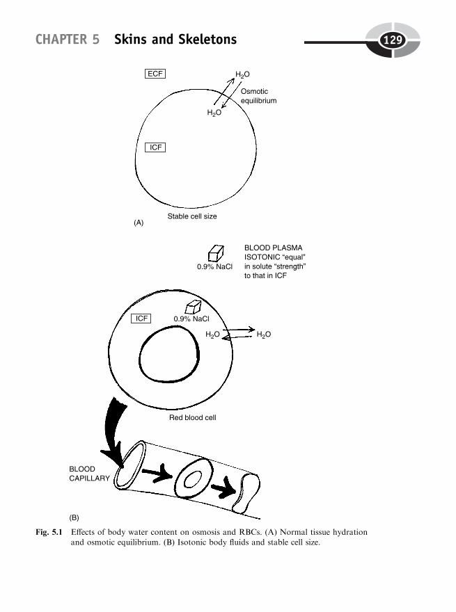
CHAPTER 5 Skins and Skeletons 129
Fig. 5.1 Effects of body water content on osmosis and RBCs. (A) Normal tissue hydrationand osmotic equilibrium. (B) Isotonic body fluids and stable cell size.
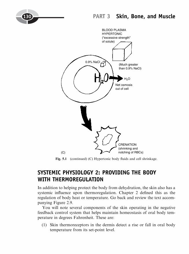
SYSTEMIC PHYSIOLOGY 2: PROVIDING THE BODYWITH THERMOREGULATION
In addition to helping protect the body from dehydration, the skin also has asystemic influence upon thermoregulation. Chapter 2 defined this as theregulation of body heat or temperature. Go back and review the text accom-panying Figure 2.9.
You will note several components of the skin operating in the negativefeedback control system that helps maintain homeostasis of oral body tem-perature in degrees Fahrenheit. These are:
(1) Skin thermoreceptors in the dermis detect a rise or fall in oral bodytemperature from its set-point level.
PART 3 Skin, Bone, and Muscle130
Fig. 5.1 (continued) (C) Hypertonic body fluids and cell shrinkage.
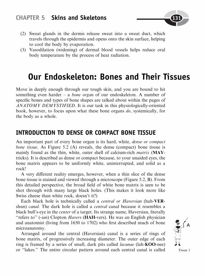
(2) Sweat glands in the dermis release sweat into a sweat duct, whichtravels through the epidermis and opens onto the skin surface, helpingto cool the body by evaporation.
(3) Vasodilation (widening) of dermal blood vessels helps reduce oralbody temperature by the process of heat radiation.
Our Endoskeleton: Bones and Their TissuesMove in deeply enough through our tough skin, and you are bound to hitsomething even harder – a bone organ of our endoskeleton. A number ofspecific bones and types of bone shapes are talked about within the pages ofANATOMY DEMYSTIFIED. It is our task in this physiologically-orientedbook, however, to focus upon what these bone organs do, systemically, forthe body as a whole.
INTRODUCTION TO DENSE OR COMPACT BONE TISSUE
An important part of every bone organ is its hard, white, dense or compactbone tissue. As Figure 5.2 (A) reveals, the dense (compact) bone tissue ismainly found as the thin, white, outer shell of calcium-rich matrix (MAY-tricks). It is described as dense or compact because, to your unaided eyes, thebone matrix appears to be uniformly white, uninterrupted, and solid as arock!
A very different reality emerges, however, when a thin slice of the densebone tissue is stained and viewed through a microscope (Figure 5.2, B). Fromthis detailed perspective, the broad field of white bone matrix is seen to beshot through with many large black holes. (This makes it look more likeSwiss cheese than white rock, doesn’t it?)
Each black hole is technically called a central or Haversian (hah-VER-shun) canal. The dark hole is called a central canal because it resembles ablack bull’s-eye in the center of a target. Its strange name, Haversian, literally‘‘refers to’’ (-ian) Clopton Havers (HAH-vers). He was an English physicianand anatomist (living from 1650 to 1702) who first described much of bonemicroanatomy.
Arranged around the central (Haversian) canal is a series of rings ofbone matrix, of progressively increasing diameter. The outer edge of eachring is framed by a series of small, dark pits called lacunae (lah-KOO-nee)or ‘‘lakes.’’ The entire circular pattern around each central canal is called
CHAPTER 5 Skins and Skeletons 131
Tissue 1
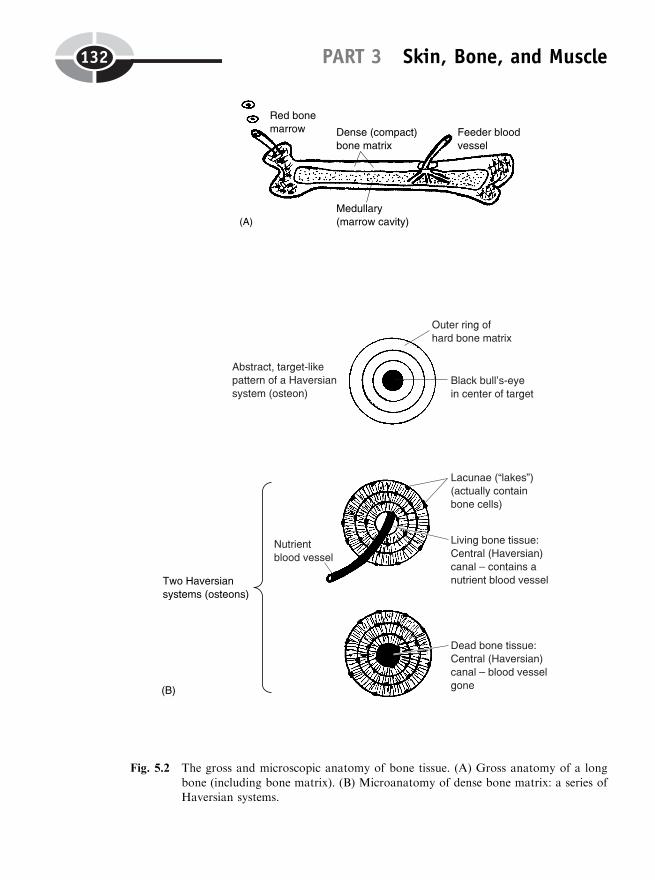
PART 3 Skin, Bone, and Muscle132
Fig. 5.2 The gross and microscopic anatomy of bone tissue. (A) Gross anatomy of a long
bone (including bone matrix). (B) Microanatomy of dense bone matrix: a series ofHaversian systems.
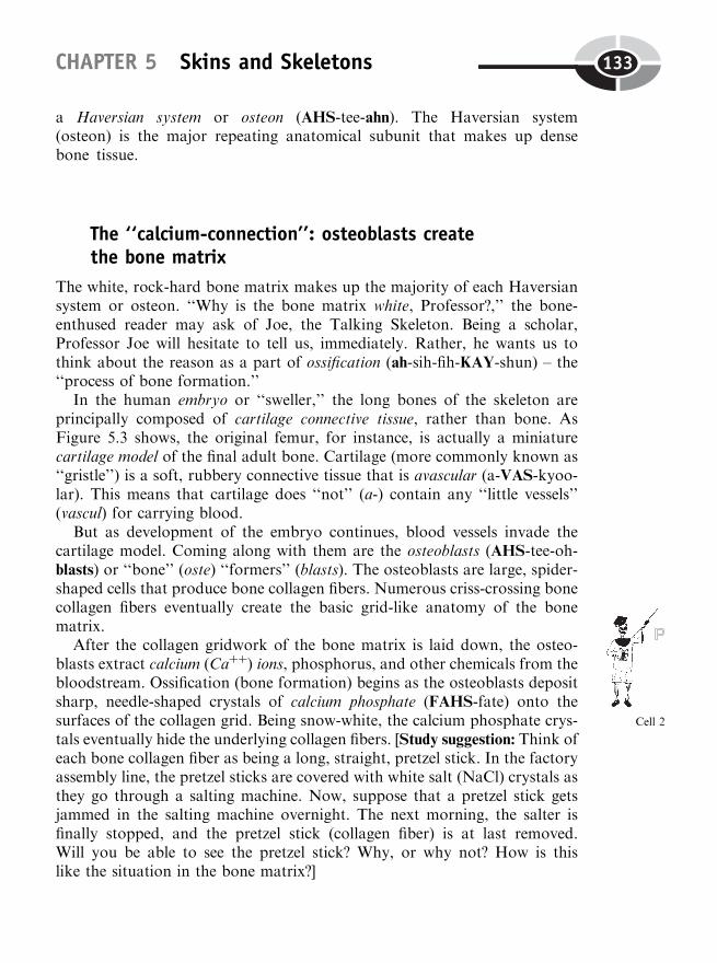
a Haversian system or osteon (AHS-tee-ahn). The Haversian system(osteon) is the major repeating anatomical subunit that makes up densebone tissue.
The ‘‘calcium-connection’’: osteoblasts createthe bone matrix
The white, rock-hard bone matrix makes up the majority of each Haversiansystem or osteon. ‘‘Why is the bone matrix white, Professor?,’’ the bone-enthused reader may ask of Joe, the Talking Skeleton. Being a scholar,Professor Joe will hesitate to tell us, immediately. Rather, he wants us tothink about the reason as a part of ossification (ah-sih-fih-KAY-shun) – the‘‘process of bone formation.’’
In the human embryo or ‘‘sweller,’’ the long bones of the skeleton areprincipally composed of cartilage connective tissue, rather than bone. AsFigure 5.3 shows, the original femur, for instance, is actually a miniaturecartilage model of the final adult bone. Cartilage (more commonly known as‘‘gristle’’) is a soft, rubbery connective tissue that is avascular (a-VAS-kyoo-lar). This means that cartilage does ‘‘not’’ (a-) contain any ‘‘little vessels’’(vascul) for carrying blood.
But as development of the embryo continues, blood vessels invade thecartilage model. Coming along with them are the osteoblasts (AHS-tee-oh-blasts) or ‘‘bone’’ (oste) ‘‘formers’’ (blasts). The osteoblasts are large, spider-shaped cells that produce bone collagen fibers. Numerous criss-crossing bonecollagen fibers eventually create the basic grid-like anatomy of the bonematrix.
After the collagen gridwork of the bone matrix is laid down, the osteo-blasts extract calcium (Caþþ) ions, phosphorus, and other chemicals from thebloodstream. Ossification (bone formation) begins as the osteoblasts depositsharp, needle-shaped crystals of calcium phosphate (FAHS-fate) onto thesurfaces of the collagen grid. Being snow-white, the calcium phosphate crys-tals eventually hide the underlying collagen fibers. [Study suggestion: Think ofeach bone collagen fiber as being a long, straight, pretzel stick. In the factoryassembly line, the pretzel sticks are covered with white salt (NaCl) crystals asthey go through a salting machine. Now, suppose that a pretzel stick getsjammed in the salting machine overnight. The next morning, the salter isfinally stopped, and the pretzel stick (collagen fiber) is at last removed.Will you be able to see the pretzel stick? Why, or why not? How is thislike the situation in the bone matrix?]
CHAPTER 5 Skins and Skeletons 133
Cell 2
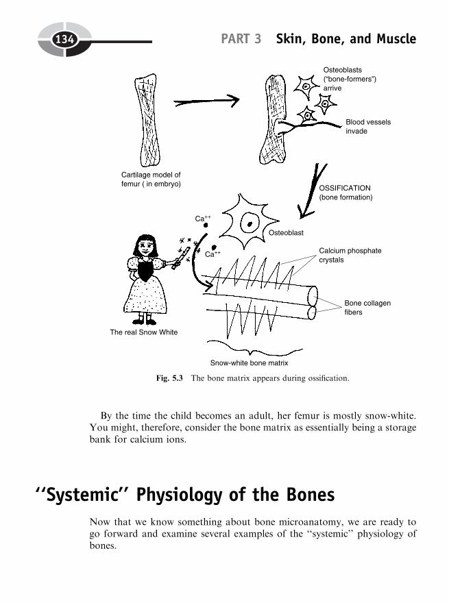
By the time the child becomes an adult, her femur is mostly snow-white.You might, therefore, consider the bone matrix as essentially being a storagebank for calcium ions.
‘‘Systemic’’ Physiology of the BonesNow that we know something about bone microanatomy, we are ready togo forward and examine several examples of the ‘‘systemic’’ physiology ofbones.
PART 3 Skin, Bone, and Muscle134
Fig. 5.3 The bone matrix appears during ossification.
SYSTEMIC BONE PHYSIOLOGY 1: MAINTAININGHOMEOSTASIS OF BLOOD [Ca11]
As a result of ossification, millions of calcium (Caþþ) ions become stored inthe form of calcium phosphate crystals on the bone collagen fibers. Eventhough the bones eventually become fully ossified, however, there is a processof bone remodeling that continues throughout life.
Bone remodeling is a change in the shape, thickness, and strength of bonesthat occurs under the influence of various factors. Important among thesefactors is the blood calcium ion concentration, which can be denoted withbrackets as blood [Caþþ].
Blood calcium ion concentration, or blood [Caþþ], is an anatomical para-meter that exists at the Chemical Level of Body Organization. We can clearlyvisualize each calcium ion as a tiny black dot with two positive (þ) charges,circulating within the bloodstream (Figure 5.4).
The blood [Caþþ] is usually measured in units of mg/dL – milligrams ofcalcium ions per deciliter (DEH-sih-lee-ter) – of blood. A deciliter is one‘‘tenth’’ (deci-) of a ‘‘liter.’’
The critical balance of normocalcemia
You may recall some important general concepts from Chapter 2, such as theideas of the normal range of a body parameter, its set-point value, its lowernormal limit (LNL), and its upper normal limit (UNL). We have alreadyoutlined these concepts for the physiological parameter of oral body tem-perature in degrees Fahrenheit.
Taking a similar approach, we can state that the normal or reference rangefor blood [Caþþ] is from a lower normal limit of 8.5 mg/dL blood up to anupper normal limit of 10.6 mg/dL blood. Using the same naming model asfor normothermia, we will state this condition as normocalcemia (nor-moh-kal-SEE-me-uh). Normocalcemia exactly translates to mean ‘‘a normal’’(normo-) ‘‘blood condition of ’’ (-emia) ‘‘calcium’’ (calc).
Assuming that normocalcemia is maintained over a definite period of time,we can further say that there is a homeostasis or relative constancy of theblood calcium ion concentration (Figure 5.4).
CHAPTER 5 Skins and Skeletons 135
Organ 2
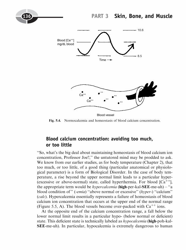
Blood calcium concentration: avoiding too much,or too little
‘‘So, what’s the big deal about maintaining homeostasis of blood calcium ionconcentration, Professor Joe?,’’ the untutored mind may be prodded to ask.We know from our earlier studies, as for body temperature (Chapter 2), thattoo much, or too little, of a good thing (particular anatomical or physiolo-gical parameter) is a form of Biological Disorder. In the case of body tem-perature, a rise beyond the upper normal limit leads to a particular hyper-(excessive or above-normal) state, called hyperthermia. For blood [Caþþ],the appropriate term would be hypercalcemia (high-per-kal-SEE-me-uh) – ‘‘ablood condition of ’’ (-emia) ‘‘above normal or excessive’’ (hyper-) ‘‘calcium’’(calc). Hypercalcemia essentially represents a failure of homeostasis of bloodcalcium ion concentration that occurs at the upper end of the normal range(Figure 5.5, A). The blood vessels become over-packed with Caþþ ions.
At the opposite end of the calcium concentration range, a fall below thelower normal limit results in a particular hypo- (below normal or deficient)state. This deficient state is technically labeled as hypocalcemia (high-poh-kal-SEE-me-uh). In particular, hypocalcemia is extremely dangerous to human
PART 3 Skin, Bone, and Muscle136
Fig. 5.4. Normocalcemia and homeostasis of blood calcium concentration.
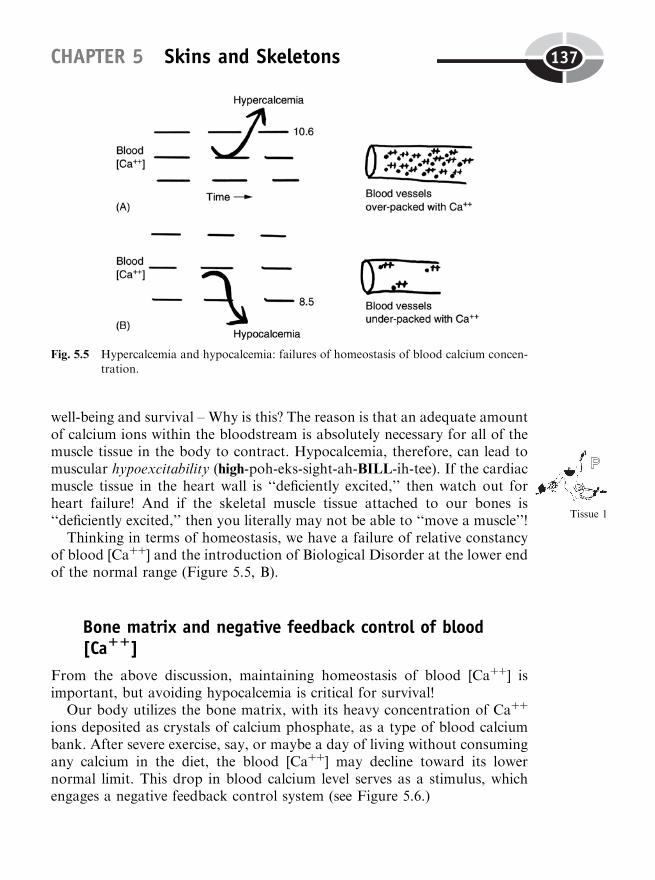
well-being and survival – Why is this? The reason is that an adequate amountof calcium ions within the bloodstream is absolutely necessary for all of themuscle tissue in the body to contract. Hypocalcemia, therefore, can lead tomuscular hypoexcitability (high-poh-eks-sight-ah-BILL-ih-tee). If the cardiacmuscle tissue in the heart wall is ‘‘deficiently excited,’’ then watch out forheart failure! And if the skeletal muscle tissue attached to our bones is‘‘deficiently excited,’’ then you literally may not be able to ‘‘move a muscle’’!
Thinking in terms of homeostasis, we have a failure of relative constancyof blood [Caþþ] and the introduction of Biological Disorder at the lower endof the normal range (Figure 5.5, B).
Bone matrix and negative feedback control of blood[Ca11]
From the above discussion, maintaining homeostasis of blood [Caþþ] isimportant, but avoiding hypocalcemia is critical for survival!
Our body utilizes the bone matrix, with its heavy concentration of Caþþ
ions deposited as crystals of calcium phosphate, as a type of blood calciumbank. After severe exercise, say, or maybe a day of living without consumingany calcium in the diet, the blood [Caþþ] may decline toward its lowernormal limit. This drop in blood calcium level serves as a stimulus, whichengages a negative feedback control system (see Figure 5.6.)
CHAPTER 5 Skins and Skeletons 137
Tissue 1
Fig. 5.5 Hypercalcemia and hypocalcemia: failures of homeostasis of blood calcium concen-tration.
This stimulus is especially strong for the osteoclasts (AHS-tee-oh-klasts) or‘‘bone-breakers’’ (-clasts). The osteoclasts are definitely not enforcers work-ing for the Mafia, who will ‘‘break your bones’’ if you don’t pay up! Inreality, the osteoclasts are large, multinucleate (mul-tee-NOO-klee-ut) cellsthat release special digestive enzymes. [Study suggestion: Look at Figure5.6. See how far you can translate the word multinucleate into its commonEnglish equivalent.]
The digestive enzymes from the osteoclasts cause a partial resorption (rih-SORP-shun) or ‘‘drinking-in again’’ of the bone matrix. This occurs becausesome of the calcium phosphate crystals on the bone collagen fibers are bro-ken down and dissolved. This releases free Caþþ ions back into the blood-stream. As a result, the blood calcium ion concentration rises back toward itsset-point (long-term average) value. By this calcium-withdrawal process fromour bone matrix ‘‘bank,’’ then, hypocalcemia is usually prevented. As aconsequence of the resorption process, however, the bones remodel and
PART 3 Skin, Bone, and Muscle138
Fig. 5.6 A negative-feedback response to a drop in blood [Caþþ].

become thinner and weaker. If the resorption process goes on long enough,as in a person whose diet is chronically low in calcium, a condition of boneatrophy (AT-roh-fee) may result. Atrophy is literally ‘‘a condition’’ (-y)‘‘without’’ (a-) ‘‘nourishment’’ (troph). In bone atrophy, the bones are notbeing ‘‘nourished,’’ in the sense that they are not being stimulated to addmore calcium phosphate crystals. If the atrophy is severe enough, the bonesare also much more vulnerable to deformity and fracture.
By reversing our previous thinking, we can trace the response of the bodyto a rise in the blood calcium ion concentration toward its upper normal limit(Figure 5.7). This rise is a stimulus for the osteoblasts (bone-makers). As
CHAPTER 5 Skins and Skeletons 139
Organ 1
Fig. 5.7 A negative feedback response to a rise in blood [Caþþ].
they do during the process of ossification, the osteoblasts extract free Caþþ
ions from the bloodstream, and supervise their deposition upon bone col-lagen fibers as more calcium phosphate crystals.
As a result, the blood [Caþþ] falls back down toward its set-point level,and the abnormal condition of hypercalcemia is usually prevented. As asecondary consequence of ‘‘depositing’’ more calcium ‘‘money’’ to the bonematrix ‘‘bank,’’ bone remodeling occurs. But this time it is in the directionof bone hypertrophy (high-PER-troh-fee) – ‘‘a condition of ’’ (-y) ‘‘excessiveor above normal’’ (hyper-) ‘‘nourishment’’ (troph) or stimulation. In short,the osteoblasts withdraw more calcium from the bloodstream, and depositit onto bone collagen fibers as more calcium phosphate crystals. With morecrystals, the bone hypertrophies (high-PER-troh-fees), becoming boththicker and stronger. This hypertrophy (excessive ‘‘nourishment’’ or stimu-lation) is completely normal and natural. Therefore, sometimes it is calledphysiological hypertrophy. This is an increase in the size of some bodystructure that is due to normal, rather than pathological, changes (suchas a bone tumor).
SYSTEMIC BONE PHYSIOLOGY 2: THE ROLE OF REDMARROW IN HEMATOPOIESIS
Let us return to the gross anatomy of a long bone. This time, though, we aregoing to cut through the entire bone, in a longitudinal (‘‘lengthwise’’) direc-tion. As Figure 5.8 reveals, there is another type of bone tissue present, inaddition to dense (compact) bone tissue. This second type is called spongy orcancellous (CAN-sih-lus) bone tissue. This type of bone tissue resembles asponge, since its hard white matrix is arranged into criss-crossing trabeculae(trah-BEK-yuh-lie) or ‘‘little beams.’’ These trabeculae (little beams) ofmatrix, since they extensively criss-cross, create a cancellous pattern, whichliterally ‘‘pertains to’’ (-ous) a ‘‘lattice’’ (cancell).
A sponge, of course, is filled with holes. In spongy or cancellous bone, theholes contain the red bone marrow. The red bone marrow largely consists of asoft, pulpy meshwork of reddish-colored blood vessels. These blood vesselssnake in and out of the holes within the latticework of slender white trabe-culae of spongy bone.
The major function performed by red bone marrow is hematopoiesis(he-muh-toh-poy-EE-sis) – the process of ‘‘blood’’ (hemat) ‘‘formation’’(-poiesis). By this translation, however, we do not mean that all of the com-ponents of the blood are manufactured by the red bone marrow. Rather, wemean the so-called formed elements of the bloodstream are manufactured
PART 3 Skin, Bone, and Muscle140
Organ 3
Tissues 1
here. In particular, these are the various types of blood cells and blood cellfragments, which are the ‘‘elements’’ of the blood having a specific shape or‘‘form.’’
Hemocytoblasts: the original parent cells
Any close, microscopic look at the red bone marrow (Figure 5.8, B), showssome of the stem or parent cells, which are the predecessor cells eventuallygiving rise to the formed elements. Everything begins with the hemocytoblasts(he-moh-SIGH-toh-blasts), literally the ‘‘blood’’ (hem) ‘‘cell’’ (cyt) ‘‘formers’’(-blasts). Each hemocytoblast is a fairly large cell having a distinct ovalnucleus and projecting arms of cytoplasm. Through a number of intermedi-ate cell stages, the hemocytoblasts eventually create all the formed elementsfound in the bloodstream.
Let us first consider the erythrocytes (eh-RITH-roh-sights) or ‘‘red’’(erythr) ‘‘cells.’’ Each mature erythrocyte is an anucleate (ay-NEW-klee-ut),biconcave (BUY-kahn-cave) disc. This means that the erythrocyte or RBC isshaped somewhat like a red hourglass (when viewed on its side), being ‘‘cavedin’’ (concave) on ‘‘both’’ (bi-) sides.
One of its intermediate stem cells is the erythroblast (eh-RITH-roh-blast) or‘‘red-former.’’ The erythroblast is a regular cell with a nucleus, but it loses thisnucleus on its way to becoming a mature erythrocyte. [Study suggestion:Whatdoes anucleate mean?] Of course, the erythrocyte looks red because it cancontain up to 280 million red-colored hemoglobin (HE-moh-gloh-bin) mole-cules, which do its main job of carrying oxygen through the bloodstream.
The leukocytes (LOO-koh-sights) or ‘‘white’’ (leuk) cells are named for theclear, whitish color of their cytoplasm. Unlike the erythrocyte, all leukocytesare regular nucleated cells. The most common type of leukocyte createdduring hematopoiesis is the neutrophil (NEW-troh-fil). The neutrophil getsits name from its apparent ‘‘love or fondness’’ (phil) for ‘‘neutral’’ (neutr)dyes during biological staining. Just after the hemocytoblast, the neutrophil’sparent cell is the myeloblast (my-EL-oh-blast) or ‘‘former’’ (blast) in the bone‘‘marrow’’ (myel).
Finally, we come to the blood platelets (PLAY-tuh-lets) or thrombocytes(THRAHM-boh-sights). The platelets are literally ‘‘small plates’’ – small,plate-like fragments of disintegrated bone marrow cells. Just after the hemo-cytoblast, the platelet’s parent cell is the megakaryoblast (meg-uh-CARE-ee-oh-blast) or ‘‘large’’ (mega-) ‘‘nucleus’’ (karyo) ‘‘former’’ (blast). The mega-rkaryoblast is an immature cell that eventually becomes a mature bone mar-row cell that breaks apart into a number of platelets. The alternative name
CHAPTER 5 Skins and Skeletons 141
Cell 3
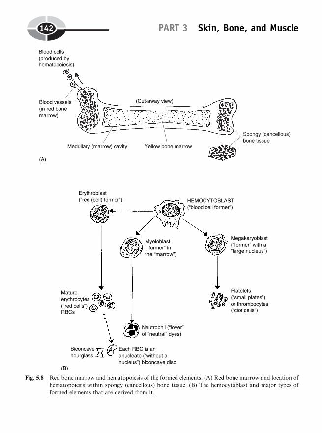
PART 3 Skin, Bone, and Muscle142
Fig. 5.8 Red bone marrow and hematopoiesis of the formed elements. (A) Red bone marrow and location of
hematopoiesis within spongy (cancellous) bone tissue. (B) The hemocytoblast and major types offormed elements that are derived from it.
for the platelets is thrombocytes, which means ‘‘clot’’ (thromb) ‘‘cells’’ (cytes).They get this name from their essential function of sticking together to helpcreate thrombi (THRAHM-buy) or blood ‘‘clots.’’
QuizRefer to the text in this chapter if necessary. A good score is at least 8 correctanswers out of these 10 questions. The answers are listed in the back of thisbook.
1. Layer of the skin containing keratin granules that provide waterproof-ing:(a) Epidermis(b) Subdermis(c) Accessory layer(d) Dermis
2. Charged solutes þ water solvent ¼ :(a) Polar ions(b) Dehydration(c) Aqueous solution(d) Isotonic ‘‘dissolver’’
3. When an osmotic equilibrium exists across the plasma membrane:(a) There is not net diffusion of H2O either into, or out of, the cell(b) A water concentration gradient is present(c) Water and dissolved solute are pumped uphill using ATP energy(d) H2O is totally blocked from moving through membrane pores
4. RBCs normally stay small enough to pass through tiny blood capil-laries:(a) Since blood plasma is usually a hypotonic solution(b) Except when the person forgets to urinate!(c) Until they explode on their way to the tissues(d) Because the ECF is an isotonic solution
5. Hemoconcentration resulting from severe dehydration creates:(a) A dramatic drop in the blood plasma NaCl concentration(b) Crenation of many circulating erythrocytes(c) An abnormally large blood [H2O](d) A huge swelling under the skin
CHAPTER 5 Skins and Skeletons 143
6. The skin’s contribution to thermoregulation is chiefly due to the:(a) Positive feedback effect of a rise in body temperature upon oral
body temperature(b) Negative feedback loops involving increased sweating and vaso-
dilation(c) Removal of all external heat-generating influences(d) Attainment of a calm, steady state involving no physical exer-
tion
7. A cartilage model in the embryo is often one of the first steps in:(a) Ossification(b) Dermabrasion(c) Leukocytosis(d) Osmotic pressure production
8. Osteoblasts deposit these on bone collagen fibers:(a) Osteons or Haversian systems(b) Pickled turnips(c) Cubic NaCl crystals(d) Slender needles of calcium phosphate material
9. Hypocalcemia is extremely dangerous to the body, considering that it:(a) Can result in total paralysis of nearly all body muscles!(b) Often leads to excessive consumption of fatty foods(c) Strongly interferes with the absorption of UV light(d) Makes bones too thick and strong for their own good!
10. The original stem or parent cell for all formed elements of theblood:(a) Lymphoblast(b) Megarkaryoblast(c) Platelet(d) Hemocytoblast
Body-Level Grids for Chapter 5Several key body facts were tagged with numbered icons in the page marginsof this chapter. Write a short summary of each of these key facts into anumbered cell or box within the appropriate Body Level-Grid that appearsbelow.
PART 3 Skin, Bone, and Muscle144
Anatomy and Biological Order Fact Grids for Chapter 5:
TISSUELevel
Physiology and Biological Order Fact Grids for Chapter 5:
CELLLevel
CHAPTER 5 Skins and Skeletons 145
1
TISSUELevel
ORGANLevel
PART 3 Skin, Bone, and Muscle146
1
1 2
3
1 2
3

Physiology and Biological Disorder Fact Grids for Chapter 5:
CELLLevel
TISSUELevel
ORGANLevel
CHAPTER 5 Skins and Skeletons 147
1
1
1

148
CHAPTER6
‘‘Gentlemen, Fire UpYour Engines!’’:The Physiology ofNeurons andMuscle Fibers
Chapter 5 covered the systemic physiology of the skin and skeleton. But it didnot include the physiology of body movement. As soon as we get into bodymovement, both the neurons (nerve cells) and muscle fibers (thin, fiber-shapedmuscle cells) become involved.
Copyright © 2004 by The McGraw-Hill Companies, Inc. Click here for terms of use.

Lever Systems: Muscles, Bones, and JointsA bone–muscle lever (LEE-ver) system is a combination of a bone, skeletalmuscle, and movable joint that together carry out some particular bodymovement. Consider, for example, the flexion (FLEK-shun) or ‘‘bending’’motion of the forearm (Figure 6.1).
A lever is a rigid bar that is acted upon by some force. In a bone–musclelever system, the bone serves as a passive lever or rigid bar that is acted uponby a pulling force (P). This pulling force is created by the contraction (short-ening) of a particular skeletal muscle. A skeletal muscle is a fleshy, reddish-colored organ that is attached to the bones of the skeleton.
For flexion of the forearm, the biceps (BUY-seps) brachii (BRAY-kee-eye)is the main skeletal muscle or prime mover. It attaches by means of tendons tothe bones of the forearm. When the biceps brachii muscle contracts, it createsa pulling force (P) upon its lower tendons, which causes the bones of thelower arm to flex or ‘‘bend’’ at the elbow joint.
A fulcrum (FULL-crumb), abbreviated as F, is a place of ‘‘support’’ uponwhich a lever turns or balances when it moves. Within a bone–muscle leversystem, a movable joint generally serves as the fulcrum. (A joint is simply aplace of union or meeting between two bones.)
CHAPTER 6 Neurons and Muscle Fibers 149
Fig. 6.1 Flexion at the elbow: model of a bone–muscle lever system.

Finally, there is always some amount of weight (W) or resistance (R) thatneeds to be lifted during the body movement. In the present case, suppose thehand was holding a 5-pound dumbbell.
In summary, as the biceps brachii muscle contracts, it pulls upon itstendons. The tendons pull upon the bones of the lower arm, and the weightor resistance in the hand is lifted as flexion occurs at the elbow joint.
Motor Neurons and Muscle Fibers:The Neuromuscular Connection
Bone-muscle lever systems, to be sure, cause portions of the human body tomove around. To review, the bones are the passive partners in body move-ment, because they merely serve as sticks or rigid bars that are pulled upon.The skeletal muscles, in contrast, are the active partners in body movement.The reason is that they are the organs that actively contract or shorten, pro-viding the pulling force (P) upon their tendons, and finally, upon the bones.
IMPORTANCE OF THE MUSCLE FIBER (MUSCLE CELL)
The detailed gross anatomy of the skeletal muscle organ and its parts istalked about within ANATOMY DEMYSTIFIED. Here in our World ofPhysiology, however, our primary focus will be upon the functions of theindividual skeletal muscle fiber. ‘‘Why is this, Professor Joe?’’, the physiolo-gically inclined reader may ask of the Good Teacher. ‘‘It is due to the factthat the individual skeletal muscle fiber or cell, as well as groups of relatedmuscle fibers, are the fundamental units of muscle contraction,’’ is the reply.
‘‘But do these muscle fibers, or groups of muscle fibers, normally contracton their own?’’, the astute reader quickly follows-up. ‘‘I see that you haveactually been doing some thinking while you have been reading!,’’ Joeresponds. ‘‘The skeletal muscle fiber has to be stimulated – ‘fired up’ or‘turned on’ – before it will contract.’’
ANATOMY AND PHYSIOLOGY OF THE MOTOR NEURON
‘‘What part of the body stimulates, fires up, or turns on the muscle fiber,Professor?’’ Adjusting his glasses to think, Joe points to Figure 6.2. ‘‘Themuscle fiber is turned on or excited by a particular motor neuron.’’
PART 3 Skin, Bone, and Muscle150
Organ 1
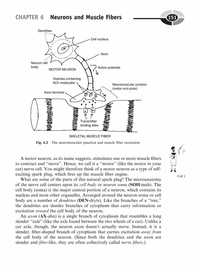
Amotor neuron, as its name suggests, stimulates one or more muscle fibersto contract and ‘‘move’’. Hence, we call it a ‘‘motor’’ (like the motor in yourcar) nerve cell. You might therefore think of a motor neuron as a type of self-exciting spark plug, which fires up the muscle fiber engine.
What are some of the parts of this natural spark plug? The microanatomyof the nerve cell centers upon its cell body or neuron soma (SOH-mah). Thecell body (soma) is the major central portion of a neuron, which contains itsnucleus and most other organelles. Arranged around the neuron soma or cellbody are a number of dendrites (DEN-dryts). Like the branches of a ‘‘tree,’’the dendrites are slender branches of cytoplasm that carry information orexcitation toward the cell body of the neuron.
An axon (AX-ahn) is a single branch of cytoplasm that resembles a longslender ‘‘axle’’ (like the axle found between the two wheels of a car). Unlike acar axle, though, the neuron axon doesn’t actually move. Instead, it is aslender, fiber-shaped branch of cytoplasm that carries excitation away fromthe cell body of the neuron. (Since both the dendrites and the axon areslender and fiber-like, they are often collectively called nerve fibers.)
CHAPTER 6 Neurons and Muscle Fibers 151
Fig. 6.2 The neuromuscular junction and muscle fiber excitation.
Cell 1
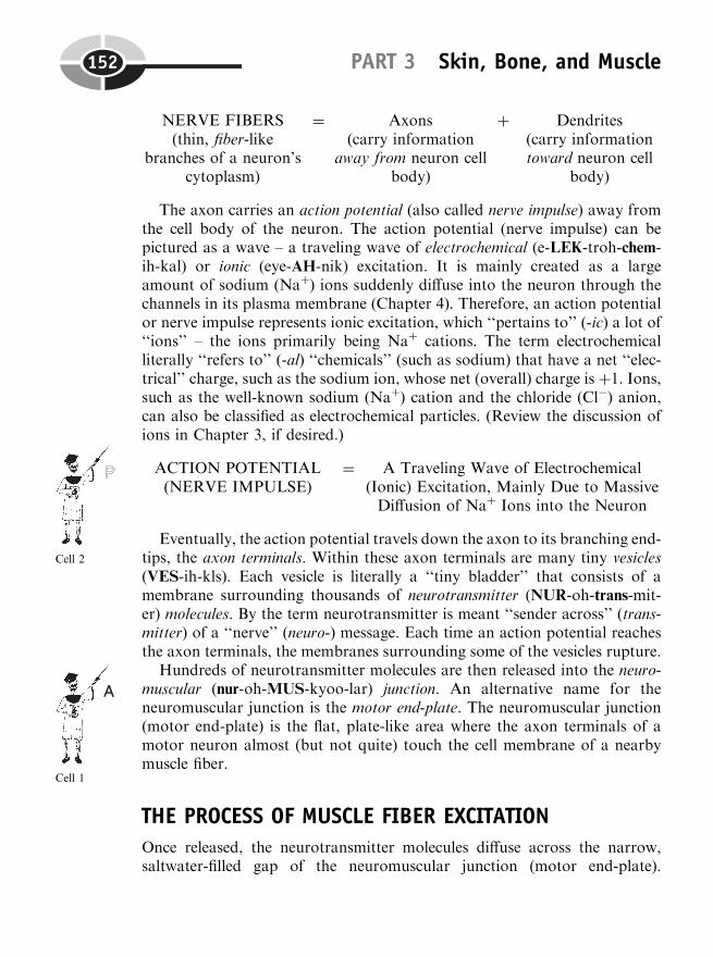
NERVE FIBERS(thin, fiber-like
branches of a neuron’scytoplasm)
¼ Axons(carry information
away from neuron cellbody)
þ Dendrites(carry informationtoward neuron cell
body)
The axon carries an action potential (also called nerve impulse) away fromthe cell body of the neuron. The action potential (nerve impulse) can bepictured as a wave – a traveling wave of electrochemical (e-LEK-troh-chem-ih-kal) or ionic (eye-AH-nik) excitation. It is mainly created as a largeamount of sodium (Naþ) ions suddenly diffuse into the neuron through thechannels in its plasma membrane (Chapter 4). Therefore, an action potentialor nerve impulse represents ionic excitation, which ‘‘pertains to’’ (-ic) a lot of‘‘ions’’ – the ions primarily being Naþ cations. The term electrochemicalliterally ‘‘refers to’’ (-al) ‘‘chemicals’’ (such as sodium) that have a net ‘‘elec-trical’’ charge, such as the sodium ion, whose net (overall) charge is þ1. Ions,such as the well-known sodium (Naþ) cation and the chloride (Cl�) anion,can also be classified as electrochemical particles. (Review the discussion ofions in Chapter 3, if desired.)
ACTION POTENTIAL(NERVE IMPULSE)
¼ A Traveling Wave of Electrochemical(Ionic) Excitation, Mainly Due to MassiveDiffusion of Naþ Ions into the Neuron
Eventually, the action potential travels down the axon to its branching end-tips, the axon terminals. Within these axon terminals are many tiny vesicles(VES-ih-kls). Each vesicle is literally a ‘‘tiny bladder’’ that consists of amembrane surrounding thousands of neurotransmitter (NUR-oh-trans-mit-er) molecules. By the term neurotransmitter is meant ‘‘sender across’’ (trans-mitter) of a ‘‘nerve’’ (neuro-) message. Each time an action potential reachesthe axon terminals, the membranes surrounding some of the vesicles rupture.
Hundreds of neurotransmitter molecules are then released into the neuro-muscular (nur-oh-MUS-kyoo-lar) junction. An alternative name for theneuromuscular junction is the motor end-plate. The neuromuscular junction(motor end-plate) is the flat, plate-like area where the axon terminals of amotor neuron almost (but not quite) touch the cell membrane of a nearbymuscle fiber.
THE PROCESS OF MUSCLE FIBER EXCITATION
Once released, the neurotransmitter molecules diffuse across the narrow,saltwater-filled gap of the neuromuscular junction (motor end-plate).
PART 3 Skin, Bone, and Muscle152
Cell 2
Cell 1
Remember that we have defined a neurotransmitter as a ‘‘sender across of anerve’’ message. In this case, the released molecules are being ‘‘sent across’’the neuromuscular junction by diffusion. The molecules then attach to trans-mitter binding sites on the muscle fiber.
The type of message being ‘‘sent across’’ the neuromuscular junction is anexcitatory (ek-SIGH-tah-tor-ee) one, because it is ‘‘characterized by’’ (-ory)‘‘excitation.’’ Specifically, the motor neuron was excited and fired a neuronaction potential. As a result, eventually the vesicles in the axon terminalsrelease excitatory neurotransmitter molecules. As their name reveals, excita-tory neurotransmitter molecules stimulate or excite the cell to which theybind or attach.
Within the neuromuscular junction, the main excitatory neurotransmitter iscalled acetylcholine (uh-see-tul-KOH-leen), abbreviated as ACh. ‘‘How do weknow that the muscle fiber or cell is excited, Professor Joe?’’ The answer to thisquestion is obvious: the muscle fiber, when it is excited, has a muscle fiberaction potential of its own, which is created in the region of the neuromuscularjunction. In summary of action potentials, here is a general rule of thumb:
Microanatomy of a Skeletal Muscle FiberIn order to fully understand how a particular muscle fiber contracts after it isstimulated or excited at the neuromuscular junction, we need to do somefurther work on the microanatomy of the muscle fiber, itself. Figure 6.3 getsus started. A thin plasmamembrane, called the sarcolemma (sar-koh-LEM-uh)or ‘‘flesh’’ (sarc) ‘‘husk’’ (lemm), surrounds the outside of the muscle fiber.Skeletal muscle fibers bear dark cross-stripes, called striations (stry-AY-shuns).
Why is the skeletal muscle fiber cross-striped? To show why, we look at theinterior of the fiber. Here are a number of myofibrils (my-uh-FEYE-brils).
CHAPTER 6 Neurons and Muscle Fibers 153

The word myofibril literally means ‘‘little fiber’’ (fibril) of a ‘‘muscle’’ (my). Inreality, however, each myofibril is just a long, thin, fiber-shaped organellewithin a muscle fiber. The myofibrils have a dark-and-light banding pattern.The dark bands are called the A bands, while the light bands are called the Ibands. The striations (cross-stripes) of the entire muscle fiber, therefore, arecreated by the stacking of the dark A bands of the myofibrils, one uponanother.
Within the middle of each light I band is a dark, zig-zagging Z-line. Thesedark, jagged lines mark off a series of sarcomeres (SAR-koh-meers). Asarcomere is a short ‘‘segment’’ (-mere) of ‘‘flesh’’ (sarc), that is, a regionof myofibril between two Z-lines. Hence, each myofibril organelle within amuscle fiber basically consists of a series of sarcomeres, attached end to end.
A LITTLE WIENIE WITH HOLES: TRANSVERSE TUBULESAND THE SARCOPLASMIC RETICULUM
Now that we have talked about some of the muscle fiber’s microanatomy, weare ready to dig deeper into its ‘‘flesh,’’ which in Greek is denoted by theword root sarc. Speaking of flesh, it would be useful to visualize each skeletalmuscle fiber as a little 3-D wienie or hot dog, with black cross-stripes (stria-tions) on it, from being cooked on an outdoor barbecue grill! We need to
PART 3 Skin, Bone, and Muscle154
Fig. 6.3 A look within a muscle fiber (cell).
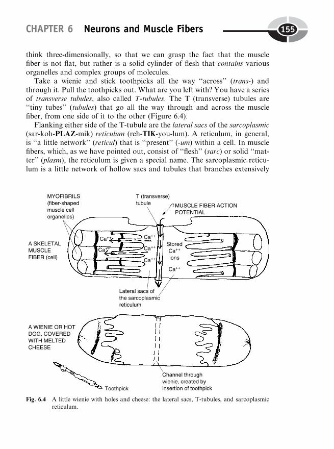
think three-dimensionally, so that we can grasp the fact that the musclefiber is not flat, but rather is a solid cylinder of flesh that contains variousorganelles and complex groups of molecules.
Take a wienie and stick toothpicks all the way ‘‘across’’ (trans-) andthrough it. Pull the toothpicks out. What are you left with? You have a seriesof transverse tubules, also called T-tubules. The T (transverse) tubules are‘‘tiny tubes’’ (tubules) that go all the way through and across the musclefiber, from one side of it to the other (Figure 6.4).
Flanking either side of the T-tubule are the lateral sacs of the sarcoplasmic(sar-koh-PLAZ-mik) reticulum (reh-TIK-you-lum). A reticulum, in general,is ‘‘a little network’’ (reticul) that is ‘‘present’’ (-um) within a cell. In musclefibers, which, as we have pointed out, consist of ‘‘flesh’’ (sarc) or solid ‘‘mat-ter’’ (plasm), the reticulum is given a special name. The sarcoplasmic reticu-lum is a little network of hollow sacs and tubules that branches extensively
CHAPTER 6 Neurons and Muscle Fibers 155
Fig. 6.4 A little wienie with holes and cheese: the lateral sacs, T-tubules, and sarcoplasmicreticulum.

within the cytoplasm of the muscle cell. [Study suggestion: It may seem a bitweird, but picture the little hot dog or wienie as having Swiss cheese meltedover it. The cheese is stringy and creates a gooey ‘‘network’’ of sorts. Whatparticular organelle is the melted, stringy Swiss cheese modeling?]
The lateral sacs serve as storage depots for calcium (Caþþ) ions within themuscle fiber. When a muscle fiber action potential is created in the region ofthe neuromuscular junction, it flows down into the T-tubule, which carries itall the way through the muscle fiber. As the action potential travels, it tem-porarily makes the lateral sacs of the sarcoplasmic reticulum much moreleaky to calcium ions. As a result, thousands of Caþþ ions leak out of thelateral sacs, and into the region of the myofibrils.
Muscle Fiber Contraction:The Sliding Filament Theory
‘‘Where are all these calcium ions going to, within each myofibril?,’’ theinquisitive brain yearns to know. As we mentioned earlier, an adequateconcentration of calcium ions within the bloodstream (and stored insidethe lateral sacs of the sarcoplasmic reticulum) is absolutely essential for theexcitation and contraction of muscle fibers! We can understand why this is soif we dig really deep into the myofibril organelles packed into each musclefiber. Here is a summary of a previous statement:
A MYOFIBRIL (fiber-shaped musclecell organelle)
¼ A series of sarcomeres, attachedend to end
Since each myofibril is essentially a bunch of sarcomeres connectedtogether, it is only logical that we should look to the individual sarcomereto discover how the entire myofibril functions.
MYOFILAMENTS WITHIN EACH SARCOMERE
A very powerful electron microscope can be used to magnify the sarcomere,tens of thousands of times! What do we see when we do this? Study Figure6.5. Here we find ourselves back down at the Chemical Level of BodyOrganization. The high end of the Chemical Level, just below the level ofthe cell organelles, consists of integrated groups of molecules.
PART 3 Skin, Bone, and Muscle156
Organelle 1
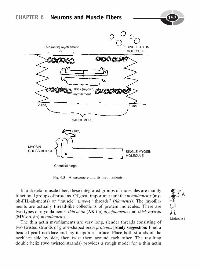
In a skeletal muscle fiber, these integrated groups of molecules are mainlyfunctional groups of proteins. Of great importance are the myofilaments (my-oh-FIL-ah-ments) or ‘‘muscle’’ (myo-) ‘‘threads’’ (filaments). The myofila-ments are actually thread-like collections of protein molecules. There aretwo types of myofilaments: thin actin (AK-tin) myofilaments and thick myosin(MY-oh-sin) myofilaments.
The thin actin myofilaments are very long, slender threads consisting oftwo twisted strands of globe-shaped actin proteins. [Study suggestion: Find abeaded pearl necklace and lay it upon a surface. Place both strands of thenecklace side by side, then twist them around each other. The resultingdouble helix (two twisted strands) provides a rough model for a thin actin
CHAPTER 6 Neurons and Muscle Fibers 157
Molecule 1
Fig. 6.5 A sarcomere and its myofilaments.
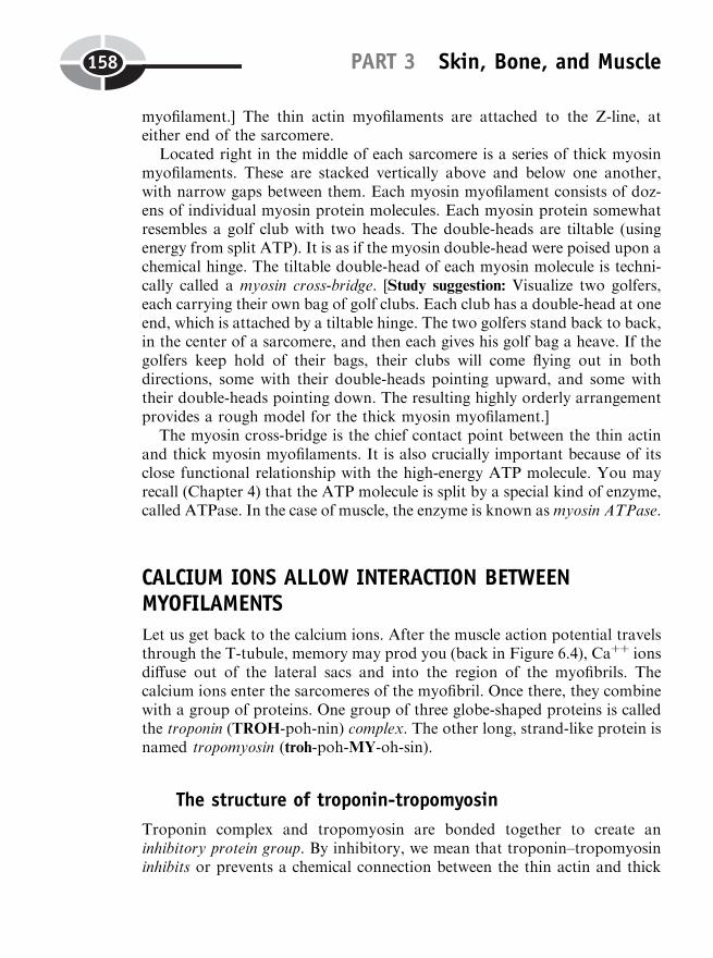
myofilament.] The thin actin myofilaments are attached to the Z-line, ateither end of the sarcomere.
Located right in the middle of each sarcomere is a series of thick myosinmyofilaments. These are stacked vertically above and below one another,with narrow gaps between them. Each myosin myofilament consists of doz-ens of individual myosin protein molecules. Each myosin protein somewhatresembles a golf club with two heads. The double-heads are tiltable (usingenergy from split ATP). It is as if the myosin double-head were poised upon achemical hinge. The tiltable double-head of each myosin molecule is techni-cally called a myosin cross-bridge. [Study suggestion: Visualize two golfers,each carrying their own bag of golf clubs. Each club has a double-head at oneend, which is attached by a tiltable hinge. The two golfers stand back to back,in the center of a sarcomere, and then each gives his golf bag a heave. If thegolfers keep hold of their bags, their clubs will come flying out in bothdirections, some with their double-heads pointing upward, and some withtheir double-heads pointing down. The resulting highly orderly arrangementprovides a rough model for the thick myosin myofilament.]
The myosin cross-bridge is the chief contact point between the thin actinand thick myosin myofilaments. It is also crucially important because of itsclose functional relationship with the high-energy ATP molecule. You mayrecall (Chapter 4) that the ATP molecule is split by a special kind of enzyme,called ATPase. In the case of muscle, the enzyme is known as myosin ATPase.
CALCIUM IONS ALLOW INTERACTION BETWEENMYOFILAMENTS
Let us get back to the calcium ions. After the muscle action potential travelsthrough the T-tubule, memory may prod you (back in Figure 6.4), Caþþ ionsdiffuse out of the lateral sacs and into the region of the myofibrils. Thecalcium ions enter the sarcomeres of the myofibril. Once there, they combinewith a group of proteins. One group of three globe-shaped proteins is calledthe troponin (TROH-poh-nin) complex. The other long, strand-like protein isnamed tropomyosin (troh-poh-MY-oh-sin).
The structure of troponin-tropomyosin
Troponin complex and tropomyosin are bonded together to create aninhibitory protein group. By inhibitory, we mean that troponin–tropomyosininhibits or prevents a chemical connection between the thin actin and thick
PART 3 Skin, Bone, and Muscle158

myosin myofilaments. How do they prevent this connection? Well, the tro-ponin–tropomyosin group is like a dark, leather, double-twisted strand (thelong, thin tropomyosin molecule) with a few dark beads (the rounded tro-ponin molecules) attached along the sides of it. This dark leather strand–darkbead combination looks like a pair of snakes, each with three buggy eyes,lying within the grooves of the double-helix molecule of the actin proteinpearl necklace! (Consult Figure 6.6, A.) We can summarize the total structureof the thin myofilament as:
THINMYOFILAMENT
¼ A Big Double Helix ofActin Molecules (likea white, twisted pearl
necklace)
þ A Smaller Double Helixof Dark Tropomyosin,with Dark Beads of
Troponin attached, lyinglike two bug-eyed snakesinside the long groovesof the Actin Double
Helix
Tropomyosin covers up the actin;troponin combines with calcium
We will add further to our snake analogy. We can say that tropomyosin, likea long, dark, sneaky snake, covers up the binding sites for myosin that arepresent on the pearl-like beads of actin! (Study Figure 6.6, B.) ‘‘So, I guessyou’re saying that tropomyosin is some kind of sneaky trouble-maker,Professor, at least as far as keeping actin and myosin apart?’’ Well, that’spretty close!
The muscle fiber stays in a state of relaxation, with its contraction beinginhibited, while tropomyosin is covering up the actin molecules’ binding sitesfor myosin.
In order to finish the process of muscle excitation, this inhibition of musclecontraction by the tropomyosin must be removed or relieved. Now, this iswhere the calcium ions finally come in! After the muscle fiber action potentialreleases stored Caþþ ions, they enter the sarcomeres of the myofibrils andbond to the buggy-eyed troponin protein complex. (View Figure 6.6, C.) Withthose calcium ions stuck to its buggy troponin eyes, the sneaky snake (longskinny tropomyosin molecule) changes its shape, breaking away from theactin molecules and falling deeper down into the groove.
At last! The sneaky snake (tropomyosin) no longer covers the binding sitesfor myosin! Hence, tiny projections on each double-headed myosin cross-bridge now fit into the binding sites on the actin pearl beads. (Examine
CHAPTER 6 Neurons and Muscle Fibers 159
Molecule 2
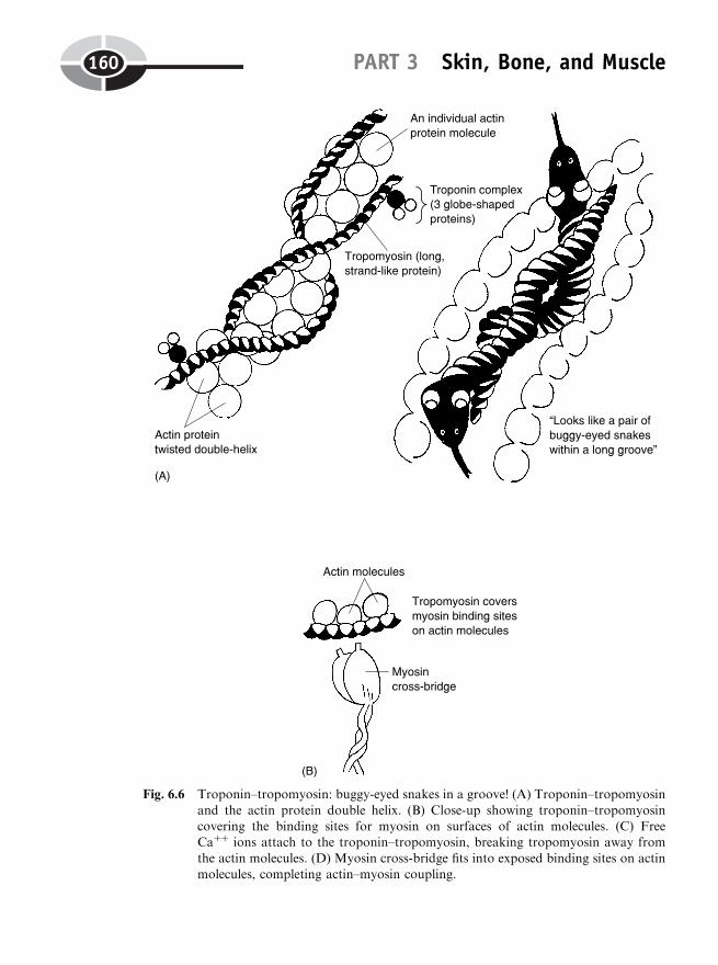
PART 3 Skin, Bone, and Muscle160
Fig. 6.6 Troponin–tropomyosin: buggy-eyed snakes in a groove! (A) Troponin–tropomyosin
and the actin protein double helix. (B) Close-up showing troponin–tropomyosincovering the binding sites for myosin on surfaces of actin molecules. (C) FreeCaþþ ions attach to the troponin–tropomyosin, breaking tropomyosin away from
the actin molecules. (D) Myosin cross-bridge fits into exposed binding sites on actinmolecules, completing actin–myosin coupling.

Figure 6.6, D.) Actin and myosin have met and bonded. Muscle fiber excita-tion is now complete, and we’re ready to contract!
READY, SET, SLIDE!
According to the sliding filament theory of muscle contraction, musclesshorten due to the inward sliding of the thin actin myofilaments over thetilted cross-bridges of the thick myosin myofilaments. ‘‘How does this slidingprocess get started, Joe?’’ The answer involves the action of the enzyme,myosin ATPase.
After actin has combined with myosin, the myosin ATPase enzymebecomes activated. It splits ATP molecules in the region of the myosincross-bridges. The resulting free energy tilts the myosin cross-bridges inward.As the cross-bridges tilt, the overhanging thin actin myofilaments slide overtheir tips. (View Figure 6.7.) The sliding occurs at both ends of the sarco-mere. Hence, each sarcomere shortens. Since each myofibril organelle con-sists of a series of sarcomeres hooked end to end, the whole myofibrilshortens. And as all of their myofibril organelles shorten, the entire musclefiber (cell) also shortens.
GETTING RID OF CALCIUM: THE PROCESS OFRELAXATION
It requires splitting of ATP to provide the energy for tilting of the myosincross-bridges during muscle contraction. Strangely enough, ATP is alsorequired for relaxation! The reason: we must get rid of the Caþþ ions, if
CHAPTER 6 Neurons and Muscle Fibers 161
Molecule 1
Fig. 6.6 (continued)
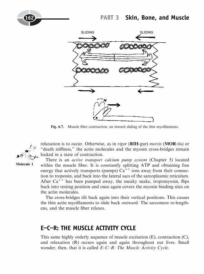
relaxation is to occur. Otherwise, as in rigor (RIH-gur) mortis (MOR-tis) or‘‘death stiffness,’’ the actin molecules and the myosin cross-bridges remainlocked in a state of contraction.
There is an active transport calcium pump system (Chapter 5) locatedwithin the muscle fiber. It is constantly splitting ATP and obtaining freeenergy that actively transports (pumps) Caþþ ions away from their connec-tion to troponin, and back into the lateral sacs of the sarcoplasmic reticulum.After Caþþ has been pumped away, the sneaky snake, tropomyosin, flipsback into resting position and once again covers the myosin binding sites onthe actin molecules.
The cross-bridges tilt back again into their vertical positions. This causesthe thin actin myofilaments to slide back outward. The sarcomere re-length-ens, and the muscle fiber relaxes.
E–C–R: THE MUSCLE ACTIVITY CYCLE
This same highly orderly sequence of muscle excitation (E), contraction (C),and relaxation (R) occurs again and again throughout our lives. Smallwonder, then, that it is called E–C–R: The Muscle Activity Cycle.
PART 3 Skin, Bone, and Muscle162
Fig. 6.7. Muscle fiber contraction: an inward sliding of the thin myofilaments.
Molecule 1
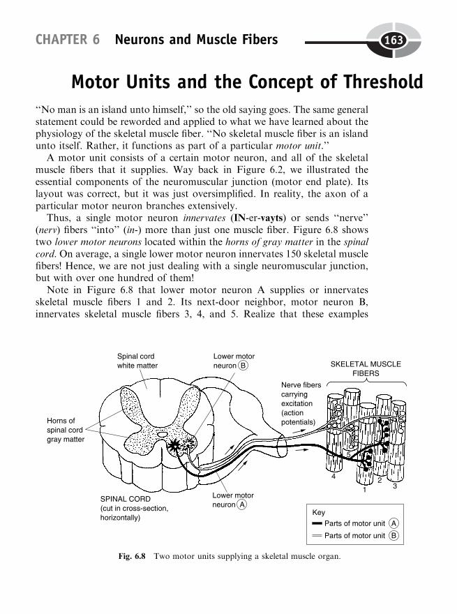
Motor Units and the Concept of Threshold‘‘No man is an island unto himself,’’ so the old saying goes. The same generalstatement could be reworded and applied to what we have learned about thephysiology of the skeletal muscle fiber. ‘‘No skeletal muscle fiber is an islandunto itself. Rather, it functions as part of a particular motor unit.’’
A motor unit consists of a certain motor neuron, and all of the skeletalmuscle fibers that it supplies. Way back in Figure 6.2, we illustrated theessential components of the neuromuscular junction (motor end plate). Itslayout was correct, but it was just oversimplified. In reality, the axon of aparticular motor neuron branches extensively.
Thus, a single motor neuron innervates (IN-er-vayts) or sends ‘‘nerve’’(nerv) fibers ‘‘into’’ (in-) more than just one muscle fiber. Figure 6.8 showstwo lower motor neurons located within the horns of gray matter in the spinalcord. On average, a single lower motor neuron innervates 150 skeletal musclefibers! Hence, we are not just dealing with a single neuromuscular junction,but with over one hundred of them!
Note in Figure 6.8 that lower motor neuron A supplies or innervatesskeletal muscle fibers 1 and 2. Its next-door neighbor, motor neuron B,innervates skeletal muscle fibers 3, 4, and 5. Realize that these examples
CHAPTER 6 Neurons and Muscle Fibers 163
Fig. 6.8 Two motor units supplying a skeletal muscle organ.

are highly simplified, in that the real-life situation would involve hundreds ofmuscle fibers within a particular muscle organ.
Returning to the idea of a motor unit, we can identify two different motorunits in Figure 6.8. Motor unit A consists of motor neuron A plus skeletalmuscle fibers 1 and 2. However, motor unit B involves motor neuron B plusskeletal muscle fibers 3, 4, and 5.
THRESHOLD, TWITCH, AND THE ‘‘ALL-OR-NONE LAW’’
‘‘Why do we bother with identifying particular motor units in a certainskeletal muscle organ, Professor Joe?’’ The answer is that it helps us under-stand the real-world contraction of skeletal muscle organs. Normally, ofcourse, we would expect a particular skeletal muscle (such as the bicepsbrachii in the upper arm) to respond and contract, if it was excited with astimulus that was sufficiently strong.
But an important question to ask ourselves here is, ‘‘Every time that thebiceps brachii muscle contracts, does the lower arm always flex with the sameamount of force?’’ The answer, of course, is ‘‘No.’’ The contractions of aparticular skeletal muscle organ are graded in their strength or intensity.What we mean by ‘‘graded’’ is that the contractions can become progressivelystronger, or progressively weaker, as if they were going up or down a gradedhill, or a series of higher and lower steps.
It takes very little mental effort and strength of biceps brachii contraction,for instance, to flex the arm and lift a piece of paper. It takes more mentaleffort, and a greater force of biceps contraction, to raise a heavy book. And ittakes supreme mental effort, and a near-maximal force of biceps contraction,to lift a dumbbell weighing 100 pounds (with one hand)! These whole-musclecontractions, then, are graded in their strength or force.
These observations lead us to state what we can call The Motor Unit Rule:
All muscle contractions are due to the contractions of a certain number ofmotor units within the muscle. The greater the number of motor units con-tracting, and the more skeletal muscle fibers these motor units contain, thegreater will be the resulting force of muscle contraction.
The concept of threshold
‘‘Okay, Professor Joe, but how do we get even one motor unit to contract?’’You have to stimulate it strongly enough to reach its threshold. A thresh-old, in general, is a place where you ‘‘tread’’ or ‘‘tramp,’’ a point where you
PART 3 Skin, Bone, and Muscle164
cross or enter a new beginning point (as in crossing the threshold of adoorway and entering a house). For our purposes, the threshold of a ske-letal muscle is the lowest strength or voltage of stimulation required to getcontraction of at least one of its motor units. Let us say that you artificiallystimulate a skeletal muscle by inserting electrodes into it. You turn on anelectric current, and zap the muscle with a shock of 1 volt (V). If nothinghappens, then this 1-volt shock is a subthreshold stimulus. It is too weak andfalls ‘‘below’’ (sub-) the intensity of stimulation required to get at least onemotor unit to contract.
What to do? Obviously, we keep raising the strength or voltage of elec-trical stimulation, and keep delivering more powerful shocks. Suppose afterdelivering a shock of 2 volts, we get a noticeable contraction response of themuscle. We can thus call this shock a threshold stimulus.
Muscle twitches
We can see the precise behavior of the muscle for ourselves by employing aninstrument called a myograph (MY-oh-graf ) – ‘‘an instrument used torecord’’ (-graph) the movements of a ‘‘muscle’’ (my). Figure 6.9 displaysthe chart tracings called a myogram (MY-oh-gram) or ‘‘graphical record’’(-gram) of ‘‘muscle’’ (my) activity.
At a threshold stimulus of, say, 2 volts, we get a very small muscle twitch.A muscle twitch is a quick, jerky contraction of one or more motor unitswhose stimulus threshold has been reached. Remembering the E–C–Rsequence of the Muscle Activity Cycle, the E (excitation) is the thresholdstimulus of 2 volts. A small number of motor units contract (C), raisingthe tracing on the myogram up from its horizontal baseline.
The muscle twitch is not very effective in human body movements, how-ever, because almost immediately after the contraction phase, relaxation (R)occurs. The myogram record of the twitch then falls back to its horizontalbaseline starting level. In intact humans, the single twitch often occurs as amuscle tic. A muscle tic is a quick, jerky spasm of a muscle (as on your cheekor eyelid). It is often due to some local irritation of the nerve endings ormuscle fibers in a body region.
The All-or-None Law
Glance back at Figure 6.8. Pretend that on the myogram recording of themuscle twitch at 2 volts (Figure 6.9), it was motor unit A and its two skeletalmuscle fibers (1 and 2) that responded. Note that there is a law, called the All-
CHAPTER 6 Neurons and Muscle Fibers 165
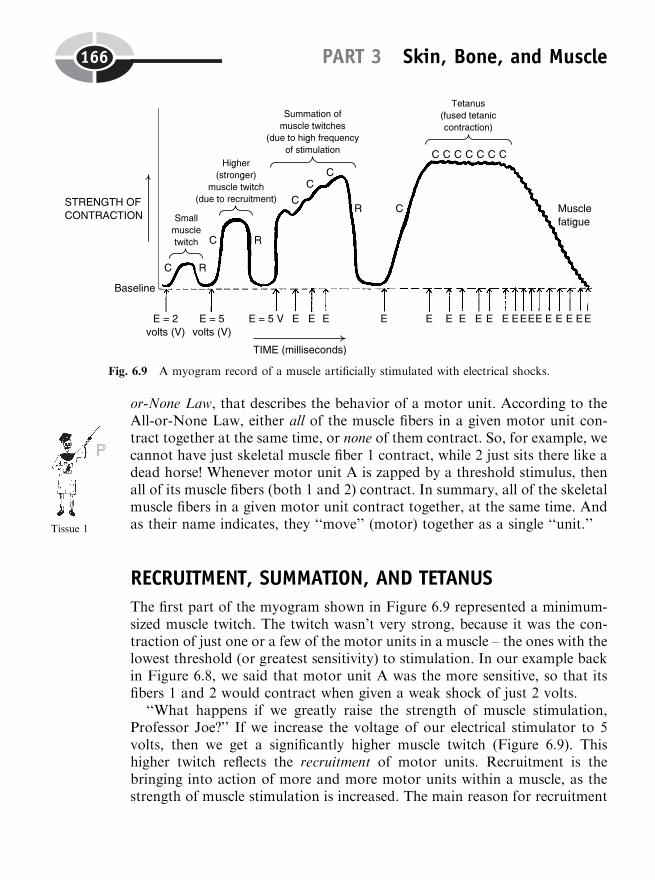
or-None Law, that describes the behavior of a motor unit. According to theAll-or-None Law, either all of the muscle fibers in a given motor unit con-tract together at the same time, or none of them contract. So, for example, wecannot have just skeletal muscle fiber 1 contract, while 2 just sits there like adead horse! Whenever motor unit A is zapped by a threshold stimulus, thenall of its muscle fibers (both 1 and 2) contract. In summary, all of the skeletalmuscle fibers in a given motor unit contract together, at the same time. Andas their name indicates, they ‘‘move’’ (motor) together as a single ‘‘unit.’’
RECRUITMENT, SUMMATION, AND TETANUS
The first part of the myogram shown in Figure 6.9 represented a minimum-sized muscle twitch. The twitch wasn’t very strong, because it was the con-traction of just one or a few of the motor units in a muscle – the ones with thelowest threshold (or greatest sensitivity) to stimulation. In our example backin Figure 6.8, we said that motor unit A was the more sensitive, so that itsfibers 1 and 2 would contract when given a weak shock of just 2 volts.
‘‘What happens if we greatly raise the strength of muscle stimulation,Professor Joe?’’ If we increase the voltage of our electrical stimulator to 5volts, then we get a significantly higher muscle twitch (Figure 6.9). Thishigher twitch reflects the recruitment of motor units. Recruitment is thebringing into action of more and more motor units within a muscle, as thestrength of muscle stimulation is increased. The main reason for recruitment
PART 3 Skin, Bone, and Muscle166
Tissue 1
Fig. 6.9 A myogram record of a muscle artificially stimulated with electrical shocks.
is that you are reaching the threshold of a greater and greater percentage ofmotor units in a muscle, as you increase the voltage of stimulation. Suppose,for instance, that motor unit B (back in Figure 6.8) has a higher thresholdthan motor unit A. If its threshold is, say, 4 volts, then it, too, will berecruited and start contracting (along with motor unit A) when a shock of5 V is delivered. If the voltage of single shocks keeps getting higher andhigher, eventually there will be a 100% recruitment of all of the motorunits in a muscle, and a maximum twitch will result. [Study suggestion:
When motor unit B is recruited, why would you expect the resulting twitchto be a lot stronger than the one produced by a shock of only 2 volts? Glanceback at Figure 6.8, if desired, to help you reason out the answer.]
Summation of muscle twitches
Now that we understand something about muscle twitches, and recruitmentof motor units at higher levels of single shocks, it is time to look at the effectof increasing the frequency of muscle stimulation. Let us go back to a singleshock of 5 volts. With one such shock, both motor units A and B (andskeletal muscle fibers 1–5) will be recruited, producing a fairly strong muscletwitch. But as we have already pointed out, individual muscle twitches have amuch too rapid onset of relaxation to allow them to be of much practical usefor body movements.
The key strategy used by the body to move its parts, then, is to create asummation of muscle twitches. As pictured in the myogram of Figure 6.9,summation is the adding together of muscle twitches, due to a high frequencyof muscle stimulation. Let us make a homely analogy. Mom yells at BabyHeinie to get off the couch and clean up the living room. If Mom yells justonce (loudly enough to reach the kid’s sensory threshold), then Baby Heiniewill just jump off the couch once, pick up one of his toys, and then immedi-ately collapse and relax back on the couch! The room is still a mess withmany scattered toys! A much better strategy (well known by all care-wornMoms and Dads) is to yell at the kid once, and (before he has had a chance torelax and flop back down) yell at him again and again!
This creates a summation effect, because Baby Heinie (just like theinvolved motor units) is stimulated to contract and move again, before hehas had a chance to completely relax and lengthen his sarcomeres! The resultis a fairly continuous body movement, one that can be used to accomplishpractical tasks like handing people a piece of paper, or picking toys up off thefloor. Therefore, most of our body movements represent the summation ofmuscle twitches.
CHAPTER 6 Neurons and Muscle Fibers 167
Tissue 2
Tetanus (tetanic contractions)
‘‘What happens if the muscle is stimulated at an extremely high frequency,say at a rate of 80 to 100 shocks every second?’’ The myogram back in Figure6.9 displays the response as tetanus (TET-ah-nus) or, using more appropriateterminology, as a fused tetanic (teh-TAN-ik) contraction. Both the wordstetanus and tetanic derive from the Greek word root tetan, which means‘‘extreme tension’’ or ‘‘convulsive tension.’’ In normal muscle physiology,tetanus (tetanic contraction) is one long, sustained muscle contraction,with no relaxation whatsoever, due to an extremely high frequency of musclestimulation.
Figure 6.9 shows this myogram as a fused tetanus, since the individualcontractions of the motor units have all been fused together into a single,‘‘extreme tension,’’ flat, plateau-like response. Tetanus (tetanic contractions)occur whenever we consciously keep stimulating our muscles, not allowingourselves to relax at all!
Muscle fatigue
‘‘Can tetanus or tetanic contractions last forever, Professor? What if we justkeep stimulating the muscle – on and on and on?’’ Well, we know that nophysiological activity of the human body can last forever, don’t we? Thesame is true for tetanus. Eventually a state of muscle fatigue sets in.Fatigue is literally ‘‘a tiring,’’ due to exhaustion of available energy and abuild-up of metabolic waste products.
Muscle Endurance Versus Fatigue:Biochemical Fiber Types
Well, did you figure out that muscle fatigue is mainly due to a using up of allavailable ATP energy within the muscle fibers? – Good for you! And we canlogically conclude that avoiding muscle fatigue, whenever possible, has defi-nite survival advantages for the human body! Pretend, for example, that youare a primitive hunter-gatherer, living long before civilization. You needgood endurance to keep tracking the deer you speared, or to keep runningaway from a snarling bear! At other times you just need to make a quick,mad dash for it, and not worry about muscle fatigue.
PART 3 Skin, Bone, and Muscle168
Organ 1
In short, we can see a physiological pattern of muscle fiber activity inoperation, here. This physiological pattern reinforces the ideas of WalterCannon (Chapter 3) about the ‘‘fight-or-flight response’’ and his book titledThe Wisdom of the Body. Let us now state a natural rule that we can imagi-natively call Muscle Fiber Wisdom:
Muscle Fiber Wisdom: All of the skeletal muscle fibers within a particularmotor unit are of a certain biochemical type. Thus, our skeletal muscle fibershave their own biochemical ‘‘wisdom.’’ This allows particular types of musclefibers to take the lead in responding to particular exercise-demanding condi-tions.
MUSCLE FIBER ENERGY STORAGE
There are three forms of potential energy storage within all skeletal musclefibers:
1. Muscle fiber ATP. There is only a small quantity of ATP moleculesstored within the muscle fiber. It provides a short-term energy supply forboth contraction and relaxation.
2. Creatine phosphate. A longer-term form of energy storage is offered bymolecules of creatine (KREE-ah-tin) phosphate, abbreviated as CP. Creatineis literally ‘‘a substance’’ (-ine) within ‘‘flesh’’ (creat), that is, muscle. Creatinephosphate, like adenosine triphosphate (ATP), contains high-energy chemi-cal bonds with phosphate. These high-energy bonds can readily be broken,resulting in free (kinetic) energy. This energy puts phosphate (P) onto ADP,thereby generating more muscle ATP.
There is about 3 to 6 times as much CP (creatine phosphate) stored withinskeletal muscle fibers, compared to ATP! Thus, creatine phosphate (CP)serves as a longer-term energy storage that helps quickly produce moreATP, thereby postponing muscle fatigue. (CP cannot be used by the musclefor energy; it just helps make ATP.)
3. Muscle fiber glycogen. Glycogen (‘‘sweetness or glucose producer’’), youmay remember (Chapter 3), is basically a large carbohydrate molecule con-sisting of many bonded glucose molecules. It thus represents a form of storedglucose (and its potential energy) that can be tapped within the muscle fiber.
RED (HO) FIBERS VERSUS WHITE (LO) FIBERS
Human skeletal muscle organs are mixtures of two basic colors or shades ofmuscle fibers. There are red or dark fibersmixed with white or light fibers. Thered (dark) muscle fibers owe their color to a high concentration of myoglobin
CHAPTER 6 Neurons and Muscle Fibers 169
(MY-oh-gloh-bin). Myoglobin is a ‘‘globe’’-shaped (glob) ‘‘muscle’’ (myo)‘‘protein’’ (-in).
Myglobin is reddish in color. Hence, red (dark) muscle fibers have a highmyoglobin concentration, while white (light) muscle fibers have a low myo-globin concentration. Myoglobin mainly functions as a temporary store foroxygen (O2) molecules within the muscle fiber. (Each myoglobin moleculetemporarily attaches and stores one O2 molecule.) It thereby helps the red ordark fiber contract aerobically, using oxygen. Red fibers are often classifiedas HO (high oxidative) or aerobic muscle fibers.
In addition, red (HO) fibers have a high density or crowding of mitochon-dria within their cytoplasm. This makes them quite resistant to fatigue, sincethe aerobic or oxidative catabolism of glucose (by the Krebs cycle and elec-tron transport system) produces a whopping net of 38 ATPs/glucose. Recall(Chapter 4) that cellular respiration or aerobic breakdown of glucose occurswithin the mitochondria.
White fibers, in contrast, contain very little myoglobin. Their mitochon-drial density is low. They are classified as LO (low oxidative) or anaerobicmuscle fibers. They chiefly break down glycogen and glucose by glycolysis inthe cytoplasm, which requires no input of oxygen. Anaerobic (LO) fibersproduce ATP quickly, because glycolysis requires very few biochemicalsteps. But they are pretty quick to fatigue, since they only get a net of 2ATPs produced/glucose.
FAST TWITCH (FT) VERSUS SLOW TWITCH (ST) FIBERS
Another way of classifying muscle fibers biochemically is according to theirfiber twitch speed (speed of twitching or contracting). Fiber twitch speed isbasically determined by a muscle fiber’s concentration of the enzyme, myosinATPase. Recall that myosin ATPase is the enzyme that splits ATP near themyosin cross-bridges, thereby releasing free energy for tilting and sliding thethin actin myofilaments. In general, the higher a skeletal muscle fiber’s con-centration of myosin ATPase, the faster it will split ATP, and the faster thefiber will twitch.
Fast twitch or FT muscle fibers thus have a high concentration of myosinATPase. They produce quick contractions, but they are more likely tofatigue, since they use up ATP at a fast rate.
Slow twitch or ST muscle fibers, in contrast, have a low concentration ofmyosin ATPase enzyme. Their contractions are fairly slow. But their payoff isbetter endurance, since they are breaking down ATP at a slow rate.
PART 3 Skin, Bone, and Muscle170
Molecule 2
Cell 3

COMBINING CHARACTERISTICS: THE THREESKELETAL MUSCLE FIBER TYPES
There are three biochemical skeletal muscle fiber types. Each of these types isa combination of two factors: oxidative or aerobic capacity (HO versus LO)and myosin ATPase concentration (FT versus ST).
Fiber type 1: HOST (High Oxidative, Slow Twitch)
These fibers are red and aerobic. Thus, they are very efficient producers ofATP. They twitch slowly, so they use up ATP at a slow rate. Hence, HOSTfibers are generally the best for slow endurance activities, because they are themost resistant to fatigue. Picture HOST as the penny-pinching miser orScrooge of the muscle fiber world – it has lots of ‘‘money’’ (ATP) but justwon’t ‘‘spend’’ it very fast! HOST fibers play an important role in yourpostural (PAHS-chur-al) muscles, such as those in your neck and lowerback, that allow you to maintain an upright posture all day long withoutcollapsing with fatigue!
Fiber type 2: HOFT (High Oxidative, Fast Twitch)
Like the HOST fibers, these are red, aerobic, and efficient ATP-producers.But since they have a high concentration of myosin ATPase enzyme, theysplit ATP at a rapid rate. These fibers are pretty good for endurance, yet stilltwitch rapidly. Picture HOFT fibers as the high-rollers or Yuppies of themuscle fiber world – they have lots of ‘‘money’’ (ATP) and they ‘‘spend’’ itrapidly (twitch fast). For example, these fibers would be important in the legmuscles of the winner of a 26-mile marathon race. The winner of the mara-thon needs good endurance to finish this long distance, but at the same timemust avoid early fatigue.
Fiber type 3: LOFT (Low Oxidative, Fast Twitch)
Unlike HOST and HOFT, these fibers produce ATP anaerobically, withoutuse of oxygen. They are thus the white muscle fibers, and the quickest fibers,because they produce 2 ATPs per glucose using just a few biochemical stepsof glycolysis. And since they are fast-twitch (FT), they split their quicklyproduced ATPs at a rapid pace. Picture LOFT fibers as the energy spend-thrifts of the muscle fiber world – they don’t ‘‘earn money’’ (produce ATP)
CHAPTER 6 Neurons and Muscle Fibers 171
Cell 4
very efficiently, but boy can they ‘‘spend it’’ (use up available ATP) veryquickly! Hence, you would vote these skeletal muscle fibers as ‘‘most likelyto fatigue (not succeed)’’!
‘‘Does this mean that LOFT fibers aren’t very useful, Professor?’’ Not atall! The LOFT fibers get things done in a big hurry, when no one is worriedabout fatigue! In a sports context, think about the leg muscles in the winnerof a 50-yard dash! The most genetically ‘‘gifted,’’ blazing-fast runners havean unusually high percentage of LOFT fibers within their leg muscles!
[Study suggestion: Look back over the three biochemical fiber types andtheir characteristics. Try to match them up with the personalities of differentpeople you know. Which one best describes you? Are you the energy miser orScrooge (HOST), the Yuppie or high-roller (HOFT), or the bankrupt energyspendthrift (LOFT)?]
QuizRefer to the text in this chapter if necessary. A good score is at least 8 correctanswers out of these 10 questions. The answers are listed in the back of thisbook.
1. The lever in any bone–muscle lever system is best represented by the:(a) Skeletal muscle(b) Bone(c) Joint(d) Pulling force
2. The nerve–muscle joining place is technically called the:(a) Knee joint(b) Axon terminal(c) Neuron soma(d) Neuromuscular junction (motor end-plate)
3. A traveling wave of electrochemical (ionic) excitation:(a) Sarcolemma(b) Dendrite(c) Action potential (nerve impulse)(d) Transverse tubule
4. A little network of hollow sacs and tubules within a skeletal musclefiber:(a) Sarcomeres
PART 3 Skin, Bone, and Muscle172
(b) Striations(c) Z-lines(d) Sarcoplasmic reticulum
5. Explains the phenomenon of muscle contraction:(a) Sliding filament theory(b) Hematopoiesis(c) Motor unit(d) Actin protein
6. Enzyme directly responsible for providing the energy for muscle con-traction:(a) Lactic anhydrase(b) Myoglobin(c) Myosin ATPase(d) Troponin–tropomyosin
7. A dead skeletal muscle fiber can’t relax, because:(a) Too much energy has pumped away all of the calcium(b) Its contractions are always isotonic(c) Actin and myosin remain locked together(d) No Naþ ions are available
8. Every time skeletal muscle fiber X contracts, skeletal muscle fiber Yalso contracts. From this information we can conclude that:(a) Both X and Y are part of the same motor unit(b) X is slow-twitch, while Y is fast-twitch(c) X is high-oxidative, while Y is low-oxidative(d) Neither fiber has an adequate energy source
9. A shock of 50 volts produces a twitch 10 millimeters high. A shock of100 volts yields a twitch 18 millimeters high. Therefore:(a) Summation is occurring(b) Fatigue is setting in(c) Tetanus has been reached(d) Recruitment has been observed
10. An alternative, high-energy source stored within all muscle fibers:(a) Vitamin K(b) Ascorbic acid(c) Creatine phosphate(d) Myoglobin
CHAPTER 6 Neurons and Muscle Fibers 173
Body-Level Grids for Chapter 6Several key body facts were tagged with numbered icons in the page marginsof this chapter. Write a short summary of each of these key facts into anumbered cell or box within the appropriate Body-Level Grid that appearsbelow.
Anatomy and Biological Order Fact Grids for Chapter 6:
MOLECULELevel
ORGANELLELevel
PART 3 Skin, Bone, and Muscle174
1 2
1
CELLLevel
Physiology and Biological Order Fact Grids for Chapter 6:
CELLLevel
CHAPTER 6 Neurons and Muscle Fibers 175
1
1 2
3 4
TISSUELevel
ORGANLevel
Physiology and Biological Disorder Fact Grids for Chapter 6:
ORGANLevel
PART 3 Skin, Bone, and Muscle176
1 2
1
1
Function and Biological Order Fact Grids for Chapter 6:
MOLECULELevel
Function and Biological Disorder Fact Grids for Chapter 6:
MOLECULELevel
CHAPTER 6 Neurons and Muscle Fibers 177
1 2
1
Test: Part 3
DO NOT REFER TO THE TEXT WHEN TAKING THIS TEST. A goodscore is at least 18 (out of 25 questions) correct. Answers are in the back ofthe book. It’s best to have a friend check your score the first time, so youwon’t memorize the answers if you want to take the test again.
1. The skin or integument consists of:(a) Malleus, incus, and stapes(b) Epidermis, osteons, and subdermis(c) Squamae, epidermal strata, and osteocytes(d) Epidermis, dermis, and hypodermis(e) Subdermis, hypodermis, and hair follicles
2. helps prevent dehydration of deep body tissues:(a) Keratin(b) Melanin(c) Glycogen(d) Sensory nerve basket(e) Ultraviolet light
Copyright © 2004 by The McGraw-Hill Companies, Inc. Click here for terms of use.
3. An isotonic solution:(a) Has a concentration of about 0.9% NaCl dissolved in 99.1% H2O(b) Seldom exists within the natural body fluids(c) Results in a net osmosis of water from the blood plasma into the
RBCs(d) Is too watery to promote homeostasis(e) Is the exact opposite of a hypotonic solution
4. When red blood cells crenate, it means that they:(a) Abnormally swell up with fluid(b) Divide extensively by mitosis(c) Clump together to create blood clots(d) Shrink and wrinkle up(e) Blow up into tiny pieces!
5. Main nerve endings in the dermis that detect a rise in body tempera-ture:(a) Thermoreceptors(b) Chemoreceptors(c) Hair follicle bulbs(d) Layered strata(e) Pores
6. Dense (compact) bone tissue consists primarily of:(a) Spongy bone(b) Yellow marrow(c) Haversian systems (osteons)(d) Osteoblasts(e) Embryonic connective tissue
7. Involves the depositing of calcium phosphate crystals:(a) Hematopoiesis(b) Bone fracture(c) Ossification(d) Body temperature homeostasis(e) Neuromuscular junction activities
8. Blood [Caþþ] measured in units of mg/dL blood is an example of:(a) An anatomical parameter(b) A functional parameter(c) Upper normal limit(d) A physiological parameter(e) The normal range of a parameter
Test: Part 3 179
9. If blood calcium ion concentration rose far beyond its upper normallimit, then the person would experience the symptoms of:(a) Normocalcemia(b) Hypertension(c) Normoglycemia(d) Hypocalcemia(e) Hypercalcemia
10. Osteoclasts primarily function in this process:(a) Extraction of sodium ions(b) Resorption of bone matrix(c) Remodeling of the skin(d) Bone formation(e) Oxygen transport
11. Usually provides evidence of physiological hypertrophy:(a) Shrinkage of a skeletal muscle down to half its normal size(b) Complete fracture of a long bone into several pieces(c) Too much sugar in the diet(d) Remodeling of bones to become thicker and stronger(e) Remodeling of bones to become thinner and weaker
12. ‘‘The erythrocyte is an anucleate, biconcave disc’’; therefore:(a) RBCs have no nucleus but are shaped like an hourglass(b) Leukocytes must have exactly the same characteristics(c) RBCs have many nuclei and are shaped like globes(d) Each erythrocyte has the form of a doughnut(e) No detailed information about its structure is available
13. The most common type of leukocyte:(a) Platelet(b) Neutrophil(c) Megakaryoblast(d) Myeloblast(e) Thrombocyte
14. The fulcrum in any bone–muscle lever system:(a) Red bone marrow(b) Tendon(c) Movable joint(d) Muscle organ(e) Ligament
PART 3 Skin, Bone, and Muscle180
15. The major central portion of the neuron:(a) Soma(b) Dendrite(c) Axon(d) Axon terminals(e) Cytoplasm
16. The two general types of cells in the body that can have action poten-tials:(a) Neurons and liver cells(b) Bone cells and muscle fibers(c) Cartilage cells and lacunae(d) Muscle fibers and nerve cells(e) Muscle cells and epithelial cells
17. Stimulate the muscle fiber across the motor end plate:(a) Lateral sacs(b) Excitatory neurotransmitter molecules(c) Glucose molecules(d) Inhibitory neurotransmitter molecules(e) I bands
18. Portion of a myofibril where contraction actually occurs:(a) T-tubules(b) Sarcoplasmic reticulum(c) Sarcomere(d) Sarcolemma(e) Lateral sacs
19. Molecules arranged into a double helix attached at one end to aZ-line:(a) Myosin(b) Creatine(c) Creatine phosphate(d) Myosin ATPase(e) Actin
20. Provides the tilting action to help myofilaments slide:(a) Myosin cross-bridges(b) Muscle ADP(c) Troponin(d) Tropomyosin(e) ACh
Test: Part 3 181

21. A quick, jerky contraction of one or more motor units whose stimulusthreshold has been reached:(a) Tetanus(b) Twitch(c) Subthreshold contraction(d) Summation(e) Relaxation response
22. can be conveniently summarized as E–C–R:(a) Neuromuscular junction(b) Diffusion of Caþþ ions(c) All-or-None Law(d) Muscle activity cycle(e) Subthreshold excitation
23. Smoothly handing a person a piece of paper would likely involve:(a) Single twitches(b) Forced expiration(c) Summation(d) Fused tetanus(e) Relaxation
24. If Alfonso, the talking parrot, who is educated in muscle physiology,says, ‘‘LO,’’ he really means:(a) Fibers with quick contractions(b) Cells which mainly rely upon glycolysis alone for their ATP
production(c) Muscle fibers with a high density or crowding of mitochondria(d) ‘‘Low ceiling, overhead!’’(e) Cells which chiefly depend upon the Krebs cycle and electron
transport system for their ATP synthesis
25. The biochemical muscle fiber type probably most important in the legmuscles of a tramp who is determined to walk across America:(a) HOFT(b) LOST(c) LOFT(d) HOST(e) TOAST
PART 3 Skin, Bone, and Muscle182

PART 4
The Physiology ofNerves and Glands
Copyright © 2004 by The McGraw-Hill Companies, Inc. Click here for terms of use.

This page intentionally left blank
185
CHAPTER7
‘‘What Happens WhenWe Step on a Nail?’’:
The Physiology ofNerves and Reflexes
Having discussed the physiology of muscles, bones, and joints, it is now timeto delve more deeply into the ‘‘spark plugs’’ and the ‘‘electrical wires’’ thatstimulate these body structures to move. Chapter 6 did bring up the conceptof the neuromuscular junction (motor end-plate). And the basic anatomy ofthe individual neuron and its nerve fibers (axon and dendrites) was provided.If the individual neuron is a ‘‘spark plug,’’ then the functional connectionsbetween the individual neurons and the associated nerves are the ‘‘electricalwires’’ that communicate with one another and carry information for long
Copyright © 2004 by The McGraw-Hill Companies, Inc. Click here for terms of use.

distances through the body. It is the main task of this chapter to explain howsuch communication of body information takes place.
Relationship of the Two Major Divisionsof the Nervous System
There are two basic anatomical divisions of the nervous system: the CentralNervous System or CNS, and the Peripheral (per-IF-er-al) Nervous System orPNS. (Consult Figure 7.1.) The Central Nervous System (CNS) is the portionof the nervous system that is ‘‘centrally’’ located, along the body midline. Itconsists of the brain or encephalon (en-SEF-ah-lahn) plus the spinal cord.
PART 4 Nerves and Glands186
Fig. 7.1 Flow of information into and out of the CNS.
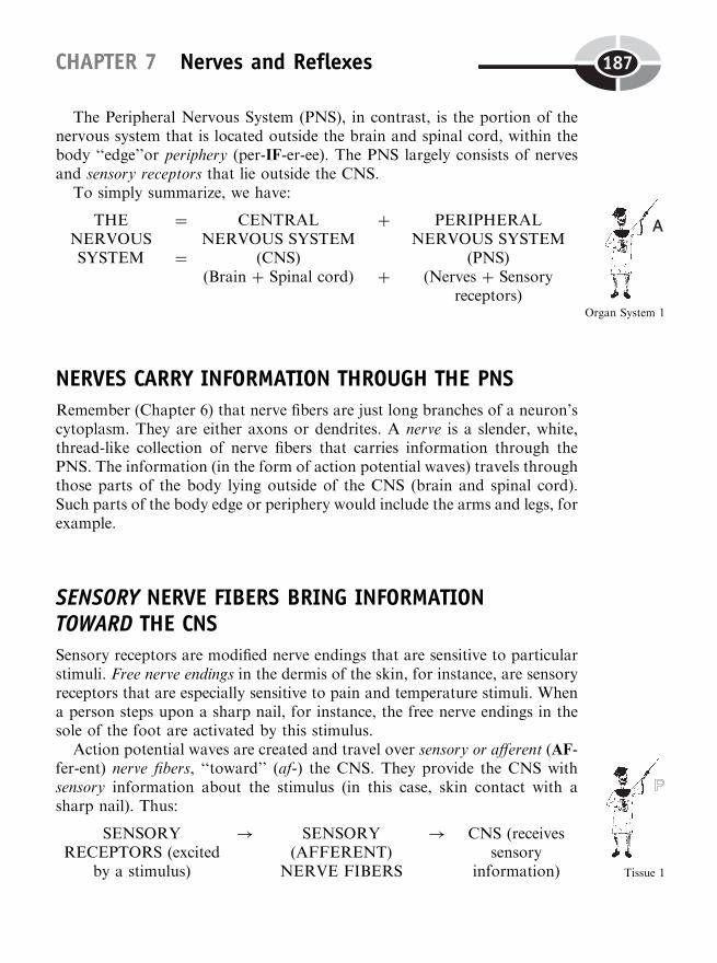
The Peripheral Nervous System (PNS), in contrast, is the portion of thenervous system that is located outside the brain and spinal cord, within thebody ‘‘edge’’or periphery (per-IF-er-ee). The PNS largely consists of nervesand sensory receptors that lie outside the CNS.
To simply summarize, we have:
THENERVOUSSYSTEM
¼
¼
CENTRALNERVOUS SYSTEM
(CNS)(Brain þ Spinal cord)
þ
þ
PERIPHERALNERVOUS SYSTEM
(PNS)(Nerves þ Sensory
receptors)
NERVES CARRY INFORMATION THROUGH THE PNS
Remember (Chapter 6) that nerve fibers are just long branches of a neuron’scytoplasm. They are either axons or dendrites. A nerve is a slender, white,thread-like collection of nerve fibers that carries information through thePNS. The information (in the form of action potential waves) travels throughthose parts of the body lying outside of the CNS (brain and spinal cord).Such parts of the body edge or periphery would include the arms and legs, forexample.
SENSORY NERVE FIBERS BRING INFORMATIONTOWARD THE CNS
Sensory receptors are modified nerve endings that are sensitive to particularstimuli. Free nerve endings in the dermis of the skin, for instance, are sensoryreceptors that are especially sensitive to pain and temperature stimuli. Whena person steps upon a sharp nail, for instance, the free nerve endings in thesole of the foot are activated by this stimulus.
Action potential waves are created and travel over sensory or afferent (AF-fer-ent) nerve fibers, ‘‘toward’’ (af-) the CNS. They provide the CNS withsensory information about the stimulus (in this case, skin contact with asharp nail). Thus:
SENSORYRECEPTORS (excited
by a stimulus)
! SENSORY(AFFERENT)
NERVE FIBERS
! CNS (receivessensory
information)
CHAPTER 7 Nerves and Reflexes 187
Tissue 1
Organ System 1

MOTOR NERVE FIBERS TAKE INFORMATIONAWAY FROM THE CNS
There is an opposite direction of information flow (action potential travel),too. There are thousands of motor neurons present within the CNS. Whensome of these motor neurons ‘‘decide’’ to fire an action potential, these waveseventually travel over motor or efferent (EE-fer-ent) nerve fibers. They arecalled efferent nerve fibers, because they ‘‘carry’’ (fer) motor (movement-related) information ‘‘away from’’ (ef-) the CNS.
‘‘Where do these motor or efferent nerve fibers eventually wind up,Professor Joe?’’ They go out to particular effectors (e-FEK-ters). An effectoris literally ‘‘something that’’ (-or) carries out a particular response, therebyhaving some ‘‘effect’’ upon the body. Specifically, the effectors in the humanbody are of two basic types: they are either muscles that contract (causing theeffect of body movement), or glands that secrete (releasing some useful sub-stance).
Summarizing the above, we have:
MOTORNEURONS
(‘‘decide’’ to fireaction potentials)
! MOTOR(EFFERENT)
NERVEFIBERS
! BODY EFFECTORSRESPOND (muscles
contract, or glands secrete)
The Synapse: Where Two Neurons‘‘Clasp Hands’’
If we are going to talk about long nerve fibers carrying motor or sensoryinformation long distances through the body, we also have to talk aboutwhat happens when the fibers finally reach their destination! Usually weare dealing with neuron axons, which have those branching axon terminalsat their tips. In the case of the axon terminals ending upon muscle effectors,the area of near-contact, you will remember, is the neuromuscular junction(motor end-plate). (Consult Figure 7.2, A.)
‘‘But what happens if the axon terminals end upon part of another neuron,instead of a muscle, Professor Joe?’’ The resulting meeting place is called asynapse (SIN-aps). A synapse is a ‘‘clasping together’’ of two neurons, asbetween the axon terminals of one neuron and the dendrite or cell body of
PART 4 Nerves and Glands188
Tissue 2
Tissue 1
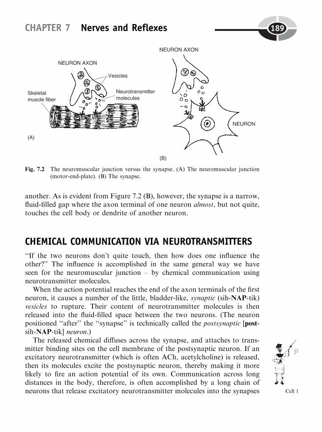
another. As is evident from Figure 7.2 (B), however, the synapse is a narrow,fluid-filled gap where the axon terminal of one neuron almost, but not quite,touches the cell body or dendrite of another neuron.
CHEMICAL COMMUNICATION VIA NEUROTRANSMITTERS
‘‘If the two neurons don’t quite touch, then how does one influence theother?’’ The influence is accomplished in the same general way we haveseen for the neuromuscular junction – by chemical communication usingneurotransmitter molecules.
When the action potential reaches the end of the axon terminals of the firstneuron, it causes a number of the little, bladder-like, synaptic (sih-NAP-tik)vesicles to rupture. Their content of neurotransmitter molecules is thenreleased into the fluid-filled space between the two neurons. (The neuronpositioned ‘‘after’’ the ‘‘synapse’’ is technically called the postsynaptic [post-sih-NAP-tik] neuron.)
The released chemical diffuses across the synapse, and attaches to trans-mitter binding sites on the cell membrane of the postsynaptic neuron. If anexcitatory neurotransmitter (which is often ACh, acetylcholine) is released,then its molecules excite the postsynaptic neuron, thereby making it morelikely to fire an action potential of its own. Communication across longdistances in the body, therefore, is often accomplished by a long chain ofneurons that release excitatory neurotransmitter molecules into the synapses
CHAPTER 7 Nerves and Reflexes 189
Fig. 7.2 The neuromuscular junction versus the synapse. (A) The neuromuscular junction(motor-end-plate). (B) The synapse.
Cell 1
between them. An action potential in one neuron is soon followed by thecreation of an action potential in the second neuron, which excites the thirdneuron in sequence, and so on.
Inhibitory neurotransmitters are a real ‘‘turn-off’’!
‘‘But if all synapses involve the release of only excitatory neurotransmitters,is there any way to turn the whole sequence off?’’, Baby Heinie asks, showingconsiderable intelligence for a little kid. Yes, there is. The flow of excitationdown a long chain of neurons can be interrupted when an inhibitory neuron isexcited. An inhibitory neuron is a neuron that releases inhibitory neurotrans-mitter molecules from its ruptured synaptic vesicles. Two chemicals that oftenact as inhibitory transmitters are called GABA and glycine (GLEYE-seen).
When either GABA or glycine is released, it diffuses across the synapseand inhibits the postsynaptic neuron. Thus, the postsynaptic neuron is lesslikely to fire off an action potential of its own, and the chain of neuronexcitation is effectively turned off.
Neuron Axons: Resting Polarized,or Active Depolarized?
If the neuron cell body is a ‘‘spark plug,’’ then the neuron axon could beconsidered a ‘‘spark plug wire.’’ Let us take a really close look at the neuronsoma (cell body) and an area called the axon hillock (HILL-ahk) (see Figure7.3). A hillock is a ‘‘little hill.’’ Therefore, the axon hillock is a little, raised,hill-like bump located near the base of the neuron axon. The axon hillock isof special functional importance, because it is the firing zone or trigger zonein the neuron where the action potential usually begins.
THE RESTING POLARIZED STATE
When a neuron is at ‘‘rest,’’ electrochemically or ionically speaking, it is notconducting an action potential (Figure 7.3, A). The cell membrane of theneuron has an electrical dipole (DIE-pohl) or ‘‘double-pole’’ distributedacross it. [Study suggestion: Visualize the positive (þ) and negative (�)poles or ends of a car battery.]
PART 4 Nerves and Glands190
Cell 2
Cell 1

The intracellular fluid (ICF) within the neuron is negatively charged (thatis, a negative pole) compared to the extracellular fluid (ECF) located outsideof the neuron. Conversely, the ECF is positively charged (that is, a positivepole) compared to the ICF. In summary, there is a dipole across the neuron’splasma membrane: the positive (þ) pole is on the outside of the neuron, whilethe negative (�) pole is on the inside of the neuron. This resting, non-conducting condition of the neuron is formally called the resting polarized(POH-lar-eyezd) state.
THE EXCITED NEURON: THE ACTIVE DEPOLARIZED STATE
When the neuron is excited, because of the binding of acetylcholine or someother excitatory neurotransmitter to it, the sodium-ion channels suddenly
CHAPTER 7 Nerves and Reflexes 191
active
Fig. 7.3. The neuron axon in its resting versus depolarized states. (A) The resting polarizedstate of a neuron. (B) The excited neuron: an active, depolarized state.

open within the axon hillock (Figure 7.3, B). There is a sudden great increasein the Naþ ion permeability of the plasma membrane. Hence, there is amassive net diffusion of sodium ions from the ECF and into the ICF ofthe neuron.
With so many new, positively charged particles (Naþ ions) now within theICF, an active depolarized (dee-POH-lar-eyezd) state is created. Since de-means ‘‘away from,’’ the word depolarized means ‘‘away from being (resting)polarized.’’ In other words, the resting polarized state across the membrane(net positive charge outside the neuron, but net negative charge inside theneuron) has been reversed. The result is the creation of an action potential,which represents a traveling wave of depolarization (net positive chargeinside of the axon, along with net negative charge outside of the axon).[Study suggestion: Picture the action potential as a þ and � wave, with theþ charge on the inside of the axon and the � charge on the outside of theaxon.]
During this active depolarized state of the neuron cell membrane, anaction potential is being actively conducted from its site of origin at the axonhillock, all the way down the axon.
Neuron Axons: Naked, or Myelinated?Once it is created, how an action potential travels largely depends uponwhether the axon is myelinated (MY-el-in-ay-ted) or naked. A myelinatedaxon is one that is covered by a myelin (MY-eh-lin) sheath. Myelin is asoft, white, fatty insulating material found on the axons of certain neurons.A naked axon, in contrast, lacks a myelin sheath.
IS IT GRAY, OR IS IT WHITE? IT ALL DEPENDSUPON MYELIN!
Thousands of neuron cell bodies and dendrites, which lack any covering ofmyelin, make up the gray matter of the brain and spinal cord. Conversely,myelinated axons, with their white myelin sheath, make up the white matterof the brain and spinal cord. Some axons, however, are naked (lackingmyelin).
PART 4 Nerves and Glands192
Organelle 1
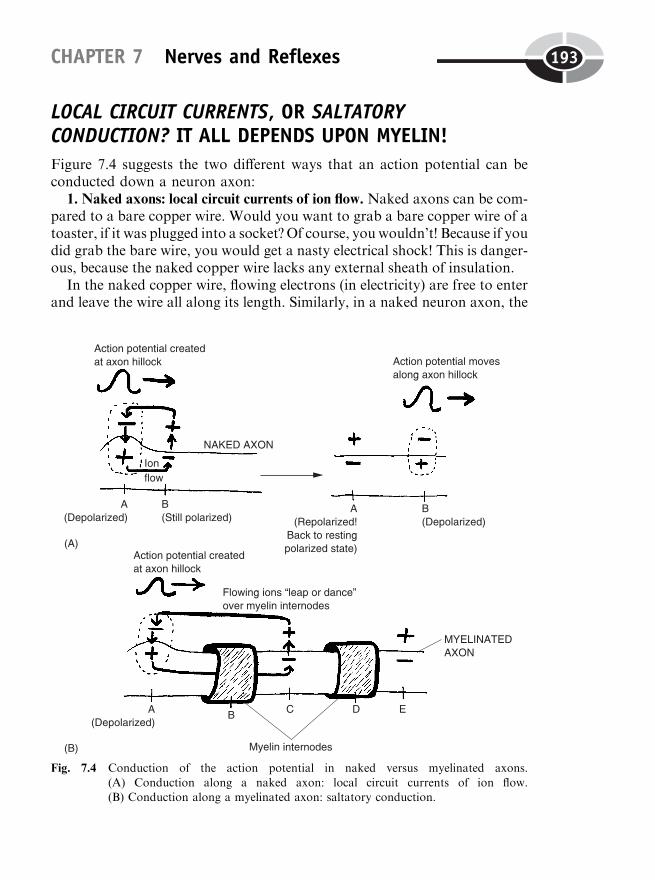
LOCAL CIRCUIT CURRENTS, OR SALTATORYCONDUCTION? IT ALL DEPENDS UPON MYELIN!
Figure 7.4 suggests the two different ways that an action potential can beconducted down a neuron axon:
1. Naked axons: local circuit currents of ion flow. Naked axons can be com-pared to a bare copper wire. Would you want to grab a bare copper wire of atoaster, if it was plugged into a socket? Of course, you wouldn’t! Because if youdid grab the bare wire, you would get a nasty electrical shock! This is danger-ous, because the naked copper wire lacks any external sheath of insulation.
In the naked copper wire, flowing electrons (in electricity) are free to enterand leave the wire all along its length. Similarly, in a naked neuron axon, the
CHAPTER 7 Nerves and Reflexes 193
Fig. 7.4 Conduction of the action potential in naked versus myelinated axons.
(A) Conduction along a naked axon: local circuit currents of ion flow.(B) Conduction along a myelinated axon: saltatory conduction.
action potential is conducted by local circuit currents of ion flow. It is best tothink of these as small loops of flowing ions, moving into and out of the axon‘‘locally’’ – at points immediately neighboring one another. There is a netinward flow of anions (such as Cl�) toward the positively charged pole withinthe axon, followed by a net outward flow of cations (such as Naþ) toward thenegatively charged pole just outside the axon. This repeating cycle of inwardand outward ion flow, therefore, creates multiple local loops or circuits. As aresult, the action potential is propagated (PRAHP-ah-gayt-ed) or ‘‘repro-duced’’ again and again, until it finally reaches the tip of the axon terminals.
2. Myelinated axons: saltatory conduction of the action potential. In myeli-nated neurons, the axon is partially coated with a myelin sheath. We mightcompare this situation with a toaster whose copper wire (naked axon) ispartially covered by an insulation of white rubber (myelin) (see Figure 7.4, B).
Suppose that our hero, Baby Heinie, being in a destructive mood, took aknife from the kitchen drawer and cut knicks out of the white rubber insula-tion that originally completely covered the toaster’s plug-in cord. The placeswhere the rotten kid cut down to bare copper wire (naked axon) we will callthe nodes of Ranvier (rahn-vee-AY). Each of these nodes is a ‘‘knot’’ ornarrowed region along the myelin sheath.
A series of myelin internodes (IN-ter-nohds) are found in the spaces‘‘between’’ (inter-) the ‘‘knots’’ (nodes) of Ranvier. Each myelin internodeis a roughly cylinder-shaped bead of myelin that occurs between two nodes ofRanvier. The overall effect is somewhat like a series of barrel-shaped Indianbeads along a string necklace.
Since we have used a toaster cord as our main model, we will return to it.Of course, Dad wouldn’t want to touch the toaster cord at any of its nodes ofRanvier! He would certainly receive an electrical shock from the flow ofcurrent leaking out of the cord at these naked points! A somewhat parallelsituation exists for the real myelinated nerve fiber. There is a saltatory (SAL-tah-toh-ree) conduction of the action potential.
Saltatory conduction is a type of conduction or carrying of the actionpotential by a process of ‘‘leaping.’’ Say that there are five adjacent points(A, B, C, D, and E) along a neuron axon. Suppose that points A, C, and Erepresent nodes of Ranvier (where ions can flow into and out of the axon).But points B and D are at myelin internodes (points of insulation where ionscannot enter or leave the axon). What happens, therefore, when an actionpotential begins at point A (the axon hillock)? The action potential ‘‘leaps’’over and under the myelin internode at point B, so that the action potential isregenerated at point C. Again, there is a saltatory (leaping) flow of ions overand under the myelin internode at point D, so the action potential is re-created at point E. By this leaping process that bypasses myelin internodes
PART 4 Nerves and Glands194
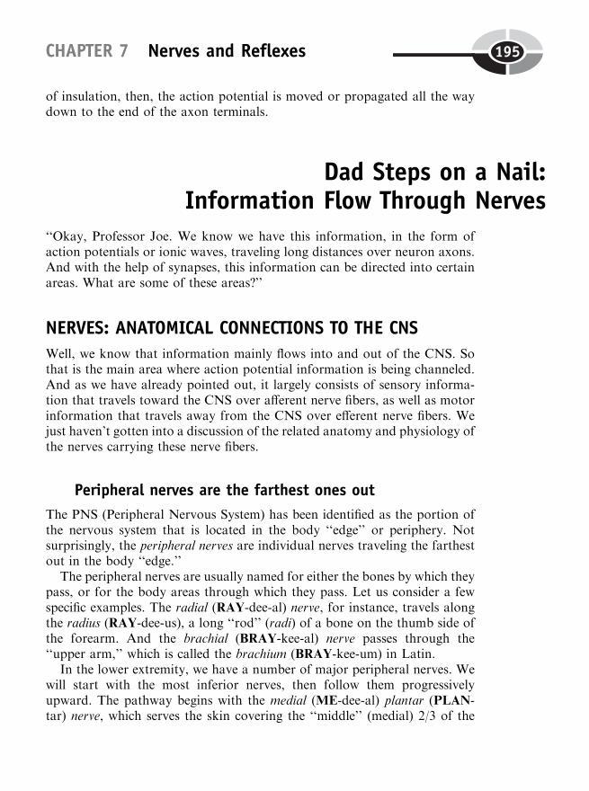
of insulation, then, the action potential is moved or propagated all the waydown to the end of the axon terminals.
Dad Steps on a Nail:Information Flow Through Nerves
‘‘Okay, Professor Joe. We know we have this information, in the form ofaction potentials or ionic waves, traveling long distances over neuron axons.And with the help of synapses, this information can be directed into certainareas. What are some of these areas?’’
NERVES: ANATOMICAL CONNECTIONS TO THE CNS
Well, we know that information mainly flows into and out of the CNS. Sothat is the main area where action potential information is being channeled.And as we have already pointed out, it largely consists of sensory informa-tion that travels toward the CNS over afferent nerve fibers, as well as motorinformation that travels away from the CNS over efferent nerve fibers. Wejust haven’t gotten into a discussion of the related anatomy and physiology ofthe nerves carrying these nerve fibers.
Peripheral nerves are the farthest ones out
The PNS (Peripheral Nervous System) has been identified as the portion ofthe nervous system that is located in the body ‘‘edge’’ or periphery. Notsurprisingly, the peripheral nerves are individual nerves traveling the farthestout in the body ‘‘edge.’’
The peripheral nerves are usually named for either the bones by which theypass, or for the body areas through which they pass. Let us consider a fewspecific examples. The radial (RAY-dee-al) nerve, for instance, travels alongthe radius (RAY-dee-us), a long ‘‘rod’’ (radi) of a bone on the thumb side ofthe forearm. And the brachial (BRAY-kee-al) nerve passes through the‘‘upper arm,’’ which is called the brachium (BRAY-kee-um) in Latin.
In the lower extremity, we have a number of major peripheral nerves. Wewill start with the most inferior nerves, then follow them progressivelyupward. The pathway begins with the medial (ME-dee-al) plantar (PLAN-tar) nerve, which serves the skin covering the ‘‘middle’’ (medial) 2/3 of the
CHAPTER 7 Nerves and Reflexes 195
plantar (PLAN-tar) surface of the foot. The word plantar literally ‘‘pertainsto the sole (plant) of the foot.’’
Suppose that Baby Heinie goes to bed, but leaves a sharp nail on thekitchen floor. Dad comes down after dark, and you know what happens!He steps on the nail! Dad steps on the nail with the middle of the sole of hisright foot. Thus, the nailhead serves as a stimulus for free nerve endings (painreceptors) in the sole. These pain receptors are connected to sensory (afferent)nerve fibers that pass through the medial plantar nerve (Figure 7.5).
Action potentials travel from the pain receptors in the sole, through themedial plantar nerve, and then up into a more superior peripheral nerve – thetibial (TIB-ee-al) nerve. The tibial nerve ascends (travels up) through thelower leg, just posterior to the tibia (TIB-ee-ah), which is the ‘‘shinbone.’’
PART 4 Nerves and Glands196
Fig. 7.5 Peripheral nerves travel up to a spinal nerve plexus.
The tibial nerve keeps ascending, snaking upward through the poples(PAHP-lees), which is also called the popliteal (pahp-lit-EE-al) space. InLatin, the word poples means ‘‘ham of the knee’’ or the ‘‘hollow of theknee.’’ [Study suggestion: Using your fingertips, probe the poples (poplitealspace) as the indented hollow region in the back of your knee. Imagine thetibial nerve passing through it, as you carefully look at Figure 7.5.]
Next in sequence is the sciatic (sigh-AT-ik) nerve. The word sciatic comesfrom the Greek for ‘‘hip joint,’’ and from ischium (IS-kee-um), which means‘‘hip.’’ Sciatic, therefore ‘‘pertains to the hip, hip joint, or ischium.’’ Theischium, of course, is not the entire bony hip. Rather, it is the most inferiorand posterior (lowest and most behind) portion of the hip bone.
Although it is named for the hip, the majority of the sciatic nerve runsfrom the poples (knee dent), up along the back of the femur (FEE-mur), or‘‘thigh’’ bone. For a short distance above the femur, the sciatic nerve (as itsname indicates) rises just posterior to the ischium region of the lower hipbone. Most interestingly, the sciatic nerve is the largest nerve in the entirebody!
Some peripheral nerves run into a spinal nerve plexus
Despite the fact that it is the biggest nerve in the body, the sciatic nervedoesn’t keep running along forever. Instead, it merges into the sacral(SAY-kral) plexus (PLEKS-us). A plexus, in general, is some kind of‘‘braid’’ or network. There are several spinal nerve plexuses – braids orbranching networks of spinal nerves supplying a particular area of thebody. (A spinal nerve is a nerve that connects directly onto the body ormain mass of the spinal cord.)
The sacral plexus, in particular, is a branching network of spinal nervesthat supply the area around the sacrum (SAY-crumb). The sacrum is literallythe ‘‘sacred’’ (sacr) bone. Because of its unique, pointed arrowhead shape, thesacrum was often considered ‘‘sacred’’ and used in magical ceremonies!
The sacral plexus is actually a branching network of spinal nerves L4 andL5 – the fourth and fifth lumbar (LUM-bar) spinal nerves – plus spinal nervesS1 through S5 – the first through fifth sacral spinal nerves.
SPINAL NERVE PLEXUS TO SPINAL CORD
From the sacral plexus, let us follow one of the spinal nerves (Figure 7.6).Each spinal nerve splits into two spinal nerve roots as it nears the body of thespinal cord. There is a dorsal root more in ‘‘back’’ (dors), and a ventral root
CHAPTER 7 Nerves and Reflexes 197

more on the front or ‘‘belly’’ (ventr) side of each spinal nerve. The dorsal rootand ventral root (like the roots of a tree) serve to anchor each spinal nerveinto the spinal cord. All of the spinal nerves, as well as their dorsal andventral roots, of course, are considered part of the Peripheral NervousSystem.
A Withdrawal Reflex ArcWhat we have been tracing, ever since Dad stepped on that sharp nail on thekitchen floor with the sole of his right foot, is a type of withdrawal reflex arc.A reflex, in general, is some type of automatic, involuntary response to aparticular stimulus. In this case, it is the automatic response to a painfulstimulus delivered to the bottom of the right foot.
PART 4 Nerves and Glands198
Fig. 7.6. Dad steps on a nail: a withdrawal reflex arc.
A withdrawal reflex is an automatic withdrawal of a body part from someharmful or annoying stimulus (such as a sharp nail pressing into the bottomof the foot). A reflex arc is a curved, arc or arch-like arrangement of thecomponents of a reflex. What we are seeing in Figure 7.6, then, is a curved,arc-like arrangement of the three major types of neurons, and their nervefibers, that are involved in automatically pulling Dad’s right foot away fromthe painful nail stimulus.
THE SENSORY NEURON AND DORSAL ROOT
The first neuron to be activated in the withdrawal reflex arc is the sensoryneuron. The dendrite of the sensory neuron has its peripheral end modified toform the sensory receptors for pain. In fact, everything back in Figure 7.5,from the peripheral nerves all the way up into the sacral plexus, was just thedendrite of a sensory neuron!
‘‘You mean, then, Professor,’’ Baby Heinie interjects, ‘‘that this wholething has been just one dendrite?’’ Well, we have been tracing the ascendingpathway followed by just one dendrite, but of many sensory neurons, run-ning side by side, and serving the sole of the right foot. And, as you canplainly see, these dendrites can be quite long, extending from the foot all theway up into the spinal cord!
The dendrite of the sensory neuron carries afferent (sensory) informationabout the painful stimulus toward the CNS. As it approaches the spinal cord,the dendrite of the sensory neuron goes up into the dorsal root of the spinalnerve. Observe in Figure 7.6 that there is a little ‘‘knot,’’ called the dorsal rootganglion (GANG-lee-ahn), present on the dorsal root. A ganglion, in general,is a knot-like collection of gray matter (neuron cell bodies and dendrites)located outside the CNS (brain and spinal cord). The dorsal root gangion inparticular, however, contains the cell bodies of the sensory neurons only.
From the dorsal root ganglion, the axon of the sensory neuron continues abit farther, finally terminating in axon terminals within the horns of graymatter in the spinal cord.
THE INTERNEURON (ASSOCIATION NEURON)
If we straighten all of the sensory nerve pathways into a smooth line, they looklike the first part of a curved arc, which has been placed on its side. ‘‘Whatabout the short bend or curve in the arc?’’, Baby Heinie follows this up.
That part is made by the interneuron (IN-ter-nur-ahn), which is alterna-tively called an association neuron. An interneuron (association neuron) is a
CHAPTER 7 Nerves and Reflexes 199
Organ 1

small, short neuron that forms a functional ‘‘association’’ or bridge‘‘between’’ (inter-) two much larger neurons. In this case, the small inter-neuron forms a functional association or linkage between the sensory neuron(bringing information into the spinal cord about the painful stimulus) andthe motor neuron, which carries information out of the spinal cord.
THE LOWER MOTOR NEURON
The final, bottom portion of the withdrawal reflex arc is a lower motorneuron. We have already discussed motor neurons and the motor end-plate. A lower motor neuron, however, is a neuron whose cell body lies inthe lower part of the CNS, often in or near the spinal cord.
In our current example, the cell body of the lower motor neuron lies in theventral (belly-side) horn of gray matter within the spinal cord. The axon ofthis lower motor neuron leaves the spinal cord and immediately enters theventral root of a spinal nerve. The ventral root of a spinal nerve, therefore, is‘‘pure,’’ in the sense that it carries motor or efferent nerve fibers only.
The ventral root hooks into a spinal nerve, which is always ‘‘mixed.’’ Thismeans that the spinal nerve essentially acts as a two-way street. It carries bothsensory (afferent) nerve fibers bringing information in about the painfulstimulus, as well as motor (efferent) nerve fibers taking information out,and directing body movement.
THE FINAL WITHDRAWAL RESPONSE
As the axon of the motor neuron leaves the individual spinal nerve, it entersthe sacral plexus. From here, the axon of the motor neuron becomes a motor(efferent) nerve fiber that basically travels in the reverse direction of thesensory (afferent) nerve fiber. But the final destination is different: the skeletalmuscle effectors of the lower leg. These skeletal muscles are stimulated tocontract, thereby withdrawing the sole of Poor Dad’s right foot away fromthe painful nail stimulus.
Sensory Pathways and Ascending Nerve Tracts‘‘But when the withdrawal reflex is over, and Dad has pulled his foot awayfrom the nail, wouldn’t he feel pain – and probably experience anger?’’, Baby
PART 4 Nerves and Glands200

Heinie somewhat guiltily whispers. ‘‘I don’t see any place in Figures 7.5 or 7.6where such pain and anger could be experienced.’’
So right you are, Baby Heinie! Dad would almost certainly experienceplantalgia (plan-TAL-jee-ah), or ‘‘pain’’ (-algia) in the ‘‘sole of the foot’’(plant). For this, the sensory areas of the brain would have to be involved.In technical terms, a sensory homunculus (hoh-MUNG-kyuh-lus) – a ‘‘littlefeeling man or dwarf ’’ – would have to be activated within the cerebral (seh-REE-bral) cortex (KOR-teks).
As Figure 7.7 shows, such a sensory homunculus can be visualized withinthe postcentral (POST-sen-tral) gyrus (JEYE-rus). This is a raised ‘‘ring’’(gyr) or fold of cerebral cortex – the thin ‘‘bark’’ of gray matter coveringthe surface of the cerebrum. It is called the postcentral gyrus because it islocated just ‘‘after’’ (post-) an important groove, called the central sulcus
CHAPTER 7 Nerves and Reflexes 201
Fig. 7.7. The spinothalamic tract ascends up to the feeling dwarf !
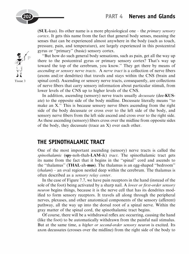
(SUL-kus). Its other name is a more physiological one – the primary sensorycortex. It gets this name from the fact that general body senses, meaning thesenses that can be experienced almost anywhere in the body (such as touch,pressure, pain, and temperature), are largely experienced in this postcentralgyrus or ‘‘primary’’ (basic) sensory cortex.
‘‘But how do such general body sensations, such as pain, get all the way upthere to the postcentral gyrus or primary sensory cortex? That’s way uptoward the top of the cerebrum, you know.’’ They get there by means ofascending or sensory nerve tracts. A nerve tract is a collection of nerve fibers(axons and/or dendrites) that travels and stays within the CNS (brain andspinal cord). Ascending or sensory nerve tracts, consequently, are collectionsof nerve fibers that carry sensory information about particular stimuli, fromlower levels of the CNS up to higher levels of the CNS.
In addition, ascending (sensory) nerve tracts usually decussate (dee-KUS-ate) to the opposite side of the body midline. Decussate literally means ‘‘tomake an X.’’ This is because sensory nerve fibers ascending from the rightside of the body decussate or cross over to the left side of the body, andsensory nerve fibers from the left side ascend and cross over to the right side.As these ascending (sensory) fibers cross over the midline from opposite sidesof the body, they decussate (trace an X) over each other.
THE SPINOTHALAMIC TRACT
One of the most important ascending (sensory) nerve tracts is called thespinothalamic (spy-noh-thah-LAM-ik) tract. The spinothalamic tract getsits name from the fact that it begins in the ‘‘spinal’’ cord and ascends tothe ‘‘thalamus’’ (THAL-ah-mus). The thalamus is an egg-shaped ‘‘bedroom’’(thalam) – an oval region nestled deep within the cerebrum. The thalamus isoften described as a sensory relay center.
In the case of Figure 7.7, we have pain receptors in the hand (instead of thesole of the foot) being activated by a sharp nail. A lower or first-order sensoryneuron begins things, because it is the nerve cell that has its dendrites mod-ified to form sensory receptors. It travels all along through the peripheralnerves, plexuses, and other anatomical components of the sensory (afferent)pathway, all the way up into the dorsal root of a spinal nerve. Within thegray matter of the spinal cord, the spinothalamic tract begins.
Of course, there will be a withdrawal reflex arc occurring, causing the hand(like the foot) to be automatically withdrawn from the painful nail stimulus.But at the same time, a higher or second-order sensory neuron is excited. Itsaxon decussates (crosses over the midline) from the right side of the body to
PART 4 Nerves and Glands202
Tissue 3

the left. The second-order sensory neuron has its axon terminal end withinthe thalamus. This is the official termination of the actual spinothalamictract.
But the thalamus is called a sensory relay center, isn’t it? The relayingaction (like passing a baton during a relay race) is by a relatively shortthird-order sensory neuron, whose cell body lies within the thalamus. Theaxon terminals of this neuron ‘‘relay’’ the sensory message up into the post-central gyrus of the cerebral cortex. It is here that the sensory homunculus(Dad’s ‘‘little feeling dwarf ’’) experiences the actual sensation of pain.Various association nerve fibers can also send the signal to nearby regionsof the cerebrum, where expressions of anger and speech can originate.
Motor Pathways and Descending Nerve TractsOf course, movements of the body do not always have to be involuntaryreflex actions (such as automatic withdrawal from a painful stimulus). Bodymovements can also be caused by a voluntary desire or will to move by thebrain. This process involves motor pathways, which make use of descendingnerve tracts (Figure 7.8).
A descending or motor nerve tract is a tract that carries motor (movement-related) information from higher levels of the CNS down to lower levels ofthe CNS. One very prominent example of such a descending (motor) nervetract is the corticospinal (kor-tih-koh-SPY-nal) tract.
As its name reveals, the corticospinal tract begins in the cerebral cortex(cortic) and descends down to the spinal cord (spinal), after it decussates tothe opposite side of the body. A key area to look at is the region around thecentral sulcus. As for the destination of the spinothalamic tract – the post-central gyrus – we again have a gyrus bordering the central sulcus. In thiscase, however, it is called the precentral gyrus. This is because the gyrus islocated ‘‘before or in front of ’’ (pre-) the central sulcus.
And for the corticospinal tract, the cerebral cortex is just the beginning ofthe tract (not its final termination). Upper motor neurons decide to voluntarilymove, say, the right hand. [Study suggestion: Speculate as to which precentralgyrus (right or left) will most likely make the decision to ‘‘bend’’ or flex theright hand, then check with Figure 7.8.]
After they decussate, the descending nerve fibers synapse onto a lowermotor neuron in the spinal cord. Its axon goes out into the PNS via theusual anatomical sequence, into a peripheral nerve, and finally to a neuro-
CHAPTER 7 Nerves and Reflexes 203
Tissue 4
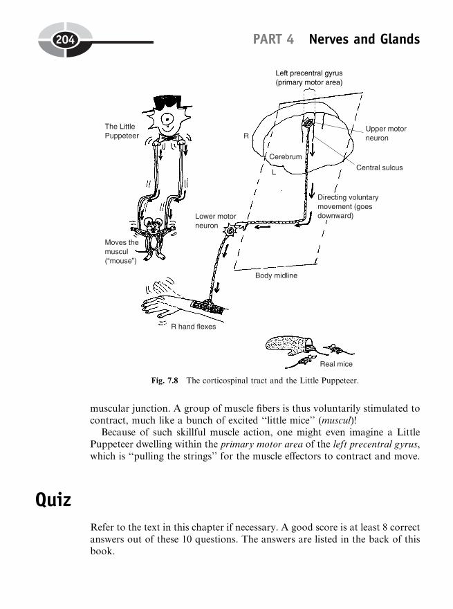
muscular junction. A group of muscle fibers is thus voluntarily stimulated tocontract, much like a bunch of excited ‘‘little mice’’ (muscul)!
Because of such skillful muscle action, one might even imagine a LittlePuppeteer dwelling within the primary motor area of the left precentral gyrus,which is ‘‘pulling the strings’’ for the muscle effectors to contract and move.
QuizRefer to the text in this chapter if necessary. A good score is at least 8 correctanswers out of these 10 questions. The answers are listed in the back of thisbook.
PART 4 Nerves and Glands204
Fig. 7.8 The corticospinal tract and the Little Puppeteer.
1. The part of the nervous system that contains the sensory receptors:(a) Nerves(b) PNS(c) Dorsal roots(d) Ganglia
2. Alternatively classified as afferent nerve fibers:(a) Motor nerve fibers(b) Axon hillocks(c) All dendrites(d) Sensory nerve fibers
3. Place where two neurons almost ‘‘clasp together’’:(a) Synapse(b) Motor end-plate(c) Neuromuscular junction(d) Myelin internode
4. Chemicals that make a postsynaptic neuron less likely to fire an actionpotential of its own:(a) ACh molecules(b) Excitatory neurotransmitters(c) Synaptic vesicles(d) GABA and glycine
5. The usual firing zone or trigger zone for an action potential:(a) Neuron soma(b) Myelin sheath(c) Node of Ranvier(d) Axon hillock
6. A stretch of a neuron cell membrane that is not currently conductingan action potential is accurately described as being:(a) Resting polarized(b) An electrical tripole(c) A neutral dipole(d) Active depolarized
7. Mode of action potential propagation that occurs along nakedaxons:(a) Saltatory conduction(b) Synaptic transmission(c) Local circuit currents(d) Ionic entrapment
CHAPTER 7 Nerves and Reflexes 205

8. The radial nerve is an example of a:(a) Cranial nerve(b) Peripheral nerve(c) Spinal nerve(d) Unmyelinated nerve
9. Consists of a branching network of spinal nerves L4 and L5, plus S1through S5:(a) Conus medullaris(b) Nodes of Ranvier(c) Sacral plexus(d) Sciatic nerve
10. Sensory homunculus:(a) Site of origin of most sensory impulses(b) Place of final destination of most ascending nerve tracts(c) Association areas of the cerebellum(d) Demyelinated neuron axons
Body-Level Grids for Chapter 7Several key body facts were tagged with numbered icons in the page marginsof this chapter. Write a short summary of each of these key facts into anumbered cell or box within the appropriate Body-Level Grid that appearsbelow.
Anatomy and Biological Order Fact Grids for Chapter 7:
CELLLevel
PART 4 Nerves and Glands206
TISSUELevel
ORGAN SYSTEMLevel
Physiology and Biological Order Fact Grids for Chapter 7:
CELLLevel
CHAPTER 7 Nerves and Reflexes 207
1
1
1
TISSUELevel
ORGANLevel
PART 4 Nerves and Glands208
1 2
1 2
3 4
1

Function and Biological Order Fact Grids for Chapter 7:
ORGANELLELevel
CHAPTER 7 Nerves and Reflexes 209
1
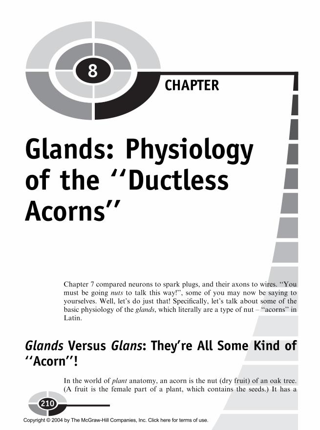
210
CHAPTER8
Glands: Physiologyof the ‘‘DuctlessAcorns’’
Chapter 7 compared neurons to spark plugs, and their axons to wires. ‘‘Youmust be going nuts to talk this way!’’, some of you may now be saying toyourselves. Well, let’s do just that! Specifically, let’s talk about some of thebasic physiology of the glands, which literally are a type of nut – ‘‘acorns’’ inLatin.
Glands Versus Glans: They’re All Some Kind of‘‘Acorn’’!
In the world of plant anatomy, an acorn is the nut (dry fruit) of an oak tree.(A fruit is the female part of a plant, which contains the seeds.) It has a
Copyright © 2004 by The McGraw-Hill Companies, Inc. Click here for terms of use.
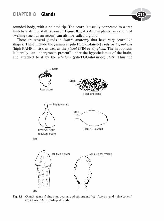
rounded body, with a pointed tip. The acorn is usually connected to a treelimb by a slender stalk. (Consult Figure 8.1, A.) And in plants, any roundedswelling (such as an acorn) can also be called a gland.
There are several glands in human anatomy that have very acorn-likeshapes. These include the pituitary (pih-TOO-ih-tair-ee) body or hypophysis(high-PAHF-ih-sis), as well as the pineal (PIN-ee-al) gland. The hypophysisis literally ‘‘an undergrowth present’’ under the hypothalamus of the brain,and attached to it by the pituitary (pih-TOO-ih-tair-ee) stalk. Thus the
CHAPTER 8 Glands 211
Fig. 8.1 Glands, glans: fruits, nuts, acorns, and sex organs. (A) ‘‘Acorns’’ and ‘‘pine cones.’’(B) Glans: ‘‘Acorn’’-shaped heads.

hypophysis (pituitary body) does, indeed, resemble a grayish-colored acornhooked onto the bottom of the brain by its stem!
A similar situation exists for the pineal gland, which is also associated withthe brain. This gland is named for its close likeness to a ‘‘pine cone’’ – stillanother nut!
GLANS LET OUR SEX ORGANS GET ‘‘A HEAD’’
A related word, glans, also means ‘‘acorn’’ in Latin. This type of ‘‘acorn,’’however, hardly grows on trees! ‘‘Why, Professor Joe?’’ Well, Baby Heinie,you are somewhat underaged, but you really need to be properly educatedabout your sex organs! ‘‘Yes, Professor Joe! If I don’t get educated about mysex organs, how am I supposed to get a head in life?’’
Precisely, Baby Heinie! A look at Figure 8.1 (B) shows two examples ofglans that serve as rounded, acorn-shaped heads of several male and femalesex organs. The glans penis (PEA-nis), for instance, is the acorn-like head ofthe male penis. Similarly, the glans clitoris (KLIT-er-is) is the acorn-shapedhead of the female clitoris. The physiology of both these glans is closelyinvolved with sexual stimulation and arousal.
In summary, the glands and heads of some of the sex organs in the humanbody are named for their anatomical shape – like an acorn – not for theirphysiology.
‘‘What is the main physiology of glands, then, Professor?’’ Glands consistof one or more epithelial cells specialized for the function of secretion – therelease of some useful substance.
Endocrine Glands Versus Exocrine GlandsEven though the entire general category of glands is named for its anatomy,the two major groups of glands are named for their differing physiology.Specifically, the two major types are named for the different destinations oftheir secretions (Figure 8.2).
Exocrine (EKS-oh-krin) glands are glands of ‘‘external’’ (exo-) ‘‘secretion’’(crin) of a useful product into a duct, which carries the secretion to some bodysurface. A good example of exocrine glands are the sweat glands of the skin.The gland cells line the walls of the sweat duct, which carries the sweat up tothe skin surface through a pore. Sweat can be considered a useful secretion,because it helps to cool the body and lower its temperature.
PART 4 Nerves and Glands212
Organ 1
Endocrine (EN-doh-krin) glands are glands of ‘‘internal’’ (endo-) ‘‘secre-tion’’ (crin) of a hormone, or chemical messenger, directly into the blood-stream, within the gland itself. Endocrine glands are therefore considered‘‘ductless,’’ since their hormone enters the bloodstream without passingthrough any duct. The hormone is then carried throughout the body.
The endocrine portion of the pancreas (PAN-kree-ahs), for instance, iscalled the pancreatic (pan-kree-AT-ik) islets (I-lets) or ‘‘little islands’’ of endo-crine gland cells. Each pancreatic islet includes the beta (BAY-tuh) cells,which are well known for their function of secreting insulin (IN-suh-lin).Many of you already know that insulin is a major hormone that causes adecrease in the blood glucose concentration.
Important Principles of Hormone ActionThe basic anatomy and descriptions of the various endocrine glands areprovided in our companion volume, ANATOMY DEMYSTIFIED. Thisbook, which emphasizes physiology, will focus upon important principlesof hormone action. And insulin from the beta cells of the pancreas, beingsuch a critical hormone, will often serve as our specific example.
HORMONE ACTION PRINCIPLE 1:HORMONES ARE ‘‘AROUSERS’’
The word hormone literally means ‘‘to arouse, set into motion, or urge on.’’Hormones, therefore, do not create any new physiological or biological
CHAPTER 8 Glands 213
Fig. 8.2 Exocrine versus endocrine glands: two different modes of secretion. (A) Exocrineglands. (B) Endocrine glands.
Organ 2
Molecule 1
processes within our cells. They merely alter (often ‘‘arousing’’ or increasing)the rate at which these processes occur.
Glucose, for instance, enters most of our cells by the process of facilitateddiffusion out of the bloodstream (Chapter 4). The protein carrier moleculeswithin cell membranes are always functioning, but at a relatively slow rate.Glucose molecules still enter our body cells by facilitated diffusion, but thisprocess does not occur at a significant rate. What insulin does, then, is‘‘arouse’’ or stimulate the carrier proteins to work much faster.Consequently, glucose molecules enter the body cells at a much faster rate,and the blood glucose concentration falls.
HORMONE ACTION PRINCIPLE 2: ENDOCRINE GLANDSARE LEAKY FAUCETS
A particular hormone (such as insulin) is always being secreted from itsendocrine gland. This makes the gland behave much like a continuouslydripping, leaky faucet. But the rate at which a hormone is secreted willchange or vary with the strength of the gland’s stimulus.
The major stimulus for the beta cells of the pancreas is a rise in the bloodglucose concentration toward its upper normal limit. This would occur aftereating a sugar-rich meal. The higher that the blood glucose concentrationgets, the more strongly the beta cells are stimulated. And the more stronglythe beta cells are stimulated, the greater will be their rate of insulin secretion(provided they are healthy, normal beta cells).
HORMONE ACTION PRINCIPLE 3: HORMONES OFTENCOMBINE WITH PLASMA PROTEINS
Most hormone molecules are not very soluble or ‘‘dissolvable’’ within theblood plasma (fluid portion of the blood). Instead, they combine withplasma proteins. The result is called a hormone–protein complex. Thehormone–protein complex is quite soluble, allowing the hormone to tra-vel long distances through the bloodstream. When the body destinationof the hormone is reached, it breaks away from the plasma proteincarrying it.
PART 4 Nerves and Glands214

HORMONE ACTION PRINCIPLE 4:‘‘A LITTLE DAB WILL DO YOU’’
Hormones are typically secreted in tiny microgram (‘‘millionths of a gram’’)quantities, but they have dramatic physiological effects. Consider the hor-mone epinephrine (ep-ih-NEF-rin), which is more popularly known as adrena-line (ah-DREN-ah-lin). The physiological response to severe stress, oftencalled the ‘‘fight-or-flight response,’’ is carried out by the sympathetic nervesand the endocrine gland called the adrenal (ah-DREE-nal) medulla (meh-DOO-lah). Both the sympathetic nerves and the adrenal medulla are acti-vated when the hypothalamus of the brain is prodded by stress (Figure 8.3).
Just a few additional micrograms of epinephrine, which is released fromthe adrenal medulla, triggers such severe ‘‘fight-or-flight’’ symptoms as
CHAPTER 8 Glands 215
Fig. 8.3 Stress and the ‘‘fight-or-flight’’ response.
tachycardia (tak-ee-KAR-dee-ah) and powerful vasoconstriction (vay-soh-kahn-STRIK-shun). This is an extremely ‘‘swift’’ (tachy-) rate of beating ofthe ‘‘heart’’ (cardi), accompanied by a dramatic ‘‘narrowing’’ (constriction) ofblood ‘‘vessels’’ (vas), which greatly raises the blood pressure.
HORMONE ACTION PRINCIPLE 5: THE BLOODSTREAM ISAN ‘‘UNSTOPPERED SINK’’
‘‘Baby Heinie, when was the very first time in your entire life that you werereally mad and upset?,’’ our Good Professor asks his somewhat difficult littlecompanion.
‘‘Oh, my Mom says my face was all red, and I was crying hysterically, thevery moment I was born!’’
Apparently, Baby Heinie (like practically all of us) experienced his firstsevere episode of the ‘‘fight-or-flight’’ response during the birth process. ‘‘Inother words, Professor, this means that I was born mean, that I’m gonna livemean, and that I’m gonna die mean?’’
‘‘It does, Baby Heinie, if the bloodstream acts like a stoppered sink forepinephrine and other stress hormones.’’ What Professor Joe is trying tosay is that if the few additional micrograms of epinephrine released duringyour very first episode of severe stress kept circulating and recirculatingthrough your bloodstream again and again, then you would always staymad. Simply speaking, you would never just be able to calm down and ‘‘getover it’’!
This condition would exist if the bloodstream were like a stoppered orplugged sink, never able to dispose of its current load of stress hormones. Butwe all know from practical experience that we usually do get over our severeepisodes of stress, don’t we?
Therefore, the bloodstream acts like an ‘‘unstoppered sink’’ for its currentload of hormones. [Study suggestion: Visualize a sink with a faucet emptyinginto it. The faucet is the endocrine gland, the water is the hormone beingreleased, and the sink represents the hormone’s receiving area, the blood-stream. After the faucet has been running for a while, pull the plug or stopperout of the bottom of the sink. What happens to the contained ‘‘water’’(current load of hormone molecules)?]
The unstoppered sink is reflected in the action of the liver and kidneys:hormone molecules are continuously inactivated by the liver, and thenexcreted into the urine by the kidneys. So this is the reason you usuallydon’t stay mad for very long!
PART 4 Nerves and Glands216
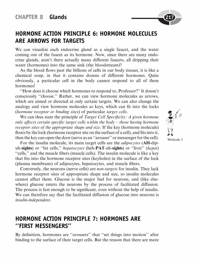
HORMONE ACTION PRINCIPLE 6: HORMONE MOLECULESARE ARROWS FOR TARGETS
We can visualize each endocrine gland as a single faucet, and the watercoming out of the faucet as its hormone. Now, since there are many endo-crine glands, aren’t there actually many different faucets, all dripping theirwater (hormones) into the same sink (the bloodstream)?
As the blood flows past the billions of cells in our body tissues, it is like achemical soup, in that it contains dozens of different hormones. Quiteobviously, a particular cell in the body cannot respond to all of thesehormones!
‘‘How does it choose which hormones to respond to, Professor?’’ It doesn’tconsciously ‘‘choose.’’ Rather, we can view hormone molecules as arrows,which are aimed or directed at only certain targets. We can also change theanalogy and view hormone molecules as keys, which can fit into the locks(hormone receptor or binding sites) of particular target cells.
We can thus state the principle of Target Cell Specificity: A given hormoneonly affects certain specific target cells within the body – those having hormonereceptor sites of the appropriate shape and size. If the key (hormone molecule)floats by the lock (hormone receptor site on the surface of a cell), and fits into it,then the key can open the door (serve as an ‘‘arouser’’ ormessenger for the cell).
For the insulin molecule, its main target cells are the adipocytes (AH-dip-uh-sights) or ‘‘fat cells,’’ hepatocytes (heh-PAT-uh-sights) or ‘‘liver’’ (hepat)‘‘cells,’’ and the muscle fibers (muscle cells). The insulin molecule is like a keythat fits into the hormone receptor sites (keyholes) in the surface of the lock(plasma membrane) of adipocytes, hepatocytes, and muscle fibers.
Conversely, the neurons (nerve cells) are non-targets for insulin. They lackhormone receptor sites of appropriate shape and size, so insulin moleculescannot affect them. Glucose is the major fuel for neurons, and (like else-where) glucose enters the neurons by the process of facilitated diffusion.The process is fast enough to be significant, even without the help of insulin.We can therefore say that the facilitated diffusion of glucose into neurons isinsulin-independent.
HORMONE ACTION PRINCIPLE 7: HORMONES ARE‘‘FIRST MESSENGERS’’
By definition, hormones are ‘‘arousers’’ that ‘‘set things into motion’’ afterbinding to the surface of their target cells. But the reason that there are more
CHAPTER 8 Glands 217
Molecule 2
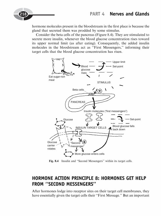
hormone molecules present in the bloodstream in the first place is because thegland that secreted them was prodded by some stimulus.
Consider the beta cells of the pancreas (Figure 8.4). They are stimulated tosecrete more insulin, whenever the blood glucose concentration rises towardits upper normal limit (as after eating). Consequently, the added insulinmolecules in the bloodstream act as ‘‘First Messengers,’’ informing theirtarget cells that the blood glucose concentration has risen.
HORMONE ACTION PRINCIPLE 8: HORMONES GET HELPFROM ‘‘SECOND MESSENGERS’’
After hormones lodge into receptor sites on their target cell membranes, theyhave essentially given the target cells their ‘‘First Message.’’ But an important
PART 4 Nerves and Glands218
Fig. 8.4 Insulin and ‘‘Second Messengers’’ within its target cells.
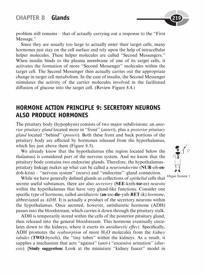
problem still remains – that of actually carrying out a response to the ‘‘FirstMessage.’’
Since they are usually too large to actually enter their target cells, manyhormones just stay on the cell surface and rely upon the help of intracellularhelper molecules. These helper molecules are called ‘‘Second Messengers.’’When insulin binds to the plasma membrane of one of its target cells, itactivates the formation of more ‘‘Second Messenger’’ molecules within thetarget cell. The Second Messenger then actually carries out the appropriatechange in target cell metabolism. In the case of insulin, the Second Messengerstimulates the activity of the carrier molecules involved in the facilitateddiffusion of glucose into the target cell. (Review Figure 8.4.)
HORMONE ACTION PRINCIPLE 9: SECRETORY NEURONSALSO PRODUCE HORMONES
The pituitary body (hypophysis) consists of two major subdivisions: an ante-rior pituitary gland located more in ‘‘front’’ (anteri), plus a posterior pituitarygland located ‘‘behind’’ (posteri). Both these front and back portions of thepituitary body are affected by hormones released from the hypothalamus,which lies just above them (Figure 8.5).
We already know that the hypothalamus (the region located below thethalamus) is considered part of the nervous system. And we know that thepituitary body contains two endocrine glands. Therefore, the hypothalamus–pituitary linkage makes up what can be called a neuroendocrine (NUR-oh-en-doh-krin) – ‘‘nervous system’’ (neuro) and ‘‘endocrine’’–gland connection.
While we have generally defined glands as collections of epithelial cells thatsecrete useful substances, there are also secretory (SEE-kreh-tor-ee) neuronswithin the hypothalamus that have very gland-like functions. Consider onespecific type of hormone, called antidiuretic (an-tee-die-yuh-RET-ik) hormone,abbreviated as ADH. It is actually a product of the secretory neurons withinthe hypothalamus. Once secreted, however, antidiuretic hormone (ADH)passes into the bloodstream, which carries it down through the pituitary stalk.
ADH is temporarily stored within the cells of the posterior pituitary gland,then released into the general bloodstream. This hormone eventually circu-lates down to the kidneys, where it exerts its antidiuretic effect. Specifically,ADH promotes the reabsorption of more H2O molecules from the kidneytubules (TWO-byools), the ‘‘tiny tubes’’ within the kidneys. As a result, itsupplies a mechanism that acts ‘‘against’’ (anti-) ‘‘excessive urination’’ (diur-esis). [Study suggestion: Look at the miniature ‘‘kidney faucet’’ model in
CHAPTER 8 Glands 219
Organ System 1
Figure 8.5. How does the literal translation of the word antidiuretic helpexplain the turning off of the faucet from full blast to a slow drip?]
HORMONE ACTION PRINCIPLE 10: RELEASINGHORMONES FROM THE HYPOTHALAMUS STIMULATE THEANTERIOR PITUITARY TO SECRETE TROPHIC HORMONES
There is another neuroendocrine relationship between the secretory neuronsin the hypothalamus and the anterior (rather than posterior) portion of the
PART 4 Nerves and Glands220
Fig. 8.5 The pituitary stalk: making a neuroendocrine connection.
pituitary body. These secretory neurons produce releasing hormones (RHs).These releasing hormones (like ADH) are secreted into the network of tinyblood vessels coursing down through the pituitary stalk. But they go downinto the anterior pituitary gland (rather than the posterior pituitary gland).The releasing hormones get their name from their primary function. Theyliterally stimulate the release of a variety of trophic (TROHF-ik) hormonesfrom the epithelial cells of the anterior pituitary gland.
HORMONE ACTION PRINCIPLE 11: TROPHIC HORMONESFROM THE ANTERIOR PITUITARY ‘‘NOURISH’’(STIMULATE) OTHER ENDOCRINE GLANDS TO SECRETE
‘‘What are these trophic hormones you are talking about, Professor Joe?What does the word trophic mean in the first place?’’
Trophic literally ‘‘pertains to’’ (-ic) ‘‘nourishment’’ (troph). But by ‘‘nour-ishment,’’ we certainly don’t mean that we are going to start handing outcandy bars! A trophic effect is ‘‘nourishing’’ only in the sense of ‘‘stimula-tion.’’
As far as glands are concerned, the trophic hormones secreted by the ante-rior pituitary cells are ‘‘nourishing or stimulating’’ for other endocrine glands.The trophic hormones are unique in that their main targets are other endo-crine glands, which are ‘‘nourished or stimulated’’ to increase the secretion oftheir own individual hormones. (Observe the little target in Figure 8.5.)
Some specific endocrine gland targets and theirhormones
Figure 8.6 shows pictures of the major endocrine gland targets being stimu-lated by various trophic hormones from the anterior pituitary. Take, forinstance, the thyroid (THIGH-royd) gland in the front of the neck. Thethyroid gland is named for its ‘‘resemblance’’ (-oid) to a broad ‘‘shield’’(thyr). The thyroid is stimulated or ‘‘nourished’’ by thyroid-stimulating hor-mone, abbreviated as TSH. The thyroid gland is stimulated by TSH toincrease the rate of secretion of its own individual hormone, thyroxine(thigh-ROCKS-in). Thyroxine, in turn, circulates throughout the blood-stream to affect most of the body cells. Thyroxine, for example, increasesthe basal (BAY-sal) metabolic rate or BMR, that is, the rate at whichbody cells burn calories during their metabolism under resting or ‘‘basal’’
CHAPTER 8 Glands 221
Molecule 3
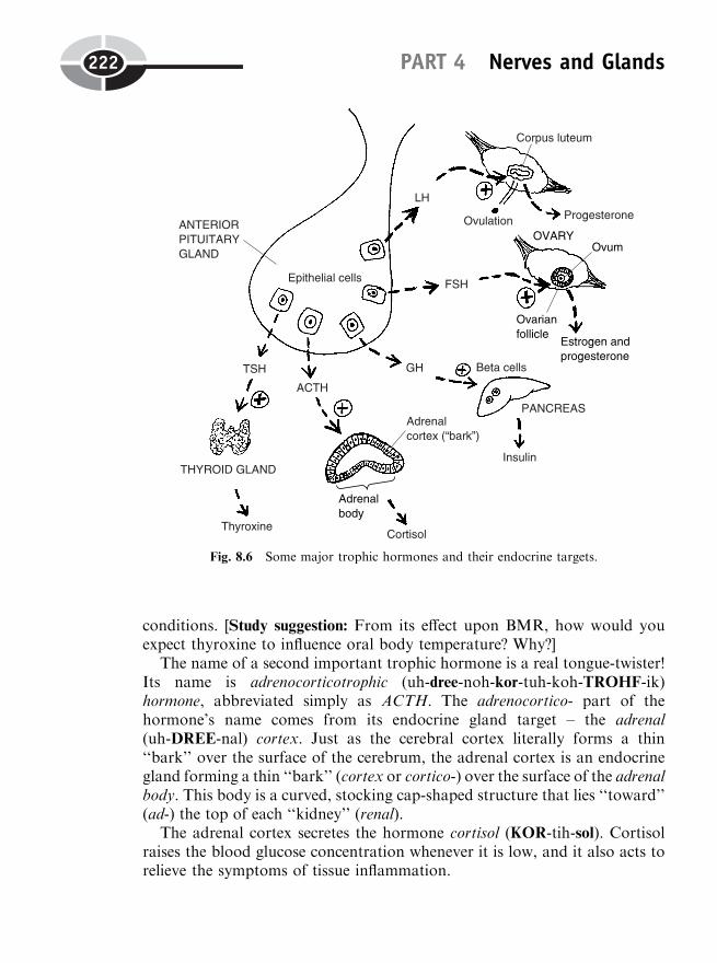
conditions. [Study suggestion: From its effect upon BMR, how would youexpect thyroxine to influence oral body temperature? Why?]
The name of a second important trophic hormone is a real tongue-twister!Its name is adrenocorticotrophic (uh-dree-noh-kor-tuh-koh-TROHF-ik)hormone, abbreviated simply as ACTH. The adrenocortico- part of thehormone’s name comes from its endocrine gland target – the adrenal(uh-DREE-nal) cortex. Just as the cerebral cortex literally forms a thin‘‘bark’’ over the surface of the cerebrum, the adrenal cortex is an endocrinegland forming a thin ‘‘bark’’ (cortex or cortico-) over the surface of the adrenalbody. This body is a curved, stocking cap-shaped structure that lies ‘‘toward’’(ad-) the top of each ‘‘kidney’’ (renal).
The adrenal cortex secretes the hormone cortisol (KOR-tih-sol). Cortisolraises the blood glucose concentration whenever it is low, and it also acts torelieve the symptoms of tissue inflammation.
PART 4 Nerves and Glands222
Fig. 8.6 Some major trophic hormones and their endocrine targets.
A third trophic hormone is called growth hormone (GH). Growth hor-mone, as its name states, circulates to most of the body cells and stimulatestheir growth by promoting such processes as protein synthesis and cell divi-sion. But the specific target gland of its trophic influence are the beta cells ofthe pancreas. The beta cells, you may recall, secrete insulin. Insulin is abso-lutely critical for human survival, because it helps transport glucose out ofthe bloodstream and into the tissue cells, thereby providing them with theirmajor energy source.
A fourth trophic hormone is follicle-stimulating hormone (FSH). A folliclein general is any ‘‘little bag.’’ Consider the case of the female ovaries (OH-var-ees), two white, ‘‘egg’’ (ovari)-shaped endocrine glands attached to bothsides of the uterus (YOU-ter-us). Each ovary contains dozens of ovarian (oh-VAIR-ee-an) follicles – tiny bags or sacs that surround the developing ovum(OH-vum) or ‘‘egg’’ (ov) cell.
Under the stimulating effect of follicle-stimulating hormone (FSH), theovarian follicles increase their secretion of the two hormones, estrogen(ES-troh-jen) and progesterone (proh-JES-ter-own). Estrogen stimulatesthe development of secondary sex characteristics in the female, such as ahigher voice and softer skin. Progesterone prepares the female body fora possible pregnancy. As a part of this reproductive role, progesteronestimulates the lining of the uterus (womb) for potential imbedding ofa fertilized ovum.
A fifth trophic hormone, luteinizing (LEW-tuh-neye-zing) hormone or LH,also acts upon the female ovary. It triggers a rupture of the mature ovarianfollicle, thereby causing ovulation (ahv-you-LAY-shun) – the release of a‘‘little egg’’ (ovul) into the abdominal cavity. But luteinizing hormone actu-ally derives its name from a structure that appears after ovulation. Thisstructure is called the corpus (KOR-pus) luteum (LEW-tee-um) or ‘‘yellowbody.’’ The corpus luteum is the tiny, yellowish-colored body that theruptured ovarian follicle transforms into after the mature ovum has beenreleased from it. LH thereby indirectly has a luteinizing or ‘‘yellowing’’ effect.It creates a yellowish body (corpus luteum) that secretes lots of progesterone(and some estrogen).
HORMONE ACTION PRINCIPLE 12: THE SECRETION OFHORMONES IS UNDER NEGATIVE FEEDBACK CONTROL
The final principle of hormone action is perhaps the most important ofthem all. The endocrine glands, much like the nervous system (and their
CHAPTER 8 Glands 223
neuroendocrine connection), are very much involved in the critical bodyfunctions of communication and control of various parameters within theinternal environment.
Head Chief in the world of control and regulation, of course, is the nega-tive feedback control system. Control of the internal environment, you mayrecall, was a major focus of our efforts back in Chapter 2. For the purposesof this chapter on glands, we can state the following general rule: The secre-tion of hormones is under negative feedback control, such that high blood levelsof a given hormone (or its predecessors) serves to ‘‘turn off’’ or inhibit furthersecretion of that hormone.
This general principle will now be traced with regard to one of thespecific anatomical parameters mentioned in Chapter 2. This was bloodglucose concentration, measured in milligrams of glucose per 100 ml ofblood.
Let us follow what happens after a person eats a sugar-rich meal, andhow negative feedback is involved with regulation of both blood glucoselevel as well as the rate of insulin secretion. Figure 8.7 starts off with themajor gland stimulus: a rise in blood glucose concentration toward itsupper normal limit of 110 mg/100 ml blood. This stimulus is detected bythe beta cells of the pancreas. They respond by dramatically increasing theirrate of insulin secretion into the bloodstream. The insulin circulates to itstarget cells, where it indirectly triggers the Second Messengers to speed upthe facilitated diffusion of glucose molecules out of the bloodstream andinto the target cells.
This enhanced movement of glucose out of the bloodstream serves todecrease the blood glucose concentration back toward its set-point level ofabout 90 mg/100 ml blood. This final response of the control system has anegative feedback effect upon the initial stimulus (a rise in blood glucosetoward its upper normal limit), acting to remove or ‘‘correct’’ it. The betacells in the pancreas, having had their major stimulus removed, are nowinhibited or ‘‘turned off ’’ via negative feedback, so that they act like agland faucet that is just barely ‘‘dripping’’ insulin!
Because insulin was present at fairly high levels, dropping the blood glu-cose significantly, it removed the stimulus for the beta cells. Hence, highblood levels of insulin eventually greatly inhibit further secretion of moreinsulin. This feedback control mechanism thus helps to prevent insulin hyper-secretion (HIGH-per-see-KREE-shun) – an ‘‘excessive or above-normal’’ (hyper-) amount of insulin release by the beta cells.
PART 4 Nerves and Glands224
Molecule 4

QuizRefer to the text in this chapter if necessary. A good score is at least 8 correctanswers out of these 10 questions. The answers are listed in the back of thisbook.
CHAPTER 8 Glands 225
Fig. 8.7 Insulin and negative feedback control of blood glucose levels.

1. An ‘‘acorn’’-shaped gland attached to the base of the hypothalamusby a stalk:(a) Pineal(b) Pancreas(c) Pituitary body(d) Mammary
2. Sweat glands are of this type:(a) Exocrine(b) Internally secreting(c) Ductless(d) Mixed
3. Responsible for secreting insulin:(a) Ovaries(b) Adrenal cortex(c) Sympathetic nerves(d) Beta cells
4. Hormones are ‘‘arousers’’ in the sense that:(a) They create new body structures and functions that were not
present earlier(b) Their influence always speeds up biological processes(c) They change the rate at which existing body processes occur(d) Removing them has no disturbing influence upon homeostasis
5. A hormone–protein complex is often essential, because:(a) No hormone can do a good job by itself(b) Many hormones are otherwise not very soluble in the bloodstream(c) All hormones are proteins anyway(d) They always carry opposite poles of charge
6. Importantly involved in the ‘‘fight-or-flight’’ response:(a) Parasympathetic nerves and sweat glands(b) The pineal gland(c) Sympathetic nerves and the adrenal medulla(d) Secretion of lots of progesterone into the bloodstream
7. Today, hormone molecule X is secreted into the bloodstream. Oneyear from today, hormone molecule X will be:(a) At the very same concentration as it is today(b) Present at a much higher concentration than it is today(c) Converted into hormone Y, which is more powerful(d) Inactivated and excreted from the body
PART 4 Nerves and Glands226
8. Neurons are non-targets for insulin, because:(a) They lack insulin receptor sites of appropriate shape and size(b) Nerve cells keep moving around too fast for insulin to catch
them!(c) Enzymes keep pumping insulin out of them(d) Insulin has no targets!
9. Hormones are ‘‘First Messengers’’ in their:(a) Ability to activate ‘‘Pre-messengers’’(b) Presence in higher concentrations within the bloodstream when
their gland is being strongly stimulated(c) Tendency to clump blood cells(d) Frequence of occurrence whenever the body is injured
10. Provides strong evidence of a neuroendocrine relationship:(a) Presence of secretory neurons within the hypothalamus(b) Storage of glucose within hepatocytes(c) Association of the adrenal cortex with the adrenal medulla(d) Occurrence of anterior and posterior pituitary together in the
hypophysis
Body-Level Grids for Chapter 8Several key body facts were tagged with numbered icons in the page marginsof this chapter. Write a short summary of each of these key facts into anumbered cell or box within the appropriate Body-Level Grid that appearsbelow.
Physiology and Biological Order Fact Grids for Chapter 8:
ORGANLevel
CHAPTER 8 Glands 227
ORGAN SYSTEMLevel
Function and Biological Order Fact Grids for Chapter 8:
MOLECULELevel
PART 4 Nerves and Glands228
1 2
1
1 2
CHAPTER 8 Glands 229
3 4
230
Test: Part 4
DO NOT REFER TO THE TEXT WHEN TAKING THIS TEST. A goodscore is at least 18 (out of 25 questions) correct. Answers are in the back ofthe book. It’s best to have a friend check your score the first time, so youwon’t memorize the answers if you want to take the test again.
1. The spinal cord is considered part of the:(a) CNS(b) ANS(c) PNS(d) SNS(e) SOS!
2. Afferent nerve fibers always travel:(a) Away from the brain(b) Through the spinal cord(c) Toward the brain(d) Parallel to the spinal cord(e) In completely unpredictable patterns
Copyright © 2004 by The McGraw-Hill Companies, Inc. Click here for terms of use.

3. The lower leg kicks out when the kneecap is hit with a hammer.The carry out this action:(a) Muscular effectors(b) Hormone binding sites(c) Sensory receptors(d) Glandular afferents(e) Motor sensations
4. Efferent nerve fibers:(a) Travel from the heart, toward the spinal cord(b) Ascend and descend within the CNS(c) Carry information away from the body of the spinal cord(d) Are never involved in reflex arcs(e) Seldom supply any viscera
5. The narrow, saltwater-filled gap between the axon terminals of oneneuron and the cell body and dendrites of another neuron:(a) Synapse(b) Node of Ranvier(c) Motor end-plate(d) Axon hillock(e) Neuromuscular junction
6. The neuron whose cell membrane receives neurotransmitter moleculesreleased from the vesicles of another neuron:(a) Presynaptic(b) Beta cell(c) Acinar cell(d) Postsynaptic(e) Fascicle
7. Neuron A fires an action potential, exciting Neuron B to fire an actionpotential, which in turn excites Neuron C to fire an action potential.However, Neuron D is influenced by Neuron C, so that it does not firean action potential. Neuron C is probably influencing Neuron D inthis way:(a) Summation of twitches(b) Destruction of its neurotransmitters(c) Releasing GABA or glycine(d) Depolarizing its plasma membrane(e) Cutting away its myelin
Test: Part 4 231

8. The axon hillock’s special significance:(a) Place where synaptic vesicles are stored(b) Contains the nucleus and most other organelles(c) Usual site of contact between neurons(d) Firing zone or trigger zone for a neuron action potential(e) Place where Baby Heinie exerts his demonic influence!
9. A neuron cell membrane is described as ‘‘resting polarized’’ when:(a) It is actively conducting a nerve impulse(b) No ions are crossing through it(c) The intracellular fluid is a negatively charged pole, while the ECF
is a positively charged pole(d) The cell is totally incapable of further excitation(e) The ICF is a positive pole, while the ECF is a negative pole
10. An action potential is best visualized as:(a) A traveling wave of depolarization(b) A stationary tree that keeps growing and growing(c) Some sick giraffe without its spots!(d) A swarm of charged cations, continually flowing out of the
neuron(e) An arrow flying straight toward its target
11. Myelin has an important role to play in:(a) Pumping poisonous materials out of nerve cells(b) Insulating neuron axons from excessive loss of electrical charge(c) Promoting homeostasis of blood Caþþ ion concentration(d) Delivering packets of neurotransmitters(e) Serving an enzyme-like role in speeding up cellular reactions
12. Nodes of Ranvier:(a) Represent bead-shaped chunks of the myelin sheath(b) Permit wave propagation via local circuit currents of ion flow(c) Are closely modeled by bare copper wires carrying electrons(d) Make up most of the white matter seen in gross anatomy(e) Allow impulse movement via saltatory conduction
13. The medial plantar nerve would most likely be stimulated by:(a) A feather stroking the middle of the sole of the foot(b) Some cold fish wrapped around your lips!(c) Hot beer being poured down your gullet!(d) A sharp nail being jabbed into the calf(e) Loud sound waves from a beating drum
PART 4 Nerves and Glands232

14. A withdrawal reflex arc involving movements of the foot:(a) Involves only the ventral root of a spinal nerve(b) Waves from sensory receptors in the sole that descend to the
spinal cord(c) A curved pathway of stimulation and automatic response(d) Voluntary lifting of the foot off some harmful or annoying stimu-
lus(e) Activation of the brachial nerve, such that the impulses travel in
an arc
15. The dorsal root ganglion represents:(a) White matter of the spinal cord(b) Cell bodies of sensory neurons lying just outside the spinal cord(c) Dendrites of motor neurons traveling up through the brainstem(d) Groups of sensory neuron cell bodies in the horns of spinal gray
matter(e) Myelinated axons entering and leaving the body of the spinal cord
16. An interneuron is best described as:(a) A small, short association neuron between two larger neurons(b) One lower motor neuron that carries movement-related informa-
tion(c) Some sensory neuron whose body lies in a horn of spinal gray
matter(d) About the same thing as an axon hillock(e) Missing the usual supply of synaptic vesicles within its axon
terminals
17. The spinothalamic tract has as its main function:(a) Transmission of ions from one spinal nerve to another(b) Carrying of nerve impulses from higher levels of the cord down to
lower ones(c) Serving as an ascending pathway for many general body sensa-
tions on their way up to the cerebral cortex(d) Decussation of descending nerve fibers through the pyramids of
the medulla oblongata(e) Providing anesthesia to the brain for otherwise intolerably painful
sensory experiences!
18. The ‘‘heads’’ of the female clitoris and male penis:(a) Secreting ‘‘acorns’’ that supply vital reproductive hormones(b) Exocrine glands associated with production of germ cells(c) Abnormal, tumor-like swellings that interfere with sexual climax
Test: Part 4 233
(d) Provide special motor pathways that relay in the thalamus(e) Glans that are named for their anatomic resemblance to real
acorns
19. A common alternative or trade name for the hormone epinephrine:(a) Norepinephrine(b) ACh(c) Synapsin(d) ACTH(e) Adrenaline
20. The odd analogy made in the text to the human bloodstream as an‘‘unstoppered sink’’ was helping to illustrate the fact that:(a) Once a hormone is secreted into the bloodstream, it never leaves(b) The secretion of hormones from glands is an on-and-off event(c) Releasing hormones from the hypothalamus stimulate the hypo-
physis(d) Some body cells are non-targets for particular hormones(e) Hormone molecules in the bloodstream are being continuously
inactivated by the liver and then excreted into the urine by thekidneys
21. Antidiuretic hormone (ADH) owes its long name to its effect of:(a) Promoting secretion of Hþ ions into the urine(b) Enhancing reabsorption of glucose out of the kidney tubules(c) Protecting ‘‘Against Diarrhea’’ that is very ‘‘Heavy’’(d) Control of blood glucose concentration(e) Increasing reabsorption of H2O from the kidney tubules
22. Insulin is essential in human physiology for its job of:(a) Unloading oxygen into the red blood cells(b) Boosting the rate of facilitated diffusion of glucose out of the
bloodstream, and into adipocytes, hepatocytes, and muscle fibers(c) Serving as a ‘‘Second Messenger’’ for anterior pituitary hormones(d) Increasing blood glucose concentration, by helping or ‘‘facilitat-
ing’’ its absorption from the stomach(e) Acting synergistically with cortisol, so as to provide the body with
a high load of available glucose circulating through the blood-stream
23. The trophic hormones are all known for their effect of:(a) Stimulating the adrenal medulla to secrete testosterone(b) Promoting storage of glucose within liver cells as glycogen
PART 4 Nerves and Glands234
(c) ‘‘Nourishing’’ or stimulating other endocrine glands(d) Inhibiting or delaying the onset of the ‘‘fight-or-flight’’ response(e) Relieving symptoms of starvation and dehydration
24. ‘‘Second Messengers’’ are often necessary, because:(a) Many hormone molecules are too large to enter their target cells(b) There is no real ‘‘First Messenger’’ available(c) Sometimes a glandular message is only imaginary, not real(d) Certain glands are starved for their normal supply of oxygen(e) The hypothalamus of the brain does not work without them
25. Releasing hormones are all characterized by their:(a) Common secretion from the epithelial cells of the anterior pitui-
tary(b) Temporary storage, then ‘‘release’’ by the posterior pituitary(c) Tendency to split into several smaller hormones of similar func-
tion(d) Ability to stimulate the anterior pituitary to release trophic hor-
mones(e) Property of exerting negative feedback control upon the rate of
secretion of all hormones within the human organism
Test: Part 4 235

This page intentionally left blank
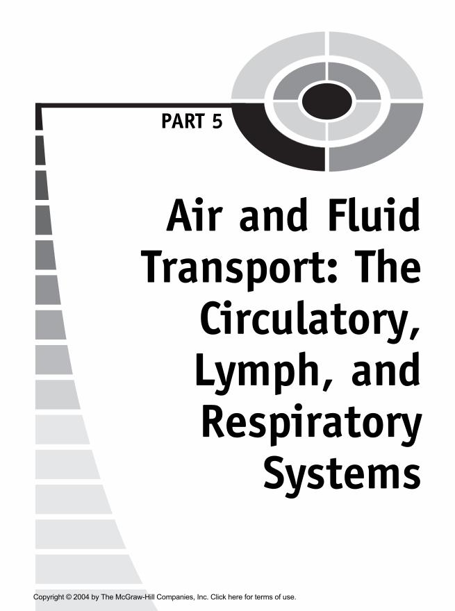
PART 5
Air and FluidTransport: The
Circulatory,Lymph, andRespiratory
Systems
Copyright © 2004 by The McGraw-Hill Companies, Inc. Click here for terms of use.

This page intentionally left blank

239
CHAPTER9
Physiology of theCirculatory System:Mover of the Bloodin a ‘‘Little Circle’’
Back in Chapter 8, we talked of the importance of the blood circulation indistributing the hormones secreted by the various endocrine glands. Now itis time for us to focus upon the circulation and pumping of the blooditself.
Copyright © 2004 by The McGraw-Hill Companies, Inc. Click here for terms of use.
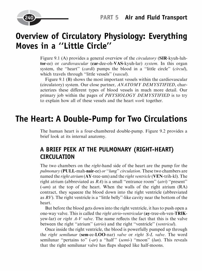
Overview of Circulatory Physiology: EverythingMoves in a ‘‘Little Circle’’
Figure 9.1 (A) provides a general overview of the circulatory (SIR-kyuh-luh-tor-ee) or cardiovascular (car-dee-oh-VAS-kyuh-lar) system. In this organsystem, the ‘‘heart’’ (cardi) pumps the blood in a ‘‘little circle’’ (circul),which travels through ‘‘little vessels’’ (vascul).
Figure 9.1 (B) shows the most important vessels within the cardiovascular(circulatory) system. Our close partner, ANATOMY DEMYSTIFIED, char-acterizes these different types of blood vessels in much more detail. Ourprimary job within the pages of PHYSIOLOGY DEMYSTIFIED is to tryto explain how all of these vessels and the heart work together.
The Heart: A Double-Pump for Two CirculationsThe human heart is a four-chambered double-pump. Figure 9.2 provides abrief look at its internal anatomy.
A BRIEF PEEK AT THE PULMONARY (RIGHT-HEART)CIRCULATION
The two chambers on the right-hand side of the heart are the pump for thepulmonary (PULL-mah-nair-ee) or ‘‘lung’’ circulation. These two chambers arenamed the right atrium (AY-tree-um) and the right ventricle (VEN-trih-kl). Theright atrium (abbreviated as RA) is a small ‘‘entrance room’’ (atri) ‘‘present’’(-um) at the top of the heart. When the walls of the right atrium (RA)contract, they squeeze the blood down into the right ventricle (abbreviatedas RV). The right ventricle is a ‘‘little belly’’-like cavity near the bottom of theheart.
But before the blood gets down into the right ventricle, it has to push open aone-way valve. This is called the right atrio-ventricular (ay-tree-oh-ven-TRIK-yew-lar) or right A-V valve. The name reflects the fact that this is the valvebetween the right ‘‘atrium’’ (atrio) and the right ‘‘ventricle’’ (ventricul).
Once inside the right ventricle, the blood is powerfully pumped up throughthe right semilunar (sem-ee-LOO-nar) valve or right S-L valve. The wordsemilunar ‘‘pertains to’’ (-ar) a ‘‘half ’’ (semi-) ‘‘moon’’ (lun). This revealsthat the right semilunar valve has flaps shaped like half-moons.
PART 5 Air and Fluid Transport240
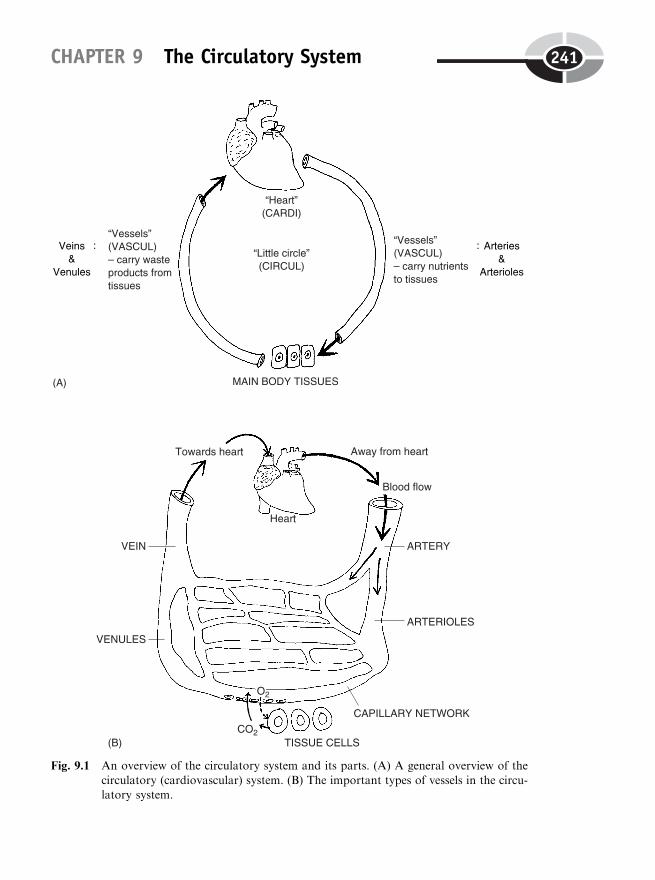
CHAPTER 9 The Circulatory System 241
Fig. 9.1 An overview of the circulatory system and its parts. (A) A general overview of thecirculatory (cardiovascular) system. (B) The important types of vessels in the circu-latory system.

The right semilunar valve is located at the base of the common pulmonaryartery, abbreviated as CPA. This artery is called a ‘‘common’’ one, because itserves as the trunk for the two smaller right and left pulmonary arteries, whichcarry the blood up into both lungs.
In summary, we have:
OUTPUT OF PULMONARY(RIGHT HEART)CIRCULATION:
RA!RIGHT A-V VALVE!RV!RIGHT S-L VALVE!CPA!
R&L PULMONARY ARTERIES!R&L ‘‘LUNGS’’ (PULMON)
PART 5 Air and Fluid Transport242
Fig. 9.2 Some internal structures and functions of the heart.
Organ System 1

A BRIEF PEEK AT THE SYSTEMIC (LEFT-HEART)CIRCULATION
The two chambers on the left-hand side of the heart are the pump for thesystemic (sis-TEM-ik) or ‘‘all organ systems’’ (except for the lungs) circula-tion. (Now, look at the left side of the heart diagram pictured in Figure 9.2.)These two chambers are named the left atrium (abbreviated as LA) and theleft ventricle (abbreviated as LV ). When the walls of the left atrium (LA)contract, they push the blood down through the left atrio-ventricular or leftA-V valve. Similar to what we saw for the right A-V valve, the left A-V valveis named for its position between the left atrium above it, and the left ventriclebelow it.
Once inside the left ventricle, the blood is powerfully pumped up throughthe left semilunar or left S-L valve. Again mirroring the situation on the rightside of the heart, the flaps of the left semilunar valves also resemble half-moons.
After the blood passes through the open flaps of the left S-L valve, it entersthe aortic (ay-OR-tik) arch. ‘‘Oh, is this like the Golden Arches, Professor?You know . . . the place where they sell all those yummy fries and hambur-gers?’’ Well, Baby Heinie, in a sense you are correct! In this case, the aorticarch might be called the Red Arch, since it is filled with red, oxygen-richblood.
‘‘Where is all this bright red blood going, Professor?’’ Now, Baby Heinie,since it is a Red Arch, you ought-er know that the aortic arch is the first partof the aorta (ay-OR-tah)! And the blood in the aorta (and its aortic arch) isliterally being ‘‘raised or lifted up’’ (aort) out of the left side of the heart.
The aorta, and its first curved part, the aortic arch, is the Great MotherArtery for the systemic circulation. There are dozens of smaller systemicarteries (such as the brachial artery and femoral artery) that supply bloodto the organs of all the major body systems (except for the lungs). Thissystemic output includes, for example, blood flowing to our skeletal mus-cles, our bones, our skin, our brains, and our many other viscera (internalorgans).
In summary, we have:
OUTPUT OFSYSTEMIC
(LEFT HEART)CIRCULATION:
LA!LEFT A-V VALVE!LV!LEFT S-L VALVE!
AORTIC ARCH (AORTA)!THE SYSTEMIC ARTERIES!
ORGANS OF ALL BODY ‘‘SYSTEMS’’(SYSTEM) EXCEPT FOR LUNGS
CHAPTER 9 The Circulatory System 243
Organ System 2
Cardiac Pacemaker Tissue: ‘‘Spark Plugs’’ inthe Heart Wall
Earlier chapters talked about the way in which skeletal muscle was stimulatedto contract by means of motor neurons. Specifically, motor neurons havetheir action potentials (traveling waves of electrochemical or ionic excitation)first. After they release excitatory neurotransmitters from their axon term-inals, the neurons stimulate the skeletal muscle fibers to fire their own actionpotentials, second. It is only after being stimulated by motor neuron ‘‘sparkplugs’’, then, that skeletal muscle fibers are excited to contract.
‘‘ONE FOR ALL, AND ALL FOR ONE’’: THE MYOCARDIUMAS THE THREE MUSKETEERS
Now, cardiac muscle fibers, like their other striated (cross-striped) relatives,the skeletal muscle fibers, have to be stimulated or excited before theycontract. In the case of the cardiac muscle tissue, however, the source ofexcitation lies within the muscle tissue itself.
This very special source of excitation is called the cardiac pacemaker tissue.It is also called nodal (NOHD-al) tissue, because it looks like a little oval‘‘knot’’ (node). The most important portion of the cardiac pacemaker (nodal)tissue is called the sinoatrial (sigh-no-AY-tree-al) or S-A node. The wordsinoatrial indicates the anatomic location of this fairly oval, ‘‘knot’’-shaped,area. (Take a look back at Figure 9.2.)
The S-A node lies in the outer wall of the right ‘‘atrium’’ (atrial, A). It isthe area just below the entrance of the superior vena (VEE-nah) cava (KAY-vah) – a major venous (VEE-nus) sinus (sino-, S). Hence we have the phrasesinoatrial (S-A). (A sinus, in general, is a large vein that is shaped like a‘‘hollow bay’’ [sin], and holding a considerable volume of blood.)
The sinoatrial node is known as the primary cardiac pacemaker. The car-diac muscle fibers within this node can, somewhat like a spark plug, sponta-neously depolarize or excite themselves. Thus the atria (upper entrancerooms) on either side of the heart are the first chambers to contract, becausethey are excited by the cardiac pacemaker cells in the S-A node. The cellmembranes of these very special muscle fibers are able to become sponta-neously leaky to sodium (Naþ) and other cations, at a certain rate or rhythm.This happens because the proteins in the membranes of the pacemaker cellstend to shift around, allowing positively charged ions to diffuse inward.
PART 5 Air and Fluid Transport244
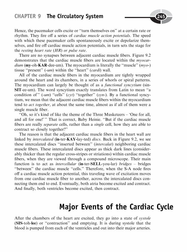
Hence, the pacemaker cells excite or ‘‘turn themselves on’’ at a certain rate orrhythm. They fire off a series of cardiac muscle action potentials. The speedwith which these pacemaker cells spontaneously excite or depolarize them-selves, and fire off cardiac muscle action potentials, in turn sets the stage forthe resting heart rate (HR) or pulse rate.
There are no synapses between adjacent cardiac muscle fibers. Figure 9.2demonstrates that the cardiac muscle fibers are located within the myocar-dium (my-oh-KAR-dee-um). The myocardium is literally the ‘‘muscle’’ (myo-)tissue ‘‘present’’ (-um) within the ‘‘heart’’ (cardi) wall.
All of the cardiac muscle fibers in the myocardium are tightly wrappedaround the heart and its chambers, in a series of whorls or spiral patterns.The myocardium can largely be thought of as a functional syncytium (sin-SIT-ee-um). The word syncytium exactly translates from Latin to mean ‘‘acondition of ’’ (-um) ‘‘cells’’ (cyt) ‘‘together’’ (syn-). By a functional syncy-tium, we mean that the adjacent cardiac muscle fibers within the myocardiumtend to act together, at about the same time, almost as if all of them were asingle muscle fiber.
‘‘Oh, so it’s kind of like the theme of the Three Musketeers – ‘One for all,and all for one!’ ’’ That is correct, Baby Heinie. ‘‘But if the cardiac musclefibers are really separate cells, rather than a single cell, how they are able tocontract so closely together?’’
The reason is that the adjacent cardiac muscle fibers in the heart wall arelinked by intercalated (in-ter-KAY-lay-ted) discs. Back in Figure 9.2, we seethese intercalated discs ‘‘inserted between’’ (intercalat) neighboring cardiacmuscle fibers. These intercalated discs appear as thick dark lines (consider-ably thicker than the regular cross-stripes or striations) within cardiac musclefibers, when they are viewed through a compound microscope. Their mainfunction is to act as intercellular (in-ter-SELL-you-lar) bridges – bridges‘‘between’’ the cardiac muscle ‘‘cells.’’ Therefore, when the S-A node firesoff a cardiac muscle action potential, this traveling wave of excitation movesfrom one cardiac muscle fiber to another, across the intercalated discs con-necting them end to end. Eventually, both atria become excited and contract.And finally, both ventricles become excited, then contract.
Major Events of the Cardiac CycleAfter the chambers of the heart are excited, they go into a state of systole(SIS-toh-lee) or ‘‘contraction’’ and emptying. It is during systole that theblood is pumped from each of the ventricles and out into their major arteries.
CHAPTER 9 The Circulatory System 245
After systole comes diastole (die-AH-stoh-lee). Whereas systole is the con-tracting and emptying phase of each chamber of the heart, diastole is the‘‘relaxation’’ and filling phase of each chamber.
AN OUTLINE OF THE CARDIAC CYCLE
The Cardiac Cycle is one heartbeat, or one complete sequence of contraction(systole) plus relaxation (diastole) of all four chambers of the heart. It is con-venient for us to construct two circles to represent the CardiacCycle – a smallerone tucked inside a larger one (Figure 9.3). The smaller, inner circle representsthe activity of both the right and left atria (upper heart chambers) during atypical Cardiac Cycle. And the larger, outer circle models the activity of theright and left ventricles (lower heart chambers) during a typical Cardiac Cycle.
You can measure the number of Cardiac Cycles (heartbeats) you are hav-ing in your own body at the present time, by palpating (touching with your
PART 5 Air and Fluid Transport246
Fig. 9.3 A model of the cardiac cycle.
fingertips) the radial artery in the wrist. [Study suggestion: Using severalfingers of one hand, gently palpate for the radial artery in the wrist on theother side of your body. It is located just proximal to the base of the thumb,within a shallow, lengthwise indentation. You should be able to feel a steadythumping of the outer wall of the radial artery against your fingertips. Canyou find it? Each thump you feel is a surge of blood passing through theradial artery during a single Cardiac Cycle. Now, count the number of‘‘thumps’’ you feel against your fingertips, for a full 60 seconds. The resultis a very important body parameter – heart rate (HR) or pulse rate (PR)measured in number of beats or Cardiac Cycles/minute. Is this an anatomicalparameter or a physiological one? Why?]
Suppose that you counted thumps and found you had a resting HR of 72beats or Cardiac Cycles/minute. This is about what the average adult has fora resting heart rate. Dividing 60 by 72, we calculate that each Cardiac Cyclelasts about 0.8 seconds. Applying this to Figure 9.3, we will cut our twocircles into halves, into quarters, and finally into eighths. Each of the eightslices of our two Cardiac Cycle ‘‘pies’’ (big ventricular circle, little atrialcircle), then, lasts for 0.1 second. Let us dig into Figure 9.3, and see whathappens inside the heart, now, during each of these 8 ‘‘pie slices’’!
During the first 0.1 second, both atria go into systole. Note that during thissame period, both ventricles are in diastole. Both A-V valves are open.Therefore, blood is pumped from the atria, down through the A-V valves,and into both resting ventricles.
A LONG ‘‘LUBB’’: THE FIRST HEART SOUND MEANSBOTH A-V VALVES ARE CLOSING
A stethoscope (STETH-oh-skohp) is ‘‘an instrument used to examine’’(-scope) the ‘‘chest’’ (steth) for various sounds. When a stethoscope bell isplaced against the chest, a health professional may perform auscultation (aws-kul-TAY-shun) – the ‘‘process of listening’’ (auscult).
After the first 0.1 second, auscultation allows us to hear the first heartsound, ‘‘lubb.’’ The ‘‘lubb’’ sound is a rather dull, prolonged vibration (sothe ‘‘lubb’’ has two b’s in it, rather than only one). ‘‘Lubb’’ is created bythe closure of both A-V valves at the beginning of ventricular (ven-TRIK-you-lar) systole. When the ventricles contract, they push blood up beneaththe flaps of the A-V valves, snapping them shut from below. The benefit, ofcourse, is that there is no leakage or regurgitation (re-gur-jih-TAY-shun) ofblood from the ventricles back up into the atria.
CHAPTER 9 The Circulatory System 247
Instead, as the ventricles powerfully contract, they push both semilunar(S-L) valves open. Blood is thus pushed up from the right ventricle, throughthe right S-L valve, and into the common pulmonary artery. On the otherside of the heart, blood is pushed up from the left ventricle, through the leftS-L valve, and into the aortic arch. This period of ventricular systole lasts fora total of 0.3 seconds. During this time, blood is being pushed up out of theventricles and into the other vessels and organs supplied by the pulmonaryand systemic circulations.
A SHORT ‘‘DUP’’: THE SECOND HEART SOUND MEANSBOTH S-L VALVES ARE CLOSING
Ventricular systole provides a vital, life-giving stream of blood to the body’sorgan systems. But, as the old saying goes, ‘‘All good things must come to anend.’’ Specifically, the end of ventricular systole comes at 0.4 seconds.
At this time, the second heart sound can be heard using auscultation. It is ashort, sharp ‘‘dup’’ sound. (Since ‘‘dup’’ is short, it ends with only one p.) Thesecond heart sound (‘‘dup’’) reflects the closing of both semilunar valves at thebeginning of ventricular diastole. The blood in the common pulmonary artery/aortic arch above each ventricle starts to fall back downward, due to the forceof gravity. As the blood falls down, it catches the edges of the semilunar valveflaps and slams them shut, thereby preventing a back-leak or regurgitation ofblood into the ventricles. [Study suggestion:Glance back at Figure 9.2 to helpyou visualize what is happening during the two heart sounds.]
In summary of the two heart sounds:
THE FIRST HEARTSOUND (‘‘lubb’’)
THE SECOND HEARTSOUND (‘‘dup’’)
¼
¼
Sound made by closure of both A-V valvesat the beginning of ventricular systole
Sound made by closure of both S-L valvesat the beginning of ventricular diastole
THE LAST HALF OF THE CARDIAC CYCLE: EVERYONE’STAKING A WELL-DESERVED NAP!
Please observe that, after the first 0.4 seconds, all four chambers of the heartare in a state of diastole. Neither the ventricles nor the atria are contracting.Therefore, for fully the last half of each Cardiac Cycle, all chambers of theheart are resting!
PART 5 Air and Fluid Transport248
Organ 1
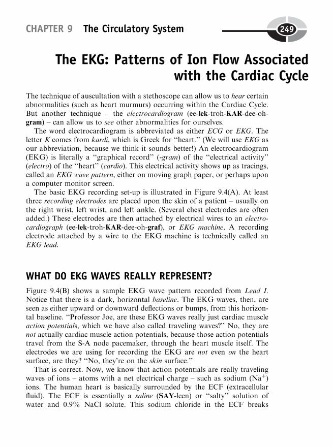
The EKG: Patterns of Ion Flow Associatedwith the Cardiac Cycle
The technique of auscultation with a stethoscope can allow us to hear certainabnormalities (such as heart murmurs) occurring within the Cardiac Cycle.But another technique – the electrocardiogram (ee-lek-troh-KAR-dee-oh-gram) – can allow us to see other abnormalities for ourselves.
The word electrocardiogram is abbreviated as either ECG or EKG. Theletter K comes from kardi, which is Greek for ‘‘heart.’’ (We will use EKG asour abbreviation, because we think it sounds better!) An electrocardiogram(EKG) is literally a ‘‘graphical record’’ (-gram) of the ‘‘electrical activity’’(electro) of the ‘‘heart’’ (cardio). This electrical activity shows up as tracings,called an EKG wave pattern, either on moving graph paper, or perhaps upona computer monitor screen.
The basic EKG recording set-up is illustrated in Figure 9.4(A). At leastthree recording electrodes are placed upon the skin of a patient – usually onthe right wrist, left wrist, and left ankle. (Several chest electrodes are oftenadded.) These electrodes are then attached by electrical wires to an electro-cardiograph (ee-lek-troh-KAR-dee-oh-graf), or EKG machine. A recordingelectrode attached by a wire to the EKG machine is technically called anEKG lead.
WHAT DO EKG WAVES REALLY REPRESENT?
Figure 9.4(B) shows a sample EKG wave pattern recorded from Lead I.Notice that there is a dark, horizontal baseline. The EKG waves, then, areseen as either upward or downward deflections or bumps, from this horizon-tal baseline. ‘‘Professor Joe, are these EKG waves really just cardiac muscleaction potentials, which we have also called traveling waves?’’ No, they arenot actually cardiac muscle action potentials, because those action potentialstravel from the S-A node pacemaker, through the heart muscle itself. Theelectrodes we are using for recording the EKG are not even on the heartsurface, are they? ‘‘No, they’re on the skin surface.’’
That is correct. Now, we know that action potentials are really travelingwaves of ions – atoms with a net electrical charge – such as sodium (Naþ)ions. The human heart is basically surrounded by the ECF (extracellularfluid). The ECF is essentially a saline (SAY-leen) or ‘‘salty’’ solution ofwater and 0.9% NaCl solute. This sodium chloride in the ECF breaks
CHAPTER 9 The Circulatory System 249
PART 5 Air and Fluid Transport250
left
Fig. 9.4 An EKG recording set-up and associated wave patterns. (A) The basic EKGstandard limb lead arrangement that picks up ion flow from the heart. (B) A normalEKG wave pattern associated with a single cardiac cycle (heartbeat). (C) A highly
disordered EKG pattern reflecting the electrical chaos of ventricular fibrillation.
down into millions of charged Naþ and Cl� ions. Together with variousother cations and anions dissolved in water, they compose the ‘‘internalsea’’ or internal environment, which lies deep to the surface of the skin.
Whenever the S-A node becomes depolarized (electrochemically or ioni-cally ‘‘excited’’), it fires off a series of cardiac muscle action potentials. Theseaction potentials quickly move throughout the heart (a functional syncy-tium), because the intercalated discs allow the action potentials to travelfrom one cardiac muscle fiber to another. After they are excited or depolar-ized by the action potential, the cardiac muscle fibers contract. This contract-ing movement makes the heart chambers pump the blood.
While all this conducting of cardiac muscle action potentials is going onwithin the heart wall, a large group of ions are diffusing away from the surfaceof the heart, and through the ECF. Remember (Chapter 4) the general prin-ciple that ‘‘like dissolves like.’’ Therefore, the charged ions diffusing awayfrom the surface of the heart, readily mix and pass through the charged,saline, watery ECF around the heart.
Eventually, the charged currents of ions diffusing from the heart surface,reach the skin surface. Here they are detected by the recording electrodes ofthe EKG machine.
Therefore, the EKG wave patterns actually represent the pattern of ionsflowing from the surface of the heart, and onto the surface of the skin. TheEKG waves indirectly represent the electrochemical activity of the heart,because the shape, size, and other characteristics of the waves are stronglyinfluenced by the ionic events going on within the heart. [Study suggestion:
Picture this abstract situation using a helpful model. An old-fashioned movieprojector is sitting in the back of a classroom. The movie projector is theheart. The electrochemical or ionic events occurring in the heart, then, are amovie on film. The heart (movie projector) casts a visual image through theair (ECF) and onto a screen. The screen is the skin surface. The visual imagesbeing viewed on the screen (observed wave patterns of an EKG) are not thetrue events actually occurring within the movie projector (heart). The trueevents are a film moving through a series of gears and sprockets. But thevisual pattern being projected onto the screen of the skin – the waves ofthe EKG – very much depend on what is happening, electrochemically orionically speaking, within the walls of the heart.]
A NORMAL EKG WAVE PATTERN
Because the electrodes in the EKG leads are arranged in a consistent manner,a consistent series of waves usually occurs within the EKG record (see Figure
CHAPTER 9 The Circulatory System 251
Organ 2

9.4, B.) These waves are identified in alphabetical sequence, starting with theletter P. (The selection of the letters from P onward is pretty arbitrary, anddoes not, itself, mean anything.) The first wave seen is usually the P wave.The P wave is a small upward bump that indirectly represents the depolar-ization (excitation with cardiac muscle action potentials) of both the rightand left atria. (The two atria are depolarized first, because the S-A node isfound in the right atrium. These two upper chambers are the heart partslocated closest to the S-A node pacemaker.)
Next in sequence is the Q-R-S wave complex. This combination of threewaves represents the depolarization (excitation) of both ventricles. Just as theP wave comes before the systole (contraction) of the atria, then, the Q-R-Swave complex comes before the systole of the ventricles and their pumpingout of blood into major arteries.
The next common event is the T wave. This wave represents the repolar-ization (REE-poh-lar-ih-zay-shun) of the ventricles. By this it is meant thatthe ventricles are going back to being resting ‘‘polarized,’’ once ‘‘again’’ (re-).This was the state they were in before they were depolarized – ‘‘taken awayfrom’’ (de-) a resting ‘‘polarized,’’ non-excited state, by cardiac action poten-tials from the nodal tissue.
Taken altogether, the resulting P, QRS, T combination represents thecomplete EKG wave pattern usually associated with each beat, CardiacCycle, or ‘‘lubb dup’’ of the heart.
In summary:
NORMAL EKGWAVE PATTERN(Pattern of ion flowassociated with one
beat, CardiacCycle, or ‘‘lubb
dup’’ of the heart)
¼ P WAVE(Depolarizationor excitationof both atria)
þ QRS WAVECOMPLEX
(Depolarization orexcitation of both
ventricles)
þ T WAVE(Repolarization or
‘‘return’’ ofventricles to restingpolarized state)
VENTRICULAR FIBRILLATION
During ventricular fibrillation (feye-bril-AY-shun), however, the normalEKG wave pattern disappears and is replaced by a highly disordered stateof random electrical chaos (Figure 9.4, C). The ventricles beat in a totallyuncoordinated manner, so that the heart looks like a bag of writhing worms,or a bunch of independently contracting ‘‘little fibers’’ (fibrils).
PART 5 Air and Fluid Transport252
Organ 3
Organ 1

The Work of the Heart: Stroke Volumeand Cardiac Output
Once the heart is properly excited and contracting normally, what influencedoes this have upon the rest of the body? What we are really asking here issomething about the work being done or achieved by the heart.
Three critically important physiological parameters associated with thework of the heart are the heart rate (HR), the stroke volume (SV), and thecardiac output (CO). We have already learned that the heart rate (also calledpulse rate) represents how fast the heart is contracting. It is measured as somany beats or Cardiac Cycles per minute. Recall that the average heart ratefor a resting adult is about 72 beats/minute.
STROKE VOLUME AND THE STRENGTH OFHEART CONTRACTION
The stroke volume, in contrast, indirectly indicates how strongly the heartmuscle (myocardium) is contracting. The stroke volume (SV) is the volume ofblood ejected from the ventricles with each beat or stroke of the heart. TheSV is usually measured as so many milliliters (ml) of blood per ‘‘stroke’’ orbeat of the heart. A typical value for SV in a resting adult is 70 ml/beat orstroke. [Study suggestion: Get a big red balloon to model a ventricle of theheart. Fill the balloon with water. Now, aim its stem over a sink. Using onehand, ‘‘stroke’’ or compress the balloon one time. The volume of water yousee being ejected from the balloon stem – the major connected artery –represents the stroke volume. Now, stroke the balloon (heart ventricle) onemore time, but with greater strength or force. What do you observe about thevolume of fluid (the stroke volume) being squirted out into the sink?]
In general, the stroke volume of the left ventricle is about equal to thestroke volume of the right ventricle. This means that, at rest, about 70 ml ofblood are being pushed out of the left ventricle and into the aortic arch. Andat the same time, about 70 ml of blood are also being pushed out of the rightventricle and into the common pulmonary artery. Thus, both sides of the heartare usually contracting with about the same amount of strength or force.
To help us see this, let us visualize the pulmonary circulation as the rightheart or lung pump, and the systemic circulation as the left heart or systemicpump. As shown in Figure 9.5, these two pumps are connected in series, oneright after the other.
CHAPTER 9 The Circulatory System 253
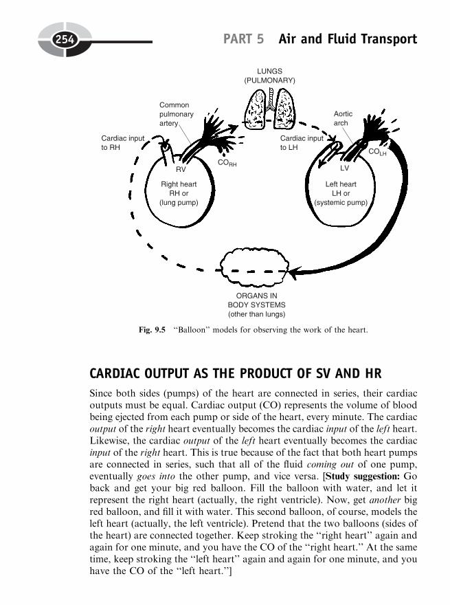
PART 5 Air and Fluid Transport254
CARDIAC OUTPUT AS THE PRODUCT OF SV AND HR
Since both sides (pumps) of the heart are connected in series, their cardiacoutputs must be equal. Cardiac output (CO) represents the volume of bloodbeing ejected from each pump or side of the heart, every minute. The cardiacoutput of the right heart eventually becomes the cardiac input of the left heart.Likewise, the cardiac output of the left heart eventually becomes the cardiacinput of the right heart. This is true because of the fact that both heart pumpsare connected in series, such that all of the fluid coming out of one pump,eventually goes into the other pump, and vice versa. [Study suggestion: Goback and get your big red balloon. Fill the balloon with water, and let itrepresent the right heart (actually, the right ventricle). Now, get another bigred balloon, and fill it with water. This second balloon, of course, models theleft heart (actually, the left ventricle). Pretend that the two balloons (sides ofthe heart) are connected together. Keep stroking the ‘‘right heart’’ again andagain for one minute, and you have the CO of the ‘‘right heart.’’ At the sametime, keep stroking the ‘‘left heart’’ again and again for one minute, and youhave the CO of the ‘‘left heart.’’]
Fig. 9.5 ‘‘Balloon’’ models for observing the work of the heart.
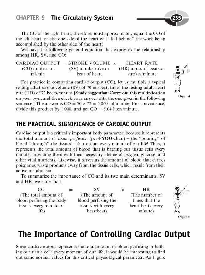
CHAPTER 9 The Circulatory System 255
Organ 4
Organ 5
The CO of the right heart, therefore, must approximately equal the CO ofthe left heart, or else one side of the heart will ‘‘fall behind’’ the work beingaccomplished by the other side of the heart!
We have the following general equation that expresses the relationshipamong HR, SV, and CO:
CARDIAC OUTPUT(CO) in liters or
ml/min
¼ STROKE VOLUME(SV) in ml/stroke or
beat of heart
� HEART RATE(HR) in no. of beats or
strokes/minute
For practice in computing cardiac output (CO), let us multiply a typicalresting adult stroke volume (SV) of 70 ml/beat, times the resting adult heartrate (HR) of 72 beats/minute. [Study suggestion: Carry out this multiplicationon your own, and then check your answer with the one given in the followingsentence.] The answer is CO ¼ 70� 72 ¼ 5,040 ml/minute. For convenience,divide this product by 1,000, and get CO ¼ 5.04 liters/minute.
THE PRACTICAL SIGNIFICANCE OF CARDIAC OUTPUT
Cardiac output is a critically important body parameter, because it representsthe total amount of tissue perfusion (per-FYOO-zhun) – the ‘‘pouring’’ ofblood ‘‘through’’ the tissues – that occurs every minute of our life! Thus, itrepresents the total amount of blood that is bathing our tissue cells everyminute, providing them with their necessary lifeline of oxygen, glucose, andother vital nutrients. Likewise, it serves as the amount of blood that carriespoisonous waste products away from the tissue cells, which result from theiractive metabolism.
To summarize the importance of CO and its two main determinants, SVand HR, we state that:
CO(The total amount of
blood perfusing the bodytissues every minute of
life)
¼ SV(The amount of
blood perfusing thetissues with every
heartbeat)
� HR(The number oftimes that the
heart beats everyminute)
The Importance of Controlling Cardiac OutputSince cardiac output represents the total amount of blood perfusing or bath-ing our tissue cells every moment of our life, it would be interesting to findout some normal values for this critical physiological parameter. As Figure
PART 5 Air and Fluid Transport256
9.6 (A) shows, the normal range for CO is from about 4 to 8 liters/minute.The average or set-point value is about 5 liters/minute. The value of 5 liters isabout the total amount of blood volume contained within the vessels of anaverage-sized adult. Therefore, at rest, just about the entire blood volume ispumped throughout the body as the cardiac output, every single minute!
If homeostasis of CO is maintained, then the result is tissue normoperfusion(nor-moh-per-FYOO-zhun) – a ‘‘normal’’ amount of blood ‘‘pouringthrough’’ or bathing our body tissues.
Under conditions of exercise or stress, both HR and SV tend to increase.Thus, CO will also tend to increase toward its upper normal limit of about 8liters/minute, because it is the product of a higher HR multiplied by a higherSV.
CARDIAC ARREST AND TISSUE ISCHEMIA(HYPOPERFUSION)
‘‘What happens if the person has a heart attack, Professor Joe?’’ Well, in acardiac arrest, the heart essentially slows down or even stops pumping andbeating. Specifically, both HR and SV greatly decline. This will cause theresulting CO to fall far below its lower normal limit of 4.0 liters/minute (seeFigure 9.6, B).
The result is tissue hypoperfusion (high-poh-per-FYOO-zhun), which ismore commonly described as tissue ischemia (is-KEE-mee-ah). Hypo-perfusion is a ‘‘below-normal or deficient’’ amount of blood ‘‘pouringthrough’’ the tissues. A synonym (word with similar meaning) is ischemia,which comes from Greek for ‘‘a holding back of blood.’’
If tissue hypoperfusion (or tissue ischemia) is very prolonged or severe,then tissue necrosis (neh-KROH-sis) or an ‘‘abnormal condition of ’’ (-osis)tissue ‘‘death’’ (necr) may result. Thus, cardiac output must be maintainedwithin its normal range, if adequate tissue perfusion and circulation of bloodthroughout all parts of the body are to be achieved.
Blood Pressure and Blood FlowWe have discussed such important physiological parameters as cardiac out-put, heart rate, and stroke volume. But at least two vital physiological para-meters associated with the cardiovascular system still need to be examined.These are blood pressure (BP) and blood flow.
CHAPTER 9 The Circulatory System 257
Fig. 9.6 Cardiac output: just enough, or too little? (A) CO in its normal range results in tissue
normoperfusion. (B) CO below its normal lower limit results in tissue hypoperfusionor ischemia.

In general, blood pressure is a pushing force exerted against the blood andagainst the walls of the blood vessels (see Figure 9.7). The BP is at its highestin the major arteries attached directly to the heart (such as the aortic arch andcommon pulmonary artery). The BP then progressively decreases withgreater distance from the heart.
Blood pressure is hugely significant because it is the main force causingblood flow. There is a blood pressure gradient (GRAY-dee-unt), a series ofdownward ‘‘steps’’ from a region of higher to a region of lower blood pres-sure. Within the systemic circulation, for example, the BP is highest within theaortic arch. As the aortic arch progressively branches into smaller and smallerarteries farther away from the heart, the BP also declines. This creates a bloodpressure gradient or series of downward steps in blood pressure in vesselsfarther from the heart. Hence, the blood is pushed down a BP gradient as itleaves the heart, so that it flows into lower-pressure arteries in a series of steps.
PART 5 Air and Fluid Transport258
Fig. 9.7 Blood pressure (BP), blood flow, and the brachial artery.
One of these farther arteries, the brachial artery (you may remember), is inthe upper ‘‘arm’’ (brachi). The brachial artery is the most frequent site usedfor taking someone’s blood pressure, utilizing a stethoscope and sphygmo-manometer (sfig-moh-muh-NAHM-uh-ter). A sphygmomanometer is literally‘‘an instrument used to measure’’ (-meter) the ‘‘throbbing pulse’’ (sphygmo)at certain ‘‘intervals’’ (mano).
To be sure, one does hear a dull throbbing sensation when the bell(expanded end) of a stethoscope is placed over the brachial artery and thearm cuff of a sphygmomanometer device is inflated around it. The sphygmo-manometer is usually marked off in units of millimeters of mercury, abbre-viated as mmHg. (Hg is the chemical symbol for the element mercury.)
As the air is slowly let out of the inflated arm cuff, the first dull throbbingnoise one hears through the stethoscope is called the systolic (sis-TAHL-ik)blood pressure, or SBP. The systolic blood pressure (SBP) is the pressurecreated by the systole (contracting and emptying phase) of the left ventricleof the heart. A slug of blood (the stroke volume) is pushed out of the leftventricle with its contraction. Flowing progressively down a BP gradient, thisslug of blood finally enters the brachial artery. The blood bulges out theartery somewhat as it passes through it, thereby creating a thumpingsound. For a resting adult, the systolic blood pressure (SBP) is usuallyrecorded as about 120 mmHg.
The diastolic (DIE-ah-stahl-ik) blood pressure, or DBP, is the blood pres-sure associated with the diastole (relaxing and filling) phase of the left ven-tricle. Upon thoughtful reflection, you might well ask, ‘‘Why isn’t thediastolic BP just 0 mmHg? Isn’t diastole the resting and filling phase, whenthe ventricle isn’t even contracting or creating any blood pressure? So, whyshould there even be any diastolic BP at all?’’
Good question! Glance back at Figure 9.7. Note that the brachial arterybulges out with the force of the systolic BP against its walls. Now, when theventricle stops contracting, and diastole begins, there is a powerful elasticrecoil, or snapping-back force, created by the stretched brachial artery wallcoming back to its non-stretched shape. This force of elastic recoil (from thepowerful snapping back of the stretched brachial artery wall) is what createsthe diastolic BP. The diastolic blood pressure is then, in a sense, a residual orleft-over blood pressure. It is the force of the blood pressure temporarily‘‘stored’’ in the bulged-out walls of the brachial artery during systole.Because it is a residual BP, the diastolic BP is normally significantly lowerthan the systolic BP. It averages about 80 mmHg in a resting adult.
The stethoscope and sphygmomanometer measure a resting adult’s bloodpressure as about 120/80 mmHg. (You can think of this as the set-point, orlong-term average value, for the blood pressure parameter.) The SBP is 120,
CHAPTER 9 The Circulatory System 259

the DBP is 80. The diastolic BP is usually recorded as the level of bloodpressure where the dull thumping sound heard through the stethoscope justdisappears. [Study suggestion: Ask yourself, ‘‘Why does the thumping sounddisappear just below the recorded level of the diastolic BP?’’ Hint: Thinkabout what is happening to the wall of the brachial artery.]
Hypertension Versus HypotensionWe have been talking about the normal blood pressure, which has an averageor set-point level of about 120/80 mmHg. The normal BP can rise to anupper normal limit of approximately 140/90 mmHg, and it can fall downto a lower normal limit of about 100/60 mmHg. The distance between theseupper and lower normal limits, of course, creates the normal range for bloodpressure. A staying of the BP within its normal range is technically callednormotension (NOR-moh-TEN-shun). We use this term because blood ‘‘pres-sure’’ represents the amount of tension exerted against the blood. Figure 9.8(A) illustrates a state of normotension, that is, a state of relative constancy orhomeostasis of blood pressure within its normal range over time.
Unfortunately, however, BP does not always remain within its normalrange. Say that a lumberjack accidentally cuts his brachial artery with achainsaw while felling a large oak tree. The blood spurts out of the arteryin hot red jets, resulting in a severe and possibly fatal hemorrhage (HEM-uh-rij) – a ‘‘bursting out’’ (-orrhage) of ‘‘blood’’ (hem). When so much bloodis lost, there isn’t much blood left to press against the arterial wall.Consequently, blood pressure steeply declines. It may even reach a state ofhypotension (HIGH-poh-TEN-shun). Hypotension is a condition of ‘‘below-normal or deficient’’ (hypo-) blood ‘‘pressure’’ (tens). Specifically, hypoten-sion is a blood pressure significantly less than 100/60 mmHg (Figure 9.8, B).
At the opposite extreme is hypertension (HIGH-per-TEN-shun) – an‘‘excessive or above normal’’ (hyper-) blood pressure. Hypertension is ablood pressure significantly above the upper normal limit of approximately140/90 mmHg (Figure 9.8, C).
‘‘So what if a person has hypotension or hypertension?’’, a skeptic mightinquire. With hypotension, the person may easily faint. And if the conditionis severe, there may be circulatory shock or coma, due to a lack of bloodpressure pushing blood up to feed the brain. In the case of hypertension, thechronically above-normal pressure may overstretch and thin out the walls ofarteries. This thinning and ballooning out creates aneurysms (AN-yuh-riz-
PART 5 Air and Fluid Transport260
Tissue 1
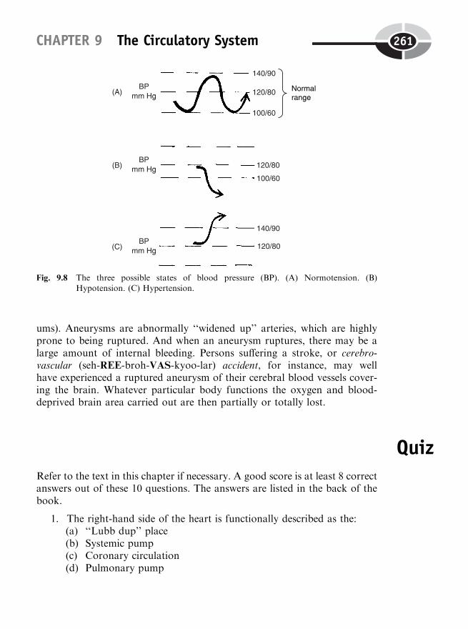
ums). Aneurysms are abnormally ‘‘widened up’’ arteries, which are highlyprone to being ruptured. And when an aneurysm ruptures, there may be alarge amount of internal bleeding. Persons suffering a stroke, or cerebro-vascular (seh-REE-broh-VAS-kyoo-lar) accident, for instance, may wellhave experienced a ruptured aneurysm of their cerebral blood vessels cover-ing the brain. Whatever particular body functions the oxygen and blood-deprived brain area carried out are then partially or totally lost.
QuizRefer to the text in this chapter if necessary. A good score is at least 8 correctanswers out of these 10 questions. The answers are listed in the back of thebook.
1. The right-hand side of the heart is functionally described as the:(a) ‘‘Lubb dup’’ place(b) Systemic pump(c) Coronary circulation(d) Pulmonary pump
CHAPTER 9 The Circulatory System 261
Fig. 9.8 The three possible states of blood pressure (BP). (A) Normotension. (B)Hypotension. (C) Hypertension.
2. When the left ventricle contracts, it pushes blood up through the:(a) Left semilunar valve(b) Left atrio-ventricular valve(c) Right auricle(d) CPA
3. The primary cardiac pacemaker in the heart wall:(a) Myelin internode(b) P wave(c) Aortic arch(d) Sinoatrial node
4. discs help make the cardiac muscle behave as a functionalsyncytium:(a) Striated(b) Smooth(c) Intercalated(d) Imaginary
5. The resting and filling phase of each chamber in the heart:(a) Systole(b) SV(c) Diastole(d) Contraction
6. Measured in number of beats/minute:(a) BP(b) HR(c) CO(d) MVP
7. The first heart sound represents the:(a) Squeezing noises of the atria(b) Ventricular repolarization(c) Closure of both A-V valves(d) Opening of both S-L valves
8. The EKG wave patterns essentially reflect:(a) Cardiac muscle action potentials occurring within the heart wall(b) Turbulent heart murmurs through damaged valves(c) Imaginary tracings having no real relationship to cardiac physiol-
ogy(d) Patterns made by ions flowing from the heart to the skin surface
PART 5 Air and Fluid Transport262
9. Indirectly indicates the strength of heart contraction:(a) P wave(b) Ventricular fibrillation(c) SV(d) Pulse rate
10. CO is calculated as:(a) TPR � SBP(b) DBP þ SV(c) HR � BP(d) SV � HR
Body-Level Grids for Chapter 9Several key body facts were tagged with numbered icons in the pagemargins ofthis chapter. Write a short summary of each of these key facts into a numberedcell or box within the appropriate Body-Level Grid that appears below.
Physiology and Biological Order Fact Grids for Chapter 9:
ORGANLevel
CHAPTER 9 The Circulatory System 263
1 2
ORGAN SYSTEMLevel
PART 5 Air and Fluid Transport264
1 2
3 4
5

Physiology and Biological Disorder Fact Grids for Chapter 9:
TISSUELevel
ORGANLevel
CHAPTER 9 The Circulatory System 265
1
1
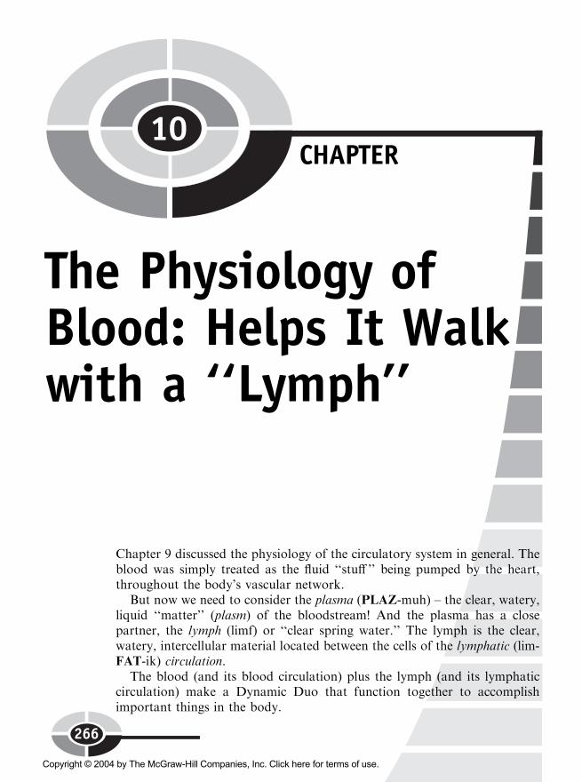
266
CHAPTER10
The Physiology ofBlood: Helps It Walkwith a ‘‘Lymph’’
Chapter 9 discussed the physiology of the circulatory system in general. Theblood was simply treated as the fluid ‘‘stuff ’’ being pumped by the heart,throughout the body’s vascular network.
But now we need to consider the plasma (PLAZ-muh) – the clear, watery,liquid ‘‘matter’’ (plasm) of the bloodstream! And the plasma has a closepartner, the lymph (limf) or ‘‘clear spring water.’’ The lymph is the clear,watery, intercellular material located between the cells of the lymphatic (lim-FAT-ik) circulation.
The blood (and its blood circulation) plus the lymph (and its lymphaticcirculation) make a Dynamic Duo that function together to accomplishimportant things in the body.
Copyright © 2004 by The McGraw-Hill Companies, Inc. Click here for terms of use.
The Blood–Lymph Physiological RelationshipTake a look at Figure 10.1. Here you see that the tiny lymphatic capillariesclosely run by or shadow the blood capillaries, and that the lymph is, in fact,a filtrate (FIL-trait), or filtration product, of the blood. This filtration ofblood occurs under the powerful pushing influence of the capillary bloodpressure. This capillary BP pushes fluid and small particles out of theblood plasma, and across the selectively permeable membranes of the thin,flat endothelial cells lining both the blood and lymphatic capillaries. Thispushed-out or filtered material from the plasma essentially becomes thelymph, which enters and flows through the lymphatic circulation.
To capsulize the blood versus lymph identities, we state two word equa-tions:
THEBLOODPLASMA
¼ THE CLEAR, FLUID, INTERCELLULAR ‘‘MATTER’’CIRCULATING THROUGH THE BLOOD VESSELS
CHAPTER 10 The Physiology of Blood 267
Fig. 10.1 The lymph: ‘‘clear water’’ (carrying dirt and contaminants) that ‘‘springs’’ from thebloodstream.
Tissue 1
Tissue 1

whileTHE
LYMPH¼ THE CLEAR, FLUID, INTERCELLULAR FILTRATE
(FILTRATION PRODUCT) OF THE BLOODPLASMA, CIRCULATING THROUGH THE
LYMPHATIC VESSELS
Eventually, the tiny lymphatic capillaries merge to become larger lympha-tic veins, and even huge lymphatic ducts. These lymphatic ducts eventuallydump their load of lymph into several large blood veins. By this means, thelymph (which was originally formed by filtration out of the blood capillaries)is returned back to the bloodstream (by being dumped into several largeblood veins) at the end of its circulation route.
Formed Elements in Blood and LymphAn important idea in the anatomy of blood connective tissue is the concept ofthe formed elements. Now, let us not confuse these with the chemical elements!An element, in general, is (according to Early Latin) some rudiment (ROO-dih-ment) – a ‘‘beginning’’ or simple stage of something.
In anatomy, the formed elements are considered to be the whole cells, andthe fragments of cells, found within the bloodstream (and to some degree,within the lymph as well). Thus, whole cells and fragments of cells are theelements or ‘‘rudiments’’ (the beginning parts) of blood connective tissue.And since each one is surrounded by a cell membrane, they also have aparticular shape or form. Hence, the formed elements are cellular elementswith a definite shape or form.
THE THREE GENERAL TYPES OF FORMED ELEMENTS
The three general types of formed elements created during hematopoiesis(Chapter 5) are the erythrocytes (red blood cells), leukocytes (white bloodcells), and platelets or thrombocytes. (Go back and review Fig 5.8,B, ifdesired).
FORMED ELEMENTS IN THE LYMPH PLAYA ROLE IN IMMUNITY
Of the three major types of formed elements in the bloodstream, only one type– the leukocyte or white blood cell – is a regular member of the lymphatic
PART 5 Air and Fluid Transport268
Cell 1

circulation as well. The leukocytes are mainly known for the importantfunctions they play in immunity (ih-MYOO-nih-tee). The word immunitytranslates to mean a ‘‘condition of ’’ (-ity) ‘‘not serving’’ (immun) disease.
There are a number of different types of leukocytes. The specific typeshown back in Figure 5.8 (B), you may recall, is called a neutrophil (NEW-troh-fil) or ‘‘lover’’ (phil) of ‘‘neutral’’ (neutro) chemical dyes. This means thatthe neutrophil is stained with neutral (neither acidic nor basic) dyes, in orderto see its major structural characteristics through a light microscope. Thenucleus of the neutrophil consists of two or three lobes connected by narrowsegments. It also has tiny granules scattered throughout its cytoplasm. Theneutrophil is the most common type of leukocyte. They usually make upmore than 50% of all white blood cells found in the bloodstream.
Of course, neutrophils and the other types of leukocytes are too large to besimply filtered across the wall of the blood capillaries. But neutrophils andother WBCs can engage in diapedesis (die-uh-ped-EE-sis). This means thatthe white blood cells literally ‘‘leap through’’ the blood capillary by crawlingin between the endothelial cells lining their walls. The crawling motion istechnically called ameboid (ah-ME-boyd) movements, since this action resem-bles the movement of real amebas.
By diapedesis and ameboid movements, then, neutrophils that were oncein the blood capillaries can cross over and enter the nearby lymphatic capil-laries. Thus, we see them as formed elements in the lymph, as well as in theblood plasma.
Neutrophils engage in phagocytosis (fag-oh-sigh-TOH-sis), a ‘‘conditionof ’’ (-osis) ‘‘cell’’ (cyt) ‘‘eating’’ (phag). In particular, neutrophils are chemi-cally attracted to sites of inflammation in the body. They leave the bloodcapillaries via ameboid movements and often enter the lymphatic circulation,which may contain invading bacteria. The neutrophils are the body’s mainbacteria-eaters, engulfing them by the thousands by the process of phago-cytosis.
The lymphocytes
After the neutrophils, the second most common type of leukocyte are thelymphocytes (LIMF-oh-sights). The lymphocytes make up about 20–45% ofthe leukocytes circulating within the bloodstream. The lymphocytes have adistinct oval or lima bean-shaped nucleus that occupies most of the cell, withonly a thin rim of cytoplasm. They lack visible granules in their cytoplasm.
The name lymphocyte means, of course, ‘‘lymph cell.’’ This reflects the factthat most of the lymphocytes are not even found in the blood plasma!
CHAPTER 10 The Physiology of Blood 269
Rather, most of them are found as formed elements within the lymph, eithercirculating within lymphatic vessels, or remaining within the lymphaticorgans.
The two main categories of lymphocytes are the B lymphocytes and the Tlymphocytes. The B or bone marrow lymphocytes are largely produced by thered bone marrow. The T or thymic (THIGH-mik) lymphocytes, in contrast,are named for the fact that they are often observed within the thymus(THIGH-mus) gland. The T lymphocytes and B lymphocytes often acttogether to fight foreign invaders by participating in the antigen (AN-tih-jen)–antibody reaction (which we will discuss shortly).
The monocytes
Finally, a third type of leukocyte (besides neutrophils and lymphocytes) is themonocyte (MAHN-oh-sight). The monocyte is a type of leukocyte namedfor its ‘‘single’’ (mono-), large, horseshoe-shaped nucleus. The monocytesfrequently leave the bloodstream via diapedesis and ameboid movements,migrating to areas of infection or inflammation. Here they differentiate(‘‘become different’’) by changing into wandering macrophages (MAH-kroh-fah-jes) or ‘‘big eaters.’’
Like the neutrophils, the monocytes (transformed into wandering macro-phages) ingest large numbers of bacteria, cancer cells, and other foreignmaterial by means of phagocytosis.
The Logical Lymph-Immune ConnectionFrom our above discussion of the formed elements, we can conclude thatmost of the activity of the leukocytes in the protective immune responseoccurs within the lymphatic vessels or organs, not the bloodstream! Hence,whenever we talk about the lymphatic system, we are essentially talking aboutthe immune system as well. It is only logical, therefore, for us to put these twoorgan systems together and describe them as a single lymphatic–immunesystem.
Capsulizing the preceding information, we have:
LYMPHATIC SYSTEM(Lymphatic vessels and
organs containing lymph)
þ IMMUNE SYSTEM(Antibodies and otherprotectors from disease)
¼ LYMPHATIC–IMMUNESYSTEM
PART 5 Air and Fluid Transport270
Organ System 1
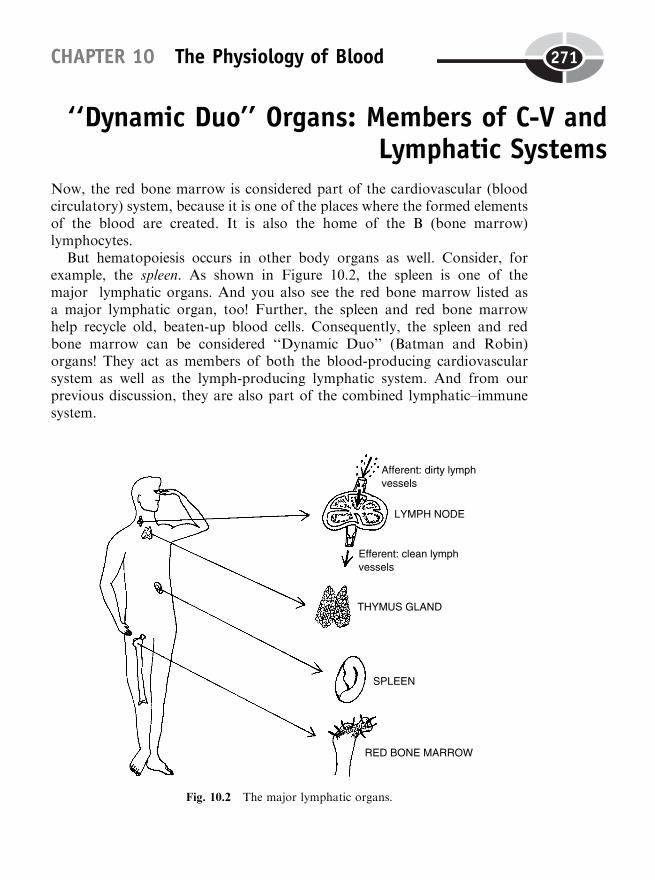
‘‘Dynamic Duo’’ Organs: Members of C-V andLymphatic Systems
Now, the red bone marrow is considered part of the cardiovascular (bloodcirculatory) system, because it is one of the places where the formed elementsof the blood are created. It is also the home of the B (bone marrow)lymphocytes.
But hematopoiesis occurs in other body organs as well. Consider, forexample, the spleen. As shown in Figure 10.2, the spleen is one of themajor lymphatic organs. And you also see the red bone marrow listed asa major lymphatic organ, too! Further, the spleen and red bone marrowhelp recycle old, beaten-up blood cells. Consequently, the spleen and redbone marrow can be considered ‘‘Dynamic Duo’’ (Batman and Robin)organs! They act as members of both the blood-producing cardiovascularsystem as well as the lymph-producing lymphatic system. And from ourprevious discussion, they are also part of the combined lymphatic–immunesystem.
CHAPTER 10 The Physiology of Blood 271
Fig. 10.2 The major lymphatic organs.

We have:
‘‘DYNAMIC DUO’’ ORGANS(MEMBERS OF BOTH THE C-VAND LYMPHATIC–IMMUNE
SYSTEMS)
¼ RED BONEMARROW þ SPLEEN
(C-V FUNCTIONS: Participate inhematopoiesis, destruction andrecycling of old blood cells);LYMPHATIC–IMMUNE
FUNCTIONS: Antigen–antibodyreactions, phagocytosis of foreign
invaders)
The Lymph Nodes and Thymus GlandTwo organs that are mainly lymphatic–immune in nature, without any directconnection to the cardiovascular system, are the lymph nodes and the thymusgland.
THE THYMUS GLAND
The thymus gland lies just deep to the sternum, in the middle of the chest.The thymus can be considered an endocrine gland of childhood and adoles-cence, because it reaches its maximum size at puberty, then progressivelydecreases in size with age. While it is still present, the thymus secretes thehormone thymosin (thigh-MOH-sin). This hormone stimulates the activity ofthe lymphocytes and other parts of the body’s immune system. It also pro-duces T (thymic) lymphocytes, which are sometimes just called T cells.
The thymus is thought to play an important role in the development ofimmune competence in youngsters – a growing ability to ward off variousdiseases. The disappearance of the thymus in adults may be related to thegradual decline of immune competence seen in older persons, thereby makingthem more susceptible to cancer and pneumonia.
THE LYMPH NODES
The most widespread lymphatic organs are the lymph nodes. The lymphnodes are a group of small, bean-like organs scattered in clusters in variousparts of the body.
PART 5 Air and Fluid Transport272
Organ 1
Afferent lymphatic vessels carry ‘‘dirty’’ lymph toward the lymph nodes. It isdirty in the sense that it often contains tiny particles of dirt, debris, or bacteria,which were filtered into the lymph from the blood capillaries. Efferentlymphatic vessels carry ‘‘clean’’ lymph away from the lymph nodes. And in‘‘cleaning up’’ the dirty lymph that passes through them, the lymph nodesare essentially carrying out the basic processes of the immune response.
The Immune Response: Antibodies andMacrophages Attack the Antigens
Most cells carry chemical markers upon their surface membranes thatuniquely identify them. Thus, cells in a particular human body have theirown chemical surface markers that tag them as ‘‘self.’’ Such cells are notattacked by the body’s immune system. Foreign cells transplanted from someother body, or a bacterium or cancer cell, however, have different surfacemarkers that tag them as ‘‘non-self.’’ The general name for such surfacemarkers is antigens (AN-tih-jens).
The word antigen means ‘‘produced’’ (-gen) ‘‘against’’ (anti-). This mean-ing reflects the fact that antigens are ‘‘non-self ’’ marker proteins that labelparticular cells as foreign. Therefore, the antigens are foreign proteins thatcause antibodies to be ‘‘produced against’’ them. Antibodies, in turn, areproteins produced by the body’s immune system that attack and destroyforeign antigens. The overall process is called an antigen–antibody reaction(as mentioned earlier).
The antigen–antibody reaction comes about after a series of precedingsteps (Figure 10.3). The first step is identification of a foreign cell and itssurface antigen by a T (thymic) lymphocyte within the lymph nodes or someother lymphatic tissue. The T lymphocytes prowl around within the networkof lymphatic vessels and act much like scouts. They send out a chemicalsignal whenever a foreign antigen is encountered.
The B (bone marrow) lymphocytes receive the chemical messages from theT lymphocyte scouts. The B lymphocytes then undergo a dramatic differen-tiation (process of becoming specialized or different). They transform into anentirely different cell type, called plasma cells. These new plasma cells have aprominent ‘‘clock face’’ nucleus when viewed through a compound lightmicroscope. There is dark chromatin (kroh-MAT-in) visible – strands ofDNA that have not yet coiled together to create chromosomes. These
CHAPTER 10 The Physiology of Blood 273

chromatin fragments are arranged in a circular fashion around the edges ofthe nucleus, giving it a distinct ‘‘clock face’’ appearance.
It is the plasma cells that actually produce the antibodies. Once produced,the individual antibody molecules attach to the foreign antigens, like twopieces of a jigsaw puzzle fitted together. The result we have called an anti-gen–antibody reaction. When the antibody combines, it causes a lysis (break-down) of the invading cell carrying the foreign antigen. Thus, millions ofinvading or abnormal cells (and their antigens) are efficiently ruptured andscattered into tiny pieces.
PART 5 Air and Fluid Transport274
Fig. 10.3 The immune response.
Moving nearby is a defensive army of neutrophils and larger wanderingmacrophages (representing transformed monocytes). The macrophages (andtheir smaller neutrophil allies) readily surround and engulf entire invadingcells and their foreign antigens by means of phagocytosis (cell eating).
SUMMARY
Hence, by two major processes – antigen-antibody reactions and phagocy-tosis – an immune response has occurred. As its name indicates, the immuneresponse produces a state of immunity from disease.
To verbally encapsule the immune response, we have:
THE IMMUNERESPONSE (STATEOF IMMUNITYFROM DISEASE)
¼ ANTIGEN–ANTIBODY
REACTIONS (Causelysis of invading cells)
þ PHAGOCYTOSIS(Eating of invadersby macrophages)
The ABO and Rh Antigen SystemsOur erythrocytes (like almost all of our other body cells) have chemical sur-face markers on their plasma membrane. But instead of being called antigens,they are called agglutinogens (ah-gloo-TIN-oh-jens). [Study suggestion:
Observe that both the word antigen and the word agglutinogen end withthe same suffix, -gen. Use this fact to help you remember that an agglutinogenis a type of antigen found on the plasma membranes of red blood cells.]
The word agglutinogen literally means ‘‘a producer’’ (-gen) of ‘‘glueing orclumping’’ (agglutin). What is meant by this is made clear by a look at Figure10.4. When two blood types are incompatible, the RBCs of the blood donorcannot be safely given to the blood recipient. The reason is that the bloodrecipient has agglutinins (a-GLOO-tin-ins) or chemical antibodies in itsserum that attack the agglutinogens on the incompatible RBCs and chemi-cally lyse (split) them. The exploded RBCs lose their hemoglobin, and theirshriveled remnants – called RBC ‘‘ghosts’’ – then ‘‘clump’’ or agglutinate (ah-GLOO-tih-nayt) together into big, sticky balls. These big balls of RBCghosts can block or occlude (ah-KLOOD) important blood vessels, therebystarving the tissue cells downstream.
In short, we have a special case of the antigen–antibody reaction. Here, it iscalled a transfusion reaction between incompatible blood types. This is the
CHAPTER 10 The Physiology of Blood 275
Cell 1
Tissue 1
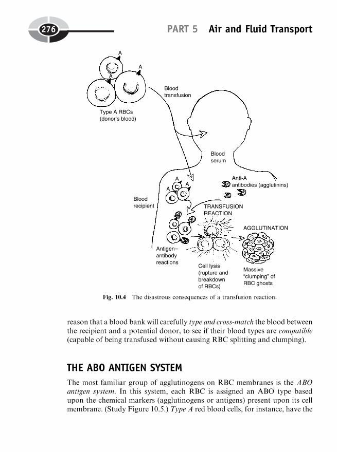
reason that a blood bank will carefully type and cross-match the blood betweenthe recipient and a potential donor, to see if their blood types are compatible(capable of being transfused without causing RBC splitting and clumping).
THE ABO ANTIGEN SYSTEM
The most familiar group of agglutinogens on RBC membranes is the ABOantigen system. In this system, each RBC is assigned an ABO type basedupon the chemical markers (agglutinogens or antigens) present upon its cellmembrane. (Study Figure 10.5.) Type A red blood cells, for instance, have the
PART 5 Air and Fluid Transport276
Fig. 10.4 The disastrous consequences of a transfusion reaction.
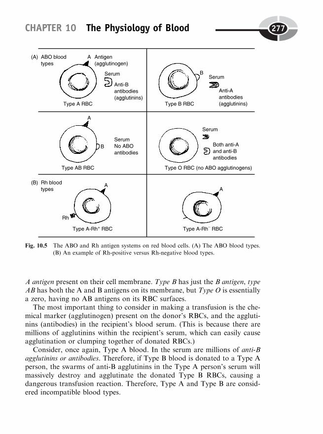
A antigen present on their cell membrane. Type B has just the B antigen, typeAB has both the A and B antigens on its membrane, but Type O is essentiallya zero, having no AB antigens on its RBC surfaces.
The most important thing to consider in making a transfusion is the che-mical marker (agglutinogen) present on the donor’s RBCs, and the aggluti-nins (antibodies) in the recipient’s blood serum. (This is because there aremillions of agglutinins within the recipient’s serum, which can easily causeagglutination or clumping together of donated RBCs.)
Consider, once again, Type A blood. In the serum are millions of anti-Bagglutinins or antibodies. Therefore, if Type B blood is donated to a Type Aperson, the swarms of anti-B agglutinins in the Type A person’s serum willmassively destroy and agglutinate the donated Type B RBCs, causing adangerous transfusion reaction. Therefore, Type A and Type B are consid-ered incompatible blood types.
CHAPTER 10 The Physiology of Blood 277
Fig. 10.5 The ABO and Rh antigen systems on red blood cells. (A) The ABO blood types.(B) An example of Rh-positive versus Rh-negative blood types.

[Study suggestion: From a careful examination of Figure 10.5, why do youthink that Type O is considered the ‘‘universal ABO donor’’? Why is TypeAB called the ‘‘universal ABO recipient’’?]
THE RH FACTOR
In addition to the ABO antigen system, there are many others present on theRBC membrane. Most important among these are the Rh factor or antigen.The Rh name comes from the fact that this agglutinogen (antigen) was firstobserved on the RBCs of a Rhesus (REE-sus) monkey. Later, it was alsofound to occur on the RBCs of about 85% of all humans.
People who have the Rh factor on their RBCs are called Rhþ or Rh-positive. The approximately 15% of people who lack the factor are calledRh� or Rh-negative. The Rh factor is usually reported in combination with aperson’s ABO antigen type. A person with both the A and Rh antigenspresent on each red blood cell, for example, is classified as A positive, or asType A, Rh positive. If they have the A antigen but lack the Rh antigen thenthey are classified as A negative, or as Type A, Rh negative.
A person who is Type A, Rh negative, automatically contains anti-Bagglutinins (antibodies) in the blood serum. But no anti-Rh antibodies oragglutinins are naturally present in the blood serum. Anti-Rh antibodies areproduced by the immune system only some time after an Rh-negative personhas somehow received a quantity of Rhþ RBCs. This can happen in theblood serum of an Rh-negative woman after she has given birth to an Rh-positive child. The reason is that some of the child’s blood may have mixedwith the mother’s during the birth process, say from small scrapes or woundson the child’s head and the women’s genital (JEN-ih-tal) area. If the mother’simmune system has been ‘‘Rh-sensitized’’ in this way, then the RBCs of anysubsequent Rhþ fetus (FEE-tus) in her womb may be attacked and destroyedby her anti-Rh antibodies.
The Process of Blood ClottingThe only type of formed element in the blood that we have not yet discussedin this chapter are the platelets or thrombocytes. The platelet name indicatestheir anatomy – shaped like ‘‘little plates.’’ The thrombocyte name indicatestheir function – participation in thrombus (THRAM-bus) or ‘‘clot’’ (thromb)formation.
PART 5 Air and Fluid Transport278
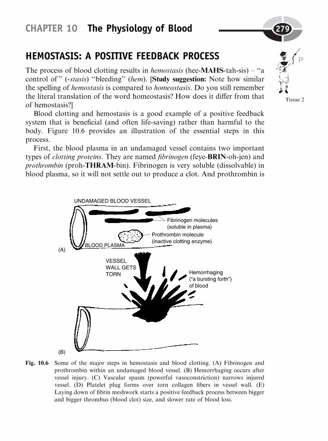
HEMOSTASIS: A POSITIVE FEEDBACK PROCESS
The process of blood clotting results in hemostasis (hee-MAHS-tah-sis) – ‘‘acontrol of ’’ (-stasis) ‘‘bleeding’’ (hem). [Study suggestion: Note how similarthe spelling of hemostasis is compared to homeostasis. Do you still rememberthe literal translation of the word homeostasis? How does it differ from thatof hemostasis?]
Blood clotting and hemostasis is a good example of a positive feedbacksystem that is beneficial (and often life-saving) rather than harmful to thebody. Figure 10.6 provides an illustration of the essential steps in thisprocess.
First, the blood plasma in an undamaged vessel contains two importanttypes of clotting proteins. They are named fibrinogen (feye-BRIN-oh-jen) andprothrombin (proh-THRAM-bin). Fibrinogen is very soluble (dissolvable) inblood plasma, so it will not settle out to produce a clot. And prothrombin is
CHAPTER 10 The Physiology of Blood 279
Tissue 2
Fig. 10.6 Some of the major steps in hemostasis and blood clotting. (A) Fibrinogen and
prothrombin within an undamaged blood vessel. (B) Hemorrhaging occurs aftervessel injury. (C) Vascular spasm (powerful vasoconstriction) narrows injuredvessel. (D) Platelet plug forms over torn collagen fibers in vessel wall. (E)
Laying down of fibrin meshwork starts a positive feedback process between biggerand bigger thrombus (blood clot) size, and slower rate of blood loss.

PART 5 Air and Fluid Transport280
Fig. 10.6 (continued)
an inactive clotting enzyme which does not promote any clotting process. Butthese clotting proteins are always there, ready to be transformed into otherchemicals that do participate in the clotting process!
Things change rapidly, then, whenever the wall of a blood vessel is tornand hemorrhaging (HEM-eh-rij-ing), or ‘‘a bursting forth’’ of bleeding,occurs. Here are some of the major steps leading to hemostasis (and completestopping of hemorrhaging), after a vessel wall is injured.
Hemostasis step 1: Occurrence of powerfulvasoconstriction (vascular spasms)
Right after a blood vessel is injured, it undergoes a powerful vasoconstriction(vessel narrowing). Because the vasoconstriction is so strong and sudden, it isoften called a vascular spasm. This spasm and narrowing of the vessel lumenimmediately slows the rate of blood loss, because less blood is now flowingthrough the constricted, injured vessel.
Hemostasis step 2: Formation of a platelet plug
When a vessel is cut open, the collagen fibers in its wall are torn. Platelets inthe surrounding blood become sticky and attach to the shredded ends of thecollagen fibers. As they pile up, a platelet plug is created that partially closesoff the hole in the vessel wall.
Hemostasis step 3: Coagulation or blood clotting
After the platelet plug is formed, coagulation (koh-ag-you-LAY-shun) – the‘‘process of ’’ (-tion) ‘‘clotting’’ (coagul) – swings into full action.
Injury to the vessel wall creates a chemical called prothrombin activator.As its name indicates, prothrombin activator converts prothrombin (aninactive clotting enzyme) into an active clotting enzyme. Now the veryword prothrombin literally means ‘‘before’’ (pro-) ‘‘thrombin.’’ Therefore,prothrombin activator converts prothrombin into what comes after it –thrombin. Thrombin is an active clotting enzyme that really gets thingsstarted!
Thrombin acts upon fibrinogen, which we said was soluble or dissolvablewithin the bloodstream. Again translating a term, note that fibrinogen means‘‘producer’’ (-gen) of ‘‘fibrin.’’ Consequently, a new protein, fibrin (FYE-brin), appears.
CHAPTER 10 The Physiology of Blood 281

Fibrin means ‘‘a fiber’’ (fibr) ‘‘substance’’ (-in). The reason for this name isthat fibrin is a thin, whitish-colored, fiber-like protein that exists as slenderfilaments. Fibrin filaments are insoluble (not dissolvable) within the bloodplasma. Hence, the fibrin filaments settle out of the blood and are depositedas a fibrin meshwork over the platelet plug.
More circulating platelets, as well as a few RBCs, get stuck in this fibrinmeshwork. A sticky, jelly-like thrombus results. A positive feedback processnow begins.
The bigger the clot (thrombus) gets, the slower that blood is lost. And theslower that blood is lost (hence allowing more time for clotting to occur), thebigger the clot or thrombus gets. This vicious cycle or vicious circle betweenever-increasing size of the thrombus, and ever-slowing rate of hemorrhagingfrom the site of the wound, keeps continuing until a dramatic climax is finallyreached – the complete sealing off of the hole in the vessel wall by a big,gooey thrombus! The rate of blood loss falls to zero, and a process of healingof the damaged vessel wall takes place, while the clot is holding back theblood like an effective beaver dam! (But this dam consists of a meshwork offibrin mixed with platelets and RBCs, rather than a dam of interlacing logs,twigs, mud, and leaves.)
QuizRefer to the text in this chapter if necessary. A good score is at least 8 correctanswers out of these 10 questions. The answers are listed in the back of thebook.
1. The major force behind the filtration of lymph:(a) Osmosis of water down its concentration gradient(b) Simple diffusion of ions through pores(c) Capillary blood pressure(d) Local circuit currents
2. Formed elements represent:(a) Cells and cell fragments(b) The atoms of particular chemical elements with distinct shapes or
forms(c) Dissolved solutes of various sizes(d) Cell organelles having distinct structures and functions
PART 5 Air and Fluid Transport282
3. Play an important role in immunity:(a) Platelets(b) Leukocytes(c) Erythrocytes(d) Thrombocytes
4. The process whereby WBCs can ‘‘leap’’ or crawl ‘‘through’’ capillarywalls:(a) Diapedesis(b) Phagocytosis(c) Filtration(d) Saltatory conduction
5. Often change into wandering macrophages:(a) Lymphocytes(b) Plasma cells(c) Monocytes(d) T cells
6. The spleen and red bone marrow are both distinct in that they:(a) Contain oxygen-rich blood(b) Belong to both the cardiovascular and lymphatic–immune
systems(c) Are exactly the same structures as lymph nodes(d) Filter lymph, rather than consume it
7. Chemical surface markers on cell membranes that tag them as ‘‘non-self ’’:(a) Hemoglobin molecules(b) Hemocytoblast fragments(c) Agglutinins(d) Antigens
8. Cells that actually produce antibodies:(a) Stem cells(b) Plasma cells(c) B lymphocytes(d) Neutrophils
9. The occurrence of a transfusion reaction indicates that:(a) An incompatible blood type has been donated to a recipient(b) Two ABO blood types have been properly cross-matched(c) No Rh factors are present(d) The immune response has become non-functional
CHAPTER 10 The Physiology of Blood 283
10. Is laid down as a skeletal meshwork during clot formation:(a) Prothrombin(b) Myosin(c) Fibrin(d) Coenzyme Q
Body-Level Grids for Chapter 10Several key body facts were tagged with numbered icons in the page marginsof this chapter. Write a short summary of each of these key facts into anumbered cell or box within the appropriate Body-Level Grid that appearsbelow.
Anatomy and Biological Order Fact Grids for Chapter 10:
CELLLevel
PART 5 Air and Fluid Transport284
1

TISSUELevel
ORGANLevel
ORGAN SYSTEMLevel
Physiology and Biological Order Fact Grids for Chapter 10:
CELLLevel
CHAPTER 10 The Physiology of Blood 285
1
1
1
TISSUELevel
Physiology and Biological Disorder Fact Grids for Chapter 10:
TISSUELevel
PART 5 Air and Fluid Transport286
1
1 2
1

287
CHAPTER11
The RespiratorySystem: Makes You‘‘Breathe Again’’
Chapter 10 in PHYSIOLOGY DEMYSTIFIED outlined some pressures,volumes, and flows of two liquids. These were the blood and the lymph.Now Chapter 11 will address still another fluid substance – the air. And,as before, we will see how its physiology behaves under pressure, at certainvolumes, and with certain rates of flow.
The basic structures of the respiratory system are thoroughly covered inANATOMY DEMYSTIFIED. In this book, we will be focusing upon thelowermost tips of the so-called respiratory tree. These tips largely consist ofthe bronchioles (BRAHNG-kee-ohls) or ‘‘little bronchi’’. The bronchioleshave a very high proportion of circular smooth muscle in their walls.(Consult Figure 11.1.)
Organ 1
Copyright © 2004 by The McGraw-Hill Companies, Inc. Click here for terms of use.
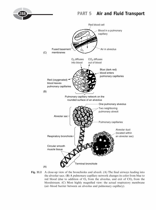
PART 5 Air and Fluid Transport288
Fig. 11.1 A close-up view of the bronchioles and alveoli. (A) The final airways leading intothe alveolar sacs. (B) A pulmonary capillary network changes its color from blue tored blood (due to addition of O2 from the alveolus, and exit of CO2 from the
bloodstream. (C) Most highly magnified view: the actual respiratory membrane(air–blood barrier between an alveolus and pulmonary capillary).
BRONCHODILATION VERSUS BRONCHOCONSTRICTION
This high amount of circular smooth muscle (plus the lack of any cartilagehorseshoes) allows the bronchioles to greatly widen (dilate), or greatly nar-row (constrict), the diameter of their lumens. During exercise or severe stress,the bronchiolar (brahng-kee-OH-lar) smooth muscle relaxes, loosening thewall of the bronchioles. This results in bronchodilation (brahng-koh-dih-LAY-shun) – the ‘‘process of bronchial tube widening’’. The amount of fric-tional resistance or ‘‘rubbing’’ of the air molecules against the greatlyenlarged tube walls is therefore reduced. Hence, air flows through the dilatedbronchioles in greater volumes.
This greater volume of air going into and out of the lungs, of course, isof considerable help in supplying the tissue cells with greater amounts of O2
to support their aerobic metabolism. It also allows greater quantities ofCO2 to flow away from the tissues as metabolic waste products. Thesegreater amounts of air flowing through dilated bronchioles are largely theresult of the increased activity of the sympathetic nerves, and of the adrenalmedulla and its secretion of epinephrine (Chapter 8, Figure 8.3).
Now, let us consider rest, relaxation, and digestion. These are the bodyconditions when the parasympathetic (PAIR-uh-SIM-pah-thet-ik) nerves aredominant. (The sympathetic and parasympathetic nerves are discussed inmore detail within the pages of ANATOMY DEMYSTIFIED.) In this situa-tion, most of the tissue cells (except those lining the digestive tract) arerelatively inactive. Hence, the circular smooth muscle in the walls of thebronchioles tends to contract with moderate force. The reason is that theparasympathetic nerves to the bronchiole wall stimulate the contraction ofthe circular smooth muscle. Like a noose lightly tightening around a cow-boy’s neck, the result is bronchoconstriction (brahng-koh-kahn-STRIK-shun)– ‘‘the process of narrowing of the bronchial tubes.’’
As a result of this bronchoconstriction, there is an increased frictionalresistance to air flowing through the bronchioles, and therefore the volumeof air moving through them goes down.
Remember:
BRONCHOCONSTRICTION:Bronchiolar smooth muscle
contracts
Airwaynarrows
! "Frictionalresistance
! #Volumeof airflow
but,BRONCHODILATION:Bronchiolar smooth muscle
relaxes
�! Airwaywidens
! #Frictionalresistance
! "Volumeof airflow
CHAPTER 11 The Respiratory System 289
Organ 1
GETTING DOWN TO THE BUSINESS OF‘‘REAL’’ RESPIRATION
There is a series of smaller and smaller bronchioles. These, of course, mayeither bronchoconstrict or bronchodilate. The smallest (and last) of them allis called the respiratory bronchiole. ‘‘Why is it called a respiratory bronchiole,Professor? None of the other bronchioles have this name.’’
That is correct, Baby Heinie. The reason for this name is that the respira-tory bronchiole is the bronchiole leading into the respiratory membrane,where real respiration occurs! You will note from the extreme close-up(back in Figure 11.1) that the respiratory bronchiole is followed by onlytwo more structures. These are the alveolar (al-VEE-oh-lar) duct, whichleads into an alveolar sac.
‘‘What’s an alveolar sac, Professor Joe?’’ Well, it looks a lot like a bunch ofgrapes, doesn’t it? In reality, however, each individual grape-like structure isreally a pulmonary alveolus (al-VEE-oh-lus). Like a bunch of grapes hangingon a hollow stem, an alveolar sac, consisting of a bunch of pulmonary alveoli(al-VEE-oh-lie), hangs from a hollow alveolar duct.
‘‘What’s an alveolus, Professor?’’ The word alveolus means ‘‘little cavity.’’So, a pulmonary alveolus is a ‘‘little cavity’’ within the ‘‘lungs’’ (pulmon). Thetwo lungs together, in fact, contain about 300 million pulmonary alveoli!
‘‘Wow! They must really be important!’’ Yes, each alveolus is important.There are two main reasons:
1. The alveolus is the very end of the respiratory ‘‘tree,’’ that is, theextensively branching series of tree-like bronchi and bronchioles.
2. The alveolus is the only structure in the entire so-called ‘‘respiratory’’system where real respiration actually occurs!
Pulmonary ventilation not the same thingas pulmonary respiration
‘‘But I thought this whole branching deal in the lungs was the respiratorysystem, which you said involved the process of breathing again and again!’’ Ican see why you are somewhat confused by this unfortunate terminology,Baby Heinie. We badly need to distinguish pulmonary respiration frompulmonary ventilation (ven-tih-LAY-shun).
Pulmonary ventilation is the process of sucking air into the lungs, andblowing air out of the lungs. (This is a lot like a ventilation system for a
PART 5 Air and Fluid Transport290

large factory building, which both sucks clean air in, from the outdoors, andthen blows dirty air out.) Therefore, when we are actually breathing, we arecarrying out pulmonary ventilation.
‘‘Okay, then what is pulmonary respiration?’’ What physiologists reallymean by respiration is gas exchange between two or more body compart-ments. ‘‘Which compartments?’’ Well, take another look back at the realclose-up in Figure 11.1. The two compartments involved in pulmonaryrespiration are the air within each alveolus, and the blood within each pul-monary capillary, lying right next to it.
‘‘Is this area what you meant when you were talking about a respiratorymembrane?’’ Yes, it is! Another name for the respiratory membrane is thealveolar–capillary membrane. Now, do you see in Figure 11.1 how thepulmonary capillaries form a webbing over the alveoli? And do you alsoobserve that the first half of the pulmonary capillaries contains blue (dark)blood, while the second half undergoes a color change to red (lighter-colored) blood? This color change in the blood is due to pulmonary respira-tion.
‘‘So, you mean that it is at the respiratory membrane, actually thealveolar–capillary membrane, where respiration really occurs?’’ Yes, this isthe place where the wall of each alveolus is tightly fused against the wall of itspulmonary capillary. Each type of chamber is lined by only a single layer ofendothelial cells. Thus, it is an excellent membrane for the diffusion of O2
molecules out of the alveolus, into the pulmonary capillary, and for CO2
molecules to diffuse in the opposite direction.‘‘What is going on in the rest of the so-called respiratory system,
Professor?’’ Well, the rest of this organ system should really be called theventilatory (VEN-tih-lah-tor-ee) system (rather than the respiratory system),because its chief function is ventilation (not respiration)!
‘‘But don’t many of the bronchi, bronchioles, and all that, run right alongblood vessels, too, so that they could exchange O2 and CO2 with them?’’ Intheory, yes. But in practical reality, no. [Study suggestion: See if you canfollow through on Professor Joe’s line of reasoning. Explain why pulmonaryrespiration cannot occur across the walls of the trachea, bronchi, and bronch-ioles. Check your reasoning with a knowledgeable friend.]
Remember:
PULMONARYRESPIRATION
¼ Gas exchange across the respiratory membrane(alveolar–capillary wall)
whilePULMONARYVENTILATION
¼ The sucking of air into the lungs, and theblowing of air out of the lungs
CHAPTER 11 The Respiratory System 291
Organ 2
Organ system 1

Ventilation: A Bulk Flow of AirIf pulmonary ventilation is the process by which we breathe, then it would begood to understand more about it. In particular, it would be good to under-stand the steps of inspiration (sucking air into the lungs), as well as those ofexpiration (eks-pir-AY-shun) or blowing air out of the lungs.
PULMONARYVENTILATION
¼ INSPIRATION(Sucking air into
lungs)
þ EXPIRATION(Blowing air out of
lungs)
VENTILATION AS A BULK FLOW OF AIR
Ventilation and the movement of air is quite similar to blood pressure andthe movement of blood. As we learned in Chapter 9, blood flows down ablood pressure (BP) gradient, from a place where the BP is higher, towardanother place where the BP is lower. In general, we can call this a bulk flowprocess. Bulk flow is the pressure-driven movement of some fluid substance(such as blood or air) from an area of greater pressure toward an area oflower pressure. In other words, bulk flow occurs down a pressure gradient.
For ventilation to occur, then, the problem becomes one of creating an airpressure gradient – a difference in air pressure – between the air in the atmo-sphere and the air within the alveoli (tiny lung air sacs). With such a gradient,there will be a bulk flow of air from the atmosphere and into the lung alveoli.
EQUAL GAS PRESSURES
There are two total gas pressures to consider. The atmospheric pressure is thepressure created by all of the gases in the atmosphere. (The atmosphere is theapproximately 1-mile-thick blanket of air covering the surface of the Earth.)At sea level, the atmospheric pressure pushes with a total force of about760 mmHg. The atmospheric pressure also pushes with this force upon thelips and nostrils. So when a person opens his mouth, the atmospheric pres-sure tends to push air down into his lung alveoli. (Consult Figure 11.2, A.)
Conversely, the intra-alveolar (in-trah-al-VEE-oh-lar) pressure is the totalpressure exerted by all of the gas molecules within the alveoli. When a personopens his mouth, the intra-alveolar pressure tends to push air out of thealveoli, and out of the nose and mouth.
PART 5 Air and Fluid Transport292
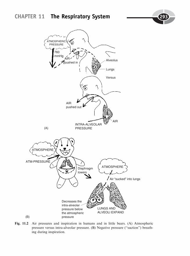
CHAPTER 11 The Respiratory System 293
Fig. 11.2 Air pressures and inspiration in humans and in little bears. (A) Atmospheric
pressure versus intra-alveolar pressure. (B) Negative pressure (‘‘suction’’) breath-ing during inspiration.
The atmospheric pressure (tending to push air into the alveoli) and theintra-alveolar pressure (tending to push air out of the alveoli) are thus twoopposing pressures. Between breaths, we have an equality of these twopressures. There is no air pressure gradient, and therefore, there is no bulkflow of air into or out of the lungs.
Summarizing:
BETWEEN BREATHS:(No air pressure gradientexists: no bulk flow into or
out of lungs)
ATMOSPHERICPRESSURE
(Tends to pushair into the lungs)
¼ INTRA-ALVEOLARPRESSURE
(Tends to push airout of the lungs)
INSPIRATION AS NEGATIVE-PRESSURE BREATHING
We humans must create an air pressure gradient between the atmosphericpressure in the air surrounding us, and the intra-alveolar pressure within ourlungs, if we are to achieve ventilation – a bulk flow of air either into, or outof, our lungs. We will trace the mechanism for creating inspiration, and thenjust reverse the process, for expiration.
Major steps in inspiration
(Study Figure 11.2, B.)
1. The diaphragm (DIE-uh-fram) or ‘‘barrier’’ muscle forming the floorof the chest cavity contracts. As the diaphragm contracts, it dropsdown in its position.
2. This makes the thoracic (chest) cavity larger at the bottom end, sinceits floor has been lowered.
3. Since the thoracic cavity becomes larger, the lungs follow suit andenlarge along with it. And because the lungs have enlarged, themillions of tiny alveoli become larger air sacs as well.
4. Because the alveoli are larger, their limited number of contained gasmolecules are pushing against the walls of a much larger sac. Now,pressure is the amount of force exerted against a certain amount ofarea. As the area of the alveoli gets larger, then, the intra-alveolarpressure within them is distributed over a considerably greater area oftheir wall.
5. As a result, the intra-alveolar pressure does something ‘‘negative’’ – itfalls below the atmospheric pressure. A pressure gradient is now
PART 5 Air and Fluid Transport294
Organ 3
created between the atmospheric pressure and the intra-alveolarpressure. Like water being sucked down into a drain having lowerpressure, the air is sucked down into the lung alveoli by inspiration.
After air has been actively sucked into the lung alveoli by the active,energy-requiring contraction of the diaphragm muscle, the diaphragm justquits contracting, and expiration occurs passively (without need of additionalenergy input). Like making a sigh of relief when you relax, this is how youbasically exhale.
[Study suggestion: Examine Figure 11.2 B, and just reverse the direction ofthe events. You will then see for yourself the mechanism of passive expiration.]
Major Lung Volumes and CapacitiesThe previous section basically told us how we breathe (ventilate air). Thissection now reveals how much we breathe. Specifically, this section deals withthe various lung volumes and capacities. We will now examine some of thesemajor physiological parameters describing various aspects of pulmonary(lung) function.
BABY HEINIE THROWING SNOWBALLS
To be entertaining, let us view a picture that might be entitled Baby HeinieThrowing Snowballs (Figure 11.3). Since we can see his frosty breath, and weknow he is getting rather excited and exercising, we can describe some of thelung volumes and capacities for an ‘‘average’’-sized (but somewhat emotion-ally immature) male like Baby Heinie.
Tidal volume (TV)
Baby Heinie takes a single breath while he is resting. This breath is called thetidal volume, abbreviated as TV. The tidal volume (TV) is named for itsresemblance to a real tide – the moving of waves of water back and forthupon the sand of a beach. (Or it can be visualized as the amount of frosty airgoing into, or out of, Baby Heinie’s mouth with each of his resting breaths.)Technically speaking, the tidal volume is the amount of air exhaled after theperson takes a normal resting inspiration. The TV amounts to about 500 ml(milliliters) in an average human adult.
CHAPTER 11 The Respiratory System 295
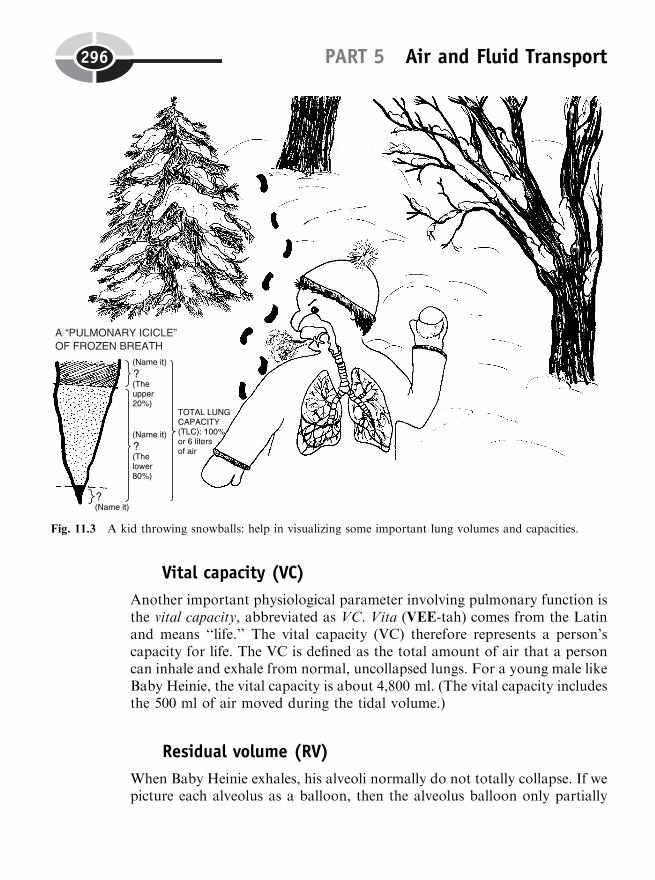
Vital capacity (VC)
Another important physiological parameter involving pulmonary function isthe vital capacity, abbreviated as VC. Vita (VEE-tah) comes from the Latinand means ‘‘life.’’ The vital capacity (VC) therefore represents a person’scapacity for life. The VC is defined as the total amount of air that a personcan inhale and exhale from normal, uncollapsed lungs. For a young male likeBaby Heinie, the vital capacity is about 4,800 ml. (The vital capacity includesthe 500 ml of air moved during the tidal volume.)
Residual volume (RV)
When Baby Heinie exhales, his alveoli normally do not totally collapse. If wepicture each alveolus as a balloon, then the alveolus balloon only partially
PART 5 Air and Fluid Transport296
Fig. 11.3 A kid throwing snowballs: help in visualizing some important lung volumes and capacities.
deflates during expiration. The balloon simply becomes smaller, rather thancompletely deflating. As a result, there is a residual volume, or RV. Theresidual volume (RV) is literally the residual or ‘‘left-over’’ volume thatstill remains within the alveoli, even after expiration has occurred. For BH(Baby Heinie), the residual volume is about 1,200 ml. This partial, residualinflation of the alveoli greatly reduces the amount of work required to com-pletely re-inflate the lungs during the next cycle of inspiration.
Total lung capacity (TLC)
If we add both the vital capacity (VC) and the residual volume (RV) together,we obtain the total lung capacity (TLC):
TOTAL LUNGCAPACITY (TLC)6 liters (or 6,000 ml)
¼ VITAL CAPACITY(VC) (4,800 ml)
þ RESIDUAL VOLUME(RV) (1,200 ml)
The total lung capacity therefore represents the total amount of air thatthe lungs can possibly hold. For BH, the TLC is about 6 liters (or 6,000 ml).[Study suggestion: Figure 11.3 pictures the various lung volumes and capa-cities that we have been studying as the ‘‘frozen breath’’ of Baby Heinie,fused together to make a Giant Pulmonary Icicle, hanging down in space.The whole pulmonary icicle (100% of its length) represents the total lungcapacity. The tiny pointed tip of 8.3% at the bottom of the icicle symbolizeswhat specific pulmonary parameter? The lower 80% of the icicle representswhat specific pulmonary parameter? Finally, the upper 20% of the icicledenotes what specific pulmonary parameter? Hint: You just need to get outyour calculator and do a little bit of simple math. When you are done, checkyour answers with that of a knowledgeable friend. Doesn’t the bottom tip ofany hanging icicle usually ‘‘melt’’ (get used up) first? How does this analogyapply to the stated lung volumes and capacities?]
Gas Transport: The Journey of O2 and CO2Well, we now know how we breathe or ventilate, and we know how much webreathe or ventilate. The next question for the inquiring mind is, ‘‘Professor,how do we carry or transport what we breathe – the respiratory gases, O2 andCO2?’’ In other words, we need to address the topic that physiologists usuallycall gas transport through the bloodstream.
CHAPTER 11 The Respiratory System 297
Organ 4
OXYHEMOGLOBIN AND THE RED BLOOD CELL
Certainly, the key issue for gas transport is the movement of oxygen (O2)molecules through the bloodstream. When fresh O2 diffuses into the bloodacross the respiratory membrane (alveolar–capillary wall), it does not mix ordissolve very well with the blood plasma itself. Instead, it diffuses into anearby red blood cell (Figure 11.4, A).
Once inside the RBC, the O2 combines with a heme (heem) group presentwithin a hemoglobin (HEE-moh-glohb-in) molecule. Hemoglobin is a reddish-colored, ‘‘globe’’-shaped (glob) ‘‘protein substance’’ (-in) found within thecytoplasm of the red ‘‘blood’’ (hem) cells. There are between 250 and 280million hemoglobin (Hb) molecules present within a single RBC!
Each heme group is an iron-containing group that can combine with oneoxygen molecule. Since there are four heme groups per hemoglobin molecule,obviously, one hemoglobin molecule can carry up to four O2 molecules.[Study suggestion: From the previous information provided, estimate howmany total O2 molecules a single red blood cell can carry.]
When hemoglobin combines with O2, the resulting combination is calledoxyhemoglobin (AHK-see-HEE-moh-glohb-in). In this new combined mole-cule, the heme group turns a bright cherry red. This is the explanation,therefore, for the bright-red color of oxygenated (AHK-suh-jen-AY-ted) oroxygen-filled blood.
Using the chemical symbol of Hb for hemoglobin, we have
Hb þ O2
(Hemoglobin)! HbO2
(Oxyhemoglobin)
The O2-loaded RBCs circulate to the tissue cells, which are actively con-suming oxygen for their aerobic metabolism. The carried oxygen moleculesthen dissociate (split off ) from the heme groups, and diffuse into the tissuecells.
BICARBONATE ION AND THE RED BLOOD CELL
The red blood cell performs a similarly important role in the transport ofcarbon dioxide (CO2) molecules excreted during cellular respiration (theKrebs cycle and other aerobic processes, Chapter 4). CO2, like O2, is notvery soluble (dissolvable) within the blood plasma. (Just about 7–10% ofCO2 is carried as gas bubbles dissolved in the blood plasma.) Hence, mostof it ends up diffusing into the erythrocytes (Figure 11.4, B).
Here, the CO2 is handled in two different ways:
PART 5 Air and Fluid Transport298

CHAPTER 11 The Respiratory System 299
Fig. 11.4 A brief look at gas transport. (A) Oxygen transport: Oxyhemoglobin and the redblood cell. (B) Carbon dioxide transport on hemoglobin: creation of a carbamino-
hemoglobin molecule. (C) Carbon dioxide transported as bicarbonate ions in theblood plasma.

1. Combines with hemoglobin. The CO2 molecules, once inside the RBC,combine with the globin (GLOH-bin), that is, the ‘‘globe’’-shaped ‘‘protein’’portion, of each hemoglobin molecule. (So, they do not compete with O2 forbinding with the heme groups.)
The combination of CO2 with hemoglobin (Hb) therefore yields a newcompound called carbaminohemoglobin (kar-bam-ih-noh-hee-moh-GLOH-bin). The prefix carbamino- (kar-bam-ih-noh) reflects the fact that ‘‘carbon
PART 5 Air and Fluid Transport300
Fig. 11.4 (continued)
dioxide’’ (carb) attaches to an amino (nitrogen-containing group) in thehemoglobin molecule. The chemical reaction is written as:
CO2
(Carbon dioxide)þ Hb (Hemoglobin) ��! HbCO2
(Carbaminohemoglobin)
Approximately 20% of the blood CO2 is carried within RBCs in the formof carbaminohemoglobin. As with O2, when the red blood cells get near thepulmonary alveoli, the carried CO2’s dissociate (break off ) from the Hb anddiffuse into the alveoli (where most are soon exhaled). This backward reac-tion is symbolized by the arrows of the reaction in the equation between CO2
and Hb, pointing in opposite directions.2. Combines with water to create bicarbonate ion. By far the largest percen-
tage (about 70%) of CO2 diffuses into the RBC and simply combines withwater. ‘‘How come the CO2 doesn’t combine with water outside of the redblood cell, Professor? Aren’t there plenty of H2O molecules available withinthe blood plasma?’’
You are correct, Baby Heinie. There is a lot of water outside of the redblood cell, but only inside the RBC do you find the right enzyme that greatlyspeeds up the combining of CO2 with H2O, so that it occurs at a significantrate. This enzyme is called carbonic (kar-BAHN-ik) anhydrase (an-HIGH-drays). (Examine Figure 11.4, C.)
When CO2 combines with H2O (under the stimulating influence of carbo-nic anhydrase enzyme), the immediate product is carbonic (kar-BAHN-ik)acid, symbolized as H2CO3. The carbonic acid (H2CO3) molecule, like allacids (Chapter 3), is a hydrogen ion (Hþ ion) donor. Thus, the H2CO3
molecule quickly decomposes (breaks down) into both Hþ and HCO�3 orbicarbonate (buy-KAR-boh-nayt) ion. We write:
CO2
(Carbondioxide)
þ H2O(water)
Carbonicanhydrase ��! H2CO3
(Carbonicacid)
So,�! � Hþ
(Hydrogenion)
þ HCO�3(Bicarbonate
ion)
Now the bicarbonate (HCO�3 ) anion, unlike plain CO2, is very soluble inthe blood plasma! Therefore, the bicarbonate ions immediately diffuse out ofthe RBC, and mix with the blood plasma. (You can think of CO2 as being‘‘buried’’ or ‘‘housed’’ within each bicarbonate, HCO�3 molecule.) The bicar-bonate ions are quickly circulated through the blood plasma, and into thelungs. Once they reach the pulmonary capillaries, the HCO�3 ions diffuse backinto the RBC, and the reaction sequence that formed them in the first place isessentially reversed. This re-creates more CO2, which then diffuses out of the
CHAPTER 11 The Respiratory System 301
Molecule 1
RBC, and finally into the pulmonary alveoli. Eventually, many of the CO2
molecules diffusing into the alveoli are exhaled out of the body. (Hence, CO2
is generally considered a body excretion – a metabolic waste product that isremoved.)
SUMMARY OF GAS TRANSPORT
Let us now briefly recap the transport of oxygen and CO2 within the blood-stream, as a couple of gas transport ‘‘rules.’’
The Blood Oxygen Rule:Most blood oxygen is carried as oxyhemoglobin (HbO2) within the red bloodcells. Each O2 molecule is attached to the heme (iron-containing group) withina hemoglobin (Hb) molecule.
The Blood Carbon Dioxide Rule:Most blood carbon dioxide is ‘‘buried’’ or ‘‘housed’’ within a larger bicarbonate(HCO�3 ) ion, which circulates freely through the blood plasma and carries itsCO2 ‘‘cargo’’ long distances, into the lungs.
Control of Respiration andBody Acid–Base Balance
The next logical question for us to ask is, ‘‘Why do we breathe? Withoutbreathing, we quickly reach a state of severe tissue hypoxia (high-PAHKS-ee-ah). This is literally a ‘condition of ’ (-ia) ‘deficient or below normal’ (hyp)blood ‘oxygen’ (oxy) levels. Doesn’t it only make sense, therefore, that themajor stimulus for inspiration would be a steep drop in the blood O2 concen-tration toward its lower normal limit? After all, doesn’t taking a nice deepbreath replenish blood oxygen?’’
Yes, that thinking makes good logical sense. But in reality it is not true. Asteep drop in the blood O2 concentration is too risky and dangerous to be themajor stimulus for taking a normal, resting inspiration! The ‘‘wisdom of thebody’’ does not allow us to wait to take our next inspiration until ouroxygen-starved brains are practically on the verge of hypoxia! This approachis foolish (physiologically speaking)!
PART 5 Air and Fluid Transport302
Molecule 2

THE BODY ACID–RESPIRATION CONNECTION
‘‘Gee, Professor Joe! If a drop in our blood oxygen level toward its lowernormal limit isn’t the major stimulus for our next resting, normal inspiration,then what is the stimulus?’’ The answer is direct. The major stimulus forinspiration is a slight increase in the carbon dioxide (CO2) concentration andacidity (Hþ ion) level within our bloodstream.
To see the mechanism, we need to go back to our old friend, the redblood cell, and peek within. Figure 11.5 (A) repeats most of the essentialsteps we talked about earlier regarding the blood transport of carbon diox-ide. CO2 produced from metabolism quickly reacts with H2O within ourerythrocytes. This reaction results in H2CO3 as its product. H2CO3, youwill remember, is the chemical shorthand for carbonic acid. These carbonicacid molecules quickly break down into hydrogen (Hþ) ions and bicarbo-nate (HCO�3 ) ions.
CHAPTER 11 The Respiratory System 303
Molecule 3
Fig. 11.5 Respiration and body acid–base balance. (A) Normoventilation achieves a healthy
state of acid–base balance. (B) Hypoventilation and respiratory acidosis. (C)Hyperventilation and respiratory alkalosis.

PART 5 Air and Fluid Transport304
Fig. 11.5 (continued)
Hydrogen (Hþ) ions and carbonic acid (H2CO3) are both classified asbody acids. Recall that body acids are substances that either donate hydro-gen ions (such as H2CO3 and HCl stomach acid) or that consist of hydro-gen ions (such as a pool of Hþ ions, itself.) [Study suggestion: Chapter 3discussed body acids, pH, and the concept of acid–base balance. It wouldbe wise for you to review this information now, if you really don’t remem-ber much of it.]
Ventilation, of course, is the process of breathing (sucking air into thelungs, and blowing air out of the lungs). Normoventilation (NOR-moh-ven-tih-LAY-shun), therefore, involves breathing at a ‘‘normal’’ rate and depthfor current metabolic conditions. In normoventilation, to be sure, the personinhales oxygen, but for the purpose of controlling breathing we are muchmore concerned with what happens during expiration. In this phase, theperson exhales just enough CO2 to prevent respiratory acidosis.
Respiratory acidosis is an ‘‘abnormal condition of ’’ (-osis) too much body‘‘acid,’’ due to ‘‘respiratory’’ (breathing-related) causes. Because the personexhales just enough CO2, there isn’t time for too much carbon dioxide toaccumulate within the RBCs and build up an excessive amount of eithercarbonic acid or hydrogen ions. Respiratory acidosis, therefore, is preventedby normoventilation, and a healthy state of acid–base balance is achieved.
HYPOVENTILATION AND RESPIRATORY ACIDOSIS
‘‘Okay, Professor Joe,’’ Baby Heinie scratches his chin while he thinks. ‘‘Ifnormoventilation blows off just the right amount of CO2 to prevent respira-tory acidosis, then what if you exhale or blow off too little CO2?’’
Blowing off or exhaling too little CO2 is what happens during hypoventila-tion (HIGH-poh-ven-tih-LAY-shun). By hypoventilation, we mean breathingat a ‘‘below-normal or deficient’’ (hypo-) rate and depth for current metabolicconditions. [Study suggestion: Try to name some specific situations where aperson who was breathing at a RR (respiratory rate) suited for normal restingconditions, was no longer willing or able to continue ventilating sufficiently.]
A person hypoventilating long enough may fall into a state of respiratoryacidosis (Figure 11.5, B). There is just too much CO2 accumulating withinthe RBCs, due to a deficient rate of excreting (exhaling) them from thelungs. Not being blown off fast enough, too many CO2 molecules react withH2O, and too many carbonic acid molecules and hydrogen ions result. Thestate of acid–base balance is broken, and a disordered condition of acidosisreigns supreme. [Study suggestion: Go back to Chapter 3, if necessary, andlook up the set-point value for blood pH, as well as its normal range.
CHAPTER 11 The Respiratory System 305
During respiratory acidosis, below what specific pH value will the bloodacidity likely fall?]
The brain can only function normally when the blood and CSF (cere-brospinal fluid) are in a state of acid–base balance. Hence, during respiratoryacidosis the person may pass out, or even collapse into a coma!
HYPERVENTILATION AND RESPIRATORY ALKALOSIS
‘‘Well, I don’t want to pass out and die, or anything, Professor! So I promisethat I won’t act like a spoiled brat and hold my breath or hypoventilate(breathe deficiently), just to scare my Mom into buying me more expensivetoys!’’ Good for you, Baby Heinie! ‘‘But what if I throw a tantrum andsuddenly start kicking and screaming and bawling uncontrollably?’’
In this case, Baby Heinie, you will be unwisely engaging in hyperventilation(HIGH-per-ven-tih-LAY-shun). Hyperventilation can often be seen in someemotionally overwrought and hysterically crying persons. In this situation,the RR and depth are at an ‘‘above-normal or excessive’’ (hyper-) level forcurrent metabolic conditions (see Figure 11.6, C).
Therefore, the hyperventilating person blows off or exhales too much CO2
from the body. Not enough CO2 is left to react with water inside of the redblood cells. And there is not enough carbonic acid or hydrogen ion produced,because of this fact.
The resulting state is called respiratory alkalosis (AL-kah-LOH-sis). This isthe technical term for an ‘‘abnormal condition of ’’ (-osis) not enough bodyacid, or too much base or alkali, due to respiratory (breathing-related)causes. Here, of course, we have pointed out emotional hyperventilation asone potential cause of respiratory alkalosis.
If a person hyperventilates for too long, body acid levels fall way belowtheir normal range, and a state of alkalosis follows. The person may wellbecome dizzy and pass out. [Study suggestion: Above what specific pH valuewould the blood pH have to rise, for the person to be classified as sufferingfrom respiratory alkalosis?]
RESPIRATION ACID–BASE SUMMARY
To capsulize, we can say that:
NORMOVENTILATION(Breathing at a normal
rate and depth)
¼ Blow offjust enough
CO2
¼ A state of acid–base (pH)balance
PART 5 Air and Fluid Transport306
Organ 5
HYPOVENTILATION(Breathing at a deficient
rate and depth)
¼ Blowing offtoo littleCO2
¼ A state of respiratoryacidosis (Too much body
acid build-up)
HYPERVENTILATION(Breathing at an excessive
rate and depth)
¼ Blowing offtoo much
CO2
¼ A state of respiratoryalkalosis (Too much body
base or alkali, or notenough acid)
When a person is stimulated to inhale by a slight rise in blood acidity, then,he or she also exhales just enough CO2 during expiration, such that a healthystate of acid–base balance is maintained. And, as a significant bonus, enoughoxygen is delivered to the brain and other vital organs during inspiration toprevent tissue hypoxia. A normal pattern of aerobic functioning of the entirebody is hopefully maintained.
QuizRefer to the text in this chapter if necessary. A good score is at least 8 correctanswers out of these 10 questions. The answers are listed in the back of thebook.
1. The first part of the respiratory system normally reached by air whenit is inhaled:(a) Pulmonary alveoli(b) Smaller bronchi and bronchioles(c) Larynx(d) Nasal and oral cavities
2. The bronchioles are special in that they have the ability to:(a) Completely collapse under pressure(b) Transport blood as well as air(c) Rigidly support the chest wall(d) Dramatically dilate or constrict their lumens
3. The location where ‘‘real’’ respiration occurs:(a) Alveolar–capillary membrane(b) Plasma membrane of the RBC(c) Inner wall of the trachea(d) Lumen of the pharynx
CHAPTER 11 The Respiratory System 307
Organ 1
4. The terminal grape-like clusters at the very distal end of the respira-tory tree:(a) Respiratory bronchioles(b) Alveolar sacs(c) Horseshoes of cartilage(d) Dense fibrous connectors
5. Pulmonary ventilation differs from pulmonary respiration in that it:(a) Consists of gas exchange between air and the tracheal lumen(b) Involves movement through all of the airways(c) Is the blowing of air into the lungs, followed by the sucking of air
out of the lungs(d) Only occurs in children
6. When the atmospheric pressure equals the intra-alveolar pressure,then:(a) No pressure gradient for bulk flow of air exists(b) Air will still move into the lungs, but slowly(c) Both CO2 and O2 will rush out of the lungs very forcefully(d) The diaphragm is probably contracting vigorously
7. Defined as the amount of air exhaled after a person takes a normalresting inspiration:(a) Residual volume(b) TLC(c) Vital capacity(d) Tidal volume
8. Most O2 molecules are carried through the bloodstream by:(a) Combining with plasma proteins(b) Dissolving as gas bubbles(c) Forming oxyhemoglobin(d) Carbaminohemoglobin
9. Carbonic anhydrase is:(a) An enzyme that speeds up the catabolism of glucose(b) The major catalyst within RBCs for the reaction of CO2 with
water(c) A form of high-energy compound useful for kinetic force(d) Mainly responsible for carrying CO2 through the bloodstream
10. The major stimulus for normal, resting inspiration:(a) A steep decline in the blood O2 level(b) Paralysis of the diaphragm muscle
PART 5 Air and Fluid Transport308

(c) An out-of-control rise in blood acidity toward acidosis(d) A slight rise in the blood or CSF Hþ ion and CO2 levels
Body-Level Grids for Chapter 11Several key body facts were tagged with numbered icons in the pagemargins ofthis chapter. Write a short summary of each of these key facts into a numberedcell or box within the appropriate Body-Level Grid that appears below.
Anatomy and Biological Order Fact Grids for Chapter 11:
ORGANLevel
Physiology and Biological Order Fact Grids for Chapter 11:
ORGANLevel
CHAPTER 11 The Respiratory System 309
1
ORGAN SYSTEMLevel
PART 5 Air and Fluid Transport310
1 2
3 4
5
1
Physiology and Biological Disorder Fact Grids for Chapter 11:
ORGANLevel
Function and Biological Order Fact Grids for Chapter 11:
MOLECULELevel
CHAPTER 11 The Respiratory System 311
1
1 2
3

This page intentionally left blank

313
Test: Part 5
DO NOT REFER TO THE TEXT WHEN TAKING THIS TEST. A goodscore is at least 18 (out of 25 questions) correct. Answers are in the back ofthe book. It’s best to have a friend check your score the first time, so youwon’t memorize the answers if you want to take the test again.
1. The circulatory system gets its name from the fact that:(a) Blood vessels circulate the blood back and forth, in a straight line(b) The spleen and liver are included in the system(c) The blood travels in a circular route, to and from the heart(d) Hematopoiesis occurs in some of its members(e) It follows a ‘‘circuitous’’ route that no has ever been able to trace!
2. The left heart is known as the pump for the:(a) Lungs and gall bladder(b) Pulmonary circulation(c) CPA(d) Systemic circulation(e) Pulmonary arterioles
Copyright © 2004 by The McGraw-Hill Companies, Inc. Click here for terms of use.
3. The S-A node plays the principal role in:(a) Excitation of the cardiac muscle(b) Carrying blood from the atria(c) Messing up EKGs!(d) T-wave inversion(e) Prolapse of the mitral valve
4. The ‘‘raises or lifts’’ blood ‘‘up’’ out of the left ventricle:(a) Aortic arch(b) Common pulmonary artery(c) Myocardium(d) Brachial artery(e) Superior vena cava
5. The contracting and emptying phase of both atria and both ventricles:(a) Diastole(b) Functional syncytium(c) Diastolic BP(d) SV(e) Systole
6. One complete sequence of excitation, contraction, and relaxation ofall four chambers of the heart:(a) ‘‘Lubb’’(b) Diastole(c) Closure of both semilunar valves(d) Snapping shut of both A-V valves(e) Cardiac Cycle
7. The ‘‘dup’’ sound generally indicates the event of:(a) Opening of both atrioventricular valves(b) Closure of both semilunar valves at the beginning of ventricular
diastole(c) The middle of the resting period for all heart chambers(d) Closure of the right A-V valve, but opening of the left(e) Closure of the left S-L valve, but opening of the right
8. The P wave in the electrocardiogram reflects:(a) Depolarization of both atria(b) Repolarization of the ventricles(c) Hyperpolarization of the sino-atrial node(d) Loss of myocardial contractility(e) Ventricular fibrillation
PART 5 Air and Fluid Transport314
9. Stroke volume � ¼ CO(a) TPR(b) HR(c) QRS(d) RR(e) TV
10. Portion of the BP that can be considered residual or ‘‘left-over’’ bloodpressure:(a) Diastolic(b) Mean(c) Systolic(d) Acidolic(e) Proximal
11. Represents a clear, watery filtrate of the blood plasma:(a) Venous blood(b) Lymph(c) Kickapoo juice(d) Formed elements(e) Chyme
12. Neutrophils help immune activity mainly by carrying out:(a) Antibody synthesis(b) Blood clotting(c) Phagocytosis(d) Hematopoiesis(e) Hemostasis
13. Formed elements that appear only in blood plasma (not the lymph):(a) Plasma proteins(b) RBCs and WBCs(c) RBCs and platelets(d) Thrombocytes(e) T-cells
14. Antigens present on cell membranes:(a) Break off and soon circulate throughout the bloodstream(b) Often label particular cells as foreign or ‘‘non-self ’’(c) Discourage powerful responses from the immune system(d) Destroy all antibodies that attack them(e) Act as hosts for viruses
Test: Part 5 315
15. An agglutinogen is just a special type of:(a) Antigen present on the plasma membrane of some erythrocytes(b) Clotter of thin blood(c) Enzyme present within all leukocytes(d) Antibody moving through the blood plasma(e) Giant wandering macrophage
16. Severe tissue ischemia or a state of hypoperfusion suggests that:(a) The CO is very high and effective(b) The heart may have gone into cardiac arrest(c) The SV has increased considerably(d) Arterioles have vasodilated(e) Venous sinuses are overfilled
17. Cerebrovascular accident (stroke) represents a:(a) Negative feedback relationship between blood pressure and heart
rate(b) Full-fledged ‘‘heart attack’’(c) Ruptured aneurysm of the cerebral blood vessels(d) Homeostasis of blood pressure and blood flow(e) A random event having no cause or pattern
18. Type O blood:(a) The universal ABO donor(b) Contains only anti-A agglutinins (antibodies) in its serum(c) The universal ABO recipient(d) Has clotting factors for all formed elements(e) Contains only anti-B agglutinins (antibodies) in its serum
19. In a group of 100 people chosen at random, % are likely to haveRh-positive blood:(a) 10(b) 15(c) 50(d) 70(e) 85
20. Blood clotting is part of the whole process of:(a) Hemostasis(b) Leukocytosis(c) Homeostasis(d) Hematopoiesis(e) Vascularization
PART 5 Air and Fluid Transport316

21. The protein laid down in a torn vessel wall as a meshwork of thinfilaments:(a) Prothrombin(b) Fibrin(c) Thrombin(d) Fibrinogen(e) Hemoglobin
22. Flips up and down over the opening of the larynx:(a) Pharynx(b) Vocal cords(c) Endothelium(d) Epiglottis(e) Nasal cavity
23. Mainly responsible for bronchoconstriction and bronchodilation:(a) Alveoli(b) Bronchioles(c) Primary bronchi(d) Trachea(e) Alveolar sacs
24. Pulmonary ventilation ¼ Inspiration þ :(a) Perspiration(b) Inhibition(c) Respiration(d) Expiration(e) Titillation
25. The total amount of air a person can inhale and exhale from normal,uncollapsed lungs:(a) Tidal volume(b) Residual air(c) Diastolic BP(d) Total lung capacity(e) Vital capacity
Test: Part 5 317

This page intentionally left blank
PART 6
Physiology from‘‘The Land Down
Under’’: TheDigestive andGenitourinary
Systems
Copyright © 2004 by The McGraw-Hill Companies, Inc. Click here for terms of use.

This page intentionally left blank

321
CHAPTER12
The Digestive System:A Miraculous‘‘Grinder’’!
Well, the shining hero of Chapter 9 (Circulatory System), of course, was theheart. And the twin heroines of Chapter 11 (Respiratory System) were cer-tainly the lungs. The ‘‘heart’’ (cardi) and the ‘‘lungs’’ (pulmon) have a veryclose cardiopulmonary (kar-dee-oh-PULL-moh-ner-ee) relationship. This istrue anatomically, as well as physiologically, since both the heart and lungsare close neighbors within the thoracic cavity, which we can call ‘‘The LandAbove the Belt.’’
Copyright © 2004 by The McGraw-Hill Companies, Inc. Click here for terms of use.

The Dark and Mysterious Organ Systems‘‘Below Our Belt’’
In this, the last major section of PHYSIOLOGY DEMYSTIFIED, we mustbe brave. ‘‘Why is that, Professor?’’ We must be brave, Baby Heinie, becausewe are going to visit the Dark and Mysterious ‘‘Land Down Under’’! By thisI simply mean that we are going to visit two major organ systems that largelylie ‘‘Below the Belt.’’ Specifically, these are the digestive system, plus thecombined genitourinary (JEN-ih-toh-ur-ih-nair-ee) system. They both liemostly below the diaphragm muscle, as close neighbors within the abdominal(ab-DAHM-ih-nal) or abdominopelvic (ab-dahm-ih-noh-PEL-vik) cavity.
TWO ORGAN SYSTEMS‘‘DOWN UNDER’’
¼ DIGESTIVESYSTEM
þ GENITOURINARYSYSTEM
‘‘How come they are so Dark and Mysterious, Professor? Our heart andlungs didn’t seem to have any problems like that!’’ You just made a veryinteresting observation, young man! Isn’t it strange that no one seems to getembarrassed about their heart pumping blood and their lungs pumping air,but many people seem really touchy and even ashamed about perfectlynormal physiological processes going on in the ‘‘Land Down Under’’!
Modern Human Feces, or Ancient RomanFasces? Our ‘‘Dregs’’ Are All Packagedinto ‘‘Bundles’’
‘‘Why is that, Professor?’’ Perhaps one reason is that certain ‘‘unmention-able’’ or ‘‘disturbing’’ things are coming out of the body, in the ‘‘Land DownUnder.’’ Specifically, for the digestive tube we have feces (FEE-seez) or‘‘dregs’’ coming out of the anus (AY-nus). Medically speaking, the anus isthe muscular ‘‘ring’’ (an) through which we defecate (DEF-eh-kayt) or‘‘remove’’ (de-) our ‘‘dregs’’ (fec).
We can better understand this whole situation if we look at a simplesummary diagram of the entire digestive tube (Figure 12.1).
The digestive system or tract is basically a long tube that begins with the‘‘mouth’’ or oral cavity, and ends at the anus. (NOTE: The basic structure of
PART 6 Digestive/Genitourinary Systems322
Organ System 1
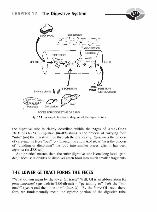
the digestive tube is clearly described within the pages of ANATOMYDEMYSTIFIED.) Ingestion (in-JES-shun) is the process of carrying food‘‘into’’ (in-) the digestive tube through the oral cavity. Egestion is the processof carrying the feces ‘‘out’’ (e-) through the anus. And digestion is the processof ‘‘dividing or dissolving’’ the food into smaller pieces, after it has beeningested (in-JES-ted).
As a practical matter, then, the entire digestive tube is one long food ‘‘grin-der,’’ because it divides or dissolves eaten food into much smaller fragments.
THE LOWER GI TRACT FORMS THE FECES
‘‘What do you mean by the lower GI tract?’’ Well, GI is an abbreviation forgastrointestinal (gas-troh-in-TES-tih-nal) – ‘‘pertaining to’’ (-al) the ‘‘sto-mach’’ (gastr) and the ‘‘intestines’’ (intestin). By the lower GI tract, there-fore, we fundamentally mean the inferior portion of the digestive tube,
CHAPTER 12 The Digestive System 323
Fig. 12.1 A simple functional diagram of the digestive tube.
consisting of everything from the stomach down through the intestines andinto the rectum (REK-tum) – a ‘‘straight’’ (rect) tube leading into the anus.Summarizing, we have:
LOWERGI
TRACT
¼ INFERIOR PORTIONOF DIGESTIVE TUBE
¼ STOMACH þINTESTINES þ RECTUM
þ ANUS
‘‘But what exactly are the intestines, Professor?’’ The intestines are thebowels – two long, hollow, cylinder-shaped tubes, attached end to end, thatextend below the stomach and look like a curled-up string of ‘‘little sausages.’’
The first 20 feet or so of the bowels are the small intestine, which resembles atightly folded string of ‘‘small’’-diameter sausages. This is followed by approxi-mately 6 feet of the large intestine or colon (KOH-lun). The large intestine(colon), of course, resembles a kinked linkage of ‘‘large’’ diameter sausages.
INTESTINES (BOWELS OR‘‘LITTLE SAUSAGES’’)
¼ SMALLINTESTINE
þ LARGEINTESTINE(COLON)
‘‘What does all this have to do with the feces or dregs?’’ Well, the intestines(bowels), especially the large intestine, absorbs lots of water and salt from thechyme (kighm). Chyme is a thick, soupy mass of partially digested materialthat is formed in the stomach, and then continues throughout most of thelower GI tract. The liquid, soupy chyme is progressively dried out as it slowlymoves through the long, kinked sections of the large intestine. More andmore water is absorbed into the bloodstream, until cylinder-shaped, semi-solid stools or feces are created.
‘‘So the chyme is like a loose, watery mud in the stomach and smallintestines? But then it gets dried out into caked mud or dregs, shaped likecylinders or sausages. And the reason is that the cylinder-shaped wall of thelarge intestine gradually presses or molds the chyme within its lumen tomatch the outline of its shape, right?’’ Correct!
ARE THEY FASCES, OR JUST ‘‘BUNDLES’’ OF FECES?
Even more fascinating is the related history behind this connection. Lookcarefully at the spelling and pronunciation of these two words: fasces (FAS-eez) and feces (FEE-seez). We have already noted that feces means ‘‘dregs’’ or‘‘mud.’’ We have added to this the fact that feces are generally shaped likesausages or cylinders. ‘‘Don’t they also look like thick sticks, or maybe,bundles of thick brown sticks?’’
PART 6 Digestive/Genitourinary Systems324
Organ Sysem 2
Organ 1

This is a correct historical observation, Baby Heinie. During the GloryDays of Ancient Rome, high authorites would carry fasces (FAS-eez) –literally, ‘‘bundles’’ of cylinder-shaped, brown wooden sticks – over oneshoulder. The bundles also contained a metal ax, with its sharp bladeprojecting out of the bundle (Figure 12.2).
These fasces were nothing to laugh about (if you valued your life!), becausethey were carried as symbols of high authority. How ironic it is, then, that thefasces or ‘‘bundles’’ of brown sticks carried by Roman officials to indicatehigh authority and imperial dignity, would eventually disappear and bereplaced by another Latin-based word with just two different letters in it –the feces – which are bundles of dregs or human excrement!
Now, we also agreed that the feces were usually greenish-brown in color,like modern mud or the Ancient Roman fasces (bundles of brown sticks).But, surprisingly, if nothing is added to them from outside of the digestivetube, the feces will be defecated as a dull slate-gray in color. ‘‘Then, how dothey go from slate-gray to brown?’’
Enter the Accessory Digestive OrgansTo answer this question, we will have to consider another major aspect of thephysiology of the digestive tube that we have not yet discussed. This functionis secretion. We have previously described secretion as being the principal
CHAPTER 12 The Digestive System 325
Fig. 12.2 The fascinating saga of the Ancient Roman fasces (‘‘bundles’’).
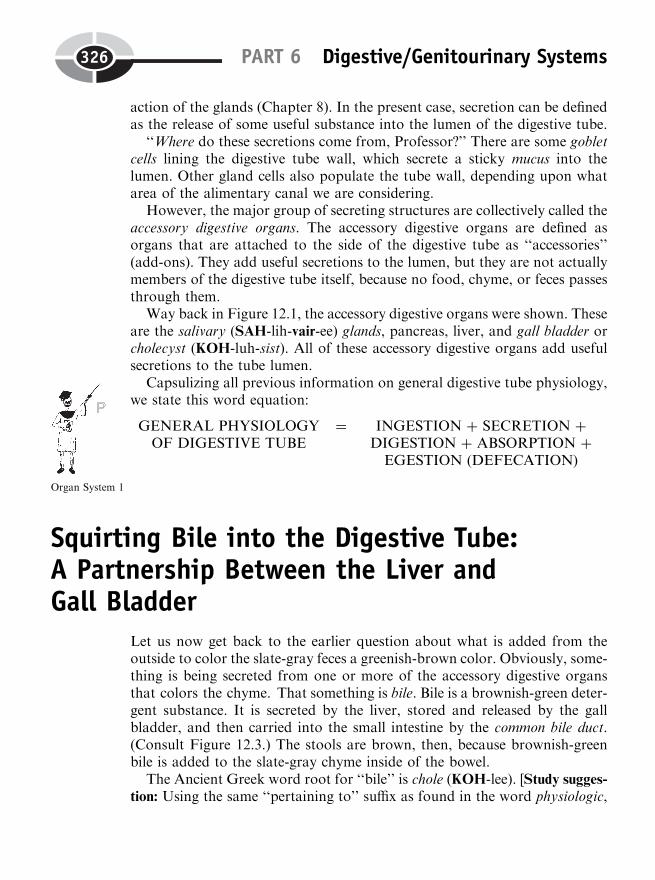
action of the glands (Chapter 8). In the present case, secretion can be definedas the release of some useful substance into the lumen of the digestive tube.
‘‘Where do these secretions come from, Professor?’’ There are some gobletcells lining the digestive tube wall, which secrete a sticky mucus into thelumen. Other gland cells also populate the tube wall, depending upon whatarea of the alimentary canal we are considering.
However, the major group of secreting structures are collectively called theaccessory digestive organs. The accessory digestive organs are defined asorgans that are attached to the side of the digestive tube as ‘‘accessories’’(add-ons). They add useful secretions to the lumen, but they are not actuallymembers of the digestive tube itself, because no food, chyme, or feces passesthrough them.
Way back in Figure 12.1, the accessory digestive organs were shown. Theseare the salivary (SAH-lih-vair-ee) glands, pancreas, liver, and gall bladder orcholecyst (KOH-luh-sist). All of these accessory digestive organs add usefulsecretions to the tube lumen.
Capsulizing all previous information on general digestive tube physiology,we state this word equation:
GENERAL PHYSIOLOGYOF DIGESTIVE TUBE
¼ INGESTION þ SECRETION þDIGESTION þ ABSORPTION þEGESTION (DEFECATION)
Squirting Bile into the Digestive Tube:A Partnership Between the Liver andGall Bladder
Let us now get back to the earlier question about what is added from theoutside to color the slate-gray feces a greenish-brown color. Obviously, some-thing is being secreted from one or more of the accessory digestive organsthat colors the chyme. That something is bile. Bile is a brownish-green deter-gent substance. It is secreted by the liver, stored and released by the gallbladder, and then carried into the small intestine by the common bile duct.(Consult Figure 12.3.) The stools are brown, then, because brownish-greenbile is added to the slate-gray chyme inside of the bowel.
The Ancient Greek word root for ‘‘bile’’ is chole (KOH-lee). [Study sugges-tion: Using the same ‘‘pertaining to’’ suffix as found in the word physiologic,
PART 6 Digestive/Genitourinary Systems326
Organ System 1
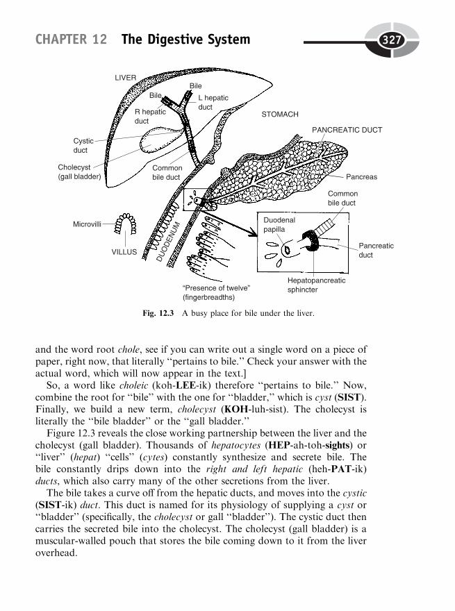
and the word root chole, see if you can write out a single word on a piece ofpaper, right now, that literally ‘‘pertains to bile.’’ Check your answer with theactual word, which will now appear in the text.]
So, a word like choleic (koh-LEE-ik) therefore ‘‘pertains to bile.’’ Now,combine the root for ‘‘bile’’ with the one for ‘‘bladder,’’ which is cyst (SIST).Finally, we build a new term, cholecyst (KOH-luh-sist). The cholecyst isliterally the ‘‘bile bladder’’ or the ‘‘gall bladder.’’
Figure 12.3 reveals the close working partnership between the liver and thecholecyst (gall bladder). Thousands of hepatocytes (HEP-ah-toh-sights) or‘‘liver’’ (hepat) ‘‘cells’’ (cytes) constantly synthesize and secrete bile. Thebile constantly drips down into the right and left hepatic (heh-PAT-ik)ducts, which also carry many of the other secretions from the liver.
The bile takes a curve off from the hepatic ducts, and moves into the cystic(SIST-ik) duct. This duct is named for its physiology of supplying a cyst or‘‘bladder’’ (specifically, the cholecyst or gall ‘‘bladder’’). The cystic duct thencarries the secreted bile into the cholecyst. The cholecyst (gall bladder) is amuscular-walled pouch that stores the bile coming down to it from the liveroverhead.
CHAPTER 12 The Digestive System 327
Fig. 12.3 A busy place for bile under the liver.
We can now state the wonderful Liver–Gall Bladder ‘‘Bile Hand-holding’’Rule: ‘‘Bile is continuously produced and secreted by the liver, but is thentemporarily stored and released into the small intestine by the cholecyst(gall bladder).’’
THE DUODENUM: FINAL DESTINATION FOR BILE ANDPANCREATIC JUICE
Note from Figure 12.3 that bile leaves the cholecyst (gall bladder) storagesac, moves out through the cystic duct, and then into the common bile duct.The common bile duct carries the bile down to its final destination – theduodenum (dew-AH-den-um). The duodenum is the first or most proximal(closest to the stomach) portion of the small intestine.
The duodenum is named for an observation made by some Ancient Latindissectors: it is about ‘‘twelve’’ (duoden) finger-widths (finger-breadths) long.For such a relatively small area, as you can plainly see from the figure, theduodenum is very busy, isn’t it! After all, not only does it receive bile from theliver and gall bladder (cholecyst), but it also gets the pancreatic (pan-kree-AT-ik) juice.
The pancreatic juice is the main secretion of the exocrine portion of thepancreas, which releases it into the pancreatic duct. (You may recollect fromChapter 8 that the endocrine or ‘‘internally secreting’’ portion of the pancreasproduces the hormones, insulin and glucagon.)
Both the bile flowing through the common bile duct, as well as the pancrea-tic juice moving through the pancreatic duct, join together at the hepatopan-creatic (heh-PAT-oh-pan-kree-AT-ik) sphincter (SFINGK-ter). (A sphincter,in general, is a ring of muscle tissue that strongly closes off the lumen of sometube or passageway.) When the hepatopancreatic sphincter relaxes, bile andpancreatic juice flow through the duodenal (dew-AH-deh-nal) papilla (pah-PIL-lah). The duodenal papilla is a ‘‘little nipple or pimple’’ (papill)-likeprojection with a hole in its center. Consequently, both bile and pancreaticjuice drip into the duodenum through this hole in the duodenal papilla.
BILE SQUIRTS OUT OF THE GALL BLADDER: IT’S ALL AMATTER OF MOTILITY!
Unfortunately, Figure 12.3 only showed us where the bile was being carried,after it was secreted (by the liver) and temporarily stored (by the gall blad-der). But it did not reveal exactly why the stored bile was suddenly released
PART 6 Digestive/Genitourinary Systems328
Organ 1

from the cholecyst in the first place. For this we need to discuss a particularphysiological control mechanism, such as negative feedback control (Chapter2) over hormone secretion.
And this negative feedback control mechanism or system needs to focusupon still another general function of the digestive tube – its motility (moh-TIL-ih-tee) or ‘‘ability’’ (-ity) to ‘‘move’’ (motil). ‘‘You mean, if I’m having abowel movement, then my intestines are carrying out the general function ofmotility?’’ Good insight, Baby Heinie!
In the case of the cholecyst or gall bladder, the motility (ability to move)lies with the capacity of its muscular wall to contract. When the muscularwall contracts, it squeezes or compresses the gall bladder lumen. This squeez-ing action squirts a slug of stored bile out of the cholecyst, and eventuallydown into the duodenum. [Study suggestion: To visualize this effect, think ofa rubber bulb attached to one end of a plastic tube, as found in the kitchen,say, on a turkey or poultry baster. When you initially squeeze the bulb, thenegative pressure (suction) draws a quantity of gravy or broth up into thebulb (gall bladder), where it is temporarily stored. The gravy will stay insidethe bulb until your hand gets ‘‘excited’’ and squeezes the filled bulb. Thissqueezing action squirts a slug of gravy (stored bile) out of the bulb (chole-cyst) and into the plastic tube (common bile duct).]
STIMULATING THE GALL BLADDER TO CONTRACT:THE DUODENUM SECRETES A HORMONE
‘‘But what is it, exactly, that stimulates or excites the hand (smooth muscletissue in gall bladder wall) to squeeze the bulb (contract the gall bladder)?’’Good question, Baby Heinie! Take a look at Figure 12.4(A).
Soon after you eat a heavy meal, especially one containing a lot of fat, alarge amount of fatty chyme leaves the stomach and enters the duodenum.The original stimulus for gall bladder contraction, then, is the presence of alarge amount of fatty chyme within the duodenum. The fatty chyme stretchesthe duodenal wall and stimulates a population of endocrine gland cellslocated there. This group of gland cells secretes a hormone called chole-cystokinin (KOH-luh-sist-oh-KIGH-nin) or CCK for short.
The word cholecystokinin literally means ‘‘a substance’’ (-in) that ‘‘moves’’(kin) the ‘‘gall bladder’’ (cholecyst). Specifically, cholecystokinin (CCK)enters the bloodstream after being secreted by the duodenal gland cells.The bloodstream carries the CCK up to the gall bladder. True to its name,the CCK stimulates the smooth muscle in the cholecyst wall to contract and‘‘move’’ (kin). Thus, cholecystokinin, because of its kinetic (‘‘pertaining to
CHAPTER 12 The Digestive System 329

movement’’) effect, triggers the motility of the gall bladder wall. As a result ofthis squeezing action, a slug of stored bile is squirted out of the gall bladder,down through the common bile duct, and into the duodenum.
The bile, as we have mentioned, is a detergent substance (somewhat likethe soapy detergents used to clean the grease from our dirty clothes anddishes). A chemical detergent (such as bile) does its job by causing emulsifica-tion (ih-mul-suh-fuh-KAY-shun). Emulsification is a ‘‘milking out’’ of fatfrom the rest of the chyme in the duodenum. The big globs of fat are thenbroken down into a foam of tiny fat globules. (Consult Figure 12.4, B.)
PART 6 Digestive/Genitourinary Systems330
Fig. 12.4 Negative feedback system for CCK secretion and contraction of gall bladder. (A) Fatty chymetriggers CCK (cholecystokinin) release, which stimulates gall bladder to squirt out stored bile. (B)Emulsification of fat inhibits duodenal gland cells. Rate of CCK secretion greatly decreases. Gall
bladder relaxes.
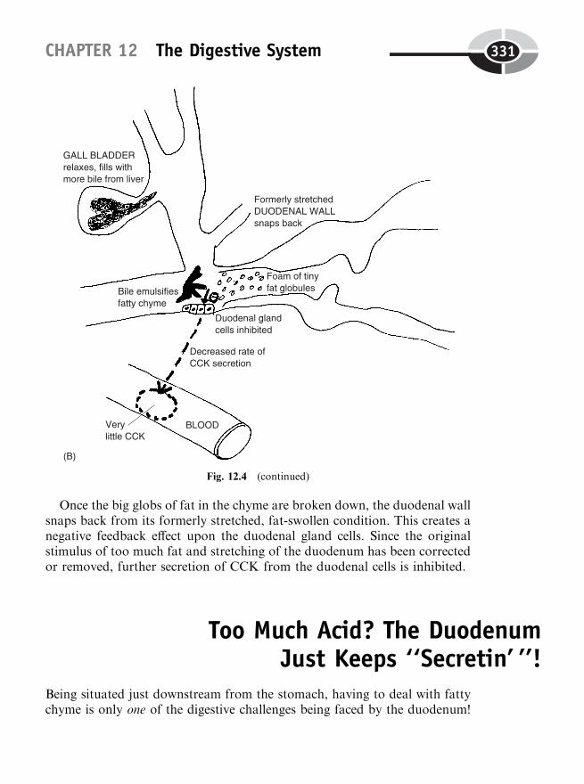
Once the big globs of fat in the chyme are broken down, the duodenal wallsnaps back from its formerly stretched, fat-swollen condition. This creates anegative feedback effect upon the duodenal gland cells. Since the originalstimulus of too much fat and stretching of the duodenum has been correctedor removed, further secretion of CCK from the duodenal cells is inhibited.
Too Much Acid? The DuodenumJust Keeps ‘‘Secretin’ ’’!
Being situated just downstream from the stomach, having to deal with fattychyme is only one of the digestive challenges being faced by the duodenum!
CHAPTER 12 The Digestive System 331
Fig. 12.4 (continued)
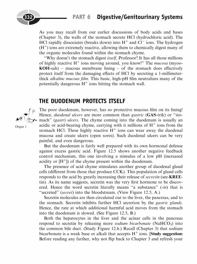
As you may recall from our earlier discussions of body acids and bases(Chapter 3), the walls of the stomach secrete HCl (hydrochloric acid). TheHCl rapidly dissociates (breaks down) into Hþ and Cl� ions. The hydrogen(Hþ) ions are extremely reactive, allowing them to chemically digest many ofthe organic molecules found within the stomach chyme.
‘‘Why doesn’t the stomach digest itself, Professor? It has all those millionsof highly reactive Hþ ions moving around, you know!’’ The mucosa (myoo-KOH-sah) – mucous membrane lining – of the stomach does effectivelyprotect itself from the damaging effects of HCl by secreting a 1-millimeter-thick alkaline mucous film. This basic, high-pH film neutralizes many of thepotentially dangerous Hþ ions hitting the stomach wall.
THE DUODENUM PROTECTS ITSELF
The poor duodenum, however, has no protective mucous film on its lining!Hence, duodenal ulcers are more common than gastric (GAS-trik) or ‘‘sto-mach’’ (gastr) ulcers. The chyme coming into the duodenum is usually anacidic or acid-bearing chyme, carrying with it millions of Hþ ions from thestomach HCl. These highly reactive Hþ ions can wear away the duodenalmucosa and create ulcers (open sores). Such duodenal ulcers can be verypainful, and even dangerous.
But the duodenum is fairly well prepared with its own hormonal defenseagainst excess gastric acid. Figure 12.5 shows another negative feedbackcontrol mechanism, this one involving a stimulus of a low pH (increasedacidity or [Hþ]) of the chyme present within the duodenum.
The presence of acid chyme stimulates another group of duodenal glandcells (different from those that produce CCK). This population of gland cellsresponds to the acid by greatly increasing their release of secretin (see-KREE-tin). As its name suggests, secretin was the very first hormone to be discov-ered. Hence the word secretin literally means ‘‘a substance’’ (-in) that is‘‘secreted’’ (secret) into the bloodstream. (View Figure 12.5, A.)
Secretin molecules are then circulated out to the liver, the pancreas, and tothe stomach. Secretin inhibits further HCl secretion by the gastric glands.Hence, the rate at which additional harmful acid moves from the stomachinto the duodenum is slowed. (See Figure 12.5, B.)
Both the hepatocytes in the liver and the acinar cells in the pancreasrespond to secretin by releasing more sodium bicarbonate (NaHCO3) intothe common bile duct. (Study Figure 12.6.) Recall (Chapter 3) that sodiumbicarbonate is a weak base or alkali that accepts Hþ ions. [Study suggestion:
Before reading any farther, why not flip back to Chapter 3 and refresh your
PART 6 Digestive/Genitourinary Systems332
Organ 1
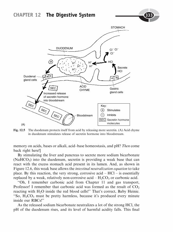
memory on acids, bases or alkali, acid–base homeostasis, and pH? Then comeback right here!]
By stimulating the liver and pancreas to secrete more sodium bicarbonate(NaHCO3) into the duodenum, secretin is providing a weak base that canreact with the excess stomach acid present in its lumen. And, as shown inFigure 12.6, this weak base allows the intestinal neutralization equation to takeplace. By this reaction, the very strong, corrosive acid – HCl – is essentiallyreplaced by a weak, relatively non-corrosive acid – H2CO3 or carbonic acid.
‘‘Oh, I remember carbonic acid from Chapter 11 and gas transport,Professor! I remember that carbonic acid was formed as the result of CO2
reacting with H2O inside the red blood cells!’’ That’s correct, Baby Heinie.‘‘So, H2CO3 must be pretty harmless, because it’s produced every minuteinside our RBCs!’’
As the released sodium bicarbonate neutralizes a lot of the strong HCl, thepH of the duodenum rises, and its level of harmful acidity falls. This final
CHAPTER 12 The Digestive System 333
Fig. 12.5 The duodenum protects itself from acid by releasing more secretin. (A) Acid chymein duodenum stimulates release of secretin hormone into bloodstream.
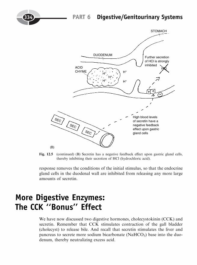
response removes the conditions of the initial stimulus, so that the endocrinegland cells in the duodenal wall are inhibited from releasing any more largeamounts of secretin.
More Digestive Enzymes:The CCK ‘‘Bonus’’ Effect
We have now discussed two digestive hormones, cholecystokinin (CCK) andsecretin. Remember that CCK stimulates contraction of the gall bladder(cholecyst) to release bile. And recall that secretin stimulates the liver andpancreas to secrete more sodium bicarbonate (NaHCO3) base into the duo-denum, thereby neutralizing excess acid.
PART 6 Digestive/Genitourinary Systems334
Fig. 12.5 (continued) (B) Secretin has a negative feedback effect upon gastric gland cells,thereby inhibiting their secretion of HCl (hydrochloric acid).
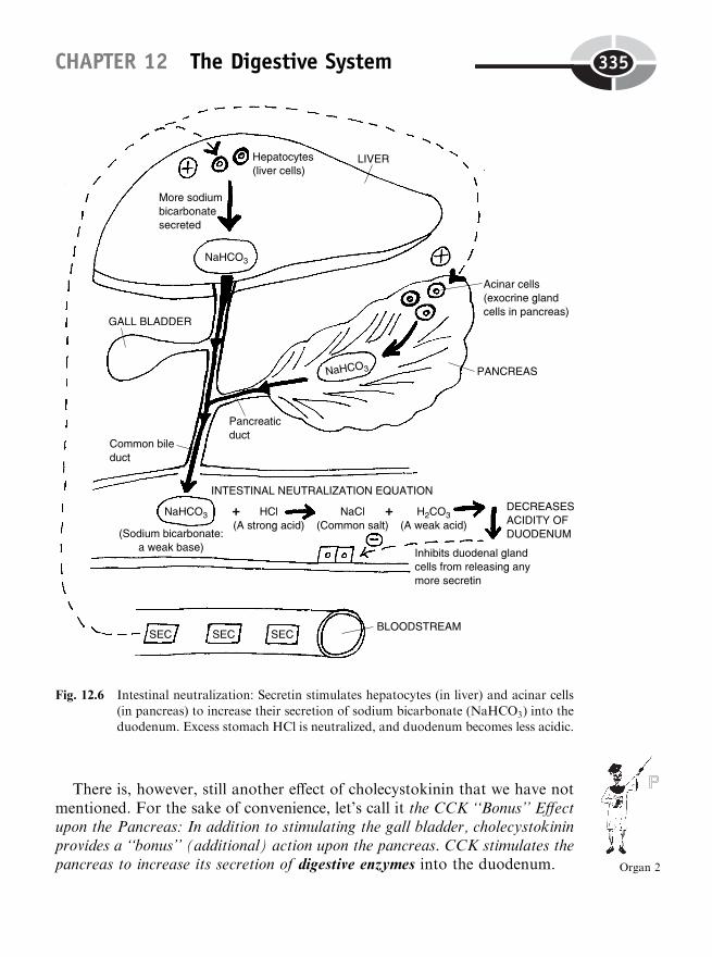
There is, however, still another effect of cholecystokinin that we have notmentioned. For the sake of convenience, let’s call it the CCK ‘‘Bonus’’ Effectupon the Pancreas: In addition to stimulating the gall bladder, cholecystokininprovides a ‘‘bonus’’ (additional) action upon the pancreas. CCK stimulates thepancreas to increase its secretion of digestive enzymes into the duodenum.
CHAPTER 12 The Digestive System 335
Fig. 12.6 Intestinal neutralization: Secretin stimulates hepatocytes (in liver) and acinar cells
(in pancreas) to increase their secretion of sodium bicarbonate (NaHCO3) into theduodenum. Excess stomach HCl is neutralized, and duodenum becomes less acidic.
Organ 2
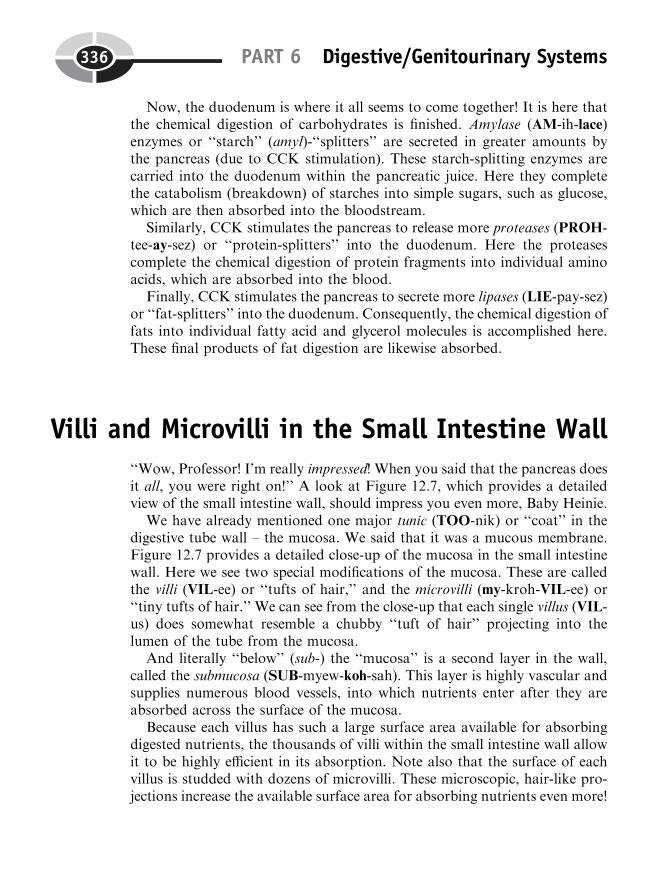
Now, the duodenum is where it all seems to come together! It is here thatthe chemical digestion of carbohydrates is finished. Amylase (AM-ih-lace)enzymes or ‘‘starch’’ (amyl)-‘‘splitters’’ are secreted in greater amounts bythe pancreas (due to CCK stimulation). These starch-splitting enzymes arecarried into the duodenum within the pancreatic juice. Here they completethe catabolism (breakdown) of starches into simple sugars, such as glucose,which are then absorbed into the bloodstream.
Similarly, CCK stimulates the pancreas to release more proteases (PROH-tee-ay-sez) or ‘‘protein-splitters’’ into the duodenum. Here the proteasescomplete the chemical digestion of protein fragments into individual aminoacids, which are absorbed into the blood.
Finally, CCK stimulates the pancreas to secrete more lipases (LIE-pay-sez)or ‘‘fat-splitters’’ into the duodenum. Consequently, the chemical digestion offats into individual fatty acid and glycerol molecules is accomplished here.These final products of fat digestion are likewise absorbed.
Villi and Microvilli in the Small Intestine Wall‘‘Wow, Professor! I’m really impressed! When you said that the pancreas doesit all, you were right on!’’ A look at Figure 12.7, which provides a detailedview of the small intestine wall, should impress you even more, Baby Heinie.
We have already mentioned one major tunic (TOO-nik) or ‘‘coat’’ in thedigestive tube wall – the mucosa. We said that it was a mucous membrane.Figure 12.7 provides a detailed close-up of the mucosa in the small intestinewall. Here we see two special modifications of the mucosa. These are calledthe villi (VIL-ee) or ‘‘tufts of hair,’’ and the microvilli (my-kroh-VIL-ee) or‘‘tiny tufts of hair.’’ We can see from the close-up that each single villus (VIL-us) does somewhat resemble a chubby ‘‘tuft of hair’’ projecting into thelumen of the tube from the mucosa.
And literally ‘‘below’’ (sub-) the ‘‘mucosa’’ is a second layer in the wall,called the submucosa (SUB-myew-koh-sah). This layer is highly vascular andsupplies numerous blood vessels, into which nutrients enter after they areabsorbed across the surface of the mucosa.
Because each villus has such a large surface area available for absorbingdigested nutrients, the thousands of villi within the small intestine wall allowit to be highly efficient in its absorption. Note also that the surface of eachvillus is studded with dozens of microvilli. These microscopic, hair-like pro-jections increase the available surface area for absorbing nutrients even more!
PART 6 Digestive/Genitourinary Systems336

GI Tube MovementsObserve in Figure 12.7 that there are two outer, more peripheral tunics in thedigestive tube wall, in addition to the mucosa and submucosa. These are calledthe muscularis (mus-kyoo-LAIR-us) and the serosa (see-ROH-sah). The mus-cularis is the smooth muscle tissue area in the digestive tube wall. The serosa,in contrast, is a serous (SEER-us) or ‘‘watery’’ membrane. While the serosa ismost important for moistening the outer surface of the GI tube wall, themuscularis is critical for its motility (ability to move and constrict the tube).
Figure 12.7 reveals that there are two thinner layers within the muscularis.These are the outer longitudinal smooth muscle layer, and the inner circular
CHAPTER 12 The Digestive System 337
Fig. 12.7 Major tunics in the small intestine wall.
smooth muscle layer. The longitudinal smooth muscle layer contains musclefibers running ‘‘lengthwise’’ (longitudinally) along the GI tube wall, while thecircular smooth muscle layer has its fibers arranged in circles around the tubelumen.
SMOOTH MUSCLE PACEMAKER CELLS
Smooth muscle tissue in the walls of the digestive tube, like the cardiacmuscle tissue in the wall of the heart (Chapter 9), contains pacemaker cellsthat automatically depolarize or excite themselves at a certain rate or rhythm.These smooth muscle pacemaker cells are modified smooth muscle fibers.They are generally located in the outer longitudinal smooth muscle layer ofthe muscularis. The smooth muscle pacemaker cells automatically depolarizeand become leaky to Naþ ions at a certain rate. This spontaneous depolar-ization of the pacemaker cells keeps summating (adding together) until thethreshold is reached for firing smooth muscle action potentials – travelingwaves of electrochemical excitation.
After they are sufficiently stimulated by smooth muscle action potentialsfrom the outer longitudinal smooth muscle layer, the inner circular smoothmuscle contracts and narrows (constricts) the digestive tube lumen. Thisconstriction or narrowing serves to push the food, chyme, or feces throughthe digestive tract.
Remember this: In general, the outer longitudinal smooth muscle tissuelayer contains pacemaker cells that create smooth muscle action potentials,thereby exciting the inner circular smooth muscle tissue layer. Once excited,the inner circular smooth muscle layer contracts and constricts the digestivetube lumen (open interior). Finally, this constriction of the lumen pushes mate-rial through the tube, toward the anus.
SPECIFIC TYPES OF DIGESTIVE TUBE MOVEMENTS
Now we will consider some specific types of movements of the digestive tube.Figure 12.8 will provide us with a toothpaste tube model of the digestive tractto help illustrate each type.
Peristalsis
The most basic type of movement of the digestive tube is peristalsis (per-ih-STAL-sis). Peristalsis is literally a ‘‘contraction around’’ a particular point of
PART 6 Digestive/Genitourinary Systems338
Organ 3
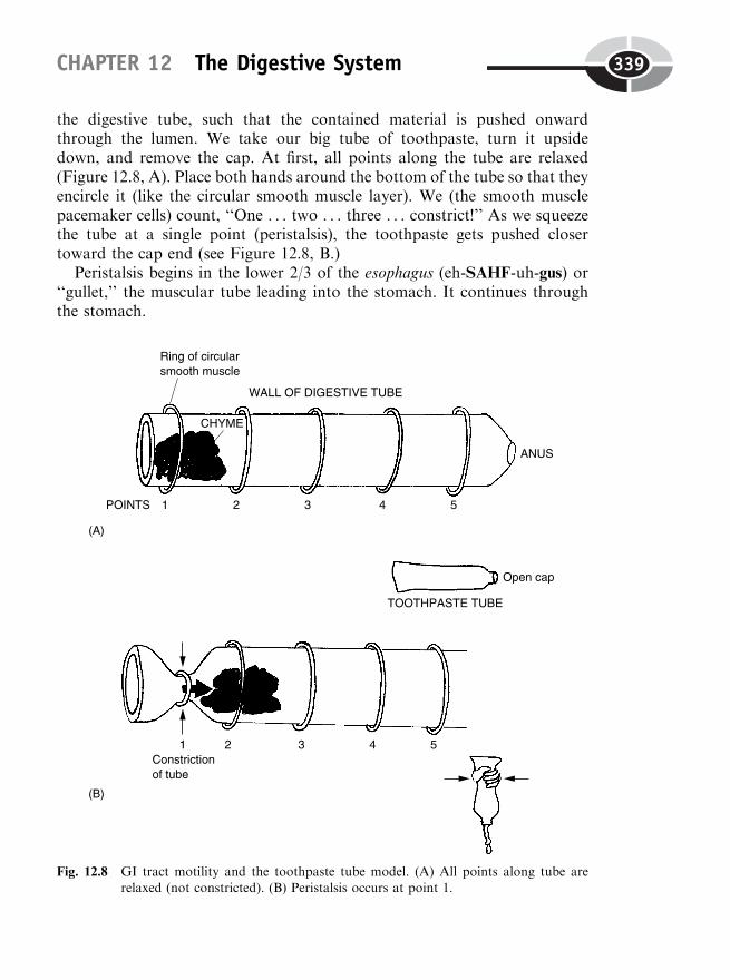
the digestive tube, such that the contained material is pushed onwardthrough the lumen. We take our big tube of toothpaste, turn it upsidedown, and remove the cap. At first, all points along the tube are relaxed(Figure 12.8, A). Place both hands around the bottom of the tube so that theyencircle it (like the circular smooth muscle layer). We (the smooth musclepacemaker cells) count, ‘‘One . . . two . . . three . . . constrict!’’ As we squeezethe tube at a single point (peristalsis), the toothpaste gets pushed closertoward the cap end (see Figure 12.8, B.)
Peristalsis begins in the lower 2/3 of the esophagus (eh-SAHF-uh-gus) or‘‘gullet,’’ the muscular tube leading into the stomach. It continues throughthe stomach.
CHAPTER 12 The Digestive System 339
Fig. 12.8 GI tract motility and the toothpaste tube model. (A) All points along tube arerelaxed (not constricted). (B) Peristalsis occurs at point 1.
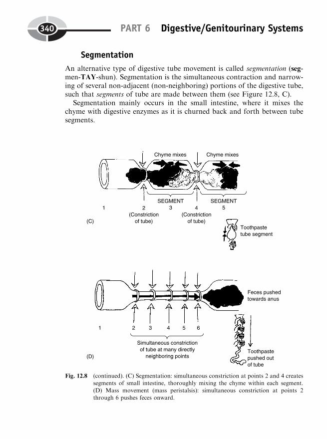
Segmentation
An alternative type of digestive tube movement is called segmentation (seg-men-TAY-shun). Segmentation is the simultaneous contraction and narrow-ing of several non-adjacent (non-neighboring) portions of the digestive tube,such that segments of tube are made between them (see Figure 12.8, C).
Segmentation mainly occurs in the small intestine, where it mixes thechyme with digestive enzymes as it is churned back and forth between tubesegments.
PART 6 Digestive/Genitourinary Systems340
Fig. 12.8 (continued). (C) Segmentation: simultaneous constriction at points 2 and 4 createssegments of small intestine, thoroughly mixing the chyme within each segment.
(D) Mass movement (mass peristalsis): simultaneous constriction at points 2through 6 pushes feces onward.

Mass movement (mass peristalsis)
The third type of digestive tube movement is called mass movement or massperistalsis. This is the simultaneous contraction and narrowing of severaldirectly adjacent (side-by-side) portions of the digestive tube, such that thefeces are pushed in a large mass from one section of the colon to another (seeFigure 12.8, D).
Mass movement (mass peristalsis) occurs in the colon (large intestine) onlythree or four times a day, usually during or soon after eating. It is the mainprocess whereby our heavy ‘‘dregs’’ (feces) are moved along, getting us readyto defecate them.
THE DEFECATION REFLEX
After mass movements powerfully propel some feces all the way through thelarge intestine, they drop down through the sigmoid (SIG-moyd) colon, andinto the rectum (REK-tum). The rectum is a ‘‘straight’’ (rect) muscular-walledtube that leads into the anus.
Two sphincters within the rectum stand between the stools and the anus. Ahigher internal anal sphincter is composed of smooth muscle, so it is notunder our voluntary control. Conversely, the lower external anal sphincteris made up of striated (cross-striped) muscle tissue. Hence, its contractionand relaxation is under our conscious control.
The wall of the upper rectum is stretched as feces enter it, thereby trigger-ing the defecation reflex. This involves the automatic relaxation of the inter-nal anal sphincter when the rectal wall has stretched out far enough with itsload of feces. The feces drop down to hit the external anal sphincter, alsoautomatically.
After the defecation reflex, it is up to the conscious will of the individual todecide when and where to relax the external anal sphincter, and thus todefecate.
Vomiting and Diarrhea: Digestive MovementsThat Can Upset Acid–Base Balance
Our earlier discussions have informed us about the role of the duodenum andof NaHCO3 base from the pancreas, in neutralizing HCl from the stomach. Afollow-up question that needs to be asked is, ‘‘What if there is an excessive
CHAPTER 12 The Digestive System 341
amount of motility in the digestive tube, either at its upper or lower end? Willthis have any effect upon body acid-base balance?’’
VOMITING AND METABOLIC ALKALOSIS
When a person vomits, a large quantity of highly acidic chyme is thrown upfrom the stomach. Hence, a big volume of HCl will tend to be lost. But mostof the NaHCO3 base in the duodenum will remain. The intestinal neutraliza-tion process can no longer work properly, and metabolic alkalosismay result.
Metabolic alkalosis is an abnormal condition of too much body base oralkali, or not enough acid, due to metabolic, that is, non-respiratory, causes.One of the most important non-respiratory causes of metabolic alkalosis isvomiting.
DIARRHEA AND METABOLIC ACIDOSIS
When a person has diarrhea, they excrete a large amount of watery stool fromtheir intestines. Because sodium bicarbonate (NaHCO3) is present in the smallintestine, much of it will be lost with the watery feces. Yet the HCl in thestomach will mainly be left behind. This can produce metabolic acidosis.
Metabolic acidosis is an abnormal condition of too much body acid, or notenough base or alkali, due to metabolic, that is, non-respiratory, causes. Oneof the chief non-respiratory causes of metabolic acidosis is diarrhea.
QuizRefer to the text in this chapter if necessary. A good score is at least 8 correctanswers out of these 10 questions. The answers are listed in the back of thebook.
1. The muscular ring through which one defecates:(a) Hyoid(b) Anus(c) Diaphragm(d) Sigmoid colon
2. Defecation is alternately known as:(a) Egestion(b) Digestion
PART 6 Digestive/Genitourinary Systems342
Organism 1
Organism 2
(c) Absorption(d) Indigestion
3. Feces are like ‘‘dregs’’ because they are:(a) Brown like mud or sediment(b) Frequently passed at the same time as one urinates(c) Held aloft by Roman scholars(d) A real drag to be around!
4. Dietary fiber is not absorbed, since:(a) People really don’t like to eat it(b) Gastric enzymes chop it up into fragments that are too tiny(c) It often contains cellulose, which is largely indigestible by humans(d) There is too much water stuck to its surface
5. If a large cholelith blocks the common bile duct, then the feces will:(a) Probably be purple and have a low fat content(b) Appear slate-gray and have fatty streaks in them(c) Likely not be affected(d) Always be passed as watery diarrhea
6. Bile is actually secreted by the:(a) Cholecyst(b) Duodenum(c) Pancreas(d) Liver
7. Cholecystokinin’s main action:(a) Release of NaHCO3 from the pancreas(b) Powerful stimulation of mass movements(c) Terrifying constipation(d) Release of bile stored in the gall bladder
8. Secretin’s primary action:(a) Causes the hypothalamus to secrete RHs(b) Removal of bile pigments from the bloodstream(c) Stimulates secretion of sodium bicarbonate by liver and pancreas(d) Activation of the defecation reflex
9. Microvilli have as their important characteristic:(a) Vastly increase surface area for absorption(b) Cut the wasting of ATP for unneeded cell repairs(c) Greater motility of the GI tract(d) Boost the rate of smooth muscle pacemaker cells
CHAPTER 12 The Digestive System 343

10. Helps efficiently mix chyme with digestive enzymes in the small intes-tine:(a) Mass peristalsis(b) Vomiting(c) Segmentation(d) Peristalsis
Body-Level Grids for Chapter 12Anatomy and Biological Order Fact Grids for Chapter 12:
ORGANLevel
ORGAN SYSTEMLevel
PART 6 Digestive/Genitourinary Systems344
1
1 2
Physiology and Biological Order Fact Grids for Chapter 12:
ORGANLevel
ORGAN SYSTEMLevel
CHAPTER 12 The Digestive System 345
1 2
3
1
Physiology and Biological Disorder Fact Grids for Chapter 12:
ORGANLevel
ORGANISMLevel
PART 6 Digestive/Genitourinary Systems346
1
1 2
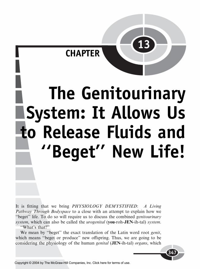
347
CHAPTER13
The GenitourinarySystem: It Allows Usto Release Fluids and
‘‘Beget’’ New Life!
It is fitting that we bring PHYSIOLOGY DEMYSTIFIED: A LivingPathway Through Bodyspace to a close with an attempt to explain how we‘‘beget’’ life. To do so will require us to discuss the combined genitourinarysystem, which can also be called the urogenital (you-roh-JEN-ih-tal) system.
‘‘What’s that?’’We mean by ‘‘beget’’ the exact translation of the Latin word root genit,
which means ‘‘beget or produce’’ new offspring. Thus, we are going to beconsidering the physiology of the human genital (JEN-ih-tal) organs, which
Copyright © 2004 by The McGraw-Hill Companies, Inc. Click here for terms of use.

allow us to carry out this critical survival function of reproduction. Ofcourse, the genital system is often referred to as the reproductive system.
But in covering the genital organs, we will also be obliged to study theurinary (YOUR-ih-nair-ee) system, because some of the organs in these twodifferent systems are shared. In the male penis (PEA-nis) or ‘‘tail,’’ especially,this fact is true. It both delivers the ‘‘seed animals’’ called spermatozoa (sper-mat-uh-ZOH-ah), as well as excreting the urine from the body. So the penis isneither just a genital organ nor just a urinary organ. Rather, it is a combinedgenitourinary organ.
Our approach is therefore summarized anatomically as:
THE GENITOURINARYSYSTEM (UROGENITAL
SYSTEM)
¼ GENITAL(REPRODUCTIVE)
SYSTEM
þ URINARYSYSTEM
For a detailed discussion of these aspects of G-U body structure, pleaseconsult our companion volume, ANATOMY DEMYSTIFIED.
The Notion of Physiological Pathways Withinthe Great Body Pyramid
As you may recollect (Chapter 1), Bodyspace was basically equated to theexistence of a highly orderly internal environment within the human organism.We modeled Bodyspace geometrically, as a series of stacked grids creating aGreat Body Pyramid. And physiology was viewed as a curving ‘‘Pathway ofLife’’ that passes through these stacked grids in the human BodyspacePyramid.
HOMEOSTASIS AND NEGATIVE FEEDBACK ASCURVING PATHWAYS
Two of the core concepts in human physiology – homeostasis and negativefeedback systems or cycles – can also be interpreted as curving pathways thatproceed through Bodyspace for certain time periods:
PART 6 Digestive/Genitourinary Systems348
Organ System 1

The Three Genitourinary PathwaysPathways are fundamentally physiological concepts, since they imply that abody function is occurring – something is moving through the pathway. In ourpresent study, we can talk about three genitourinary pathways. It will also beof great interest to examine what is moving through these G-U pathways, aswell as how and why they are moving through them.
1. THE URINARY PATHWAY AND ITS EXITFROM THE PYRAMID
The urinary pathway is simply the pathway followed by the urine as it is beingexcreted from the body. In an abstract sense (Figure 13.1), the urinary path-way can be drawn as a curving line that leaves the Great Body Pyramid at theChemical Level of organization (molecules, atoms, and subatomic particles).Urine is technically defined as an amber (‘‘yellowish-brown’’), salty, slightlyacidic, watery fluid excreted by the kidneys.
The actual word, urine, however, can also translate from ancient Greek tomean ‘‘presence of urea’’ (you-REE-ah). Urea is the main nitrogen-contain-ing solute found within the urine. It is a white crystal molecule that is formed
CHAPTER 13 The Genitourinary System 349
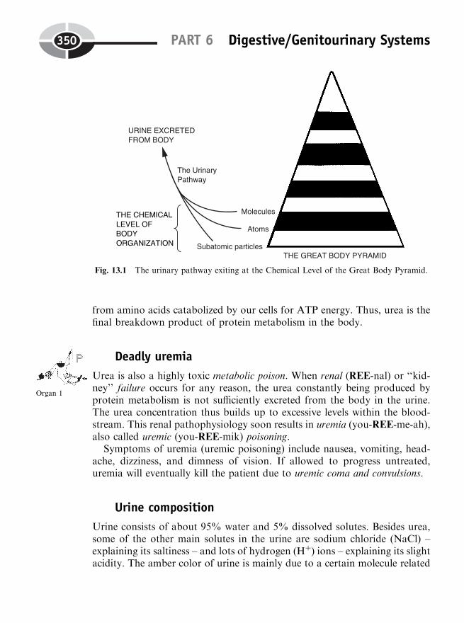
from amino acids catabolized by our cells for ATP energy. Thus, urea is thefinal breakdown product of protein metabolism in the body.
Deadly uremia
Urea is also a highly toxic metabolic poison. When renal (REE-nal) or ‘‘kid-ney’’ failure occurs for any reason, the urea constantly being produced byprotein metabolism is not sufficiently excreted from the body in the urine.The urea concentration thus builds up to excessive levels within the blood-stream. This renal pathophysiology soon results in uremia (you-REE-me-ah),also called uremic (you-REE-mik) poisoning.
Symptoms of uremia (uremic poisoning) include nausea, vomiting, head-ache, dizziness, and dimness of vision. If allowed to progress untreated,uremia will eventually kill the patient due to uremic coma and convulsions.
Urine composition
Urine consists of about 95% water and 5% dissolved solutes. Besides urea,some of the other main solutes in the urine are sodium chloride (NaCl) –explaining its saltiness – and lots of hydrogen (Hþ) ions – explaining its slightacidity. The amber color of urine is mainly due to a certain molecule related
PART 6 Digestive/Genitourinary Systems350
Fig. 13.1 The urinary pathway exiting at the Chemical Level of the Great Body Pyramid.
Organ 1
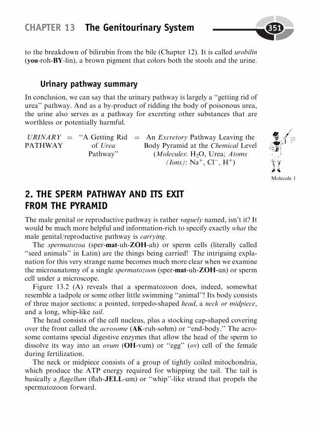
to the breakdown of bilirubin from the bile (Chapter 12). It is called urobilin(you-roh-BY-lin), a brown pigment that colors both the stools and the urine.
Urinary pathway summary
In conclusion, we can say that the urinary pathway is largely a ‘‘getting rid ofurea’’ pathway. And as a by-product of ridding the body of poisonous urea,the urine also serves as a pathway for excreting other substances that areworthless or potentially harmful.
URINARYPATHWAY
¼ ‘‘A Getting Ridof UreaPathway’’
¼ An Excretory Pathway Leaving theBody Pyramid at the Chemical Level
(Molecules: H2O, Urea; Atoms(Ions): Naþ, Cl�, Hþ)
2. THE SPERM PATHWAY AND ITS EXITFROM THE PYRAMID
The male genital or reproductive pathway is rather vaguely named, isn’t it? Itwould be much more helpful and information-rich to specify exactly what themale genital/reproductive pathway is carrying.
The spermatozoa (sper-mat-uh-ZOH-ah) or sperm cells (literally called‘‘seed animals’’ in Latin) are the things being carried! The intriguing expla-nation for this very strange name becomes much more clear when we examinethe microanatomy of a single spermatozoon (sper-mat-uh-ZOH-un) or spermcell under a microscope.
Figure 13.2 (A) reveals that a spermatozoon does, indeed, somewhatresemble a tadpole or some other little swimming ‘‘animal’’! Its body consistsof three major sections: a pointed, torpedo-shaped head, a neck or midpiece,and a long, whip-like tail.
The head consists of the cell nucleus, plus a stocking cap-shaped coveringover the front called the acrosome (AK-ruh-sohm) or ‘‘end-body.’’ The acro-some contains special digestive enzymes that allow the head of the sperm todissolve its way into an ovum (OH-vum) or ‘‘egg’’ (ov) cell of the femaleduring fertilization.
The neck or midpiece consists of a group of tightly coiled mitochondria,which produce the ATP energy required for whipping the tail. The tail isbasically a flagellum (flah-JELL-um) or ‘‘whip’’-like strand that propels thespermatozoon forward.
CHAPTER 13 The Genitourinary System 351
Molecule 1
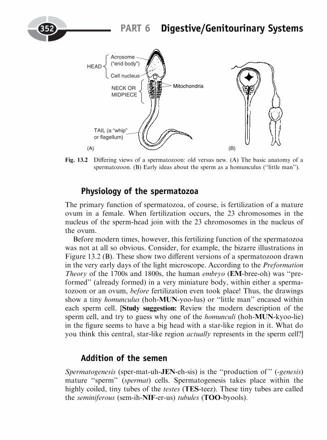
Physiology of the spermatozoa
The primary function of spermatozoa, of course, is fertilization of a matureovum in a female. When fertilization occurs, the 23 chromosomes in thenucleus of the sperm-head join with the 23 chromosomes in the nucleus ofthe ovum.
Before modern times, however, this fertilizing function of the spermatozoawas not at all so obvious. Consider, for example, the bizarre illustrations inFigure 13.2 (B). These show two different versions of a spermatozoon drawnin the very early days of the light microscope. According to the PreformationTheory of the 1700s and 1800s, the human embryo (EM-bree-oh) was ‘‘pre-formed’’ (already formed) in a very miniature body, within either a sperma-tozoon or an ovum, before fertilization even took place! Thus, the drawingsshow a tiny homunculus (hoh-MUN-yoo-lus) or ‘‘little man’’ encased withineach sperm cell. [Study suggestion: Review the modern description of thesperm cell, and try to guess why one of the homunculi (hoh-MUN-kyoo-lie)in the figure seems to have a big head with a star-like region in it. What doyou think this central, star-like region actually represents in the sperm cell?]
Addition of the semen
Spermatogenesis (sper-mat-uh-JEN-eh-sis) is the ‘‘production of ’’ (-genesis)mature ‘‘sperm’’ (spermat) cells. Spermatogenesis takes place within thehighly coiled, tiny tubes of the testes (TES-teez). These tiny tubes are calledthe seminiferous (sem-ih-NIF-er-us) tubules (TOO-byools).
PART 6 Digestive/Genitourinary Systems352
Fig. 13.2 Differing views of a spermatozoon: old versus new. (A) The basic anatomy of aspermatozoon. (B) Early ideas about the sperm as a homunculus (‘‘little man’’).

A healthy young male in his 20s may produce up to 400 million sperma-tozoa every day! They are ejaculated (ih-JACK-yuh-lay-ted) or ‘‘thrown out’’of the urethra (yew-REETH-rah) at the tip of the penis. When the swimmingof the spermatozoa is not assisted by ejaculation, they move at a maximumspeed of about 3 millimeters every hour. These sperm do not have very longto reach an ovum and attempt to fertilize it! Ejaculated spermatozoa have alife expectancy of just 48 to 72 hours.
The spermatozoa, however, are not the only things present within theejaculate (ee-JACK-yoo-lut). After all, they do have to swim through some-thing wet – not just thrash their tails around within hollow tubes filled withair! What the sperm are swimming through is the seminal (SEM-ih-nal) fluidor semen (SEE-mun). The semen (seminal fluid) is the fluid portion of theejaculate. Semen is the thick, milky, sugar-rich, basic pH fluid which sus-pends and provides nutrients for the ‘‘seeds’’ (living sperm cells).
Semen is the fluid added to the sperm when they are being ejaculated. Mostof the fluid is secreted by two types of accessory (‘‘added-on’’) reproduciveorgans: the pair of seminal vesicles (‘‘seed bladders’’) and the single prostate(PRAH-state) gland.
The male ejaculate is about 2 to 5 milliliters in volume. Approximately90% of this ejaculate is semen (seminal fluid), with the other 10% beingspermatozoa. The semen or seminal fluid, in turn, consists of about 60%seminal vesicle fluid and 30% prostate gland fluid.
The sperm count has a lower normal limit of 50 million spermatozoa/mlof ejaculate, and an upper normal limit of 120 million spermatozoa/ml ofejaculate.
Semen summary
To sum things up about the spermatozoa and semen, we state the followingregarding the ejaculate:
THEEJACULATE(2–5 ml involume)
¼ SEMEN OR SEMINALFLUID (60% seminal vesicle
fluid; plus 30% prostategland fluid; 10% from other
sources)
þ SPERMATOZOA(Ejaculated sperm
cells)
The sperm pathway toward the ovum
The sperm pathway leading to fertilization is the pathway followed by thespermatozoa within the semen after they have been ejaculated. When werefer back to the model of the Great Body Pyramid, the male ejaculate can
CHAPTER 13 The Genitourinary System 353
Tissue 1
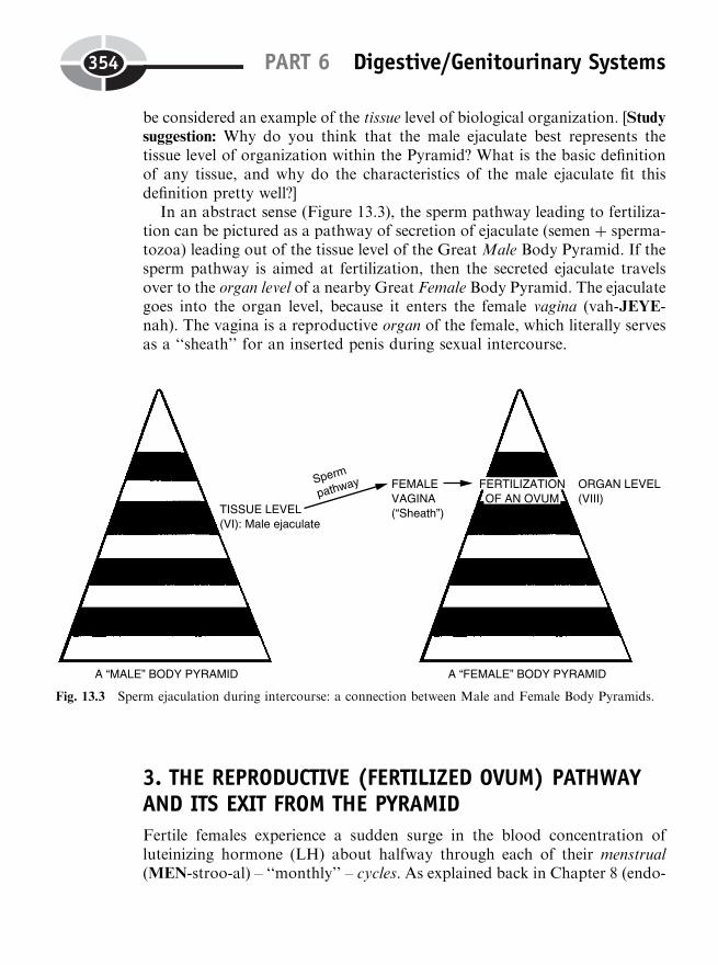
be considered an example of the tissue level of biological organization. [Studysuggestion: Why do you think that the male ejaculate best represents thetissue level of organization within the Pyramid? What is the basic definitionof any tissue, and why do the characteristics of the male ejaculate fit thisdefinition pretty well?]
In an abstract sense (Figure 13.3), the sperm pathway leading to fertiliza-tion can be pictured as a pathway of secretion of ejaculate (semen þ sperma-tozoa) leading out of the tissue level of the Great Male Body Pyramid. If thesperm pathway is aimed at fertilization, then the secreted ejaculate travelsover to the organ level of a nearby Great Female Body Pyramid. The ejaculategoes into the organ level, because it enters the female vagina (vah-JEYE-nah). The vagina is a reproductive organ of the female, which literally servesas a ‘‘sheath’’ for an inserted penis during sexual intercourse.
3. THE REPRODUCTIVE (FERTILIZED OVUM) PATHWAYAND ITS EXIT FROM THE PYRAMID
Fertile females experience a sudden surge in the blood concentration ofluteinizing hormone (LH) about halfway through each of their menstrual(MEN-stroo-al) – ‘‘monthly’’ – cycles. As explained back in Chapter 8 (endo-
PART 6 Digestive/Genitourinary Systems354
Fig. 13.3 Sperm ejaculation during intercourse: a connection between Male and Female Body Pyramids.
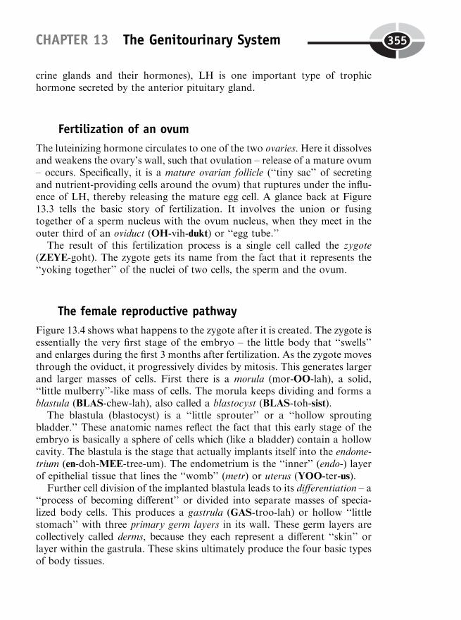
crine glands and their hormones), LH is one important type of trophichormone secreted by the anterior pituitary gland.
Fertilization of an ovum
The luteinizing hormone circulates to one of the two ovaries. Here it dissolvesand weakens the ovary’s wall, such that ovulation – release of a mature ovum– occurs. Specifically, it is a mature ovarian follicle (‘‘tiny sac’’ of secretingand nutrient-providing cells around the ovum) that ruptures under the influ-ence of LH, thereby releasing the mature egg cell. A glance back at Figure13.3 tells the basic story of fertilization. It involves the union or fusingtogether of a sperm nucleus with the ovum nucleus, when they meet in theouter third of an oviduct (OH-vih-dukt) or ‘‘egg tube.’’
The result of this fertilization process is a single cell called the zygote(ZEYE-goht). The zygote gets its name from the fact that it represents the‘‘yoking together’’ of the nuclei of two cells, the sperm and the ovum.
The female reproductive pathway
Figure 13.4 shows what happens to the zygote after it is created. The zygote isessentially the very first stage of the embryo – the little body that ‘‘swells’’and enlarges during the first 3 months after fertilization. As the zygote movesthrough the oviduct, it progressively divides by mitosis. This generates largerand larger masses of cells. First there is a morula (mor-OO-lah), a solid,‘‘little mulberry’’-like mass of cells. The morula keeps dividing and forms ablastula (BLAS-chew-lah), also called a blastocyst (BLAS-toh-sist).
The blastula (blastocyst) is a ‘‘little sprouter’’ or a ‘‘hollow sproutingbladder.’’ These anatomic names reflect the fact that this early stage of theembryo is basically a sphere of cells which (like a bladder) contain a hollowcavity. The blastula is the stage that actually implants itself into the endome-trium (en-doh-MEE-tree-um). The endometrium is the ‘‘inner’’ (endo-) layerof epithelial tissue that lines the ‘‘womb’’ (metr) or uterus (YOO-ter-us).
Further cell division of the implanted blastula leads to its differentiation – a‘‘process of becoming different’’ or divided into separate masses of specia-lized body cells. This produces a gastrula (GAS-troo-lah) or hollow ‘‘littlestomach’’ with three primary germ layers in its wall. These germ layers arecollectively called derms, because they each represent a different ‘‘skin’’ orlayer within the gastrula. These skins ultimately produce the four basic typesof body tissues.
CHAPTER 13 The Genitourinary System 355
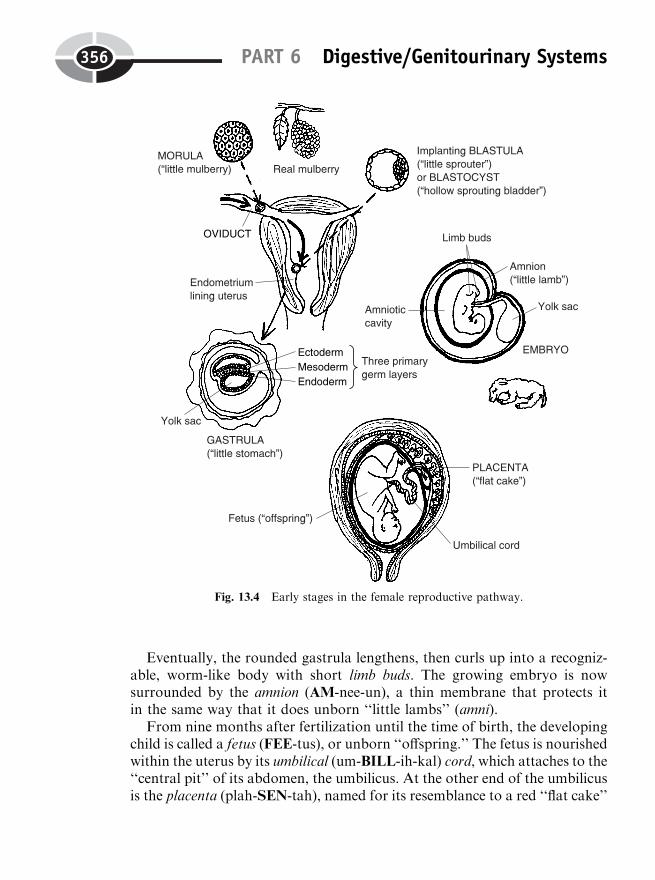
Eventually, the rounded gastrula lengthens, then curls up into a recogniz-able, worm-like body with short limb buds. The growing embryo is nowsurrounded by the amnion (AM-nee-un), a thin membrane that protects itin the same way that it does unborn ‘‘little lambs’’ (amni).
From nine months after fertilization until the time of birth, the developingchild is called a fetus (FEE-tus), or unborn ‘‘offspring.’’ The fetus is nourishedwithin the uterus by its umbilical (um-BILL-ih-kal) cord, which attaches to the‘‘central pit’’ of its abdomen, the umbilicus. At the other end of the umbilicusis the placenta (plah-SEN-tah), named for its resemblance to a red ‘‘flat cake’’
PART 6 Digestive/Genitourinary Systems356
Fig. 13.4 Early stages in the female reproductive pathway.
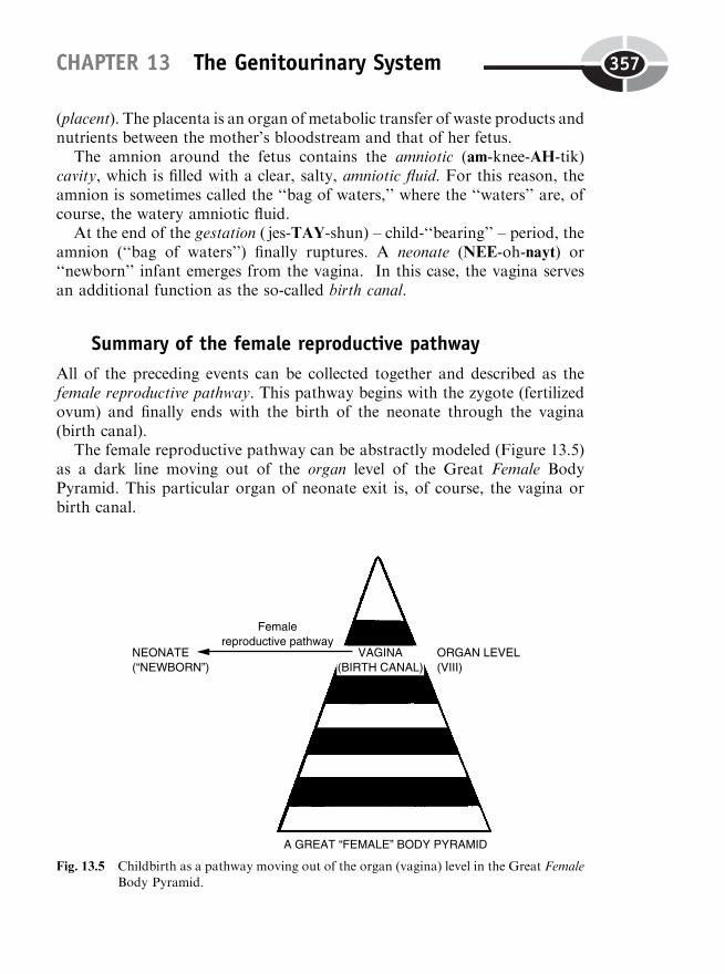
(placent). The placenta is an organ of metabolic transfer of waste products andnutrients between the mother’s bloodstream and that of her fetus.
The amnion around the fetus contains the amniotic (am-knee-AH-tik)cavity, which is filled with a clear, salty, amniotic fluid. For this reason, theamnion is sometimes called the ‘‘bag of waters,’’ where the ‘‘waters’’ are, ofcourse, the watery amniotic fluid.
At the end of the gestation ( jes-TAY-shun) – child-‘‘bearing’’ – period, theamnion (‘‘bag of waters’’) finally ruptures. A neonate (NEE-oh-nayt) or‘‘newborn’’ infant emerges from the vagina. In this case, the vagina servesan additional function as the so-called birth canal.
Summary of the female reproductive pathway
All of the preceding events can be collected together and described as thefemale reproductive pathway. This pathway begins with the zygote (fertilizedovum) and finally ends with the birth of the neonate through the vagina(birth canal).
The female reproductive pathway can be abstractly modeled (Figure 13.5)as a dark line moving out of the organ level of the Great Female BodyPyramid. This particular organ of neonate exit is, of course, the vagina orbirth canal.
CHAPTER 13 The Genitourinary System 357
Fig. 13.5 Childbirth as a pathway moving out of the organ (vagina) level in the Great FemaleBody Pyramid.
THE FEMALEREPRODUCTIVE
PATHWAY
¼ PATHWAY OF THEZYGOTE
(FERTILIZEDOVUM) AND ITSSUCCESSORS
¼ A PATHWAYLEADING TO BIRTH
(EXIT OF THENEONATE)
THROUGH THEVAGINA
Labor and Birth: A BeneficialPositive Feedback Cycle
Finally, after nine months of gestation, a neonate will be expelled from thebirth canal of a pregnant woman by the positive feedback process of labor andnatural childbirth.
Way back in Chapter 2, we introduced the concept of feedback controlsystems or cycles. As you may recall, a positive feedback system or cycle wasconsidered an ‘‘out-of-control’’ system. [Study suggestion: If you don’t remem-ber very much about positive feedback systems, it would be a very good ideafor you to review Chapter 2, before you go further on with this chapter.]
In Chapter 2, we did mention several examples where positive feedbackcycles going to some dramatic climax were beneficial, rather than harmful, tothe body. Sexual arousal leading to eventual climax or orgasm (OR-gazm),for instance, is a positive feedback cycle in the male that normally must occurif he is to ejaculate spermatozoa and fertilize an ovum. (Ejaculating spermand getting a woman pregnant are pretty dramatic events!)
The other main example of beneficial positive feedback cited in Chapter 2was contractions of the ‘‘womb’’ (uterus) that lead to childbirth. We will nowexamine this amazing process in more detail (see Figure 13.6).
Step 1: Estrogen increases irritability of the uterine smooth muscle to oxy-
tocin. The best place to start is where the action is! This is the myometrium(my-oh-MEE-tree-um), a thick layer of smooth ‘‘muscle’’ (myo-) within thewall of the ‘‘uterus’’ (metr). The smooth muscle fibers of the myometriumencircle the uterus, so that when they contract, they constrict and powerfullypush upon the fetus inside.
During the last few weeks of pregnancy, the very large placenta starts todramatically increase its secretion of the hormone estrogen into the mother’sbloodstream. Estrogen molecules circulate to the uterine wall. Here theystimulate the smooth muscle fibers in the myometrium to increase their
PART 6 Digestive/Genitourinary Systems358
Organism 1
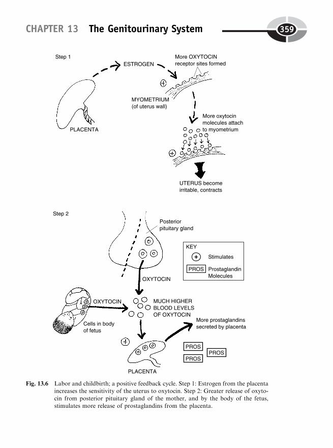
CHAPTER 13 The Genitourinary System 359
���� �
Fig. 13.6 Labor and childbirth; a positive feedback cycle. Step 1: Estrogen from the placentaincreases the sensitivity of the uterus to oxytocin. Step 2: Greater release of oxyto-
cin from posterior pituitary gland of the mother, and by the body of the fetus,stimulates more release of prostaglandins from the placenta.
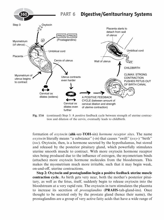
formation of oxytocin (ahk-see-TOH-sin) hormone receptor sites. The nameoxytocin literally means ‘‘a substance’’ (-in) that causes ‘‘swift’’ (oxy-) ‘‘birth’’(toc). Oxytocin, then, is a hormone secreted by the hypothalamus, but storedand released by the posterior pituitary gland, which powerfully stimulatesuterine smooth muscle to contract. With more oxytocin hormone receptorsites being produced due to the influence of estrogen, the myometrium binds(attaches) more oxytocin hormone molecules from the bloodstream. Thismakes the myometrium much more irritable, such that it may begin weak,on-and-off, uterine contractions.
Step 2: Oxytocin and prostaglandins begin a positive feedback uterine muscle
contraction cycle. As birth gets very near, both the mother’s posterior pitui-tary, as well as the fetus, itself, suddenly begin to release oxytocin into thebloodstream at a very rapid rate. The oxytocin in turn stimulates the placentato increase its secretion of prostaglandins (PRAHS-tah-gland-ins). Oncethought to be secreted only by the prostate gland (hence their name), theprostaglandins are a group of very active fatty acids that have a wide range of
PART 6 Digestive/Genitourinary Systems360
Fig. 13.6 (continued) Step 3: A positive feedback cycle between strength of uterine contrac-tion and dilation of the cervix, eventually leads to childbirth.

important biological effects. One of the most important effects of theprostaglandins is to act as a synergist with oxytocin, powerfully boostingits stimulating effect upon the contraction of the myometrium. Therefore,oxytocin and prostaglandins act together as the hormones that start labor.
Step 3: A positive feedback cycle occurs between the amount of cervical
dilation and the strength of uterine contraction. The myometrium (uterinesmooth muscle) begins to forcefully contract due to strong stimulation byboth oxytocin and prostaglandins. The uterus has a cervix (SIR-viks) – atapered, inferior ‘‘neck’’ area. The cervical os is the tiny ‘‘mouth’’ at the tip ofthe uterus, which opens into the vagina or birth canal.
As the uterus begins to contract, the cervical os begins to dilate. And as thecervical os dilates, the uterus contracts even harder. A positive feedback cycletherefore kicks in, between the degree of dilation (widening) of the cervicalos, and the strength of uterine smooth muscle contraction. The wider thecervical os dilates, the harder the uterus contracts. And the harder that theuterus contracts, the wider the os dilates. Finally, when the os is fully dilated,a strong uterine contraction propels the fetus out through the os, then outthrough the birth canal. The positive feedback cycle thus ends with adramatic climax – the birth of a new human being!
Controlling the Output of Urine:The Urinary Excretion Equation
The excretion (E) of urine, like the process of labor and childbirth, is impor-tantly influenced by several hormones. The job of urine formation is mainlydone by the nephrons (nef-RAHNS), the microscopic structural and func-tional units of the kidney. (Consult Figure 13.7, A.)
Each nephron begins with a glomerulus (gluh-MAHR-yew-lus). The glo-merulus is a tiny, red-colored collection of renal capillaries. This structurederives its name from its striking resemblance to a little red ‘‘ball of yarn’’(glomerul). The blood pressure pushing against the inner walls of the capil-laries in each glomerulus causes a filtration (F) of fluid out of the glomerulus,and into the adjoining group of urinary tubules – ‘‘tiny urine tubes.’’
Figure 13.7 (B) shows the processes involving the structures of eachnephron in creating urine from the blood plasma. The summary equationexpressing the formation of urine by the nephrons is:
E ¼ F�Rþ S
CHAPTER 13 The Genitourinary System 361
Molecule 2
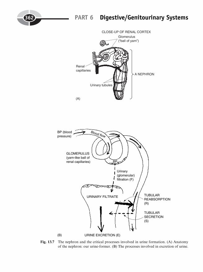
PART 6 Digestive/Genitourinary Systems362
Fig. 13.7 The nephron and the critical processes involved in urine formation. (A) Anatomyof the nephron: our urine-former. (B) The processes involved in excretion of urine.

In this equation, E stands for ‘‘excretion,’’ F for the amount of blood‘‘filtered,’’ R for the amount of tubular reabsorption, and S for tubular secre-tion.
The urinary filtrate (FIL-trayt), or filtration product, comes from thepushing force of the blood pressure against the walls of the renal capillariesin the glomeruli (glah-MEHR-you-lie). This quantity of filtrate is huge, aver-aging about 180 liters of fluid per day in an average adult! [Study suggestion:
If a person’s blood volume is 6 liters, then how many times is the person’stotal blood volume filtered into the kidney tubules every single day?]
While it may seem wasteful, the huge volume of urinary filtrate (F) acts asthe starting pool of fluid and solutes for what eventually becomes excreted asthe urine. Because there is so much of this filtrate, the body can adjust manyfactors to influence how much urine is actually excreted, under particularphysiological conditions.
After urinary filtration, one of the chief processes is tubular reabsorption(R). Reabsorption is the movement of material out of the filtrate, across thewalls of the urinary tubules, and finally back into the bloodstream. Consider,for example, the tubular reabsorption of glucose. Under normal conditions,almost 100% of the glucose that is filtered into the kidney tubules is even-tually reabsorbed back into the bloodstream. As a result, the urine excretedfrom the body is nearly free of glucose.
Under typical conditions, about 99%, or 179 liters, of the urinary filtrate(mostly water) is reabsorbed. [Study suggestion: If a person becomes extre-mely dehydrated, as after excessive sweating, then what do you predict willhappen to the amount of H2O reabsorbed? Will the percent (%) reabsorbedincrease above typical amounts, or decrease below it? Why?]
Another process, tubular secretion (S), involves the active (ATP-requiring)addition of small quantities of particular chemicals from the bloodstreaminto the urinary tubules. Molecules of penicillin (pen-ih-SILL-in) and manyother antibiotics (an-tih-buy-AH-ticks), for instance, are just too large to befiltered across the walls of the glomeruli. Hence, the epithelial cells lining theblood vessels actively pump the penicillin out of the bloodstream and into theurinary tubules. Therefore, penicillin is excreted (E) out of the body, via theurine. Other substances that can be filtered across the glomerular wall, butare still present in huge quantities within the blood plasma, can also besecreted into the urine. An example of this are the millions of Hþ (acid)ions removed from the bloodstream by tubular secretion. Although only afew milliliters of total fluid are generally secreted each day, they still have asignificant influence.
Summarizing our previous urinary excretion equation and plugging insome numbers, we obtain:
CHAPTER 13 The Genitourinary System 363
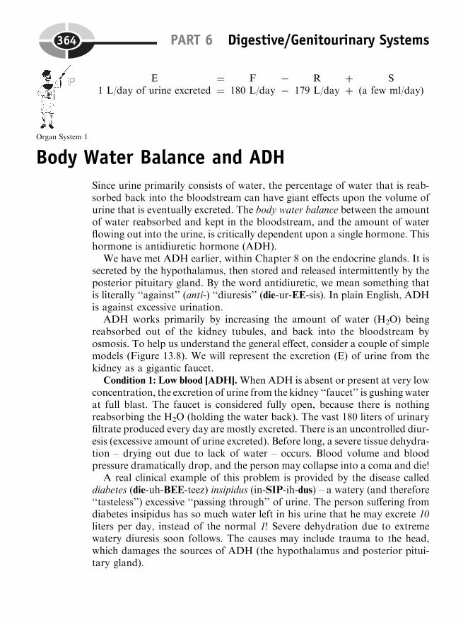
E1 L/day of urine excreted
¼¼
F180 L/day
��
R179 L/day
þþ
S(a few ml/day)
Body Water Balance and ADHSince urine primarily consists of water, the percentage of water that is reab-sorbed back into the bloodstream can have giant effects upon the volume ofurine that is eventually excreted. The body water balance between the amountof water reabsorbed and kept in the bloodstream, and the amount of waterflowing out into the urine, is critically dependent upon a single hormone. Thishormone is antidiuretic hormone (ADH).
We have met ADH earlier, within Chapter 8 on the endocrine glands. It issecreted by the hypothalamus, then stored and released intermittently by theposterior pituitary gland. By the word antidiuretic, we mean something thatis literally ‘‘against’’ (anti-) ‘‘diuresis’’ (die-ur-EE-sis). In plain English, ADHis against excessive urination.
ADH works primarily by increasing the amount of water (H2O) beingreabsorbed out of the kidney tubules, and back into the bloodstream byosmosis. To help us understand the general effect, consider a couple of simplemodels (Figure 13.8). We will represent the excretion (E) of urine from thekidney as a gigantic faucet.
Condition 1: Low blood [ADH].When ADH is absent or present at very lowconcentration, the excretion of urine from the kidney ‘‘faucet’’ is gushingwaterat full blast. The faucet is considered fully open, because there is nothingreabsorbing the H2O (holding the water back). The vast 180 liters of urinaryfiltrate produced every day are mostly excreted. There is an uncontrolled diur-esis (excessive amount of urine excreted). Before long, a severe tissue dehydra-tion – drying out due to lack of water – occurs. Blood volume and bloodpressure dramatically drop, and the person may collapse into a coma and die!
A real clinical example of this problem is provided by the disease calleddiabetes (die-uh-BEE-teez) insipidus (in-SIP-ih-dus) – a watery (and therefore‘‘tasteless’’) excessive ‘‘passing through’’ of urine. The person suffering fromdiabetes insipidus has so much water left in his urine that he may excrete 10liters per day, instead of the normal 1! Severe dehydration due to extremewatery diuresis soon follows. The causes may include trauma to the head,which damages the sources of ADH (the hypothalamus and posterior pitui-tary gland).
PART 6 Digestive/Genitourinary Systems364
Organ System 1
Condition 2: Normal blood [ADH]. Suppose that ADH is being secreted innormal amounts. When a giant kidney faucet is operating at full blast, thelevel of water in the big jug (body fluid content) attached to the faucet soonbegins to run low. (This models a drop in the blood H2O concentrationtoward its lower normal limit.) Such a situation provides a stimulus forgreater ADH secretion.
The ADH hormone molecules can be visualized as a big hand that stronglyturns the giant kidney faucet down to a slow trickle. This dramatic reductionin urine excretion will tend to raise the blood H2O concentration back towardits set-point level. The fluid reservoir attached to the faucet does not rundown too low. Therefore, tissue dehydration is prevented.
ADH SUMMARY
In summary,
CHAPTER 13 The Genitourinary System 365
Fig. 13.8 Two giant body water faucets: with and without ADH. (1) Low blood [ADH]:
diabetes insipidus and an uncontrolled high volume of urine excretion from thewide-open kidney ‘‘faucet.’’ (2) Normal blood [ADH]: as blood [H2O] falls towardits lower normal limit, the hypothalamus and posterior pituitary gland increase
their release of ADH; greatly increased reabsorption of water turns kidney faucetdown to excreting urine at a slow drip.
A D H
means Anti-‘‘Against’’
diuretic‘‘Excessive urination’’
hormone
and, therefore, might also be called:
Anti-‘‘Against’’
dehydration‘‘Excessive bodytissue drying out’’
hormone
Body Salt (Electrolyte) BalanceIn addition to body water balance, body salt or electrolyte balance is likewisecritical for human survival. To be specific, the concentration of sodium (Naþ)ions within the bloodstream is vital. Why? Remember that sodium ions (inaddition to calcium, Caþþ ions) are the main chemical particles responsiblefor neuromuscular excitation! Upsets in their balance (too high or too low anion or electrolyte concentration) therefore affect how nerve fibers excite mus-cles to contract, how well we can think, and even whether our heart continuesto beat or not!
NORMONATREMIA THE IDEAL
The ideal state of affairs for maintaining normal, healthy levels of neuromus-cular excitability, then, is a maintenance of normonatremia (nor-moh-nah-TREE-me-uh). Translated into everyday English, normonatremia is a ‘‘nor-mal’’ (normo-) ‘‘blood concentration of ’’ (-emia) ‘‘sodium’’ (natr). With con-ditions of normonatremia, our heart muscle, neurons, and other ionicallyexcitable tissues usually operate smoothly and with great cooperation andcoordination.
THE DEADLY THREAT OF HYPONATREMIA
‘‘What happens if I work out and lose lots and lots of sodium in my sweat,Professor?’’
In that case, Baby Heinie, we would be looking for renal compensation, or‘‘kidney payback,’’ for help in correcting such a large Naþ loss. Rememberthat urine is a very salty fluid, with a high NaCl content. So we need amechanism to increase the tubular reabsorption (R) of sodium ions fromthe urinary filtrate.
PART 6 Digestive/Genitourinary Systems366
Molecule 3
CHAPTER 13 The Genitourinary System 367
Tissue 1
Molecule 4
‘‘Well, what if I just keep sweating and sweating, don’t take any salt pills,and don’t reabsorb any more Naþ ions?’’
In this case, you may well end up with hyponatremia (high-poh-nah-TREE-me-ah) – ‘‘a blood condition of ’’ (-emia) ‘‘deficient or below-normal’’ (hypo-)‘‘sodium’’ (natr) concentration. Now, why should you be concerned aboutsuffering from hyponatremia, Baby Heinie? [Study suggestion: From yourcurrent reading, why do you think that Baby Heinie should be worriedabout suffering from hyponatremia?]
ALDOSTERONE: THE SALT-CONSERVING HORMONE
Renal compensation for excessive body Naþ loss usually comes in the formof an increased secretion of aldosterone (al-DAHS-ter-ohn). The hormone,aldosterone, is a steroid secreted by the adrenal cortex. The stimulus for itssecretion is a drop in the blood sodium ion concentration toward its lowernormal limit (as after a bout of excessive sweating).
When the appropriate gland cells in the adrenal cortex are stimulated(through an indirect process), they increase their secretion of aldosteroneinto the bloodstream. Aldosterone functions by increasing the reabsorptionof Naþ ions from the urinary filtrate in the kidney tubules, back into thebloodstream. [Study suggestion: Think of a giant hand with the word, aldos-terone, written on the back of it. The hand reaches down into the urinaryfiltrate (wearing a glove, of course) and grabs bunches of salt crystals, pullingthem out of the filtrate before it goes down the drain as urine.]
As a result, the blood [Naþ] usually rises back up toward its set-point level,and the threat of hyponatremia is prevented. In summary, we can think ofaldosterone as our main ‘‘salt-conserving’’ hormone.
A FINAL GOOD-BYE
Congratulations! You have now finished reading PHYSIOLOGYDEMYSTIFIED! (Have you checked out ANATOMY DEMYSTIFIED, itspairmate?) ‘‘Now, goodnight, Baby Heinie!’’ ‘‘Goodnight and thanks,Professor Joe!’’
QuizRefer to the text in this chapter if necessary. A good score is at least 8 correctanswers out of these 10 questions. The answers are listed in the back of thebook.

1. The G part of the G-U system is primarily involved in the function of:(a) Urinary excretion(b) Respiration(c) Reproduction(d) Blood pressure
2. The main nitrogen-containing solute found within the urine:(a) Urea(b) NaCl(c) Ammonia(d) Peptides
3. The urinary pathway leaves the Great Body Pyramid at thelevel:(a) Organelle(b) Cell(c) Organ(d) Chemical
4. The end-body or cap on the head of a sperm:(a) Acrosome(b) Chromosome(c) Mitochondrion(d) Flagellum
5. The fluid added to the sperm when they are ejaculated:(a) Blood serum(b) Lymph(c) Semen(d) Bilirubin
6. The actual body structure that releases an ovum for possible fertiliza-tion:(a) Penis(b) Fallopian tube(c) Urethra(d) Mature follicle
7. The child-bearing period:(a) Ejaculation(b) Gestation(c) Emasculation(d) A long vacation!
PART 6 Digestive/Genitourinary Systems368
8. Two substances critical for starting labor:(a) ADH and aldosterone(b) Insulin and testosterone(c) Estrogen and progesterone(d) Prostaglandins and estrogen
9. The muscular wall of the uterus:(a) Myometrium(b) Dorsal venous sinus(c) Endometrium(d) Corpus striatum
10. The salt-conserving hormone:(a) Vasopressin(b) ADH(c) Insulin(d) Aldosterone
Body-Level Grids for Chapter 13Anatomy and Biological Order Fact Grids for Chapter 13:
TISSUELevel
CHAPTER 13 The Genitourinary System 369
1
ORGAN SYSTEMLevel
Anatomy and Biological Disorder Fact Grids for Chapter 13:
TISSUELevel
Physiology and Biological Order Fact Grids for Chapter 13:
MOLECULELevel
PART 6 Digestive/Genitourinary Systems370
1
1
ORGAN SYSTEMLevel
ORGANISMLevel
CHAPTER 13 The Genitourinary System 371
1 2
3 4
1
1
Physiology and Biological Disorder Fact Grids for Chapter 13:
ORGANLevel
PART 6 Digestive/Genitourinary Systems372
1
Test: Part 6
DO NOT REFER TO THE TEXT WHEN TAKING THIS TEST. A goodscore is at least 18 (out of 25 questions) correct. Answers are in the back ofthe book. It’s best to have a friend check your score the first time, so youwon’t memorize the answers if you want to take the test again.
1. In Ancient Rome, fasces were:(a) Dregs(b) Defecated material(c) Salivary glands(d) Cheek cell smears(e) Bundles of brown sticks
2. The release of some useful substance into the lumen of the digestivetract:(a) Reabsorption(b) Secretion(c) Digestion(d) Ingestion(e) Desiccation
373
Copyright © 2004 by The McGraw-Hill Companies, Inc. Click here for terms of use.
3. Hepatocytes would be found in the:(a) Liver(b) Skeletal muscles(c) Duodenal wall(d) Dirty wash(e) Spleen
4. Carries bile into the gall bladder:(a) Cystic duct(b) Jejunal fold(c) Serosa lumen(d) Common bile duct(e) Pancreatic duct
5. Sphincter guarding entrances of both the common bile duct andpancreatic duct:(a) Cardiac(b) Lower esophageal(c) Internal anal(d) Hepatopancreatic(e) Biliary
6. CCK represents a:(a) Symbol for a liquid detergent substance(b) Type of enzyme found in RBCs(c) Hormone that stimulates the release of stored bile(d) Chemical messenger that promotes gastric secretions(e) Species of intestinal parasites
7. Results from a state of overhydration:(a) Low blood [ADH](b) Constipation(c) Joint crystals(d) High blood [ADH](e) High blood [aldosterone]
8. Secretion is inhibited whenever duodenal pH rises beyond 7:(a) Insulin(b) Secretin(c) Glycogen(d) Amylase(e) Starch
PART 6 Digestive/Genitourinary Systems374
9. Proteases have as their main targets:(a) Protein fragments(b) Carbohydrate polymers(c) Simple sugars(d) Membrane lipids(e) Cell mitochondria
10. The final products of fat digestion, which are absorbed:(a) Amino acids such as glycine(b) Chunks of chyme(c) Frozen slush(d) Fatty acids and glycerol(e) Carbonic anhydrase
11. Greatly increase surface area for absorption within the small intestine:(a) Rugae(b) Submucosae(c) Villi and microvilli(d) Gastric enzymes(e) Mucosal peptides
12. Thought to be mainly responsible for excitation of the GI tube wall:(a) Longitudinal smooth muscle(b) Goblet cells(c) Circular smooth muscle(d) Tunica adventitia(e) Cholecyst
13. Constriction around a single point of the digestive tube:(a) Mass movements(b) Duodenal stretching with chyme(c) Vasoconstriction(d) Valsalva’s maneuver(e) Peristalsis
14. Produces thorough mixing of chyme with digestive enzymes:(a) Segmentation(b) Gastric bypass(c) Cholecystectomy(d) Sphincter closure(e) Appendectomy
15. Mainly responsible for coloring urine an amber hue:(a) Urobilin
Test: Part 6 375

(b) Acetyl CoA(c) Lysosomes(d) Gut bacteria(e) Ammonia
16. According to the Preformation Theory:(a) Sperm cells contained homunculi(b) Mature ova sometimes talked amongst themselves(c) Eggs and sperm cells were already formed in the blastula(d) People were born with tails, then lost them(e) Spermatozoa were always ejaculated as pre-formed pairs
17. Ejaculated spermatozoa have a life expectancy of about hours:(a) 2(b) 48–72(c) 100–300(d) 5,000–10,000(e) 10 years
18. Makes up about 60% of the semen:(a) Salivary exudate(b) Seminal vesicle fluid(c) Prostate fluid(d) Female vaginal mucus(e) Spermatozoa
19. Represents the ‘‘yoking together’’ of a sperm plus ovum:(a) Morula(b) Blastocyst(c) Ectoderm(d) Zygote(e) Chromosome bead
20. The ‘‘bag of waters’’:(a) Amnion(b) Umbilical cord(c) Blastula(d) Urocyst(e) Dura mater
21. A tubal ligation essentially interrupts:(a) Photosynthesis(b) Spermatogenesis(c) Cell division
PART 6 Digestive/Genitourinary Systems376
(d) The female reproductive pathway(e) Entry of a sperm into the vagina
22. Labor and childbirth are good examples of:(a) Negative feedback systems(b) Homeostatic mechanisms(c) Cybernetic cycles(d) Linear programs(e) Positive feedback
23. Starts secreting large amounts of estrogen during the final weeks ofpregnancy:(a) Dermis(b) Umbilical cord(c) Liver(d) Placenta(e) Stomach
24. Smooth muscle portion of the uterine wall:(a) Endometrium(b) Glandular epithelium(c) Renal capsule(d) Mesoderm(e) Myometrium
25. Tiny yarn-like ball of renal capillaries:(a) Collecting duct(b) Glomerulus(c) Transverse colon(d) Cardia(e) Genital tubercle
Test: Part 6 377
378
Final Exam
DONOT REFER TO THE TEXTWHEN TAKING THIS EXAM. A goodscore is at least 75 correct. Answers are in the back of the book. It’s best tohave a friend check your score the first time, so you won’t memorize theanswers if you want to take the test again.
1. Speaking broadly, an organism is:(a) A collection of unrelated organs and organ systems(b) Some multi-celled creature with no apparent design(c) Any living body with a high degree of recognizable pattern(d) Several associated body structures whose functions are unrelated(e) One or more apparently randomly constructed bodies
2. To many early Greek thinkers, the human body was a microcosm.This implies that:(a) Living things do not appear to follow fixed laws(b) The internal environment is a tiny representative of the external
environment(c) A macrocosm existed that directly contradicted whatever hap-
pened at the microcosm
Copyright © 2004 by The McGraw-Hill Companies, Inc. Click here for terms of use.
(d) The Laws of Nature were apparently suspended when humanswere around
(e) No larger Universe exists
3. First coined the word physiology:(a) Hippocrates(b) Galen(c) Aristotle(d) Claude Bernard(e) Socrates
4. Plain functions are:(a) Actions that non-living structures in the body perform(b) Structures that occupy space(c) Best represented by nouns rather than verbs(d) Whatever all living or non-living bodies do(e) Seldom apparent to outside observers
5. A roughly S-shaped pattern of relative constancy:(a) Anatomy(b) Hemostasis(c) State of chaos(d) Morbidity(e) Homeostasis
6. According to the Law of Complementarity:(a) We should give each other compliments(b) Metabolism is a ‘‘state of fixity’’(c) The existence of body structures having particular characteristics
limits or determines the possible functions those structures mighthave
(d) Oral body temperature is absolutely constant(e) ‘‘Monkey see, monkey do’’
7. A stimulus is:(a) Something that inhibits the activities of a sensory receptor(b) A group of nerve impulses that always result in a corrective action(c) Any creature that causes others to withdraw(d) A detectable change in the body’s internal environment, or within
the external one(e) Generally preceded by a response
8. Pathophysiology represents a:(a) Highly orderly progression of changes in body structure
Final Exam 379
(b) Change in body structure that reflects normal body function(c) State wherein physiology is diseased and greatly abnormal(d) Type of untreatable medical condition(e) Bit of order within an otherwise diseased organism
9. A Great Pyramid of Structure-Function Disorder would involve:(a) Regular arrangements of body structures to perform functions
that were also regular(b) Loss of pattern between body structures and their functions(c) Maintenance of homeostasis across various organ systems(d) Stacked grids of body structures that fit nicely into healthy groups(e) Many levels of biological organization that interacted normally
with one another
10. Physiology is limited to these levels of body organization:(a) Atoms and subatomic particles(b) Organ systems and everything below them(c) Living cells and everything above them(d) Molecules, but not atoms(e) Most subatomic particles (except for electrons)
11. The concept of a lower normal limit (LNL) is valuable because:(a) Accurate verbal descriptions of people and processes are obtained(b) Sensitivity to the personal feelings of the subject are observed(c) It provides a floor, below which the parameter is in a state of
deficiency(d) Qualitative thinking subject to verification and checking by others
is attained(e) It provides a ceiling, above which the parameter is in a state of
excess
12. Parameters:(a) Are nothing but recorded data(b) Seldom provide reliable information(c) Are produced by measuring and recording the results in certain
numbers of units(d) Are the same things as body structures(e) Frequently involve functions that cannot be tied down
13. Shoe weight in pounds:(a) Anatomical parameter(b) Physiology(c) Function
Final Exam380
(d) Structure(e) Structural parameter
14. An aspect of living body function that has been measured andexpressed in a certain number of units:(a) Structural parameter(b) Feedback loop(c) Functional parameter(d) Negative change(e) Physiological parameter
15. The book said that the phrase ‘‘Compu-think’’ can be interpreted tomean:(a) Computer-like modes or ways of human thinking(b) An over-reliance upon technology is foolish(c) Science is a bad thing(d) More attention should be given to arts in the school curriculum(e) Teachers should be paid higher salaries
16. ‘‘Regression towards the mean,’’ or ‘‘things usually tend to balance-out’’:(a) A hypo-state(b) Set-point value(c) Diabetes insipidus(d) Upper normal limit(e) Random ‘‘noise’’ within an otherwise controlled environment
17. Milliliters (ml) and liters (L) are typical units of:(a) Mass or weight(b) Concentration and density(c) Volume(d) Area(e) Length
18. The typical units for recording blood pressure (BP):(a) Degrees Fahrenheit(b) Cubic centimetres(c) Grams per liter of blood(d) Millimeters of mercury (Hg)(e) pH units
19. The ancient Humoral Doctrine was somewhat like the modern con-cept of:(a) Absolute constancy
Final Exam 381
(b) Balance as a rough equality between two or more different things(c) Strict following of laws that are essentially meaningless(d) An imbalance between the Universe and ourselves(e) Wisdom is found in the details!
20. The Father of Modern Medicine:(a) Basel(b) Plato(c) Vesalius(d) Hippocrates(e) Galileo
21. An upside-down, dead giraffe without spots is most closely modeling:(a) Pathophysiology(b) Normal function(c) Homeostasis(d) Negative feedback(e) Biological order
22. Provided the first strong evidence of natural chemical synthesis in thebody, by performing experiments on living animals:(a) Barnum & Bailey(b) Schleiden & Schwann(c) Claude Bernard(d) William Harvey(e) Sir Robin of Loxley
23. The phrase milieu interieur comes from the French for:(a) ‘‘Nourishing substances’’(b) ‘‘A state of change’’(c) ‘‘External surroundings’’(d) ‘‘The raging sea’’(e) ‘‘Internal environment’’
24. The fluid lying outside of our cells:(a) Organic paste(b) Intracellular fluid(c) Organelles(d) Extracellular fluid(e) Solid rock
25. Originated the concept of a ‘‘fight-or-flight’’ response:(a) Walter Cannon(b) Claude Bernard
Final Exam382

(c) Ernest Starling(d) Hippocrates(e) Connie Chung
26. First used the term homeostasis:(a) Walter Mondale(b) Galen(c) Leonardo da Vinci(d) Albrecht Durer(e) Walter Cannon
27. The use of parameters generally improves the quality of informationregarding homeostasis, because:(a) More vagueness of definition allows more ease in translation(b) Structure determines function (except when both are being
measured)(c) Whether a relative constancy is being maintained can be directly
assessed(d) They provide less information, but it is more honest(e) Some people can’t be trusted!
28. Cybernetics is the:(a) Ancient art of translating writing(b) Modern science of communication and control in man and
machine(c) Relative constancy of thought waves(d) Same thing as kinetics(e) Opposite of control system theory
29. For a pendulum swinging from a clock, the set-point is:(a) A way of reading the time(b) The far-left swing(c) The far-right swing(d) The average position of the pendulum over time(e) The place where the pendulum swings so far that it breaks
30. Stressors:(a) Are exactly the same things as stimuli(b) Can effectively be removed from the environment(c) Occupy space, have mass and weight, and assume a definite form(d) Tend to disturb parameters from their set-point levels(e) Can always be corrected or removed
Final Exam 383
31. System whose output or response curves back upon the start to inhibitfurther activity of the system:(a) Negative feedback(b) Linear(c) Summative(d) Partially movable(e) Positive feedback
32. An afferent pathway:(a) Carries information away from a control center(b) Means a motor channel(c) Is generally a sensory pathway(d) Acts like a boomerang(e) Is just an efferent pathway
33. Establishes 98.6 degrees F as the set-point for oral body temperature:(a) Effector(b) Free nerve endings in the skin(c) Response(d) Stimulus(e) Control center
34. An ‘‘out-of-control’’ system:(a) Anatomical(b) Homeostatic(c) Normal immune(d) Positive feedback(e) Neutral
35. Body temperature way below the lower normal limit:(a) Normothermia(b) Hypotension(c) Euthyroidism(d) Hyperthermia(e) Hypothermia
36. A system that tends to maximize its own errors:(a) Blood glucose control(b) Vicious cycle(c) Benign line(d) Cybernetic(e) Missile guidance
37. If a state of homeostasis exists, then clinical health:
Final Exam384
(a) Is absent(b) Probably also exists(c) Is totally unaffected(d) May never come into being(e) Has been irreversibly lost
38. A beneficial example of positive feedback:(a) Climbing a tree higher and higher(b) Singing so loudly that no one can stand it!(c) Washing the dishes less, the more that someone complains(d) Firing and transmission of a neuron action potential(e) Blood pressure getting so low that a person faints
39. The Chemical Level of body organization:(a) Atoms, but not cells(b) Atoms, molecules, electrons, protons, and neutrons(c) Mitochondria and cell nuclei(d) Heart and cardiac muscle tissue(e) Soup to nuts
40. The class of molecules rich in carbon (C) atoms:(a) Crystals(b) Electrolytes(c) Inorganic(d) Hydrated(e) Organic
41. The chemical bond between H and O in H2O:(a) Polar covalent(b) Ionic(c) Non-electric(d) Nonpolar covalent(e) Double
42. Cations:(a) Negatively charged ions(b) Nonpolar solutes(c) Positively charged ions(d) Peptide bonds(e) The same thing as anions
43. Anabolism is:(a) Synthesis or build-up(b) Exchange
Final Exam 385
(c) Enzyme reaction(d) Breakdown or decomposition(e) Replacement
44. HCl þ NaHCO3:(a) Hþ þ Na2CO3
(b) NaCl þ H2CO3
(c) H2SO4 � HCl(d) CO2 þ H2O(e) ATP þ Phosphate
45. Hþ ion acceptors:(a) Bases (alkali)(b) H2CO3
(c) HCl(d) Acids(e) Neutral substances
46. Has a low pH, but a high [Hþ]:(a) Baking soda(b) Albumen(c) Vinegar(d) Blood(e) Saliva
47. Average or set-point blood pH level:(a) 4.8(b) 6.2(c) 7.3(d) 7.4(e) 7.5
48. Baby Heinie drinks a bottle of lye. He would likely suffer:(a) Hallucinations(b) Metabolic alkalosis(c) Delusions of grandeur(d) Metabolic acidosis(e) Neutral liquid overdose
49. Microphysiology can also be legitimately called:(a) Organelle physiology(b) Cell anatomy(c) Microbial fine structure(d) Cell functions
Final Exam386
(e) Molecular interactions
50. Fluid mosaic:(a) Describes nature of the blood plasma(b) Structure of plasma membrane(c) The intercellular protein matrix(d) Pretty colored pictures on the body wall(e) Cell membrane protein bilayer
51. ‘‘Like dissolves like’’ means:(a) Oil and water don’t mix!(b) Soap, like car wash liquid, can clean family pets(c) NaCl dissolves in CH3CH2CH3
(d) Nonpolar covalent substances bond with polar ones(e) Ionic compounds dissolve in hydrocarbon solvents
52. Glucose enters most cells by this process:(a) Osmosis(b) Facilitated diffusion(c) Simple diffusion through pores(d) Active transport(e) Phagocytosis
53. The ATP–ADP cycle:(a) Involves the splitting of ADP by ATPase(b) The combination of phosphate with ADP(c) Glucose combines with phosphorus(d) ATP sops up free energy and becomes AQP(e) H2O and H2CO3 ¼ ATP
54. Cellular respiration occurs in this part:(a) Cytoplasm(b) Lysosome(c) Glycogen droplet(d) Mitochondrion(e) ER
55. A single gene provides the:(a) Chemical code for the synthesis of a certain protein(b) List of ingredients for making an entire new organism(c) Structure of a whole DNA double helix(d) Codons for a particular lipid(e) Basis for absorbing most alcohols into the liver cells
Final Exam 387
56. The main dense fibrous connective tissue portion of the skin:(a) Hypodermis(b) Dermis(c) Hair follicles(d) Sebaceous glands(e) Subdermis
57. Blood plasma and other parts of the ECF are normally solu-tions:(a) Hypertonic(b) Salt and hemoglobin(c) Isotonic(d) Keratin(e) Hypotonic
58. Cells that create the bone matrix:(a) Endothelial(b) Simple squamous epithelial(c) Neurons(d) Muscle fibers(e) Osteoblasts
59. Hypocalcemia is very dangerous, because:(a) Too little calcium in the urine may cause kidney stones(b) Potassium is also found at high concentrations within most cells(c) Excessive calcium may dull the sense of taste, leading to starva-
tion(d) Normocalcemic states may just be taken for granted(e) Adequate blood Caþþ levels are absolutely necessary for muscles
to contract
60. The major body function performed by red bone marrow:(a) Transmission of action potentials(b) Hematopoiesis(c) Thermoregulation(d) Water reabsorption(e) Naþ excretion
61. In a bone–muscle lever system, the serves as a lever:(a) Joint(b) Bone(c) Skin(d) Skeletal muscle
Final Exam388
(e) Blood vessel
62. The major central portion of a neuron, which contains its nucleus andmost other organelles:(a) Soma(b) Dendrite(c) Axon(d) Synapse(e) Vesicles
63. An alternative name for the neuromuscular junction is the:(a) Axon hillock(b) Transmitter binding sites(c) Motor end-plate(d) Myofibril(e) Synapse
64. Within muscle fibers, the lateral sacs of the sarcoplasmic reticulumserve as:(a) Sliding filaments between molecules(b) Passageways for excitation(c) Terminals that release neurotransmitter molecules(d) Hþ ion acceptors(e) Caþþ ion storage depots
65. Myosin cross-bridges:(a) Places where actin changes into myosin(b) Chief points of contact between thin actin and thick myosin myo-
filaments(c) Are another form of myosin ATPase enzyme(d) Water-filled channels through muscle fibers(e) The same thing as sarcomeres
66. Troponin–tropomyosin serves to:(a) Attach a muscle fiber to a motor neuron(b) Stimulate fibers across the neuromuscular junction(c) Open up gaps within neuron cell membranes(d) Inhibit muscle contraction by preventing a chemical connection
between actin and myosin(e) Excite or agitate ribosomes to synthesize more proteins
67. Electrical shock not producing any observable response from amuscle:(a) Maximal stressor
Final Exam 389

(b) Subthreshold stimulus(c) Threshold prod(d) Twitch(e) Sarcomere
68. Either all of the muscle fibers in a given motor unit contract togetherat the same time, or none of them contract:(a) Summation(b) Recruitment of motor units(c) The All-or-None Law(d) Threshold(e) Tetanus
69. Most continuous body movements represent these types of musclecontractions:(a) Tetanus(b) Fatigue(c) Single twitches(d) Inverted twitches(e) Summations of single twitches
70. Serves as a reserve muscle energy storage, that can quickly be con-verted to ATP:(a) Creatine phosphate(b) Myoglobin(c) Heme iron(d) Calcium ions(e) Myosin ATPase
71. Low oxidative (LO) muscle fibers are basically:(a) Fast twitch(b) Aerobic(c) Slow twitch(d) Myosin-deficient(e) Anaerobic
72. A narrow, fluid-filled gap, where two neurons almost (but not quite)have their axons and cell body touch:(a) Tight junction(b) Axon hillock(c) Synapse(d) Nephron(e) Ganglion
Final Exam390
73. Myelinated nerve fibers:(a) Usually have a dull, grayish appearance(b) Saltatory conduction(c) Do not conduct action potentials at all(d) Local circuit currents(e) Intercalated discs
74. The automatic pulling back of some body part from a harmful orannoying stimulus:(a) Ventral rooting of a spinal nerve(b) Effector activation(c) Withdrawal reflex(d) Sensory pathway(e) Descending nerve tracting
75. ‘‘Ductless’’ glands of internal secretion:(a) Mixed(b) Salivary(c) Exocrine(d) Mammary(e) Endocrine
76. A given hormone only affects certain cells within the body, thosehaving hormone receptor sites of appropriate shape and size:(a) Concept of a ‘‘Second Messenger’’(b) ‘‘A little dab will do you’’(c) Endocrine glands are leaky faucets(d) Hormones are ‘‘arousers’’(e) Target cell specificity
77. Releasing hormones are secreted from the:(a) Anterior pituitary(b) Thyroid gland(c) Adrenal cortex(d) Hypophysis(e) Hypothalamus
78. The Great Mother Artery for the systemic circulation:(a) CPA(b) RA(c) Aortic arch(d) R pulmonary artery(e) Brachial artery
Final Exam 391
79. One complete sequence of systole and diastole of all four chambers ofthe heart:(a) ‘‘Lubb’’(b) Cardiac Cycle(c) ‘‘Dup’’(d) Auscultation response(e) Valve regurgitation
80. The normal EKG wave pattern:(a) M, S, Q, T(b) RS, VS, TPR(c) P, QRS, T(d) EDV, SV, RA(e) T, MAD, IVP
81. The amount of blood ejected from the ventricles with each beat of theheart:(a) CO(b) HR(c) SBP(d) SV(e) DBP
82. A residual or ‘‘left-over’’ blood pressure due to the force of snappingback of the stretched brachial artery wall:(a) Arteriosclerosis(b) Cardiac output(c) Blood pressure gradient(d) Diastolic BP(e) Systolic BP
83. A clear, watery filtrate of the blood plasma:(a) Serum(b) Bile(c) Pancreatic juice(d) Bicarbonate(e) Lymph
84. The most common formed element in the bloodstream:(a) Erythrocyte(b) Neutrophil(c) Platelet(d) Lymphocyte
Final Exam392
(e) Globulin
85. The plasma cells have the important immune function of:(a) Presenting surface antigens(b) Releasing toxic chemicals that kill invading bacteria(c) Production of antibodies(d) Secretion of thymosin(e) Promotion of blood clotting
86. A lady with ‘‘A-positive’’ blood type would have on her RBCs:(a) B agglutinogen, no Rh factor present(b) AB agglutinogens, Rh factor present(c) O agglutinogen, no Rh factor present(d) A agglutinogen, Rh factor present(e) A agglutinogen, no Rh factor present
87. The bigger a clot gets, the slower the blood is lost from a cut vessel,and the slower that blood is lost, the bigger the clot gets:(a) Law of Complementarity(b) ‘‘Fight-or-flight’’ response(c) Negative feedback process(d) Trophic hormone influence(e) Positive feedback cycle
88. Bronchodilation involves the following general sequence of events:(a) Bronchiole narrows, increased resistance, decreased air flow(b) Blood vessel narrows, decreased resistance, increased blood flow(c) Trachea enlarges, decreased resistance, increased air flow(d) Primary bronchus narrows, increased resistance, decreased air
flow(e) Bronchiole widens, decreased resistance, increased air flow
89. Negative-pressure breathing:(a) Artificial respiration(b) Shoving a bicycle pump hose between somebody’s lips, then
pumping like mad!(c) Normal, resting expiration(d) Using oxygen tanks to breathe while climbing Mount Everest(e) Normal, unassisted inspiration
90. The total amount of air a person can inhale and exhale from normal,uncollapsed lungs:(a) Total lung capacity(b) Residual volume
Final Exam 393
(c) Tidal volume(d) Expiratory reserve volume(e) Vital capacity
91. The enzyme within red blood cells that allows H2O and CO2 to com-bine rapidly:(a) Myosin ATPase(b) Lactic dehydrogenase(c) Carbonic anhydrase(d) CO(e) Acetyl CoA
92. The major stimulus for inspiration:(a) A slight drop in blood [O2](b) A huge increase in blood pH(c) Accumulation of carbon monoxide within the alveoli(d) A slight increase in blood CO2 and Hþ concentrations within the
blood(e) Reduction in alkali level due to vomiting
93. Defecation is the same thing as:(a) Ingestion(b) Egestion(c) Secretion(d) Digestion(e) Absorption
94. Dietary fiber eaten as whole wheat cereal is:(a) Absorbed, but not secreted(b) Ingested, but not absorbed(c) Digested, but not defecated(d) Egested, then ingested(e) Digested, then has gestation!
95. Bile is continuously produced and secreted by the:(a) Cholecyst(b) Pancreatic acinar cells(c) Hepatocytes(d) Chondrocytes(e) Zygotes
96. The major stimulus for gall bladder contraction:(a) Increased secretin levels in the blood(b) Lower bicarbonate level within the pancreatic juice
Final Exam394

(c) Elevated bilirubin concentration inside the bloodstream(d) A blockage of the common bile duct(e) Raised blood CCK levels
97. The main poisonous, nitrogen-containing solute excreted in the urine:(a) Ammonia(b) Sodium chloride(c) Penicillin(d) Glucose(e) Urea
98. About % of the ejaculate actually consists of sperm cells:(a) 5(b) 10(c) 25(d) 50(e) 60
99. The female reproductive pathway begins with the zygote and endswith the:(a) Placenta(b) Neonate(c) Embryo(d) Fetus(e) Amnion
100. The two primary hormones that act synergistically together to power-fully start labor:(a) CCK and GH(b) Insulin and glucagon(c) Protein and vitamin C(d) Oxytocin and prostaglandins(e) Oxytocin and ADH
Final Exam 395
396
Answers to Quiz,Test, and ExamQuestions
Chapter 1
1. B 2. C 3. A 4. D5. D 6. C 7. D 8. A9. B 10. A
Chapter 2
1. C 2. A 3. B 4. D5. C 6. B 7. A 8. B9. A 10. B
Copyright © 2004 by The McGraw-Hill Companies, Inc. Click here for terms of use.

Test: Part 1
1. B 2. D 3. E 4. A5. B 6. C 7. B 8. A9. E 10. E 11. C 12. A
13. D 14. A 15. A 16. D17. B 18. B 19. C 20. E21. D 22. A 23. C 24. B25. D
Chapter 3
1. D 2. B 3. D 4. C5. B 6. D 7. D 8. B9. C 10. D
Chapter 4
1. B 2. C 3. A 4. A5. D 6. D 7. D 8. A9. C 10. C
Test: Part 2
1. C 2. A 3. C 4. C5. E 6. B 7. E 8. C9. C 10. A 11. C 12. D
13. C 14. E 15. A 16. C17. E 18. B 19. A 20. C21. D 22. C 23. D 24. E25. B
Chapter 5
1. A 2. C 3. A 4. D5. B 6. B 7. A 8. D9. A 10. D
Answers 397
Chapter 6
1. B 2. D 3. C 4. D5. A 6. C 7. C 8. A9. D 10. C
Test: Part 3
1. D 2. A 3. A 4. D5. A 6. C 7. C 8. A9. E 10. B 11. D 12. A
13. B 14. C 15. A 16. D17. B 18. C 19. E 20. A21. B 22. D 23. C 24. B25. D
Chapter 7
1. B 2. D 3. A 4. D5. D 6. A 7. C 8. B9. C 10. B
Chapter 8
1. C 2. A 3. D 4. C5. B 6. C 7. D 8. A9. B 10. A
Test: Part 4
1. A 2. C 3. A 4. C5. A 6. D 7. C 8. D9. C 10. A 11. B 12. E
13. A 14. C 15. B 16. A17. C 18. E 19. E 20. E21. E 22. B 23. C 24. A25. D
Answers398
Chapter 9
1. D 2. A 3. D 4. C5. C 6. B 7. C 8. D9. C 10. D
Chapter 10
1. C 2. A 3. B 4. A5. C 6. B 7. D 8. B9. A 10. C
Chapter 11
1. D 2. D 3. A 4. B5. B 6. A 7. D 8. C9. B 10. D
Test: Part 5
1. C 2. D 3. A 4. A5. E 6. E 7. B 8. A9. B 10. A 11. B 12. C
13. C 14. B 15. A 16. B17. C 18. A 19. E 20. A21. B 22. D 23. B 24. D25. E
Chapter 12
1. B 2. A 3. A 4. C5. B 6. D 7. D 8. C9. A 10. C
Answers 399
Chapter 13
1. C 2. A 3. D 4. A5. C 6. D 7. B 8. D9. A 10. D
Test: Part 6
1. E 2. B 3. A 4. A5. D 6. C 7. A 8. B9. A 10. D 11. C 12. A
13. E 14. A 15. A 16. A17. B 18. B 19. D 20. A21. D 22. E 23. D 24. E25. B
Final Exam
1. C 2. B 3. C 4. A5. E 6. C 7. D 8. C9. B 10. C 11. C 12. C
13. E 14. E 15. A 16. B17. C 18. D 19. B 20. D21. A 22. C 23. E 24. D25. A 26. E 27. C 28. B29. D 30. D 31. A 32. C33. E 34. D 35. E 36. B37. B 38. D 39. B 40. E41. A 42. C 43. A 44. B45. A 46. C 47. D 48. B49. D 50. B 51. A 52. B53. B 54. D 55. A 56. B57. C 58. E 59. E 60. B61. B 62. A 63. C 64. E65. B 66. D 67. B 68. C69. E 70. A 71. E 72. C73. B 74. C 75. E 76. E77. E 78. C 79. B 80. C81. D 82. D 83. E 84. A85. C 86. D 87. E 88. E
Answers400
89. E 90. E 91. C 92. D93. B 94. B 95. C 96. E97. E 98. B 99. B 100. D
Answers 401

This page intentionally left blank

403
INDEX
a-, 70, 133A bands, 154
ABO antigen system, 276–277absorption, 323, 326, 336accessory digestive organs, 323, 325–326
accessory reproductive organs, 353acetylcholine (ACh), 151, 153acid, 79–81:amino, 81
carbonic, 80–81hydrochloric, 80–81strong, 80
weak, 81acid-base balance, 79–83acidosis, 85
action potential (nerve impulse), 151–152active transport, 99, 101–102adenosine diphosphate (ADP), 103
adenosine triphosphate (ATP), 102–103adipocytes, 217adrenal:cortex, 215
medulla, 215adrenocorticotrophic hormone (ACTH),
222
aerobic, 105af-, 43afferent (sensory) pathway, 43, 45
agglutination, 276agglutinins, 275–276agglutinogens, 275–276
aldosterone, 367alkali, 81
alkalosis, 83–85All-or-None Law, 166amino acid language, 110
ammonia, 81amnion, 356amniotic fluid, 357amylase, 336
ana-, 8anabol, 78anabolism, 78–79
anal sphincter:external, 341internal, 341
anatomy, 8, 14, 25aneurysm, 260–261anode, 75–77
anti-, 219antibiotics, 363antibodies, 273–275antidiuretic hormone (ADH), 219–220,
364–366antigen-antibody reaction, 270, 273–275antigens, 273–275
anus, 322–323aorta, 242–243aortic arch, 242–243
-ar, 240Aristotle, 8–9, 11arteries, 12
Copyright © 2004 by The McGraw-Hill Companies, Inc. Click here for terms of use.
atmospheric pressure, 292–294
Atomic Theory, 69atoms, 21–23, 68–70ATP–ADP Cycle, 102–103
avascular, 133axon, 151–152axon hillock, 190–191
axon terminals, 151–152
Baby Heinie, 295–296
balance, 31banding pattern, 154basal metabolic rate (BMR), 221–222base, 79–81
Bernard, Claude, 33–36beta cells, 213, 222, 224–225bi, 4
bicarbonate ion (HCO�3 ), 300–302biceps brachii, 149bile, 326, 328–331
Biological Disorder, 5–7, 20Biological Order, 4–5, 7biology, 4
birth canal, 357blastula (blastocyst), 355–356blood capillaries, 127blood clotting, 52
‘‘blood hydrogen homeostasis,’’ 79blood plasma, 126, 266blood pressure (BP), 37, 256, 258–261
body effectors, 19Bodyspace, 9–11, 37(body) structures, 8
bone atrophy, 139bone hypertrophy, 140bone–muscle lever system, 149
bone remodeling, 135bronchioles, 287–288bronchoconstriction, 289bronchodilation, 289
calcium (Caþþ) ions, 133–134calcium phosphate, 133–134
Cannon, Walter, 36–37carbaminohemoglobin, 299–300carbohydrates, 31
carbon atom, 69–70carbon dioxide (CO2), 35, 94–95, 298–304
carbonic acid (H2CO3), 300–301
carbonic anhydrase, 300–301cardi, 240cardiac arrest, 256–257
Cardiac Cycle, 246–248cardiac muscle action potentials, 245cardiac output (CO), 252–256
cardiac pathophysiology, 20cardiac physiology, 20cartilage model, 133–134
catabol, 78catabolism, 79cathode, 75–77CCK ‘‘Bonus Effect,’’ 335–336
cell body (neuron soma), 151Cell Level, 21–23cell nucleus, 108
cell physiology, 92cell (plasma) membrane, 93–94cellul, 36
cellular respiration, 105–106central (Haversian) canal, 131–132Central Nervous System (CNS), 186–187
central sulcus, 201–202cerebral cortex, 201cerebrovascular accident (stroke), 261cervical os, 360–361
cervix, 360–361chemical bonds, 71:carbon–carbon (C–C), 72
carbon–hydrogen (C–H), 72hydrogen–oxygen (H–O), 72–73ionic, 74–75
nonpolar covalent, 72–74peptide, 110polar covalent, 72–74
Chemical Level, 67–68
chemical reactions, 78chlorine (Cl), 74–75cholecystokinin (CCK), 329–331
chondr, 105chrom, 108chromosome, 108–109
chyme, 324circulatory (cardiovascular) system,
240–241
circulatory shock (coma), 260clinical health, 50
INDEX404
common bile duct, 326–327
common pulmonary artery (CPA), 242Complementarity, Law of, 17–19Compu-think, 38
concentration gradient, 96conformation, 97control center, 43, 45:
temperature, 46control of respiration, 302–307corpus luteum, 222–223
corticospinal tract, 203–204cortisol, 222creatine phosphate, 169cren, 128
crenation, 128, 130crystal lattice, 74–75cybernetics, 38
cytoplasm, 104
de-, 108, 126
decussate, 202defecation reflex, 341dehydration, 126
Democritus, 69dendrites, 151–152dense (compact) bone tissue, 131–132deoxyribonucleic acid (DNA), 108–109
deoxyribose, 108descending nerve tract, 203–204diabetes insipidus, 364
diapedesis, 269diarrhea, 341–342diastole, 246–248
diastolic blood pressure (DBP), 259–261differentiation, 355digestive system, 322–342
dilat, 46dissection, 8DNA codons, 108–109dorsal root ganglion, 198–199
duodenal papilla, 327–328duodenum, 327–328‘‘Dup’’ (second heart sound), 246–247
E–C–R (The Muscle Activity Cycle), 162ef-, 43
efferent (motor) pathway, 43, 45egestion (defecation), 323
ejaculate, 353–354
electrical chaos, 5, 20electro-, 20, 75electrocardiogram (ECG or EKG), 5, 20,
249–252electrodes, 75–77electrolysis, 75–76
electrolyte, 75electron, 70–71electron microscope, 69
electron transport system, 107-elle, 93embryo, 133, 355–356-emia, 51
emulsification, 330–331endo-, 125endometrium, 355–356
endoskeleton, 125energy, 77:kinetic (free), 77–79
potential, 77–79enzymes, 78:anabolic, 78
catabolic, 78epinephrine (adrenaline), 36–37, 215–216erythrocytes, 141–142estrogen, 222–223, 358–359
exo-, 212experiment, 12experimental physiology, 12
expiration, 292external, 9external environment, 9
extra-, 35extracellular fluid (ECF), 35, 126eyeball, 18
facilitated diffusion (of glucose), 97–99fasces, 324–325fast twitch (FT) fibers, 170
fatty acid tails, 94feces, 322–324feedback system/cycle, 42, 44:
negative feedback (control), 43–47,224–225
positive feedback (out-of-control), 45,
47–50female reproductive pathway, 357–358
INDEX 405

femor, 18
femur, 18fetus, 356, 358–361fibrin, 279–280
fibrinogen, 279–280‘‘fight-or-flight’’ response, 36–37, 215‘‘First Messengers,’’ 217–218
Fluid Mosaic Model, 94follicle-stimulating hormone (FSH),
222–223
formed elements, 140, 267fulcrum, 149function, 10functional syncytium, 245
GABA, 190Galen, Claudius, 11–13
gall bladder (cholecyst), 326–331gas transport, 297–302gastrula, 355–356
-gen, 79gene, 109–110-genesis, 34
genital organs, 347genital (reproductive) system, 348genitourinary (G-U)/ urogenital:pathways, 349–358
system, 347gestation, 357glands, 210:
anterior pituitary, 219–220endocrine, 212–213exocrine, 212–213
pineal, 211pituitary body (hypophysis), 211posterior pituitary, 219–220
glans, 211–212:clitoris, 211–212penis, 211–212
glomerulus, 361–362
gluc, 33gluconeogenesis, 33–34glucose, 33–34, 51, 104
glyc, 51, 79glycine, 190glycogen, 169
glycolysis, 104, 106Great (Body) Pyramid, 10
Great Pyramid of Structure-Function:
Disorder, 21, 23Order, 21–22
grids, 10, 26
growth hormone (GH), 222–223
Havers, Clopton, 131
Haversian system (osteon), 132–133heart rate (HR), 253–256hem, 141
heme group, 298–299hemoconcentration, 128hemocytoblasts, 141–142
hemoglobin, 111hemorrhage, 260hemostasis, 279–282hepatic veins, 33
hepatocytes, 217hepatopancreatic sphincter, 327–328high-energy bonds, 102–103
Hippocrates, 31–32homeostasis, 17, 36–38, 40hormone, 213
Hormone Action Principles, 213–225hormone-protein complex, 214hormone receptor (binding) sites, 217horns of gray matter, 198–199
Humoral Doctrine (balance of the fourbody humors), 31–33
hydr, 126
hydration, 127hydrogen-carrier molecules, 105–106hydrogen ion donor, 80
hyper-, 48, 51hypercalcemia, 136–137hyperglycemia, 51
hypertension, 260–261hyperthermia, 48–49hypertonic solution, 128, 130hyperventilation, 304–307
hypo-, 48, 51hypocalcemia, 136–137hypoglycemia, 52
hyponatremia, 366–367hypotension, 260–261hypothalamus, 215
hypothermia, 48–49hypoventilation, 304–307
INDEX406

-ia, 37
I-bands, 154-ic, 77icons, 7, 14–16
immunity, 269ingestion, 323inhibitory neurotransmitters, 190
inspiration, 292, 294–295insulin, 213insulin hypersecretion, 224–225
inter-, 200intercalated discs, 242, 245internal, 9internal environment, 9, 37–38
‘‘internal sea,’’ 77interneuron (association neuron), 198–200intestinal neutralization equation, 80,
333–335intra-, 36, 126intra-alveolar pressure, 292–294
intracellular fluid (ICF), 35–36ions, 74:anions, 75–77
cations, 75–77ischemia, 128iso-, 127isotonic solution, 127, 129
-ity, 52
joint, 149
keratin granules, 128Krebs cycle, 105–106
labor and birth, 358–361lacunae, 131–132
lateral sacs, 155–156left atrio-ventricular (A-V) valve, 242–243left atrium (LA), 242–243left semilunar (S-L) valve, 242–243
left ventricle (LV), 12, 243lemm, 153leukocytes (white blood cells), 268–270
Levels of Body Organization, 20–23, 26‘‘Life-line,’’ 22–23‘‘Like Dissolves Like,’’ 95
lipases, 336lipids, 31
Little Puppeteer, 204
living, 12–13, 23local circuit currents, 193–194lower GI (gastrointestinal) tract, 323–324
lower motor neuron, 198, 200, 203–204lower normal limit (LNL), 17‘‘Lubb’’ (first heart sound), 246–248
lun, 240luteinizing hormone (LH), 222–223,
354–355
lymph, 268lymphatic capillary, 267lymphatic-immune system, 270lymphatic organs, 271
lymph node, 271lymphocytes, 269:B (bone marrow), 270
T (thymic), 270
macr, 9
macrocosm, 9mass movement (mass peristalsis), 340–341matrix (bone), 131–132
mediated transport, 97mega-, 141megakaryoblast, 141–142messenger RNA (mRNA), 109–110
metabolic acidosis, 342metabolic alkalosis, 342metabolism, 19, 78–79
micr, 9microanatomy (microscopic anatomy), 92microcosm, 9, 68–69
microphysiology, 92microvilli, 336–337milieu interieur (internal environment),
34–36mitochondrial matrix, 105mitochondrion, 105–107molecules, 21–23, 71:
inorganic, 72–73organic, 72–73
monocytes, 270
morbid, 52morbidity, 52mortal, 52
mortality, 52motor (efferent) nerve fibers, 188
INDEX 407
motor neuron, 150–151
motor pathway, 203–204Motor Unit Rule, 164motor units, 163–165
mucosa, 336–337multinucleate, 138muscle fatigue, 166, 168
muscle fibers, 148, 150–151Muscle Fiber Wisdom, 169muscle twitch, 165–166
muscularis, 337:inner circular smooth muscle layer, 338outer longitudinal smooth muscle layer,
337–338
mutation, 111myelin:internodes, 193–194
sheath, 192myeloblast, 141–142myofibrils, 153–154, 156
myofilaments, 157:thick myosin, 157thin actin, 157, 159
myoglobin, 169–170myogram, 165myometrium, 358myosin ATPase, 158
myosin cross-bridges, 157–158
Natural History, 8
neo-, 34neonate, 357nephron, 361–362
nerve impulses, 19nerves:brachial, 195
medial plantar, 195–196motor, 46peripheral, 195radial, 195
sciatic, 196–197sensory, 46spinal, 197
tibial, 196–197net diffusion (or net osmosis), 96neuroendocrine connection, 219–220
neuromuscular junction (motor end-plate),150–152
neuron, 148
neurotransmitter molecules, 151–153neutral solution/fluid, 82–83neutron, 70–71
neutrophil, 141–142, 269nodes of Ranvier, 193–194normal range, 17
normo-, 38, 50normocalcemia, 135–136normoglycemia, 51
normonatremia, 366normotension, 260–261normothermia, 37–38, 50–51normoventilation, 303
noun, 8nucleus (atomic), 71nutrients, 35
-ology, 4or, 17
oral, 17oral body temperature, 17, 37–38, 40, 42organ, 21–23
organelles, 21–23, 93organism, 4–6, 8, 21–23organ system, 21–23orgasm (sexual climax), 52, 358
-osis, 83osm, 96osmosis, 96
osmotic equilibrium, 127ossification, 133–134oste, 133
osteoblasts, 133–134osteoclasts, 138ovarian follicle, 223, 355
ovulation, 223ovum, 223, 355oxygen (O2), 94–95, 297–299, 302oxyhemoglobin, 298–299
oxytocin, 359–360
pancreas, 213
pancreatic:duct, 327–328islets, 213
juice, 328parameters, 37–38:
INDEX408

anatomical, 37–38
physiological, 37–38path, 20pathophysiology, 20
Pathway of Life, 13patterns, 4–6pendulum, 38–40
penicillin, 363Peripheral Nervous System (PNS), 186–187peristalsis, 338–340
pH, 81–83:optimum, 81permissible range of, 81scale, 83–84
phagocytosis, 269phosphate-nitrogen head group, 94phospholipid bilayer, 94
physi, 11physiological:hypertrophy, 140
sequence, 19physiology, 4, 8, 11–13, 15, 25placenta, 356–357
plain functions, 13, 16, 22–23plasma cell, 273–274platelet plug, 280–281platelets (thrombocytes), 141–143
-poiesis, 140poly-, 110polypeptide, 110
postcentral gyrus, 201precentral gyrus, 203–204primary cardiac pacemakers, 244
primary germ layers, 355–356primary motor area, 204progesterone, 222–223prostaglandins, 359–361
prostate gland, 353proteases, 336proteins, 31:
‘‘carrier’’ molecules, 97structural, 94, 96synthesis, 107–110
transport, 94, 96prothrombin, 279proton, 70–71
pulling force (P), 149pulmonary:
alveolus, 288–290
arteries, 242(lung or right heart) circulation, 240–242respiration, 290–292
ventilation, 290–291P wave, 5, 20, 250, 252
QRS wave complex/sequence, 5, 20, 252
radiation, 47
recruitment, 166–167rectum, 341red blood cells, 111red bone marrow, 140, 142, 271
red (HO) fibers, 169–179releasing hormones (RHs), 220–221renal compensation, 366–367
residual volume (RV), 296–297resorption, 138respiratory:
acidosis, 305–307alkalosis, 305–307
response, 19
Rh factor/antigen, 277–278ribose, 110ribosome, 110right atrio-ventricular (right A-V) valve,
240, 242right atrium (RA), 240, 242right semilunar (right S-L) valve, 240, 242
right ventricle (RV), 240, 242
salt, 79–80
salt (electrolyte) balance, 366–367saltatory conduction, 193–194salt cube, 74–75
sarc, 153sarcolemma, 153sarcomeres, 154sarcoplasmic reticulum, 154–156
scientific notation, 82secondary sex characteristics, 223‘‘Second Messenger,’’ 218–219
secretion, 212, 332–333segmentation, 340semen (seminal fluid), 353
semi-, 240seminal vesicles, 353
INDEX 409
seminiferous tubules, 352
sensory:(afferent) nerve fibers, 187areas of brain, 201
(ascending) nerve tracts, 200–203cortex, 201–202homunculus, 201
neuron (first, second, and third order),201–203
receptors, 19, 187
relay center, 202serosa, 337set-point, 38–40sickle cell disease, 111
sigmoid colon, 341simple diffusion, 94–95sinoatrial (S-A) node, 242, 244
skeletal muscle (biochemical) fiber types,171–172
sliding filament theory (of muscle
contraction), 161–162slow twitch (ST) fibers, 170small intestine, 80
smooth muscle:action potentials, 338pacemaker cells, 338
sodium bicarbonate (NaHCO3), 80,
332–333sodium chloride (NaCl), 74–75sodium (Naþ) ions, 99sodium-potassium exchange pump, 99–102solute, 126solution, 126
solvent, 75, 126-some, 110spermatogenesis, 352–353spermatozoon (sperm cell), 351–353
sperm pathway, 353–354spinal nerve plexus, 197spinal nerve roots:
dorsal, 197–198ventral, 197–198
spinothalamic tract, 201–202
spleen, 270spongy (cancellous) bone tissue, 140, 142stimulus, 19, 41
stress, 36, 215stressors, 41:
cold, 41–42
heat, 41–42striations, 153–154stroke volume (SV), 253–256
subatomic particles, 21–23, 70–71submucosa, 336–337subthreshold stimulus, 165
summation, 167superior vena cava, 242, 244sympathetic nerves, 215
synapse, 188–189synaptic vesicles, 189–190systemic:arteries, 243
(left heart) circulation, 243systole, 245–248systolic blood pressure (SBP), 259–261
tachycardia, 215–216
Target Cell Specificity, 217tendons, 149tetanus (tetanic contractions), 166, 168
thalam, 202thalamus, 202therm, 38, 45thermoreceptors, 19
thermoregulation, 45three-way system of fact classification,
23–26
threshold stimulus, 164–165thromb, 143thrombus, 143, 278
thymus gland, 271–272thyroid gland, 221–222thyroid stimulating hormone (TSH),
221–222tidal volume (TV), 295–296-tion, 46tissue ischemia (hypoperfusion), 256
tissue necrosis, 256tissues, 21–23tom, 8
total lung capacity (TLC), 296–297trabeculae, 140trans-, 155
transcription, 109–110transfer RNA (tRNA), 109–110
INDEX410
transfusion reaction, 275–276
translation, 109–110transverse tubule (T-tubule), 154–156troph, 139
trophic hormones, 220–221tropomyosin, 158troponin (protein) complex, 158–160
troponin-tropomyosin, 158–160tubular:reabsorption (R), 361–364
secretion (S), 361–364T wave, 5, 20, 250, 252
umbilical cord, 356upper motor neuron, 203–204upper normal limit (UNL), 17
urea, 81, 349–351uremia, 350urinary:excretion equation, 361–364
filtrate, 361–364(glomerular) filtration (F), 361–364pathway, 349–351
system, 348tubules, 361
urine, 349–351
uterus, 355
vagina, 354
vas, 46vascular spasm, 280vasoconstriction, 215–216
vasodilation, 46–47verbs, 10vital capacity (VC), 296–297
vivi, 12vivisection, 12vomiting, 341–342
wandering macrophages, 270waste products, 35water balance, 364–366
weight (W) or resistance (R), 150white (LO) fibers, 169–170Wisdom of the Body, The, 37
withdrawal reflex arc, 198
-y, 8
z-line, 154zygote, 355
INDEX 411

This page intentionally left blank

ABOUT THE AUTHOR
Dale Layman, Ph.D., Grand Ph.D. in Medicine, is a popular professor ofbiology and human anatomy and physiology at Joliet Junior College inIllinois. He is the author of many articles, as well as ANATOMYDEMYSTIFIED and BIOLOGY DEMYSTIFIED, two closely relatedbooks in this series. He is the first Grand Doctor of Philosophy inMedicine for the United States, and has received many other internationalhonors and awards for his writing, teaching, and highly creative thinking. Dr.Layman has more than 28 years of experience in the biological sciences.
Copyright © 2004 by The McGraw-Hill Companies, Inc. Click here for terms of use.